Review

Progress in Electrochemical Immunosensors with Alkaline Phosphatase as the Signal Label

1
College of Chemical and Environmental Engineering, Pingdingshan University, Pingdingshan 476000, China
2
College of Chemistry and Chemical Engineering, Central South University, Changsha 410083, China
3
College of Chemistry and Chemical Engineering, Anyang Normal University, Anyang 455000, China
*
Author to whom correspondence should be addressed.
These authors contributed equally to this work.
Biosensors 2023, 13(9), 855; https://doi.org/10.3390/bios13090855
Submission received: 4 July 2023 / Revised: 18 August 2023 / Accepted: 22 August 2023 / Published: 29 August 2023
(This article belongs to the Special Issue Biosensors Based on Self-Assembly and Boronate Affinity Interaction)

Abstract

:
Electrochemical immunosensors have shown great potential in clinical diagnosis, food safety, environmental protection, and other fields. The feasible and innovative combination of enzyme catalysis and other signal-amplified elements has yielded exciting progress in the development of electrochemical immunosensors. Alkaline phosphatase (ALP) is one of the most popularly used enzyme reporters in bioassays. It has been widely utilized to design electrochemical immunosensors owing to its significant advantages (e.g., high catalytic activity, high turnover number, and excellent substrate specificity). In this work, we summarized the achievements of electrochemical immunosensors with ALP as the signal reporter. We mainly focused on detection principles and signal amplification strategies and briefly discussed the challenges regarding how to further improve the performance of ALP-based immunoassays.

1. Introduction

Electrochemical immunosensors are considered as the most widely used detection techniques in the fields of food safety, disease diagnosis, and environmental monitoring because of their intrinsic merits of high selectivity and sensitivity, rapid response, and ease of miniaturization [1,2,3,4,5,6]. In order to improve the sensitivity, particular attention is paid to exploit a series of effective signal amplification strategies for the detection of low-abundance targets, including enzyme catalysis, DNA assembly, and nanomaterials [7,8,9,10]. For example, when DNA–antibody conjugates are introduced into immunosensors, the previously reported amplification techniques for DNA detection can be employed for the ultrasensitive detection of antigens, such as the hybridization chain reaction (HCR), strand displacement amplification (SDA), and rolling circle amplification (RCA) [11,12]. Magnetic nanoparticles are widely used in electrochemical immunoassays due to their remarkable merits in the separation and pre-concentration of targets from complex biological samples [13,14,15]. Enzyme catalysis can be perfectly integrated with these amplification techniques to improve the sensitivity of electrochemical immunosensors, thus favoring their applications in bioanalytical fields [16,17,18].
Nowadays, enzymes such as horseradish peroxidase (HRP), alkaline phosphatase (ALP), glucose oxidase, and tyrosinase are successfully utilized as signal labels to amplify electrochemical signals [19,20]. Among them, HRP and ALP are two of the most popularly used enzyme reporters [21]. Nonetheless, the application of HRP may be affected by several inherent problems, such as a non-specific staining response, activity inhibition by Cu+ ions, various microorganisms and antibiotics, and high background from the electrochemical reduction of the H2O2 substrate. In contrast, ALP has attracted considerable attention as a reporter enzyme used for signal amplification due to its excellent advantages of high catalytic activity, a high turnover number (1000-fold higher than that of HRP), and broad substrate specificity [22]. Despite all this, the sensitivity of ALP-based electrochemical immunosensors is still relatively limited. Aiming to successfully achieve high sensitivity and a low detection limit (LOD), other strategies or devices can be elaborately combined with ALP to boost performance [23]. For example, nanolabels modified with a large number of ALP molecules and cascade reactions between ALP and nanocatalysts/nanozymes are successfully used to construct highly sensitive electrochemical immunosensors. Immunoreaction-triggered DNA nanostructures can capture plenty of ALP molecules via avidin–biotin interactions. Several reviews introduce the role of ALP in electrochemical bioassays [24,25,26]. However, there are few systematic reviews that focus on the advancements of electrochemical immunosensors with ALP as the signal label. In this work, we aim to comprehensively summarize the development of ALP-based electrochemical immunosensors from three sections according to the types of signal outputs, including the electrochemical, photoelectrochemical (PEC), and electrochemiluminescent (ECL) methods. In each section, the works are carefully categorized based on the differences of the catalytic reactions and signal outputs. The main problems and future perspectives are also discussed. Due to space limitations and incomprehensive bibliographic retrieval, we apologize for the omission of some interesting and important works.

2. Electrochemical Methods

Although antigen–antibody interactions can be monitored by label-free electrochemical methods, the small physicochemical changes derived from the binding of an antigen to an antibody lead to a weak electrochemical signal [27]. For the sensitive detection of analytes at low concentrations, enzymes are frequently used as labels to modify an antibody or an antigen for converting binding events to detectable electrochemical signals. Among the kinds of reporting enzymes, ALP is one of the most popularly used signal labels in electrochemical immunoassays because of its high turnover number, low cost, and high stability. The detection principles of ALP-based electrochemical immunosensors are mainly dependent upon enzymatic products that are linearly proportional to the target concentration [28]. The simplest detection principle is to directly quantify the electroactive products generated by ALP catalysis through different electrochemical techniques. By taking advantages of the characteristics of ALP substrates and products, different signal amplification strategies, including redox cycling and product-triggered in situ metallization, are introduced into detection systems to amplify the signals [29].Thus, ALP-based electrochemical immunoassays are widely constructed to determine various targets, including nucleic acids, proteins, bacteria, viruses, biological toxins, andother microorganisms in food matrices [30,31]. In addition, such immunoassays are also developed to determine the pesticides and antibiotics in the environment and agriculture [32,33,34]. The detailed signal amplification strategies and principles are reviewed in the following subsections, and the analytical performance of typical examples is shown in Table 1.

2.1. Direct Detection of ALP-Catalyzed Products

ALP-based immunosensors can be constructed on the electrode surface in which the enzymatic products are directly electrochemically reduced or oxidized. The electrochemical response is related to the antigen concentration, and the sensitivity is dependent on the catalytic ability of ALP for substrate hydrolysis. To obtain a high signal-to-background ratio, ALP substrate should be electrochemically inactive within the scanning potential window [35]. To date, a lot of substrate/product pairs for ALP-based electrochemical immunoassays have been exploited with the structures shown in Figure 1. Initially, phenyl phosphate was used as the substrate to construct electrochemical immunosensors for the detection of digoxin and progesterone [36,37]. However, the high oxidative potential of phenol led to a high background signal, and the electropolymerization of phenol radicals fouled the electrode surface, resulting in a loss of sensing performance.
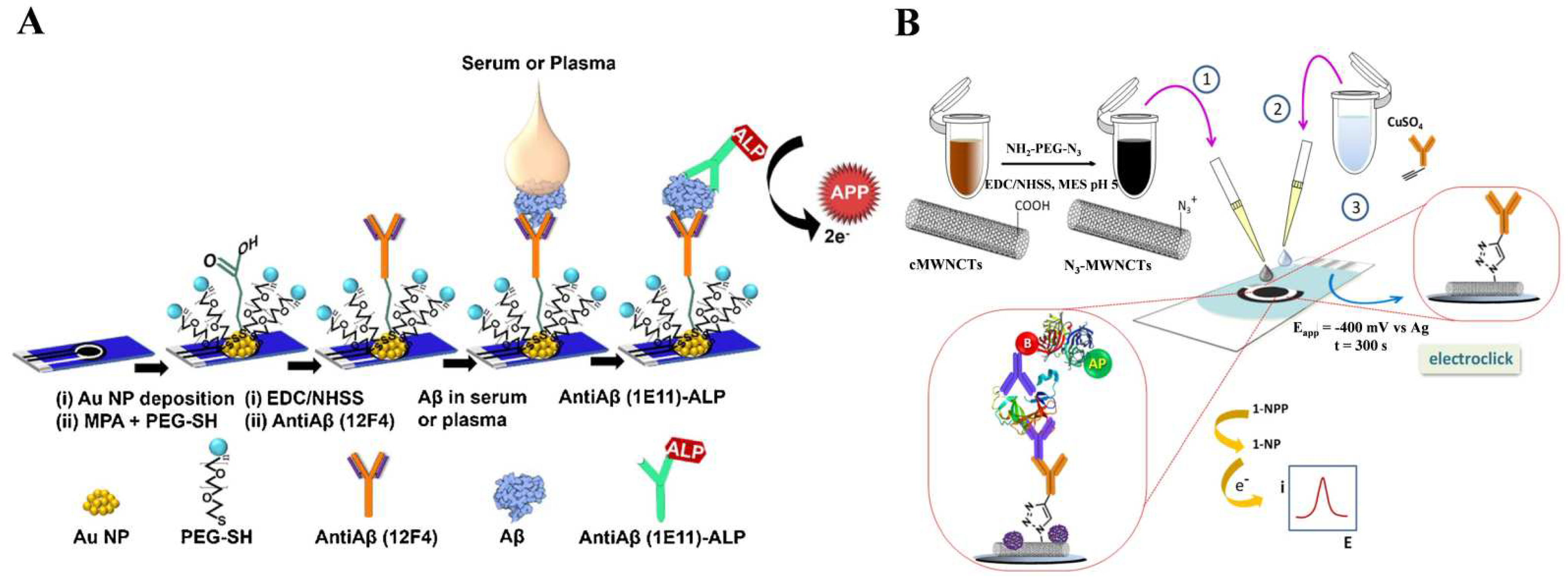
Aiming to reduce the oxidation potential of enzyme products and avoid electrode fouling, a wide variety of alternative substrate/product pairs were developed for ALP-based electrochemical immunosensors, including p-aminophenyl phosphate (PAPP)/p-aminophenol (PAP) [38,39,40,41,42,43,44,45,46,47,48,49,50,51,52,53], hydroquinone diphosphate(HQDP)/hydroquinone (HQ) [54,55,56,57,58], p-nitrophenylphosphate (PNPP)/p-nitrophenol (PNP) [59,60,61,62], α-naphthyl phosphate (NPP)/α-naphthol [63,64,65,66,67,68,69,70,71,72,73,74,75,76,77,78,79,80,81,82,83,84], 3-indoxyl phosphate (3-IP)/indigo blue [85,86,87,88,89], ascorbic acid 2-phosphate (AAP)/ascorbic acid (AA) [90,91,92,93,94], and disodium phenyl phosphate (DPP)/phenol [95,96,97,98]. For instance, Preechaworapun et al. reported an ALP-based immunosensor for the amperometric detection of mouse IgG using AAP as the substrate [99]. PAPP was utilized as the substrate for the electrochemical determination of cancer cells, immunoglobulin E, and the amyloid-beta 1–42 (Aβ) peptide [100,101,102]. As profiled in Figure 2A, gold nanoparticles (AuNPs)-decorated screen-printed carbon electrode (SPCE) was modified with mercaptopropionic acid (MPA) and thiol-functionalized polyethylene glycol (PEG-SH). The antibody toward Aβ, antiAβ(12F4), was immobilized on the surface of SPCE through N-(3-dimethylaminopropyl)-N’-ethylcarbodiimide hydrochloride/N-hydroxysulfosuccinimide (EDC/NHSS)-mediated coupling chemistry. The peptides of Aβ in biological samples were captured and then labeled with ALP-conjugated antiAβ(1E11). The ALP-catalyzed hydrolysis of PAPP to PAP was electrochemically monitored. In addition, Guerrero et al. developed an electrochemical immunosensor for the detection of IL-1β cytokine through electro-click chemistry [103]. As depicted in Figure 2B, ethynylated IgG was immobilized on azide-functionalized multi-walled carbon nanotubes (MWCNTs)-modified SPCE through the Cu(I)-catalyzed cycloaddition reaction for the capture of the IL-1β cytokine and ALP-linked detection antibody. After the sandwich immunoreaction, ALP catalyzed the hydrolysis of NPP to NP that could be determined by differential pulse voltammetry (DPV).
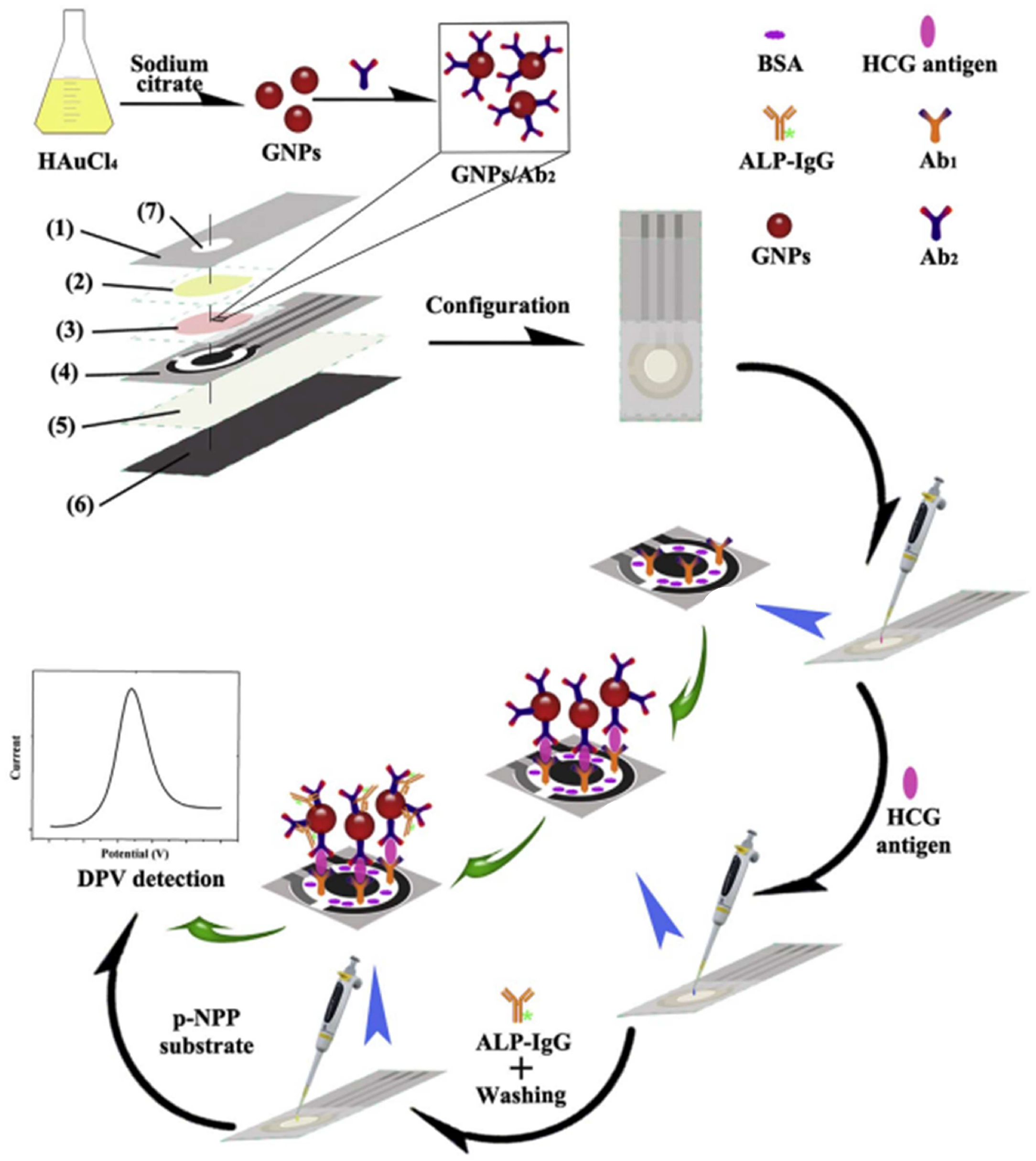
In order to further improve the detection sensitivity, multi-enzyme strategies were proposed with a variety of nanomaterials as the carriers to load plenty of enzymes [104,105,106,107,108]. For instance, Cao et al. fabricated a microfluidic paper-based device (μPAD) for the electrochemical immunoassay of human chorionic gonadotropin (HCG) using Ab2-AuNPs as the recognition elements [109]. As illustrated in Figure 3, Ab2-AuNPs were used to label the captured HCG, and many ALP-linked secondary antibodies (ALP-IgG) were specifically adsorbed on Ab2-AuNPs. DPV was used to measure the electrochemical signal from the ALP-catalyzed conversion of PNPP to PNP. Hou et al. reported an electrochemical immunosensor for the detection of tumor necrosis factor α (TNF-α) based on ALP-catalyzed generation of AA and hydrogel prepared from ferrocene (Fc)-modified amino acids [110]. In this work, the hydrogel formed from the self-assembly of Fc-modified phenylalanine showed high redox activity due to the large number of Fc moieties. However, the ALP-catalyzed generation of AA led to the reduction of the Fc moieties in hydrogel, which was accompanied by a decrease in the redox current.

2.2. ALP Catalysis plus Redox Cycling

Redox cycling involves the repetitive generation of signal molecules in the presence of additional reducing or oxidative species [111]. In this process, a small number of signal molecules can produce a significantly enhanced electrochemical signal. Nowadays, electrochemical immunosensors based on the signal amplification of enzymes plus electrochemical–chemical (EC) or electrochemical–chemical–chemical (ECC) redox cycling arouse wide interest due to their excellent sensitivity and selectivity [112,113,114,115,116,117].

2.2.1. EC Redox Cycling

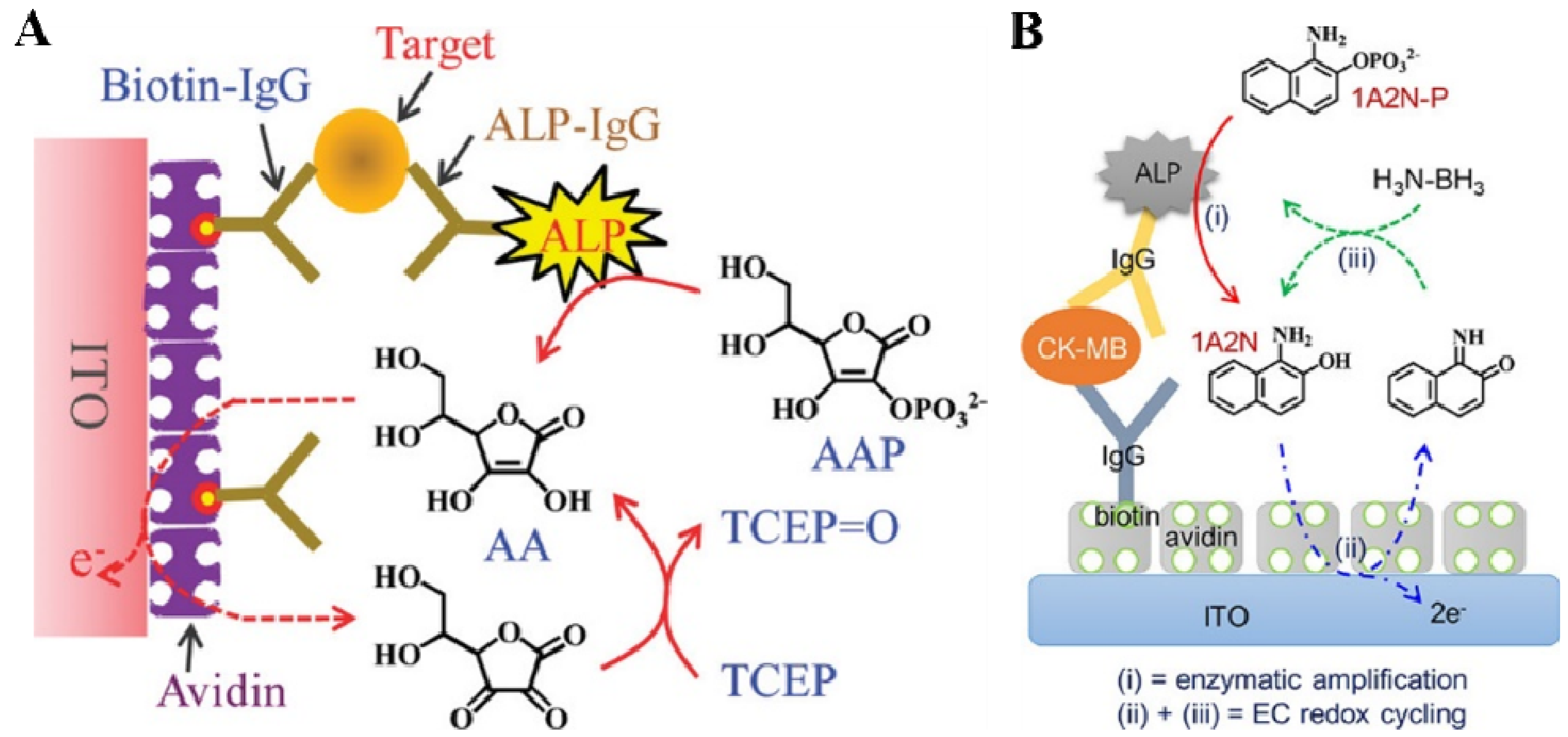
In typical ALP plus EC redox cycling, the substrate should be electrochemically inactive, and the product can be electrochemically oxidized on the electrode surface at a relatively low potential with a high reaction rate. Then, the oxidized product could be immediately reduced by a reducing agent for the next electrochemical oxidation. Thus, a few enzymatic products can be repeatedly regenerated through chemical redox cycling reactions by excessive reducing agents, ultimately producing a strongly amplified electrochemical signal. Meanwhile, the reducing agents can protect the enzymatic products from oxidation in the ambient air. In order to minimize the background current, indium–tin oxide (ITO) electrodes are widely used in EC redox-cycling-based immunoassays because of their low electrocatalytic activity toward reducing agents. Akanda et al. developed an ALP and EC redox-cycling-based immunosensor for the detection of troponin I (Figure 4A) [118]. The performance of AAP was compared with that of other ALP substrates (e.g., PAPP, NPP, and 4-amino-1-naphthyl phosphate). It was found that the AAP/AA (substrate/product) pair was better than the others in terms of the formal potential and the electro-oxidation rate. The ITO electrode without immobilization of the electrocatalyst or electron mediator exhibited a good voltammetric behavior for the fast electro-oxidation of AA. Meanwhile, tris(2-carboxyethyl)phosphine (TCEP) allowed for fast redox cycling with a very low anodic current at the electrode. In the presence of TCEP, the enzymatic product (AA) produced from the ALP-catalyzed hydrolysis of AAP triggered the redox cycling reaction.
The electrochemical oxidation of diaromatic substances is faster than that of monoaromatic reagents with lower electrocatalytic ability. However, such substances are easily oxidized by dissolved oxygen, limiting their applications in EC redox cycling. To address this problem, Seo et al. developed an electrochemical immunosensor for the detection of creatine kinase-MB (CK-MB) using 1-amino-2-naphthyl phosphate (1A2N-P) and H3N-BH3 as the ALP substrate and reducing reagent for EC redox cycling, respectively (Figure 4B) [119]. In this study, ALP catalyzed the hydrolysis of stable 1A2N-P to 1-amino-2-naphthol (1A2N). The oxygen-caused oxidation and polymerization of 1A2N was prevented by excessive H3N-BH3. Meanwhile, the EC redox cycling between 1A2N and H3N-BH3 produced a strong electrochemical signal for CK-MB determination.
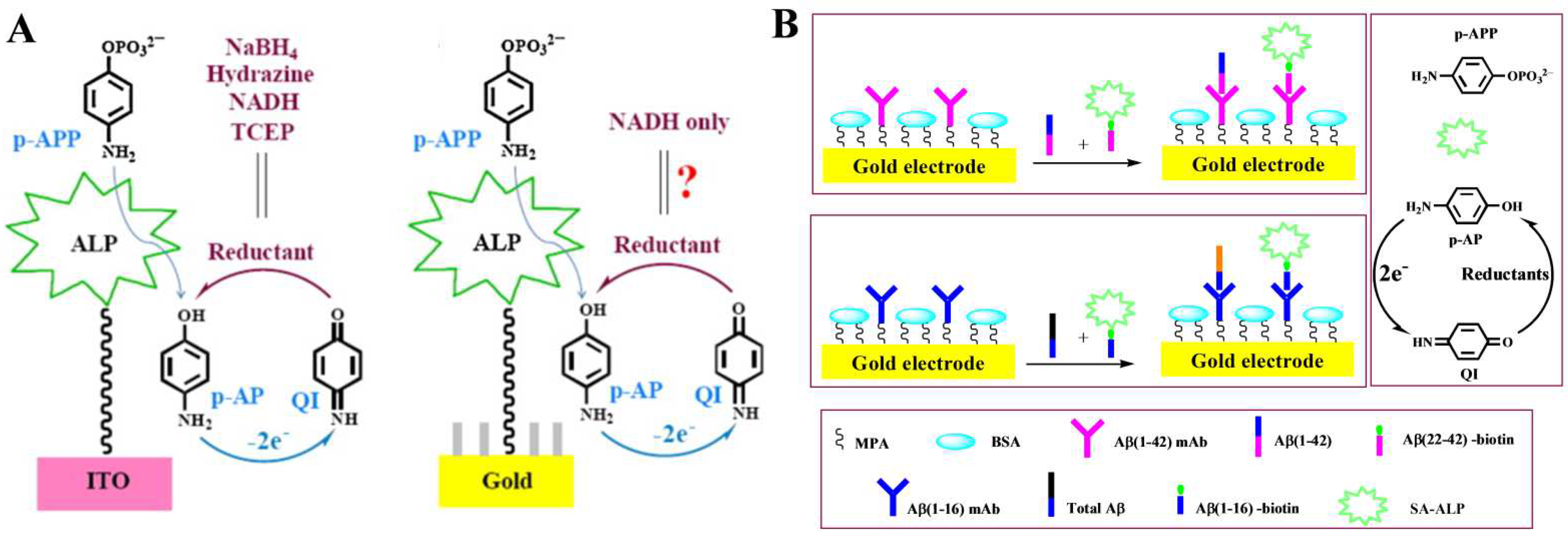
The low activity of the ITO electrode may result in a low electro-oxidation rate for enzymatic products, leading to a weak signal. Therefore, it is important to select appropriate ALP products, reducing agents, and sensing electrodes. Usually, a gold electrode with electrocatalytic ability is not suitable for redox cycling because the reducing agent may exhibit a relatively low potential at the electrode surface, causing a high background current. Challenges remain to find appropriate reducing agents for redox cycling with a low background current. One of our groups systematically evaluated the performance of biosensors with different reducing agents, including NaBH4, hydrazine, TCEP, nicotinamide adenine dinucleotide (NADH), Na2SO3, and cysteamine on analkanethiol self-assembled monolayers (SAMs)-modified gold electrode (Figure 5A) [120]. The result suggested that NADH, TCEP, and cysteamine were suitable for PAP-mediated EC redox cycling because of their low background current on the SAMs-modified gold electrode [121]. Based on PAP-mediated EC redox cycling, Liu’s group developed a competitive electrochemical immunosensor for the detection of Aβ(1–42) and total Aβ peptides [122]. As displayed in Figure 5B, Aβ(1–42) could compete with biotin-Aβ(22–42) to bind Aβ(1–42)mAb on the electrode through the immunoreaction. Similarly, total Aβ peptides could compete with biotin-Aβ(1–16) to bind Aβ(1–16) mAb on the electrode. After the competitive immunoreaction, the streptavidin (SA)-conjugated ALP (SA-ALP) captured by the sensor electrode catalyzed the hydrolysis of PAPP to PAP, initiating the EC redox cycling in the presence of TCEP to generate an amplified anodic current. In addition, to avoid the potential impact of redox species in samples, an immunomagnetic pre-concentration was combined with ALP and EC redox cycling for the immunoassay of Salmonella [123]. After magnetic separation, the enzymatic product (AA) triggered the EC redox cycling in the presence of TCEP. The immunoassay showed a detection limit of 6.0 × 10² CFU/mL in agricultural water.
The electrochemical detection of enzymatic products by direct oxidation at the sensor electrode exhibits slow electron-transfer kinetics and high surface fouling from oxidation products, thus resulting in poor sensitivity, selectivity, and reproducibility. In order to avoid or minimize this effect, redox mediators or nanocatalysts can be introduced to accelerate the oxidation of enzymatic products, in the form of surface-confined layers or the electrolyte solution phase [124]. Actually, the redox mediators or the nanocatalysts-accelerated oxidation of ALP products also involve the redox cycling process. Unlike the abovementioned works, the mediator is electro-oxidized on the electrode surface, and its oxidized form is reduced back to the reduced form by the abundant ALP products accumulated in the solution. Then, the regenerated mediator in the reduced form is electrochemically oxidized again, eventually producing an amplified signal. Notably, the mediator should exhibit faster electron-transfer kinetics and lower formal reversible potential than the enzymatic products, as well as good stability in both oxidized and reduced forms. It was shown that Fc and its derivatives can electrochemically catalyze the oxidation of AA, thereby amplifying the current [125,126]. Zhong et al. developed an electrochemical immunosensor for human apurinic/apyrimidinic endonuclease 1 (APE-1) detection using ALP and Fc-tagged Ab2-modified AuNPs-decorated graphene nanosheets as the signal labels [127]. In this work, the Fc/Fc+ couple effectively catalyzed the electrochemical oxidation of the enzymatic product (AA), further increasing the current response.
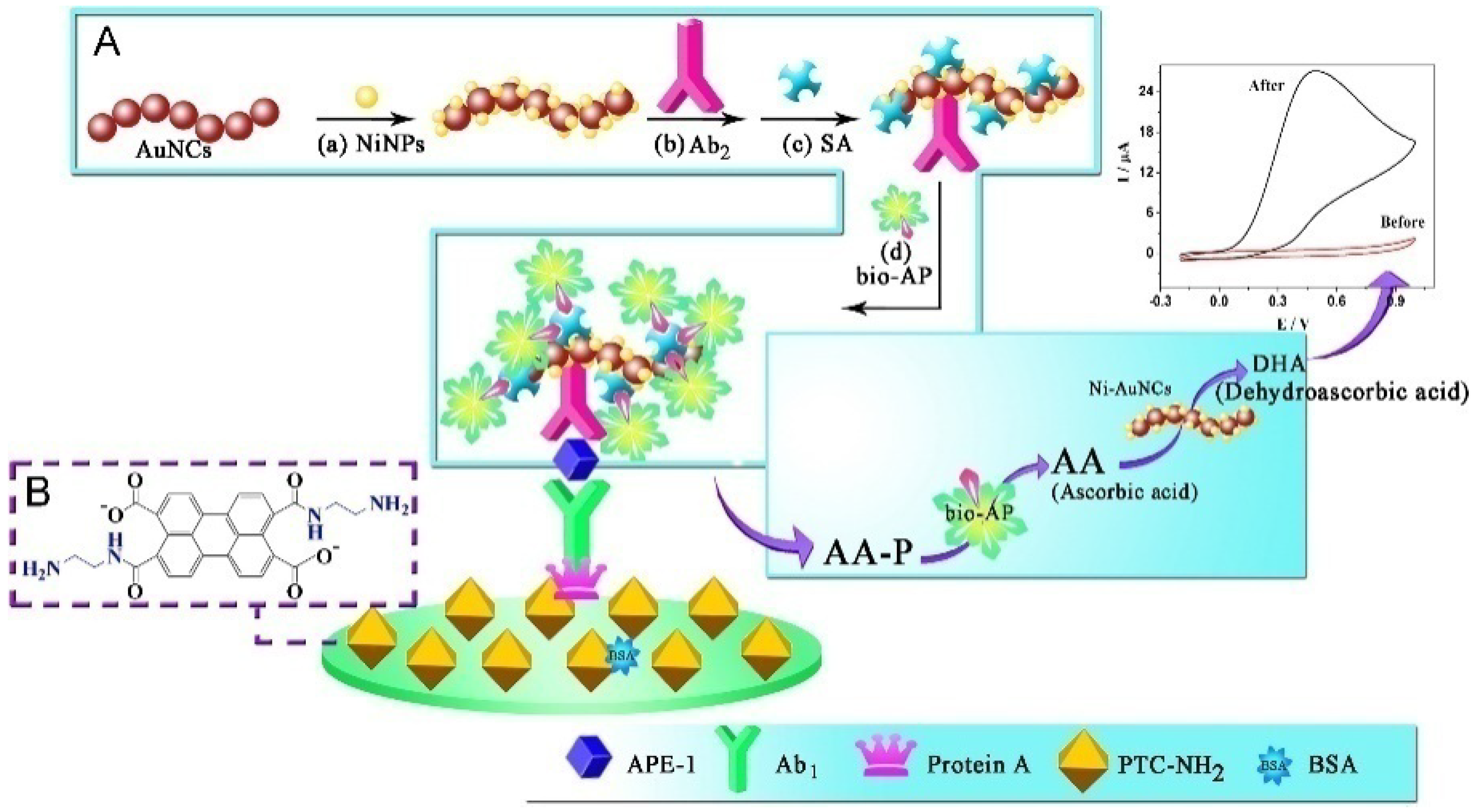
Like the mediator, nanomaterials can also serve as electrocatalysts to accelerate the electrochemical oxidation of enzymatic products, in the form of electrode substrates or signal labels [7]. For this consideration, ALP-based electrochemical immunoassays were extensively developed by coupling the ALP-catalyzed in situ generation of enzymatic products with nanomaterials-assisted electrocatalysis [128,129]. For instance, Hayat demonstrated that nanoceria particles could catalyze the oxidation of enzymatic product 1-naphthol [130]. Han et al. proposed an ultrasensitive ALP-based electrochemical approach for the detection of APE-1 via the triple signal amplification strategy [131]. As shown in Figure 6, the organic compound PTC-NH2 was synthesized by 3,4,9,10-perylene tetracarboxylic dianhydride and ethylenediamine. A glass carbon electrode (GCE) was modified with PTC-NH2 for the covalent immobilization of protein A, which could lower the background current signal. This step was followed by incubation with bovine serum albumin (BSA) solution to block the surface. ALP and Ab2 were co-immobilized on the nickel hexacyanoferrates nanoparticle-decorated Au nanochains (Ni–AuNCs) as multienzyme labels, achieving the first signal amplification. After the immunoreaction, the biocatalysis of ALP toward the conversion of AAP to AA achieved the second signal amplification. Concomitantly, the third signal amplification was achieved with the EC redox cycling process among AA, Ni–AuNCs, and the electrode. Based on triple signal amplification, a significantly enhanced electrochemical signal was obtained for APE-1 detection.

2.2.2. ECC Redox Cycling

ECC redox cycling can be achieved in the presence of an oxidant and a reducing reagent. Usually, the oxidant serves as an electron mediator to catalyze the electrochemical oxidation of ALP products. The generated oxidation products are reduced by excess reducing reagents and then electrochemically oxidized again with the aid of the oxidant mediator. In ECC redox cycling, a few ALP products can greatly promote the current signal. The electrocatalytic activity of the sensor electrode toward the reducing reagent used in the ECC redox cycling may influence the background current and the signal-to-noise ratio. Highly electrocatalytic electrode can lead to a strong background current due to the oxidation of the reducing reagent at a low potential. Although a partially ferrocene-functionalized dendrimer (Fc-D)-modified ITO was employed to design a ECC redox cycling system with NaBH4 as the reducing reagent, a gold nanoparticle as the nanocatalyst, and PNP as the substrate [132], the background current for the reduction of NaBH4 at the modified ITO is still high. Thus, it is necessary to explore electrochemically inactive and more stable reducing agents for ECC redox cycling. Das et al. developed an electrochemical immunosensor for the detection of mouse IgG based on PAP-mediated ECC redox cycling using hydrazine as the reducing agent (Figure 7A) [133]. In this study, hydrazine was used as a reducing agent for ECC redox cycling because it was electrochemically inactive and exhibited slow electro-oxidation kinetics on the Fc-D-modified ITO electrode. After the formation of a sandwich immunecomplex on the ITO electrode, PAPP was converted to PAP under the catalysis of ALP for a certain incubation time, accumulating a large number of electroactive PAP species. The generated PAP was electrochemically oxidized to p-quinoneimine (PQI) under the electrocatalysis of the Fc moieties in Fc-D as the redox mediator. Then, the oxidized PQI was reduced back to PAP by excess hydrazine, and the resulting PAP was again electrochemically oxidized to PQI. Through repeated electrocatalytic reactions, the oxidation current of PAP was dramatically increased, and the sensitivity of the ALP-based immunosensor was significantly improved.
After that, more efforts were put into the selection of suitable ECC systems composed of a reducing reagent, an enzymatic product, a redox mediator, and a sensor electrode. For example, Kwon et al. proposed an ECC redox cycling system with NADH as the reductant [134]. In this study, a gold electrode was modified with the SAMs of long thiol molecules to reduce the background current and was further modified with Fc-D to promote the oxidation of p-AP. NADH showed a slow electrochemical oxidation rate at the electrode and a fast chemical reaction with PAP during the redox cycling. Akanda et al. reported an ECC redox cycling system for an ultrasensitive immunoassay of cardiac troponin I (cTnI) [135]. As shown in Figure 7B, outer-sphere-reaction (OSR)-philic Ru(NH3)63+, innersphere-reaction (ISR)-philic TCEP, and an OSR- and ISR-philic QI/AP couple were used as the oxidant, reductant, and ALP substrate/product to design an “outer-sphere to inner-sphere” redox cycling system. The QI/AP couple exhibited fast redox reactions with both Ru(NH3)63+ and TCEP. A highly OSR-philic ITO electrode minimized the unwanted electrochemical reaction with TCEP. The immunosensor showed a detection limit of 10 fg/mL for the detection of troponin I in serum.
Figure 7. (A) Schematic representation of an electrochemical immunosensor for the detection of mouse IgG based on PAP-mediated ECC redox cycling using hydrazine as the reductant. (a) Schematic representation of the preparation of an immunosensing layer. (b) Schematic view of electrochemical detection for mouse IgG [133]. Copyright 2007 American Chemical Society. (B) Schematic representation of “outer-sphere to inner-sphere” redox cycling for ultrasensitive immunosensors [135]. Copyright 2012 American Chemical Society.
Biosensors 13 00855 g007
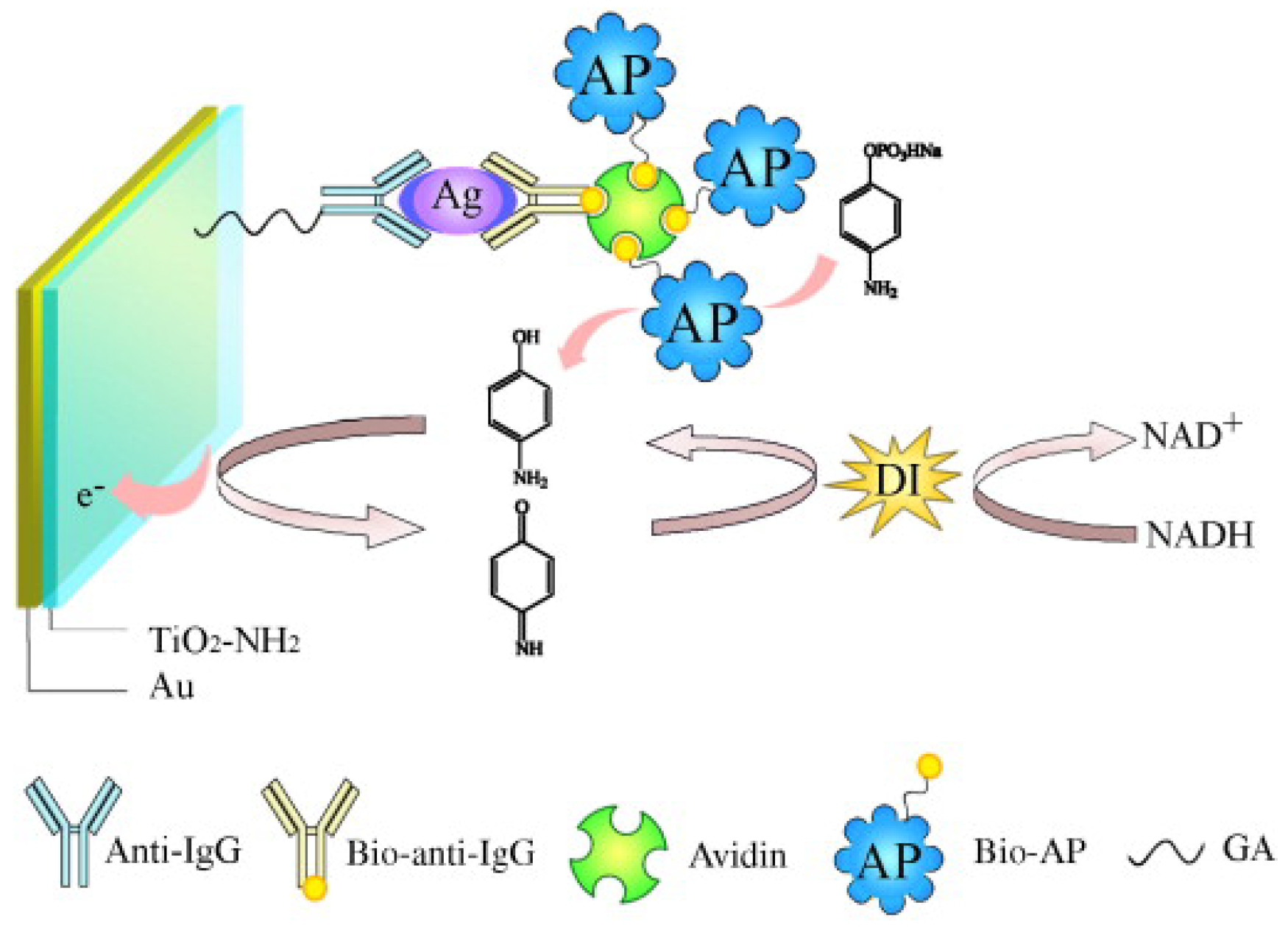
In ALP-mediated enzymatic–enzymatic (EE) redox cycling, appropriate oxidoreductases, such as tyrosinase and diaphorase (DI), are required to regenerate ALP products with the consumption of enzyme substrates [136,137]. For example, Yuan et al. developed an ALP-mediated electrochemical immunosensor for the determination of human IgG based on bienzyme redox cycling [138]. As displayed in Figure 8, under the catalysis of ALP, electroactive PAP was produced from the inactive substrate of PAPP and was concomitantly oxidized at the electrode surface to p-quinoneimine (PQI). PQI was then reduced to PAP by DI, leading to the repeated generation of PQI. The oxidized state of DI changed to its native state by the substrate of NADH. Under ALP/DI EE redox cycling, the immunosensor exhibited a wide linear range from 1 × 10⁻¹⁴ to 1 × 10⁻⁵ g/mL with a detection limit of 3.5 × 10⁻¹⁵ g/mL.

2.3. ALP-Catalyzed Metal Deposition

The enzymatic control of metal precipitation is an effective approach to decrease the background signal in contrast to conventional AuNPs-catalyzed silver electrodeposition. ALP-catalyzed reductive products, such as indoxyl intermediate, PAP, and AA, can reduce Ag+ ions to Ag deposited on the electrode with a low background. The amount of Ag deposition can be increased by accumulating the enzymatic products over a long period of incubation, and the oxidation peak can be monitored by anodic stripping voltammetry (ASV). A well-defined ASV peak allowed for the electrochemical immunoassays of various targets with ALP-linked detection antibodies [139,140,141,142]. For example, Chen et al. developed an electrochemical immunosensor for human IgG detection based on ALP-catalyzed Ag deposition [143]. In this work, ALP catalyzed the hydrolysis of AAP to AA that could reduce Ag+ ions to Ag deposited on the electrode. Although the stripping current peak of Ag on a gold electrode was divisive from gold, it was still difficult to directly measure the Ag signal. Therefore, the deposited Ag was stripped from the gold electrode at 0.7 V and then accumulated on a glassy carbon electrode for ASV measurement.
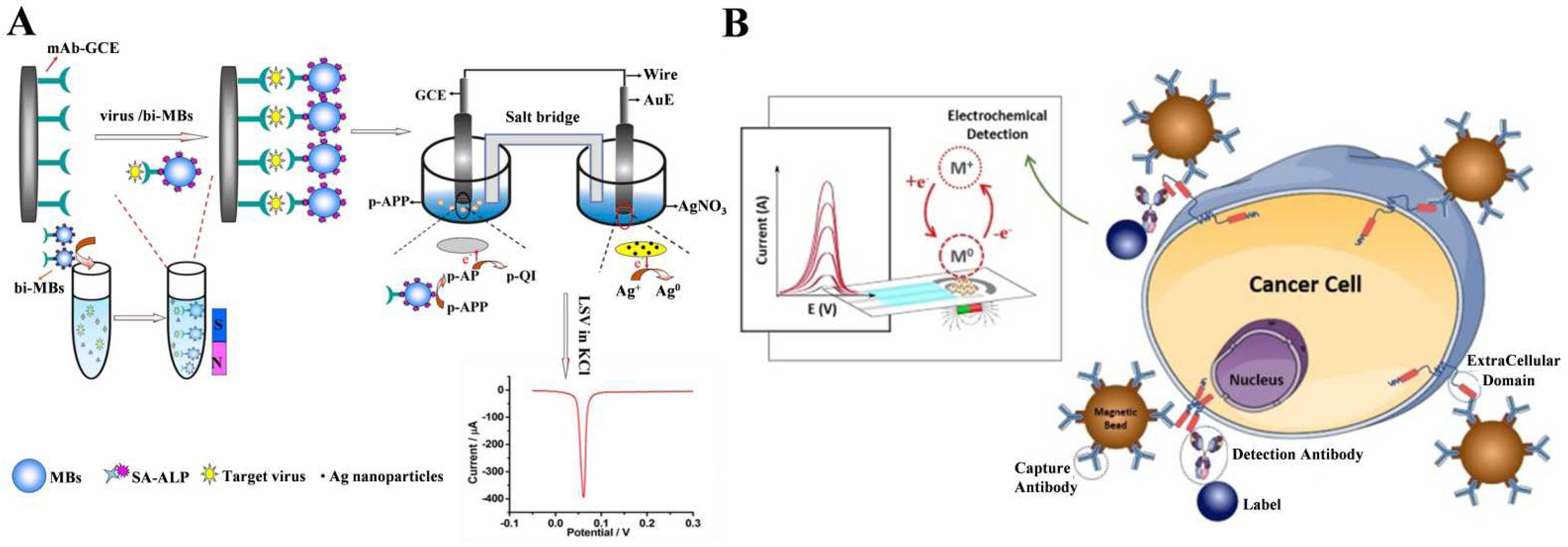
Nanomaterials with a large surface area and plenty of functional groups can serve as nanocarriers to load numerous enzymes for signal amplification [144,145,146,147,148]. Qu et al. used ALP-modified silica nanoparticles (SiO2 NPs) and ALP-encapsulating liposomes as the signal labels to trigger enzymatic silver metallization for the detection of prostate-specific antigen (PSA) [149,150]. Due to their large surface area and abundant surface functional groups, magnetic nanoparticles (MBs) can serve as carriers to simultaneously load antibodies and a large number of ALP species, increasing the signal-to-noise ratio and improving the sensitivity [151,152,153]. Moreover, thanks to their good magnetic response ability, MBs were popularly utilized as the separation and enrichment supports to capture targets from complex samples. As an example, Wu et al. reported an electrochemical immunosensor for the detection of avian influenza A (H7N9) virus based on immunomagnetic separation and ALP-induced metallization [154]. As shown in Figure 9A, MBs modified with antibodies and ALP were used to capture virus from the samples. After the sandwich immunoreaction, ALP catalyzed the transformation of PAPP to PAP. In the galvanic cell, Ag+ ions could be reduced to Ag0 by PAP and then deposited on the gold electrode. The dual-electrode signal conversion approach eliminated the potential effect of Ag+ ions or silver deposition on enzyme activity. In addition, the combination of 3-indoxyl phosphate (3-IP, the enzymatic substrate) and Ag+ allowed the development of versatile ALP-based electrochemical immunosensors for the detection of human anti-gliadin antibodies, cancer antigen 15-3, human epidermal growth factor receptor 2 (HER2-ECD), amyloid-beta 1–42, and so on [155,156,157,158,159,160]. For example, Freitas et al. reported the electrochemical immunomagnetic analysis of HER2-ECD in human serum and cancer cells [161]. As illustrated in Figure 9B, MBs were modified with capture antibody, and the detection antibody was conjugated with ALP. In the presence of HER2-ECD, the sandwich immunecomplex formed on the surface of the MBs that catalyzed the hydrolysis of 3-IP. The products could reduce Ag+ ions to Ag deposition on the SPCE surface.
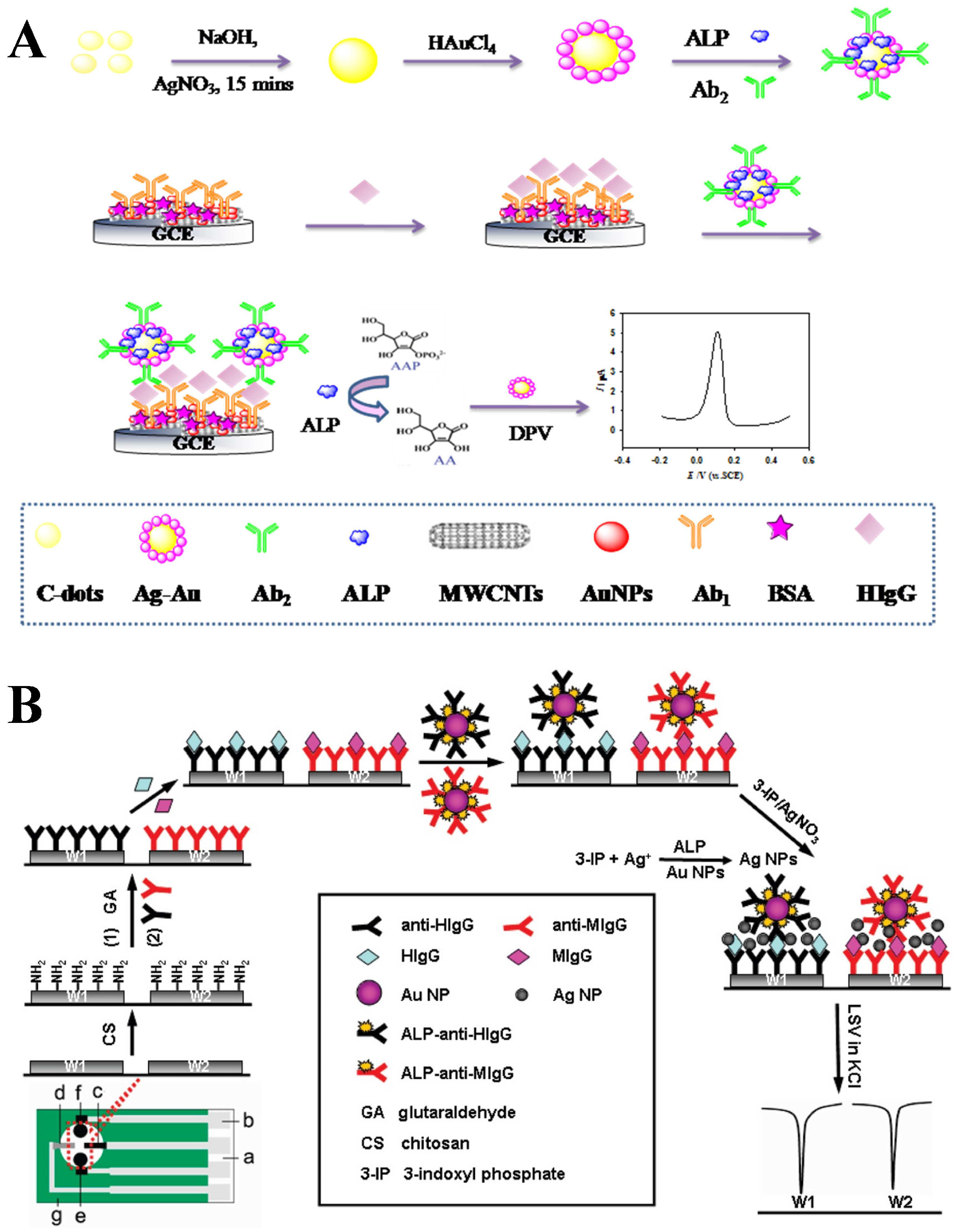
The accumulation of metal silver on the electrode may block the diffusing of enzymatic products into a solution. Thus, different nanomaterials such as AuNPs, carbon nanotubes (CNTs), and platinum nanoparticles (PtNPs) were used for Ag deposition [162,163,164,165,166]. In an ALP-linked AuNPs-based immunoassay, AuNPs can serve as the nanocarriers for ALP loading and the supports, as well as catalysts for Ag deposition [167,168,169]. Zhang et al. reported an electrochemical immunosensor for human IgG detection based on ALP-triggered Ag deposition and Ag–Au bimetallic nanoparticles as the catalysts (Figure 10A) [170]. In this study, the Ag–Au bimetallic nanoparticles were synthesized with carbon dots (CDs) as the reductant and stabilizer and were further modified with ALP and a detection antibody. After the immunoreaction, the ALP on the electrode catalyzed the hydrolysis of AAP to generate AA, which could induce Ag deposition on the surface of Ag–Au bimetallic nanoparticles. Lai et al. reported a multiplexed immunoassay by integration of ALP-functionalized AuNPs with Ag deposition [171]. As illustrated in Figure 10B, capture antibodies were covalently immobilized on the surface of chitosan-modified SPCE. After the sandwich-type immunoreaction, ALP attached on the SPCE catalyzed the hydrolysis of 3-IP, which could reduce Ag+ ions to Ag. Both ALP and AuNPs promoted the deposition of Ag, amplifying the detection signal. This multiplexed immunosensor exhibited a wide linear range over four orders of magnitude for human and mouse IgG detection. To achieve the best performance, the authors further investigated the influence of AuNPs morphology on the detection performance and found that the protocol with irregular-shaped AuNPs showed a better analytical result than that with spherical AuNPs [172].
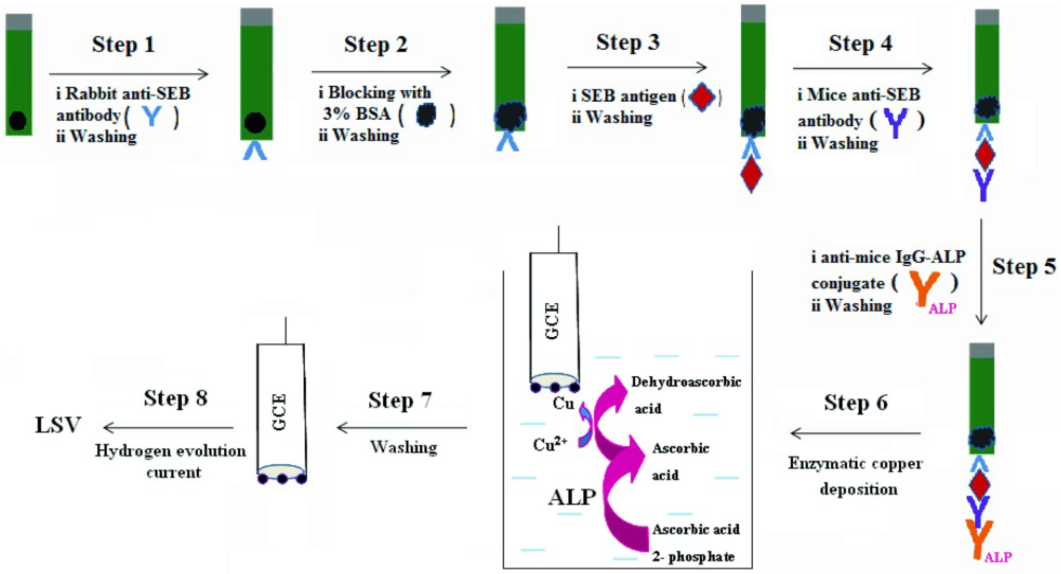
PtNPs modified on the electrode can generate a strong electrocatalytic current through the hydrogen evolution reaction (HER). AA-induced copper deposition on the PtNPs-modified electrode can lead to a negative shift of the hydrogen evolution potential by the catalytic poisoning of PtNPs. Thus, the ALP-catalyzed generation of AA can be combined with copper deposition and HER for the construction of electrochemical immunosensors [173]. As proof, Sharma et al. developed an electrochemical immunosensor for Staphylococcal Enterotoxin B (SEB) detection based on HER inhibition by ALP-catalyzed copper deposition on PtNPs-modified GCE (Figure 11) [174]. After the sandwich immunoreaction, ALP catalyzed the hydrolysis of AAP to AA. Then, the PtNPs-modified electrode was immersed in the resulting solution containing ALP-catalyzed product AA and Cu2+ ions for copper deposition. The potential shift value exhibited a linear relationship with SEB concentration in the range of 1 ng/mL–1 μg/mL, with a detection limit of 1 ng/mL.
Table 1. Analytical performance of ALP-based electrochemical biosensors.
Detection PrincipleALP SubstrateTargetLinear RangeLODRef.
Direct detection of enzymatic productsPAPPDigoxin0.5–2.0 ng/mL30 pg/mL[38]
PAPPMouse IgG10–1000 ng/mL10 ng/mL[39]
PAPPhCG0.8–40 units/L0.8 units/L[40]
PAPPTBG and cortisol31–1000 μg/L
and 1 × 10²–2000 nM
NA[41]
NPPPCBs1 × 10−5–1 U/mL2.1 × 10−6 U/mL[63]
PNPP5-methylcytosine0.01–50 nM3.2 pM[70]
3-IPEscherichia coli O157:H76 × 104–6 × 10⁷ cells/mL6 × 103 cells/mL[89]
AAPMouse IgG1–1000 ng/mL0.3 ng/mL[99]
PNPPhCG1–100 mIU/mL0.36 mIU/mL[109]
ALP catalysis plus EC redox cyclingAAPTroponin I100 fg/mL–1 μg/mL10 fg/mL[118]
1A2N-PCK-MB100 fg/mL–1 μg/mL80 fg/mL[119]
AAPAPE-110 fg/mL–100 pg/mL3.9 fg/mL[131]
ALP catalysis plus ECC redox cyclingPAPPMouse IgG0.1–1 × 10⁵ pg/mL100 fg/mL[133]
PAPPMouse IgG1 pg/mL–1 μg/mL1 pg/mL[134]
PAPPTroponin I10 fg/mL–1 μg/mL1 fg/mL[135]
ALP-based EE redox cyclingPAPPCEA5 pg/mL–50 ng/mL2 pg/mL[137]
PAPPHuman IgG10 fg/mL–1 μg/mL3.5 fg/mL[138]
ALP-catalyzed metal depositionAAPHuman IgG0.1–50 ng/mL0.03 ng/mL[142]
AAPHuman IgG5–1000 ng/mL2.2 ng/mL[143]
PAPPH7N9 virus0.01–20 ng/mL6.8 pg/mL[154]
3-IPHER25–50 ng/mL2.8 ng/mL[161]
AAPHuman IgG0.005–100 ng/mL0.9 pg/mL[170]
3-IPHuman and mouse IgG0.01–250 ng/mL4.8 pg/mL[171]
AAPHuman IgG10 pg/mL–1 μg/mL2 pg/mL[173]
AAPSEB1 ng/mL–1 μg/mL1 ng/mL[174]
Abbreviations: PAPP, p-aminophenyl phosphate; IgG, immunoglobulin G; hCG, humanchorionic gonadotropin; TBG, thyroxine-binding globulin; NPP, α-naphthyl phosphate; PCBs, polychlorinated biphenyls; PNPP, p-nitrophenyl phosphate; AAP, 2-phospho-L-ascorbic acid; 1A2N-P, 1-amino-2-naphthyl phosphate; APE-1, apurnic/apyrimidinicendonuclease; 3-IP, 5-bromo-4-chloro-3-indolyl phosphate; CEA, carcinoembryonic antigen; SEB, Staphylococcal Enterotoxin B; HER2, human epidermal growth factor receptor 2.

3. PEC Methods

PEC techniques are widely used in various analytical applications due to their impressive advantages in terms of high sensitivity, a simple instrument, and easy operation [175]. The PEC process mainly involves photo-to-electric conversion on a photoelectrode under the illustration of an applied light. The PEC sensing interface and sensing strategy play a major role in the construction of highly efficient PEC biosensors [176]. In recent years, the combination of ALP-based immunoassays and PEC techniques has sparked significant excitement. The electroactive or reducing species produced by ALP catalysis can modulate PEC signals through different mechanisms, including ALP-catalyzed products as electron donors, ALP-mediated redox cycling, and ALP-mediated in situ growth or the bioetching of the photoelectrode (Table 2) [177,178].

3.1. ALP-Catalyzed Products as Electron Donors
The photoelectrode can be coupled with enzyme catalysis through the interactions between electrons/holes and enzymatic products (oxidative/reductive species). The exciting and transfer of electrons can be promoted or hindered, and the enzymatic reaction-modulated change in the generated photocurrent indirectly reflects the concentration of analytes. ALP can be integrated into PEC immunoassays as an enzyme unit for signal amplification [179,180]. Generally, enzymatic products such as AA could be in situ produced to act as hole-trapping reagents for the capture of the photogenerated holes (h+) of photoactive materials, providing electrons to hamper the photogenerated electron–hole recombination and resulting in an increase in the photocurrent signal [181,182,183,184,185]. For example, Yang et al. developed a signal-on PEC immunosensor for the detection of M.SssI methyltransfease activity based on the ALP-catalyzed in situ production of AA as an electron donor [186]. Zhang et al. reported a simultaneous PEC immunosensor for dual-cardiac marker detection using ALP and acetylcholineesterase (AChE) as the enzyme tags [187]. Ai et al. developed a PEC biosensor for N6-methyladenosine (m6A) detection based on ALP catalysis [188]. As presented in Figure 12A, black titanium dioxide (TiO2−x) and a molybdenum sulfide (MoS2) heterojunction (TiO2−x-MoS2) were used as the photoactive materials and were further modified with an m6A antibody through the interaction between the boronic acid group in p-mercaptophenylboronic acid (MPBA) and the glycosyl group in the antibody. After the capture of m6ATP by the immunoreaction, Phos-tag-biotin was added to specifically label the phosphate group of m6ATP, allowing for the immobilization of avidin-ALP. The attached ALP could catalyze the conversion of AAP toAA by serving as the electron donor to inhibit the recombination of the photogenerated electron and hole of the photoactive material, greatly improving the PEC response. Nevertheless, the 1:1 ration of the target and signal amplification unit (ALP) limited the sensitivity of the method. To improve the ratio of the target and enzyme label, different nanomaterials were used as the carriers to load recognition elements and enzymes [189,190,191]. For instance, Yin et al. developed a PEC immunosensor for microRNA detection using IgG–ALP-modified AuNPs as multi-enzyme labels [192]. As illustrated in Figure 12B, DNA capture probes were immobilized on the AuNPs-g-C3N4-decorated ITO surface. After hybridization between the capture probe and the target microRNA, the DNA:RNA hybrids were labeled with an anti-DNA:RNA antibody. Then, the IgG–ALP conjugates attached on AuNPs were captured through the specific interaction between the antibody and IgG. ALP immobilized on the electrode catalyzed the hydrolysis of AAP to AA as the electron donor to produce a strong photocurrent. However, the binding of biomolecules (e.g., antibodies, antigens, and enzymes) and nanomaterials on the photoelectrode may decrease the PEC signal because of the steric hindrance effect to mass transfer and the interference in the light harvest of the photoactive substrate [193]. Furthermore, the oxidation capacity of the photovoltaic substrate and light radiation in the PEC system may damage the structure of biomolecules.
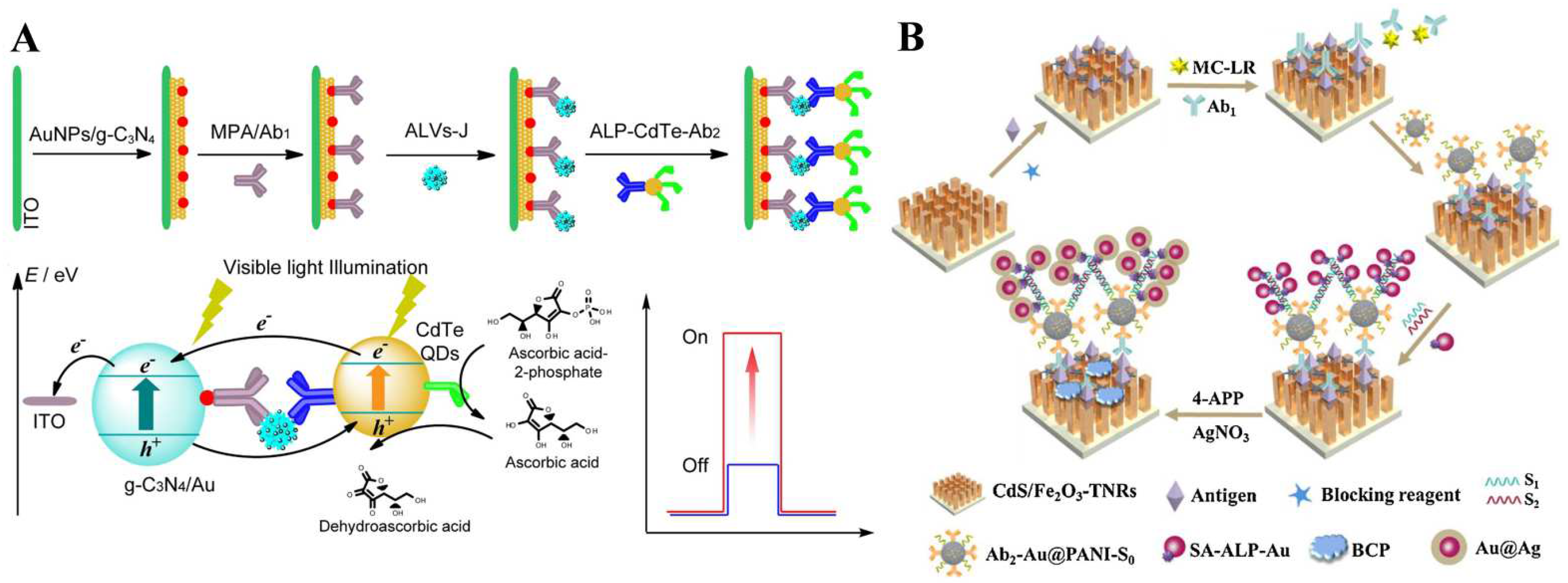
It is an effective “signal-on” strategy to modify photoactive nanoparticles with detection antibodies and enzymes. After the immune-recognition event, the signal labels can significantly enhance the PEC responses through the synergistic interaction of enzymatic catalysis and photoactive species. Sun et al. developed a dual “signal-on” PEC immunosensor for the detection of subgroup J avian leucosis viruses (ALV-J) based on the integration of AuNPs/g-C3N4 and CdTe QDs as well as the in situ enzymatic generation of electron donors [194]. As illustrated in Figure 13A, AuNPs/g-C3N4 as the photoactive electrode materials were modified with capture antibodies. CdTe QDs with light-harvesting ability were functionalized with a detection antibody and ALP. After the immunoreaction, the photocurrent was enhanced due to the matched energy level between the CdTe QDs and AuNPs/g-C3N4. Meanwhile, the ALP-catalyzed in situ produced AA further enhanced the photocurrent response, realizing the dual “signal-on” mode for the PEC assay.
Coupling the enzymatic reaction or plasmonic metal nanoparticles with the steric hindrance effect is also an effective strategy to reduce the PEC response through multiple amplification strategies. For example, Zhang et al. reported the PEC and visualized immunoassay of β-human chorionic gonadotrophin based on enzymatic biocatalytic precipitation [195]. In this work, ALP catalyzed the oxidative hydrolysis of 5-bromo-4-chloro-3-indoyl phosphate (BCIP) to form indigo precipitates. The in situ performed insulating layer reduced the photocurrent by impeding the interfacial mass and electron transfer. The exciton–plasmon interaction (EPI) between photoactive CdS QDs and plasmonic Ag/Au NPs caused significant photocurrent attenuation through the energy transfer effect. The CdS QDs- and AgNPs/AuNPs-based system was well-exploited for PEC analysis [196]. Wei et al. reported a PEC immunosensor for the detection of microcystin-LR (MC-LR) based on the hybridization chain reaction (HCR)-assisted EPI effect and enzymatic precipitation [197]. As displayed in Figure 13B, the sandwich immunoreaction occurred on the surface of CdS/Fe2O3 co-sensitized TiO2NR arrays/the ITO electrode. After the HCR reaction on DNA-primer-modified Au@polyaniline nanocomposites, the resulting DNA polymers with multiple biotin labels could capture an increasing number of SA-ALP-modified AuNPs via specific biotin–SA interactions. Then, ALP catalyzed the transformation of PAPP to PAP. In the presence of Ag+ ions, PAP induced the Ag deposition reaction on the photoelectrode to generate Au@Ag for the PEI effect and an insoluble biocatalytic precipitation (BCP) of benzoquinone serving as an insulating layer and an electron acceptor to inhibit the electron transfer between the solid–liquid interface.
Figure 13. (A) Schematic illustration of a dual signal-on PEC immunosensor for detection of ALV-J based on AuNPs/g-C3N4 coupling with CdTe QDs and in situ enzymatic generation of electron donor [194]. Copyright 2016 Elsevier. (B) Schematic illustration of the PEC immunoassay for detection of MC-LR based on HCR-assisted EPI effect and enzymatic biocatalytic precipitation [197]. Copyright 2018 Elsevier.
Biosensors 13 00855 g013
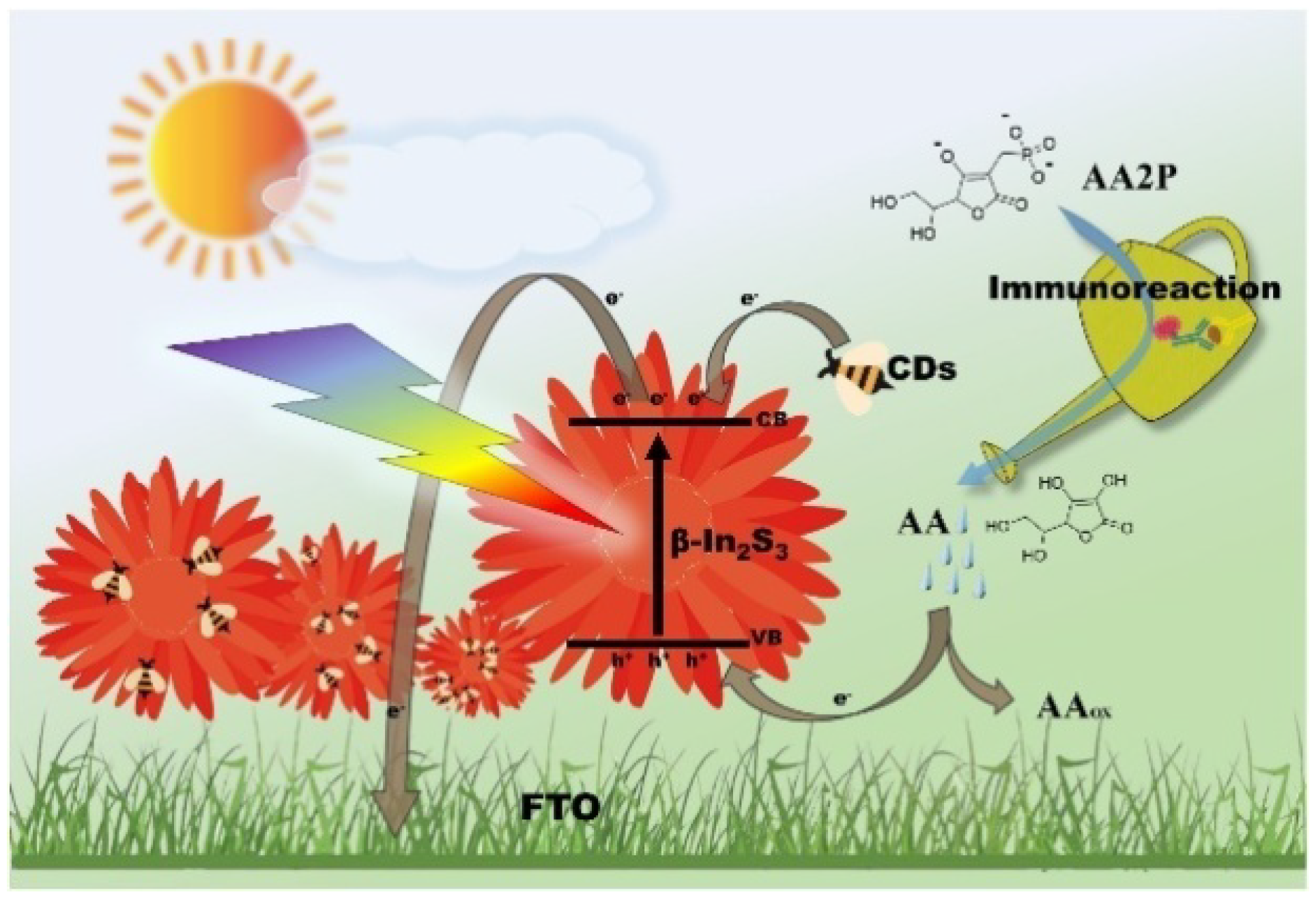
To avoid the steric hindrance effect and the potential damage of biomolecules, the split-type detection mode was widely adopted for various PEC immunoassays, in which the immunoreaction process was separated from the PEC detection system [198]. The utilized optical spectrum region of most photoactive materials is always in the limited region like ultraviolet (UV) and visible (Vis) light. To take advantage of the overall luminous energy, Yu et al. developed a full-spectrum-responsive PEC immunosensor for the detection of alpha-fetoprotein (AFP) based on β-In2S3@CDs nanoflowers [199]. As illustrated in Figure 14, the flower-like β-In2S3@CDs hybrid materials prepared via a one-pot hydrothermal method could enhance the photocurrent signal under UV, Vis, and near-infrared (NIR) irradiation. AA generated in the ALP-mediated immunoreaction served as a photoanode sacrificial agent to reduce the electron–hole pair recombination, increasing the PEC signal in the glare of axenon lamp.
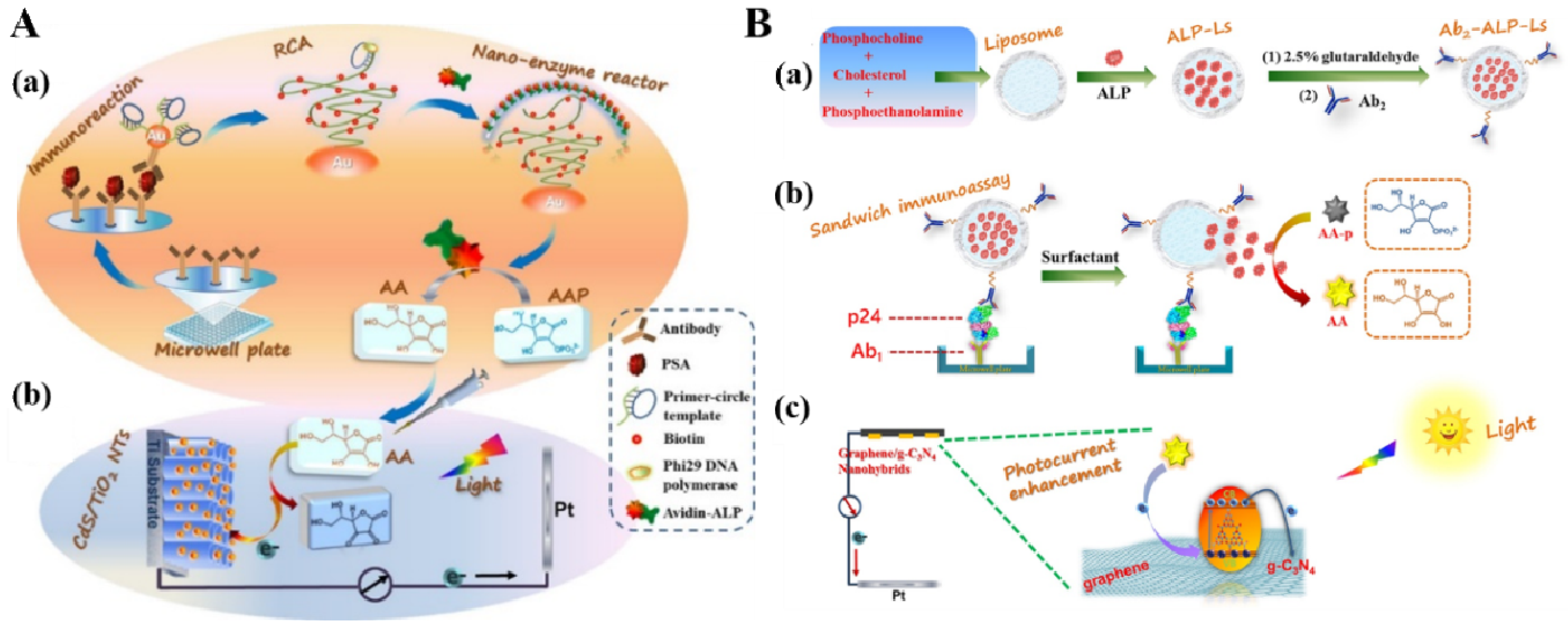
DNA-based amplification techniques are considered as powerful tools to amplify the signals of different biosensors [200]. Such techniques can also be introduced into PEC immunoassays for signal enhancement. Zhuang et al. developed a split-type PEC immunosensor for PSA detection by combining the RCA reaction with the ALP-triggered in situ electron donor-producing strategy [201]. As presented in Figure 15A, the immunoreaction was conducted on a microplate with secondary antibody/primer-circular DNA-labeled AuNPs as the detection tags. After the RCA reaction, a large number of the repeated biotin-functionalized DNA sequences were in situ generated on AuNPs to capture a large number of avidin–ALP conjugates. Next, the resulting solution containing enzymatic products (AA) was transferred to the PEC cell, greatly quenching the photogenerated holes in the CdS QDs-sensitized TiO2 nanotube arrays. However, DNA-based PEC immunosensors are always limited by the time-consuming reactions and the use of extra enzyme-conjugated labels.
Liposome composed of phospholipid bilayers with hollow cavity can carry various guest species for biosensing applications, such as small molecules, enzymes, and nanomaterials [202,203,204,205,206]. They were also used in ALP-linked liposomal PEC immunoassays for multiple signal amplification. For example, Zhuang et al. reported a split-type ALP-encapsulated liposomal PEC immunoassay for HIV-p24 antigen (p24) detection [207]. As displayed in Figure 15B, liposome (Ls) was loaded with ALP in its aqueous cavity and then modified with detection antibody Ab2 to form an Ab2–ALP–Ls signal label. After the immunoreaction in the plate, ALP molecules released from Ls under the treatment with Tween 20 catalyzed the hydrolysis of AAP to AA. When the resulting solution was added to the PEC detection cell, the produced AA restrained the electron–hole recombination in g-C3N4, increasing the photocurrent signal of the graphene/g-C3N4 nanohybrids (GR/g-C3N4)-based photoelectrode.
Figure 15. (A) Schematic illustration of (a) immunoreaction-induced ALP-mediated nanoenzyme reactor formation through RCA, and (b) enzymatic product AA-mediated hole-trapping in CdS QD-sensitized TiO2nanotube array for the amplification of PEC response [201]. Copyright 2015 American Chemical Society. (B) Schematic illustration of (a) preparation process of Ab2–ALP–Ls signal label, (b) sandwich immunoassay based on Ab2–ALP–Ls signal label coupling with ALP-catalyzed generation of AA, and (c) amplification of photocurrent signal based on AA-mediated hole-trapping in GR/g-C3N4 electrode [207]. Copyright 2017 Elsevier.
Biosensors 13 00855 g015
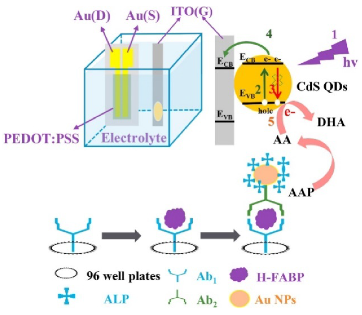
Recently, the organic PEC transistor (OPECT) technique, by the integration of PEC analysis with an organic electrochemical transistor, showed extraordinarily high sensitivity in the determination of low-abundance analytes. With a photoelectrode as the gate electrode, the enzymatic-reaction-triggered small change in photovoltage can be significantly amplified by the channel current (IDS) between the source electrode and the drain electrode [208]. For this view, Shi et al. reported an ALP-mediated OPECT sensing strategy for the detection of the heart-type fatty acid binding protein (H-FABP) [209]. Referring to Figure 16, the primary antibody/H-FABP/secondary antibody–AuNPs–ALP sandwich immunecomplexes were produced in a 96-well plate via the specific immunoreaction. Then, the reaction solution was transferred to the OPECT cell, and the enzymatic product AA, serving as a sacrificial reagent, scavenged the photogenerated hole on the valence band (VB) of CdS QDs, leading to a change in the effective gate voltage ( V G eff ) and IDS of the devices. The concentration of H-FABP was sensitively determined by measuring the corresponding IDS.
Most PEC immunosensors were designed based on the individual signal change caused by the recognition event, in which the response may be influenced by external interferences, such as operating personnel and different experimental environments. As a result, the dual-signal detection mode was widely adopted for PEC immunoassays to improve the accuracy and sensitivity [210,211]. Wei et al. constructed a dual-modal split-type PEC immunosensor for the detection of MC-LR based on HCR and ALP catalysis [212]. As showed in Figure 17A, the immunoreaction was conducted in amicroplate well, and mesoporous silica nanospheres were employed as the carriers to simultaneously load Ab2 and DNA primers to initiate the HCR reaction. The formed biotin-suspended DNA polymers could capture many ALP molecules to catalyze the hydrolysis of AAP. The generated AA could quench the holes generated from CdS/ZnO hollow nanorod arrays (HNRs), achieving a “signal-on” PEC assay. Meanwhile, AA serving as a reducing reagent promoted the in situ formation of silver shells on Au nanobipyramids (Au NBPs), resulting in a series of vivid color variations and blue shifts of the localized surface plasmon resonance (LSPR) band. Recently, machine learning was combined with different analytical methods to develop novel biosensors. Qileng et al. presented imaging-matching-based machine learning for the development of a three-mode broad-specificity immunosensor for the detection of multiple ochratoxins, including ochratoxin A (OTA), ochratoxin B, and ochratoxin C [213]. As shown in Figure 17B, after the enzymatic hydrolysis of AAP during the immunoreaction, the generated AA was used to induce PEC, fluorescence, and the colorimetric reaction. In this method, AA serving as a reducing agent could quench the photogenerated hole and enhance the photocurrent of CdS QDs, inducing the Ag metallization of AuNPs with a color change from blue to red and reducing Ce4+ to Ce3+ with an intense fluorescence at 360 nm.

3.2. ALP-Mediated Redox Cycling

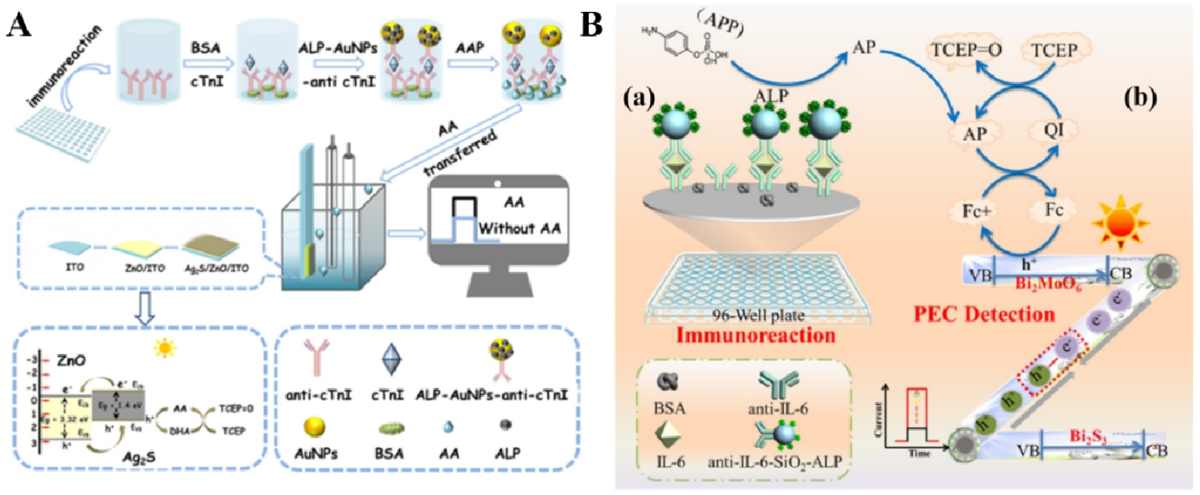
As mentioned above, redox-cycling-based amplification can be perfectly combined with an enzymatic reaction to repeatedly regenerate the consumed catalytic products based on well-coupled oxidation–reduction reactions. Given this concept, Cao and co-workers constructed a series of novel PEC platforms based on the fusion of redox cycling amplification and an appropriate photoelectrode for the detection of myoglobin and interleukin-6 (IL-6) [214,215,216,217]. A typical example is the detection of cTnI based on photogenerated hole-induced chemical redox cycling amplification [218]. As shown in Figure 18A, during the immunoreaction in a 96-well plate, ALP attached on AuNPs catalyzed the hydrolysis of AAP tothe signal-reporting species AA. Subsequently, AA served as the electron donor to quench the photogenerated holes of Ag2S/ZnO nanocomposites. The oxidation product (dehydroascorbic acid, DHA) at the electrode was repeatedly reduced to AA under the TCEP-mediated chemical redox cycling reaction, eventually leading to an enhanced PEC response. Comparatively, photogenerated hole-induced chemical−chemical (PECCC) redox cycling amplification involving the signaling species recycled by two different reducing (or oxidizing) agents and an appropriate photoelectrode can lead to much faster redox reactions and the regeneration of signaling species. Cao et al. reported a PEC method for IL-6 detection based on PECCC redox cycling for advanced signal amplification [219]. As shown in Figure 18B, the oxidation of Fc by the holes in the Z-scheme Bi2S3/Bi2MoO6 heterostructure photoelectrode under illumination triggered the PECCC redox cycling amplification system among the redox mediator Fc, the ALP-participated enzymatic generation of signaling unit AA, and the reducing agent TCEP. Under triple signal amplification, the proposed method exhibited a very low detection limit (2 × 10⁻¹⁴ g/mL)and a wide linear range (5 × 10⁻¹⁴ to 1 × 10⁻⁸ g/mL).

3.3. ALP-Mediated In Situ Growth or Bioetching of Photoelectrode

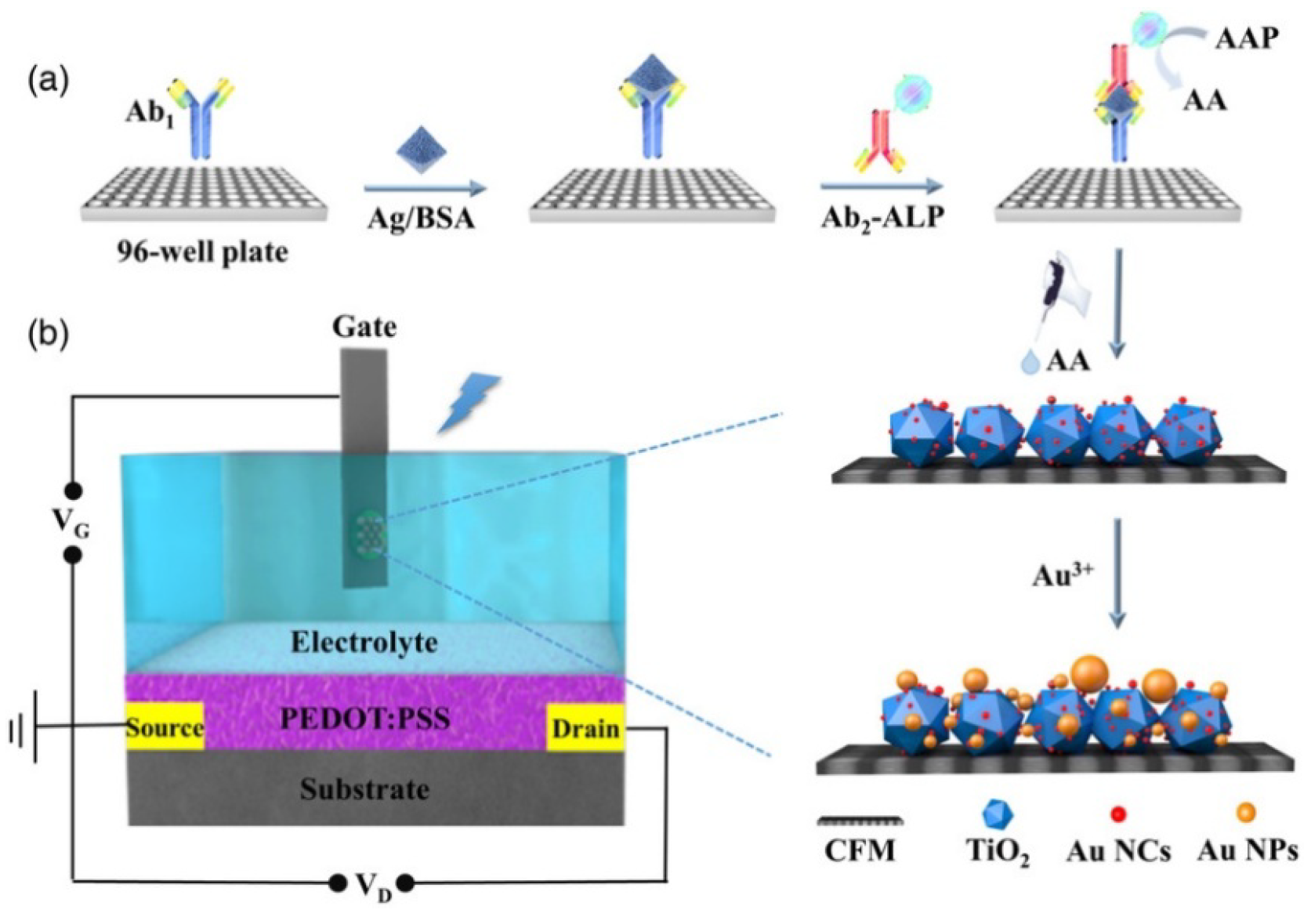
Apart from serving as electrode substrates or signal labels in sandwich assays, photoactive materials can be in situ grown on the surface of a photoelectrode to regulate PEC signals. The ALP-mediated in situ enzymatic growth of photoactive materials was popularly combined with PEC techniques for signal amplification. ALP-catalyzed reductive products, AA and IP, can reduce Au3+ and Ag+ ions to AuNPs and AgNPs on the photoelectrode, leading to a change in PEC signals [220]. Lu et al. developed an OPECT immunosensor for the detection of C-reactive protein (CRP) based on the ALP-mediated regulation of a light-sensitive gate electrode [221]. As displayed in Figure 19, ALP-conjugated mAb2 was used in the sandwich immunoassay. AuNCs as the photosensitizers were immobilized on TiO2 supported by a 3D carbon fiber matrix (CFM) to improve photon-to-electron conversion efficiency. The enzymatic product AA could reduce Au3+ions to AuNCs as the crystalline seeds to promote the formation of plasmonic AuNPs. The light source used was unable to effectively trigger the SPR effect, thereby decreasing the photon-to-electron conversion efficiency and weakening the PEC signal [222].
ALP can catalyze the decomposition of sodium thiophosphate (Na3SPO3, TP) into orthophosphate (PO43−) and H2S. The resulting H2S can react with metal ions or active molecules to in situ prepare materials that can change the PEC signal [223]. PEC immunosensors based on the ALP-mediated enzymatic in situ generation of QDs were developed for the detection of antibody and human serum albumin, in which the produced H2S interacted with Cd2+ to form CdS QDs that could be determined by the PEC technique [224,225]. Gao et al. developed a tunable competitive absorption-induced “signal-on” PEC immunosensor for cTnI detection based on the Zr-scheme MOF heterojunction and the enzyme-triggered growth of photoactive materials [226]. As shown in Figure 20A, a Zr-MOFs@TiO2 nanorods (NRs) electrode exhibiting a high photoelectric response was synthesized by a solvothermal method. The electrode modified with Cu(II) by an electrostatic interaction quenched the PEC signal. Zeolitic imidazolate framework-8 nanoparticles (ZIF-8 NMOFs) were loaded with ALP and mAb2 to form ZIF-8@ALP–mAb2 complexes. After the formation of the sandwich immunocomplexes in 96-well plates, the enzymatic product H2S competitively reacted with Cu(II) to quickly form CuS with a high negative potential of the conduction band (CB). The formed charge-carrier migration pathway resulted in the enhancement of the PEC signal. However, the type-I heterojunction was not a suitable candidate for PEC biosensing due to the unmatched CB and valence band (VB) levels of semiconductors A and B. The in situ combination or growth on a photoelectrode can endow a target-dependent type-I heterojunction with more possibilities in PEC bioassays. Gao et al. reported a liposome-aided type-I heterojunction growth method for a PEC immunoassay of h-FABP [227]. As illustrated in Figure 20B, a fluorine-doped tin oxide (FTO)/ZnInS nanosheets (ZIS NSs)-Sn(IV) electrode was fabricated as the working electrode. ALP-loaded liposome was used as the signal label in the sandwich immunoassay. Under the lysis treatment, the released ALP could catalyze the production of H2S to immediately react with Sn(IV) for the in situ formation of the ZIS NSs/SnS2 type-I heterojunction on the FTO/ZIS NSs-Sn(IV) electrode. This changed the photogenerated electron−hole transfer path of the photoelectrode, leading to a reduction in the current intensity.
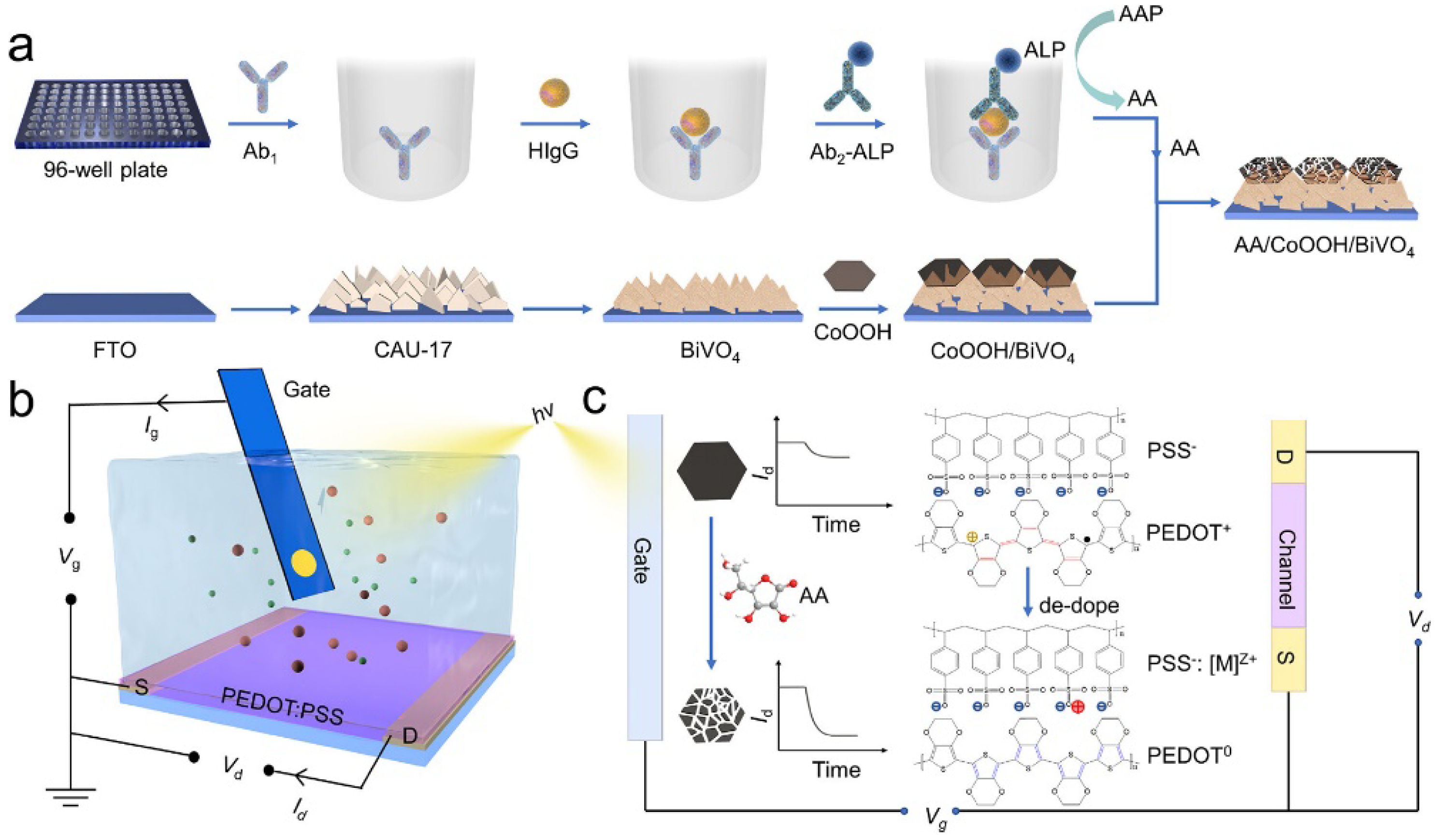
The enzymatic-bioetching-mediated photocurrent change/shift is a useful approach for PEC biosensing via the controllable dissociation of photoactive materials and the manipulation of the light-harvesting gates [204,228]. Cobalt oxyhydroxide (CoOOH) NSs exhibit an excellent light absorption capacity. They can be easily decomposed by the enzymatic product AA. CoOOH NSs coated on the electrode surface can block the electrolyte contact and light accessibility to the photoelectrode, leading to a decrease in the PEC signal. ALP-catalyzed product AA can etch CoOOH NSs and restore the PEC signal [229]. Zhang et al. developed a PEC biosensor for carcinoembryonic antigen (CEA) detection by coupling HCR with the ALP-mediated bioetching of CoOOH NSs [230]. Ban et al. reported a “signal-on” OPECT immunosensor for human IgG detection based on the ALP-mediated bioetching of the CoOOH/BiVO4 gate [231]. As shown in Figure 21, the FTO electrode was gradually covered with CAU-17 MOF-derived BiVO4 and CoOOH NSs, resulting in a PEC “signal-off” mode. After the completion of the ALP-linked sandwich immunoassay, the catalytic product AA was collected and then dropped onto the CoOOH/BiVO4-modified gate electrode. CoOOH NSs were etched by AA, and BiVO4 was partially exposed, resulting in the recovery of the PEC signal. The current change during the ALP-mediated bioetching process was monitored by the polymeric poly(3,4-ethylenedioxythiophene):poly(styrene sulfonate) (PEDOT:PSS) channel.
MnO2 NSs, with a band gap of about 2.1 eV and an absorption peak at around 380 nm near the visible regime, can be reduced to Mn2+ ions by the enzymatic product, such as H2O2 and AA. This mechanism was successfully used to develop MnO2 NSs-based biosensors [232,233]. For example, Lin et al. developed a PEC immunosensor for AFP detection based on the ALP-mediated bioetching of photoactive carbon quantum dots (CQDs)-functionalized MnO2 NSs [234]. In this work, the ALP-triggered dissolution of MnO2 NSs led to the release of CQDs from the electrode, followed by a decrease in the PEC current.
Table 2. Analytical performance of ALP-based PEC biosensors.
Detection PrincipleALP SubstratesTargetLinear RangeLODRef.
ALP-catalyzed products as electron donorsAAPM.SssIMTs1–50 unit/mL0.33 unit/mL[186]
AAPN6-methyladenosine0.005–35 nM2.57 pM[188]
AAPMiRNA5–3000 fM2.26 fM[192]
AAPAFP0.5–50 ng/mL37.9 pg/mL[199]
AAPPSA0.001–3 ng/mL0.32 pg/mL[201]
AAPHIV-p24 antigen1 pg/mL–50 ng/mL0.63 pg/mL[207]
AAPMicrocystin-LR0.05 ng/mL–5 μg/mL0.03 pg/mL[212]
ALP-mediated redox cyclingAAPMyoglobin1 × 10−4–100 ng/mL0.1 pg/mL[214]
AAPMyoglobin1 × 10−4–100 ng/mL0.03 pg/mL[215]
AAPTroponin I10 fg/mL–1 ng/mL3 fg/mL[218]
PAPPInterleukin-650 fg/mL–10 ng/mL20 fg/mL[219]
ALP-mediated in situ growth or bioetching of photoelectrodeAAPCRP1 pg/mL–200 ng/mL0.1 pg/mL[221]
AAPh-FABP0.5 pg/mL–50 ng/mL0.1 pg/mL[222]
thiophosphateTroponin I0.01–10 ng/mL8.6 pg/mL[226]
thiophosphateh-FABP0.1–1000 pg/mL55 fg/mL[227]
AAPAflatoxin B10.01–10 ng/mL2.6 pg/mL[229]
AAPCEA0.01–100 ng/mL5.2 pg/mL[230]
AAPHuman IgG1 × 10−4–100 ng/mL25 fg/mL[231]
AAPAlpha-fetoprotein0.01–100 ng/mL9.3 pg/mL[234]
Abbreviations: MTs, methyltransfease; AFP, alpha-fetoprotein; PSA, prostate specific antigen; h-FABP, heart-type fatty acid binding protein; AAP, 2-phospho-L-ascorbic acid; CEA, carcinoembryonic antigen.

4. ECL Methods

ECL is a type of luminescence produced from the combination of an electrochemical reaction and a chemiluminescence (CL) reaction. The technique possesses the advantages of both electrochemical and CL methods. During the ECL process, the electrochemically produced species at/near the electrode surface can react with each other to form the excited state of luminophore-related compounds, thus lighting up the luminescence. Unlike electrochemical biosensors, ECL assays are scarcely influenced by the background current and the potential window of the electrode. Thus, the ECL technique was successfully integrated with enzyme-linked immunoassays [235,236,237,238,239]. As the key components of ECL sensing platforms, various nanomaterials with good chemical stability and excellent signals were used as luminophores to construct various immunosensors. Like the Förster resonance energy transfer (FRET) mechanism in fluorescence biosensors, the excited-state nanomaterials can be quenched through a charge transfer or energy transfer. Given this concept, Yang et al. reported an ECL immunosensor for the detection of CEA based on the ALP-catalyzed in situ generation of molecular quenchers [240]. As shown in Figure 22A, a mixture of chitosan-multiwalled CNTs (MWCNTs) and CdTe QDs was deposited on the electrode. ALP-modified AuNPs were used to increase the number of enzyme labels per immunoreaction event. ALP-catalyzed product PNP was electrochemically oxidized to p-benzoquinone (PBQ), which could quench the luminescence of excited CdTe QDs through the energy transfer from QDs to PBQ.
AuNCs as fluorophores can produce an ECL signal and, meanwhile, serve as the seeds for gold metallization. Thus, an ALP-mediated immunoassay can be integrated with a AuNCs-based ECL system to modulate the signal through in situ metallization. Cao et al. proposed a novel strategy by coupling liposome, ALP catalysis, chemical redox cycling, and the in situ growth of AuNPs to develop an ECL immunosensor for PSA detection [241] As shown in Figure 22B, after the immunoreaction, many ALP molecules released from the liposome catalyzed the conversion of AAP to AA. The produced AA promoted the growth of AuNCs to AuNPs in the presence of Au3+ ions. In this process, AA was oxidized to DHA that could be immediately reduced back to AA by excess TCEP for the next reduction of Au3+. Under chemical redox cycling, the repeated regeneration of AA resulted in the formation of AuNPs and greatly enhanced the ECL intensity.

5. Conclusions and Perspectives

In recent years, in parallel with the significant progress in nanotechnology and bioconjugation chemistry, great advances in the development of ALP-based electrochemical immunoassays have been achieved. To increase the sensitivity and reduce the background, significant efforts have been made to integrate various signal-amplified strategies with electrochemical immunoassays. Many novel works and promising results were systematically summarized in this work. In particular, there was a significant achievement in the strategies by combining ALP catalysis with EC and ECC redox cycling for improving sensitivity. The loading of multiple ALP molecules on the nanomaterials with excellent electrocatalytic activity also significantly improved detection performance under the synergistic catalysis. Split-type immunoassays, by separating the immunoreaction from the detection system, can avoid the interference from complex samples and decrease the background signal. Abundant substrate/product pairs and effective ALP catalysis provide more promising ways to perfectly couple electrochemical immunoassays with emerging powerful strategies.
In spite of these advancements, there are still some challenges facing the applications of ALP-based electrochemical immunosensors. First, the requirement of multiple incubation and washing steps may limit the application of ALP-based immunoassays in terms of fast, on-line, and automated analyte detection. In addition, some methods were only used for the assays of samples in buffer solutions but not of real biological matrices. Second, for the preparation of enzyme-modified nanomaterials, existing immobilization methods may face several problems such as enzyme leaching and denaturation, complex processes, and decreased recognition ability. The in situ formation or encapsulation of enzymes on nanocomposites through mild and one-pot methods may be an effective solution to this problem. Third, considering the widespread application of nanomaterials and a nanostructured surface in multienzyme labeling and antibody immobilization, the size, shape, or composition of nanomaterials and the bioconjugation efficiency during the preparation of ALP-modified nanocomposites may affect the reproducibility and accuracy of measurement results. Fourth, although smartphone-based Point-of-Care Testing (POCT) has become a popular research hotspot due to its portability and low cost, most ALP-based immunoassays are limited to the requirements of strict experimental conditions, specialized instruments, and professional personnel, which may hamper the realization of POCT. Thus, greater efforts should be made to integrate ALP-based immunoassays into smartphone-based devices (e.g., lab-on-a-chip, microfluidic analysis, and μPAD).

Author Contributions

Conceptualization, C.C. and X.Y.; writing—original draft preparation, C.C., M.L. and M.H.; writing—review and editing, X.Y., N.X. and Y.Z.; project administration, Y.Z.; funding acquisition, Y.Z. All authors have read and agreed to the published version of the manuscript.

Funding

This research was funded by the Fund Project for Young Scholars Sponsored by Henan Province (No. 2020JS227) and the Science and Technology Development Program of Henan Province (Nos. 222102320057 and 232102321043).

Data Availability Statement

Not applicable.

Conflicts of Interest

The authors declare no conflict of interest.

References

  1. Chikkaveeraiah, B.V.; Bhirde, A.A.; Morgan, N.Y.; Eden, H.S.; Chen, X. Electrochemical immunosensors for detection of cancer protein biomarkers. ACS Nano 2012, 6, 6546–6561. [Google Scholar] [CrossRef] [PubMed]
  2. Kokkinos, C.; Economou, A.; Prodromidis, M.I. Electrochemical immunosensors: Critical survey of different architectures and transduction strategies. TrAC-Trend. Anal. Chem. 2016, 79, 88–105. [Google Scholar] [CrossRef]
  3. Felix, F.S.; Angnes, L. Electrochemical immunosensors—A powerful tool for analytical applications. Biosens. Bioelectron. 2018, 102, 470–478. [Google Scholar] [CrossRef] [PubMed]
  4. Puiu, M.; Nativi, C.; Bala, C. Early detection of tumour-associated antigens: Assessment of point-of-care electrochemical immunoassays. TrAC-Trend. Anal. Chem. 2023, 160, 116981. [Google Scholar] [CrossRef]
  5. Kim, J.; Park, M. Recent progress in electrochemical immunosensors. Biosensors 2021, 11, 360. [Google Scholar] [CrossRef] [PubMed]
  6. Zherdev, A.V.; Dzantiev, B.B. Detection limits of immunoanalytical systems: Limiting factors and methods of reduction. J. Anal. Chem. 2022, 77, 391–401. [Google Scholar] [CrossRef]
  7. Campuzano, S.; Pedrero, M.; Yáñez-Sedeño, P.; Pingarrón, J.M. Nanozymes in electrochemical affinity biosensing. Microchim. Acta 2020, 187, 423–438. [Google Scholar] [CrossRef] [PubMed]
  8. Zheng, Y.; Li, J.; Zhou, B.; Ian, H.; Shao, H. Advanced sensitivity amplification strategies for voltammetric immunosensors of tumor marker: State of the art. Biosens. Bioelectron. 2021, 178, 113021–113042. [Google Scholar] [CrossRef]
  9. Police Patil, A.V.; Chuang, Y.S.; Li, C.; Wu, C.C. Recent advances in electrochemical immunosensors with nanomaterial assistance for signal amplification. Biosensors 2023, 13, 125. [Google Scholar] [CrossRef]
  10. Xia, N.; Huang, Y.; Zhao, Y.; Wang, F.; Liu, L.; Sun, Z. Electrochemical biosensors by in situ dissolution of self-assembled nanolabels into small monomers on electrode surface. Sens. Actuators B Chem. 2020, 325, 128777. [Google Scholar] [CrossRef]
  11. Jarczewska, M.; Malinowska, E. The application of antibody-aptamer hybrid biosensors in clinical diagnostics and environmental analysis. Anal. Methods 2020, 12, 3183–3199. [Google Scholar] [CrossRef] [PubMed]
  12. Liu, L.; Deng, D.; Wu, D.; Hou, W.; Wang, L.; Li, N.; Sun, Z. Duplex-specific nuclease-based electrochemical biosensor for the detection of microRNAs by conversion of homogeneous assay into surface-tethered electrochemical analysis. Anal. Chim. Acta 2021, 1149, 338199. [Google Scholar] [CrossRef] [PubMed]
  13. Kuramitz, H. Magnetic microbead-based electrochemical immunoassays. Anal. Bioanal. Chem. 2009, 394, 61–69. [Google Scholar] [CrossRef] [PubMed]
  14. Masud, M.K.; Na, J.; Younus, M.; Hossain, M.S.A.; Bando, Y.; Shiddiky, M.J.A.; Yamauchi, Y. Superparamagnetic nanoarchitectures for disease-specific biomarker detection. Chem. Soc. Rev. 2019, 48, 5717–5751. [Google Scholar] [CrossRef]
  15. Kavetskyy, T.; Alipour, M.; Smutok, O.; Mushynska, O.; Kiv, A.; Fink, D.; Farshchi, F.; Ahmadian, E.; Hasanzadeh, M. Magneto-immunoassay of cancer biomarkers: Recent progress and challenges in biomedical analysis. Microchem. J. 2021, 167, 106320–106332. [Google Scholar] [CrossRef]
  16. Li, X.-M.; Yang, X.-Y.; Zhang, S.-S. Electrochemical enzyme immunoassay using model labels. TrAC-Trend. Anal. Chem. 2008, 27, 543–553. [Google Scholar] [CrossRef]
  17. Yang, H. Enzyme-based ultrasensitive electrochemical biosensors. Curr. Opin. Chem. Biol. 2012, 16, 422–428. [Google Scholar] [CrossRef] [PubMed]
  18. Pollap, A.; Kochana, J. Electrochemical immunosensors for antibiotic detection. Biosensors 2019, 9, 61. [Google Scholar] [CrossRef]
  19. Noji, H.; Minagawa, Y.; Ueno, H. Enzyme-based digital bioassay technology-key strategies and future perspectives. Lab. Chip 2022, 22, 3092–3109. [Google Scholar] [CrossRef]
  20. Xia, N.; Deng, D.; Yang, S.; Hao, Y.; Wang, L.; Liu, Y.; An, C.; Han, Q.; Liu, L. Electrochemical immunosensors with protease as the signal label for the generation of peptide-Cu(II) complexes as the electrocatalysts toward water oxidation. Sens. Actuators. B Chem. 2019, 291, 113–119. [Google Scholar] [CrossRef]
  21. Wignarajah, S.; Chianella, I.; Tothill, I.E. Development of electrochemical immunosensors for HER-1 and HER-2 analysis in serum for breast cancer patients. Biosensors 2023, 13, 355. [Google Scholar] [CrossRef] [PubMed]
  22. Manes, T.; Hoylaerts, M.F.; Müller, R.; Lottspeich, F.; Holke, W.; Millán, J.L. Genetic complexity, structure, and characterization of highly active bovine intestinal alkaline phosphatases. J. Biol. Chem. 1998, 273, 23353–23360. [Google Scholar] [CrossRef] [PubMed]
  23. Gu, C.; Gai, P.; Hou, T.; Li, H.; Xue, C.; Li, F. Enzymatic fuel cell-based self-powered homogeneous immunosensing platform via target-induced glucose release: An appealing alternative strategy for turn-on melamine assay. ACS Appl. Mater. Interfaces 2017, 9, 35721–35728. [Google Scholar] [CrossRef] [PubMed]
  24. Shaban, S.M.; Byeok Jo, S.; Hafez, E.; Ho Cho, J.; Kim, D.-H. A comprehensive overview on alkaline phosphatase targeting and reporting assays. Coordin. Chem. Rev. 2022, 465, 214567–214604. [Google Scholar] [CrossRef]
  25. Kanno, Y.; Zhou, Y.; Fukuma, T.; Takahashi, Y. Alkaline phosphatase-based electrochemical analysis for point-of-care testing. Electroanalysis 2021, 34, 161–167. [Google Scholar] [CrossRef]
  26. Nsabimana, A.; Lan, Y.; Du, F.; Wang, C.; Zhang, W.; Xu, G. Alkaline phosphatase-based electrochemical sensors for health applications. Anal. Methods 2019, 11, 1996–2006. [Google Scholar] [CrossRef]
  27. Xia, N.; Huang, Y.; Cui, Z.; Liu, S.; Deng, D.; Liu, L.; Wang, J. Impedimetric biosensor for assay of caspase-3 activity and evaluation of cell apoptosis using self-assembled biotin-phenylalanine network as signal enhancer. Sens. Actuators B Chem. 2020, 320, 128436. [Google Scholar] [CrossRef]
  28. Mollarasouli, F.; Kurbanoglu, S.; Ozkan, S.A. The role of electrochemical immunosensors in clinical analysis. Biosensors 2019, 9, 86. [Google Scholar] [CrossRef]
  29. Plikusiene, I.; Ramanaviciene, A. Investigation of biomolecule interactions: Optical-, electrochemical-, and acoustic-based biosensors. Biosensors 2023, 13, 292. [Google Scholar] [CrossRef]
  30. Zhang, X.; Li, C.R.; Wang, W.C.; Xue, J.; Huang, Y.L.; Yang, X.X.; Tan, B.; Zhou, X.P.; Shao, C.; Ding, S.J.; et al. A novel electrochemical immunosensor for highly sensitive detection of aflatoxin B1 in corn using single-walled carbon nanotubes/chitosan. Food Chem. 2016, 192, 197–202. [Google Scholar] [CrossRef]
  31. Freitas, M.; Neves, M.; Nouws, H.P.A.; Delerue-Matos, C. Electrochemical immunosensor for the simultaneous determination of two main peanut allergenic proteins (Ara h 1 and Ara h 6) in food matrices. Foods 2021, 10, 1718. [Google Scholar] [CrossRef] [PubMed]
  32. El-Moghazy, A.Y.; Huo, J.; Amaly, N.; Vasylieva, N.; Hammock, B.D.; Sun, G. An innovative nanobody-based electrochemical immunosensor using decorated nylon nanofibers for point-of-care monitoring of human exposure to pyrethroid insecticides. ACS Appl. Mater. Interfaces 2020, 12, 6159–6168. [Google Scholar] [CrossRef] [PubMed]
  33. Bauer, C.G.; Eremenko, A.V.; Ehrentreich-Forster, E.; Bier, F.F.; Makower, A.; Halsall, H.B.; Heineman, W.R.; Scheller, F.W. Zeptomole-detecting biosensor for alkaline phosphatase in an electrochemical immunoassay for 2,4-dichlorophenoxyacetic acid. Anal. Chem. 1996, 68, 2453–2458. [Google Scholar] [CrossRef] [PubMed]
  34. Sharma, P.; Bhalla, V.; Tuteja, S.; Kukkar, M.; Suri, C.R. Rapid extraction and quantitative detection of the herbicide diuron in surface water by a hapten-functionalized carbon nanotubes based electrochemical analyzer. Analyst 2012, 137, 2495–2502. [Google Scholar] [CrossRef] [PubMed]
  35. Kanso, H.; Barthelmebs, L.; Inguimbert, N.; Noguer, T. Immunosensors for estradiol and ethinylestradiol based on new synthetic estrogen derivatives: Application to wastewater analysis. Anal. Chem. 2013, 85, 2397–2404. [Google Scholar] [CrossRef] [PubMed]
  36. Wehmeyer, K.R.; Halsall, H.B.; Heineman, W.R.; Volle, C.P.; Chen, I.W. Competitive heterogeneous enzyme immunoassay for digoxin with electrochemical detection. Anal. Chem. 1986, 58, 135–139. [Google Scholar] [CrossRef] [PubMed]
  37. Hart, J.P.; Pemberton, R.M.; Luxton, R.; Wedge, R. Studies towards a disposable screen-printed amperometric biosensor for progesterone. Biosens. Bioelectron. 1997, 12, 1113–1121. [Google Scholar] [CrossRef]
  38. Tang, H.T.; Lunte, C.E.; Halsall, H.B.; Heineman, W.R. p-Aminophenyl phosphate: An improved substrate for electrochemical enzyme immnoassay. Anal. Chim. Acta 1988, 214, 187–195. [Google Scholar] [CrossRef]
  39. Niwa, O.; Xu, Y.; Halsall, H.B.; Heineman, W.R. Small-volume voltammetric detection of 4-aminophenol with interdigitated array electrodes and its application to electrochemical enzyme immunoassay. Anal. Chem. 1993, 65, 1559–1563. [Google Scholar] [CrossRef]
  40. Duan, C.; Meyerhoff, M.E. Separation-free sandwich enzyme immunoassays using microporous gold electrodes and self-assembled monolayer/immobilized capture antibodies. Anal. Chem. 1994, 66, 1369–1377. [Google Scholar] [CrossRef]
  41. Treloar, P.H.; Nkohkwo, A.a.T.; Kane, J.W.; Barber, D.; Vadgama, P.M. Electrochemical immunoassay: Simple kinetic detection of alkaline phosphatase enzyme labels in limited and excess reagent systems. Electroanalysis 1994, 6, 561–566. [Google Scholar] [CrossRef]
  42. Yu, Z.; Xu, Y.; Ip, M.P. An ultra-sensitive electrochemical enzyme immunoassay for thyroid stimulating hormone in human serum. J. Pharm. Biomed. Anal. 1994, 12, 787–793. [Google Scholar] [CrossRef]
  43. Pemberton, R.M.; Hart, J.P.; Foulkes, J.A. Development of a sensitive, selective electrochemical immunoassay for progesterone in cow’s milk based on a disposable screen-printed amperometric biosensor. Electrochim. Acta 1998, 43, 3567–3574. [Google Scholar] [CrossRef]
  44. Wang, J.; Ibanez, A.; Chatrathi, M.P.; Escarpa, A. Electrochemical enzyme immunoassays on microchip platforms. Anal. Chem. 2001, 73, 5323–5327. [Google Scholar] [CrossRef] [PubMed]
  45. O’Regan, T.M.; Pravda, M.; O’Sullivan, C.K.; Guilbault, G.G. Development of a disposable immunosensor for the detection of human heart fatty-acid binding protein in human whole blood using screen-printed carbon electrodes. Talanta 2002, 57, 501–510. [Google Scholar] [CrossRef] [PubMed]
  46. Tang, T.C.; Deng, A.; Huang, H.J. Immunoassay with a microtiter plate incorporated multichannel electrochemical detection system. Anal. Chem. 2002, 74, 2617–2621. [Google Scholar] [CrossRef] [PubMed]
  47. Rao, A.K.; Creager, S.E. Superporous agarose-reticulated vitreous carbon electrodes for electrochemical sandwich bioassays. Anal. Chim. Acta 2008, 628, 190–197. [Google Scholar] [CrossRef]
  48. Thompson, R.Q.; Porter, M.; Stuver, C.; Halsall, H.B.; Heineman, W.R.; Buckley, E.; Smyth, M.R. Zeptomole detection limit for alkaline phosphatase using 4-aminophenylphosphate, amperometric detection, and an optimal buffer system. Anal. Chim. Acta 1993, 271, 223–229. [Google Scholar] [CrossRef]
  49. Regiart, M.; Rinaldi-Tosi, M.; Aranda, P.R.; Bertolino, F.A.; Villarroel-Rocha, J.; Sapag, K.; Messina, G.A.; Raba, J.; Fernandez-Baldo, M.A. Development of a nanostructured immunosensor for early and in situ detection of Xanthomonas arboricola in agricultural food production. Talanta 2017, 175, 535–541. [Google Scholar] [CrossRef]
  50. Liu, Y.; Wang, H.; Chen, J.; Liu, C.; Li, W.; Kong, J.; Yang, P.; Liu, B. A sensitive microchip-based immunosensor for electrochemical detection of low-level biomarker S100B. Electroanalysis 2013, 25, 1050–1055. [Google Scholar] [CrossRef]
  51. Serafin, V.; Ubeda, N.; Agui, L.; Yanez-Sedeno, P.; Pingarron, J.M. Ultrasensitive determination of human growth hormone (hGH) with a disposable electrochemical magneto-immunosensor. Anal. Bioanal. Chem. 2012, 403, 939–946. [Google Scholar] [CrossRef] [PubMed]
  52. Pandey, B.; Demchenko, A.V.; Stine, K.J. Nanoporous gold as a solid support for protein immobilization and development of an electrochemical immunoassay for prostate specific antigen and carcinoembryonic antigen. Microchim. Acta 2012, 179, 71–81. [Google Scholar] [CrossRef] [PubMed]
  53. Hu, Y.; Zhao, Z.; Wan, Q. Facile preparation of carbon nanotube-conducting polymer network for sensitive electrochemical immunoassay of Hepatitis B surface antigen in serum. Bioelectrochemistry 2011, 81, 59–64. [Google Scholar] [CrossRef] [PubMed]
  54. Wilson, M.S. Electrochemical immunosensors for the simultaneous detection of two tumor markers. Anal. Chem. 2005, 77, 1496–1502. [Google Scholar] [CrossRef] [PubMed]
  55. Wilson, M.S.; Rauh, R.D. Hydroquinone diphosphate: An alkaline phosphatase substrate that does not produce electrode fouling in electrochemical immunoassays. Biosens. Bioelectron. 2004, 20, 276–283. [Google Scholar] [CrossRef] [PubMed]
  56. Wilson, M.S.; Nie, W. Electrochemical multianalyte immunoassays using an array-based sensor. Anal. Chem. 2006, 78, 2507–2513. [Google Scholar] [CrossRef]
  57. Wilson, M.S.; Nie, W. Multiplex measurement of seven tumor markers using an electrochemical protein chip. Anal. Chem. 2006, 78, 6476–6483. [Google Scholar] [CrossRef] [PubMed]
  58. Wilson, M.S.; Rauh, R.D. Novel amperometric immunosensors based on iridium oxide matrices. Biosens. Bioelectron. 2004, 19, 693–699. [Google Scholar] [CrossRef]
  59. Zhang, Y.; Fan, Y.; Wu, J.; Wang, X.; Liu, Y. An amperometric immunosensor based on an ionic liquid and single-walled carbon nanotube composite electrode for detection of tetrodotoxin in pufferfish. J. Agric. Food Chem. 2016, 64, 6888–6894. [Google Scholar] [CrossRef]
  60. Yang, Z.; Jiang, W.; Liu, F.; Zhou, Y.; Yin, H.; Ai, S. A novel electrochemical immunosensor for the quantitative detection of 5-hydroxymethylcytosine in genomic DNA of breast cancer tissue. Chem. Commun. 2015, 51, 14671–14673. [Google Scholar] [CrossRef]
  61. Ding, J.; Wang, X.; Qin, W. Pulsed galvanostatic control of a polymeric membrane ion-selective electrode for potentiometric immunoassays. ACS Appl. Mater. Interfaces 2013, 5, 9488–9493. [Google Scholar] [CrossRef] [PubMed]
  62. Moreno-Guzman, M.; Ojeda, I.; Villalonga, R.; Gonzalez-Cortes, A.; Yanez-Sedeno, P.; Pingarron, J.M. Ultrasensitive detection of adrenocorticotropin hormone (ACTH) using disposable phenylboronic-modified electrochemical immunosensors. Biosens. Bioelectron. 2012, 35, 82–86. [Google Scholar] [CrossRef] [PubMed]
  63. Del Carlo, M.; Lionti, I.; Taccini, M.; Cagnini, A.; Mascini, M. Disposable screen-printed electrodes for the immunochemical detection of polychlorinated biphenyls. Anal. Chim. Acta 1997, 342, 189–197. [Google Scholar] [CrossRef]
  64. Masson, M.; Runarsson, O.V.; Johannson, F.; Aizawa, M. 4-Amino-1-naphthylphosphate as a substrate for the amperometric detection of alkaline phosphatase activity and its application for immunoassay. Talanta 2004, 64, 174–180. [Google Scholar] [CrossRef]
  65. Carralero, V.; González-Cortés, A.; Yáñez-Sedeño, P.; Pingarrón, J.M. Development of a progesterone immunosensor based on a colloidal gold-graphite-teflon composite electrode. Electroanalysis 2007, 19, 853–858. [Google Scholar] [CrossRef]
  66. Aziz, M.A.; Jo, K.; Qaium, M.A.; Huh, C.-H.; Hong, I.S.; Yang, H. Platform for highly sensitive alkaline phosphatase-based immunosensors using 1-naphthyl phosphate and an avidin-modified indium tin oxide electrode. Electroanalysis 2009, 21, 2160–2164. [Google Scholar] [CrossRef]
  67. Colozza, N.; Mazzaracchio, V.; Kehe, K.; Tsoutsoulopoulos, A.; Schioppa, S.; Fabiani, L.; Steinritz, D.; Moscone, D.; Arduini, F. Development of novel carbon black-based heterogeneous oligonucleotide-antibody assay for sulfur mustard detection. Sens. Actuators B Chem. 2021, 328, 129054–129064. [Google Scholar] [CrossRef]
  68. Yugender Goud, K.; Sunil Kumar, V.; Hayat, A.; Vengatajalabathy Gobi, K.; Song, H.; Kim, K.-H.; Marty, J.L. A highly sensitive electrochemical immunosensor for zearalenone using screen-printed disposable electrodes. J. Electroanal. Chem. 2019, 832, 336–342. [Google Scholar] [CrossRef]
  69. Nelis, J.L.D.; Migliorelli, D.; Muhlebach, L.; Generelli, S.; Stewart, L.; Elliott, C.T.; Campbell, K. Highly sensitive electrochemical detection of the marine toxins okadaic acid and domoic acid with carbon black modified screen printed electrodes. Talanta 2021, 228, 122215–122223. [Google Scholar] [CrossRef]
  70. Zhou, Y.; Jiang, W.; Wu, H.; Liu, F.; Yin, H.; Lu, N.; Ai, S. Amplified electrochemical immunoassay for 5-methylcytosine using a nanocomposite prepared from graphene oxide, magnetite nanoparticles and β-cyclodextrin. Microchim. Acta 2019, 186, 488–497. [Google Scholar] [CrossRef]
  71. Lin, J.; He, C.; Zhang, S. Immunoassay channels for α-fetoprotein based on encapsulation of biorecognition molecules into SBA-15 mesopores. Anal. Chim. Acta 2009, 643, 90–94. [Google Scholar] [CrossRef] [PubMed]
  72. Liu, X.P.; Deng, Y.J.; Jin, X.Y.; Chen, L.G.; Jiang, J.H.; Shen, G.L.; Yu, R.Q. Ultrasensitive electrochemical immunosensor for ochratoxin A using gold colloid-mediated hapten immobilization. Anal. Biochem. 2009, 389, 63–68. [Google Scholar] [CrossRef] [PubMed]
  73. Bonel, L.; Vidal, J.C.; Duato, P.; Castillo, J.R. Ochratoxin A nanostructured electrochemical immunosensors based on polyclonal antibodies and gold nanoparticles coupled to the antigen. Anal. Methods 2010, 2, 335–341. [Google Scholar] [CrossRef]
  74. Xu, W.; Qing, Y.; Chen, S.; Chen, J.; Qin, Z.; Qiu, J.; Li, C. Electrochemical indirect competitive immunoassay for ultrasensitive detection of zearalenone based on a glassy carbon electrode modified with carboxylated multi-walled carbon nanotubes and chitosan. Microchim. Acta 2017, 184, 3339–3347. [Google Scholar] [CrossRef]
  75. Karczmarczyk, A.; Baeumner, A.J.; Feller, K.-H. Rapid and sensitive inhibition-based assay for the electrochemical detection of ochratoxin A and aflatoxin M1 in red wine and milk. Electrochim. Acta 2017, 243, 82–89. [Google Scholar] [CrossRef]
  76. Yang, X.; Zhou, X.; Zhang, X.; Qing, Y.; Luo, M.; Liu, X.; Li, C.; Li, Y.; Xia, H.; Qiu, J. A highly sensitive electrochemical immunosensor for fumonisin B1 detection in corn using single-walled carbon nanotubes/chitosan. Electroanalysis 2015, 27, 2679–2687. [Google Scholar] [CrossRef]
  77. Ojeda, I.; Moreno-Guzmán, M.; González-Cortés, A.; Yáñez-Sedeño, P.; Pingarrón, J.M. Electrochemical magnetic immunosensors for the determination of ceruloplasmin. Electroanalysis 2013, 25, 2166–2174. [Google Scholar] [CrossRef]
  78. Ojeda, I.; Moreno-Guzmán, M.; González-Cortés, A.; Yáñez-Sedeño, P.; Pingarrón, J.M. A disposable electrochemical immunosensor for the determination of leptin in serum and breast milk. Analyst 2013, 138, 4284–4291. [Google Scholar] [CrossRef]
  79. Al-Khafaji, Q.A.M.; Harris, M.; Tombelli, S.; Laschi, S.; Turner, A.P.F.; Mascini, M.; Marrazza, G. An electrochemical immunoassay for HER2 detection. Electroanalysis 2012, 24, 735–742. [Google Scholar] [CrossRef]
  80. Yin, Z.; Liu, Y.; Jiang, L.P.; Zhu, J.J. Electrochemical immunosensor of tumor necrosis factor α based on alkaline phosphatase functionalized nanospheres. Biosens. Bioelectron. 2011, 26, 1890–1894. [Google Scholar] [CrossRef]
  81. Chen, S.P.; Yu, X.D.; Xu, J.J.; Chen, H.Y. Gold nanoparticles-coated magnetic microspheres as affinity matrix for detection of hemoglobin A1c in blood by microfluidic immunoassay. Biosens. Bioelectron. 2011, 26, 4779–4784. [Google Scholar] [CrossRef]
  82. Liu, X.; Wu, H.; Zheng, Y.; Wu, Z.; Jiang, J.; Shen, G.; Yu, R. A sensitive electrochemical immunosensor for α-fetoprotein detection with colloidal gold-based dentritical enzyme complex amplification. Electroanalysis 2010, 22, 244–250. [Google Scholar] [CrossRef]
  83. Lin, J.; He, C.; Zhang, L.; Zhang, S. Sensitive amperometric immunosensor for α-fetoprotein based on carbon nanotube/gold nanoparticle doped chitosan film. Anal. Biochem. 2009, 384, 130–135. [Google Scholar] [CrossRef] [PubMed]
  84. Hao, J.; Li, C.; Wu, K.; Hu, C.; Yang, N. Detection of tumor marker using ZnO@reduced graphene oxide decorated with alkaline phosphatase-labeled magnetic beads. ACS Appl. Nano Mater. 2019, 2, 7747–7754. [Google Scholar] [CrossRef]
  85. Fernández-Sánchez, C.; Costa-García, A. 3-Indoxyl phosphate: An alkaline phosphatase substrate for enzyme immunoassays with voltammetric detection. Electroanalysis 1998, 10, 249–255. [Google Scholar] [CrossRef]
  86. Fanjul-Bolado, P.; Gonzalez-Garcia, M.B.; Costa-Garcia, A. Voltammetric determination of alkaline phosphatase and horseradish peroxidase activity using 3-indoxyl phosphate as substrate Application to enzyme immunoassay. Talanta 2004, 64, 452–457. [Google Scholar] [CrossRef] [PubMed]
  87. Diaz-Gonzalez, M.; Hernandez-Santos, D.; Gonzalez-Garcia, M.B.; Costa-Garcia, A. Development of an immunosensor for the determination of rabbit IgG using streptavidin modified screen-printed carbon electrodes. Talanta 2005, 65, 565–573. [Google Scholar] [CrossRef] [PubMed]
  88. Fernandez-Sanchez, C.; Gonzalez-Garcia, M.B.; Costa-Garcia, A. AC voltammetric carbon paste-based enzyme immunosensors. Biosens. Bioelectron. 2000, 14, 917–924. [Google Scholar] [CrossRef]
  89. Ruan, C.; Yang, L.; Li, Y. Immunobiosensor chips for detection of Escherichia coil O157:H7 using electrochemical impedance spectroscopy. Anal. Chem. 2002, 74, 4814–4820. [Google Scholar] [CrossRef]
  90. Song, S.; Kim, Y.J.; Shin, I.-S.; Kim, W.-H.; Lee, K.-N.; Seong, W.K. Electrochemical immunoassay based on indium tin oxide activity toward a alkaline phosphatase. BioChip J. 2019, 13, 387–393. [Google Scholar] [CrossRef]
  91. Kokado, A.; Arakawa, H.; Maeda, M. New electrochemical assay of alkaline phosphatase using ascorbic acid 2-phosphate and its application to enzyme immunoassay. Anal. Chim. Acta 2000, 407, 119–125. [Google Scholar] [CrossRef]
  92. Kim, S.-E.; Kim, Y.J.; Song, S.; Lee, K.-N.; Seong, W.K. A simple electrochemical immunosensor platform for detection of Apolipoprotein A1 (Apo-A1) as a bladder cancer biomarker in urine. Sens. Actuators B Chem. 2019, 278, 103–109. [Google Scholar] [CrossRef]
  93. Lee, S.E.; Jeong, S.E.; Hong, J.S.; Im, H.; Hwang, S.Y.; Oh, J.K.; Kim, S.E. Gold-nanoparticle-coated magnetic beads for ALP-enzyme-based electrochemical immunosensing in human plasma. Materials 2022, 15, 6875. [Google Scholar] [CrossRef] [PubMed]
  94. Lin, J.; Liu, J.; Xu, J. Ultrasensitive electrochemical immunoassay for screening the influenza A (H1N1) virus based on atomically Ru-dispersed nitrogen-doped carbon. New J. Chem. 2023, 47, 1685–1690. [Google Scholar] [CrossRef]
  95. Pemberton, R.M.; Hart, J.P.; Stoddard, P.; Foulkes, J.A. A comparison of 1-naphthyl phosphate and 4 aminophenyl phosphate as enzyme substrates for use with a screen-printed amperometric immunosensor for progesterone in cows’ milk. Biosens. Bioelectron. 1999, 14, 495–503. [Google Scholar] [CrossRef] [PubMed]
  96. Kreuzer, M.P.; O’Sullivan, C.K.; Guilbault, G.G. Alkaline phosphatase as a label for immunoassay using amperometric detection with a variety of substrates and an optimal buffer system. Anal. Chim. Acta 1999, 393, 95–102. [Google Scholar] [CrossRef]
  97. Preechaworapun, A.; Dai, Z.; Xiang, Y.; Chailapakul, O.; Wang, J. Investigation of the enzyme hydrolysis products of the substrates of alkaline phosphatase in electrochemical immunosensing. Talanta 2008, 76, 424–431. [Google Scholar] [CrossRef]
  98. Chen, J.; Zou, G.; Zhang, X.; Jin, W. Ultrasensitive electrochemical immunoassay based on counting single magnetic nanobead by a combination of nanobead amplification and enzyme amplification. Electrochem. Commun. 2009, 11, 1457–1459. [Google Scholar] [CrossRef]
  99. Preechaworapun, A.; Ivandini, T.A.; Suzuki, A.; Fujishima, A.; Chailapakul, O.; Einaga, Y. Development of amperometric immunosensor using boron-doped diamond with poly(o-aminobenzoic acid). Anal. Chem. 2008, 80, 2077–2083. [Google Scholar] [CrossRef]
  100. Nam, E.J.; Kim, E.J.; Wark, A.W.; Rho, S.; Kim, H.; Lee, H.J. Highly sensitive electrochemical detection of proteins using aptamer-coated gold nanoparticles and surface enzyme reactions. Analyst 2012, 137, 2011–2016. [Google Scholar] [CrossRef]
  101. Safaei, T.S.; Mohamadi, R.M.; Sargent, E.H.; Kelley, S.O. In situ electrochemical ELISA for specific identification of captured cancer cells. ACS Appl. Mater. Interfaces 2015, 7, 14165–14169. [Google Scholar] [CrossRef]
  102. Diba, F.S.; Kim, S.; Lee, H.J. Electrochemical immunoassay for amyloid-beta 1–42 peptide in biological fluids interfacing with a gold nanoparticle modified carbon surface. Catal. Today 2017, 295, 41–47. [Google Scholar] [CrossRef]
  103. Guerrero, S.; Agui, L.; Yanez-Sedeno, P.; Pingarron, J.M. Design of electrochemical immunosensors using electro-click chemistry. Application to the detection of IL-1β cytokine in saliva. Bioelectrochemistry 2020, 133, 107484–107490. [Google Scholar] [CrossRef] [PubMed]
  104. Li, S.; Yan, Y.; Zhong, L.; Liu, P.; Sang, Y.; Cheng, W.; Ding, S. Electrochemical sandwich immunoassay for the peptide hormone prolactin using an electrode modified with graphene, single walled carbon nanotubes and antibody-coated gold nanoparticles. Microchim. Acta 2015, 182, 1917–1924. [Google Scholar] [CrossRef]
  105. Thiruppathiraja, C.; Saroja, V.; Kamatchiammal, S.; Adaikkappan, P.; Alagar, M. Development of electrochemical based sandwich enzyme linked immunosensor for Cryptosporidium parvum detection in drinking water. J. Environ. Monit. 2011, 13, 2782–2787. [Google Scholar] [CrossRef] [PubMed]
  106. Dzantiev, B.B. New and improved nanomaterials and approaches for optical bio- and immunosensors. Biosensors 2023, 13, 443. [Google Scholar] [CrossRef]
  107. Xia, N.; Wu, D.; Sun, T.; Wang, Y.; Ren, X.; Zhao, F.; Liu, L.; Yi, X. Magnetic bead-based electrochemical and colorimetric methods for the detection of poly(ADP-ribose) polymerase-1 with boronic acid derivatives as the signal probes. Sens. Actuators B. Chem. 2021, 327, 128913. [Google Scholar] [CrossRef]
  108. Xia, N.; Wu, D.; Yu, H.; Sun, W.; Yi, X.; Liu, L. Magnetic bead-based electrochemical and colorimetric assays of circulating tumor cells with boronic acid derivatives as the recognition elements and signal probes. Talanta 2021, 221, 121640. [Google Scholar] [CrossRef]
  109. Cao, L.; Fang, C.; Zeng, R.; Zhao, X.; Jiang, Y.; Chen, Z. Paper-based microfluidic devices for electrochemical immunofiltration analysis of human chorionic gonadotropin. Biosens. Bioelectron. 2017, 92, 87–94. [Google Scholar] [CrossRef]
  110. Hou, Y.; Li, T.; Huang, H.; Quan, H.; Miao, X.; Yang, M. Electrochemical immunosensor for the detection of tumor necrosis factor α based on hydrogel prepared from ferrocene modified amino acid. Sens. Actuators B Chem. 2013, 182, 605–609. [Google Scholar] [CrossRef]
  111. Xia, N.; Deng, D.; Mu, X.; Liu, A.; Xie, J.; Zhou, D.; Yang, P.; Xing, Y.; Liu, L. Colorimetric immunoassays based on pyrroloquinoline quinone-catalyzed generation of Fe(II)-ferrozine with tris(2-carboxyethyl)phosphine as the reducing reagent. Sens. Actuators B Chem. 2020, 306, 127571. [Google Scholar] [CrossRef]
  112. Kang, C.; Kang, J.; Lee, N.S.; Yoon, Y.H.; Yang, H. DT-diaphorase as a bifunctional enzyme label that allows rapid enzymatic amplification and electrochemical redox cycling. Anal. Chem. 2017, 89, 7974–7980. [Google Scholar] [CrossRef] [PubMed]
  113. Ito, K.Y.; Inoue, K.; Ito-Sasaki, T.; Ino, K.; Shiku, H. Electrochemical immunoassay with dual-signal amplification for redox cycling within a nanoscale gap. ACS Appl. Nano Mater. 2021, 4, 12393–12400. [Google Scholar] [CrossRef]
  114. Ito, K.K.Y.I.; Ino, K.; Shiku, H. High-sensitivity amperometric dual immunoassay using two cascade reactions with signal amplification of redox cycling in nanoscale gap. Anal. Chem. 2022, 94, 16451–16460. [Google Scholar] [CrossRef] [PubMed]
  115. Park, S.; Singh, A.; Kim, S.; Yang, H. Electroreduction-based electrochemical-enzymatic redox cycling for the detection of cancer antigen 15-3 using graphene oxide-modified indium-tin oxide electrodes. Anal. Chem. 2014, 86, 1560–1566. [Google Scholar] [CrossRef]
  116. Kim, K.J.; Song, Y.; Park, S.; Oh, S.J.; Kwon, S.J. Immunosensor for human IgE detection using electrochemical redox cycling with ferrocene-mixed self-assembled monolayers modified Au electrode. Bull. Korean Chem. Soc. 2022, 44, 141–146. [Google Scholar] [CrossRef]
  117. Yan, K.; Liu, Y.; Guan, Y.; Bhokisham, N.; Tsao, C.Y.; Kim, E.; Shi, X.W.; Wang, Q.; Bentley, W.E.; Payne, G.F. Catechol-chitosan redox capacitor for added amplification in electrochemical immunoanalysis. Colloids Surf. B Biointerfaces 2018, 169, 470–477. [Google Scholar] [CrossRef] [PubMed]
  118. Akanda, M.R.; Aziz, M.A.; Jo, K.; Tamilavan, V.; Hyun, M.H.; Kim, S.; Yang, H. Optimization of phosphatase- and redox cycling-based immunosensors and its application to ultrasensitive detection of troponin I. Anal. Chem. 2011, 83, 3926–3933. [Google Scholar] [CrossRef]
  119. Seo, J.; Ha, H.; Park, S.; Haque, A.J.; Kim, S.; Joo, J.M.; Yang, H. Immunosensor employing stable, solid 1-amino-2-naphthyl phosphate and ammonia-borane toward ultrasensitive and simple point-of-care testing. ACS Sens. 2017, 2, 1240–1246. [Google Scholar] [CrossRef]
  120. Xia, N.; Ma, F.; Zhao, F.; He, Q.; Du, J.; Li, S.; Chen, J.; Liu, L. Comparing the performances of electrochemical sensors using p-aminophenol redox cycling by different reductants on gold electrodes modified with self-assembled monolayers. Electrochim. Acta 2013, 109, 348–354. [Google Scholar] [CrossRef]
  121. Liu, L.; Xia, N.; Liu, H.; Kang, X.; Liu, X.; Xue, C.; He, X. Highly sensitive and label-free electrochemical detection of microRNAs based on triple signal amplification of multifunctional gold nanoparticles, enzymes and redox-cycling reaction. Biosens. Bioelectron. 2014, 53, 399–405. [Google Scholar] [CrossRef] [PubMed]
  122. Liu, L.; He, Q.; Zhao, F.; Xia, N.; Liu, H.; Li, S.; Liu, R.; Zhang, H. Competitive electrochemical immunoassay for detection of beta-amyloid (1–42) and total beta-amyloid peptides using p-aminophenol redox cycling. Biosens. Bioelectron. 2014, 51, 208–212. [Google Scholar] [CrossRef] [PubMed]
  123. Wang, D.; Wang, Z.; Chen, J.; Kinchla, A.J.; Nugen, S.R. Rapid detection of Salmonella using a redox cycling-based electrochemical method. Food Control 2016, 62, 81–88. [Google Scholar] [CrossRef]
  124. Kitani, A.; Miller, L.L. Fast oxidants for NADH and electrochemical discrimination between ascorbic acid and NADH. J. Am. Chem. Soc. 1981, 103, 3595–3597. [Google Scholar] [CrossRef]
  125. Lertanantawong, B.; O’Mullane, A.P.; Zhang, J.; Surareungchai, W.; Somasundrum, M.; Bond, A.M. Investigation of mediated oxidation of ascorbic acid by ferrocenemethanol using large-amplitude Fourier transformed ac voltammetry under quasi-reversible electron-transfer conditions at an indium tin oxide electrode. Anal. Chem. 2008, 80, 6515–6525. [Google Scholar] [CrossRef]
  126. Rahman, M.A.; Son, J.I.; Won, M.S.; Shim, Y.B. Gold nanoparticles doped conducting polymer nanorod electrodes: Ferrocene catalyzed aptamer-based thrombin immunosensor. Anal. Chem. 2009, 81, 6604–6611. [Google Scholar] [CrossRef] [PubMed]
  127. Zhong, Z.; Li, M.; Qing, Y.; Dai, N.; Guan, W.; Liang, W.; Wang, D. Signal-on electrochemical immunoassay for APE1 using ionic liquid doped Au nanoparticle/graphene as a nanocarrier and alkaline phosphatase as enhancer. Analyst 2014, 139, 6563–6568. [Google Scholar] [CrossRef] [PubMed]
  128. Han, J.; Zhuo, Y.; Chai, Y.; Yuan, R.; Zhang, W.; Zhu, Q. Simultaneous electrochemical detection of multiple tumor markers based on dual catalysis amplification of multi-functionalized onion-like mesoporous graphene sheets. Anal. Chim. Acta 2012, 746, 70–76. [Google Scholar] [CrossRef]
  129. Yang, Z.H.; Zhuo, Y.; Yuan, R.; Chai, Y.Q. An amplified electrochemical immunosensor based on in situ-produced 1-naphthol as electroactive substance and graphene oxide and Pt nanoparticles functionalized CeO2 nanocomposites as signal enhancer. Biosens. Bioelectron. 2015, 69, 321–327. [Google Scholar] [CrossRef]
  130. Hayat, A.; Andreescu, S. Nanoceria particles as catalytic amplifiers for alkaline phosphatase assays. Anal. Chem. 2013, 85, 10028–10032. [Google Scholar] [CrossRef]
  131. Han, J.; Zhuo, Y.; Chai, Y.; Xiang, Y.; Yuan, R.; Yuan, Y.; Liao, N. Ultrasensitive electrochemical strategy for trace detection of APE-1 via triple signal amplification strategy. Biosens. Bioelectron. 2013, 41, 116–122. [Google Scholar] [CrossRef]
  132. Das, J.; Aziz, M.A.; Yang, H. A nanocatalyst-based assay for proteins: DNA-free ultrasensitive electrochemical detection using catalytic reduction of p-nitrophenol by gold-nanoparticle labels. J. Am. Chem. Soc. 2006, 128, 16022–16023. [Google Scholar] [CrossRef] [PubMed]
  133. Das, J.; Jo, K.; Lee, J.W.; Yang, H. Electrochemical immunosensor using p-aminophenol redox cycling by hydrazine combined with a low background current. Anal. Chem. 2007, 79, 2790–2796. [Google Scholar] [CrossRef] [PubMed]
  134. Kwon, S.J.; Yang, H.; Jo, K.; Kwak, J. An electrochemical immunosensor using p-aminophenol redox cycling by NADH on a self-assembled monolayer and ferrocene-modified Au electrodes. Analyst 2008, 133, 1599–1604. [Google Scholar] [CrossRef] [PubMed]
  135. Akanda, M.R.; Choe, Y.L.; Yang, H. Outer-sphere to inner-sphere redox cycling for ultrasensitive immunosensors. Anal. Chem. 2012, 84, 1049–1055. [Google Scholar] [CrossRef] [PubMed]
  136. Carralero, V.; Gonzalez-Cortes, A.; Yanez-Sedeno, P.; Pingarron, J.M. Nanostructured progesterone immunosensor using a tyrosinase-colloidal gold-graphite-Teflon biosensor as amperometric transducer. Anal. Chim. Acta 2007, 596, 86–91. [Google Scholar] [CrossRef]
  137. Zhang, Y.; Xiang, Y.; Chai, Y.; Yuan, R.; Qian, X.; Zhang, H.; Chen, Y.; Su, J.; Xu, J. Gold nanolabels and enzymatic recycling dual amplification-based electrochemical immunosensor for the highly sensitive detection of carcinoembryonic antigen. Sci. China Chem. 2011, 54, 1770–1776. [Google Scholar] [CrossRef]
  138. Yuan, Y.; Yuan, R.; Chai, Y.; Zhuo, Y.; Bai, L.; Liao, Y. An electrochemical enzyme bioaffinity electrode based on biotin-streptavidin conjunction and bienzyme substrate recycling for amplification. Anal. Biochem. 2010, 405, 121–126. [Google Scholar] [CrossRef]
  139. Fanjul-Bolado, P.; Hernandez-Santos, D.; Gonzalez-Garcia, M.B.; Costa-Garcia, A. Alkaline phosphatase-catalyzed silver deposition for electrochemical detection. Anal. Chem. 2007, 79, 5272–5277. [Google Scholar] [CrossRef]
  140. Hwang, S.; Kim, E.; Kwak, J. Electrochemical detection of DNA hybridization using biometallization. Anal. Chem. 2005, 77, 579–584. [Google Scholar] [CrossRef]
  141. Tan, Y.; Chu, X.; Shen, G.L.; Yu, R.Q. A signal-amplified electrochemical immunosensor for aflatoxin B1 determination in rice. Anal. Biochem. 2009, 387, 82–86. [Google Scholar] [CrossRef] [PubMed]
  142. Chen, Z.P.; Peng, Z.F.; Luo, Y.; Qu, B.; Jiang, J.H.; Zhang, X.B.; Shen, G.L.; Yu, R.Q. Successively amplified electrochemical immunoassay based on biocatalytic deposition of silver nanoparticles and silver enhancement. Biosens. Bioelectron. 2007, 23, 485–491. [Google Scholar] [CrossRef] [PubMed]
  143. Chen, Z.-P.; Peng, Z.-F.; Jiang, J.-H.; Zhang, X.-B.; Shen, G.-L.; Yu, R.-Q. An electrochemical amplification immunoassay using biocatalytic metal deposition coupled with anodic stripping voltammetric detection. Sens. Actuators B Chem. 2008, 129, 146–151. [Google Scholar] [CrossRef]
  144. Ma, X.; Hao, Y.; Dong, X.; Xia, N. Biosensors with metal ion–phosphate chelation interaction for molecular recognition. Molecules 2023, 28, 4394. [Google Scholar] [CrossRef] [PubMed]
  145. Zhu, L.; Chang, Y.; Li, Y.; Qiao, M.; Liu, L. Biosensors based on the binding events of nitrilotriacetic acid–metal complexes. Biosensors 2023, 13, 507. [Google Scholar] [CrossRef] [PubMed]
  146. Chang, Y.; Liu, G.; Li, S.; Liu, L.; Song, Q. Biorecognition element-free electrochemical detection of recombinant glycoproteins using metal-organic frameworks as signal tags. Anal. Chim. Acta 2023, 1273, 341540–341546. [Google Scholar] [CrossRef] [PubMed]
  147. Chang, Y.; Liu, M.; Wu, T.; Lin, R.; Liu, L.; Song, Q. Competitive electrochemical immunosensors by immobilization of hexahistidine-rich recombinant proteins on the signal labels. J. Electroanal. Chem. 2023, 944, 117662–117667. [Google Scholar] [CrossRef]
  148. Liu, L.; Ma, X.; Chang, Y.; Guo, H.; Wang, W. Biosensors with boronic acid-based materials as the recognition elements and signal labels. Biosensors 2023, 13, 785. [Google Scholar] [CrossRef]
  149. Qu, B.; Chu, X.; Shen, G.; Yu, R. A novel electrochemical immunosensor based on colabeled silica nanoparticles for determination of total prostate specific antigen in human serum. Talanta 2008, 76, 785–790. [Google Scholar] [CrossRef]
  150. Qu, B.; Guo, L.; Chu, X.; Wu, D.H.; Shen, G.L.; Yu, R.Q. An electrochemical immunosensor based on enzyme-encapsulated liposomes and biocatalytic metal deposition. Anal. Chim. Acta 2010, 663, 147–152. [Google Scholar] [CrossRef]
  151. Peng, X.; Luo, G.; Wu, Z.; Wen, W.; Zhang, X.; Wang, S. Fluorescent-magnetic-catalytic nanospheres for dual-modality detection of H9N2 avian influenza virus. ACS Appl. Mater. Interfaces 2019, 11, 41148–41156. [Google Scholar] [CrossRef] [PubMed]
  152. Wang, H.; Zhao, X.; Yang, H.; Cao, L.; Deng, W.; Tan, Y.; Xie, Q. Three-dimensional macroporous gold electrodes superior to conventional gold disk electrodes in the construction of an electrochemical immunobiosensor for Staphylococcus aureus detection. Analyst 2020, 145, 2988–2994. [Google Scholar] [CrossRef] [PubMed]
  153. Chang, Y.; Wang, Y.; Zhang, J.; Xing, Y.; Li, G.; Deng, D.; Liu, L. Overview on the design of magnetically assisted electrochemical biosensors. Biosensors 2022, 12, 954. [Google Scholar] [CrossRef] [PubMed]
  154. Wu, Z.; Zhou, C.H.; Chen, J.J.; Xiong, C.; Chen, Z.; Pang, D.W.; Zhang, Z.L. Bifunctional magnetic nanobeads for sensitive detection of avian influenza A (H7N9) virus based on immunomagnetic separation and enzyme-induced metallization. Biosens. Bioelectron. 2015, 68, 586–592. [Google Scholar] [CrossRef] [PubMed]
  155. Neves, M.M.P.S.; González-García, M.B.; Santos-Silva, A.; Costa-García, A. Voltammetric immunosensor for the diagnosis of celiac disease based on the quantification of anti-gliadin antibodies. Sens. Actuators B Chem. 2012, 163, 253–259. [Google Scholar] [CrossRef]
  156. Marques, R.C.B.; Costa-Rama, E.; Viswanathan, S.; Nouws, H.P.A.; Costa-García, A.; Delerue-Matos, C.; González-García, M.B. Voltammetric immunosensor for the simultaneous analysis of the breast cancer biomarkers CA 15-3 and HER2-ECD. Sens. Actuators B Chem. 2018, 255, 918–925. [Google Scholar] [CrossRef]
  157. Rama, E.C.; González-García, M.B.; Costa-García, A. Competitive electrochemical immunosensor for amyloid-beta 1–42 detection based on gold nanostructurated Screen-Printed Carbon Electrodes. Sens. Actuators B Chem. 2014, 201, 567–571. [Google Scholar] [CrossRef]
  158. Alves, R.C.; Pimentel, F.B.; Nouws, H.P.; Marques, R.C.; Gonzalez-Garcia, M.B.; Oliveira, M.B.; Delerue-Matos, C. Detection of Ara h 1 (a major peanut allergen) in food using an electrochemical gold nanoparticle-coated screen-printed immunosensor. Biosens. Bioelectron. 2015, 64, 19–24. [Google Scholar] [CrossRef]
  159. Neves, M.M.P.S.; Nouws, H.P.A.; Santos-Silva, A.; Delerue-Matos, C. Neutrophil gelatinase-associated lipocalin detection using a sensitive electrochemical immunosensing approach. Sens. Actuators B Chem. 2020, 304, 127285–127292. [Google Scholar] [CrossRef]
  160. Silva, N.F.D.; Neves, M.; Magalhaes, J.; Freire, C.; Delerue-Matos, C. Electrochemical immunosensor towards invasion-associated protein p60: An alternative strategy for Listeria monocytogenes screening in food. Talanta 2020, 216, 120976–120982. [Google Scholar] [CrossRef]
  161. Freitas, M.; Nouws, H.P.A.; Keating, E.; Delerue-Matos, C. High-performance electrochemical immunomagnetic assay for breast cancer analysis. Sens. Actuators B Chem. 2020, 308, 127667–127676. [Google Scholar] [CrossRef]
  162. Wang, J.; Polsky, R.; Xu, D. Silver-enhanced colloidal gold electrochemical stripping detection of DNA hybridization. Langmuir 2001, 17, 5739–5741. [Google Scholar] [CrossRef]
  163. Chu, X.; Fu, X.; Chen, K.; Shen, G.L.; Yu, R.Q. An electrochemical stripping metalloimmunoassay based on silver-enhanced gold nanoparticle label. Biosens. Bioelectron. 2005, 20, 1805–1812. [Google Scholar] [CrossRef] [PubMed]
  164. Yang, X.; Wang, Q.; Wang, K.; Tan, W.; Li, H. Enhanced surface plasmon resonance with the modified catalytic growth of Au nanoparticles. Biosens. Bioelectron. 2007, 22, 1106–1110. [Google Scholar] [CrossRef] [PubMed]
  165. Liao, K.-T.; Huang, H.-J. Femtomolar immunoassay based on coupling gold nanoparticle enlargement with square wave stripping voltammetry. Anal. Chim. Acta 2005, 538, 159–164. [Google Scholar] [CrossRef]
  166. Zhang, P.; Guo, X.; Wang, H.; Sun, Y.; Kang, Q.; Shen, D. An electrode-separated piezoelectric immunosensor array with signal enhancement based on enzyme catalytic deposition of palladium nanoparticles and electroless deposition nickel-phosphorus. Sens. Actuators B Chem. 2017, 248, 551–559. [Google Scholar] [CrossRef]
  167. Li, X.-Y.; Yi, Z.; Tang, H.; Chu, X.; Yu, R.-Q. A novel electrochemical immunosensor based on dual signal amplification of gold nanoparticles and telomerase extension reaction. Anal. Methods 2014, 6, 2221–2226. [Google Scholar] [CrossRef]
  168. Narayanan, J.; Sharma, M.K.; Ponmariappan, S.; Sarita; Shaik, M.; Upadhyay, S. Electrochemical immunosensor for botulinum neurotoxin type-E using covalently ordered graphene nanosheets modified electrodes and gold nanoparticles-enzyme conjugate. Biosens. Bioelectron. 2015, 69, 249–256. [Google Scholar] [CrossRef]
  169. Liu, C.; Dong, J.; Waterhouse, G.I.N.; Cheng, Z.; Ai, S. Electrochemical immunosensor with nanocellulose-Au composite assisted multiple signal amplification for detection of avian leukosis virus subgroup. J. Biosens. Bioelectron. 2018, 101, 110–115. [Google Scholar] [CrossRef]
  170. Zhang, S.; Li, R.; Liu, X.; Yang, L.; Lu, Q.; Liu, M.; Li, H.; Zhang, Y.; Yao, S. A novel multiple signal amplifying immunosensor based on the strategy of in situ-produced electroactive substance by ALP and carbon-based Ag-Au bimetallic as the catalyst and signal enhancer. Biosens. Bioelectron. 2017, 92, 457–464. [Google Scholar] [CrossRef]
  171. Lai, G.; Yan, F.; Wu, J.; Leng, C.; Ju, H. Ultrasensitive multiplexed immunoassay with electrochemical stripping analysis of silver nanoparticles catalytically deposited by gold nanoparticles and enzymatic reaction. Anal. Chem. 2011, 83, 2726–2732. [Google Scholar] [CrossRef] [PubMed]
  172. Lai, W.; Tang, D.; Que, X.; Zhuang, J.; Fu, L.; Chen, G. Enzyme-catalyzed silver deposition on irregular-shaped gold nanoparticles for electrochemical immunoassay of alpha-fetoprotein. Anal. Chim. Acta 2012, 755, 62–68. [Google Scholar] [CrossRef] [PubMed]
  173. Huang, Y.; Wen, Q.; Jiang, J.-H.; Shen, G.-L.; Yu, R.-Q. A novel electrochemical immunosensor based on hydrogen evolution inhibition by enzymatic copper deposition on platinum nanoparticle-modified electrode. Biosen. Bioelectron. 2008, 24, 600–605. [Google Scholar] [CrossRef] [PubMed]
  174. Sharma, A.; Kameswara Rao, V.; Vrat Kamboj, D.; Jain, R. Electrochemical immunosensor for Staphylococcal Enterotoxin B (SEB) based on platinum nanoparticles-modified electrode using hydrogen evolution inhibition approach. Electroanalysis 2014, 26, 2320–2327. [Google Scholar] [CrossRef]
  175. Lu, F.; Yang, L.; Hou, T.; Li, F. Label-free and “signal-on” homogeneous photoelectrochemical cytosensing strategy for ultrasensitive cancer cell detection. Chem. Commun. 2020, 56, 11126–11129. [Google Scholar] [CrossRef] [PubMed]
  176. Wang, R.; Ma, H.; Zhang, Y.; Wang, Q.; Yang, Z.; Du, B.; Wu, D.; Wei, Q. Photoelectrochemical sensitive detection of insulin based on CdS/polydopamine co-sensitized WO3 nanorod and signal amplification of carbon nanotubes@polydopamine. Biosens. Bioelectron. 2017, 96, 345–350. [Google Scholar] [CrossRef]
  177. Gu, T.; Gu, M.; Liu, Y.L.; Dong, Y.; Zhu, L.B.; Li, Z.; Wang, G.L.; Zhao, W.W. In situ chemical redox and functionalization of graphene oxide: Toward new cathodic photoelectrochemical bioanalysis. Chem. Commun. 2019, 55, 10072–10075. [Google Scholar] [CrossRef] [PubMed]
  178. Wang, H.; Yuan, F.; Wu, X.; Dong, Y.; Wang, G.L. Enzymatic in situ generation of covalently conjugated electron acceptor of PbSe quantum dots for high throughput and versatile photoelectrochemical bioanalysis. Anal. Chim. Acta 2019, 1058, 1–8. [Google Scholar] [CrossRef]
  179. Liu, K.; Deng, H.; Wang, Y.; Cheng, S.; Xiong, X.; Li, C. A sandwich-type photoelectrochemical immunosensor based on ReS2 nanosheets for high-performance determination of carcinoembryonic antigen. Sens. Actuators B Chem. 2020, 320, 128341–128348. [Google Scholar] [CrossRef]
  180. Su, L.; Tong, P.; Zhang, L.; Luo, Z.; Fu, C.; Tang, D.; Zhang, Y. Photoelectrochemical immunoassay of aflatoxin B1 in foodstuff based on amorphous TiO2 and CsPbBr3 perovskite nanocrystals. Analyst 2019, 144, 4880–4886. [Google Scholar] [CrossRef]
  181. Wang, F.X.; Ye, C.; Mo, S.; Liao, L.L.; Zhang, X.F.; Ling, Y.; Lu, L.; Luo, H.Q.; Li, N.B. A novel signal-on photoelectrochemical sensor for ultrasensitive detection of alkaline phosphatase activity based on a TiO2/g-C3N4 heterojunction. Analyst 2018, 143, 3399–3407. [Google Scholar] [CrossRef]
  182. Zhu, Y.C.; Zhang, N.; Ruan, Y.F.; Zhao, W.W.; Xu, J.J.; Chen, H.Y. Alkaline phosphatase tagged antibodies on gold nanoparticles/TiO2 nanotubes electrode: A plasmonic strategy for label-free and amplified photoelectrochemical immunoassay. Anal. Chem. 2016, 88, 5626–5630. [Google Scholar] [CrossRef] [PubMed]
  183. Lin, Q.; Yu, Z.; Lu, L.; Huang, X.; Wei, Q.; Tang, D. Smartphone-based photoelectrochemical immunoassay of prostate-specific antigen based on Co-doped Bi2O2S nanosheets. Biosens. Bioelectron. 2023, 230, 115260–115266. [Google Scholar] [CrossRef] [PubMed]
  184. Lin, X.; You, L.; He, Q.; Zhuang, W.; Huang, B.; Zheng, D. Surface plasmon resonance-enhanced photoelectrochemical immunoassay of prostate-specific antigen based on BiOCl-Au heterojunction. Electroanalysis 2023, 35, e202300001–e202300007. [Google Scholar] [CrossRef]
  185. Wang, F.X.; Ye, C.; Mo, S.; Liao, L.L.; Luo, H.Q.; Li, N.B. A novel photoelectrochemical sensing platform based on Fe2O3@Bi2S3 heterojunction for an enzymatic process and enzyme activity inhibition reaction. Sens. Actuators B Chem. 2019, 288, 202–209. [Google Scholar] [CrossRef]
  186. Yang, Z.; Wang, F.; Wang, M.; Yin, H.; Ai, S. A novel signal-on strategy for M.SssI methyltransfease activity analysis and inhibitor screening based on photoelectrochemical immunosensor. Biosens. Bioelectron. 2015, 66, 109–114. [Google Scholar] [CrossRef] [PubMed]
  187. Zhang, N.; Ma, Z.Y.; Ruan, Y.F.; Zhao, W.W.; Xu, J.J.; Chen, H.Y. Simultaneous photoelectrochemical immunoassay of dual cardiac markers using specific enzyme tags: A proof of principle for multiplexed bioanalysis. Anal. Chem. 2016, 88, 1990–1994. [Google Scholar] [CrossRef]
  188. Ai, L.; Wang, Y.; Zhou, Y.; Yin, H. Photoelectrochemical biosensor for N6-methyladenosine detection based on enhanced photoactivity of TiO2−X and MoS2 nanocomposite. J. Electroanal. Chem. 2021, 895, 115444–115452. [Google Scholar] [CrossRef]
  189. Wang, Y.; Zhao, G.; Zhang, Y.; Du, B.; Wei, Q. Ultrasensitive photoelectrochemical immunosensor based on Cu-doped TiO2 and carbon nitride for detection of carcinoembryonic antigen. Carbon 2019, 146, 276–283. [Google Scholar] [CrossRef]
  190. Wei, Q.; Wang, C.; Li, P.; Wu, T.; Yang, N.; Wang, X.; Wang, Y.; Li, C. ZnS/C/MoS2 nanocomposite derived from metal-organic framework for high-performance photo-electrochemical immunosensing of carcinoembryonic antigen. Small 2019, 15, e1902086–e1902095. [Google Scholar] [CrossRef]
  191. Sun, B.; Qiao, F.; Chen, L.; Zhao, Z.; Yin, H.; Ai, S. Effective signal-on photoelectrochemical immunoassay of subgroup J avian leukosis virus based on Bi2S3 nanorods as photosensitizer and in situ generated ascorbic acid for electron donating. Biosens. Bioelectron. 2014, 54, 237–243. [Google Scholar] [CrossRef]
  192. Yin, H.; Zhou, Y.; Li, B.; Li, X.; Yang, Z.; Ai, S.; Zhang, X. Photoelectrochemical immunosensor for microRNA detection based on gold nanoparticles-functionalized g-C3N4 and anti-DNA:RNA antibody. Sens. Actuators B Chem. 2016, 222, 1119–1126. [Google Scholar] [CrossRef]
  193. Zeng, X.; Bao, J.; Han, M.; Tu, W.; Dai, Z. Quantum dots sensitized titanium dioxide decorated reduced graphene oxide for visible light excited photoelectrochemical biosensing at a low potential. Biosens. Bioelectron. 2014, 54, 331–338. [Google Scholar] [CrossRef] [PubMed]
  194. Sun, B.; Dong, J.; Cui, L.; Feng, T.; Zhu, J.; Liu, X.; Ai, S. A dual signal-on photoelectrochemical immunosensor for sensitively detecting target avian viruses based on AuNPs/g-C3N4 coupling with CdTe quantum dots and in situ enzymatic generation of electron donor. Biosens. Bioelectron. 2019, 124–125, 1–7. [Google Scholar] [CrossRef] [PubMed]
  195. Zhang, N.; Ruan, Y.F.; Ma, Z.Y.; Zhao, W.W.; Xu, J.J.; Chen, H.Y. Simultaneous photoelectrochemical and visualized immunoassay of β-human chorionic gonadotrophin. Biosens. Bioelectron. 2016, 85, 294–299. [Google Scholar] [CrossRef] [PubMed]
  196. Ma, Z.Y.; Xu, F.; Qin, Y.; Zhao, W.W.; Xu, J.J.; Chen, H.Y. Invoking direct exciton-plasmon interactions by catalytic Ag deposition on Au nanoparticles: Photoelectrochemical bioanalysis with high efficiency. Anal. Chem. 2016, 88, 4183–4187. [Google Scholar] [CrossRef] [PubMed]
  197. Wei, J.; Xie, X.; Chang, W.; Yang, Z.; Liu, Y. Ultrasensitive photoelectrochemical detection of microcystin-LR based on hybridization chain reaction assisted exciton-plasmon interaction and enzymatic biocatalytic precipitation. Sens. Actuators B Chem. 2018, 276, 180–188. [Google Scholar] [CrossRef]
  198. Yang, Z.; Shi, Y.; Liao, W.; Yin, H.; Ai, S. A novel signal-on photoelectrochemical biosensor for detection of 5-hydroxymethylcytosine based on in situ electron donor producing strategy and all wavelengths of light irradiation. Sens. Actuators B Chem. 2016, 223, 621–625. [Google Scholar] [CrossRef]
  199. Yu, Z.; Huang, L.; Chen, J.; Tang, Y.; Xia, B.; Tang, D. Full-spectrum responsive photoelectrochemical immunoassay based on β-In2S3@carbon dot nanoflowers. Electrochim. Acta 2020, 332, 135473–135480. [Google Scholar] [CrossRef]
  200. Li, N.; Fu, C.; Wang, F.; Sun, Y.; Zhang, L.; Ge, S.; Zhu, P.; Yu, J. Photoelectrochemical detection of let-7a based on toehold-mediated strand displacement reaction and Bi2S3 nanoflower for signal amplification. Sens. Actuators B Chem. 2020, 323, 128655–128661. [Google Scholar] [CrossRef]
  201. Zhuang, J.; Tang, D.; Lai, W.; Xu, M.; Tang, D. Target-induced nano-enzyme reactor mediated hole-trapping for high-throughput immunoassay based on a split-type photoelectrochemical detection strategy. Anal. Chem. 2015, 87, 9473–9480. [Google Scholar] [CrossRef] [PubMed]
  202. Mei, L.P.; Liu, F.; Pan, J.B.; Zhao, W.W.; Xu, J.J.; Chen, H.Y. Enediol-ligands-encapsulated liposomes enables sensitive immunoassay: A proof-of-concept for general liposomes-based photoelectrochemical bioanalysis. Anal. Chem. 2017, 89, 6300–6304. [Google Scholar] [CrossRef] [PubMed]
  203. Lin, Y.; Zhou, Q.; Tang, D. Dopamine-loaded liposomes for in-situ amplified photoelectrochemical immunoassay of AFB1 to enhance photocurrent of Mn2+-doped Zn3OH2V2O7 nanobelts. Anal. Chem. 2017, 89, 11803–11810. [Google Scholar] [CrossRef]
  204. Wei, J.; Chen, H.; Chen, H.; Cui, Y.; Qileng, A.; Qin, W.; Liu, W.; Liu, Y. Multifunctional peroxidase-encapsulated nanoliposomes: Bioetching-induced photoelectrometric and colorimetric immunoassay for broad-spectrum detection of ochratoxins. ACS Appl. Mater. Interfaces 2019, 11, 23832–23839. [Google Scholar] [CrossRef]
  205. Yu, Z.; Gong, H.; Li, M.; Tang, D. Hollow prussian blue nanozyme-richened liposome for artificial neural network-assisted multimodal colorimetric-photothermal immunoassay on smartphone. Biosens. Bioelectron. 2022, 218, 114751–114759. [Google Scholar] [CrossRef] [PubMed]
  206. Yu, Z.; Gong, H.; Xu, J.; Li, Y.; Xue, F.; Zeng, Y.; Liu, X.; Tang, D. Liposome-embedded Cu2−xAgxS nanoparticle-mediated photothermal immunoassay for daily monitoring of cTnI protein using a portable thermal imager. Anal. Chem. 2022, 94, 7408–7416. [Google Scholar] [CrossRef] [PubMed]
  207. Zhuang, J.; Han, B.; Liu, W.; Zhou, J.; Liu, K.; Yang, D.; Tang, D. Liposome-amplified photoelectrochemical immunoassay for highly sensitive monitoring of disease biomarkers based on a split-type strategy. Biosens. Bioelectron. 2018, 99, 230–236. [Google Scholar] [CrossRef] [PubMed]
  208. Li, Z.; Hu, J.; Gao, G.; Liu, X.-N.; Wu, J.-Q.; Xu, Y.-T.; Zhou, H.; Zhao, W.-W.; Xu, J.-J.; Chen, H.-Y. Organic photoelectrochemical transistor detection of tear lysozyme. Sens. Diagn. 2022, 1, 294–300. [Google Scholar] [CrossRef]
  209. Shi, Z.; Xu, Z.; Hu, J.; Wei, W.; Zeng, X.; Zhao, W.W.; Lin, P. Ascorbic acid-mediated organic photoelectrochemical transistor sensing strategy for highly sensitive detection of heart-type fatty acid binding protein. Biosens. Bioelectron. 2022, 201, 113958–113964. [Google Scholar] [CrossRef]
  210. Mei, L.P.; Jiang, X.Y.; Yu, X.D.; Zhao, W.W.; Xu, J.J.; Chen, H.Y. Cu nanoclusters-encapsulated liposomes: Toward sensitive liposomal photoelectrochemical immunoassay. Anal. Chem. 2018, 90, 2749–2755. [Google Scholar] [CrossRef]
  211. Wei, J.; Liu, S.; Qileng, A.; Qin, W.; Liu, W.; Wang, K.; Liu, Y. A photoelectrochemical/colorimetric immunosensor for broad-spectrum detection of ochratoxins using bifunctional copper oxide nanoflowers. Sens. Actuators B Chem. 2021, 330, 129380–129389. [Google Scholar] [CrossRef]
  212. Wei, J.; Chang, W.; Qileng, A.; Liu, W.; Zhang, Y.; Rong, S.; Lei, H.; Liu, Y. Dual-modal split-type immunosensor for sensitive detection of microcystin-LR: Enzyme-induced photoelectrochemistry and colorimetry. Anal. Chem. 2018, 90, 9606–9613. [Google Scholar] [CrossRef] [PubMed]
  213. Qileng, A.; Zhu, H.; Liu, S.; He, L.; Qin, W.; Liu, W.; Xu, Z.; Liu, Y. Machine learning: Assisted multivariate detection and visual image matching to build broad-specificity immunosensor. Sens. Actuators B Chem. 2021, 339, 129872–129881. [Google Scholar] [CrossRef]
  214. Cao, J.T.; Wang, B.; Dong, Y.X.; Wang, Q.; Ren, S.W.; Liu, Y.M.; Zhao, W.W. Photogenerated hole-induced chemical redox cycling on Bi2S3/Bi2Sn2O7 heterojunction: Toward general amplified split-type photoelectrochemical immunoassay. ACS Sens. 2018, 3, 1087–1092. [Google Scholar] [CrossRef] [PubMed]
  215. Wang, B.; Mei, L.P.; Ma, Y.; Xu, Y.T.; Ren, S.W.; Cao, J.T.; Liu, Y.M.; Zhao, W.W. Photoelectrochemical-chemical-chemical redox cycling for advanced signal amplification: Proof-of-concept toward ultrasensitive photoelectrochemical bioanalysis. Anal. Chem. 2018, 90, 12347–12351. [Google Scholar] [CrossRef] [PubMed]
  216. Cao, J.T.; Lv, J.L.; Liao, X.J.; Ma, S.H.; Liu, Y.M. A membraneless self-powered photoelectrochemical biosensor based on Bi2S3/BiPO4 heterojunction photoanode coupling with redox cycling signal amplification strategy. Biosens. Bioelectron. 2022, 195, 113651–113656. [Google Scholar] [CrossRef] [PubMed]
  217. Wang, B.; Xu, Y.T.; Lv, J.L.; Xue, T.Y.; Ren, S.W.; Cao, J.T.; Liu, Y.M.; Zhao, W.W. Ru(NH3)63+/Ru(NH3)62+-mediated redox cycling: Toward enhanced triple signal amplification for photoelectrochemical immunoassay. Anal. Chem. 2019, 91, 3768–3772. [Google Scholar] [CrossRef]
  218. Liao, X.J.; Xiao, H.J.; Cao, J.T.; Ren, S.W.; Liu, Y.M. A novel split-type photoelectrochemical immunosensor based on chemical redox cycling amplification for sensitive detection of cardiac troponin I. Talanta 2021, 233, 122564–122570. [Google Scholar] [CrossRef]
  219. Cao, J.T.; Lv, J.L.; Liao, X.J.; Ma, S.H.; Liu, Y.M. Photogenerated hole-induced chemical-chemical redox cycling strategy on a direct Z-scheme Bi2S3/Bi2MoO6 heterostructure photoelectrode: Toward an ultrasensitive photoelectrochemical immunoassay. Anal. Chem. 2021, 93, 9920–9926. [Google Scholar] [CrossRef]
  220. Lv, J.L.; Wang, B.; Liao, X.J.; Ren, S.W.; Cao, J.T.; Liu, Y.M. Chemical-chemical redox cycling amplification strategy in a self-powered photoelectrochemical system: A proof of concept for signal amplified photocathodic immunoassay. Chem. Commun. 2021, 57, 1883–1886. [Google Scholar] [CrossRef]
  221. Lu, M.-J.; Chen, F.-Z.; Hu, J.; Zhou, H.; Chen, G.; Yu, X.-D.; Ban, R.; Lin, P.; Zhao, W.-W. Regulating light-sensitive gate of organic photoelectrochemical transistor toward sensitive biodetection at Zero gate bias. Small Struct. 2021, 2, 2100087–2100093. [Google Scholar] [CrossRef]
  222. Chen, F.Z.; Han, D.M.; Chen, H.Y. Liposome-assisted enzymatic modulation of plasmonic photoelectrochemistry for immunoassay. Anal. Chem. 2020, 92, 8450–8458. [Google Scholar] [CrossRef] [PubMed]
  223. Chen, J.H.; Wang, C.S.; Li, Z.; Hu, J.; Yu, S.Y.; Xu, Y.T.; Lin, P.; Zhao, W.W. Dual functional conjugated acetylenic polymers: High-efficacy modulation for organic photoelectrochemical transistors and structural evolution for bioelectronic detection. Anal. Chem. 2023, 95, 4243–4250. [Google Scholar] [CrossRef] [PubMed]
  224. Barroso, J.; Saa, L.; Grinyte, R.; Pavlov, V. Photoelectrochemical detection of enzymatically generated CdS nanoparticles: Application to development of immunoassay. Biosens. Bioelectron. 2016, 77, 323–329. [Google Scholar] [CrossRef]
  225. Diez-Buitrago, B.; Fernandez-San Argimiro, F.J.; Lorenzo, J.; Bijelic, G.; Briz, N.; Pavlov, V. Design of a photoelectrochemical lab-on-a-chip immunosensor based on enzymatic production of quantum dots in situ. Analyst 2022, 147, 3470–3477. [Google Scholar] [CrossRef] [PubMed]
  226. Gao, Y.; Li, M.; Zeng, Y.; Liu, X.; Tang, D. Tunable competitive absorption-induced signal-on photoelectrochemical immunoassay for cardiac troponin I based on Z-scheme metal-organic framework heterojunctions. Anal. Chem. 2022, 94, 13582–13589. [Google Scholar] [CrossRef] [PubMed]
  227. Gao, Y.; Zeng, Y.; Liu, X.; Tang, D. Liposome-mediated in situ formation of type-I heterojunction for amplified photoelectrochemical immunoassay. Anal. Chem. 2022, 94, 4859–4865. [Google Scholar] [CrossRef] [PubMed]
  228. Wang, Y.; Gao, C.; Ge, S.; Yu, J.; Yan, M. Platelike WO3 sensitized with CdS quantum dots heterostructures for photoelectrochemical dynamic sensing of H2O2 based on enzymatic etching. Biosens. Bioelectron. 2016, 85, 205–211. [Google Scholar] [CrossRef]
  229. Su, L.; Song, Y.; Fu, C.; Tang, D. Etching reaction-based photoelectrochemical immunoassay of aflatoxin B1 in foodstuff using cobalt oxyhydroxide nanosheets-coating cadmium sulfide nanoparticles as the signal tags. Anal. Chim. Acta 2019, 1052, 49–56. [Google Scholar] [CrossRef]
  230. Zhang, K.; Lv, S.; Zhou, Q.; Tang, D. CoOOH nanosheets-coated g-C3N4/CuInS2 nanohybrids for photoelectrochemical biosensor of carcinoembryonic antigen coupling hybridization chain reaction with etching reaction. Sens. Actuators B Chem. 2020, 307, 127631–127638. [Google Scholar] [CrossRef]
  231. Ban, R.; Li, C.J.; Xu, Y.T.; Zhu, Y.Y.; Ju, P.; Li, Y.M.; Du, H.J.; Hu, J.; Chen, G.; Lin, P.; et al. Alkaline phosphatase-nediated bioetching of CoOOH/BiVO4 for signal-on organic photoelectrochemical transistor bioanalysis. Anal. Chem. 2023, 95, 1454–1460. [Google Scholar] [PubMed]
  232. Lin, Y.; Zhou, Q.; Tang, D.; Niessner, R.; Knopp, D. Signal-on photoelectrochemical immunoassay for aflatoxin B1 based on enzymatic product-etching MnO2 nanosheets for dissociation of carbon dots. Anal. Chem. 2017, 89, 5637–5645. [Google Scholar] [CrossRef] [PubMed]
  233. Lv, Z.; Zhu, L.; Yin, Z.; Li, M.; Tang, D. Signal-on photoelectrochemical immunoassay mediated by the etching reaction of oxygen/phosphorus co-doped g-C3N4/AgBr/MnO2 nanohybrids. Anal. Chim. Acta 2021, 1171, 338680–338687. [Google Scholar] [CrossRef] [PubMed]
  234. Lin, J.; Liu, G.; Qiu, Z.; Huang, L.; Weng, S. Etching reaction of carbon quantum dot-functionalized MnO2 nanosheets with an enzymatic product for photoelectrochemical immunoassay of alpha-fetoprotein. New J. Chem. 2022, 46, 12836–12843. [Google Scholar] [CrossRef]
  235. Fu, Y.Z.; Liu, X.M.; Ma, S.H.; Cao, J.T.; Liu, Y.M. Liposome-assisted enzyme catalysis: Toward signal amplification for sensitive split-type electrochemiluminescence immunoassay. Analyst 2021, 146, 3918–3923. [Google Scholar] [CrossRef] [PubMed]
  236. Zhao, Y.; Wang, R.; Xue, Y.; Jie, G. Versatile Au nanoclusters/Au-MnO2 nanoflowers electrochemiluminescence energy transfer platform coupled with rolling circle amplification for dual-targets biosensing of PSA and Let-7a. Sens. Actuators B Chem. 2022, 369, 132397–132404. [Google Scholar] [CrossRef]
  237. Lv, W.; Yang, Q.; Li, Q.; Li, H.; Li, F. Quaternary ammonium salt-functionalized tetraphenylethene derivative boosts electrochemiluminescence for highly sensitive aqueous-phase biosensing. Anal. Chem. 2020, 92, 11747–11754. [Google Scholar] [CrossRef]
  238. Lin, Z.; Cheng, W.; Liu, C.; Zhao, M.; Ding, S.; Deng, Z. Coordination-induced self-assembly based carbon dot dendrimers as efficient signal labels for electrochemiluminescent immunosensor construction. Talanta 2023, 254, 124101–124110. [Google Scholar] [CrossRef]
  239. Qi, W.; Fu, Y.; Zhao, M.; He, H.; Tian, X.; Hu, L.; Zhang, Y. Electrochemiluminescence resonance energy transfer immunoassay for alkaline phosphatase using p-nitrophenyl phosphate as substrate. Anal. Chim. Acta 2020, 1097, 71–77. [Google Scholar] [CrossRef]
  240. Yang, M.; Chen, Y.; Xiang, Y.; Yuan, R.; Chai, Y. In situ energy transfer quenching of quantum dot electrochemiluminescence for sensitive detection of cancer biomarkers. Biosens. Bioelectron. 2013, 50, 393–398. [Google Scholar] [CrossRef]
  241. Cao, J.T.; Fu, Y.Z.; Wang, Y.L.; Zhang, H.D.; Liu, X.M.; Ren, S.W.; Liu, Y.M. Liposome-assisted chemical redox cycling strategy for advanced signal amplification: A proof-of-concept toward sensitive electrochemiluminescence immunoassay. Biosens. Bioelectron. 2022, 214, 114514–114520. [Google Scholar] [CrossRef] [PubMed]
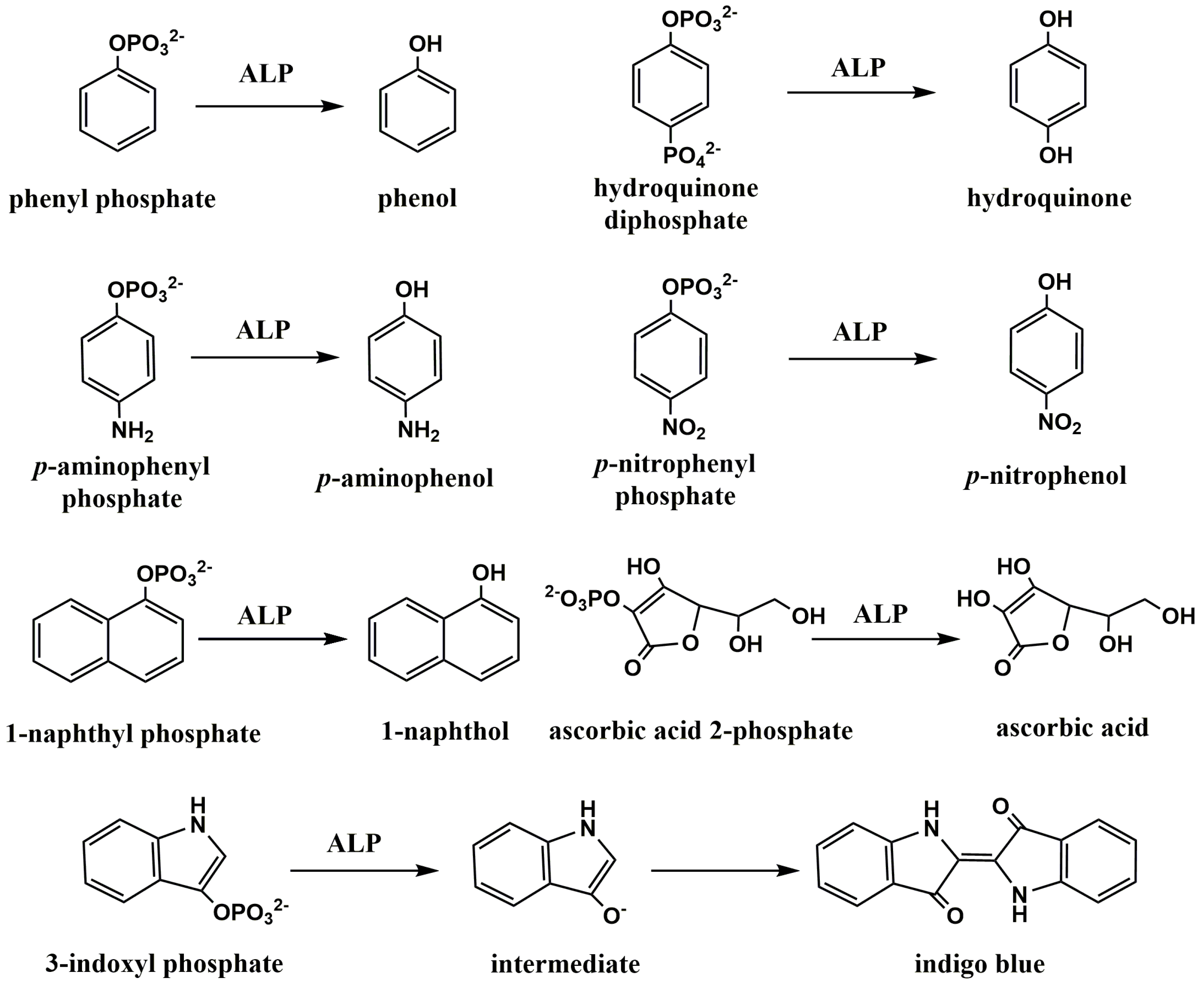
Figure 1. The chemical structure of several typical ALP substrates and products.
Biosensors 13 00855 g001
Figure 2. (A) Schematic illustration of the electrochemical immunoassay for detection of Aβ peptide using PAPP as the ALP substrate [102]. Copyright 2017 Elsevier. (B) Schematic illustration of the preparation process of the ALP-based electrochemical immunosensor for detection of IL-1β cytokine [103]. Copyright 2020 Elsevier.
Biosensors 13 00855 g002
Figure 3. Schematic illustration of assembly processes and assay procedure of the μPAD-based electrochemical immunosensor. (1) ABS top cover, (2) cellulose paper-based sample pad, (3) glass fiber-based conjugate pad, (4) paper-based SPEs, (5) blotting paper (6) single-sided adhesive backing layer, and (7) sample injection hole [109]. Copyright 2017 Elsevier.
Biosensors 13 00855 g003
Figure 4. (A) Schematic illustration of the electrochemical immunosensor for troponin I detection using the generation of AA by ALP and the redox cycling of AA by TCEP [118]. Copyright 2011 American Chemical Society. (B) Schematic illustration of the electrochemical immunosensor for the detection of CK-MB using (i) enzymatic amplification and (ii) + (iii) EC redox cycling [119]. Copyright 2017 American Chemical Society.
Biosensors 13 00855 g004
Figure 5. (A) Schematic illustration of the process of PAP production and its electrocatalytic reaction by reductants on ITO and gold electrode [120]. Copyright 2013 Elsevier. (B) Schematic illustration of the detection of Aβ(1–42) and total Aβ using PAP-mediated redox cycling by chemical reductants [122]. Copyright 2014 Elsevier.
Biosensors 13 00855 g005
Figure 6. Schematic illustration of the prepared immunosensor for APE-1 detection and the triple signal amplification mechanism: (A) the stepwise bio-AP/SA/Ab2/Ni–AuNCs bioconjugates fabrication process: (a) absorption of NiNPs, (b) Ab2 loading, (c) blocking with SA, and (d) binding bio-AP; (B) the molecular structure of PTC-NH2 [131]. Copyright 2013 Elsevier.
Biosensors 13 00855 g006
Figure 8. Schematic illustration of assembly process with the enzyme bioaffinity immunosensor for human IgG detection based on bienzyme substrate recycling for amplification [138]. Copyright 2010 Elsevier.
Biosensors 13 00855 g008
Figure 9. (A) Schematic illustration of the protocol for the immunoassay of H7N9 AIV: the target virus was captured by MBs and the sandwich immunoreaction, enzyme-induced metallization reaction mechanism [154]. Copyright 2015 Elsevier. (B) Schematic illustration of the electrochemical immunomagnetic assay for the analysis of HER2-ECD based on ALP-induced metallization reaction [161]. Copyright 2020 Elsevier.
Biosensors 13 00855 g009
Figure 10. (A) Schematic illustration of the electrochemical immunosensor for human IgG detection based on ALP-triggered silver deposition and Ag–Au bimetallic NPs as the catalyst [170]. Copyright 2017 Elsevier. (B) Schematic illustration of preparation of immunosensor array and detection strategy by sandwich-type immunoassay and linear sweep voltammetric stripping analysis of enzymatically deposited silver nanoparticles (AgNPs) [171]. Copyright 2011 American Chemical Society.
Biosensors 13 00855 g010
Figure 11. Schematic illustration of the electrochemical immunosensor for SEB detection based on HER inhibition by ALP-catalyzed copper deposition on PtNPs-modified GCE [174]. Copyright 2014 Wiley-VCH.
Biosensors 13 00855 g011
Figure 12. (A) Schematic illustration of the preparation process of TiO2−x-MoS2 and the construction process of the biosensor [188]. Copyright 2021 Elsevier. (B) Schematic illustration of the PEC immunosensor for microRNA detection based on IgG–ALP-modified AuNPs. (a) DNA/g-C3N4-AuNPs/ITO; (b) ALP/antibody/RNA-DNA/g-C3N4-AuNPs/ITO [192]. Copyright 2016 Elsevier.
Biosensors 13 00855 g012
Figure 14. Schematic illustration of the PEC immunosensing system for the detection of AFP on β-In2S3@CDs photoelectrode by coupling with enzyme immunoassay format [199]. Copyright 2017 Elsevier.
Biosensors 13 00855 g014
Figure 16. Schematic illustration of the AA-mediated OPECT sensing strategy (process 1: light irradiation, process 2: excitation of valence band electrons, process 3: recombination of photogenerated holes and electrons, process 4: electrons transfer from CdS QDs to ITO electrode, and process 5: scavenge of photogenerated holes by AA) [209]. Copyright 2022 Elsevier.
Biosensors 13 00855 g016
Figure 17. (A) Schematic illustration of the construction (a) and the response mechanism (b) of dual-modal HCR and ALP catalysis-based PEC and colorimetric immunosensor [212]. Copyright 2018 American Chemical Society. (B) Schematic illustration of the construction of the immunosensor for multiple ochratoxins; the signal generation of PEC, fluorescence, and colorimetry; the signal transformation; and machine learning [213]. Copyright 2021 Elsevier.
Biosensors 13 00855 g017
Figure 18. (A) Schematic illustration of the split-type chemical redox-cycling-amplification-based PEC immunosensor for detection of cTnI [218]. Copyright 2021 Elsevier. (B) Schematic illustration of the PEC bioanalysis (a) for IL-6 based on PECCC redox cycling amplification (b) [219]. Copyright 2021 American Chemical Society.
Biosensors 13 00855 g018
Figure 19. Schematic illustration of (a) the sandwich immunorecognition with ALP labels to catalyze the growth of Au NCs toAu NPs in 96-well plate and (b) the operation mechanism of the OPECT biosensor with a bio-regulated gate photoanode [221]. Copyright 2021 Wiley-VCH.
Biosensors 13 00855 g019
Figure 20. (A) Schematic illustration of the signal-on PEC immunoassay based on the tunable competitive absorption of Cu(II) ions onto a MOF-based heterojunction [226]. Copyright 2022 American Chemical Society. (B) Schematic illustration of the ALP-loaded liposome-mediated PEC immunoassay based on the in situ formation of type-I heterojunction on an FTO electrode [227]. Copyright 2022 American Chemical Society.
Biosensors 13 00855 g020
Figure 21. Schematic illustration of (a) ALP-labeled sandwich immunocomplexing in a 96-well plate to produce AA for bioetching of the as-fabricated CoOOH/BiVO4, (b) the OPECT configuration, and (c) the corresponding modulation mechanism [231]. Copyright 2023 American Chemical Society.
Biosensors 13 00855 g021
Figure 22. (A) Schematic illustration ofthe principle for amplified energy transfer ECL quenching for sensitive detection of CEA based on the ALP-catalyzed in situ generation of molecular quenchers [240]. Copyright 2013 Elsevier. (B) Schematic illustration of the ECL immunoassay for PSA detection based on liposome, ALP catalysis, chemical redox cycling, and in situ growth of AuNPs [241]. Copyright 2022 Elsevier.
Biosensors 13 00855 g022
Disclaimer/Publisher’s Note: The statements, opinions and data contained in all publications are solely those of the individual author(s) and contributor(s) and not of MDPI and/or the editor(s). MDPI and/or the editor(s) disclaim responsibility for any injury to people or property resulting from any ideas, methods, instructions or products referred to in the content.