Fluorescent Biosensors for the Detection of Viruses Using Graphene and Two-Dimensional Carbon Nanomaterials


Abstract
1. Introduction
2. Graphene Oxide Fluorescent Biosensor for Human Virus Detection
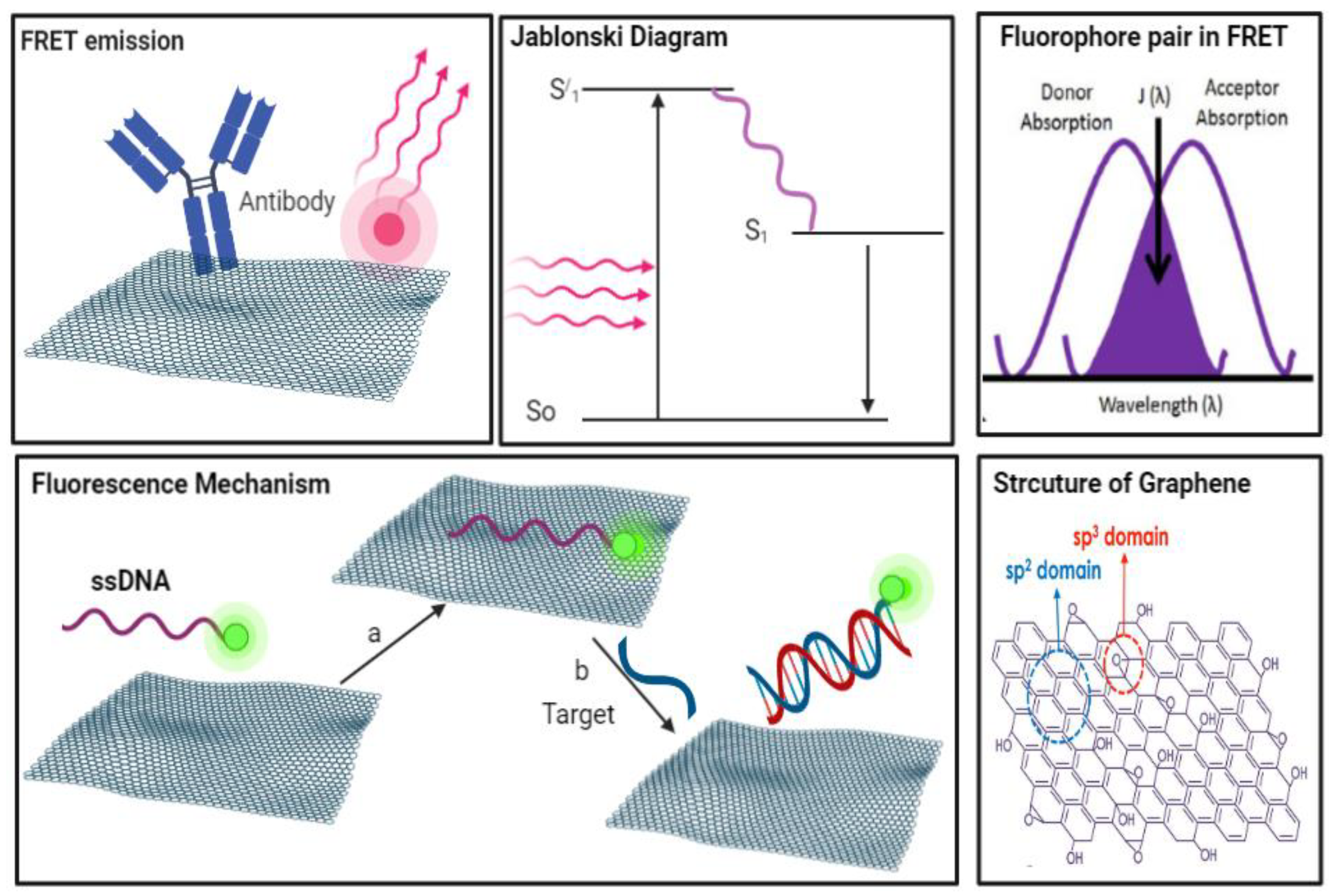
2.1. Fluorescence Resonance Energy Transfer Mechanism (FRET)
2.2. Characteristics of Graphene Material and Biomolecule Interaction
2.3. Virus Detection
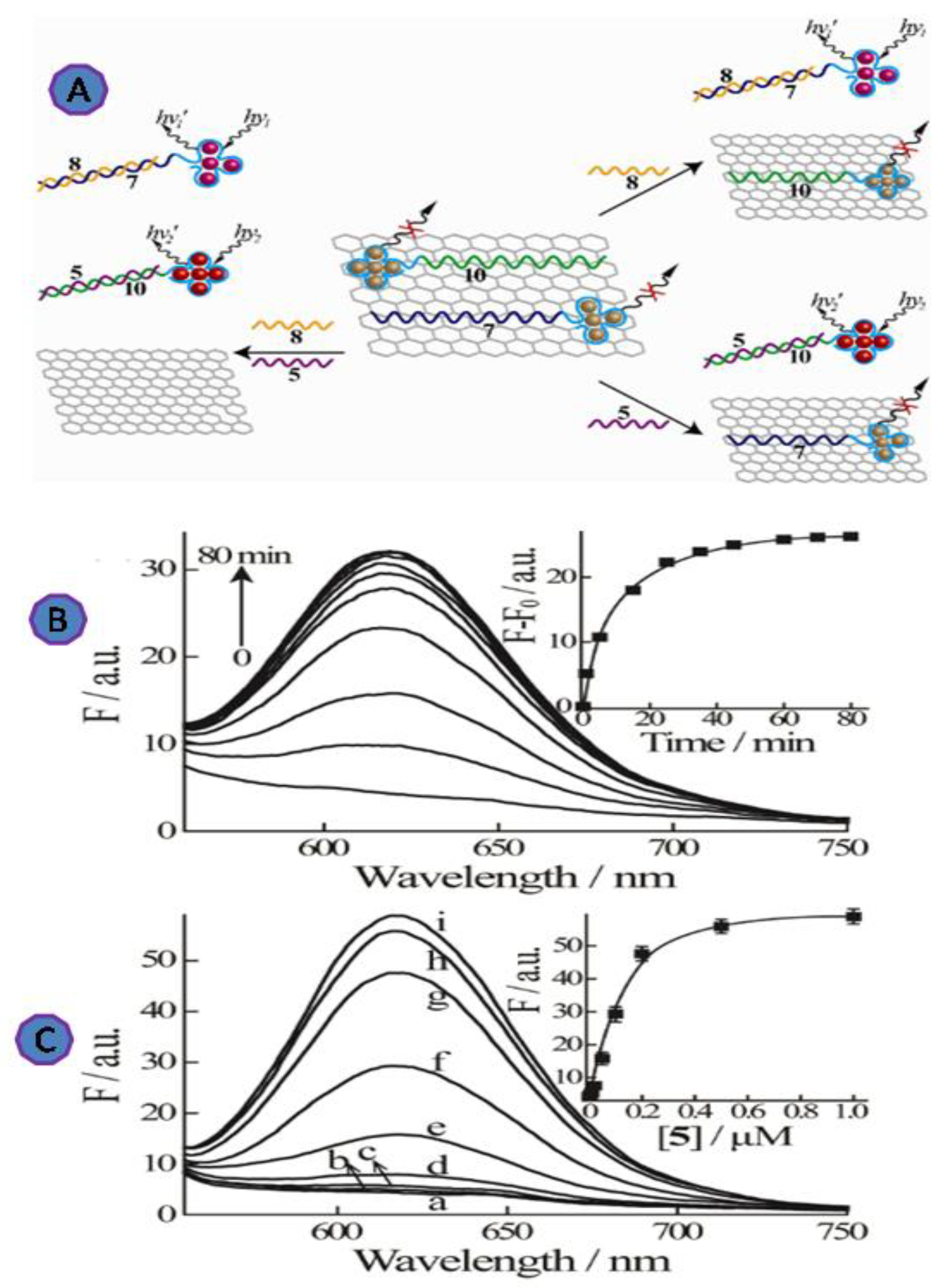
2.3.1. Rotavirus
2.3.2. Ebolavirus
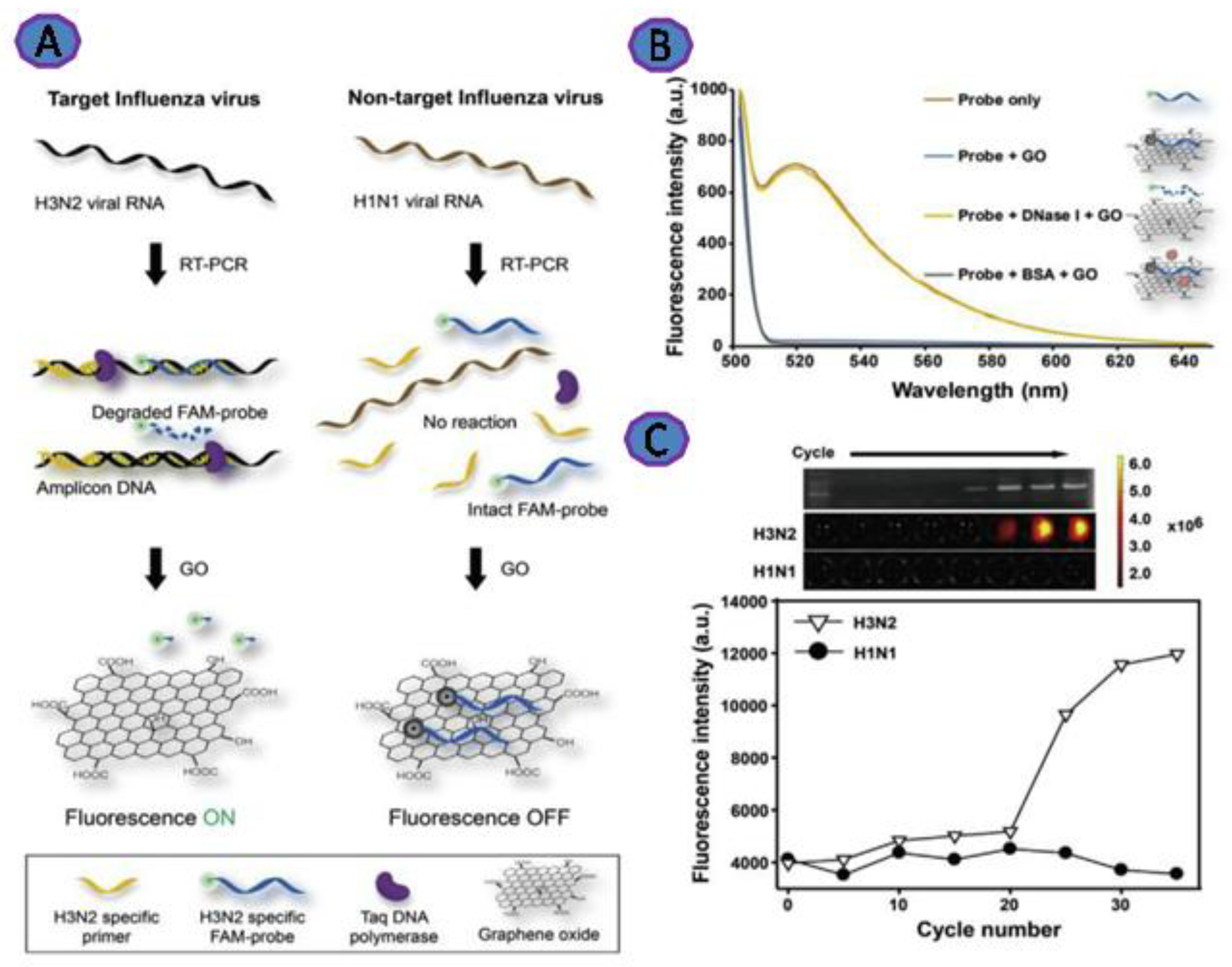
2.3.3. Influenza
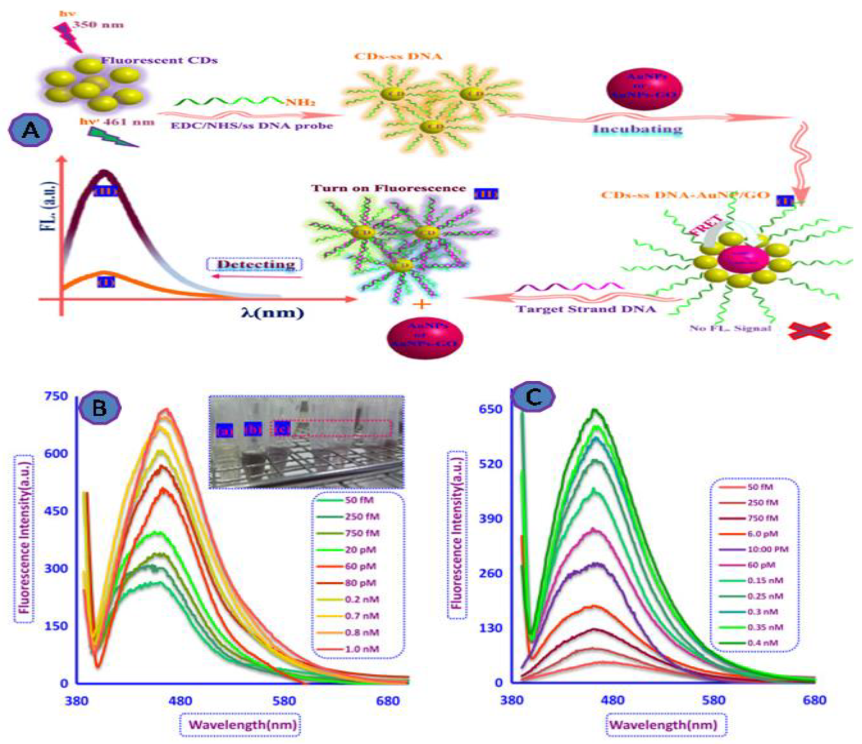
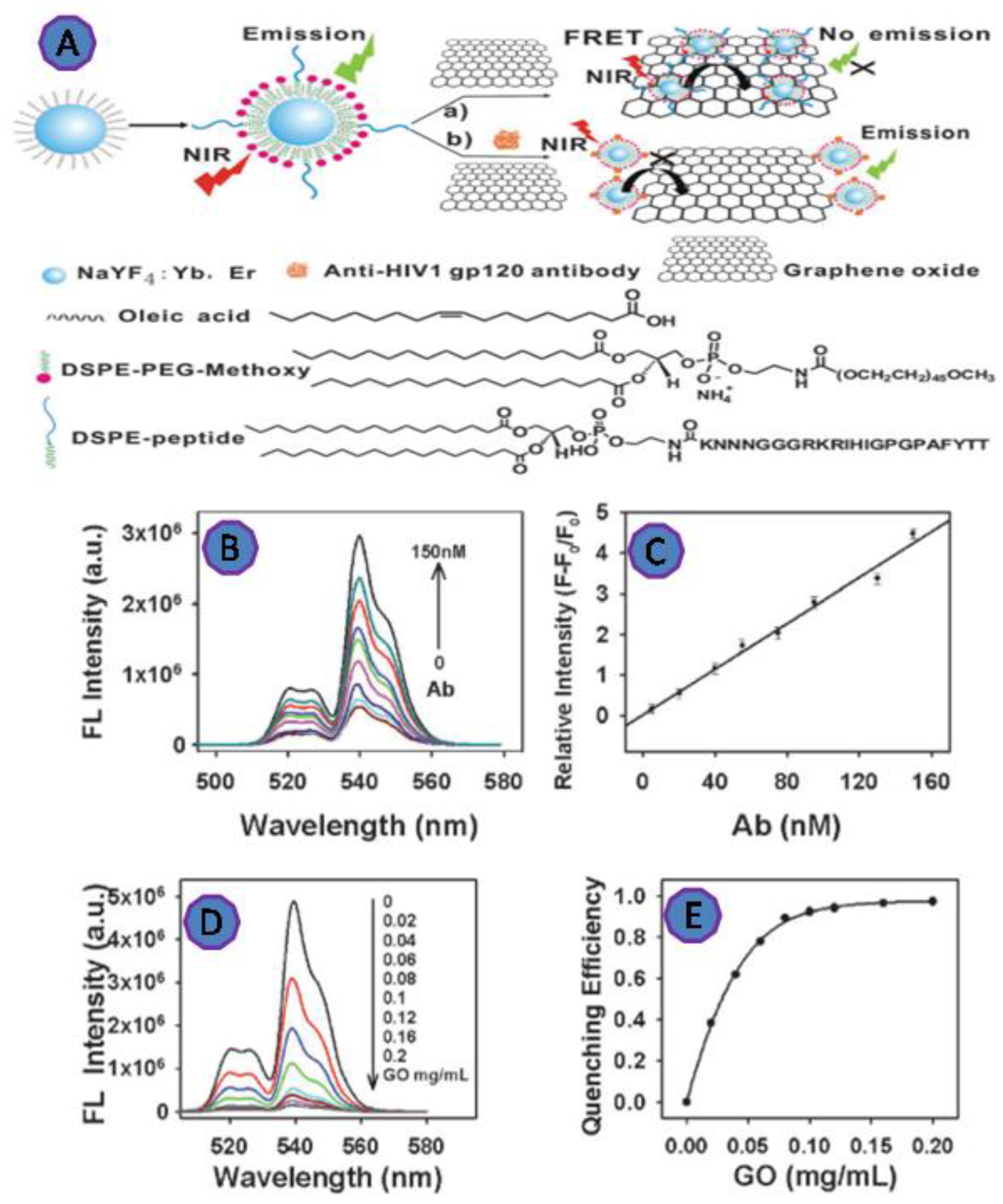
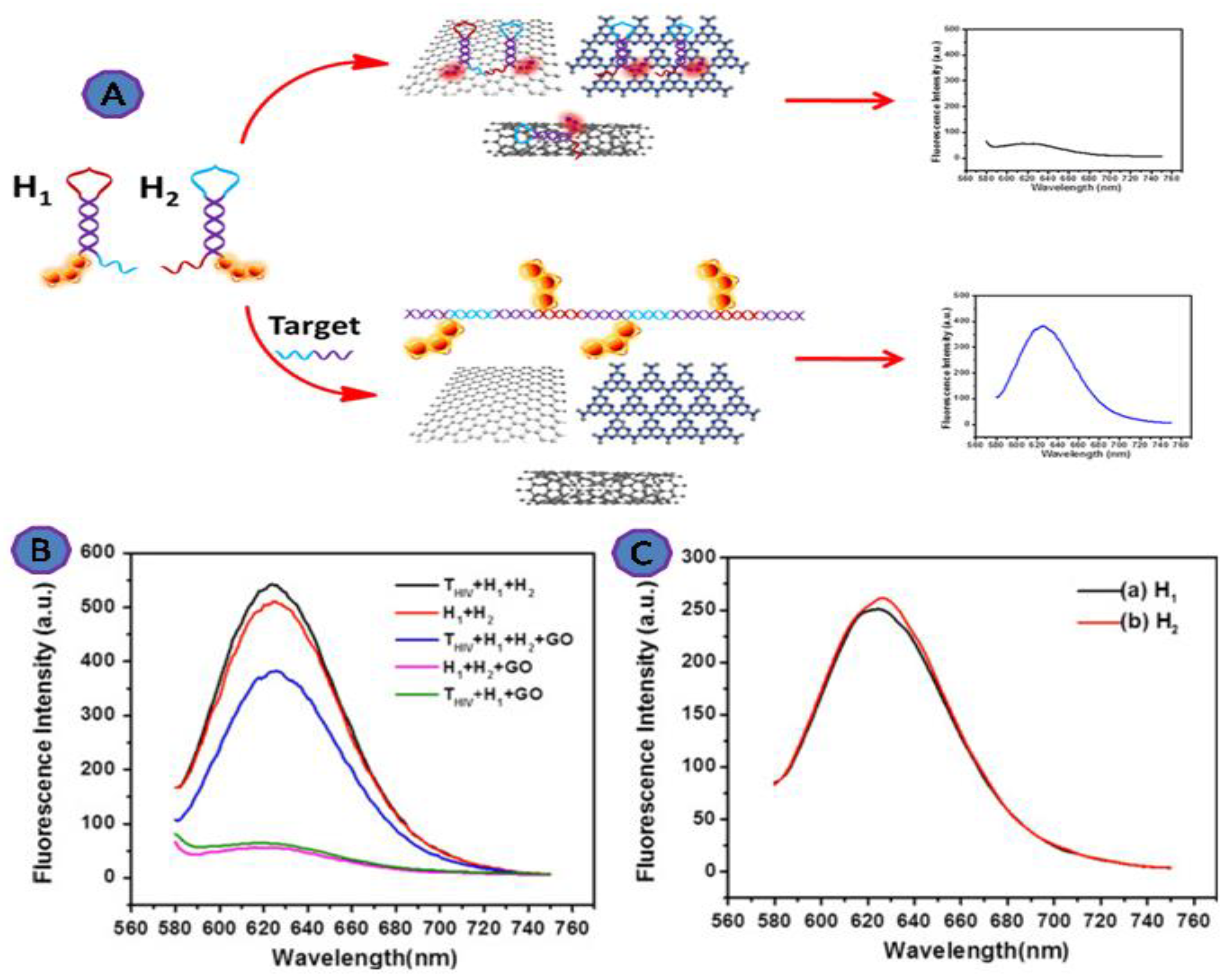
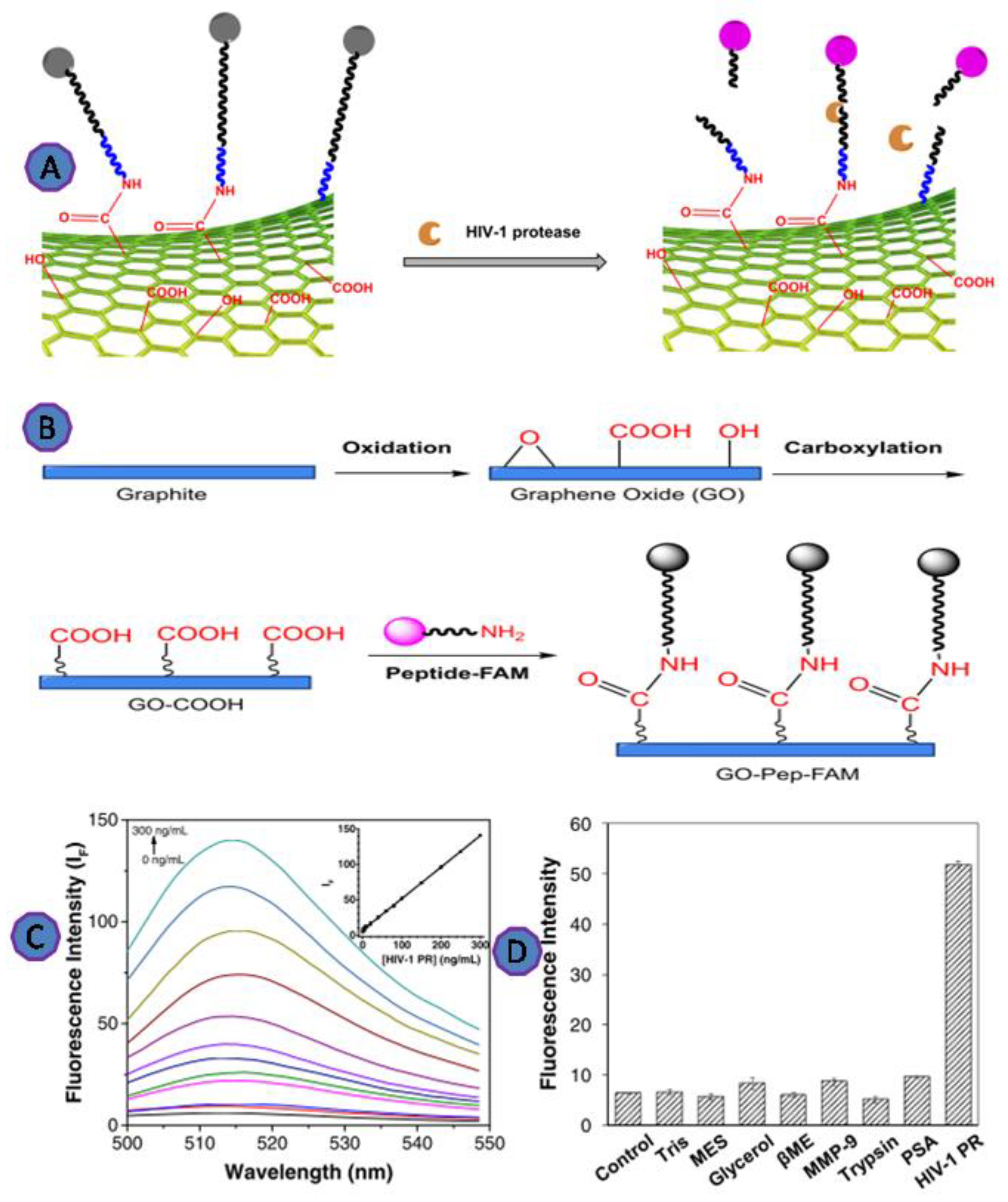
2.3.4. HIV
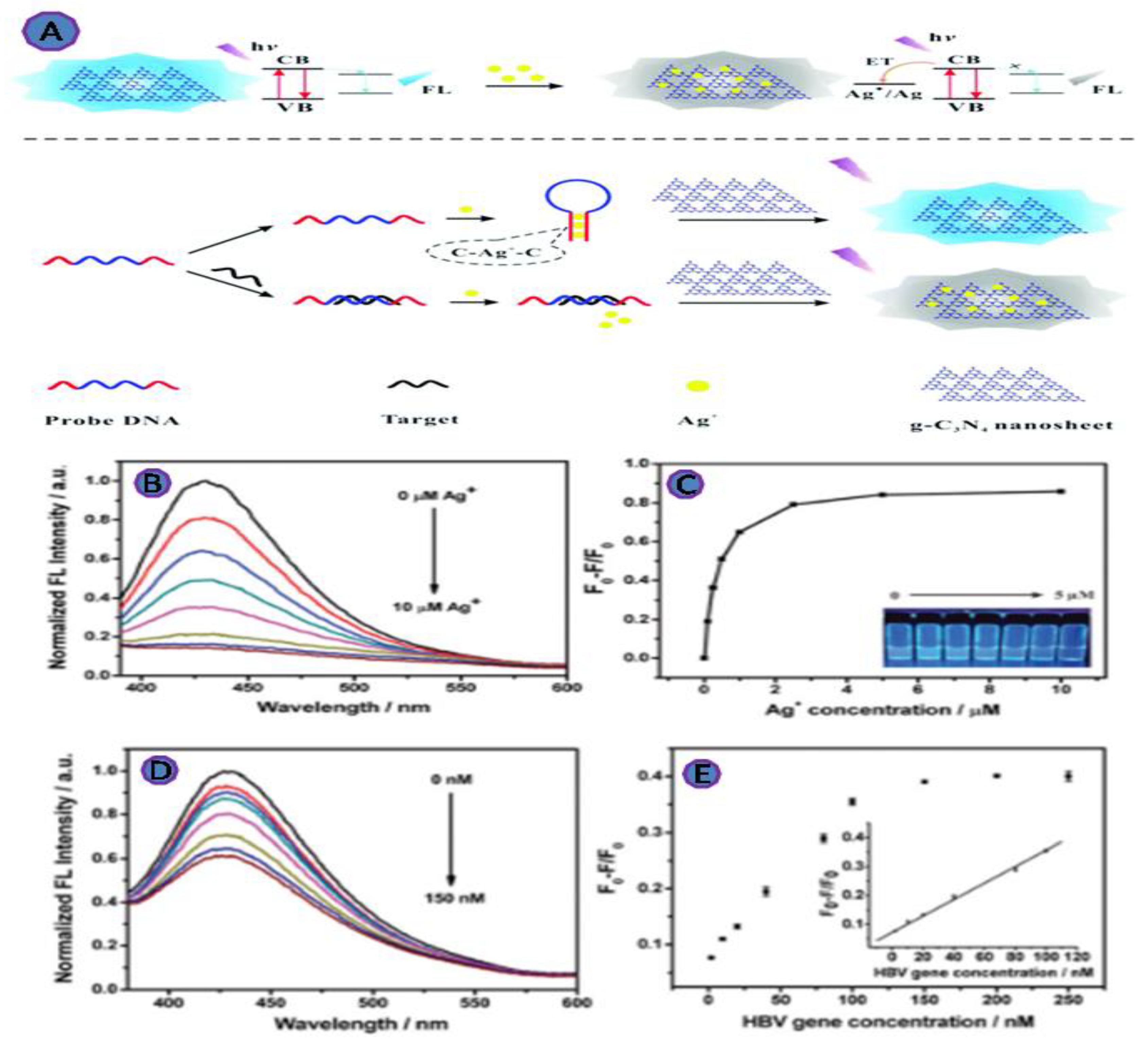
2.3.5. Hepatitis B Virus
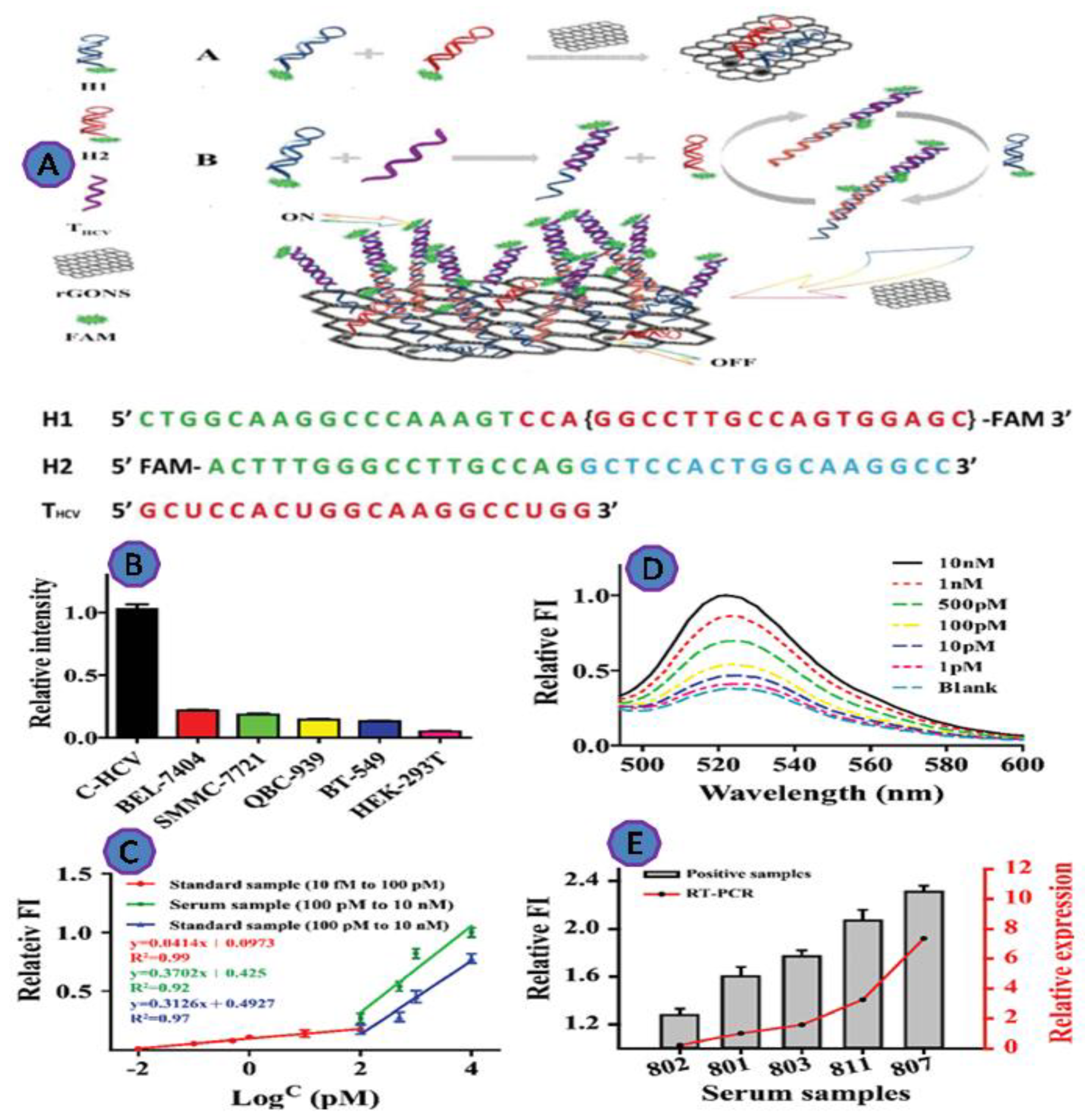
2.3.6. Hepatitis C Virus (HCV)

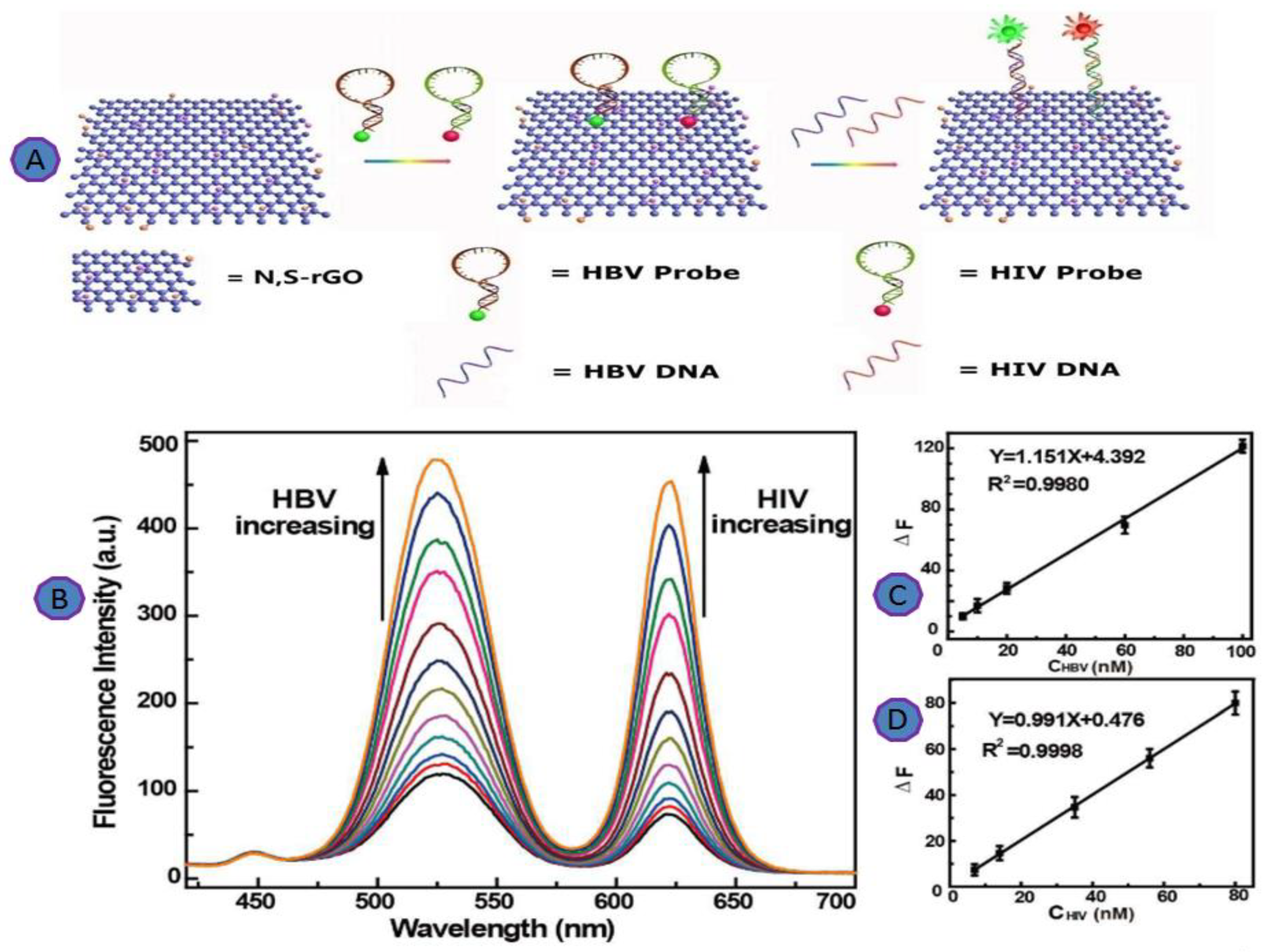
2.4. Multiplexing Viruses Detection
3. Literature on 2D Fluorescent Biosensors for the detection of Viruses
3.1. Overcoming the Drawbacks in Carbon Nanomaterials
3.2. Detection Limit and Analysis Time
3.3. Non-Specific Binding
3.4. Cytotoxicity
4. Future Outlook and Challenges
5. Conclusions
Author Contributions
Funding
Acknowledgments
Conflicts of Interest
References
- Ahmed, S.R.; Mogus, J.; Chand, R.; Nagy, E.; Neethirajan, S. Optoelectronic fowl adenovirus detection based on local electric field enhancement on graphene quantum dots and gold nanobundle hybrid. Biosens. Bioelectron. 2018, 103, 45–53. [Google Scholar] [CrossRef] [PubMed]
- Bianying, F.; Linjie, G.; Lihua, W.; Fan, L.; Jianxin, L.; Jimin, G.; Chunhai, F.; Qing, H. A graphene oxide-based fluorescent biosensor for the analysis of peptide-receptor interactions and imaging in somatostatin receptor subtype 2 overexpressed tumor cells. Anal. Chem. 2013, 85, 7732–7737. [Google Scholar] [CrossRef] [PubMed]
- Constantinou, A.; Polizzi, K.M. Opportunities for bioprocess monitoring using FRET biosensors. Biochem. Soc. Trans. 2013, 41, 1146–1151. [Google Scholar] [CrossRef] [PubMed][Green Version]
- Dong, Y.; Zhang, T.; Lin, X.; Feng, J.; Luo, F.; Gao, H.; Wu, Y.; Deng, R.; He, Q. Graphene/aptamer probes for small molecule detection: From in vitro test to in situ imaging. Microchim. Acta 2020, 187, 179. [Google Scholar] [CrossRef]
- Ravikumar, A.; Panneerselvam, P.; Radhakrishnan, K. Fluorometric determination of lead(II) and mercury(II) based on their interaction with a complex formed between graphene oxide and a DNAzyme. Microchim. Acta 2018, 185, 2. [Google Scholar] [CrossRef]
- Chen, J.; Nugen, S.R. Detection of protease and engineered phage-infected bacteria using peptide-graphene oxide nanosensors. Anal. Bioanal. Chem. 2019, 411, 2487–2492. [Google Scholar] [CrossRef]
- Nguyen, E.P.; de Carvalho Castro Silva, C.; Merkoçi, A. Recent advancement in biomedical applications on the surface of two-dimensional materials: From biosensing to tissue engineering. Nanoscale 2020, 12, 19043–19067. [Google Scholar] [CrossRef]
- Sohail Haroone, M.; Li, L.; Ahmad, A.; Huang, Y.; Ma, R.; Zhang, P.; Hu, Y.; Muhammad Kaleem, Q.; Lu, J. Luminous composite ultrathin films of CdTe quantum dots/silk fibroin co-assembled with layered doubled hydroxide: Enhanced photoluminescence and biosensor application. J. Mater. 2018, 4, 165–171. [Google Scholar] [CrossRef]
- Ryoo, S.R.; Yim, Y.; Kim, Y.K.; Park, I.S.; Na, H.K.; Lee, J.; Jang, H.; Won, C.; Hong, S.; Kim, S.Y.; et al. High-throughput chemical screening to discover new modulators of microRNA expression in living cells by using graphene-based biosensor. Sci. Rep. 2018, 8, 11413. [Google Scholar] [CrossRef]
- He, Y.; Xing, X.; Tang, H.; Pang, D. Graphene oxide-based fluorescent biosensor for protein detection via terminal protection of small-molecule-linked DNA. Small 2013, 9, 2097–2101. [Google Scholar] [CrossRef]
- Zhang, Z.; Xia, X.; Xiang, X.; Huang, F.; Han, L. Conjugated cationic polymer-assisted amplified fluorescent biosensor for protein detection via terminal protection of small molecule-linked DNA and graphene oxide. Sens. Actuators B Chem. 2017, 249, 8–13. [Google Scholar] [CrossRef]
- Girigoswami, K.; Akhtar, N. Nanobiosensors and fluorescence based biosensors: An overview. Int. J. Nano Dimens. 2019, 10, 1–17. [Google Scholar]
- Rabiee, N.; Bagherzadeh, M.; Ghadiri, A.M.; Salehi, G. ZnAl nano layered double hydroxides for dual functional CRISPR/Cas9 delivery and enhanced green fluorescence protein biosensor. Sci. Rep. 2020, 10, 20672. [Google Scholar] [CrossRef]
- Morales, M.A.; Halpern, J.M. Guide to Selecting a Biorecognition Element for Biosensors. Bioconjug. Chem. 2018, 29, 3231–3239. [Google Scholar] [CrossRef]
- Sang, S.; Zhang, W.; Zhao, Y. Review on the Design Art of Biosensors. State Art Biosens.-Gen. Asp. 2013. [Google Scholar] [CrossRef]
- Zhou, L.; Mao, H.; Wu, C.; Tang, L.; Wu, Z.; Sun, H.; Zhang, H.; Zhou, H.; Jia, C.; Jin, Q.; et al. Label-free graphene biosensor targeting cancer molecules based on non-covalent modification. Biosens. Bioelectron. 2017, 87, 701–707. [Google Scholar] [CrossRef]
- Shirani, M.; Kalantari, H.; Khodayar, M.J.; Kouchak, M.; Rahbar, N. A novel strategy for detection of small molecules based on aptamer/gold nanoparticles/graphitic carbon nitride nanosheets as fluorescent biosensor. Talanta 2020, 219, 121235. [Google Scholar] [CrossRef] [PubMed]
- Zhang, Z.; Liu, J. An engineered one-site aptamer with higher sensitivity for label-free detection of adenosine on graphene oxide. Can. J. Chem. 2018, 96, 957–963. [Google Scholar] [CrossRef]
- Damborský, P.; Švitel, J.; Katrlík, J. Optical biosensors. Essays Biochem. 2016, 60, 91–100. [Google Scholar] [CrossRef] [PubMed]
- Mehrotra, P. ScienceDirect Biosensors and their applications—A review. J. Oral Biol. Craniofacial Res. 2016, 6, 153–159. [Google Scholar] [CrossRef] [PubMed]
- Meyer, D.; Hagemann, A.; Kruss, S. Kinetic Requirements for Spatiotemporal Chemical Imaging with Fluorescent Nanosensors. ACS Nano 2017, 11, 4017–4027. [Google Scholar] [CrossRef]
- Ehtesabi, H. Application of carbon nanomaterials in human virus detection. J. Sci. Adv. Mater. Devices 2020, 5, 436–450. [Google Scholar] [CrossRef]
- Devi, M. Application of 2D Nanomaterials as Fluorescent Biosensors. ACS Symp. Ser. 2020, 1353, 117–141. [Google Scholar] [CrossRef]
- Hwang, H.S.; Jeong, J.W.; Kim, Y.A.; Chang, M. Carbon nanomaterials as versatile platforms for biosensing applications. Micromachines 2020, 11, 814. [Google Scholar] [CrossRef]
- Ramnani, P.; Saucedo, N.M.; Mulchandani, A. Carbon nanomaterial-based electrochemical biosensors for label-free sensing of environmental pollutants. Chemosphere 2016, 143, 85–98. [Google Scholar] [CrossRef]
- Rajakumar, G.; Zhang, X.H.; Gomathi, T.; Wang, S.F.; Ansari, M.A.; Mydhili, G.; Nirmala, G.; Alzohairy, M.A.; Chung, I.M. Current use of carbon-based materials for biomedical applications-A prospective and review. Processes 2020, 8, 355. [Google Scholar] [CrossRef]
- Kour, R.; Arya, S.; Young, S.-J.; Gupta, V.; Bandhoria, P.; Khosla, A. Review—Recent Advances in Carbon Nanomaterials as Electrochemical Biosensors. J. Electrochem. Soc. 2020, 167, 037555. [Google Scholar] [CrossRef]
- Zhu, C.; Du, D.; Lin, Y. Graphene-like 2D nanomaterial-based biointerfaces for biosensing applications. Biosens. Bioelectron. 2017, 89, 43–55. [Google Scholar] [CrossRef]
- Vermisoglou, E.; Panáček, D.; Jayaramulu, K.; Pykal, M.; Frébort, I.; Kolář, M.; Hajdúch, M.; Zbořil, R.; Otyepka, M. Human virus detection with graphene-based materials. Biosens. Bioelectron. 2020, 166, 112436. [Google Scholar] [CrossRef]
- Tan, J.; Lai, Z.; Zhang, Z.; Zheng, R.; Su, J.; Huang, Y.; Huang, P.; Song, H.; Yang, N.; Zhou, S.; et al. A Graphene Oxide-Based Fluorescent Aptasensor for the Turn-on Detection of CCRF-CEM. Nanoscale Res. Lett. 2018, 13, 66. [Google Scholar] [CrossRef]
- Liu, F.; Choi, J.Y.; Seo, T.S. Biosensors and Bioelectronics Graphene oxide arrays for detecting specific DNA hybridization by fluorescence resonance energy transfer. Biosens. Bioelectron. 2010, 25, 2361–2365. [Google Scholar] [CrossRef]
- Tehrani, Z.; Burwell, G.; Mohd Azmi, M.A.; Castaing, A.; Rickman, R.; Almarashi, J.; Dunstan, P.; Miran Beigi, A.; Doak, S.H.; Guy, O.J. Generic epitaxial graphene biosensors for ultrasensitive detection of cancer risk biomarker. 2D Mater. 2014, 1, 025004. [Google Scholar] [CrossRef]
- Cui, F.; Ji, J.; Sun, J.; Wang, J.; Wang, H.; Zhang, Y.; Ding, H.; Lu, Y.; Xu, D.; Sun, X. A novel magnetic fluorescent biosensor based on graphene quantum dots for rapid, efficient, and sensitive separation and detection of circulating tumor cells. Anal. Bioanal. Chem. 2019, 411, 985–995. [Google Scholar] [CrossRef] [PubMed]
- Liu, B.; Huang, P.J.J.; Kelly, E.Y.; Liu, J. Graphene oxide surface blocking agents can increase the DNA biosensor sensitivity. Biotechnol. J. 2016, 11, 780–787. [Google Scholar] [CrossRef] [PubMed]
- Wang, L.; Wu, A.; Wei, G. Graphene-based aptasensors: From molecule-interface interactions to sensor design and biomedical diagnostics. Analyst 2018, 143, 1526–1543. [Google Scholar] [CrossRef]
- Waiwijit, U.; Phokaratkul, D.; Kampeera, J.; Lomas, T.; Wisitsoraat, A.; Kiatpathomchai, W.; Tuantranont, A. Graphene oxide based fluorescence resonance energy transfer and loop-mediated isothermal amplification for white spot syndrome virus detection. J. Biotechnol. 2015, 212, 44–49. [Google Scholar] [CrossRef]
- Bayraktutan, T.; Meral, K. Merocyanine 540 adsorbed on polyethylenimine-functionalized graphene oxide nanocomposites as a turn-on fluorescent sensor for bovine serum albumin. Phys. Chem. Chem. Phys. 2016, 18, 23400–23406. [Google Scholar] [CrossRef]
- Jeong, S.; Kim, D.; Kim, D.H.; Kim, D. Fluorometric detection of in fluenza viral RNA using graphene oxide. Anal. Biochem. 2018, 561–562, 66–69. [Google Scholar] [CrossRef]
- Hochreiter, B.; Garcia, A.P.; Schmid, J.A. Fluorescent proteins as genetically encoded FRET biosensors in life sciences. Sensors 2015, 15, 26281–26314. [Google Scholar] [CrossRef]
- Salihoglu, O.; Kakenov, N.; Balci, O.; Balci, S.; Kocabas, C. Graphene as a Reversible and Spectrally Selective Fluorescence Quencher. Sci. Rep. 2016, 6, srep33911. [Google Scholar] [CrossRef]
- Huang, H.; Su, S.; Wu, N.; Wan, H.; Wan, S.; Bi, H.; Sun, L. Graphene-based sensors for human health monitoring. Front. Chem. 2019, 7, 399. [Google Scholar] [CrossRef]
- Kalkal, A.; Pradhan, R.; Kadian, S.; Manik, G.; Packirisamy, G. Biofunctionalized Graphene Quantum Dots Based Fluorescent Biosensor toward Efficient Detection of Small Cell Lung Cancer. ACS Appl. Bio Mater. 2020, 3, 4922–4932. [Google Scholar] [CrossRef]
- Zhang, H.; Zhang, H.; Aldalbahi, A.; Zuo, X.; Fan, C.; Mi, X. Fluorescent biosensors enabled by graphene and graphene oxide. Biosens. Bioelectron. 2017, 89, 96–106. [Google Scholar] [CrossRef]
- Alhazmi, H.A.; Ahsan, W.; Mangla, B.; Javed, S.; Hassan, M.Z. Graphene-based biosensors for disease theranostics: Development, applications, and recent advancements. Nanotechnol. Rev. 2022, 11, 96–116. [Google Scholar] [CrossRef]
- Wang, W.; Su, H.; Wu, Y.; Zhou, T.; Li, T. Review—Biosensing and Biomedical Applications of Graphene: A Review of Current Progress and Future Prospect. J. Electrochem. Soc. 2019, 166, B505–B520. [Google Scholar] [CrossRef]
- Di Pietrantonio, F.; Cannatà, D.; Benetti, M. Biosensor Technologies Based on Nanomaterials; Elsevier Inc.: Amsterdam, The Netherlands, 2019. [Google Scholar]
- Peltomaa, R.; Glahn-Martínez, B.; Benito-Peña, E.; Moreno-Bondi, M.C. Optical Biosensors for Label-Free Detection of Small Molecules. Sensors 2018, 18, 4126. [Google Scholar] [CrossRef]
- Gosai, A.; Khondakar, K.R.; Ma, X.; Ali, M.A. Application of functionalized graphene oxide based biosensors for health monitoring: Simple graphene derivatives to 3D printed platforms. Biosensors 2021, 11, 384. [Google Scholar] [CrossRef]
- Kim, T.H.; Lee, D.; Choi, J.W. Live cell biosensing platforms using graphene-based hybrid nanomaterials. Biosens. Bioelectron. 2017, 94, 485–499. [Google Scholar] [CrossRef]
- Zhao, H.; Ding, R.; Zhao, X.; Li, Y.; Qu, L.; Pei, H.; Yildirimer, L.; Wu, Z.; Zhang, W. Graphene-based nanomaterials for drug and/or gene delivery, bioimaging, and tissue engineering. Drug Discov. Today 2017, 22, 1302–1317. [Google Scholar] [CrossRef]
- Jiang, Z.; Feng, B.; Xu, J.; Qing, T.; Zhang, P.; Qing, Z. Graphene biosensors for bacterial and viral pathogens. Biosens. Bioelectron. 2020, 166, 112471. [Google Scholar] [CrossRef]
- Ozcelik, D.; Parks, J.W.; Wall, T.A.; Stott, M.A.; Cai, H.; Parks, J.W.; Hawkins, A.R.; Schmidt, H. Optofluidic wavelength division multiplexing for single-virus detection. Proc. Natl. Acad. Sci. USA 2015, 112, 12933–12937. [Google Scholar] [CrossRef]
- Torrente-Rodríguez, R.M.; Lukas, H.; Tu, J.; Min, J.; Yang, Y.; Xu, C.; Rossiter, H.B.; Gao, W. SARS-CoV-2 RapidPlex: A Graphene-Based Multiplexed Telemedicine Platform for Rapid and Low-Cost COVID-19 Diagnosis and Monitoring. Matter 2020, 3, 1981–1998. [Google Scholar] [CrossRef]
- Tymm, C.; Zhou, J.; Tadimety, A.; Burklund, A.; Zhang, J.X.J. Scalable COVID-19 Detection Enabled by Lab-on-Chip Biosensors. Cell. Mol. Bioeng. 2020, 13, 313–329. [Google Scholar] [CrossRef]
- Hao, X.; Liu, R.; He, Y.; Xiao, X.; Xiao, W.; Zheng, Q.; Lin, X.; Tao, P.; Zhou, P.; Li, S. Multiplex PCR methods for detection of several viruses associated with canine respiratory and enteric diseases. PLoS ONE 2019, 14, e0213295. [Google Scholar] [CrossRef]
- Zhang, X.; Hu, Y.; Yang, X.; Tang, Y.; Han, S.; Kang, A.; Deng, H.; Chi, Y.; Zhu, D.; Lu, Y. FÖrster resonance energy transfer (FRET)-based biosensors for biological applications. Biosens. Bioelectron. 2019, 138, 111314. [Google Scholar] [CrossRef]
- Zhang, Z.; Tang, Z.; Farokhzad, N.; Chen, T.; Tao, W. Sensitive, Rapid, Low-Cost, and Multiplexed COVID-19 Monitoring by the Wireless Telemedicine Platform. Matter 2020, 3, 1818–1820. [Google Scholar] [CrossRef]
- Li, Z.; Yin, X.; Sun, Y.; Qu, L.; Du, D.; Lin, Y. Functionalized Two-Dimensional Nanomaterials for Biosensing and Bioimaging. ACS Symp. Ser. 2020, 1353, 143–165. [Google Scholar] [CrossRef]
- Xiao, Y.; Sheng, Y.; Zhou, J.; Chen, M.; Wen, W.; Zhang, X.; Wang, S. A novel label-free strategy for pathogenic DNA detection based on metal ions binding-induced fluorescence quenching of graphitic carbon nitride nanosheets. Analyst 2017, 142, 2617–2623. [Google Scholar] [CrossRef]
- Bányai, K.; Estes, M.K.; Martella, V.; Parashar, U.D. Viral gastroenteritis. Lancet 2018, 392, 175–186. [Google Scholar] [CrossRef]
- Tian, F.; Lyu, J.; Shi, J.; Yang, M. Graphene and graphene-like two-denominational materials based fluorescence resonance energy transfer (FRET) assays for biological applications. Biosens. Bioelectron. 2017, 89, 123–135. [Google Scholar] [CrossRef]
- Chang, X.H.; Zhang, J.; Wu, L.H.; Peng, Y.K.; Yang, X.Y.; Li, X.L.; Ma, A.J.; Ma, J.C.; Chen, G.Q. Research progress of near-infrared fluorescence immunoassay. Micromachines 2019, 10, 422. [Google Scholar] [CrossRef]
- Shi, J.; Tian, F.; Lyu, J.; Yang, M. Nanoparticle based fluorescence resonance energy transfer (FRET) for biosensing applications. J. Mater. Chem. B 2015, 3, 6989–7005. [Google Scholar] [CrossRef]
- Morales-Narváez, E.; Merkoçi, A. Graphene oxide as an optical biosensing platform. Adv. Mater. 2012, 24, 3298–3308. [Google Scholar] [CrossRef]
- Jung, J.H.; Cheon, D.S.; Liu, F.; Lee, K.B.; Seo, T.S. A Graphene Oxide Based Immuno-biosensor for Pathogen Detection. Angew. Chem. 2010, 49, 5708–5711. [Google Scholar] [CrossRef]
- James, A.S.; Todd, S.; Pollak, N.M.; Marsh, G.A.; Macdonald, J. Ebolavirus diagnosis made simple, comparable and faster than molecular detection methods: Preparing for the future. Virol. J. 2018, 15, 75. [Google Scholar] [CrossRef]
- Wen, J.; Li, W.; Li, J.; Tao, B.; Xu, Y.; Li, H.; Lu, A.; Sun, S. Study on rolling circle amplification of Ebola virus and fluorescence detection based on graphene oxide. Sens. Actuators B Chem. 2016, 227, 655–659. [Google Scholar] [CrossRef]
- Krammer, F.; Smith, G.J.D.; Fouchier, R.A.M.; Peiris, M.; Kedzierska, K.; Doherty, P.C.; Palese, P.; Shaw, M.L.; Treanor, J.; Webster, R.G.; et al. Influenza. Nat. Rev. Dis. Primers 2018, 4, 4. [Google Scholar] [CrossRef]
- Srivastava, S.; Singh, P.K.; Vatsalya, V.; Karch, R.C. Developments in the Diagnostic Techniques of Infectious Diseases: Rural and Urban Prospective. Adv. Infect. Dis. 2018, 8, 121–138. [Google Scholar] [CrossRef]
- Kabir, M.A.; Zilouchian, H.; Caputi, M.; Asghar, W. Advances in HIV diagnosis and monitoring. Crit. Rev. Biotechnol. 2020, 40, 623–638. [Google Scholar] [CrossRef]
- Qaddare, S.H.; Salimi, A. Amplified fluorescent sensing of DNA using luminescent carbon dots and AuNPs/GO as a sensing platform: A novel coupling of FRET and DNA hybridization for homogeneous HIV-1 gene detection at femtomolar level. Biosens. Bioelectron. 2017, 89, 773–780. [Google Scholar] [CrossRef]
- Wu, Y.; Cen, Y.; Huang, L.; Yu, R.; Chu, X. Upconversion fluorescence resonance energy transfer biosensor for sensitive detection of human immunodeficiency virus antibodies in human serum. Chem. Commun. 2014, 50, 4759–4762. [Google Scholar] [CrossRef]
- Zhang, S.; Wang, K.; Li KBin Shi, W.; Jia, W.P.; Chen, X.; Sun, T.; Han, D.M. A DNA-stabilized silver nanoclusters/graphene oxide-based platform for the sensitive detection of DNA through hybridization chain reaction. Biosens. Bioelectron. 2017, 91, 374–379. [Google Scholar] [CrossRef]
- Arora, K. Recent Biosensing Applications of Graphene-Based Nanomaterials. Handb. Graphene 2019, 6, 297–348. [Google Scholar]
- Zhang, Y.; Chen, X.; Roozbahani, G.M.; Guan, X. Graphene oxide-based biosensing platform for rapid and sensitive detection of HIV-1 protease. Anal. Bioanal. Chem. 2018, 410, 6177–6185. [Google Scholar] [CrossRef]
- Abd Muain, M.F.; Cheo, K.H.; Omar, M.N.; Amir Hamzah, A.S.; Lim, H.N.; Salleh, A.B.; Tan, W.S.; Ahmad Tajudin, A. Gold nanoparticle-decorated reduced-graphene oxide targeting anti hepatitis B virus core antigen. Bioelectrochemistry 2018, 122, 199–205. [Google Scholar] [CrossRef]
- Xavier, M.M.; Nair, P.R.; Mathew, S. Emerging trends in sensors based on carbon nitride materials. Analyst 2019, 144, 1475–1491. [Google Scholar] [CrossRef]
- Fan, J.; Yuan, L.; Liu, Q.; Tong, C. An ultrasensitive and simple assay for the Hepatitis C virus using a reduced graphene oxide-assisted hybridization chain reaction. Analyst 2019, 144, 3972–3979. [Google Scholar] [CrossRef]
- Ozcelik, D.; Jain, A.; Stambaugh, A.; Stott, M.A.; Parks, J.W.; Hawkins, A.; Schmidt, H. Scalable Spatial-Spectral Multiplexing of Single-Virus Detection Using Multimode Interference Waveguides. Sci. Rep. 2017, 7, 12199. [Google Scholar] [CrossRef]
- Liu, X.; Wang, F.; Aizen, R.; Yehezkeli, O.; Willner, I. Graphene oxide/nucleic-acid-stabilized silver nanoclusters: Functional hybrid materials for optical aptamer sensing and multiplexed analysis of pathogenic DNAs. J. Am. Chem. Soc. 2013, 135, 11832–11839. [Google Scholar] [CrossRef]
- Gingrich, J.C.; Davis, D.R.; Nguyen, Q. Multiplex detection and quantitation of proteins on Western blots using fluorescent probes. Biotechniques 2000, 29, 636–642. [Google Scholar] [CrossRef]
- Hsu, C.C.; Franklin, C.; Riley, L.K. Multiplex Fluorescent Immunoassay for the simultaneous detection of serum antibodies to multiple rodent pathogens. Lab Anim. 2007, 36, 36–38. [Google Scholar] [CrossRef]
- Chen, L.; Song, L.; Zhang, Y.; Wang, P.; Xiao, Z.; Guo, Y.; Cao, F. Nitrogen and Sulfur Codoped Reduced Graphene Oxide as a General Platform for Rapid and Sensitive Fluorescent Detection of Biological Species. ACS Appl. Mater. Interfaces 2016, 8, 11255–11261. [Google Scholar] [CrossRef]
- Jin, Z.; Geißler, D.; Qiu, X.; Wegner, K.D.; Hildebrandt, N. A Rapid, Amplification-Free, and Sensitive Diagnostic Assay for Single-Step Multiplexed Fluorescence Detection of MicroRNA. Angew. Chem. Int. Ed. 2015, 54, 10024–10029. [Google Scholar] [CrossRef]
- Zheng, Q.; Wu, H.; Wang, N.; Yan, R.; Ma, Y.; Guang, W.; Wang, J.; Ding, K. Graphene-based Biosensors for Biomolecules Detection. Curr. Nanosci. 2014, 10, 627–637. [Google Scholar] [CrossRef]
- Perumal, V.; Hashim, U. ScienceDirect Advances in biosensors: Principle, architecture and. J. Econ. Financ. Adm. Sci. 2013, 12, 1–15. [Google Scholar] [CrossRef]
- Kasry, A.; Ardakani, A.A.; Tulevski, G.S.; Menges, B.; Copel, M. Highly Efficient Fluorescence Quenching with Graphene. J. Phys. Chem. C 2012, 4, 2858–2862. [Google Scholar] [CrossRef]
- Liu, M.; Zhang, Q.; Brennan, J.D.; Li, Y. Graphene-DNAzyme-based fluorescent biosensor for Escherichia coli detection. MRS Commun. 2018, 8, 687–694. [Google Scholar] [CrossRef]
- Bitounis, D.; Ali-Boucetta, H.; Hong, B.H.; Min, D.H.; Kostarelos, K. Prospects and challenges of graphene in biomedical applications. Adv. Mater. 2013, 25, 2258–2268. [Google Scholar] [CrossRef]
- Banerjee, A.N. Graphene and its derivatives as biomedical materials: Future prospects and challenges. Interface Focus 2018, 8, 20170056. [Google Scholar] [CrossRef]
- Agarwal, S.; Zhou, X.; Ye, F.; He, Q.; Chen, G.C.K.; Soo, J.; Boey, F.; Zhang, H.; Chen, P. Interfacing live cells with nanocarbon substrates. Langmuir 2010, 26, 2244–2247. [Google Scholar] [CrossRef]
- Zhang, L.; Xia, J.; Zhao, Q.; Liu, L.; Zhang, Z. Functional graphene oxide as a nanocarrier for controlled loading and targeted delivery of mixed anticancer drugs. Small 2010, 6, 537–544. [Google Scholar] [CrossRef]
- Lammel, T.; Boisseaux, P.; Fernández-Cruz, M.L.; Navas, J.M. Internalization and cytotoxicity of graphene oxide and carboxyl graphene nanoplatelets in the human hepatocellular carcinoma cell line Hep G2. Part. Fibre Toxicol. 2013, 10, 27. [Google Scholar] [CrossRef]
- Liao, C.; Li, Y.; Tjong, S.C. Graphene nanomaterials: Synthesis, biocompatibility, and cytotoxicity. Int. J. Mol. Sci. 2018, 19, 3564. [Google Scholar] [CrossRef]
- Guo, S.; Dong, S. Graphene and its derivative-based sensing materials for analytical devices. J. Mater. Chem. 2011, 21, 18503–18516. [Google Scholar] [CrossRef]
- Sengupta, J.; Hussain, C.M. Graphene and its derivatives for Analytical Lab on Chip platforms. TrAC-Trends Anal. Chem. 2019, 114, 326–337. [Google Scholar] [CrossRef]
- Yu, X.; Cheng, H.; Zhang, M.; Zhao, Y.; Qu, L.; Shi, G. Graphene-based smart materials. Nat. Rev. Mater. 2017, 2, 17046. [Google Scholar] [CrossRef]
- Chircov, C.; Grumezescu, A.M.; Andronescu, E. Biosensors-on-Chip: An Up-to-Date Review. Molecules 2020, 25, 6013. [Google Scholar] [CrossRef]
- Huang, G.; Huang, Q.; Xie, L.; Xiang, G.; Wang, L.; Xu, H.; Ma, L. A rapid, low-cost, and microfluidic chip-based system for parallel identification of multiple pathogens related to clinical pneumonia. Sci. Rep. 2017, 7, 6441. [Google Scholar] [CrossRef]
- Pang, B.; Fu, K.; Liu, Y.; Ding, X.; Hu, J.; Wu, W.; Xu, K.; Song, X.; Wang, J.; Mu, Y.; et al. Development of a self-priming PDMS/paper hybrid microfluidic chip using mixed-dye-loaded loop-mediated isothermal amplification assay for multiplex foodborne pathogens detection. Anal. Chim. Acta 2018, 1040, 81–89. [Google Scholar] [CrossRef]
- Sengupta, P.; Khanra, K.; Chowdhury, A.R.; Datta, P. Lab-on-a-Chip Sensing Devices for Biomedical Applications; Elsevier Ltd.: Amsterdam, The Netherlands, 2019. [Google Scholar]
- Kujawska, M.; Bhardwaj, S.K.; Mishra, Y.K.; Kaushik, A. Using graphene-based biosensors to detect dopamine for efficient parkinson’s disease diagnostics. Biosensors 2021, 11, 433. [Google Scholar] [CrossRef]
- Kaushik, A.; Khan, R.; Solanki, P.; Gandhi, S.; Gohel, H.; Mishra, Y.K. From nanosystems to a biosensing prototype for an efficient diagnostic: A special issue in honor of professor Bansi D. Malhotra. Biosensors 2021, 11, 359. [Google Scholar] [CrossRef]
- Sengupta, J.; Adhikari, A.; Hussain, C.M. Graphene-based analytical lab-on-chip devices for detection of viruses: A review. Carbon Trends 2021, 4, 100072. [Google Scholar] [CrossRef]
- Soomro, R.A. Development of Biosensors for Drug Detection Applications. Nanobiosensors 2020, 203–222. [Google Scholar] [CrossRef]
- Xiang, X.; Luo, M.; Shi, L.; Ji, X.; He, Z. Droplet-based microscale colorimetric biosensor for multiplexed DNA analysis via a graphene nanoprobe. Anal. Chim. Acta 2012, 751, 155–160. [Google Scholar] [CrossRef]
- Kenry Yeo, J.C.; Yu, J.; Shang, M.; Loh, K.P.; Lim, C.T. Highly Flexible Graphene Oxide Nanosuspension Liquid-Based Microfluidic Tactile Sensor. Small 2016, 12, 1593–1604. [Google Scholar] [CrossRef]
- Hashmi, A.; Nayak, V.; Rb, K.; Jain, B.; Baid, M.; Alexis, F.; Kumar, A. Potentialities of graphene and its allied derivatives to combat against SARS-CoV-2 infection. Mater. Today Adv. 2022, 13, 100208. [Google Scholar] [CrossRef]
- Roberts, A.; Chauhan, N.; Islam, S.; Mahari, S.; Ghawri, B. Graphene functionalized field-effect transistors for ultrasensitive detection of Japanese encephalitis and Avian influenza virus. Sci. Rep. 2020, 10, 14546. [Google Scholar] [CrossRef]
- Jin, X.; Zhang, H.; Li, Y.T.; Xiao, M.M.; Zhang, Z.L.; Pang, D.W.; Wong, G.; Zhang, Z.Y.; Zhang, G.J. A field effect transistor modified with reduced graphene oxide for immunodetection of Ebola virus. Microchim. Acta 2019, 186, 223. [Google Scholar] [CrossRef]
- Simone, G. Graphene-based optical sensors for the pre- vention of SARS-CoV-2 viral dissemination. arXiv 2020, arXiv:2011.02181. [Google Scholar]


| 2D Carbon Structure | Virus Sensing | Recognition Element | Type of Interaction | Dynamic Range | Limit of Detection | REF |
|---|---|---|---|---|---|---|
| GO | Rotavirus | Antibodies | Carbodiimide-assisted amidation reaction | 103 to 105 pfu mL−1 | 105 pfu mL−1 | [64,65] |
| GO | Ebola virus gene Influenza | dsDNA | π–π stacking interaction | 30 fM–3 nM | 1.4 Pm | [67] |
| GO | Influenza virus H3N2 hemagglutinin gene | RNA | π–π stacking interaction and hydrogen bonding | 37–9400 pg | 3.8 pg | [38] |
| GO | HIV-1 gene | dsDNA | carbodiimide-assisted amidation reaction | 50.0 fM–1.0 nM | 15 fM | [71] |
| GO | HIV | Antibodies | π–π interaction | 5–150 nM | 2 nM | [72] |
| GO | HIV | dsDNA | π–π interaction | - | 1.18 nM | [73,74] |
| GO | HIV | Enzyme | π–π stacking and/or electrostatic interaction between | 5–300 ng/mL | 109 pM | [75] |
| rGO | HCV | dsDNA | π–π interaction | 10 fM | [78] | |
| g-C3N4 | HBV gene | DNA | π–π stacking | 2–100 nM | 1.0 nM | [59] |
| rGO | HIV | Aptamer | π–π interaction | - | 3.0 nM | [79] |
| rGO | HBV | Aptamer | π–π interaction | - | 2.4 nM | [79] |
| GO | HBV | ssDNA | π–π stacking | - | 0.5 nM | [84] |
| GO | HIV | ssDNA | π–π stacking | 1 nm | [84] |
Publisher’s Note: MDPI stays neutral with regard to jurisdictional claims in published maps and institutional affiliations. |
© 2022 by the authors. Licensee MDPI, Basel, Switzerland. This article is an open access article distributed under the terms and conditions of the Creative Commons Attribution (CC BY) license (https://creativecommons.org/licenses/by/4.0/).
Share and Cite
Salama, A.M.; Yasin, G.; Zourob, M.; Lu, J. Fluorescent Biosensors for the Detection of Viruses Using Graphene and Two-Dimensional Carbon Nanomaterials. Biosensors 2022, 12, 460. https://doi.org/10.3390/bios12070460
Salama AM, Yasin G, Zourob M, Lu J. Fluorescent Biosensors for the Detection of Viruses Using Graphene and Two-Dimensional Carbon Nanomaterials. Biosensors. 2022; 12(7):460. https://doi.org/10.3390/bios12070460
Chicago/Turabian StyleSalama, Ahmed M., Ghulam Yasin, Mohammed Zourob, and Jun Lu. 2022. "Fluorescent Biosensors for the Detection of Viruses Using Graphene and Two-Dimensional Carbon Nanomaterials" Biosensors 12, no. 7: 460. https://doi.org/10.3390/bios12070460
APA StyleSalama, A. M., Yasin, G., Zourob, M., & Lu, J. (2022). Fluorescent Biosensors for the Detection of Viruses Using Graphene and Two-Dimensional Carbon Nanomaterials. Biosensors, 12(7), 460. https://doi.org/10.3390/bios12070460