A Study of the Direct Effect of Pegylated Graphene Oxide Nanoparticles and Fullerenol C60(OH)24 on the Differentiation of Regulatory T Cells In Vitro

 ,
,  , , , ,
  and
and
Abstract
1. Introduction
2. Materials and Methods
3. Results
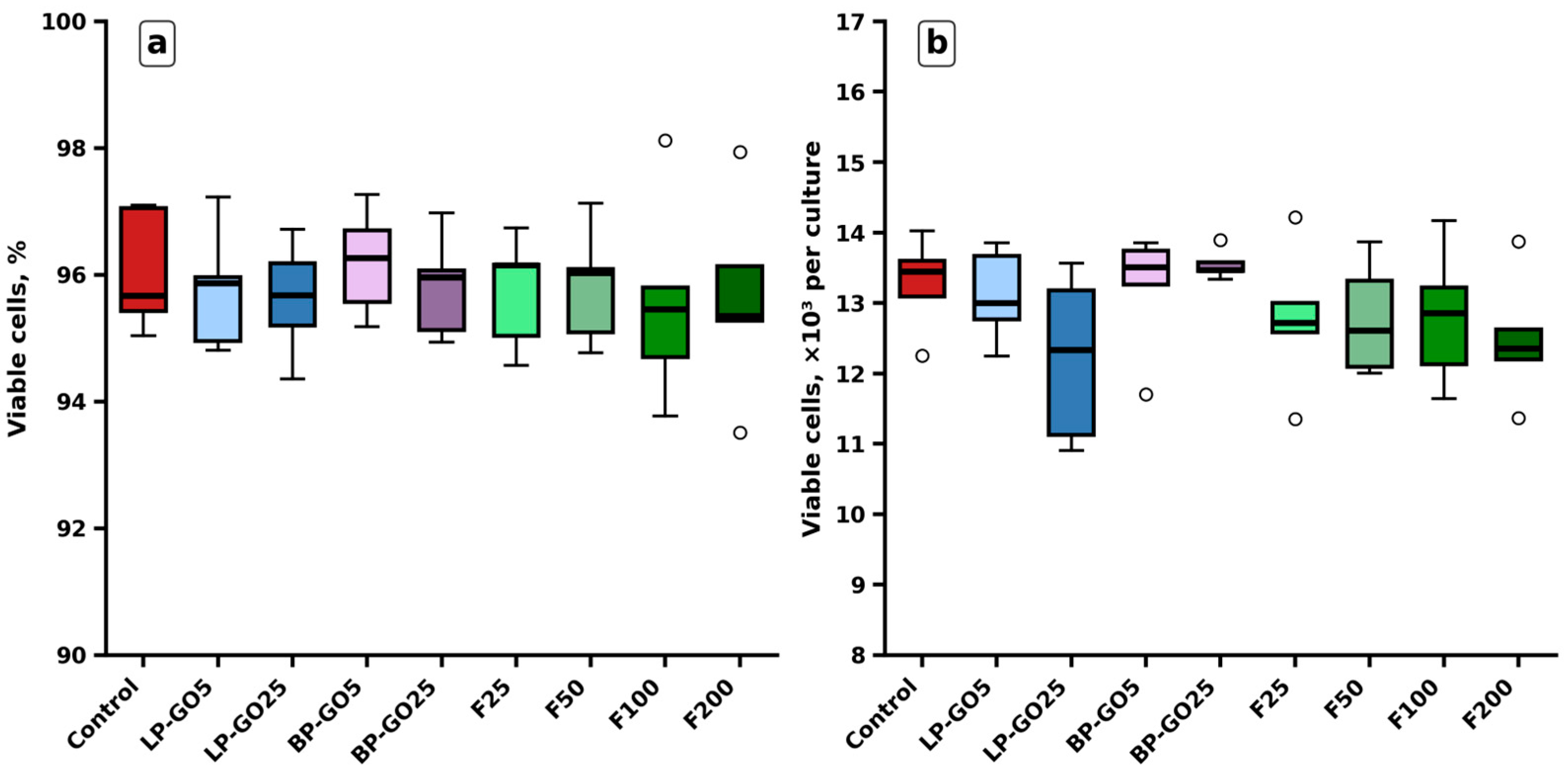
3.1. Effects of Pegylated Graphene Oxide Nanoparticles and Fullerenol C60(OH)24 on Viability of CD4+ Cells Polarized to the Treg Phenotype
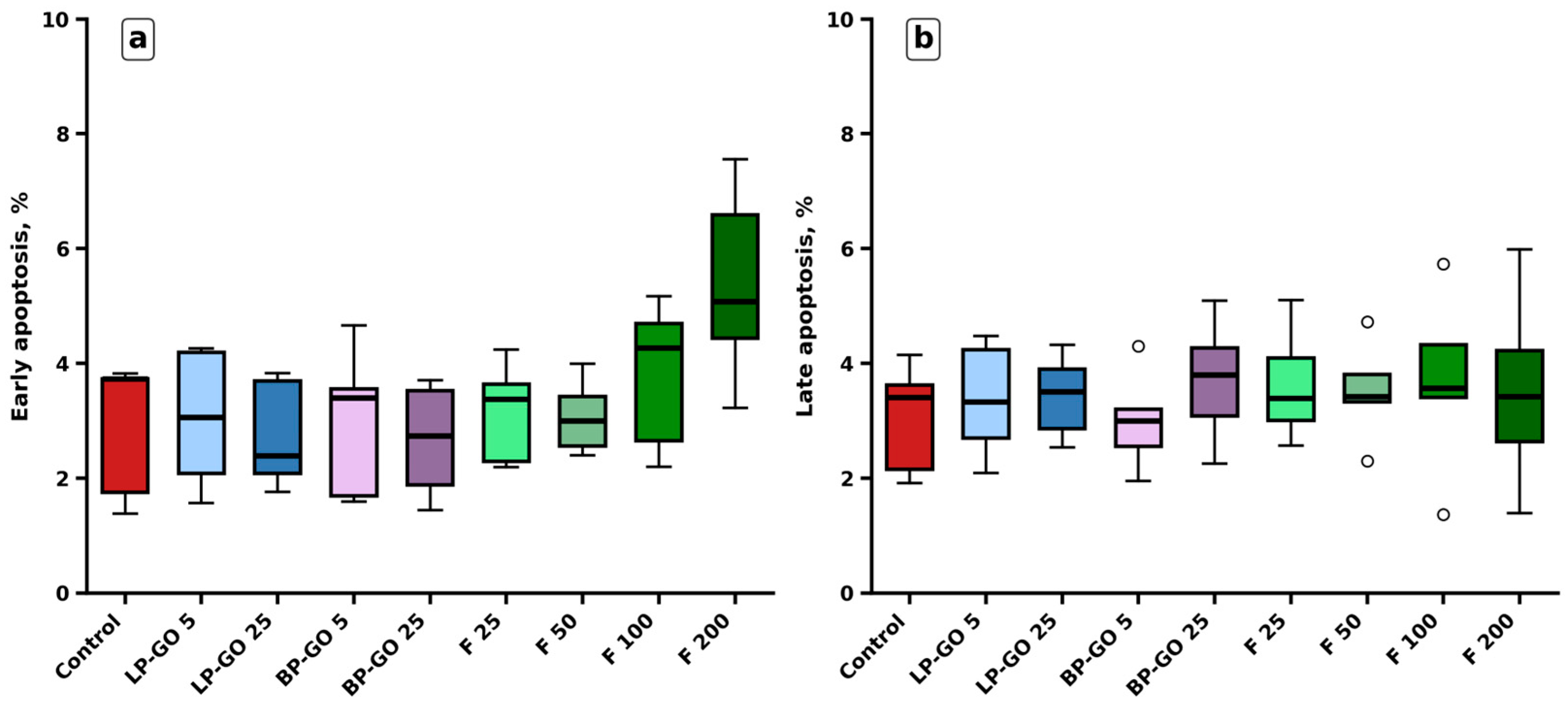
3.2. Effects of Pegylated Graphene Oxide Nanoparticles and Fullerenol C60(OH)24 on Apoptosis of CD4+ Cells Polarized to the Treg Phenotype
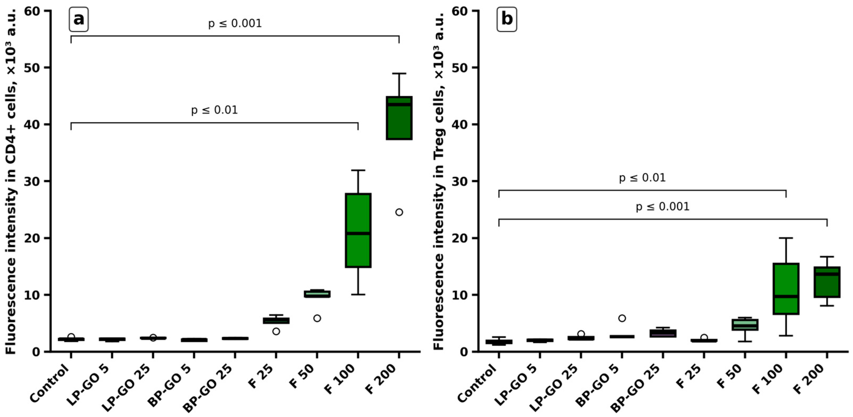
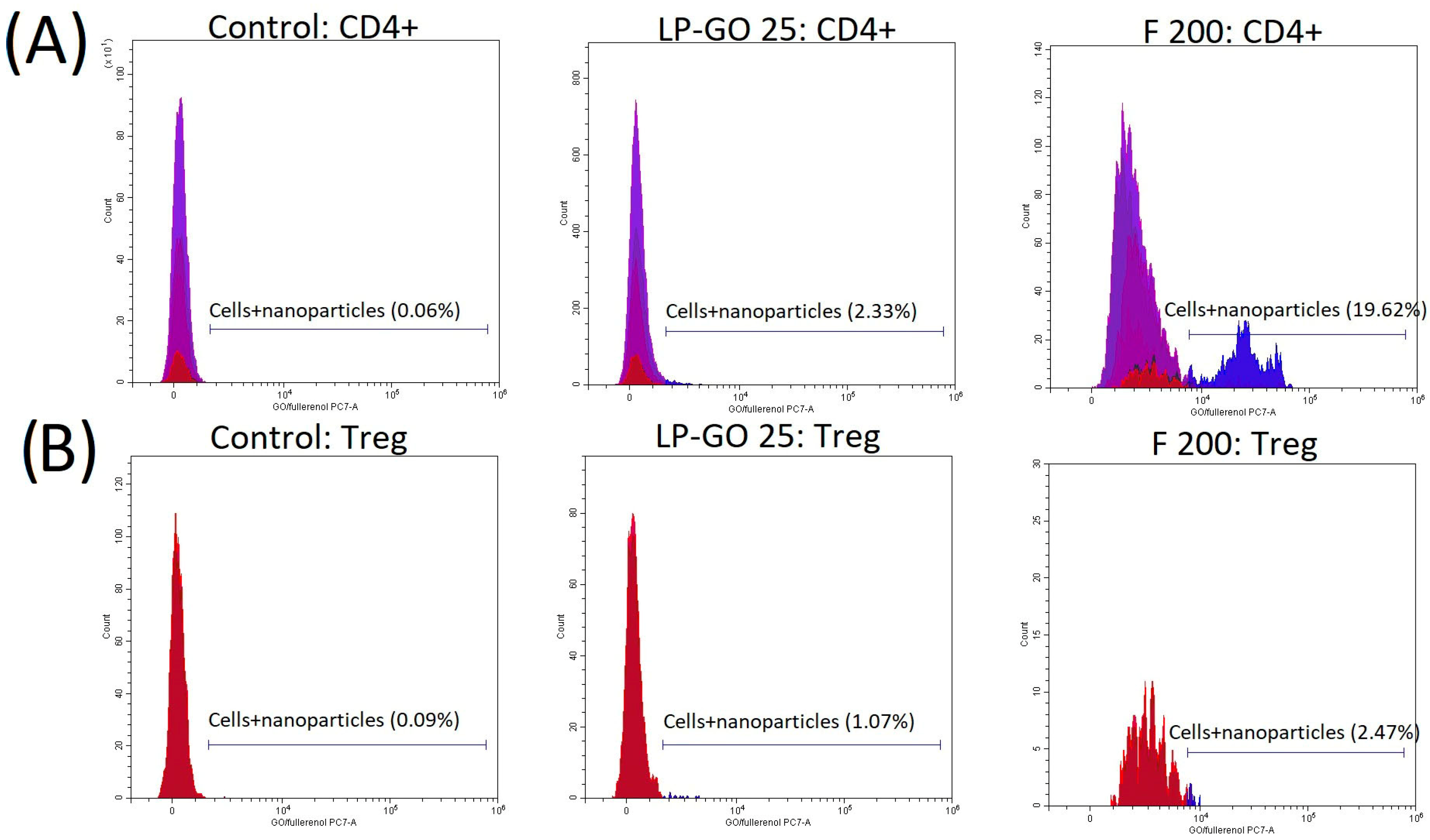
3.3. T-Helper Sorption of Pegylated Graphene Oxide Nanoparticles and Fullerenol C60(OH)24
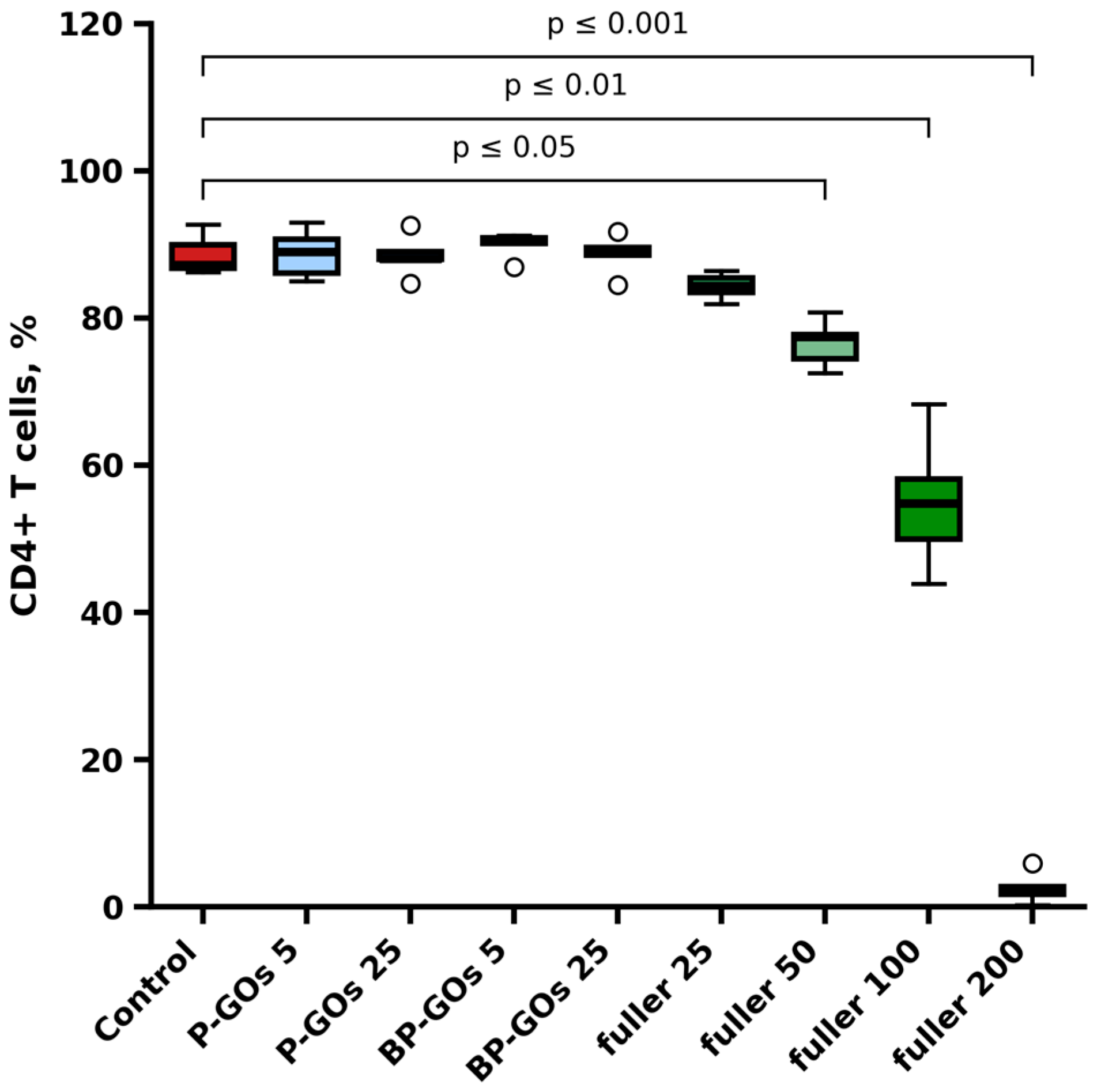
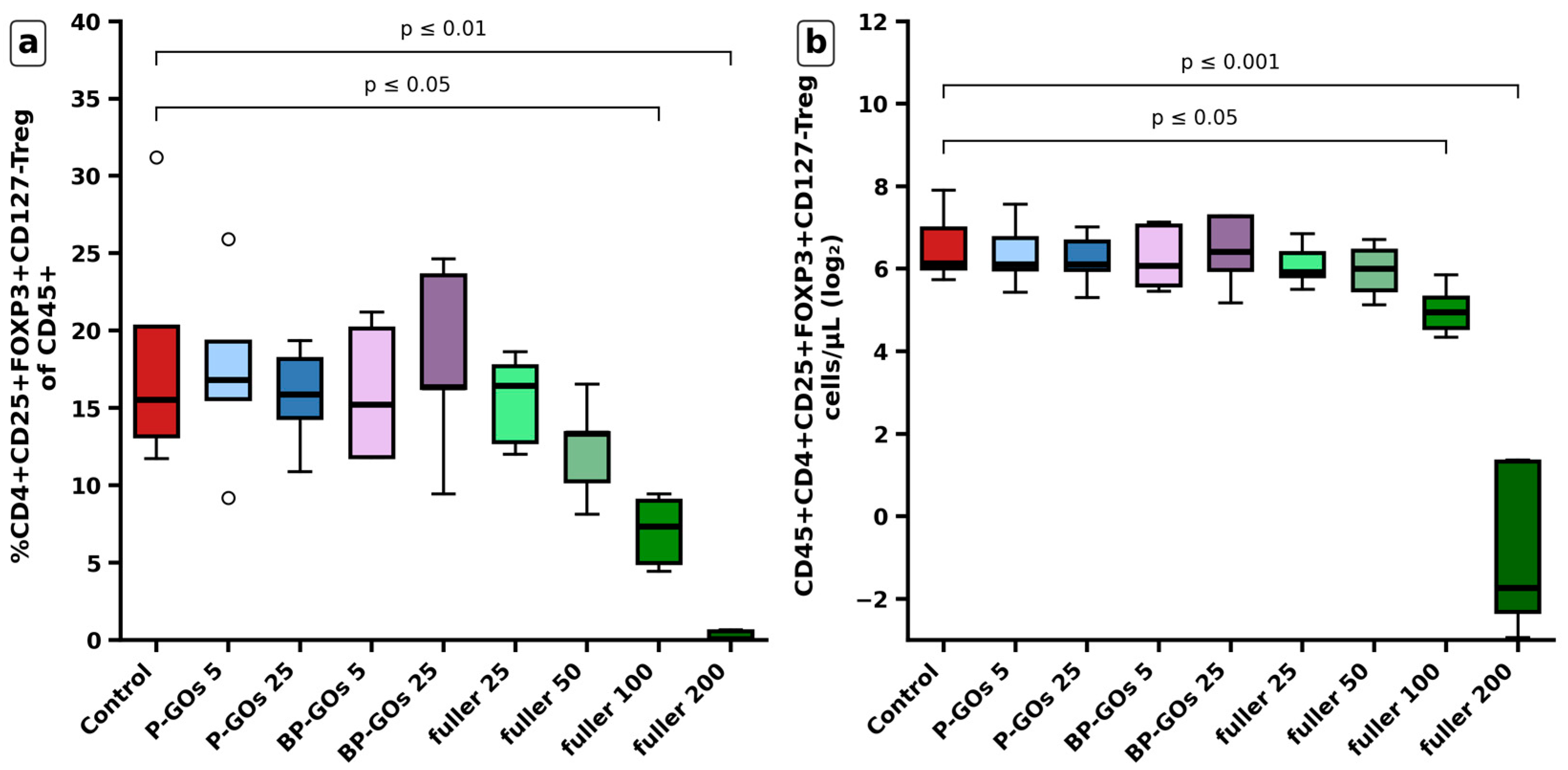
3.4. Effects of Pegylated Graphene Oxide Nanoparticles and Fullerenol C60(OH)24 on Treg Differentiation
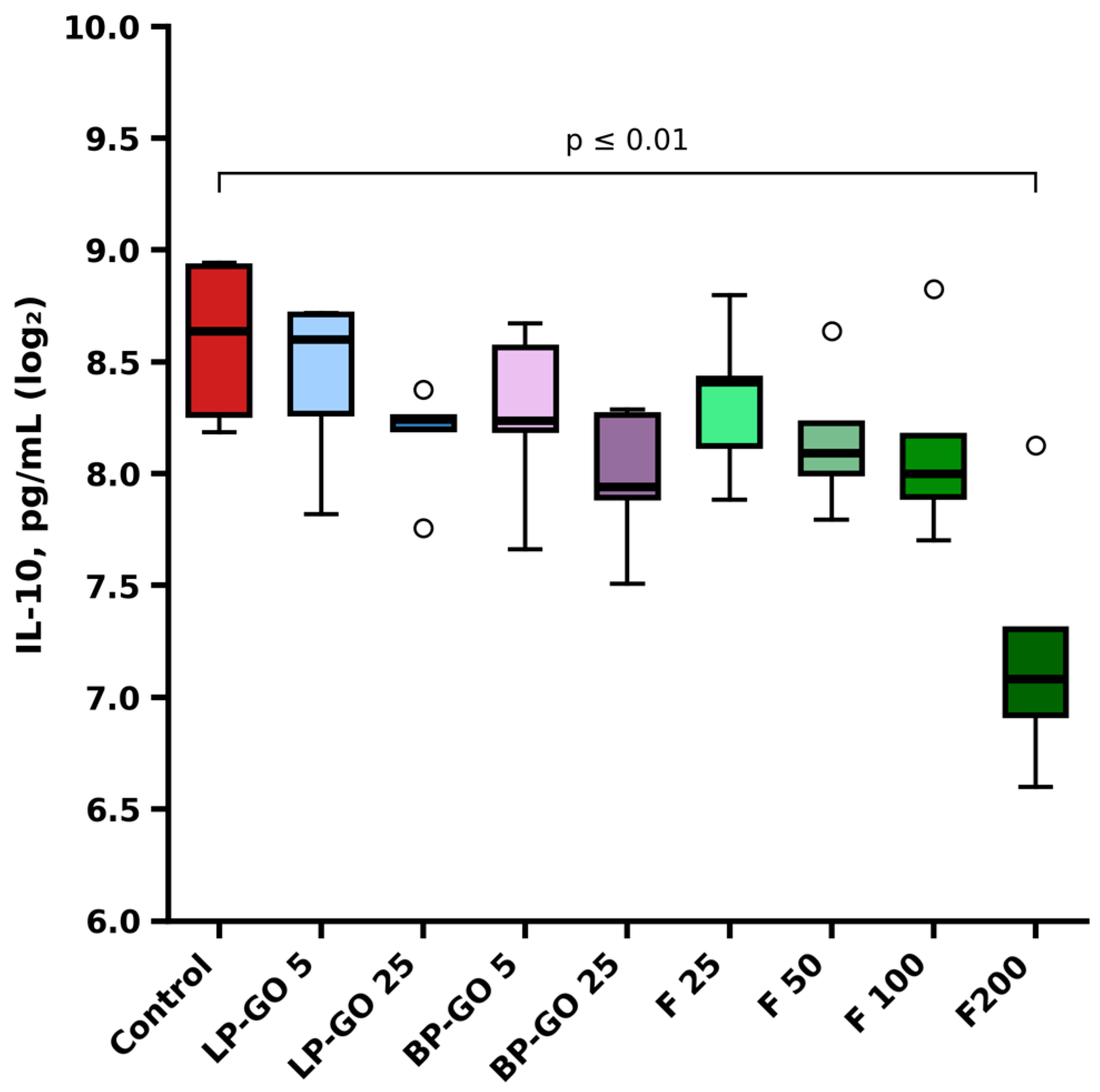
3.5. Effects of Pegylated Graphene Oxide and Fullerenol C60(OH)24 Nanoparticles on IL-10 Production by Tregs
4. Discussion
5. Conclusions
Supplementary Materials
Author Contributions
Funding
Data Availability Statement
Conflicts of Interest
Abbreviations
| BP-GO | Branched polyethylene glycol-coated graphene oxide nanoparticles |
| CNP | Carbon nanoparticles |
| CTLA-4 | Cytotoxic T-lymphocyte–associated protein 4 |
| GITR | Glucocorticoid-induced TNFR-related protein |
| GO | Graphene oxide |
| IL | Interleukin |
| LP-GO | Linear polyethylene glycol-coated graphene oxide nanoparticles |
| PEG | Polyethylene glycol |
| TGF-β | Transforming growth factor β |
| Th | T helper cell |
| Treg | Regulatory T cell |
References
- Vignali, D.A.A.; Collison, L.W.; Workman, C.J. How Regulatory T Cells Work. Nat. Rev. Immunol. 2008, 8, 523–532. [Google Scholar] [CrossRef] [Scilit]
- Okeke, E.B.; Uzonna, J.E. The Pivotal Role of Regulatory T Cells in the Regulation of Innate Immune Cells. Front. Immunol. 2019, 10, 680. [Google Scholar] [CrossRef] [Scilit] [PubMed]
- Goldmann, O.; Nwofor, O.V.; Chen, Q.; Medina, E. Mechanisms Underlying Immunosuppression by Regulatory Cells. Front. Immunol. 2024, 15, 1328193. [Google Scholar] [CrossRef] [Scilit]
- Ge, J.; Yin, X.; Chen, L. Regulatory T Cells: Masterminds of Immune Equilibrium and Future Therapeutic Innovations. Front. Immunol. 2024, 15, 1457189. [Google Scholar] [CrossRef] [Scilit] [PubMed]
- Hori, S.; Nomura, T.; Sakaguchi, S. Control of Regulatory T Cell Development by the Transcription Factor Foxp3. Science 2003, 299, 1057–1061. [Google Scholar] [CrossRef] [Scilit]
- Rudensky, A.Y. Regulatory T Cells and Foxp3. Immunol. Rev. 2011, 241, 260–268. [Google Scholar] [CrossRef] [Scilit]
- Carrette, F.; Surh, C.D. IL-7 Signaling and CD127 Receptor Regulation in the Control of T Cell Homeostasis. Semin. Immunol. 2012, 24, 209–217. [Google Scholar] [CrossRef] [Scilit]
- Staats, J. Immunophenotyping of Human Regulatory T Cells. Methods Mol. Biol. 2019, 2032, 141–177. [Google Scholar] [CrossRef] [Scilit]
- Liu, W.; Putnam, A.L.; Xu-Yu, Z.; Szot, G.L.; Lee, M.R.; Zhu, S.; Gottlieb, P.A.; Kapranov, P.; Gingeras, T.R.; Fazekas de St Groth, B.; et al. CD127 Expression Inversely Correlates with FoxP3 and Suppressive Function of Human CD4+ T Reg Cells. J. Exp. Med. 2006, 203, 1701–1711. [Google Scholar] [CrossRef] [Scilit]
- Jung, K.; Kim, J.-A.; Kim, Y.-J.; Lee, H.W.; Kim, C.-H.; Haam, S.; Kim, Y.-S. A Neuropilin-1 Antagonist Exerts Antitumor Immunity by Inhibiting the Suppressive Function of Intratumoral Regulatory T Cells. Cancer Immunol. Res. 2020, 8, 46–56. [Google Scholar] [CrossRef] [Scilit] [PubMed]
- Sacchetti, C.; Rapini, N.; Magrini, A.; Cirelli, E.; Bellucci, S.; Mattei, M.; Rosato, N.; Bottini, N.; Bottini, M. In Vivo Targeting of Intratumor Regulatory T Cells Using PEG-Modified Single-Walled Carbon Nanotubes. Bioconjugate Chem. 2013, 24, 852–858. [Google Scholar] [CrossRef] [Scilit]
- Yang, J.; Bae, H. Drug Conjugates for Targeting Regulatory T Cells in the Tumor Microenvironment: Guided Missiles for Cancer Treatment. Exp. Mol. Med. 2023, 55, 1996–2004. [Google Scholar] [CrossRef] [Scilit]
- Ronchetti, S.; Ricci, E.; Petrillo, M.G.; Cari, L.; Migliorati, G.; Nocentini, G.; Riccardi, C. Glucocorticoid-Induced Tumour Necrosis Factor Receptor-Related Protein: A Key Marker of Functional Regulatory T Cells. J. Immunol. Res. 2015, 2015, 171520. [Google Scholar] [CrossRef] [Scilit]
- Wang, L.; Liang, Y.; Zhao, C.; Ma, P.; Zeng, S.; Ju, D.; Zhao, M.; Yu, M.; Shi, Y. Regulatory T Cells in Homeostasis and Disease: Molecular Mechanisms and Therapeutic Potential. Signal Transduct. Target. Ther. 2025, 10, 345. [Google Scholar] [CrossRef] [Scilit]
- Ma, S.; Ming, Y.; Wu, J.; Cui, G. Cellular Metabolism Regulates the Differentiation and Function of T-Cell Subsets. Cell. Mol. Immunol. 2024, 21, 419–435. [Google Scholar] [CrossRef] [Scilit]
- Michalek, R.D.; Gerriets, V.A.; Jacobs, S.R.; Macintyre, A.N.; MacIver, N.J.; Mason, E.F.; Sullivan, S.A.; Nichols, A.G.; Rathmell, J.C. Cutting Edge: Distinct Glycolytic and Lipid Oxidative Metabolic Programs Are Essential for Effector and Regulatory CD4+ T Cell Subsets. J. Immunol. 2011, 186, 3299–3303. [Google Scholar] [CrossRef] [Scilit]
- Fortune, A.; Aime, A.; Raymond, D.; Kumar, S. Nanotechnology in Medicine: A Double-Edged Sword for Health Outcomes. Health Nanotechnol. 2025, 1, 9. [Google Scholar] [CrossRef] [Scilit]
- Hou, Y.; Zhu, C.; Shen, Z.; Xu, Y.; Zhou, S.; Zhou, X. Carbon Nanomaterials in Biomedicine: Opportunities and Toxicological Concerns. Int. J. Nanomed. 2025, 20, 14707–14730. [Google Scholar] [CrossRef] [Scilit]
- Chen, X.; Zou, M.; Liu, S.; Cheng, W.; Guo, W.; Feng, X. Applications of Graphene Family Nanomaterials in Regenerative Medicine: Recent Advances, Challenges, and Future Perspectives. Int. J. Nanomed. 2024, 19, 5459–5478. [Google Scholar] [CrossRef] [Scilit]
- Huang, X.; Zhao, W.; Khalilov, F.; Xu, N. Graphene Oxide: Preparation and Medical Research. Materials 2025, 18, 2855. [Google Scholar] [CrossRef] [Scilit]
- Liu, J.; Feng, X.; Chen, Z.; Yang, X.; Shen, Z.; Guo, M.; Deng, F.; Liu, Y.; Zhang, H.; Chen, C. The Adjuvant Effect of C60(OH)22 Nanoparticles Promoting Both Humoral and Cellular Immune Responses to HCV Recombinant Proteins. Mater. Sci. Eng. C Mater. Biol. Appl. 2019, 97, 753–759. [Google Scholar] [CrossRef] [Scilit] [PubMed]
- Seke, M.; Zivkovic, M.; Stankovic, A. Versatile Applications of Fullerenol Nanoparticles. Int. J. Pharm. 2024, 660, 124313. [Google Scholar] [CrossRef] [Scilit] [PubMed]
- Liu, Y.; Hardie, J.; Zhang, X.; Rotello, V.M. Effects of Engineered Nanoparticles on the Innate Immune System. Semin. Immunol. 2017, 34, 25–32. [Google Scholar] [CrossRef] [Scilit] [PubMed]
- Khramtsov, P.; Bochkova, M.; Timganova, V.; Nechaev, A.; Uzhviyuk, S.; Shardina, K.; Maslennikova, I.; Rayev, M.; Zamorina, S. Interaction of Graphene Oxide Modified with Linear and Branched PEG with Monocytes Isolated from Human Blood. Nanomaterials 2021, 12, 126. [Google Scholar] [CrossRef] [Scilit]
- Usanina, D.; Zamorina, S.; Bochkova, M.; Timganova, V.; Vlasova, V.; Ponomareva, V.; Dolgikh, M.; Lazarev, S.; Rayev, M. Effect of Fullerenol C60(OH)24 on the Viability and Metabolism of THP-1 Cells. Molecules 2025, 30, 4407. [Google Scholar] [CrossRef] [Scilit]
- Jeong, Y.H.; Lennon, G.; Veldman, G.; Serna, D.M.; Ibrahimov, A. Establishing Endotoxin Limits to Enhance the Reliability of in Vitro Immunogenicity Risk Assessments. mAbs 2025, 17, 2458627. [Google Scholar] [CrossRef] [Scilit]
- Zamorina, S.; Timganova, V.; Bochkova, M.; Shardina, K.; Uzhviyuk, S.; Khramtsov, P.; Usanina, D.; Rayev, M. The Effect of PEGylated Graphene Oxide Nanoparticles on the Th17-Polarization of Activated T Helpers. Materials 2023, 16, 877. [Google Scholar] [CrossRef] [Scilit]
- Lee, G.R. The Balance of Th17 versus Treg Cells in Autoimmunity. Int. J. Mol. Sci. 2018, 19, 730. [Google Scholar] [CrossRef] [Scilit]
- Bochkova, M.S.; Timganova, V.P.; Usanina, D.I.; Zamorina, S.A.; Rayev, M.B. Evaluation of Immunotoxicity of Fullerenol C60(OH)22-24 on Human Peripheral Blood T and B Lymphocytes. Bull. Exp. Biol. Med. 2025, 180, 164–169. [Google Scholar] [CrossRef] [Scilit]
- Ding, Z.; Zhang, Z.; Ma, H.; Chen, Y. In Vitro Hemocompatibility and Toxic Mechanism of Graphene Oxide on Human Peripheral Blood T Lymphocytes and Serum Albumin. ACS Appl. Mater. Interfaces 2014, 6, 19797–19807. [Google Scholar] [CrossRef] [Scilit]
- Wlodkowic, D.; Telford, W.; Skommer, J.; Darzynkiewicz, Z. Apoptosis and Beyond: Cytometry in Studies of Programmed Cell Death. In Methods in Cell Biology; Academic Press: Cambridge, MA, USA, 2011; Volume 103, pp. 55–98. [Google Scholar]
- Giang, S.; Horwitz, D.A.; Bickerton, S.; La Cava, A. Nanoparticles Engineered as Artificial Antigen-Presenting Cells Induce Human CD4+ and CD8+ Tregs That Are Functional in Humanized Mice. Front. Immunol. 2021, 12, 628059. [Google Scholar] [CrossRef] [Scilit] [PubMed]
- Serrano, A.; Casares, N.; Trocóniz, I.F.; Lozano, T.; Lasarte, J.J.; Zalba, S.; Garrido, M.J. Foxp3 Inhibitory Peptide Encapsulated in a Novel CD25-Targeted Nanoliposome Promotes Efficient Tumor Regression in Mice. Acta Pharmacol. Sin. 2025, 46, 171–183. [Google Scholar] [CrossRef] [Scilit] [PubMed]



| LP-GO | BP-GO | Fullerenol C60(OH)24 | |
|---|---|---|---|
| Diameter, nm | 184 ± 73 | 287 ± 52 | 186 (DLS, in water) 356 ± 167 (TEM, in water) 308 ± 110 (TEM, in the complete culture medium) |
| Polydispersity index | 0.25 ± 0.02 | 0.23 ± 0.02 | 0.45 (DLS, in water) |
| Zeta potential, mV | −31.70 ± 1.70 | −34.28 ± 0.41 | −26.0 ± 6.31 |
| PEG mass fraction, % | 17.2 ± 1.4 | 20.5 ± 1.8 | — |
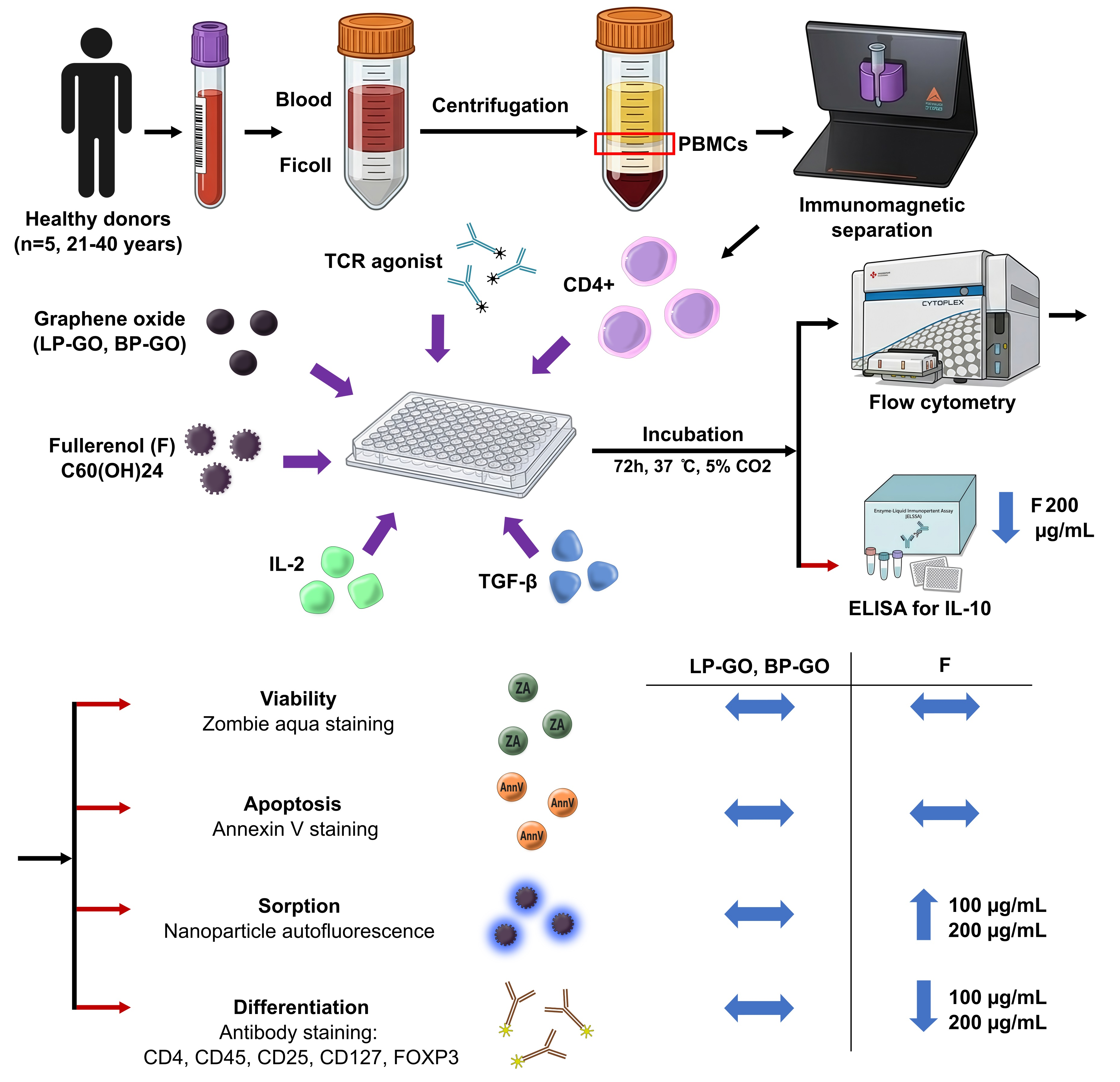
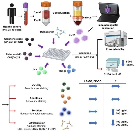
| Parameters | Graphene Oxide Nanoparticles LP-GO and BP-GO 5 and 25 μg/mL | Fullerenol C60(OH)24 |
|---|---|---|
| T-helper viability, % | ↔ | ↔ |
| T-helper viability, abs. | ↔ | ↔ |
| Early apoptosis, % | ↔ | ↔ |
| Late apoptosis, % | ↔ | ↔ |
| Sorption, T-helpers | ↔ | 100, 200 μg/mL ↑ |
| Sorption, Treg | ↔ | 100, 200 μg/mL ↑ |
| Treg, % | ↔ | 100, 200 μg/mL ↓ |
| T-helpers, % | ↔ | 50, 100, 200 μg/mL ↓ |
| IL-10, pg/mL, | ↔ | 100, 200 μg/mL ↓ |
Disclaimer/Publisher’s Note: The statements, opinions and data contained in all publications are solely those of the individual author(s) and contributor(s) and not of MDPI and/or the editor(s). MDPI and/or the editor(s) disclaim responsibility for any injury to people or property resulting from any ideas, methods, instructions or products referred to in the content. |
© 2026 by the authors. Licensee MDPI, Basel, Switzerland. This article is an open access article distributed under the terms and conditions of the Creative Commons Attribution (CC BY) license.
Share and Cite
Zamorina, S.; Usanina, D.; Devyatova, K.; Bochkova, M.; Nikitina, M.; Rayev, M.; Timganova, V. A Study of the Direct Effect of Pegylated Graphene Oxide Nanoparticles and Fullerenol C60(OH)24 on the Differentiation of Regulatory T Cells In Vitro. Nanomaterials 2026, 16, 667. https://doi.org/10.3390/nano16110667
Zamorina S, Usanina D, Devyatova K, Bochkova M, Nikitina M, Rayev M, Timganova V. A Study of the Direct Effect of Pegylated Graphene Oxide Nanoparticles and Fullerenol C60(OH)24 on the Differentiation of Regulatory T Cells In Vitro. Nanomaterials. 2026; 16(11):667. https://doi.org/10.3390/nano16110667
Chicago/Turabian StyleZamorina, Svetlana, Darya Usanina, Kseniya Devyatova, Maria Bochkova, Maria Nikitina, Mikhail Rayev, and Valeria Timganova. 2026. "A Study of the Direct Effect of Pegylated Graphene Oxide Nanoparticles and Fullerenol C60(OH)24 on the Differentiation of Regulatory T Cells In Vitro" Nanomaterials 16, no. 11: 667. https://doi.org/10.3390/nano16110667
APA StyleZamorina, S., Usanina, D., Devyatova, K., Bochkova, M., Nikitina, M., Rayev, M., & Timganova, V. (2026). A Study of the Direct Effect of Pegylated Graphene Oxide Nanoparticles and Fullerenol C60(OH)24 on the Differentiation of Regulatory T Cells In Vitro. Nanomaterials, 16(11), 667. https://doi.org/10.3390/nano16110667