Article

Optimized Construction of Highly Efficient P-Bi2MoO6/g-C3N4 Photocatalytic Bactericide: Based on Source Material and Synthesis Process

1
College of Chemistry and Molecular Engineering, Qingdao University of Science and Technology, Qingdao 266042, China
2
State Key Laboratory of Advanced Marine Materials, Key Laboratory of Marine Environmental Corrosion and Bio-Fouling, Institute of Oceanology, Chinese Academy of Sciences, Qingdao 266071, China
3
University of Chinese Academy of Sciences, 19 (Jia) Yuquan Road, Beijing 100049, China
*
Authors to whom correspondence should be addressed.
Nanomaterials 2025, 15(11), 834; https://doi.org/10.3390/nano15110834
Submission received: 1 May 2025 / Revised: 24 May 2025 / Accepted: 28 May 2025 / Published: 30 May 2025
(This article belongs to the Special Issue Heterogeneous Photocatalysts Based on Nanocomposites)

Abstract

In this study, Bi2MoO6 nanoflowers with different molybdenum sources were in situ grown on the surface of g-C3N4 nanosheets (OCN) by a simple one-step solvothermal method. The effects of doping and different molybdenum sources on the photocatalytic degradation and bactericidal activity of Bi2MoO6/OCN were discussed. Among them, the solvothermal preparation of P-Bi2MoO6/OCN using phosphomolybdic acid as molybdenum source can make up for the shortcomings caused by the destruction of OCN structure by generating more lattice defects to promote charge separation and constructing Lewis acid/base sites to effectively improve the photocatalytic performance. In addition, by adding phosphoric acid to increase the P-doped content, more exposed alkaline active sites are induced on the surface of P-Bi2MoO6/OCN, as well as larger specific surface area and charge transfer efficiency, which further improve the photocatalytic performance. Finally, the optimized 16P-Bi2MoO6/OCN showed a degradation rate of 99.7% for 20 mg/L rhodamine B (RhB) within 80 min under visible light, and the antibacterial rates against E. coli, S. aureus and P. aeruginosa within 300 min were 99.58%, 98.20% and 97.48%, respectively. This study provides a reference for optimizing the synthesis of environmentally friendly, solar-responsive, photocatalytic sterilization materials from the perspective of preparation, raw materials and structure.

Graphical Abstract

1. Introduction

The threat of microbial contamination to human health and the economy has attracted more and more attention in many fields. In addition to daily life and medical disinfection, the key role of bacteria in the biological pollution of water environments such as oceans has also prompted the development of new antibacterial and antifouling methods. Although traditional antibacterial materials have effective bactericidal effects, the production cost of many materials is high. The heavy metal ions and toxic substances exuded during the action process can easily cause secondary pollution to the environment and are also prone to biological resistance, laying hidden dangers for long-term prevention and control. Looking for green, efficient and low-cost antibacterial and anti-fouling methods has become an important issue that is closely related to social development and people’s life safety and cannot be ignored.
In recent years, photocatalytic technology has attracted more and more attention in the fields of green energy, environmental management, pollution control and medical and health care [1,2,3]. Its unique photo-induced redox reaction mechanism has wide applicability, and the controllability of photocatalytic materials also provides great potential for research and development. At present, the most studied photocatalytic materials are TiO2 [4], graphitic carbon nitride (g-C3N4) [5], metal-organic frameworks (MOFs) [6], MXene [7], etc., which are widely used in the photocatalytic decomposition of water to produce hydrogen, degradation of dyes or antibiotics, photocatalytic nitrogen fixation and CO2 conversion. With the development of the marine economy, the application potential of photocatalytic technology in the field of prevention and control of biological fouling has also begun to receive attention. However, the development of photocatalytic materials is still limited by the shortcomings of low separation efficiency and easy recombination of photogenerated carriers [8,9,10]. It is still a huge challenge to develop efficient and stable new photocatalytic materials suitable for various fields.
g-C3N4 is a non-metallic, two-dimensional semiconductor material with excellent stability, environmental protection performance and low preparation cost. As a photocatalytic material, it also has an appropriate band gap and a wide light response range. The modification of pure g-C3N4 is often carried out by adjusting the morphology. For instance, g-C3N4 with special morphologies, such as porous, single-layer nanosheets, hollow spheres and nanotubes, is designed and synthesized [11,12,13]. Ultimately, g-C3N4 with larger specific surface area, smaller pore size and higher crystallinity is prepared. In this regard, more exposed active sites are the key to improving photocatalytic performance. However, subject to the disadvantages of slow charge separation and transport and easy recombination of free carriers, g-C3N4 is more suitable for constructing heterojunctions with other materials to obtain more efficient photocatalytic performance.
In addition to typical catalytic materials such as TiO2, MOFs and ZnO2, non-toxic bismuth-based photocatalysts such as Bi2WO6 [14], BiVO4 [15,16] and Bi2MoO6 [17,18] are also regarded as environmentally friendly, non-toxic photocatalysts and have received extensive attention. Bi2MoO6 is an Aurivillius-type oxide formed by alternating layers of [MoO4]2− and [Bi2O2]2+, which are connected by oxygen atoms [19]. Similarly to g-C3N4, its non-toxicity, cost-effectiveness and broad-spectrum light response make it suitable as an environmentally friendly photocatalytic antifouling material. However, similarly, the photocatalytic performance of Bi2MoO6 monomer is also not good, due to its low quantum yield and rapid recombination of photogenerated carriers [20,21]. The modification of Bi2MoO6 monomer is mainly to improve the photocatalytic efficiency by doping and inducing lattice defects. The different doping elements may affect the band structure, charge transfer and surface adsorption of the catalyst. Chang et al., by doping In3+, directly replaced Bi3+ ions in the [Bi2O2]2+ layer, resulting in surface trapping traps to reduce the recombination rate of photogenerated carriers, causing the lattice compressive strain to induce the formation of a local built-in electric field to improve the charge transfer efficiency, so that the optimized Bi2MoO6 exhibits enhanced photocatalytic degradation of antibiotics [22]. By doping Fe3+ to partially replace Bi3+, Li et al. enhanced the asymmetry inside the Bi2MoO6 lattice, improved the surface separation efficiency of photogenerated electron–hole pairs and significantly improved the photocatalytic dye degradation and water splitting oxygen production capacity [23]. However, in previous studies on the doping modification of Bi2MoO6, although sodium molybdate dihydrate or ammonium molybdate tetrahydrate was used as the molybdenum source to prepare Bi2MoO6 photocatalyst, the effect of introducing Na and N doping from the reaction source on the photocatalytic performance of the system was rarely discussed. In order to design and construct a better Bi2MoO6-based photocatalytic material, it is obviously necessary to compare and analyze the effects of doping atoms introduced by different molybdenum sources in detail. In this paper, phosphomolybdic acid (H3PO4·12MoO3) is selected as the optimized molybdenum source. This is because P doping often associates to produce acidic and alkaline active centers, which can interact with g-C3N4 with strong Lewis bases to form sterically hindered Lewis bases and Lewis acids, which greatly enhances the adsorption and activation of reactant molecules [24]. At the same time, previous studies on Bi2MoO6 and g-C3N4 composites have also shown that they have a suitable band gap structure and can form type I [25], type II [26], type Z [27] and type S [28,29] heterojunctions to construct new carrier transfer channels to promote separation.
It cannot be ignored that for the composite materials of the doped system, due to the dual effects of doping modification and composite modification, the influencing factors are more complex, and it is difficult to obtain the target material with improved performance. In this paper, the Bi2MoO6/g-C3N4 system is taken as an example to analyze and explore this complex process in detail. Firstly, Bi2MoO6 nanoflowers with different molybdenum sources were in situ grown on the surface of g-C3N4 nanosheets (OCN) by a simple one-step solvothermal method under the same conditions. P-Bi2MoO6/OCN (phosphomolybdate as molybdenum source), Na-Bi2MoO6/OCN (sodium molybdate as molybdenum source) and N-Bi2MoO6/OCN (ammonium molybdate as molybdenum source) heterojunction composites were obtained. The effects of different molybdenum source doping on the structure and photocatalytic performance of Bi2MoO6/g-C3N4 heterojunction were studied, and the potential reaction mechanism was analyzed. After solvothermal treatment, large-area OCN nanosheets shrank into small-area interlaced g-C3N4 nanosheets, and the specific surface area and active sites were reduced. After comparing the photocatalytic degradation performance of dyes under visible light, it was found that although the heterojunction of Bi2MoO6 doped with Na, N and contracted OCN could effectively inhibit the recombination of photogenerated carriers to a certain extent, the photocatalytic performance was still lower than that of pure OCN due to the inability to strongly compensate for the reduction of contracted OCN active sites. The difference was that the P-doped Bi2MoO6/g-C3N4 system prepared with phosphomolybdic acid as the molybdenum source effectively compensated for the unfavorable factors caused by the structural damage of OCN by generating more Bi vacancy lattice defects to capture holes and introducing highly active reaction sites, such as Lewis acid/base sites, and significantly improved the photocatalytic activity. The degradation rate of 20 mg/L RhB was 98.8% under visible light for 120 min, and the antibacterial rates against E. coli, S. aureus and P. aeruginosa were 93.09%, 92.81% and 91.13% within 300 min, respectively. For further optimization, an appropriate amount of phosphoric acid was added to increase the content of phosphorus doping, induce more [Bi2O2]2+ layer lattice bismuth to form vacancy defects and cover the surface with oxides such as metal Bi and Bi2O3 to form impurity levels to promote charge separation, with faster charge-transfer efficiency. At the same time, there were more alkaline active sites on the surface of OCN, and the synergistic effect improved the photocatalytic performance. Finally, the optimized 16P-Bi2MoO6/OCN showed a degradation rate of 99.7% for 20 mg/L RhB within 80 min under visible light, and the antibacterial rates against E. coli, S. aureus and P. aeruginosa within 300 min were 99.58%, 98.20% and 97.48%, respectively. This study provides a valuable reference for the synthesis of environmentally friendly, solar-responsive, photocatalytic sterilization materials from the perspective of reaction source selection and optimization of the structure of complex systems.

2. Experimental Section

2.1. Preparation of OCN

A certain amount of urea was put into an alumina crucible, which was wrapped with tin foil, heated from room temperature to 550 °C at 5 °C/min and kept for 2 h in a muffle furnace. After cooling, it was not necessary to take out the crucible. Then, the muffle furnace was heated to 500 °C at the same speed and kept for 2 h. Then, the light-yellow g-C3N4 was obtained and labeled as OCN.

2.2. Preparation of Three Types of Nanostructured Flower-like Bi2MoO6 Particles

For P-Bi2MoO6, Na-Bi2MoO6 and N-Bi2MoO6, bismuth nitrate pentahydrate (0.485 g) was used as the Bi source, while phosphomolybdate (0.076 g), sodium molybdate dihydrate (0.121 g) and ammonium molybdate tetrahydrate (0.0883 g) were utilized as the Mo sources for the synthesis of Bi2MoO6 nanoflower particles.
Initially, the aforementioned reactants were dissolved in 5 mL of ethylene glycol and ultrasonically treated for 5 min to ensure thorough dissolution. Subsequently, the Bi source and Mo source were mixed evenly before being slowly added to 20 mL anhydrous ethanol with continuous stirring for 1 h. The resulting mixture was then transferred into a reaction vessel and maintained at 160 °C for a duration of 10 h. Following cooling, it underwent washing with anhydrous ethanol, centrifugation, drying at 60 °C and subsequent grinding into powder. Based on their respective Mo sources representing different doping types, they were denoted as P-Bi2MoO6, Na-Bi2MoO6 and N-Bi2MoO6.

2.3. Preparation of Doped-Bi2MoO6/g-C3N4 Heterojunction

A total of 0.3 g of OCN was added to 20 mL of anhydrous ethanol, and the g-C3N4 ethanol dispersion was obtained by ultrasonication for 30 min. The Bi source and Mo source were mixed and slowly added to the g-C3N4 ethanol dispersion, and the other steps were the same as those used to prepare Bi2MoO6. According to the mass ratio of g-C3N4:Bi2MoO6 = 1:2, OCN composites with three different doped Bi2MoO6 nanoflower spheres were prepared, labeled as P-Bi2MoO6/OCN, Na-Bi2MoO6/OCN and N-Bi2MoO6/OCN. In addition, in order to optimize P-Bi2MoO6/OCN heterojunction, a small amount of phosphoric acid was added to the mixture and stirred together for 1 h before the solvothermal reaction. It was named according to the amount of phosphoric acid added. For example, P-Bi2MoO6/OCN with 16 μL phosphoric acid was named 16P-Bi2MoO6/OCN.

2.4. Characterization of the Materials

Sample characterization methods were as follows: The morphology and structure of the samples were characterized by scanning electron microscopy (SEM, Hitachi Regulus8100, Tokyo, Japan), energy dispersive spectrometer (Thermo Fisher Ultradry Series Noran System7, MA, USA), transmission electron microscopy (TEM, Hitachi HT7700, Tokyo, Japan) and high-resolution transmission electron microscopy (HRTEM, JEM-2100F, NEC Co., Tokyo, Japan). The X-ray diffraction (XRD) patterns were recorded on a Riken Smart Lab diffractometer. The X-ray photoelectron spectroscopy (XPS) results were recorded on a Thermo ESCALAB 250 Xi instrument (Thermo, MA, USA). At the liquid nitrogen temperature (77 K), the Brunauer–Emmett–Teller (BET) specific surface area of the samples was determined by a nitrogen adsorption apparatus (Micromeritics ASAP 2460, GA, USA). The pore size distribution was calculated from the desorption branch of the isotherm according to the Barrett–Joyner–Halenda (BJH) model. Photoluminescence (PL) analysis was performed on a F-4700 (Hitachi, Tokyo, Japan) fluorescence spectrophotometer. The Fourier-transform infrared (FT-IR) spectra were collected with a Thermo Fisher IS5 spectrometer (MA, USA), with KBr as the diluent. CO2 and NH3 temperature-programmed desorption (CO2/NH3-TPD) were measured by using a Micromeritics Autochem II2920 temperature programming detector, USA. Superoxide radicals (·O2) and hydroxyl radicals (·OH) generated in the photocatalytic reaction were detected by DMPO in methanol and DMPO in water, respectively, and the corresponding EPR signals were recorded on a Bruker EMXplus spectrometer (Bruker Co., MA, USA).

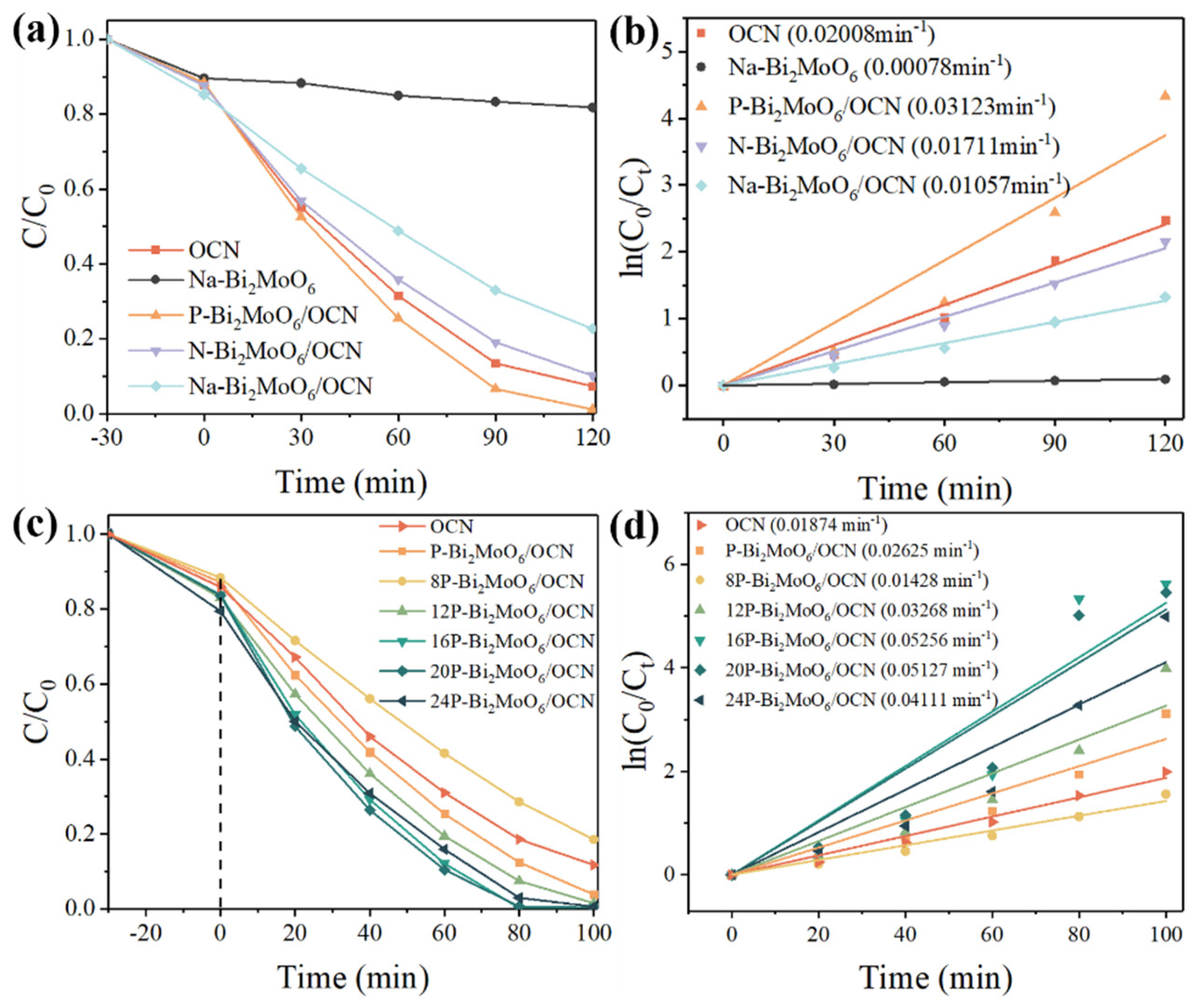
2.5. Photocatalytic Degradation Performance

The photocatalytic degradation experiment was carried out using an 800 W xenon lamp with a light intensity of 80 mW/cm2 and a 420 nm filter. Rhodamine B (RhB) was chosen as the pollutant dye for degradation in the photocatalytic experiment, with 25 mg of photocatalyst added to a 50 mL solution containing RhB at a concentration of 20 mg/L. Prior to illumination, the photocatalyst was stirred in the RhB solution for 30 min to achieve adsorption–desorption equilibrium. Subsequently, samples were collected every 30 min, and after removing the catalyst by centrifugation, the absorbance of the residual solution was measured at 554 nm using an ELISA meter (AKTAavant25, Thermo Fisher). The concentration of RhB at each sampling time point was determined based on the absorbance–concentration standard curve of the RhB solution.

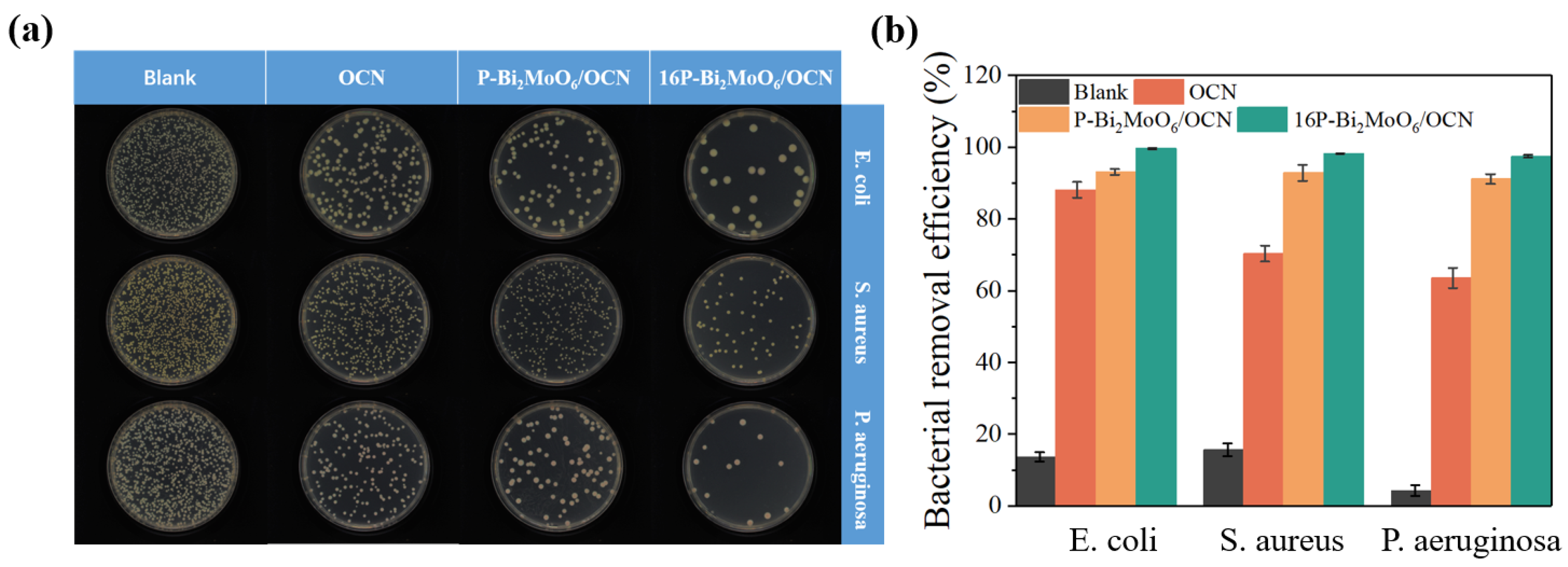
2.6. Photocatalytic Antibacterial Performance

In the photocatalytic antibacterial experiment, the light intensity was the same as that of the degradation experiment. Escherichia coli (E. coli), Staphylococcus aureus (S. aureus) and Pseudomonas aeruginosa (P. aeruginosa) were selected as the typical strains of broad-spectrum antibacterial, and the plate colony counting method was used to calculate the number of colonies to evaluate the antibacterial effect of each photocatalyst. The single colony was inoculated into the LB liquid medium, which had been killed by bacteria, and cultured at 37 °C for 24 h to obtain the original bacterial solution. Five mL of the original bacterial solution was put into a 10 mL centrifuge tube, centrifuged to remove the LB liquid medium, washed with 0.01 mol/L PBS (pH = 7.4) buffer solution twice. Five mL of PBS buffer solution was added, and the solution was shaken well. A total of 49.5 mL of PBS buffer solution and 0.5 mL of the washed original bacterial solution were added into a 50 mL quartz tube, along with 50 mg of photocatalyst. The blank experiment without photocatalyst was used as the control experiment. After adding the photocatalyst, it was stirred in the dark for 1 h to achieve the adsorption–desorption balance. After illumination, it was sampled every 100 min. One hundred μL of the reaction solution was taken each time, several gradients were diluted with 0.01 mol/L PBS buffer solution according to the series dilution method. Then, 100 μL was taken from the solution with different dilution times to the prepared sterile LB solid medium, and the bacterial solution was evenly smeared on the medium by rolling small glass beads. Finally, the LB solid medium was put into a constant temperature incubator at 37 °C for 24 h. The bacterial concentration was calculated by counting the number of colonies grown on the culture medium, with 30~300 colonies on the culture medium as the best counting range. The survival rate and sterilization rate of each group of bacteria were calculated. Each group of experiments was measured three times in parallel, and the average value was taken as the final result. The survival rate of bacteria was calculated according to the following formula:
survival rate   ( % ) = C t C 0 × 100 %
C0 is the initial number of colonies, and Ct is the number of colonies at the time of illumination t. The antibacterial rate is
antibacterial rate   ( % ) = 100 % survival rate   ( % )

2.7. Photoelectrochemical (PEC) Performance

The photoelectrochemical (PEC) and electrochemical performance of the prepared samples were measured under a three-electrode system using CHI660E electrochemical workstation (Shanghai Chenhua Instrument Co., Ltd., Shanghai, China). The 5 mg powder sample was mixed with isopropanol and 5% Nafion solution to form a uniform and fine slurry, which was then coated on the conductive side of FTO conductive glass (coated area: 1 × 1 cm2) and was dried naturally to obtain the working electrode for testing. The PEC performance test used a 300 W xenon lamp light source, and a 420 nm filter was installed on it to filter ultraviolet light. The test position was fixed, and the light intensity of the illuminated surface was adjusted to 100 mW/cm2. The Ag/AgCl (saturated KCl) electrode was used as the reference electrode, the platinum plate as the counter electrode, and the FTO conductive glass coated with the sample as the working electrode. The photoelectric response i-t curve was tested by 50 s-switching on and off light. The electrochemical impedance spectroscopy (EIS) of the sample was tested under the illumination condition at open circuit potential over frequency range of 100 kHz~0.1 Hz and at the amplitude of the AC voltage of 5 mV. The Mott–Schottky plot was measured under the potential range of −1.0~1.0 V and the frequency of 1000 Hz in the dark. The electrolyte used for the above measurements was 0.1 M Na2SO4 aqueous solution.

3. Results and Discussion

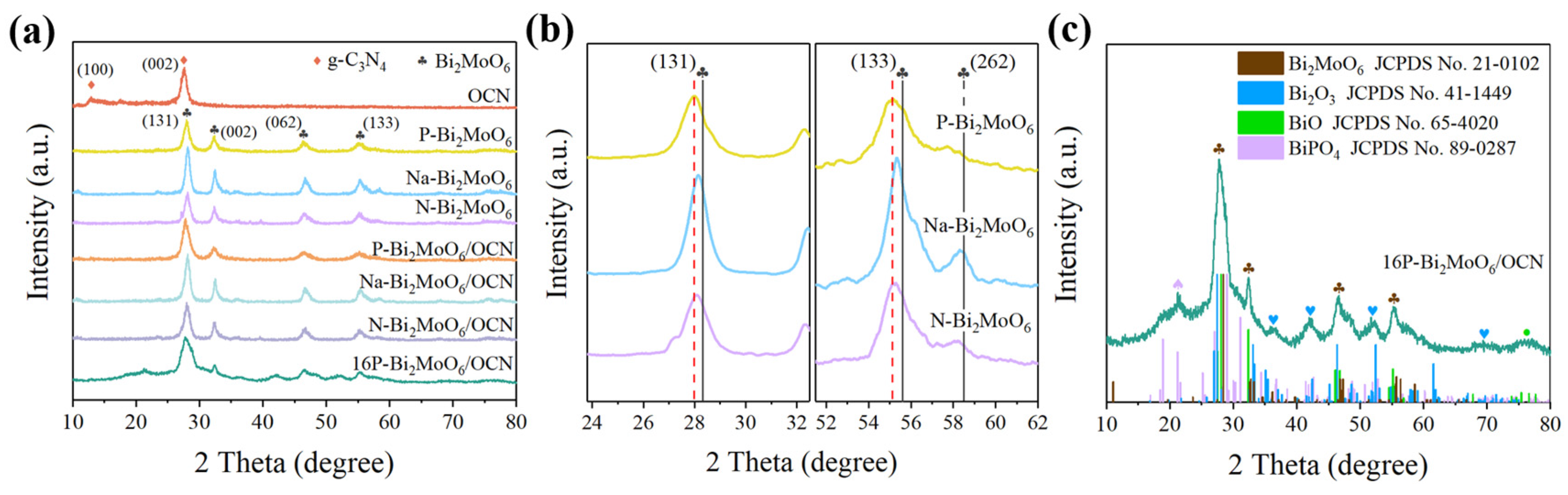
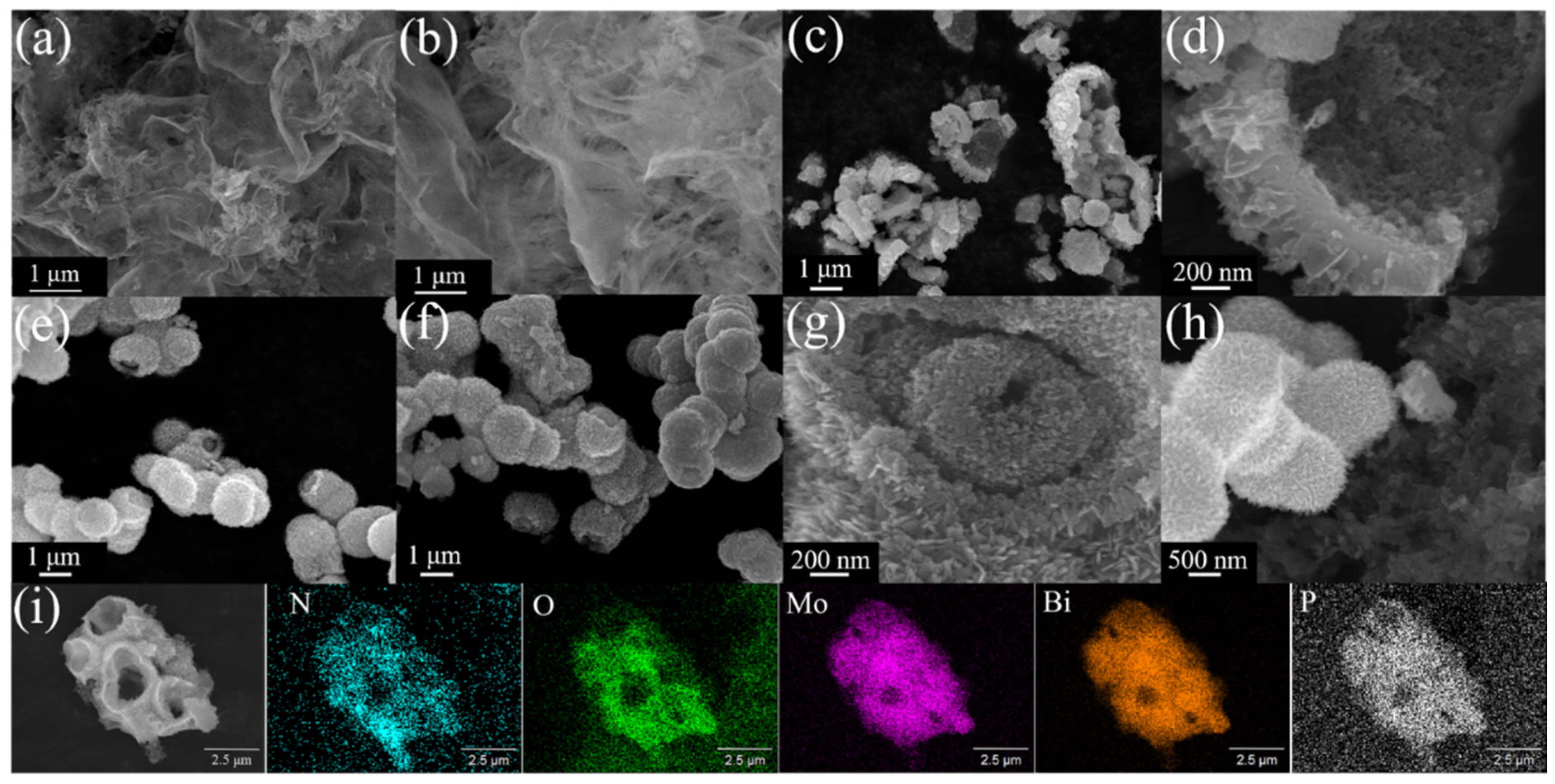
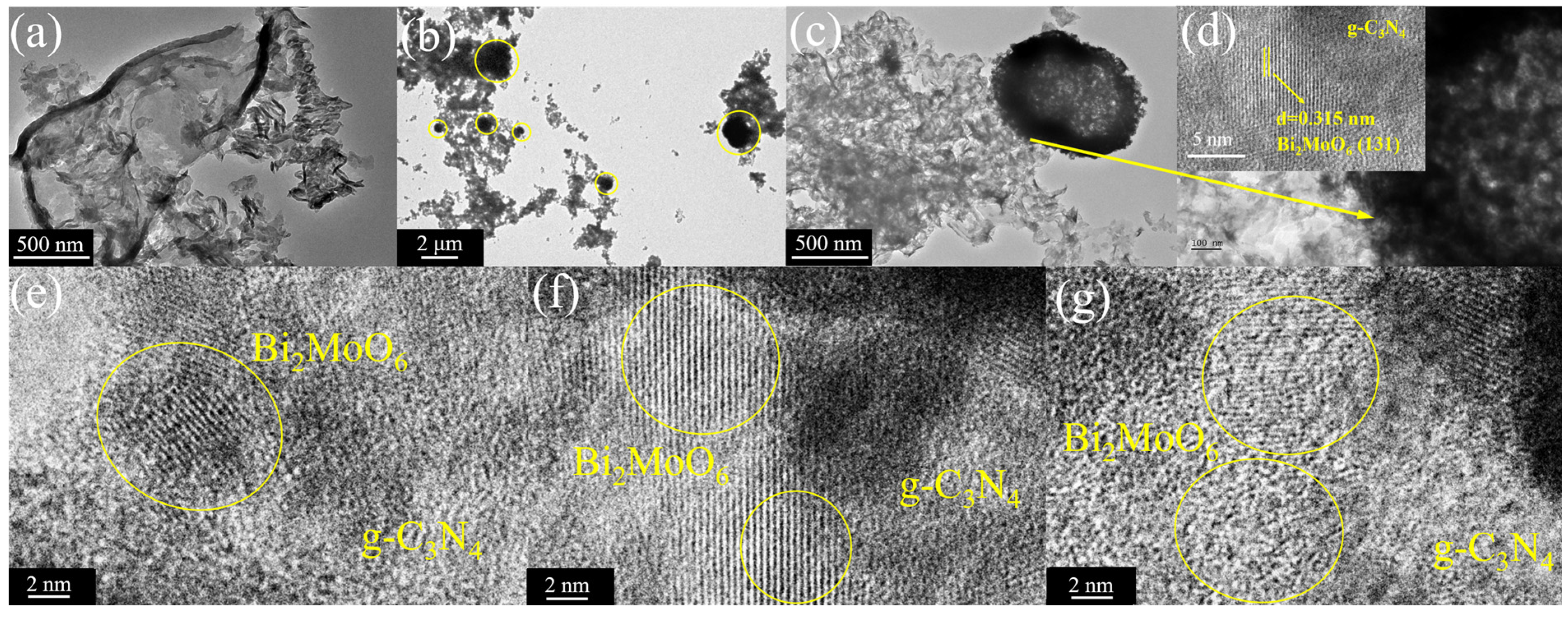
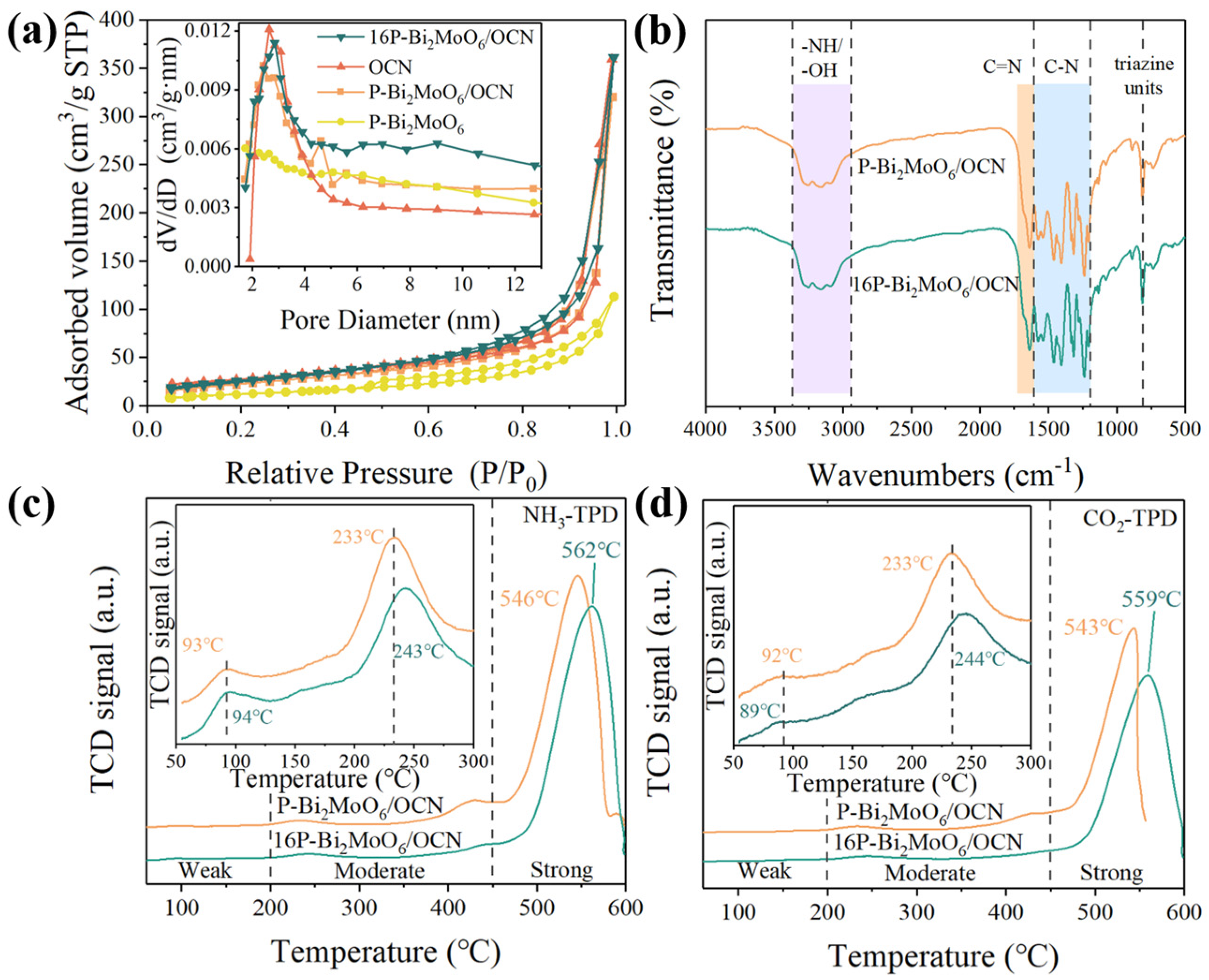
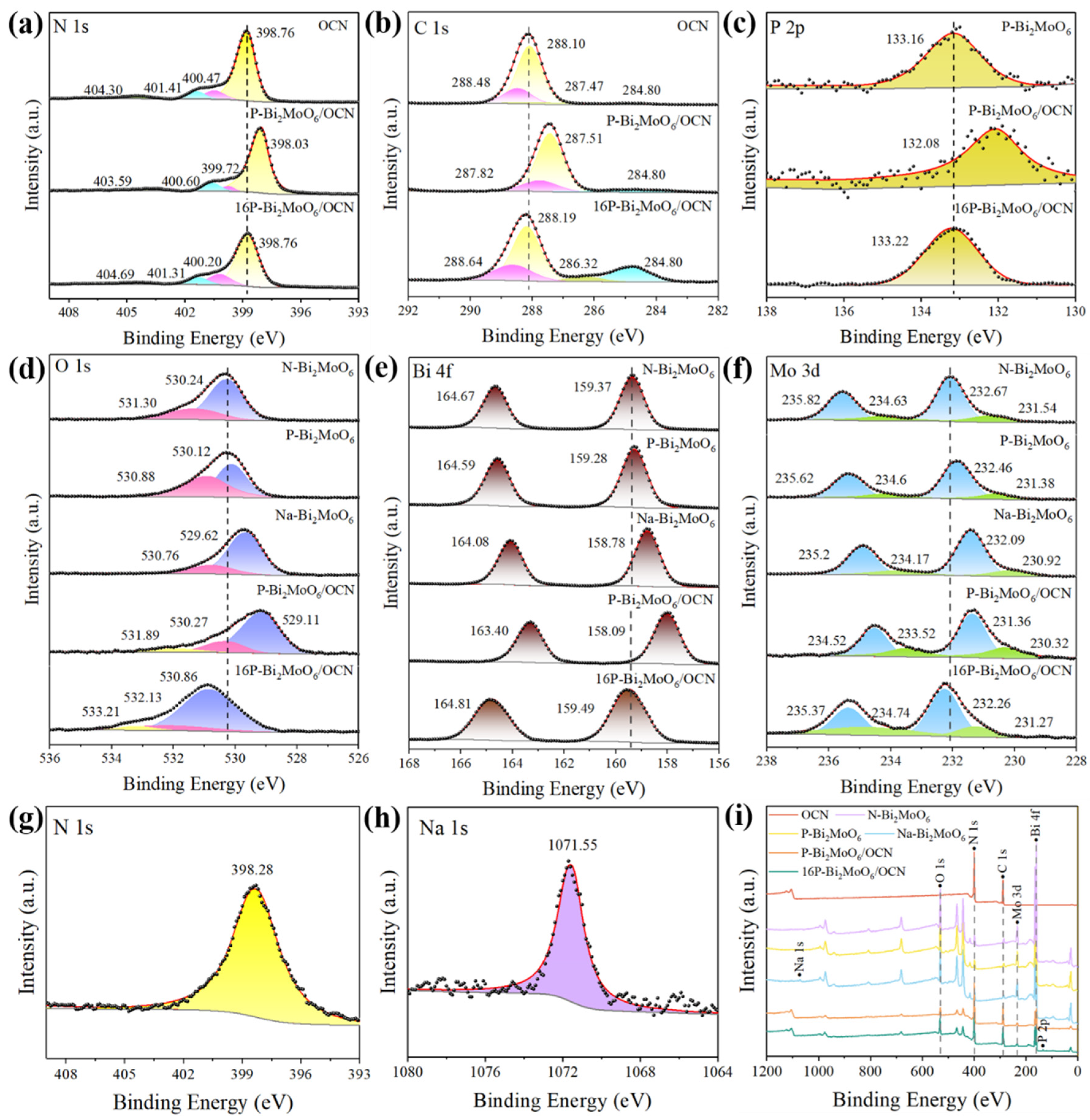
3.1. Analyses of Structure and Surface States

The samples were characterized by XRD to determine the crystal structure and phase details. Figure 1a shows the XRD patterns of three kinds of doped Bi2MoO6 and their combination with OCN and gives the XRD patterns of pure OCN. OCN exhibits characteristic peaks at about 12.7° and 27.5°, corresponding to the (100) and (002) diffraction planes of graphite-phase carbon nitride, respectively [30]. This is consistent with the crystal plane of the hexagonal g-C3N4 (JCPDS No. 87-1526). The XRD spectra of pure Bi2MoO6 prepared by three different molybdenum sources showed diffraction peaks at 27.9°, 32.4°, 46.5° and 55.2°, corresponding to the (131), (002), (062) and (133) crystal planes of orthorhombic bismuth molybdate (JCPDS No.21-0102) [31]. However, compared with the standard Bi2MoO6 card, the diffraction peaks at (131) and (133) are significantly shifted to a lower angle (Figure 1b), which is due to the increase of the interplanar spacing of Bi2MoO6 caused by Na, N and P doping. Among them, compared with the XRD spectra of Na-Bi2MoO6 and N-Bi2MoO6, P-Bi2MoO6 has a greater degree of left shift and a greater influence on the interplanar spacing. In addition, compared with the small peak at (262), it is obvious that P doping reduces the crystallinity of Bi2MoO6 and may introduce more defects. As shown in the Table 1, the calculated cell parameters of several Bi2MoO6 are not significantly different. Compared to other samples, the unit cell volume of P-Bi2MoO6 is the largest. The XRD spectra of three kinds of Bi2MoO6/OCN all show the characteristic peaks of Bi2MoO6. Due to the overlap of the characteristic peaks of OCN at (002) and Bi2MoO6 at (131), the decrease of OCN content in the composites, the secondary peak near 12.7° representing the (100) crystal plane of graphite phase carbon nitride becomes less prominent. In contrast, other characterization techniques such as SEM can better show the close bonding between Bi2MoO6 and OCN. It is worth noting that after adding phosphoric acid to increase the P doping content, taking 16P-Bi2MoO6/OCN as an example, other characteristic peaks such as Bi2O3 appeared. As shown in Figure 1c, the 16P-Bi2MoO6/OCN sample contains four different phases, namely Bi2MoO6, Bi2O3, BiO and BiPO4, and the main characteristic peaks of each phase are distinguished by color in the figure. The increase of P doping content induces more lattice defects such as Bi vacancies. The free Bi finally tends to combine with oxygen and regenerates other oxides at the layer edge. At the same time, P replaces Mo in the [MoO4]2− layer, which is the reason for the formation of BiPO4 phase.
The morphology of the samples was characterized by scanning electron microscopy (SEM) and transmission electron microscopy (TEM). As shown in Figure 2a,b, it can be observed that OCN is irregularly folded together by thin, few-layer, large-area nanosheets and only modified with small curved nanosheets at some endpoints or staggered positions. Figure 3c–g depict the SEM images of P-, Na- and N-doped Bi2MoO6 nanoflowers. Different states of nanoflowers can be observed; however, they have a common feature: the surface of Bi2MoO6 nanoflowers is composed of densely packed, fine nanosheet unit structures. In contrast, P-Bi2MoO6 exhibits more cracked, hollow nanoflowers, resulting in an increase in the total surface area after cracking. This is consistent with the results of XRD peaks, indicating that P-Bi2MoO6 has a larger interplanar spacing and more lattice defects and distortions. From Figure 3c–g, it can be inferred that after the formation process is completed, the internal structure of Bi2MoO6 nanoflowers has collapsed to varying degrees, resulting in the formation of hollow structures to varying degrees. Among them, the collapse rate of P-Bi2MoO6 is significantly faster than that of sodium-doped and nitrogen-doped Bi2MoO6, which may be related to the lower pH value caused by phosphomolybdic acid (H3PO4·12MoO3). However, the collapse modes of these three materials are similar. They all pierce from the outermost layer to the inner layer and then gradually expand, eventually forming a hollow sphere structure with a rough inner surface and fine pore distribution. This special structure is conducive to multiple scattering and slow photon effect in the process of illumination, so that more light energy can be captured and collected [4].
From Figure 2h,i, it can be inferred that after the formation process is completed, the internal structures of the Bi2MoO6 nanoflowers have collapsed to varying degrees, resulting in the formation of hollow structures to varying degrees. This is the SEM image and corresponding element mapping of OCN and P-Bi2MoO6 in situ composites. There are two composite structures of complete P-Bi2MoO6 nanoflowers and highly collapsed P-Bi2MoO6 nanoflowers hollow spheres. It can be seen that after solvent heating, the original large-area OCN nanosheet structure was destroyed and turned into a compressed small and irregularly staggered structure. From this point of view, OCN is not suitable as an improved method for long-term solvothermal heating in situ growth. In addition, in the element mapping diagram of P-Bi2MoO6/OCN, the signal intensities of N, O, Mo, Bi and P elements are represented by different colors. It can be seen that P is uniformly distributed in Bi2MoO6. In summary, through SEM and element mapping, it is effectively proven that P-Bi2MoO6 is successfully combined with OCN.
Figure 3a is the TEM image of OCN, and the transparent nanosheet image similar to the background color again proves the successful synthesis of g-C3N4 with the large area and less-thin layer. Figure 3b,c show the interface combination of P-Bi2MoO6 and OCN. Due to the existence of the internal hollow structure, the center color of P-Bi2MoO6 is lighter. In addition, there are also tiny, layered units of P-Bi2MoO6 nanoflowers on OCN nanosheets. Compared with pure OCN, the surface is rougher, and the structure is more complex, indicating that the interface between P-Bi2MoO6 and OCN is more fully bonded, which promotes the separation and transfer of carriers in the heterogeneous junction. The comparison of OCN in Figure 3a–c also clearly shows that large-area nanosheets shrink into small and tortuous structures. The HRTEM image in Figure 3d–g further proves the tightly bonded interface structure between the two. The spherical structure in the yellow circle in Figure 3b is P-Bi2MoO6, which has less light transmission than the few-layer OCN, so it is represented in the form of superimposed black shadows in the low magnification image. P-Bi2MoO6 mainly binds to OCN in the (131) crystal plane [32]. Due to the thin grain size of OCN, no obvious lattice fringes can be seen under the 2 nm high resolution transmission electron microscope. In addition, HRTEM images show the irregular and disordered lattice fringes (yellow circle out part) of P-Bi2MoO6 at the interface with OCN, which is due to lattice defects caused by P doping. These defects can effectively capture the photogenerated electrons in OCN, thereby inhibiting the recombination of excitons, enhancing the production of free radicals and ultimately improving the photocatalytic performance.
The results of XRD and SEM confirmed the simultaneous existence and close combination of the two materials. At the beginning, a large area of few-layer OCN nanosheet powder was dispersed in the synthesis solution of P-Bi2MoO6. After the beginning of the solvothermal reaction, P-Bi2MoO6 nanoflowers were gradually grown on OCN nanosheets as the substrate in a high-pressure reactor at 160 °C. Both of them are two-dimensional layered materials that are conducive to the superposition of [MoO4]2− and [Bi2O2]2+ layers. Finally, with the increase of reaction time, the two are closely combined. At the same time, the shrinkage of OCN nanosheets and the hollow collapse of P-Bi2MoO6 nanoflowers appear, thus obtaining the composite materials with complex structure.
As shown in Figure 4a, the specific surface area and pore structure of the OCN, P-Bi2MoO6, P-Bi2MoO6/OCN and 16P-Bi2MoO6/OCN composites were studied by N2 adsorption–desorption isotherm analysis. All materials exhibit distinct IV-type isotherm [33,34], indicating that they had typical layered structure and mesoporous characteristics. The specific surface area of P-Bi2MoO6/OCN is 94.78 m2/g (Table 2), which is significantly higher than that of P-Bi2MoO6 (46.89 m2/g) and P-Bi2MoO6/OCN (85.54 m2/g). This is due to the fact that the solvothermal treatment destroys the original structure of OCN to a certain extent. In addition to the direct observation in the SEM and TEM characterization results, the BJH pore size distribution curve is drawn. Compared with OCN and P-Bi2MoO6 monomers, the pore size distribution of P-Bi2MoO6/OCN has an additional protrusion at 4~6 nm, which is also evidence of structural changes. It is worth noting that the 16P-Bi2MoO6/OCN optimized by adding phosphoric acid does not have this protrusion, but the curve strength in the range of 4~12 nm increases uniformly, which indicates that the structure of OCN in 16P-Bi2MoO6/OCN changes more. In general, 16P-Bi2MoO6/OCN has the largest specific surface area and pore volume (97.10 m2/g), as well as a smaller average pore size, which is related to the fact that H3PO4 promotes the formation of more etching channels and porous structures at high temperatures, which provides more active sites for the adsorption of reactants [34]. In order to further confirm the structure of 16P-Bi2MoO6/OCN optimized by adding phosphoric acid, we compared the Fourier transform infrared spectroscopy (FT-IR) of P-Bi2MoO6/OCN and 16P-Bi2MoO6/OCN. As shown in Figure 4b, the broad peaks of the two spectra between 3364 and 2940 cm−1 are attributed to -NH and -OH bonds, which are related to the surface amino groups and adsorbed water molecules [35]. The peak at about 1637 cm−1 is the C=N bond of the g-C3N4 host. The absorption band in the range of 1600~1200 cm−1 is the stretching vibration peak of the aromatic C-N heterocycle. The peak at 810 cm−1 is a unique vibration peak of the tri-s-triazine ring system [36]. The peak observed in the range of 770–560 cm−1 belongs to the special absorption band of Bi2MoO6 [37]. Comparing the two spectra, it was found that the addition of phosphoric acid did not affect the characteristic structure of P-Bi2MoO6/OCN.
In order to better understand the catalytic nature, the surface acidity and basicity of P-Bi2MoO6/OCN and 16P-Bi2MoO6/OCN were evaluated by NH3 and CO2-TPD tests and were divided into three acid/basic site regions according to temperature [38,39]. The higher the desorption temperature of NH3 and CO2, the stronger the acidity and alkalinity, the larger the area of desorption peak and the greater the number of acid/base sites. The acidity and alkalinity of the material (Table 3 and Table 4) can be calculated by integrating the TPD response peaks. It can be seen from Figure 4c,d that the weak acidic and weak alkaline sites of the two materials are not obvious, and the desorption temperatures of the two materials are not much different, which indicates that the weak physical adsorption of CO2 and NH3 by the materials is less, mainly chemical adsorption. The number of weak intermediate acidic and weak intermediate alkaline sites of 16P-Bi2MoO6/OCN is lower than that of Bi2MoO6/OCN, but it has more strong acidic and strong alkaline sites. Especially, the strong alkaline sites are 16.49155 mmol/g, which is significantly higher than that of Bi2MoO6/OCN: 9.35347 mmol/g. In addition, the desorption temperature of 16P-Bi2MoO6/OCN for CO2 and NH3 is significantly higher than that of P-Bi2MoO6/OCN, which indicates that the acidic and alkaline strength of its active sites are higher. The addition of phosphoric acid changed the number and strength of acid/base sites, which may be attributed to the induced bismuth oxides such as Bi2O3 and the change of nitrogen-containing bases in OCN [40,41]. Therefore, the catalytic activity of 16P-Bi2MoO6/OCN is stronger, which may be related to the enriched basic sites on its surface. A number of studies have shown that acid/base sites are important reasons for the adsorption of water molecules on the catalyst surface, such as the formation of hydrogen bond adsorption mode, which can promote h+ to activate water molecules to generate more free ·OH radicals, thereby improving photocatalytic performance [42].
The XPS spectra were used to characterize the chemical bond state on the surface of the prepared material. Figure 5 shows the comparison of XPS spectra of P, Na and N doped Bi2MoO6, OCN, P-Bi2MoO6/OCN and 16P-Bi2MoO6/OCN heterojunctions. Figure 5a shows the N 1s spectra of OCN, P-Bi2MoO6/OCN and 16P-Bi2MoO6/OCN. The main peak marked yellow at about 398.76 eV belongs to the C-N=C bond in the six-membered ring of g-C3N4, which belongs to sp2 hybridization [43,44]. The peak at about 400.47 eV represents the graphite nitrogen connected to three carbon atoms in the g-C3N4 six-membered ring [45]. The blue-labeled peak is N-Hx (x = 1,2) at the edge of the g-C3N4 structure [46]. The peak at about 404.30 eV (marked as green) is related to the charge effect, which is consistent with previous reports. After the combination of OCN and P-Bi2MoO6, the overall N 1s moves to the direction of low binding energy, which is due to the fact that OCN has many pyridine-N (C-N=C) with strong electron-withdrawing properties. The difference is that the proportion of pyridine-N in 16P-Bi2MoO6/OCN is significantly reduced, and the proportion of graphite-N (N-(C)3) and amino-N (N-Hx) is increased. The final peak position is not much different from that of OCN and P-Bi2MoO6 monomers. It can be seen from Table 5 that the C-N=C/N-(C)3 of P-Bi2MoO6/OCN is significantly higher than that of OCN, but the C/N ratio of the two is basically the same, which indicates that the original tri-s-triazine structure of OCN is destroyed after solvothermal treatment, increasing the abundance of s-triazine structure. According to previous studies [47], the tri-s-triazine structure has better photocatalytic performance than the s-triazine structure, which makes it necessary to pay more attention to the control of reaction conditions when OCN is treated by solvothermal method. In addition, graphite-N and pyridine-N are considered to play a vital role in improving the catalytic performance and are important active centers. Although the size and mechanism of the contribution to the reaction have not yet been fully agreed upon, it is generally believed that graphite-N can effectively increase the free carrier density and promote charge transfer [48,49]. Amino-N also has a benign effect on the reaction and can improve the separation ability of photogenerated electrons and holes [50].
As shown in Figure 5b, the C 1s spectrum is fitted by peak separation, and 284.8 eV is the C-C bond, which is used as the standard reference carbon for XPS analysis. The main peak at about 288.10 eV (yellow marker) and the sub-peak at about 287.47 eV (green marker) are attributed to the main skeleton carbon N=C-N bond and the relatively small C-NHx of g-C3N4, respectively. The peak at about 288.48 eV corresponds to the C-O bond, which is partly derived from the adsorption of CO2 in the air. The increase of C-C and C-O in 16P-Bi2MoO6/OCN may be due to the substitution of pyridine-N by C and O atoms. Figure 5d compares the O 1s spectra of N-Bi2MoO6, P-Bi2MoO6, Na-Bi2MoO6 and 16P-Bi2MoO6/OCN. The peak at about 529~532 eV belongs to the lattice oxygen, and the main peak marked as purple corresponds to the Bi-O bond in Bi2MoO6. The sub-peaks near 530~531 eV (marked as pink) are attributed to the Mo-O bond [26,51]. The small peak near the lattice oxygen is surface adsorbed oxygen, which is marked as yellow. The surface oxygen is related to the adsorption of water molecules in the air after Bi2MoO6 composite OCN, indicating the existence of O-H bond. From Table 6, it can be seen that the Bi/Mo of P-Bi2MoO6 and 16 P-Bi2MoO6/OCN is significantly higher than that of the other materials, but the O-Bi/O-Mo on the surface is significantly lower, which indicates that P doping will lead to the generation of a large number of Bi vacancies and the accumulation of surface metal Bi (Bi0). More lattice defects also correspond to the characteristics of the microstructure of P-Bi2MoO6 in the SEM test, that is, more collapsing of nanoflowers at the same reaction time. Compared with P-Bi2MoO6, the increase of O-Bi/O-Mo in 16P-Bi2MoO6/OCN may be attributed to the production of other Bi oxides, which would be consistent with the results of XRD. Bi vacancies have been shown to effectively promote charge transfer and separation by temporarily trapping carriers [51,52,53], thereby improving photocatalytic performance. At the same time, the metal Bi formed in situ on the surface can further promote the transfer of charge and can be used as an active center for the formation of ·OH [54]. This paper provides a method for constructing a large number of bismuth vacancies in Bi2MoO6 materials and the synergistic effect of in situ metal bismuth.
As shown in Figure 5f, the Mo 3d peak shows a pair of main peaks at 235 eV and 232 eV, indicating that it mainly exists in the form of Mo6+, and there are a pair of secondary peaks at about 234 eV and 231 eV, representing Mo4+ [55,56]. Obviously, compared with P and N doped Bi2MoO6, the Mo 3d peak of Na doped Bi2MoO6 has a more obvious right shift, which is due to the fact that Na ions have more difficulty obtaining electrons, so that the charge shifts more to Mo ions; thus, the binding energy is reduced. After the complexation of P-Bi2MoO6 with OCN, the peak position of Mo moves to a lower binding energy, which is attributed to the fact that Mo6+ captures the electron cloud after combining with the conjugated structure interface of OCN, which promotes the conversion of some Mo6+ to Mo4+, and the peak area ratio representing Mo4+ increases significantly. The proportion of Mo4+ in several materials is shown in Table 6. In the solvothermal process of preparing 16P-Bi2MoO6/OCN, the addition of phosphoric acid promotes the reduction of lattice bismuth to precipitate surface metal Bi, and the pyridine-N in OCN is attacked and destroyed, which are the reasons for reducing the proportion of Mo4+. The XPS spectrum of P in Figure 5c proves the successful introduction of P, and the peak around 133 eV is attributed to phosphates. Therefore, P tends to replace Mo in the form of P5+ and embed into the Bi2MoO6 lattice or interlayer gap. The N peak at 398.28 eV in Figure 5g shows that in the process of preparing N-Bi2MoO6, N is introduced in the form of carbon nitride as impurity doping. The characteristic peak of Na 1s at 1071.55 eV in Figure 5h indicates that Na was successfully doped in Na-Bi2MoO6 [57].

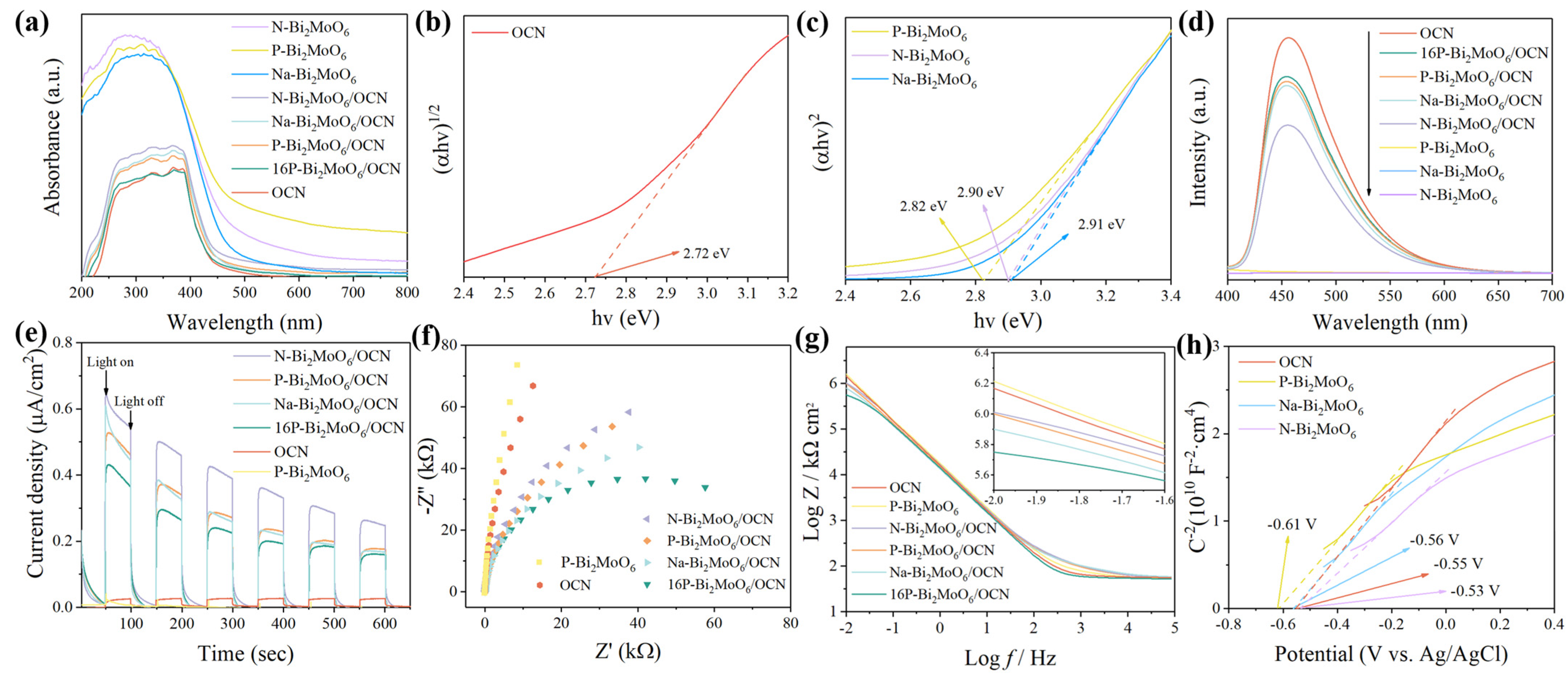
3.2. Optical Absorption Property Analysis

Figure 6a shows the UV-Vis DRS of a series of samples. All three doping types of Bi2MoO6 demonstrate high absorption strength within the 200~800 nm range. Among them, the absorption edge of Na-Bi2MoO6 is close to 480 nm, and the absorption edge of P-Bi2MoO6 is close to 500 nm, which is much larger than the absorption edge of OCN (around 430 nm). The coupling of OCN with Bi2MoO6 improves its light absorption ability in the visible region. As shown in Figure 6b,c, the Kubelka–Munk transformation function is used to evaluate the optical band gap (Eg) of the prepared material:
( α h ν ) 1 n A = ( h ν E g )
Bi2MoO6 is an indirect semiconductor, and g-C3N4 is a direct semiconductor. According to the type of semiconductor, the n value is different. The results are shown in Figure 6b,c. The band gap of OCN is 2.72 eV, and the band gap of Bi2MoO6 is 2.82~2.91 eV, which are similar to those reported in the literature [21,58]. The band gap of N-Bi2MoO6 is similar to that of Na-Bi2MoO6; they are 2.90 eV and 2.91 eV, respectively, but the band gap of P-Bi2MoO6 is narrower (2.82 eV). This may be due to the more lattice distortion caused by P doping, which adjusts the electronic structure and narrows the band gap of Bi2MoO6.
Figure 6d shows the photoluminescence spectra of samples such as OCN, P-Bi2MoO6, P-Bi2MoO6/OCN and 16P-Bi2MoO6/OCN to evaluate the recombination efficiency of photogenerated charge carriers. Generally, the stronger the peak, the easier it is for photogenerated electron–hole pairs to recombine. The fluorescence radiation intensity emitted by carrier recombination of Bi2MoO6 monomer is the lowest, which may be due to the fact that the absorbed photon energy is mostly released through nonradiative recombination, such as vibration, regardless of the type of doping element. Therefore, the photocatalytic degradation ability, antibacterial performance and photocurrent density of Bi2MoO6 itself are not significant. Pure OCN exhibits higher emission intensity, attributed to the easier recombination of photogenerated electron–hole pairs during photoexcitation. After compounding with Bi2MoO6, the doping types are different, showing different degrees of inhibition of carrier recombination. Among them, the fluorescence emission intensity of 16P-Bi2MoO6/OCN and P-Bi2MoO6/OCN is higher than that of other composite materials, P doping produces more Bi vacancies, and the capture effect of h+ makes the PL peak higher [52]. In addition, the fluorescence intensity of N-Bi2MoO6/OCN is significantly lower than that of other doped composites. This may be due to the heterojunction band gap structure formed by N-Bi2MoO6 and OCN, which is more conducive to charge transfer and makes it more effective in photocatalytic reaction.
In order to further evaluate the reactivity of the material and analyze the internal reasons for the improvement of performance, the instantaneous photocurrent curve under visible light and the electrochemical impedance spectroscopy (EIS) under illumination were measured by photoelectric measurement technology. As shown in Figure 6e, N-Bi2MoO6/OCN shows higher photocurrent intensity. The photocurrent intensity of P and Na doped Bi2MoO6/OCN composites is similar. The photocurrent response ability of 16P-Bi2MoO6/OCN decreases slightly, but all composites show stronger photoelectric conversion ability than OCN, which is consistent with the law of photoluminescence spectrum. In Figure 6f,g, the combination with the two-dimensional layered material Bi2MoO6 is obviously beneficial to reduce the impedance of OCN, and the cyclic transformation of Mo6+/Mo4+ plays an important role [59]. Compared with different doping types, sodium doped Bi2MoO6 has more advantages in promoting charge transfer than N and P doping. When studying the metal–organic interface, Blowey et al. found that alkali doping can form a two-dimensional charge transfer salt with high binding energy and increase the transfer rate by adjusting the charge barrier [60]. Therefore, Na doping can promote the charge transfer rate at the interface between Bi and Mo metal ions and OCN by acting as a connection node with special strong charge transfer ability. However, in general, the optimized 16-Bi2MoO6/OCN exhibits the smallest semicircle radius and the smallest impedance value in the low frequency region (0.01 Hz), indicating that the addition of phosphoric acid can effectively improve the charge transfer efficiency in P-Bi2MoO6/OCN, which is related to the increase of the content of surface metal Bi and the significant increase of the proportion of graphite-N in the OCN structure. As shown in Figure 6h, the flat band potentials of OCN and three doped Bi2MoO6 monomers were calculated according to the Mott–Schottky curve. The slope of the curves of several materials is positive, indicating that they are n-type semiconductors. In summary, P, Na and N doping have their own advantages, but in the actual photocatalytic reaction, they are affected by various factors. How to rationally modify and construct photocatalytic materials to improve their comprehensive performance is still a complex process that needs to be continuously explored.

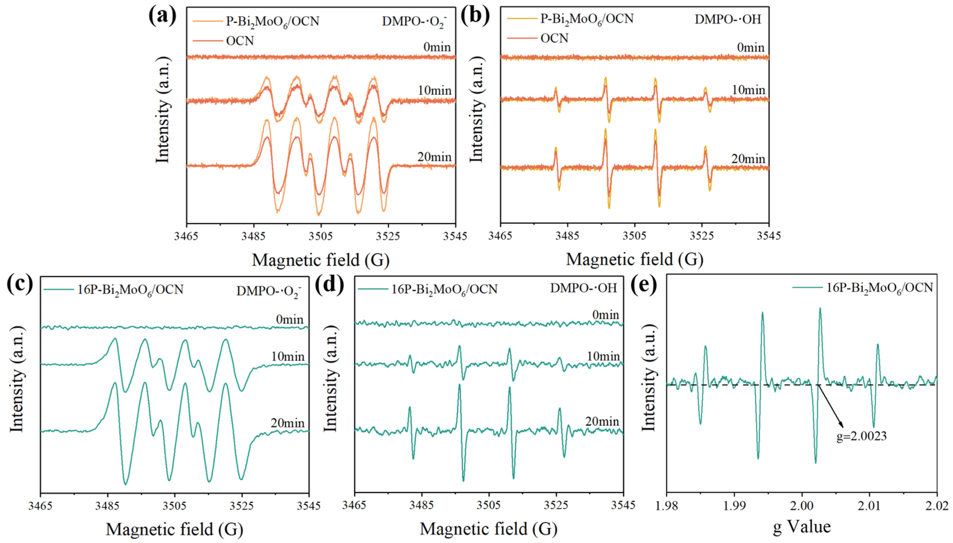
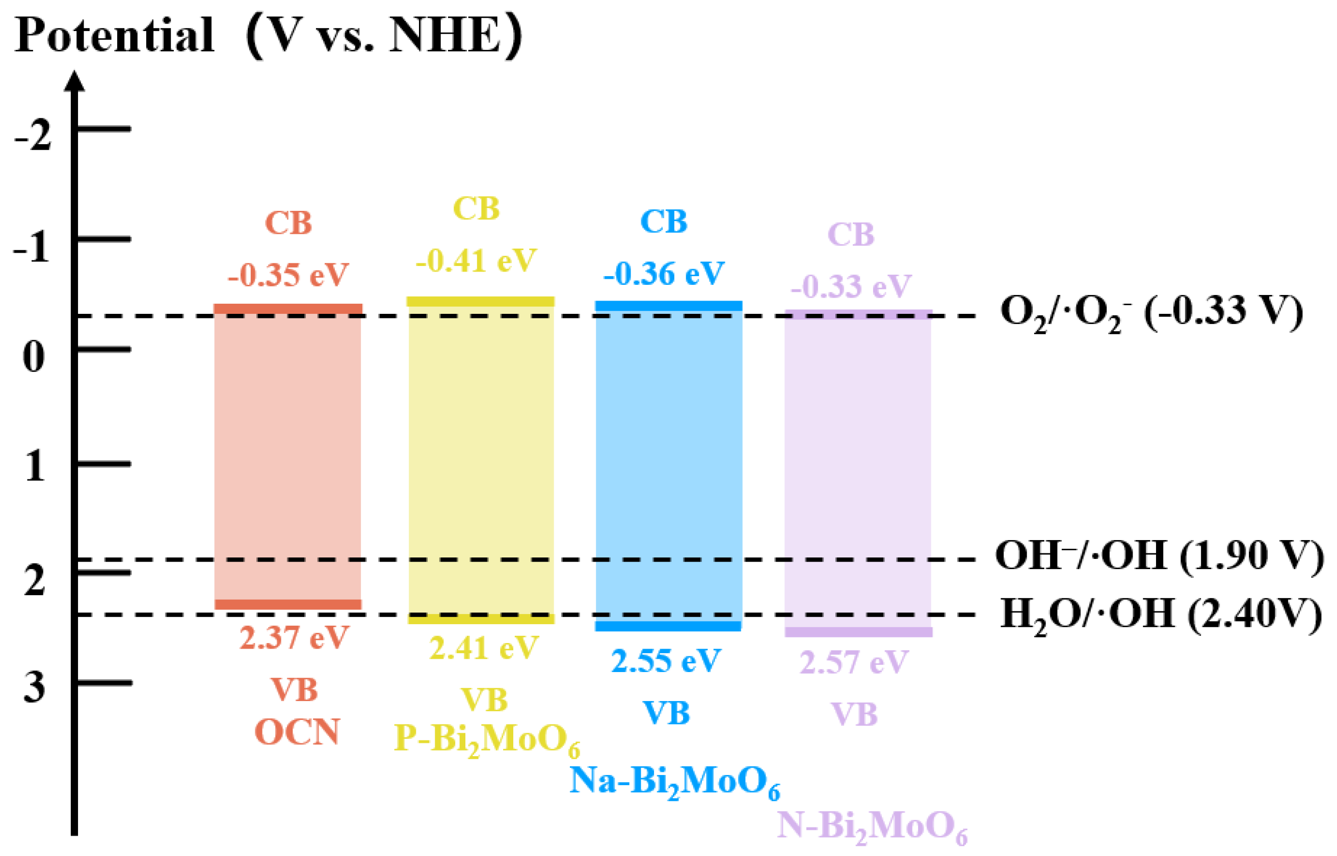
3.3. Photo-Generated Free Radical Analysis

The role of active free radicals in the photocatalytic process was discussed by using EPR technology. Figure 7 shows the free radical paramagnetic response of OCN and P-Bi2MoO6 under the same xenon lamp. There is no paramagnetic response in the dark state (time 0), and then it is illuminated with a xenon lamp equipped with a 420 nm filter and tested every 10 min. According to the flat band potential determined by the electrochemical Mott–Schottky curve and the band gap width obtained by the Kubelka–Munk transform function, the band gap structure fitted indicates that OCN has the thermodynamic condition for photocatalytic generation of ·OH and ·O2. This is because after forming heterojunctions with P-Bi2MoO6, OCN and P-Bi2MoO6, the lattice defects caused by P doping promote the separation and transfer of photogenerated carriers, promote the generation of more free radicals and improve the photocatalytic performance. After adding an appropriate amount of phosphoric acid, 16P-Bi2MoO6/OCN can not only achieve faster charge transfer and more surface reactive active sites but also have a band gap structure that can produce two free radicals, with strong DMPO-·O2 and DMPO-·OH signal values (Figure 7c,d). In addition, the g value of EPR signal is 2.0023, which indicates that there are a large number of free electrons and oxygen vacancies in 16P-Bi2MoO6/OCN [61].
Figure 8 shows the visible light degradation results of 20 mg/L RhB (50 mL) by the heterojunction composed of OCN and three doping types of Bi2MoO6 at 25 mg catalyst dosage. Na-Bi2MoO6 was used as the representative material for the degradation experiment of Bi2MoO6 monomer. This is because the three doping types have little difference in the characteristics of optical characterization. The results show that Bi2MoO6 monomer has almost no degradation performance under visible light. This is due to the low quantum yield and low activity of Bi2MoO6. In the previous literature, pure Bi2MoO6 does not show obvious photocatalytic performance [62,63]. Compared with different doping, P-Bi2MoO6/OCN showed the highest photocatalytic degradation performance, and the degradation rate of 20 mg/L RhB was 98.8% within 2 h.
It is worth noting that although N-Bi2MoO6/OCN and Na-Bi2MoO6/OCN exhibit low peak intensity in photoluminescence spectra, their actual photocatalytic degradation performance is lower than that of pure OCN. This is not only because the specific surface area of Bi2MoO6/OCN doped with Na and N is lower than that of OCN, which is conducive to the lack of active sites adsorbed by the reactants, so that the photogenerated carriers cannot play the role of oxidation and reduction faster, limiting the photocatalytic degradation rate. At the same time, the structure of OCN is destroyed after solvothermal treatment, which reduces the abundance of tri-s-triazine structure, which is more conducive to photocatalytic reaction. These factors make OCN have higher requirements for composite materials. The difference is that due to the more lattice defects of P-Bi2MoO6, a large number of Bi vacancies can promote the transfer of photogenerated carriers by capturing the carriers of OCN and coordinating with the in situ metal Bi. In addition, as a dopant and reaction source, phosphomolybdate itself has abundant Lewis and Brønsted acid sites. Many studies have shown that the introduction of P can produce strong Lewis and Brønsted acid sites, especially highly active Lewis acid sites [64], which interact with strong Lewis base OCN to increase the adsorption and activation of O2 and other reactants, thereby improving photocatalytic performance. A series of optimized P-Bi2MoO6/OCN were obtained by adding different amounts of phosphoric acid to increase the P doping content in the reaction, and the degradation performance of 20 ppm RhB with the same amount of catalyst was tested. It should be noted that, taking OCN as an example, the rate constant (k) obtained by fitting the degradation kinetics of Figure 8d was slightly lower than that of the previous group of experiments (Figure 8b), which is a systematic error and environmental reason such as the decrease of light intensity caused by the lifetime loss of xenon lamp. After optimization, 16P-Bi2MoO6/OCN had the best degradation performance, and the dye was completely degraded within 80 min. This shows that after optimization, the improvement of the specific surface area of the material, the improvement of the charge transfer rate and more active sites effectively improve the photocatalytic performance.
By the photocatalytic degradation of RhB, 16P-Bi2MoO6/OCN, with high photocatalytic performance, was selected for the comparative antibacterial test. Figure 9a,b show the plate antibacterial experiments and antibacterial rate comparison of E. coli, S. aureus and P. aeruginosa to evaluate their photocatalytic, broad-spectrum antibacterial properties under visible light. The experimental conditions such as the light conditions and catalyst delivery quality of each group of antibacterial experiments are the same. In the blank sample without catalyst input, the number of colonies did not decrease significantly after 300 min of illumination, indicating that visible light had little effect on E. coli, S. aureus or P. aeruginosa in this experiment. The antibacterial rates of 16P-Bi2MoO6/OCN against E. coli, S. aureus and P. aeruginosa in 300 min were 99.58%, 98.20% and 97.48%, respectively, which were significantly higher than those of OCN against the three bacteria (88.09%, 70.31% and 63.55%, respectively). Compared with P-Bi2MoO6/OCN, the antibacterial properties of 16P-Bi2MoO6/OCN were further improved. The composite of P-Bi2MoO6 and OCN effectively suppressed the recombination of photogenerated carriers and made carriers more efficiently transfer to the surface of the material to participate in the reaction. The capture of Bi vacancies caused by P doping, as well as high active sites such as surface metal Bi and acid/base sites, was conducive to consuming O2 and other substances, producing more reactive oxygen radicals, oxidizing and attacking bacterial cell membranes and cell walls and leading to bacterial rupture and death. The addition of phosphoric acid-optimized 16P-Bi2MoO6/OCN induced the regulation of the nitrogen ratio of OCN and increased the basic active sites and surface metal Bi, which was more conducive to the adsorption and activation of reactants. Finally, the photocatalytic performance was significantly improved by fine-tuning the P doping content. In addition, the electron transfer between Mo6+ and Mo4+ in P-Bi2MoO6/OCN also accelerated the charge conduction. Several studies have shown that the redox cycle reaction between Mo6+ and Mo4+ continuously stimulates more ROS production [59,65,66].
Through a series of characterization experiments, as well as the degradation experiments of RhB under visible light and the antibacterial experiments of three strains, the photocatalytic reaction mechanism of P-Bi2MoO6/OCN was proposed. Firstly, the band gap structure relationship of P-Bi2MoO6/OCN heterojunction was obtained by UV-Vis DRS and electrochemical Mott–Schottky curve. It should be noted that on the basis of the flat band potential obtained by Ag/AgCl reference, 0.199 eV was added to convert into a standard hydrogen electrode as a reference, and the band structure relationship shown in Figure 10 was obtained after fitting. It can be seen from the energy band relationship that the conduction band and valence band of OCN were contained in the band gap of P-Bi2MoO6 and Na-Bi2MoO6, which is a typical feature of type I heterojunction, but it was a type II heterojunction with staggered band gap with Na-Bi2MoO6. Under light excitation, the electrons on the valence band of P-Bi2MoO6 absorbed light energy, activated the transition to the conduction band and then released the energy back to the conduction band of OCN with a corrected potential to generate free radicals to participate in the redox reaction. Although the quantum yield of photoexcited P-Bi2MoO6 was very low, so that it could only transfer few photogenerated electrons and holes to OCN in photocatalytic reaction, the photoluminescence, photo electrochemistry, degradation and antibacterial results under visible light all proved that the composite material significantly inhibits the recombination rate of photogenerated carriers and has higher photocatalytic performance. Based on the above results, this indicates that the P-Bi2MoO6/OCN heterojunction not only improves the performance by constructing the traditional charge transfer form of type I heterojunction. Considering that P doping can endow Bi2MoO6 with abundant Lewis acid sites and Brønsted acid sites, the former (Lewis acid sites) especially have higher activity and electron absorption capacity, while OCN itself has strong Lewis basic sites, both of which form sterically hindered Lewis bases and Lewis acids, which greatly enhance the adsorption and activation of reactant molecules and improve the conversion efficiency of O2 to ·O2 [42,67,68]. At the same time, the lattice defects (Bi3+ vacancies) caused by P doping are greater than the other two doping types, which tend to capture the holes of OCN as the active sites for the generation of ·OH and cooperate with the in situ metal Bi to promote the separation and transfer of photogenerated charge carriers. In addition, the cyclic transformation of Mo6+ and Mo4+ in P-Bi2MoO6 provides more opportunities for electron transfer, so the electrons consumed on the surface of P-Bi2MoO6 can also be supplemented by rapid interlayer transfer. After re-optimization, 16P-Bi2MoO6/OCN with higher P doping amount was obtained. The phosphoric acid etching effect increased the specific surface area and induced the increase of the proportion of graphite nitrogen on OCN and the precipitation of Bi2MoO6. More metal Bi and related oxides made the adjusted structure more conducive to charge separation and transfer. Compared with other doping types of Bi2MoO6/OCN, the band gap structure of Na-Bi2MoO6/OCN conforms to the type I heterojunction. However, in addition to the faster charge transfer rate, there is no other mechanism that can further improve the photocatalytic performance. N-Bi2MoO6 and OCN can form a type II heterojunction, so that part of the photogenerated electrons on the OCN conduction band first fall into the conduction band of N-Bi2MoO6, reducing the recombination with holes on the valence band, thereby achieving effective separation of photogenerated carriers. However, N-Bi2MoO6/OCN also does not have enough active sites as a carrier for photocatalytic reactions, making it still unable to compensate for the performance degradation caused by structural damage of OCN after solvothermal treatment. In summary, the combination of OCN and P-Bi2MoO6 effectively inhibited the recombination of OCN photogenerated carriers. Compared with N and Na doping, it compensated for the decrease of photocatalytic activity caused by the decrease of active sites after OCN solvothermal treatment and finally achieved high photocatalytic degradation and antibacterial properties.

4. Conclusions

In this paper, three molybdenum sources and three doping types (P, Na and N) of Bi2MoO6 nanoflowers were successfully designed and synthesized for in situ growth on g-C3N4 nanosheets. The effects of different molybdenum sources and corresponding doping on the photocatalytic performance of doped Bi2MoO6/g-C3N4 composite system were studied.
  • Firstly, by comparing the composite of Bi2MoO6 with different doping elements grown on the surface of OCN under the same conditions, it was found that after compounding, the OCN structure collapsed and shrunk into small, interlaced g-C3N4 nanosheets, and the structure was destroyed, which increased the s-triazine structure, but the photocatalytic performance was low and brought disadvantages.
  • Furthermore, it is worth noting that in Bi2MoO6 prepared from different molybdenum sources, compared with OCN/P or Na or N doped Bi2MoO6, it is found that the collapsed P-Bi2MoO6/OCN still shows further improved performance compared with Na-Bi2MoO6/OCN and N-Bi2MoO6/OCN, which is significantly higher than any other system. This is because P doping brings more lattice defects and active sites, which helps to promote the separation of photogenerated carriers and the adsorption and activation of reactants. Due to the introduction of abundant Lewis acid sites and other highly active reaction sites by phosphomolybdate, it interacts with OCN of strong Lewis base to effectively promote the generation of superoxide anion radicals by photogenerated electrons. Therefore, it effectively compensates for the unfavorable factors caused by the structural damage of OCN and further improves the photocatalytic activity of the P-Bi2MoO6/g-C3N4 system. In contrast, although the heterojunction of Na- and N-doped Bi2MoO6 prepared from sodium molybdate and ammonium molybdate as molybdenum sources with the shrunken OCN can effectively suppress the recombination of photogenerated carriers, it still reduces the photocatalytic performance due to the inability to strongly compensate for the reduction of active sites.
  • Finally, in order to further optimize, the P doping content in P-Bi2MoO6/OCN was increased. The etching effect of phosphoric acid increased the specific surface area of the material, promoted more Bi vacancies and was also accompanied by more metal Bi and Bi oxide precipitation surface, which promoted the charge transfer efficiency. At the same time, the proportion of N atom distribution in OCN was induced and regulated, and more basic sites were obtained. These factors further improve the photocatalytic performance of P-Bi2MoO6/OCN.
In summary, the optimal construction process of photocatalytic materials will be affected by various factors; in the actual photocatalytic reaction, the reaction sites with higher activity may have a more important role in promoting the photocatalytic performance. Finally, an environmentally friendly P-Bi2MoO6/g-C3N4 with high photocatalytic antibacterial activity was successfully prepared by a simple preparation method using phosphomolybdic acid as the molybdenum source and dopant. The degradation rate of 20 mg/L RhB by 16P-Bi2MoO6/OCN with the best performance after optimization was 99.7% in 80 min under visible light. The antibacterial rates of E. coli, S. aureus and P. aeruginosa were 99.58%, 98.20% and 97.48%, respectively, in 300 min. This provides a valuable reference for optimizing environmentally friendly and efficient photocatalytic antibacterial materials.

Author Contributions

L.X.: writing—original draft, writing—review and editing, conceptualization, methodology, investigation, formal analysis; J.Z. and D.W.: writing—review and editing, supervision, resources, funding acquisition; M.S., H.Z. and K.W.: software, investigation, project administration; R.Z.: funding acquisition, project administration, resources. All authors have read and agreed to the published version of the manuscript.

Funding

This work was financially supported by the Major Basic Research Project of Natural Science Foundation of Shandong Province, China (No. ZR2023ZD31), National Natural Science Foundation of China (No. 42176049, 42076043), the Shandong Provincial Natural Science Foundation, China (No. ZR2022YQ39) and the Key Research Program of Frontier Sciences, Chinese Academy of Sciences (No. ZDBS-LY-DQC025).

Data Availability Statement

The data are contained within the article.

Conflicts of Interest

The authors declare no conflicts of interest.

References

  1. Thangudu, S.; Su, C.H. Review of Light Activated Antibacterial Nanomaterials in the Second Biological Window. J. Nanobiotechnol. 2025, 23, 293. [Google Scholar] [CrossRef] [PubMed]
  2. Malik, S.; Muhammad, K.; Waheed, Y. Emerging Applications of Nanotechnology in Healthcare and Medicine. Molecules 2023, 28, 6624. [Google Scholar] [CrossRef]
  3. Khan, S.; Noor, T.; Iqbal, N.; Yaqoob, L. Photocatalytic Dye Degradation from Textile Wastewater: A Review. ACS Omega 2024, 9, 21751–21767. [Google Scholar] [CrossRef]
  4. Jiang, Z.; Long, Q.; Cheng, B.; He, R.; Wang, L. 3D Ordered Macroporous Sulfur-Doped g-C3N4/TiO2 S-Scheme Photocatalysts for Efficient H2O2 Production in Pure Water. J. Mater. Sci. Technol. 2023, 162, 1–10. [Google Scholar] [CrossRef]
  5. Wang, J.; Wang, S. A Critical Review on Graphitic Carbon Nitride (g-C3N4)-Based Materials: Preparation, Modification and Environmental Application. Coord. Chem. Rev. 2022, 453, 214338. [Google Scholar] [CrossRef]
  6. Yao, T.; Wang, H.; Qin, Y.; Shi, J.W.; Cheng, Y. Enhancing Pseudocapacitive Behavior of MOF-Derived TiO2-X@Carbon Nanocubes via Mo-Doping for High-Performance Sodium-Ion Capacitors. Compos. Part B Eng. 2023, 253, 110557. [Google Scholar] [CrossRef]
  7. Murali, G.; Reddy Modigunta, J.K.; Park, Y.H.; Lee, J.H.; Rawal, J.; Lee, S.Y.; In, I.; Park, S.J. A Review on MXene Synthesis, Stability, and Photocatalytic Applications. ACS Nano 2022, 16, 13370–13429. [Google Scholar] [CrossRef]
  8. Ding, J.; He, D.; Du, P.; Wu, J.; Hu, Q.; Chen, Q.; Jiao, X. Design Photocatalysts to Boost Carrier Dynamics in Plastics Photoconversion into Fuels. ACS Appl. Mater. Interfaces 2024, 16, 35865–35873. [Google Scholar] [CrossRef]
  9. Li, Z.; Liu, F.; Lu, Y.; Hu, J.; Feng, J.; Shang, H.; Sun, B.; Jiang, W. Molecular Design of Perylene Diimide Derivatives for Photocatalysis. ACS Catal. 2025, 15, 1829–1840. [Google Scholar] [CrossRef]
  10. Sun, Y.D.; Zeng, C.; Zhang, X.; Zhang, Z.Q.; Yang, B.; Guo, S.Q. Tendencies of Alloyed Engineering in BiOX-Based Photocatalysts: A State-of-the-Art Review. Rare Met. 2024, 43, 1488–1512. [Google Scholar] [CrossRef]
  11. Zhang, Y.; Chen, Z.; Li, J.; Lu, Z.; Wang, X. Self-Assembled Synthesis of Oxygen-Doped g-C3N4 Nanotubes in Enhancement of Visible-Light Photocatalytic Hydrogen. J. Energy Chem. 2021, 54, 36–44. [Google Scholar] [CrossRef]
  12. Zhu, X.; Luo, J.; Dong, Z.; Li, Z.; Wu, Y.; Cheng, Z.; Cao, X.; Wang, Y.; Liu, Y.; Zhang, Z. Synthesis of High Crystallinity G-C3N4 Hollow Spheres for Efficient Photocatalytic Removal of U(VI) under Visible Light. Sep. Purif. Technol. 2023, 324, 124235. [Google Scholar] [CrossRef]
  13. Li, D.; Wen, C.; Huang, J.; Zhong, J.; Chen, P.; Liu, H.; Wang, Z.; Liu, Y.; Lv, W.; Liu, G. High-Efficiency Ultrathin Porous Phosphorus-Doped Graphitic Carbon Nitride Nanosheet Photocatalyst for Energy Production and Environmental Remediation. Appl. Catal. B-Environ. Energy 2022, 307, 121099. [Google Scholar] [CrossRef]
  14. Liu, C.; Dai, H.; Tan, C.; Pan, Q.; Hu, F.; Peng, X. Photo-Fenton Degradation of Tetracycline over Z-Scheme Fe-g-C3N4/Bi2WO6 Heterojunctions: Mechanism Insight, Degradation Pathways and DFT Calculation. Appl. Catal. B-Environ. Energy 2022, 310, 121326. [Google Scholar] [CrossRef]
  15. Xie, Q.; He, W.; Liu, S.; Li, C.; Zhang, J.; Wong, P.K. Bifunctional S-Scheme g-C3N4/Bi/BiVO4 Hybrid Photocatalysts toward Artificial Carbon Cycling. Chin. J. Catal. 2020, 41, 140–153. [Google Scholar] [CrossRef]
  16. Liu, X.; Gu, S.; Zhao, Y.; Zhou, G.; Li, W. BiVO4, Bi2WO6 and Bi2MoO6 Photocatalysis: A Brief Review. J. Mater. Sci. Technol. 2020, 56, 45–68. [Google Scholar] [CrossRef]
  17. Di, G.; Zhu, Z.; Zhang, H.; Qiu, Y.; Yin, D.; Crittenden, J. Simultaneous Sulfamethazine Oxidation and Bromate Reduction by Pd-Mediated Z-Scheme Bi2MoO6/g-C3N4 Photocatalysts: Synergetic Mechanism and Degradative Pathway. Chem. Eng. J. 2020, 401, 126061. [Google Scholar] [CrossRef]
  18. Li, Q.; Zhao, W.; Zhai, Z.; Ren, K.; Wang, T.; Guan, H.; Shi, H. 2D/2D Bi2MoO6/g-C3N4 S-Scheme Heterojunction Photocatalyst with Enhanced Visible-Light Activity by Au Loading. J. Mater. Sci. Technol. 2020, 56, 216–226. [Google Scholar] [CrossRef]
  19. Huang, J.; Qiu, J.; Yang, Y.; Li, B.; Wang, L.; Cheng, H.M.; Liu, G. Interface Regulation of Bi3TiNbO9/Rh Photocatalysts by Introducing Ultrathin Heterolayer to Enhance Overall Water Splitting. Adv. Funct. Mater. 2024, 34, 2402711. [Google Scholar] [CrossRef]
  20. Yu, H.; Jiang, L.; Wang, H.; Huang, B.; Yuan, X.; Huang, J.; Zhang, J.; Zeng, G. Modulation of Bi2MoO6-Based Materials for Photocatalytic Water Splitting and Environmental Application: A Critical Review. Small 2019, 15, 1901008. [Google Scholar] [CrossRef]
  21. Lyu, M.; Wang, C.; Rong, Y.; Wei, J.; Yang, Y.; Liu, Y.; Wei, G.; Zhang, Q.; Wang, C.; Xiu, J. Advances in Modification of Bi2MoO6 and Its Photocatalysis: A Review. J. Alloys Compd. 2024, 982, 173759. [Google Scholar] [CrossRef]
  22. Chang, X.; Liu, J.; Guo, Z.; Cheng, Y.; Yan, Q.; Li, Y.Y. Enhanced Photocatalytic Performance of Bi2MoO6 via Strain Engineering through Collaborative Optimization of Indium Doping and Oxygen Vacancies. Appl. Catal. B-Environ. Energy 2024, 349, 123858. [Google Scholar] [CrossRef]
  23. Li, J.; Chen, C.; Bai, J.; Jin, Y.; Guo, C. Boosting of the Piezoelectric Photocatalytic Performance of Bi2MoO6 by Fe3+ Doping and Construction S-Scheme Heterojunction Using WO3. J. Colloid Interface Sci. 2025, 683, 574–584. [Google Scholar] [CrossRef]
  24. Heo, J.; Roh, Y.; Shin, K.; Lee, C.; Wang, C.; Kim, H.T. Versatile Zn Hosting Lewis-Basic Interfacial Layer for High-Performant Zn Reversibility in Aqueous Zn Ion Battery. Energy Storage Mater. 2024, 71, 103580. [Google Scholar] [CrossRef]
  25. Wang, Q.; Zhang, Y.; Li, J.; Liu, N.; Jiao, Y.; Jiao, Z. Construction of Electron Transport Channels in Type-I Heterostructures of Bi2MoO6/BiVO4/g-C3N4 for Improved Charge Carriers Separation Efficiency. J. Colloid Interface Sci. 2020, 567, 145–153. [Google Scholar] [CrossRef]
  26. Vesali-Kermani, E.; Habibi-Yangjeh, A.; Diarmand-Khalilabad, H.; Ghosh, S. Nitrogen Photofixation Ability of G-C3N4 Nanosheets/Bi2MoO6 Heterojunction Photocatalyst under Visible-Light Illumination. J. Colloid Interface Sci. 2020, 563, 81–91. [Google Scholar] [CrossRef]
  27. Ma, D.; Wu, J.; Gao, M.; Xin, Y.; Chai, C. Enhanced Debromination and Degradation of 2,4-Dibromophenol by an Z-Scheme Bi2MoO6/CNTs/g-C3N4 Visible Light Photocatalyst. Chem. Eng. J. 2017, 316, 461–470. [Google Scholar] [CrossRef]
  28. Wang, Y.; Xing, Z.; Yang, Y.; Kong, W.; Wu, C.; Peng, H.; Li, Z.; Xie, Y.; Zhou, W. Oxygen-Defective Bi2MoO6/g-C3N4 Hollow Tubulars S-Scheme Heterojunctions toward Optimized Photocatalytic Performance. J. Colloid Interface Sci. 2024, 653, 1566–1576. [Google Scholar] [CrossRef]
  29. Fan, Y.; Xiong, J.; Cai, Y.; Huang, S.; Liang, Y.; Jiang, S.; Chen, G. Bi2MoO6/g-C3N4 S-Type Heterojunction with Oxygen Vacancies for Enhanced Removal of Ciprofloxacin: Investigation of Degradation Efficiency and Mechanism. J. Water Process Eng. 2025, 71, 107157. [Google Scholar] [CrossRef]
  30. Yu, H.; Shi, R.; Zhao, Y.; Bian, T.; Zhao, Y.; Zhou, C.; Waterhouse, G.I.N.; Wu, L.Z.; Tung, C.H.; Zhang, T. Alkali-Assisted Synthesis of Nitrogen Deficient Graphitic Carbon Nitride with Tunable Band Structures for Efficient Visible-Light-Driven Hydrogen Evolution. Adv. Mater. 2017, 29, 1605148. [Google Scholar] [CrossRef]
  31. He, W.; Wei, Y.; Xiong, J.; Tang, Z.; Song, W.; Liu, J.; Zhao, Z. Insight into Reaction Pathways of CO2 Photoreduction into CH4 over Hollow Microsphere Bi2MoO6-Supported Au Catalysts. Chem. Eng. J. 2022, 433, 133540. [Google Scholar] [CrossRef]
  32. Li, J.; Yin, Y.; Liu, E.; Ma, Y.; Wan, J.; Fan, J.; Hu, X. In Situ Growing Bi2MoO6 on G-C3N4 Nanosheets with Enhanced Photocatalytic Hydrogen Evolution and Disinfection of Bacteria under Visible Light Irradiation. J. Hazard. Mater. 2017, 321, 183–192. [Google Scholar] [CrossRef] [PubMed]
  33. Li, C.; Sun, Z.; Zhang, W.; Yu, C.; Zheng, S. Highly Efficient G-C3N4/TiO2/Kaolinite Composite with Novel Three-Dimensional Structure and Enhanced Visible Light Responding Ability towards Ciprofloxacin and S. Aureus. Appl. Catal. B-Environ. Energy 2018, 220, 272–282. [Google Scholar] [CrossRef]
  34. Xu, C.; Wang, Y.; Ma, X.; Wang, X.; Yang, Y.; Zhang, Q. High-Efficient Removal of Tebuconazole from Aqueous Solutions Using P-Doped Corn Straw Biochar: Performance, Mechanism and Application. Bioresour. Technol. 2024, 412, 131387. [Google Scholar] [CrossRef]
  35. Yussuf, N.M.; Ismail, A.F.; Rahmat, M.A.; Mohamed, N.A. Electrosorption-Driven Selective Thorium Removal from Radioactive Wastewater with Phosphate—Incorporated g-C3N4 Electrode. J. Environ. Chem. Eng. 2024, 12, 113440. [Google Scholar] [CrossRef]
  36. Luo, W.; Li, Y.; Wang, J.; Liu, J.; Zhang, N.; Zhao, M.; Wu, J.; Zhou, W.; Wang, L. Asymmetric Structure Engineering of Polymeric Carbon Nitride for Visible-Light-Driven Reduction Reactions. Nano Energy 2021, 87, 106168. [Google Scholar] [CrossRef]
  37. Yang, J.; Xie, T.; Mei, Y.; Chen, J.; Sun, H.; Feng, S.; Zhang, Y.; Zhao, Y.; Wang, J.; Li, X.; et al. High-Efficiency V-Mediated Bi2MoO6 Photocatalyst for PMS Activation: Modulation of Energy Band Structure and Enhancement of Surface Reaction. Appl. Catal. B-Environ. Energy 2023, 339, 123149. [Google Scholar] [CrossRef]
  38. Faizan, M.; Li, Y.; Wang, X.; Song, P.; Zhang, R.; Liu, R. Rare Earth Metal Based DES Assisted the VPO Synthesis for N-Butane Selective Oxidation toward Maleic Anhydride. Green Energy Environ. 2023, 8, 1737–1752. [Google Scholar] [CrossRef]
  39. Sahar, J.; Farooq, M.; Ramli, A.; Naeem, A.; Khattak, N.S.; Ghazi, Z.A. Highly Efficient Heteropoly Acid Decorated SnO2@Co-ZIF Nanocatalyst for Sustainable Biodiesel Production from Nannorrhops Ritchiana Seeds Oil. Renew. Energy 2022, 198, 306–318. [Google Scholar] [CrossRef]
  40. Liu, Y.; Guo, J.-g.; Wang, Y.; Hao, Y.-j.; Liu, R.-h.; Li, F.-t. One-Step Synthesis of Defected Bi2Al4O9/β-Bi2O3 Heterojunctions for Photocatalytic Reduction of CO2 to CO. Green Energy Environ. 2021, 6, 244–252. [Google Scholar] [CrossRef]
  41. Wong, K.T.; Brigljević, B.; Lee, J.H.; Yoon, S.Y.; Jang, S.B.; Choong, C.E.; Nah, I.; Kim, H.; Roh, H.S.; Kwak, S.K.; et al. Highly Exposed -NH2 Edge on Fragmented g-C3N4 Framework with Integrated Molybdenum Atoms for Catalytic CO2 Cycloaddition: DFT and Techno-Economic Assessment. Small 2023, 19, 2204336. [Google Scholar] [CrossRef] [PubMed]
  42. Ding, L.; Li, M.; Zhao, Y.; Zhang, H.; Shang, J.; Zhong, J.; Sheng, H.; Chen, C.; Zhao, J. The Vital Role of Surface Brönsted Acid/Base Sites for the Photocatalytic Formation of Free ·OH Radicals. Appl. Catal. B-Environ. Energy 2020, 266, 118634. [Google Scholar] [CrossRef]
  43. Chen, W.; Duan, G.R.; Liu, T.Y.; Chen, S.M.; Liu, X.H. Fabrication of Bi2MoO6 Nanoplates Hybridized with G-C3N4 Nanosheets as Highly Efficient Visible Light Responsive Heterojunction Photocatalysts for Rhodamine B Degradation. Mater. Sci. Semicond. Process. 2015, 35, 45–54. [Google Scholar] [CrossRef]
  44. Mao, S.; Zhao, P.; Wu, Y.; Liu, C.; Xia, M.; Wang, F. Promoting Charge Migration of Co(OH)2/g-C3N4 by Hydroxylation for Improved PMS Activation: Catalyst Design, DFT Calculation and Mechanism Analysis. Chem. Eng. J. 2023, 451, 138503. [Google Scholar] [CrossRef]
  45. Dong, P.; Zhang, A.; Cheng, T.; Pan, J.; Song, J.; Zhang, L.; Guan, R.; Xi, X.; Zhang, J. 2D/2D S-Scheme Heterojunction with a Covalent Organic Framework and g-C3N4 Nanosheets for Highly Efficient Photocatalytic H2 Evolution. Chin. J. Catal. 2022, 43, 2592–2605. [Google Scholar] [CrossRef]
  46. Yang, X.; Qian, F.; Zou, G.; Li, M.; Lu, J.; Li, Y.; Bao, M. Facile Fabrication of Acidified G-C3N4/g-C3N4 Hybrids with Enhanced Photocatalysis Performance under Visible Light Irradiation. Appl. Catal. B-Environ. Energy 2016, 193, 22–35. [Google Scholar] [CrossRef]
  47. Xu, T.; Xia, Z.; Li, H.; Niu, P.; Wang, S.; Li, L. Constructing Crystalline G-C3N4/g-C3N4−xSx Isotype Heterostructure for Efficient Photocatalytic and Piezocatalytic Performances. Energy Environ. Mater. 2022, 6, e12306. [Google Scholar] [CrossRef]
  48. Schiros, T.; Nordlund, D.; Pálová, L.; Prezzi, D.; Zhao, L.; Kim, K.S.; Wurstbauer, U.; Gutiérrez, C.; Delongchamp, D.; Jaye, C.; et al. Connecting Dopant Bond Type with Electronic Structure in N-Doped Graphene. Nano Lett. 2012, 12, 4025–4031. [Google Scholar] [CrossRef]
  49. Qian, Y.; Lu, T.; Liu, K.; Lu, G.; Xiao, J.; Zeng, X. Nickel Metal Nanoparticles Encapsulated by N-Doped Carbon Nanotubes Derived from g-C3N4 for Efficient Electrocatalytic Oxygen Reduction Reaction. Diam. Relat. Mater. 2025, 154, 112220. [Google Scholar] [CrossRef]
  50. Tao, K.Y.; Yuan, K.; Yang, W.; Zhong, D.C.; Lu, T.B. A Template Co-Pyrolysis Strategy towards the Increase of Amino/Imino Content within g-C3N4 for Efficient CO2 Photoreduction. Chem. Eng. J. 2023, 455, 140630. [Google Scholar] [CrossRef]
  51. Zhang, L.; Wang, Z.; Hu, C.; Shi, B. Enhanced Photocatalytic Performance by the Synergy of Bi Vacancies and Bi0 in Bi0-Bi2-δMoO6. Appl. Catal. B-Environ. Energy 2019, 257, 117785. [Google Scholar] [CrossRef]
  52. Kang, Z.; Lin, E.; Qin, N.; Wu, J.; Bao, D. Bismuth Vacancy-Mediated Quantum Dot Precipitation to Trigger Efficient Piezocatalytic Activity of Bi2WO6 Nanosheets. ACS Appl. Mater. Interfaces 2022, 14, 11375–11387. [Google Scholar] [CrossRef] [PubMed]
  53. Li, Y.; Xu, K.; Hu, H.; Jia, L.; Zhang, Y.; Zhuoga, C.; Yang, P.; Tan, X.; Guo, W.; Hao, W.; et al. Bi Vacancy Simultaneous Manipulation of Bulk Adsorption and Carrier Utilization to Replenish the Mechanism of Cr(VI) Photoreduction at Universal PH. Chem. Eng. J. 2022, 450, 138106. [Google Scholar] [CrossRef]
  54. Li, N.; Gao, H.; Wang, X.; Zhao, S.; Lv, D.; Yang, G.; Gao, X.; Fan, H.; Gao, Y.; Ge, L. Novel Indirect Z-Scheme g-C3N4/Bi2MoO6/Bi Hollow Microsphere Heterojunctions with SPR-Promoted Visible Absorption and Highly Enhanced Photocatalytic Performance. Chin. J. Catal. 2020, 41, 426–434. [Google Scholar] [CrossRef]
  55. Chen, X.; Sun, H.; Kuo, D.H.; Abdeta, A.B.; Zelekew, O.A.; Guo, Y.; Zhang, J.; Yuan, Z.; Lin, J. Spherical Nanoflower-like Bimetallic (Mo,Ni)(S,O)3-x Sulfo-Oxide Catalysts for Efficient Hydrogen Evolution under Visible Light. Appl. Catal. B-Environ. Energy 2021, 287, 119992. [Google Scholar] [CrossRef]
  56. Zhang, L.; Hao, X.; Wang, Y.; Jin, Z.; Ma, Q. Construction Strategy of Mo-S@Mo-P Heterojunction Formed with in-Situ Phosphating Mo-S Nanospheres toward Efficient Photocatalytic Hydrogen Production. Chem. Eng. J. 2020, 391, 123545. [Google Scholar] [CrossRef]
  57. Zhou, X.; Wang, T.; Zhang, L.; Che, S.; Liu, H.; Liu, S.; Wang, C.; Su, D.; Teng, Z. Highly Efficient Ag2O/Na-g-C3N4 Heterojunction for Photocatalytic Desulfurization of Thiophene in Fuel under Ambient Air Conditions. Appl. Catal. B-Environ. Energy 2022, 316, 121614. [Google Scholar] [CrossRef]
  58. Hu, K.; Li, R.; Ye, C.; Wang, A.; Wei, W.; Hu, D.; Qiu, R.; Yan, K. Facile Synthesis of Z-Scheme Composite of TiO2 Nanorod/g-C3N4 Nanosheet Efficient for Photocatalytic Degradation of Ciprofloxacin. J. Clean. Prod. 2020, 253, 120055. [Google Scholar] [CrossRef]
  59. He, W.; Wei, Y.; Xiong, J.; Tang, Z.; Wang, Y.; Wang, X.; Xu, H.; Zhang, X.; Yu, X.; Zhao, Z.; et al. Variable Valence Mo5+/Mo6+ Ionic Bridge in Hollow Spherical g-C3N4/Bi2MoO6 Catalysts for Promoting Selective Visible Light-Driven CO2 Photoreduction into CO. J. Energy Chem. 2023, 80, 361–372. [Google Scholar] [CrossRef]
  60. Blowey, P.J.; Blowey, P.J.; Sohail, B.; Rochford, L.A.; Lafosse, T.; Duncan, D.A.; Ryan, P.T.P.; Ryan, P.T.P.; Warr, D.A.; Lee, T.L.; et al. Alkali Doping Leads to Charge-Transfer Salt Formation in a Two-Dimensional Metal-Organic Framework. ACS Nano 2020, 14, 7475–7483. [Google Scholar] [CrossRef]
  61. Deng, X.; Chen, P.; Cui, R.; Gong, X.; Li, X.; Wang, X.; Deng, C. Synergistic Polarity Interaction and Structural Reconstruction in Bi2MoO6/C3N4 Heterojunction for Enhancing Piezo-Photocatalytic Nitrogen Oxidation to Nitric Acid. Appl. Catal. B-Environ. Energy 2024, 351, 123977. [Google Scholar] [CrossRef]
  62. Ma, T.; Li, R.; Huang, Y.C.; Lu, Y.; Guo, L.; Niu, M.; Huang, X.; Soomro, R.A.; Ren, J.; Wang, Q.; et al. Interfacial Chemical-Bonded MoS2/In-Bi2MoO6 Heterostructure for Enhanced Photocatalytic Nitrogen-to-Ammonia Conversion. ACS Catal. 2024, 14, 6292–6304. [Google Scholar] [CrossRef]
  63. He, J.; Lin, J.; Zhang, Y.; Hu, Y.; Huang, Q.; Zhou, G.; Li, W.; Hu, J.; Hu, N.; Yang, Z. Interfacial Mo-S Bond-Reinforced Hierarchical S-Scheme Heterostructure of Bi2MoO6@ZnIn2S4 for Highly-Selective and Efficient CO2 Photoreduction into CO. Chem. Eng. J. 2024, 480, 148036. [Google Scholar] [CrossRef]
  64. Yan, T.; Liu, Q.; Wang, S.; Xu, G.; Wu, M.; Chen, J.; Li, J. Promoter Rather than Inhibitor: Phosphorus Incorporation Accelerates the Activity of V2O5-WO3/TiO2 Catalyst for Selective Catalytic Reduction of NOx by NH3. ACS Catal. 2020, 10, 2747–2753. [Google Scholar] [CrossRef]
  65. Yang, Z.; Fu, X.; Ma, D.; Wang, Y.; Peng, L.; Shi, J.; Sun, J.; Gan, X.; Deng, Y.; Yang, W. Growth Factor-Decorated Ti3C2 MXene/MoS2 2D Bio-Heterojunctions with Quad-Channel Photonic Disinfection for Effective Regeneration of Bacteria-Invaded Cutaneous Tissue. Small 2021, 17, 2103993. [Google Scholar] [CrossRef]
  66. Tian, Y.; Zhou, M.; Pan, Y.; Du, X.; Wang, Q. MoS2 as Highly Efficient Co-Catalyst Enhancing the Performance of Fe0 Based Electro-Fenton Process in Degradation of Sulfamethazine: Approach and Mechanism. Chem. Eng. J. 2021, 403, 126361. [Google Scholar] [CrossRef]
  67. Zhu, L.; Cheng, P.; Xiao, Z.; Lu, C.; Li, B.; Jiang, X.; Shen, Z.; Qian, N.; Zhong, W.; He, Y. Incorporation of Phenolic Skeleton into Imidazolium Ionic Polymers as Recyclable Catalysts for Efficient Fixation of CO2 into Cyclic Carbonates. Chem. Eng. J. 2024, 481, 148359. [Google Scholar] [CrossRef]
  68. Ma, L.; Gao, Y.; Wei, B.; Huang, L.; Zhang, N.; Weng, Q.; Zhang, L.; Liu, S.F.; Jiang, R. Visible-Light Photocatalytic H2O2 Production Boosted by Frustrated Lewis Pairs in Defected Polymeric Carbon Nitride Nanosheets. ACS Catal. 2024, 14, 2775–2786. [Google Scholar] [CrossRef]
Figure 1. XRD patterns and some details of the as-prepared samples (a,b); XRD spectrum of 16P-Bi2MoO6/OCN contains different phases (c).
Nanomaterials 15 00834 g001
Figure 2. SEM images of OCN (a,b), P-Bi2MoO6 (c,d), Na-Bi2MoO6 (e), N-Bi2MoO6 (f,g), P-Bi2MoO6/OCN (h); EDS elemental mapping images of P-Bi2MoO6/OCN (i).
Nanomaterials 15 00834 g002
Figure 3. TEM and HRTEM images of OCN (a), P-Bi2MoO6/OCN (bd); HRTEM images of lattice defects in P-Bi2MoO6/OCN (eg).
Nanomaterials 15 00834 g003
Figure 4. (a) N2 adsorption–desorption isotherms and BJH pore size distribution curves (inset); (b) FT-IR spectra of P-Bi2MoO6/OCN and 16P-Bi2MoO6/OCN; (c) NH3-TPD profile; (d) CO2-TPD profile.
Nanomaterials 15 00834 g004
Figure 5. XPS spectra of N 1s (a,g), C 1s (b), P (c), O 1s (d), Bi 4f (e), Mo 3d (f), Na 1s (h) and total spectrum (i) of the as-synthesized samples.
Nanomaterials 15 00834 g005
Figure 6. The UV-Vis DRS spectra (a) and the estimated band gaps of the prepared samples (b,c); PL spectra (d); the transient photocurrent response of the samples under intermittent visible light (e); the EIS plots of the samples under visible light (f); Bode plot (g); Mott–Schottky plots of the samples in the dark (h).
Nanomaterials 15 00834 g006
Figure 7. EPR signals of DMPO-·O2 (a,c) and DMPO-·OH (b,d) for the samples. EPR spectra of the oxygen vacancy in 16P-Bi2MoO6/OCN (e).
Nanomaterials 15 00834 g007
Figure 8. The photocatalytic degradation rate of RhB solution of the samples under visible light irradiation (a,c) and the corresponding first-order kinetic plots (b,d).
Nanomaterials 15 00834 g008
Figure 9. (a) The colony image of the antibacterial experiment coated on agar plates after 300 min of visible light illumination; (b) after 300 min of visible light illumination, the photocatalytic sterilization rate of the material on E. coli (around 2.5 × 10⁶ CFU/mL, 50 mL), S. aureus (around 2.0 × 10⁶ CFU/mL, 50 mL) and P. aeruginosa (around 2.1 × 10⁶ CFU/mL, 50 mL).
Nanomaterials 15 00834 g009
Figure 10. The schematic of energy band alignments for the prepared OCN, P-Bi2MoO6, Na-Bi2MoO6 and N-Bi2MoO6.
Nanomaterials 15 00834 g010
Table 1. Cell parameter data of several Bi2MoO6 with orthorhombic crystal structure.
Samplea (Å)b (Å)c (Å)Vol (Å3)
P-Bi2MoO65.5516.235.44490.70
Na-Bi2MoO65.5016.215.48489.11
N-Bi2MoO65.5316.145.49490.38
Table 2. Specific surface area, pore volume and average pore radius of the as-prepared materials.
SampleSBET (m2/g)Pore Volume (cm3/g)Average Pore Radius (nm)
OCN94.780.1612.48
P-Bi2MoO646.890.104.54
P-Bi2MoO6/OCN85.540.1710.69
16P-Bi2MoO6/OCN97.100.1910.65
Table 3. NH3-TPD quantification results of the catalysts.
SampleQuantity of Weak and Medium Acid Sites (mmol/g)Quantity of Strong Acid Sites (mmol/g)Total Quantity (mmol/g)
P-Bi2MoO6/OCN0.182540.786670.96921
16P-Bi2MoO6/OCN0.129390.84230.97169
Table 4. CO2-TPD quantification results of the catalysts.
SampleQuantity of Weak and Medium Base Sites (mmol/g)Quantity of Strong Base Sites (mmol/g)Total Quantity (mmol/g)
P-Bi2MoO6/OCN0.010259.343229.35347
16P-Bi2MoO6/OCN0.0019716.4895816.49155
Table 5. Atomic distribution of N and C atoms based on XPS N 1s and C 1s spectra.
SampleC-N=CN-(C)3NHxC-N=C/N-(C)3C/N
OCN76.44%9.99%6.28%7.650.45
P-Bi2MoO6/OCN80.81%3.05%8.05%26.500.46
16P-Bi2MoO6/OCN57.25%22.11%12.45%2.590.57
Table 6. Atomic distribution of O, Bi and Mo atoms based on XPS O 1s, Bi 4f and Mo 3d spectra.
SampleBi/MoO-Bi/O-MoMo4+/Mo6+
P-Bi2MoO68.131.0424.01
Na-Bi2MoO67.103.5230.79
N-Bi2MoO66.752.9827.18
P-Bi2MoO6/OCN6.943.5241.12
16P-Bi2MoO6/OCN9.522.7220.79
Disclaimer/Publisher’s Note: The statements, opinions and data contained in all publications are solely those of the individual author(s) and contributor(s) and not of MDPI and/or the editor(s). MDPI and/or the editor(s) disclaim responsibility for any injury to people or property resulting from any ideas, methods, instructions or products referred to in the content.

