Plasmonic Nanomaterials in Dark Field Sensing Systems
Abstract
1. Introduction
2. LSPR and DFM
2.1. The Theory of LSPR
2.2. DFM
2.2.1. Mechanical Scanning Dark-Field Microscopic Imaging System
2.2.2. Dark-Field Microscopic Imaging System with Incident Wavelength Modulation
2.2.3. Summary of DFM
3. Materials and Applications
3.1. Single-Component Plasmonic Nanomaterial
3.1.1. Nanospheres
3.1.2. Nanorods
3.1.3. Nanoplate
3.1.4. Core–Satellite Structure
3.2. Plasmonic of Composite Materials
3.2.1. Bimetallic Composite Plasmonic
3.2.2. Other Multi-Component Nanomaterial Plasmonic Probes
4. Conclusions
Author Contributions
Funding
Data Availability Statement
Conflicts of Interest
References
- Guo, L.; Jackman, J.A.; Yang, H.-H.; Chen, P.; Cho, N.-J.; Kim, D.-H. Strategies for enhancing the sensitivity of plasmonic nanosensors. Nano Today 2015, 10, 213–239. [Google Scholar] [CrossRef]
- Zheng, J.; Cheng, X.; Zhang, H.; Bai, X.; Ai, R.; Shao, L.; Wang, J. Gold Nanorods: The Most Versatile Plasmonic Nanoparticles. Chem. Rev. 2021, 121, 13342–13453. [Google Scholar] [CrossRef] [PubMed]
- Ha, M.; Kim, J.H.; You, M.; Li, Q.; Fan, C.; Nam, J.M. Multicomponent Plasmonic Nanoparticles: From Heterostructured Nanoparticles to Colloidal Composite Nanostructures. Chem. Rev. 2019, 119, 12208–12278. [Google Scholar] [CrossRef] [PubMed]
- Gao, P.F.; Lei, G.; Huang, C.Z. Dark-Field Microscopy: Recent Advances in Accurate Analysis and Emerging Applications. Anal. Chem. 2021, 93, 4707–4726. [Google Scholar] [CrossRef]
- Xia, C.; He, W.; Yang, X.F.; Gao, P.F.; Zhen, S.J.; Li, Y.F.; Huang, C.Z. Plasmonic Hot-Electron-Painted Au@Pt Nanoparticles as Efficient Electrocatalysts for Detection of H2O2. Anal. Chem. 2022, 94, 13440–13446. [Google Scholar] [CrossRef]
- Du, B.; Lin, L.; Liu, W.; Zu, S.; Yu, Y.; Li, Z.; Kang, Y.; Peng, H.; Zhu, X.; Fang, Z. Plasmonic hot electron tunneling photodetection in vertical Au-graphene hybrid nanostructures. Laser Photonics Rev. 2017, 11, 1600148. [Google Scholar] [CrossRef]
- Feng, Q.; Qin, L.; Dou, B.; Han, X.; Wang, P. Plasmon-Tunable Ag@Au Bimetallic Core–Shell Nanostructures to Enhance the Electrochemiluminescence of Quantum Dots for MicroRNA Sensing. ACS Appl. Nano Mater. 2022, 5, 16325–16331. [Google Scholar] [CrossRef]
- Ye, W.; Götz, M.; Celiksoy, S.; Tüting, L.; Ratzke, C.; Prasad, J.; Ricken, J.; Wegner, S.V.; Ahijado-Guzmán, R.; Hugel, T.; et al. Conformational Dynamics of a Single Protein Monitored for 24 h at Video Rate. Nano Lett. 2018, 18, 6633–6637. [Google Scholar] [CrossRef]
- Henkel, A.; Ye, W.; Khalavka, Y.; Neiser, A.; Lambertz, C.; Schmachtel, S.; Ahijado-Guzmán, R.; Sönnichsen, C. Narrowing the Plasmonic Sensitivity Distribution by Considering the Individual Size of Gold Nanorods. J. Phys. Chem. C 2018, 122, 10133–10137. [Google Scholar] [CrossRef]
- Gong, L.J.; Li, Y.F.; Zou, H.Y.; Huang, C.Z. Resonance light scattering technique for sensitive detection of heparin using plasmonic Cu2−xSe nanoparticles. Talanta 2020, 216, 120967. [Google Scholar] [CrossRef]
- Lee, H.E.; Ahn, H.Y.; Mun, J.; Lee, Y.Y.; Kim, M.; Cho, N.H.; Chang, K.; Kim, W.S.; Rho, J.; Nam, K.T. Amino-acid- and peptide-directed synthesis of chiral plasmonic gold nanoparticles. Nature 2018, 556, 360–365. [Google Scholar] [CrossRef] [PubMed]
- Hoener, B.S.; Kirchner, S.R.; Heiderscheit, T.S.; Collins, S.S.E.; Chang, W.-S.; Link, S.; Landes, C.F. Plasmonic Sensing and Control of Single-Nanoparticle Electrochemistry. Chem 2018, 4, 1560–1585. [Google Scholar] [CrossRef]
- Zhang, Y.; McKelvie, I.D.; Cattrall, R.W.; Kolev, S.D. Colorimetric detection based on localised surface plasmon resonance of gold nanoparticles: Merits, inherent shortcomings and future prospects. Talanta 2016, 152, 410–422. [Google Scholar] [CrossRef] [PubMed]
- Jackson, D. Classical Electrodynamics. Am. J. Phys. 1999, 67, 841–842. [Google Scholar] [CrossRef]
- Maier, S. Plasmonics Fundamentals and Applications; Southeast University Press: Nanjing, China, 2014. [Google Scholar]
- Feliu, N.; Hassan, M.; Garcia Rico, E.; Cui, D.; Parak, W.; Alvarez-Puebla, R. SERS Quantification and Characterization of Proteins and Other Biomolecules. Langmuir 2017, 33, 9711–9730. [Google Scholar] [CrossRef]
- Goodacre, R.; Graham, D.; Faulds, K. Recent developments in quantitative SERS: Moving towards absolute quantification. TrAC Trends Anal. Chem. 2018, 102, 359–368. [Google Scholar] [CrossRef]
- Song, H.D.; Choi, I.; Yang, Y.I.; Hong, S.; Lee, S.; Kang, T.; Yi, J. Picomolar selective detection of mercuric ion (Hg(2+)) using a functionalized single plasmonic gold nanoparticle. Nanotechnology 2010, 21, 145501. [Google Scholar] [CrossRef]
- Zhang, W.J.; Du, Q.; Dou, Z.; Ning, S.P.; Wang, Q.Z.; Dong, Y.M.; Yue, Z.; Ye, W.X.; Liu, G.H. Ultrasensitive detection of lead (II) ion by dark-field spectroscopy and glutathione modified gold nanoparticles. Sens. Actuators B Chem. 2020, 321, 128548. [Google Scholar] [CrossRef]
- Singh Sekhon, J.; Verma, S.S. Refractive Index Sensitivity Analysis of Ag, Au, and Cu Nanoparticles. Plasmonics 2011, 6, 311–317. [Google Scholar] [CrossRef]
- Dou, Z.; Zhang, W.; Du, Q.; Liu, G. An in-situ plasmonic spectroscopy based biosensor for detection of copper (II) ions highlighting analytical specifications. Sens. Actuators B Chem. 2021, 329, 129103. [Google Scholar] [CrossRef]
- Liu, X.; Wu, Z.; Zhang, Q.; Zhao, W.; Zong, C.; Gai, H. Single Gold Nanoparticle-Based Colorimetric Detection of Picomolar Mercury Ion with Dark-Field Microscopy. Anal. Chem. 2016, 88, 2119–2124. [Google Scholar] [CrossRef] [PubMed]
- Guo, Y.; Liu, F.; Hu, Y.; Zheng, X.; Cao, X.; Zhu, Y.; Zhang, X.; Li, D.; Zhang, Z.; Chen, S.K. Activated Plasmonic Nanoaggregates for Dark-Field in Situ Imaging for HER2 Protein Imaging on Cell Surfaces. Bioconjugate Chem. 2020, 31, 631–638. [Google Scholar] [CrossRef] [PubMed]
- Qi, F.; Han, Y.; Liu, H.; Meng, H.; Li, Z.; Xiao, L. Localized surface plasmon resonance coupled single-particle galactose assay with dark-field optical microscopy. Sens. Actuators B Chem. 2020, 320, 128347. [Google Scholar] [CrossRef]
- Novo, C.; Funston, A.M.; Mulvaney, P. Direct observation of chemical reactions on single gold nanocrystals using surface plasmon spectroscopy. Nat. Nanotechnol. 2008, 3, 598–602. [Google Scholar] [CrossRef] [PubMed]
- Mulvaney, P.; Pérez-Juste, J.; Giersig, M.; Liz-Marzán, L.M.; Pecharromán, C. Drastic Surface Plasmon Mode Shifts in Gold Nanorods Due to Electron Charging. Plasmonics 2006, 1, 61–66. [Google Scholar] [CrossRef]
- Pan, Z.Y.; Gao, P.F.; Jing, C.J.; Zhou, J.; Liang, W.T.; Lei, G.; Feng, W.; Li, Y.F.; Huang, C.Z. Microscopic electron counting during plasmon-driven photocatalytic proton coupled electron transfer on a single silver nanoparticle. Appl. Catal. B Environ. 2021, 291, 120090. [Google Scholar] [CrossRef]
- Lee, K.S.; El-Sayed, M.A. Gold and Silver Nanoparticles in Sensing and Imaging Sensitivity of Plasmon Response to Size, Shape, and Metal Composition. J. Phys. Chem. B 2006, 110, 19220–19225. [Google Scholar] [CrossRef]
- Song, S.; Lee, J.U.; Kang, J.; Park, K.H.; Sim, S.J. Real-time monitoring of distinct binding kinetics of hot-spot mutant p53 protein in human cancer cells using an individual nanorod-based plasmonic biosensor. Sens. Actuators B Chem. 2020, 322, 128584. [Google Scholar] [CrossRef]
- Zhu, J.; He, J.; Verano, M.; Brimmo, A.T.; Glia, A.; Qasaimeh, M.A.; Chen, P.; Aleman, J.O.; Chen, W. An integrated adipose-tissue-on-chip nanoplasmonic biosensing platform for investigating obesity-associated inflammation. Lab Chip 2018, 18, 3550–3560. [Google Scholar] [CrossRef]
- Zhang, Q.; Yan, H.H.; Ru, C.; Zhu, F.; Zou, H.Y.; Gao, P.F.; Huang, C.Z.; Wang, J. Plasmonic biosensor for the highly sensitive detection of microRNA-21 via the chemical etching of gold nanorods under a dark-field microscope. Biosens. Bioelectron. 2022, 201, 113942. [Google Scholar] [CrossRef]
- Ye, W.; Celiksoy, S.; Jakab, A.; Khmelinskaia, A.; Heermann, T.; Raso, A.; Wegner, S.V.; Rivas, G.; Schwille, P.; Ahijado-Guzman, R.; et al. Plasmonic Nanosensors Reveal a Height Dependence of MinDE Protein Oscillations on Membrane Features. J. Am. Chem. Soc. 2018, 140, 17901–17906. [Google Scholar] [CrossRef] [PubMed]
- Yan, H.H.; Huang, M.; Zhu, F.; Cheng, R.; Wen, S.; Li, L.T.; Liu, H.; Zhao, X.H.; Luo, F.K.; Huang, C.Z.; et al. Two-Dimensional Analysis Method for Highly Sensitive Detection of Dual MicroRNAs in Breast Cancer Cells. Anal. Chem. 2023, 95, 3968–3975. [Google Scholar] [CrossRef] [PubMed]
- Zhu, L.; Li, G.; He, Y.; Tan, H.; Sun, S. Scattering measurement of single particle for highly sensitive homogeneous detection of DNA in serum. Talanta 2018, 178, 545–551. [Google Scholar] [CrossRef] [PubMed]
- Ryu, K.R.; Ha, J.W. Enhanced detection sensitivity of the chemisorption of pyridine and biotinylated proteins at localized surface plasmon resonance inflection points in single gold nanorods. Analyst 2021, 146, 3543–3548. [Google Scholar] [CrossRef]
- Kim, G.W.; Ha, J.W. Single-particle study: Effects of oxygen plasma treatment on structural and spectral changes of anisotropic gold nanorods. Phys. Chem. Chem. Phys. 2020, 22, 11767–11770. [Google Scholar] [CrossRef]
- Gu, X.Y.; Liu, J.J.; Gao, P.F.; Li, Y.F.; Huang, C.Z. Gold Triangular Nanoplates Based Single-Particle Dark-Field Microscopy Assay of Pyrophosphate. Anal. Chem. 2019, 91, 15798–15803. [Google Scholar] [CrossRef]
- Gu, X.Y.; Gao, P.F.; Zou, H.Y.; Liu, J.H.; Li, Y.F.; Huang, C.Z. The localized surface plasmon resonance induced edge effect of gold regular hexagonal nanoplates for reaction progress monitoring. Chem. Commun. 2018, 54, 13359–13362. [Google Scholar] [CrossRef]
- Chan, G.H.; Zhao, J.; Schatz, G.C.; Van Duyne, R.P. Localized Surface Plasmon Resonance Spectroscopy of Triangular Aluminum Nanoparticles.pdf. J. Phys. Chem. C 2008, 112, 13958–13963. [Google Scholar] [CrossRef]
- Clausen, J.S.; Hojlund-Nielsen, E.; Christiansen, A.B.; Yazdi, S.; Grajower, M.; Taha, H.; Levy, U.; Kristensen, A.; Mortensen, N.A. Plasmonic metasurfaces for coloration of plastic consumer products. Nano Lett. 2014, 14, 4499–4504. [Google Scholar] [CrossRef]
- Wang, K.; Shangguan, L.; Liu, Y.; Jiang, L.; Zhang, F.; Wei, Y.; Zhang, Y.; Qi, Z.; Wang, K.; Liu, S. In Situ Detection and Imaging of Telomerase Activity in Cancer Cell Lines via Disassembly of Plasmonic Core-Satellites Nanostructured Probe. Anal. Chem. 2017, 89, 7262–7268. [Google Scholar] [CrossRef]
- Prasad, J.; Zins, I.; Branscheid, R.; Becker, J.; Koch, A.H.R.; Fytas, G.; Kolb, U.; Sönnichsen, C. Plasmonic Core–Satellite Assemblies as Highly Sensitive Refractive Index Sensors. J. Phys. Chem. C 2015, 119, 5577–5582. [Google Scholar] [CrossRef]
- Markhali, B.P.; Sriram, M.; Bennett, D.T.; Khiabani, P.S.; Hoque, S.; Tilley, R.D.; Bakthavathsalam, P.; Gooding, J.J. Single particle detection of protein molecules using dark-field microscopy to avoid signals from nonspecific adsorption. Biosens. Bioelectron. 2020, 169, 112612. [Google Scholar] [CrossRef] [PubMed]
- Zhang, D.; Wang, K.; Wei, W.; Liu, S. Single-Particle Assay of Poly(ADP-ribose) Polymerase-1 Activity with Dark-Field Optical Microscopy. ACS Sens. 2020, 5, 1198–1206. [Google Scholar] [CrossRef] [PubMed]
- Zhang, W.; Liu, G.; Bi, J.; Bao, K.; Wang, P. In-situ and ultrasensitive detection of mercury (II) ions (Hg2+) using the localized surface plasmon resonance (LSPR) nanosensor and the microfluidic chip. Sens. Actuators A Phys. 2023, 349, 114074. [Google Scholar] [CrossRef]
- Liu, F.; Guo, Y.; Hu, Y.; Zhang, X.; Zheng, X. Intracellular dark-field imaging of ATP and photothermal therapy using a colorimetric assay based on gold nanoparticle aggregation via tetrazine/trans-cyclooctene cycloaddition. Anal. Bioanal. Chem. 2019, 411, 5845–5854. [Google Scholar] [CrossRef]
- Zhou, J.; Gao, P.F.; Zhang, H.Z.; Lei, G.; Zheng, L.L.; Liu, H.; Huang, C.Z. Color resolution improvement of the dark-field microscopy imaging of single light scattering plasmonic nanoprobes for microRNA visual detection. Nanoscale 2017, 9, 4593–4600. [Google Scholar] [CrossRef]
- Yang, X.F.; He, W.; Li, Y.F.; Gao, P.F.; Huang, C.Z. Telomerase Activity Assay via 3,3′,5,5′-Tetramethylbenzidine Dilation Etching of Gold Nanorods. ACS Appl. Nano Mater. 2022, 5, 1484–1490. [Google Scholar] [CrossRef]
- Park, J.H.; Byun, J.Y.; Mun, H.; Shim, W.B.; Shin, Y.B.; Li, T.; Kim, M.G. A regeneratable, label-free, localized surface plasmon resonance (LSPR) aptasensor for the detection of ochratoxin A. Biosens. Bioelectron. 2014, 59, 321–327. [Google Scholar] [CrossRef]
- Ma, J.; Hou, S.; Lu, D.; Zhang, B.; Xiong, Q.; Chan-Park, M.B.; Duan, H. Caging Cationic Polymer Brush-Coated Plasmonic Nanostructures for Traceable Selective Antimicrobial Activities. Macromol. Rapid Commun. 2022, 43, e2100812. [Google Scholar] [CrossRef]
- Soares, L.; Csaki, A.; Jatschka, J.; Fritzsche, W.; Flores, O.; Franco, R.; Pereira, E. Localized surface plasmon resonance (LSPR) biosensing using gold nanotriangles: Detection of DNA hybridization events at room temperature. Analyst 2014, 139, 4964–4973. [Google Scholar] [CrossRef]
- Campu, A.; Lerouge, F.; Chateau, D.; Chaput, F.; Baldeck, P.; Parola, S.; Maniu, D.; Craciun, A.M.; Vulpoi, A.; Astilean, S.; et al. Gold NanoBipyramids Performing as Highly Sensitive Dual-Modal Optical Immunosensors. Anal. Chem. 2018, 90, 8567–8575. [Google Scholar] [CrossRef] [PubMed]
- Sriram, M.; Markhali, B.P.; Nicovich, P.R.; Bennett, D.T.; Reece, P.J.; Brynn Hibbert, D.; Tilley, R.D.; Gaus, K.; Vivekchand, S.R.C.; Gooding, J.J. A rapid readout for many single plasmonic nanoparticles using dark-field microscopy and digital color analysis. Biosens. Bioelectron. 2018, 117, 530–536. [Google Scholar] [CrossRef] [PubMed]
- Huang, M.; Fan, Y.; Liu, H.; Ma, Y.; Wei, L. Single-particle cadmium(II) and chromium(III) ions assay using GNP@Ag core−shell nanoparticles with dark-field optical microscopy. Sens. Actuators B Chem. 2021, 346, 130471. [Google Scholar] [CrossRef]
- Wang, H.; Zhao, W.; Zhao, Y.; Xu, C.H.; Xu, J.J.; Chen, H.Y. Real-Time Tracking the Electrochemical Synthesis of Au@Metal Core-Shell Nanoparticles toward Photo Enhanced Methanol Oxidation. Anal. Chem. 2020, 92, 14006–14011. [Google Scholar] [CrossRef] [PubMed]
- Wang, K.; Jiang, L.; Zhang, F.; Wei, Y.; Wang, K.; Wang, H.; Qi, Z.; Liu, S. Strategy for In Situ Imaging of Cellular Alkaline Phosphatase Activity Using Gold Nanoflower Probe and Localized Surface Plasmon Resonance Technique. Anal. Chem. 2018, 90, 14056–14062. [Google Scholar] [CrossRef] [PubMed]
- Wang, K.; Zhang, F.; Wei, Y.; Wei, W.; Jiang, L.; Liu, Z.; Liu, S. In Situ Imaging of Cellular Reactive Oxygen Species and Caspase-3 Activity Using a Multifunctional Theranostic Probe for Cancer Diagnosis and Therapy. Anal. Chem. 2021, 93, 7870–7878. [Google Scholar] [CrossRef]
- Zhou, J.; Yang, T.; He, W.; Pan, Z.Y.; Huang, C.Z. A galvanic exchange process visualized on single silver nanoparticles via dark-field microscopy imaging. Nanoscale 2018, 10, 12805–12812. [Google Scholar] [CrossRef]
- Li, M.; Cheng, J.; Yuan, Z.; Shen, Q.; Fan, Q. DNAzyme-catalyzed etching process of Au/Ag nanocages visualized via dark-field imaging with time elapse for ultrasensitive detection of microRNA. Sens. Actuators B Chem. 2021, 330, 129347. [Google Scholar] [CrossRef]
- Wang, F.; Li, Y.; Han, Y.; Ye, Z.; Wei, L.; Luo, H.B.; Xiao, L. Single-Particle Enzyme Activity Assay with Spectral-Resolved Dark-Field Optical Microscopy. Anal. Chem. 2019, 91, 6329–6339. [Google Scholar] [CrossRef]
- Xu, W.; Ouyang, M.; Luo, H.; Xu, D.; Lin, Q. Single Au@MnO2 nanoparticle imaging for sensitive glucose detection based on H2O2-mediated etching of the MnO2 layer. New J. Chem. 2022, 46, 15473–15480. [Google Scholar] [CrossRef]
- Li, Q.; Peng, M.; Wang, C.; Li, C.; Li, K.; Lin, Y. Sensitive Sulfide Monitoring in Live Cells by Dark-Field Microscopy Based on the Formation of Ag2S on Au@AgI Core–Shell Nanoparticles. ACS Sustain. Chem. Eng. 2019, 7, 19338–19343. [Google Scholar] [CrossRef]
- Wang, H.; Xu, C.H.; Zhao, W.; Chen, H.Y.; Xu, J.J. Alkaline Phosphatase-Triggered Etching of Au@FeOOH Nanoparticles for Enzyme Level Assay under Dark-Field Microscopy. Anal. Chem. 2021, 93, 10727–10734. [Google Scholar] [CrossRef] [PubMed]
- Wang, H.; Zhao, W.; Xu, C.H.; Chen, H.Y.; Xu, J.J. Electrochemical synthesis of Au@semiconductor core-shell nanocrystals guided by single particle plasmonic imaging. Chem. Sci. 2019, 10, 9308–9314. [Google Scholar] [CrossRef] [PubMed]
- Feng, W.; He, W.; Zhou, J.; Gu, X.Y.; Li, Y.F.; Huang, C.Z. Inconspicuous Reactions Identified by Improved Precision of Plasmonic Scattering Dark-Field Microscopy Imaging Using Silver Shell-Isolated Nanoparticles as Internal References. Anal. Chem. 2019, 91, 3002–3008. [Google Scholar] [CrossRef]
- Sikes, J.C.; Wonner, K.; Nicholson, A.; Cignoni, P.; Fritsch, I.; Tschulik, K. Characterization of Nanoparticles in Diverse Mixtures Using Localized Surface Plasmon Resonance and Nanoparticle Tracking by Dark-Field Microscopy with Redox Magnetohydrodynamics Microfluidics. ACS Phys. Chem. Au 2022, 2, 289–298. [Google Scholar] [CrossRef] [PubMed]
- Zhao, Y.; Zhao, W.; Chen, H.Y.; Xu, J.J. Dark-field microscopic real-time monitoring the growth of Au on Cu(2)O nanocubes for ultra-sensitive glucose detection. Anal. Chim. Acta 2021, 1162, 338503. [Google Scholar] [CrossRef]
- Fan, X.; Xu, C.; Hao, X.; Tian, Z.; Lin, Y. Synthesis and optical properties of Janus structural ZnO/Au nanocomposites. EPL (Europhys. Lett.) 2014, 106, 67001. [Google Scholar] [CrossRef]
- Xu, G.; Zhu, Y.H.; Pang, J. Sensitive and Simple Detection of Glucose Based on Single Plasmonic Nanorod. Anal. Sci. 2017, 33, 223–227. [Google Scholar] [CrossRef]
- Shen, J.; Zhang, L.; Liu, L.; Wang, B.; Bai, J.; Shen, C.; Chen, Y.; Fan, Q.; Chen, S.; Wu, W.; et al. Revealing Lectin-Sugar Interactions with a Single Au@Ag Nanocube. ACS Appl. Mater. Interfaces 2019, 11, 40944–40950. [Google Scholar] [CrossRef]
- Zhang, D.; Jiang, N.; Li, P.; Zhang, Y.; Sun, S.; Mao, J.; Liu, S.; Wei, W. Detection of monoamine oxidase B using dark-field light scattering imaging and colorimetry. Chem. Commun. 2022, 58, 12329–12332. [Google Scholar] [CrossRef]
- Xia, C.; He, W.; Gao, P.F.; Wang, J.R.; Cao, Z.M.; Li, Y.F.; Wang, Y.; Huang, C.Z. Nanofabrication of hollowed-out Au@AgPt core-frames via selective carving of silver and deposition of platinum. Chem Commun 2020, 56, 2945–2948. [Google Scholar] [CrossRef] [PubMed]
- Ye, Z.; Weng, R.; Ma, Y.; Wang, F.; Liu, H.; Wei, L.; Xiao, L. Label-Free, Single-Particle, Colorimetric Detection of Permanganate by GNPs@Ag Core-Shell Nanoparticles with Dark-Field Optical Microscopy. Anal. Chem. 2018, 90, 13044–13050. [Google Scholar] [CrossRef] [PubMed]


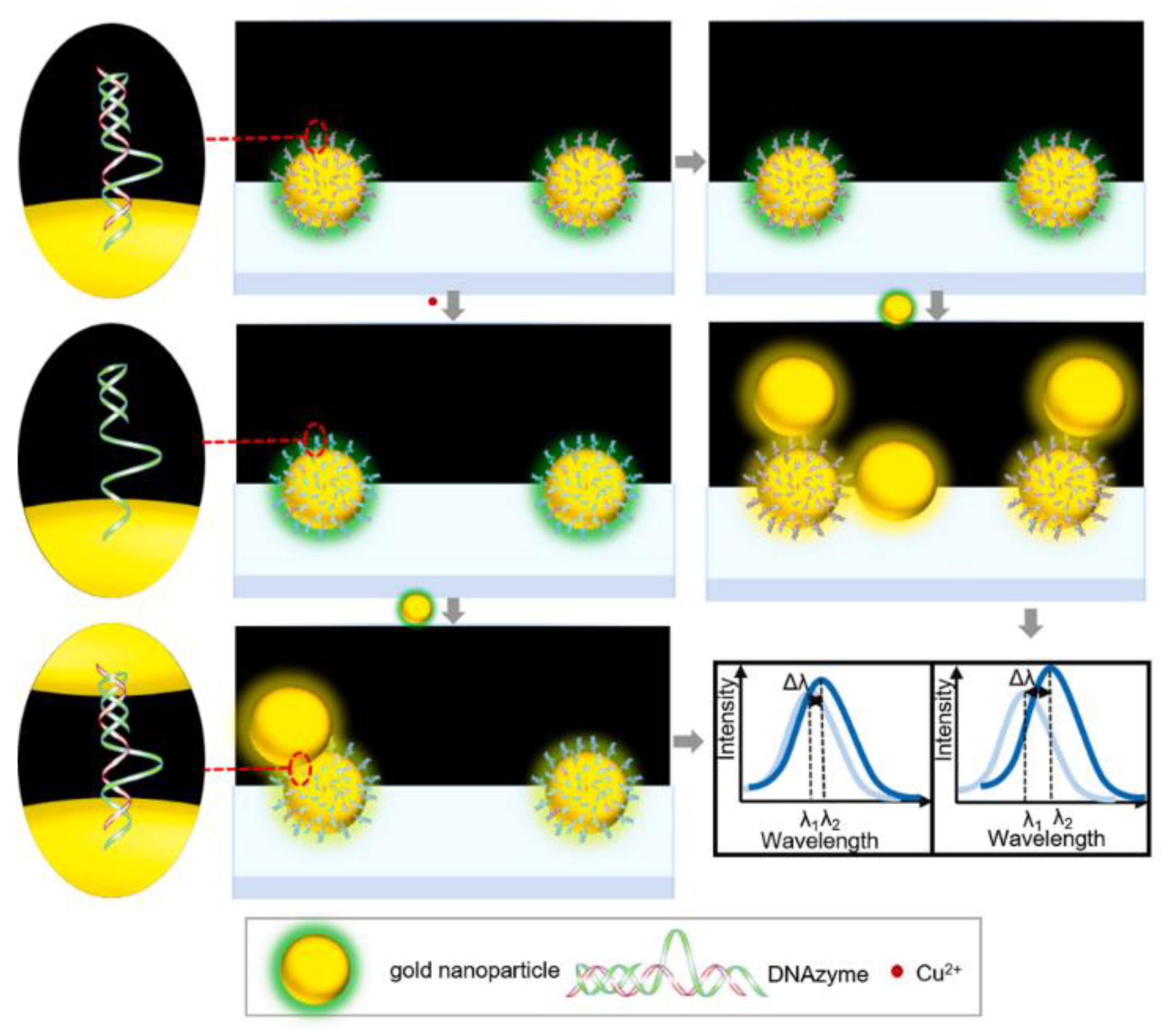
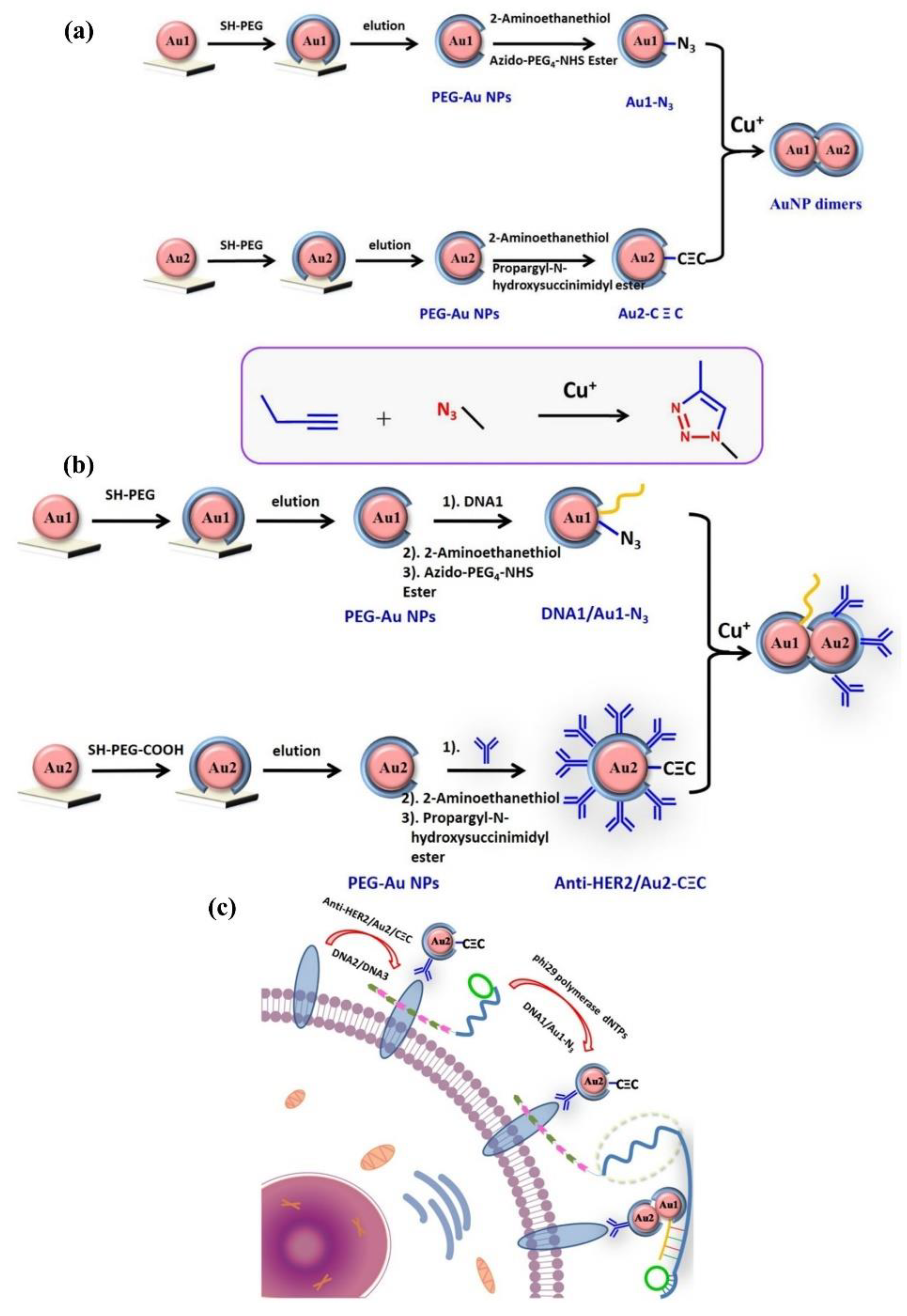
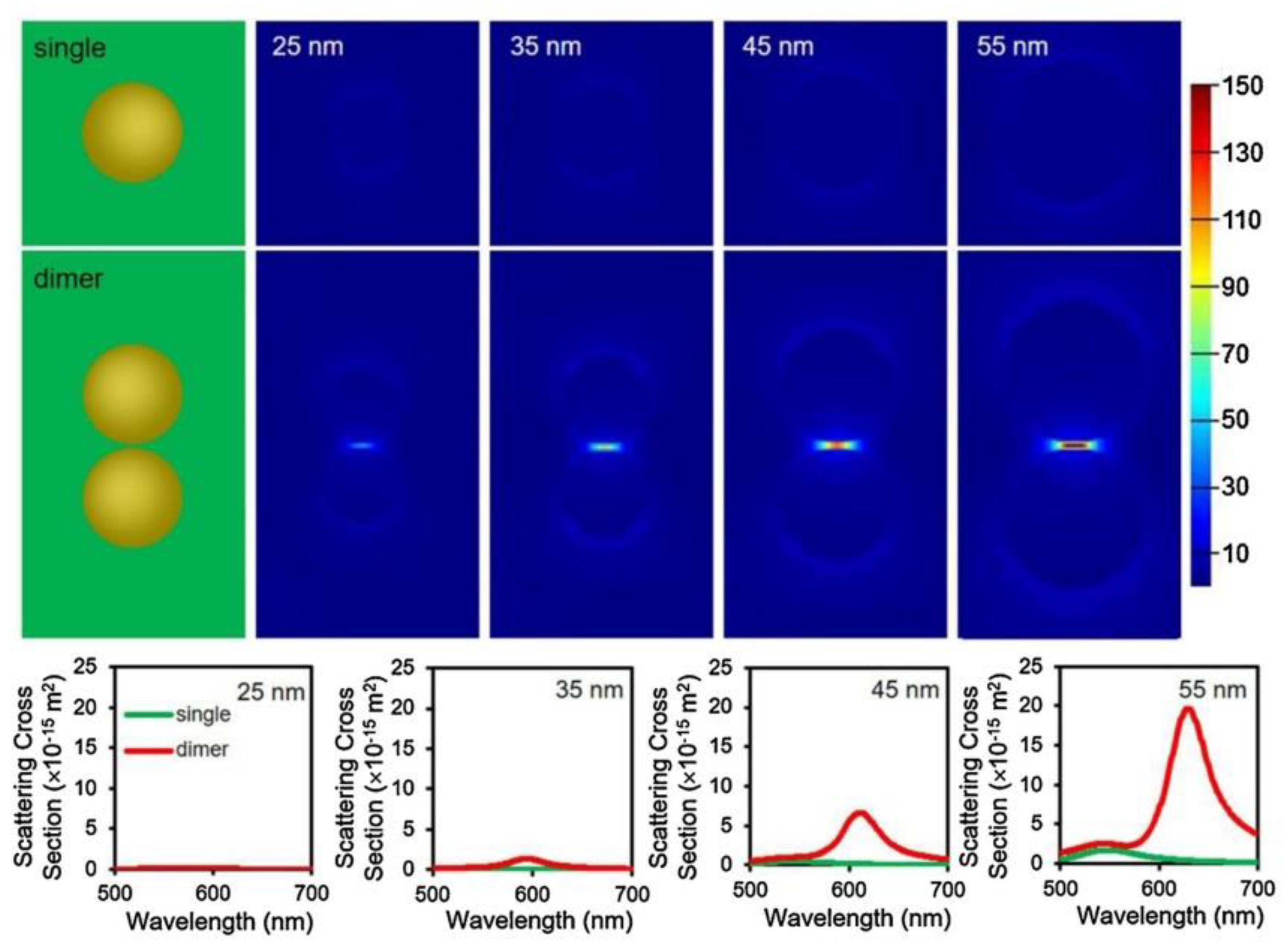

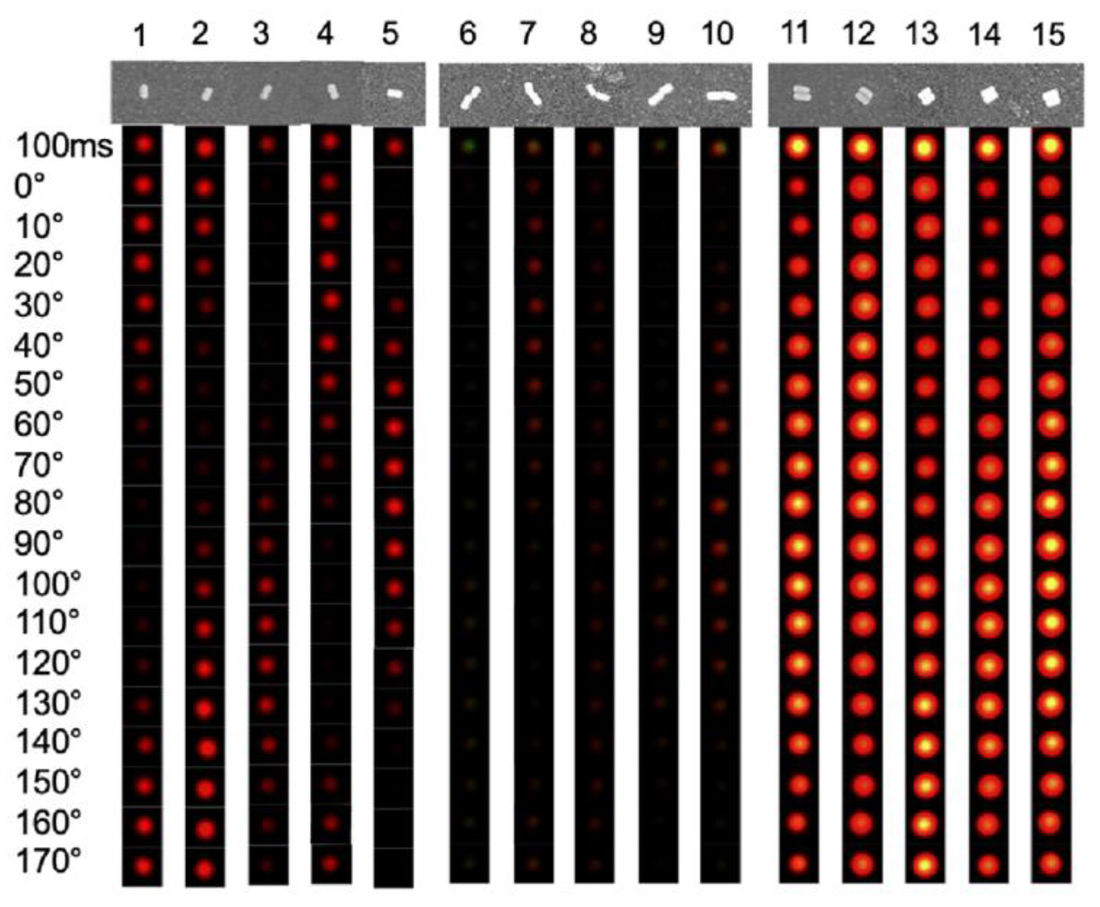
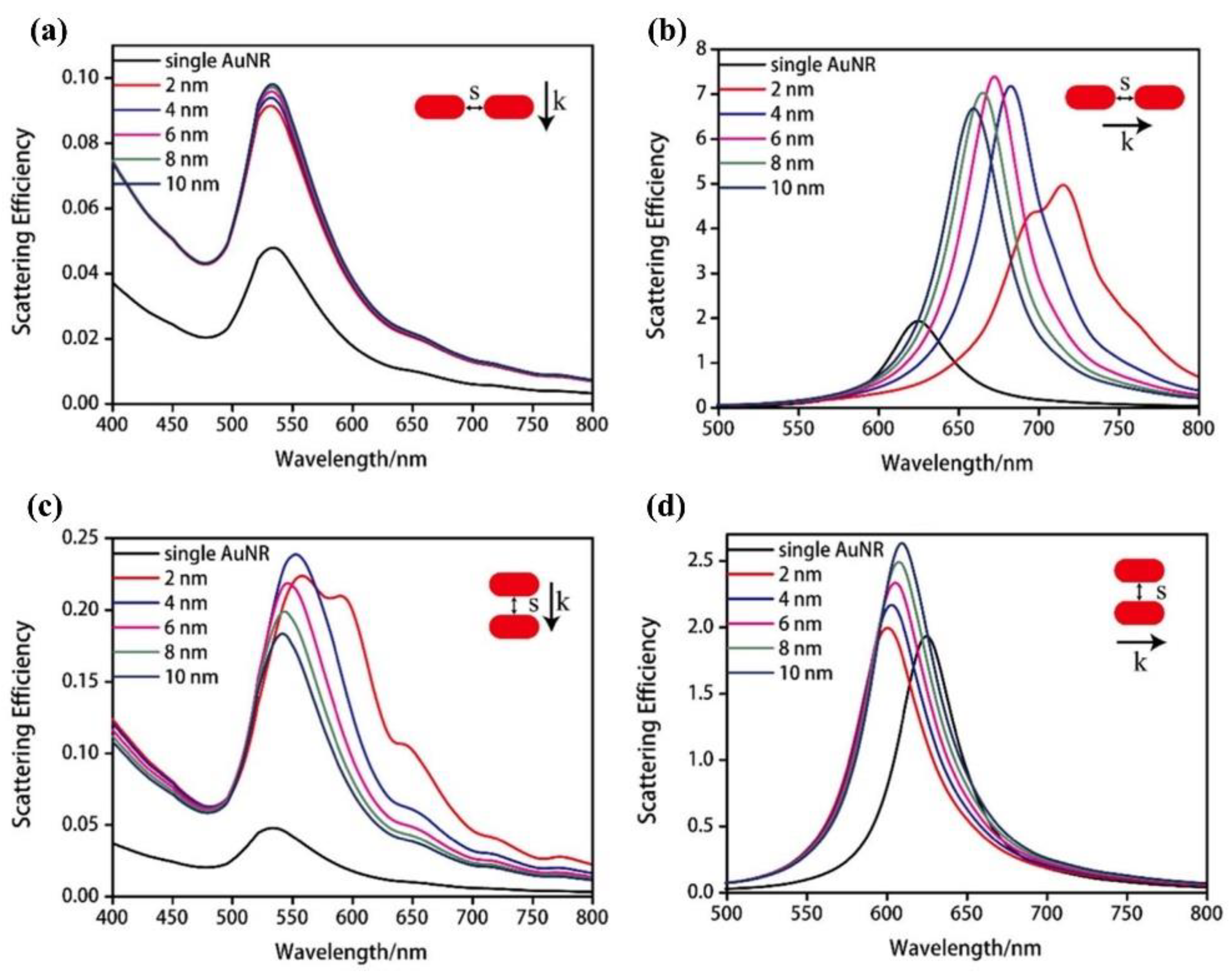
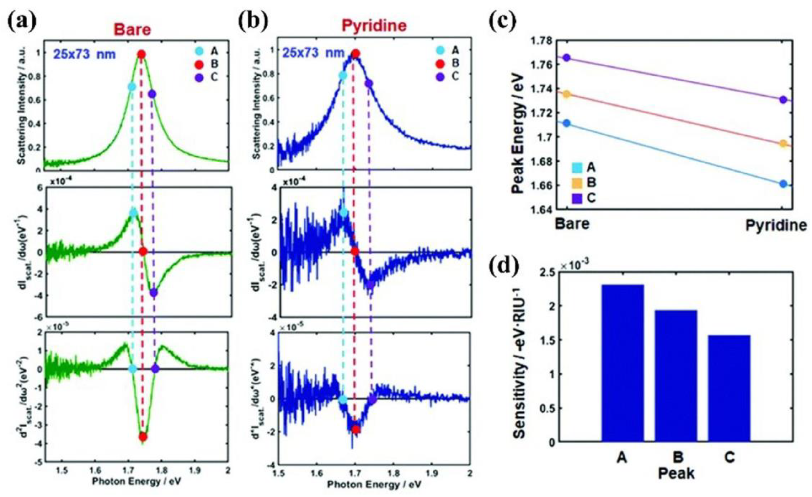
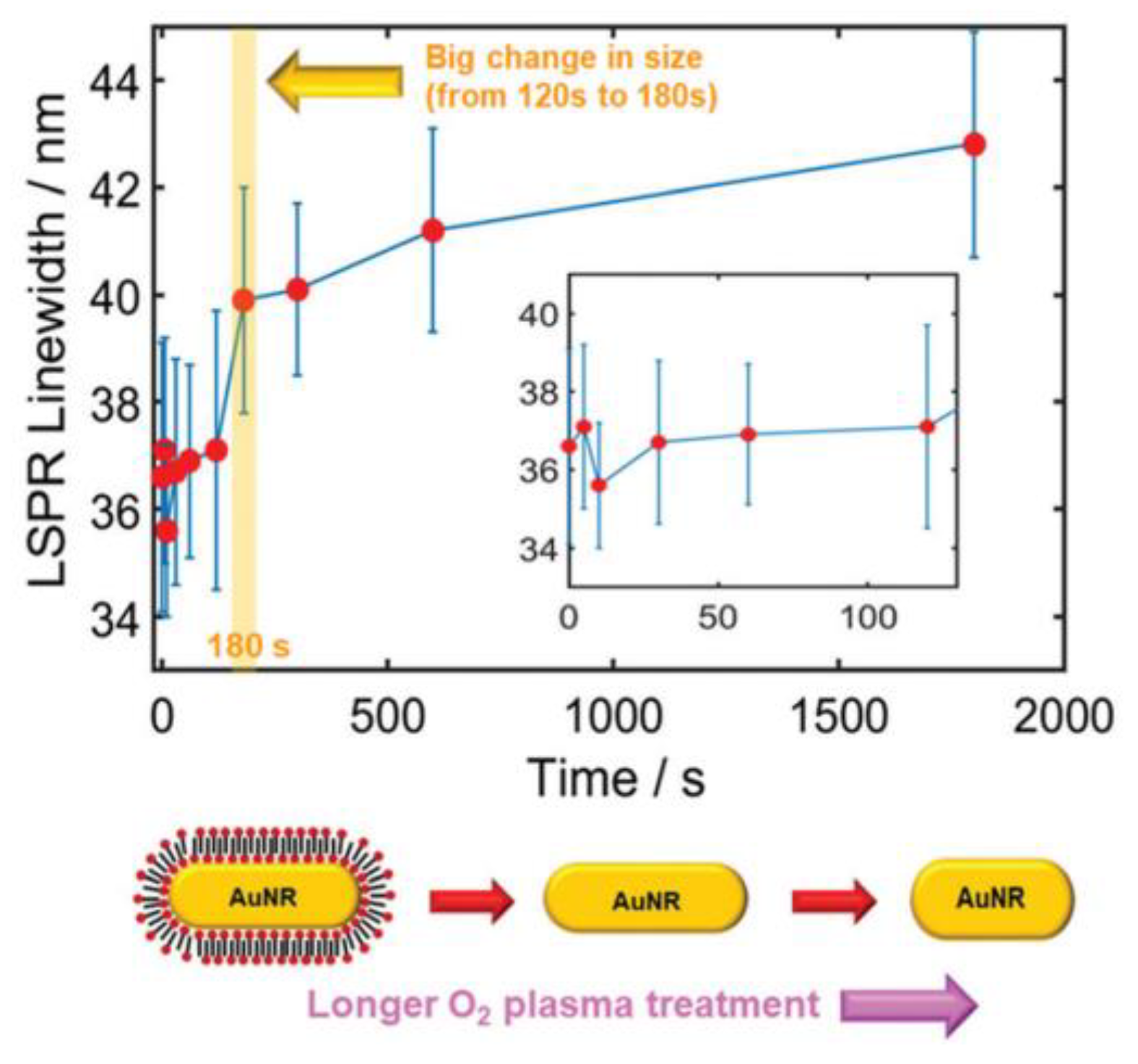
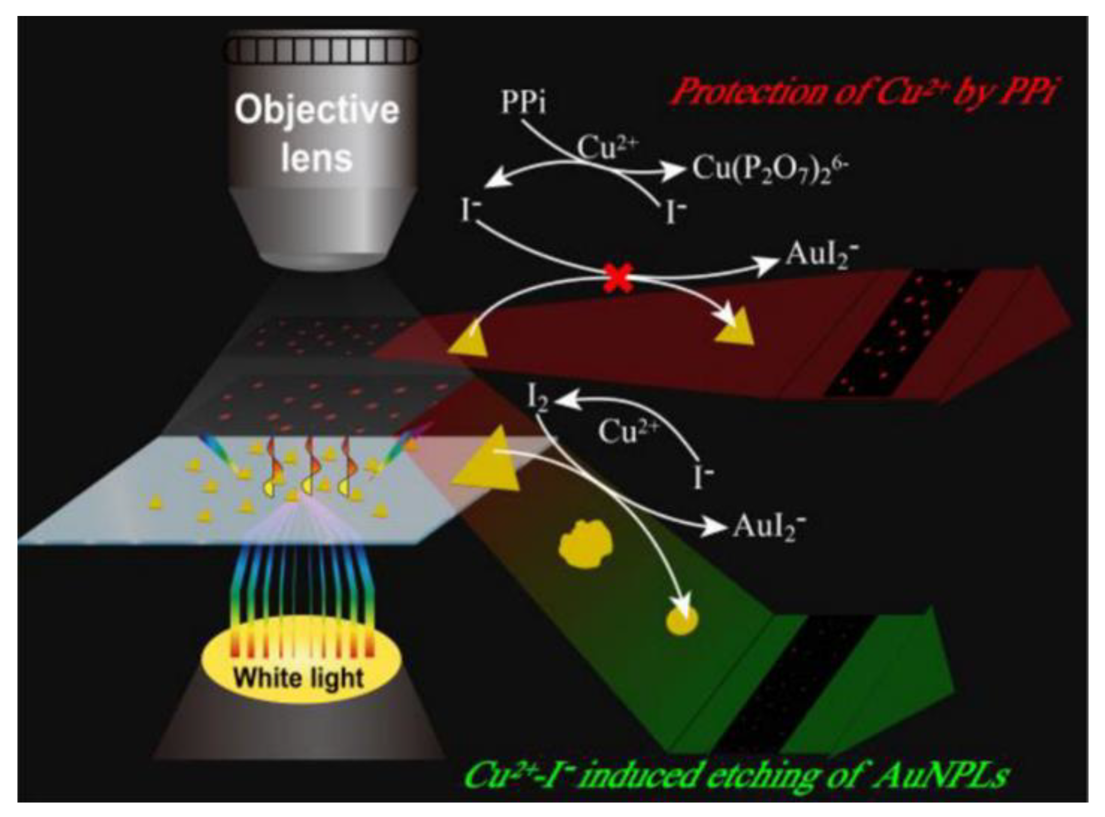




| Structure | Nanoparticle | Probe | Analyte | Detection Range | LOD | Ref. |
|---|---|---|---|---|---|---|
| nanospheres | AuNPs | DNA | Cu2+ | 0.1 Nm−5 μM | 0.082 nM | [21] |
| AuNPs | MPBA | galactose | 1−75 nM | 0.83 nM | [24] | |
| AuNPs | Tetrazine, trans−cycloctene | ATP | - | - | [46] | |
| AuNPs | oligonucleotide | Hg2+ | 0.005−25.0 nM | 1.4 pM | [22] | |
| AuNPs | dsDNA | PARP−1 | 0.2−10 mU | - | [44] | |
| AuNPs | DNA | miRNA−122 | 100 pm−100 nM | - | [47] | |
| nanorods | AuNRs | CTAB | telomerase | 100−24,000 cells | 43 cells | [48] |
| AuNRs | GADD45 | p53 protein | 10−106 fM | 11.47 fM | [29] | |
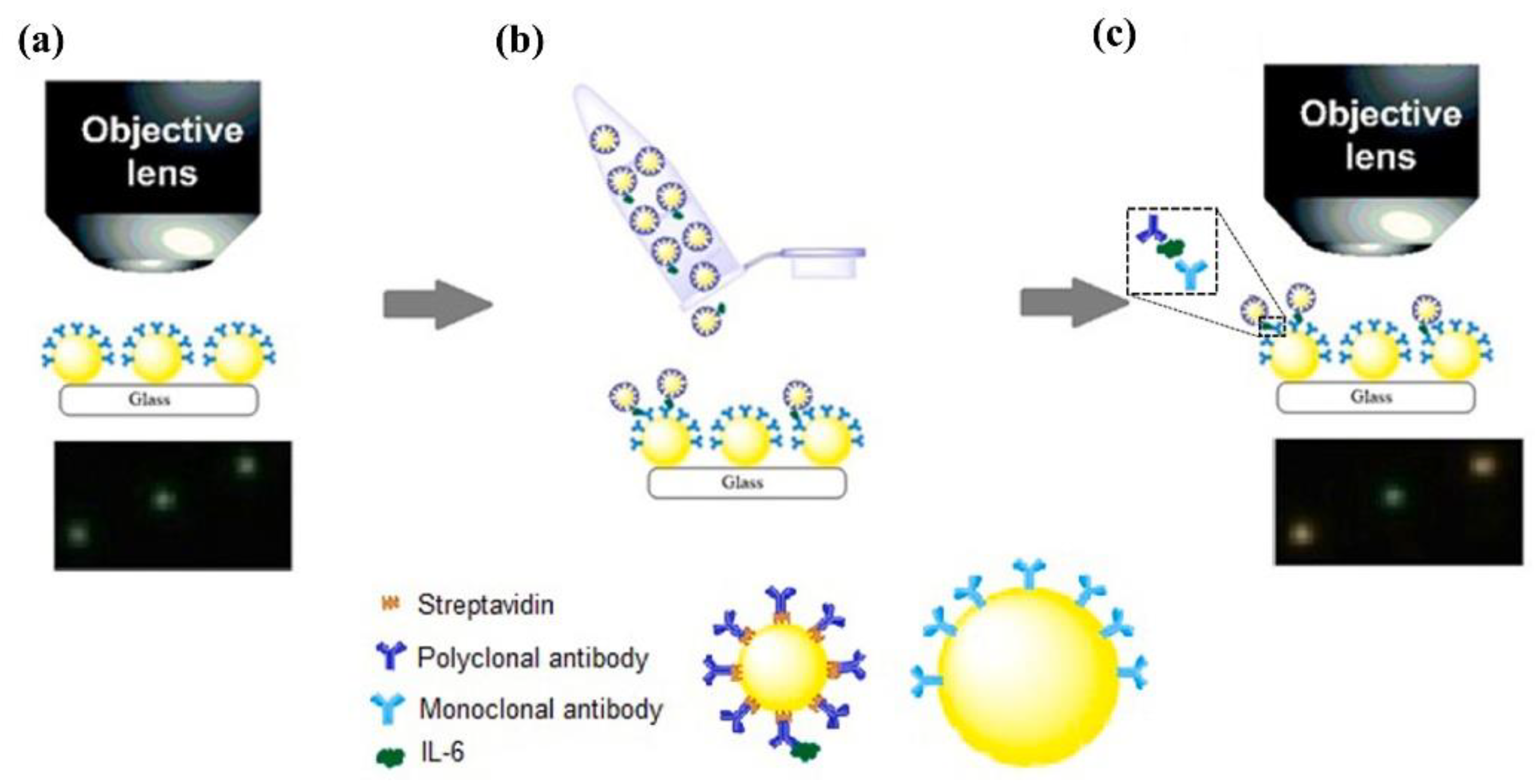
| AuNRs | antibody | TNF−α, IL−4, IL−6, IL−10 | - | - | [30] | |
| AuNRs | DNA | miRNA−21 | 50−2500 fM | 1.72 fM | [33] | |
| AuNRs | DNA | miRNA−Let−7a | 2−2000 fM | 0.53 fM | [33] | |
| AuNRs | DNA | DNA in serum | 0.1 pM−1 nM | 30 fM | [34] | |
| AuNRs | OTA Aptamer, Poly A | OTA | 0.1 nM−10 μM | <1 nM | [49] | |
| AuNRs | - | miRNA−21 | 0.1 pM−10 nM | 71.22 fM | [31] | |
| AuNRs | PDMA, PCM−b−PEG | Bacterial lipase | - | - | [50] | |
| nanosheets | AuNTs | - | PPi | 7–100 nM | 1.09 nM | [37] |
| AuNTs | oligonucleotides | DNA | - | - | [51] | |
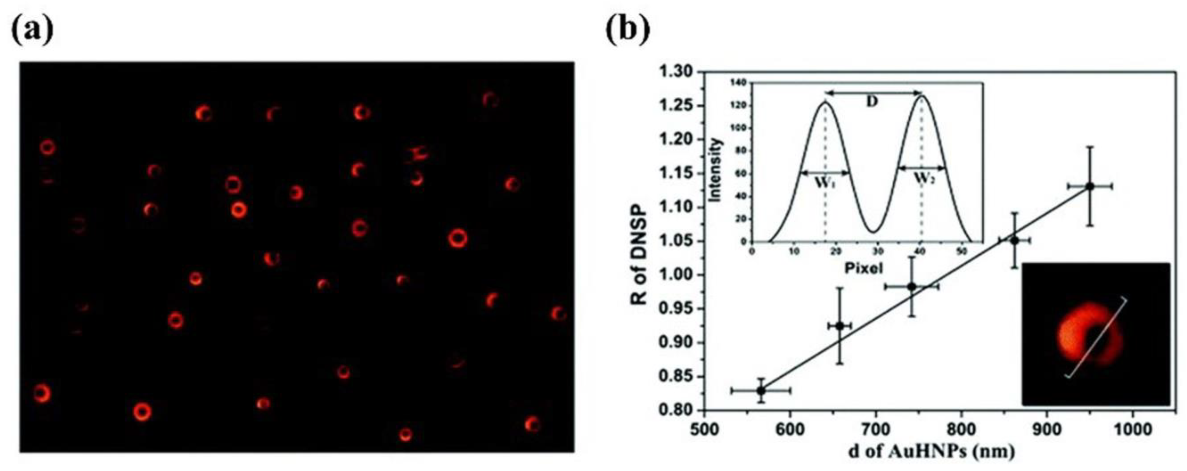
| AuHNPs | - | Au@Hg | - | - | [38] | |
| AuBPs | p−ATP@Biotin | Streptavidin | - | - | [52] | |
| AuBPs | p−ATP@Anti−IgG | IgG | - | - | [52] | |
| Nanocore–satellite | AuNPs | L−DNA, S−DNA | telomerase | 3.8 × 10⁻¹³−1.9 × 10⁻¹¹ IU | 1.3 × 10⁻¹³ IU | [41] |
| AuNPs | Anti IL−6 | IL−6 | - | 0.01 ng/mL | [43,53] | |
| AuNR, AuNPs | oligonucleotides | Hg2+ | 10 pM−10 μM | 2.7 pM | [45] |
| Structure | Nanoparticle | Probe | Analyte | Detection Range | LOD | Ref. |
|---|---|---|---|---|---|---|
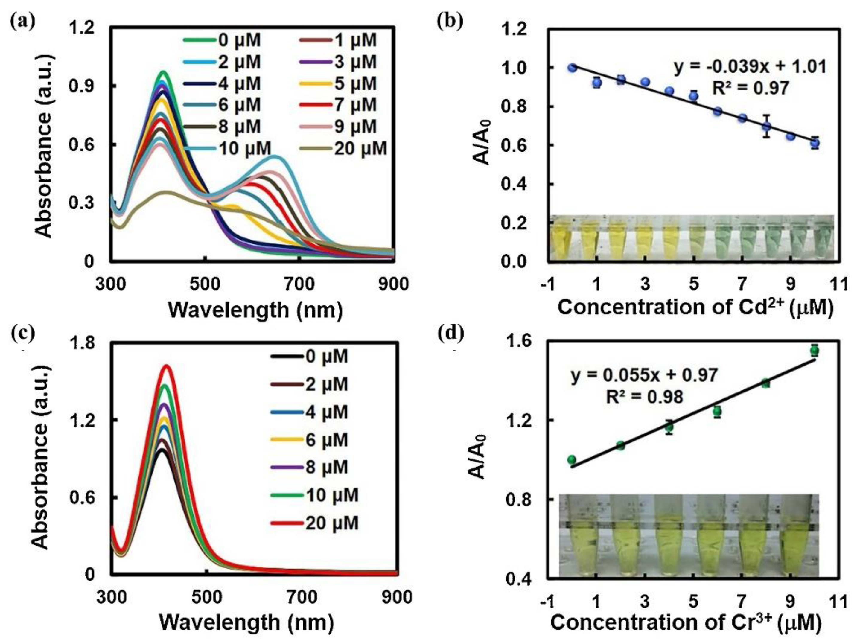
| composite plasmonic materials | AuNP@Ag | - | Cd2+ | 1−5 μM | 11.5 nM | [54] |
| AuNP@Ag | - | Cr3+ | 2−10 μM | 26.8 nM | [54] | |
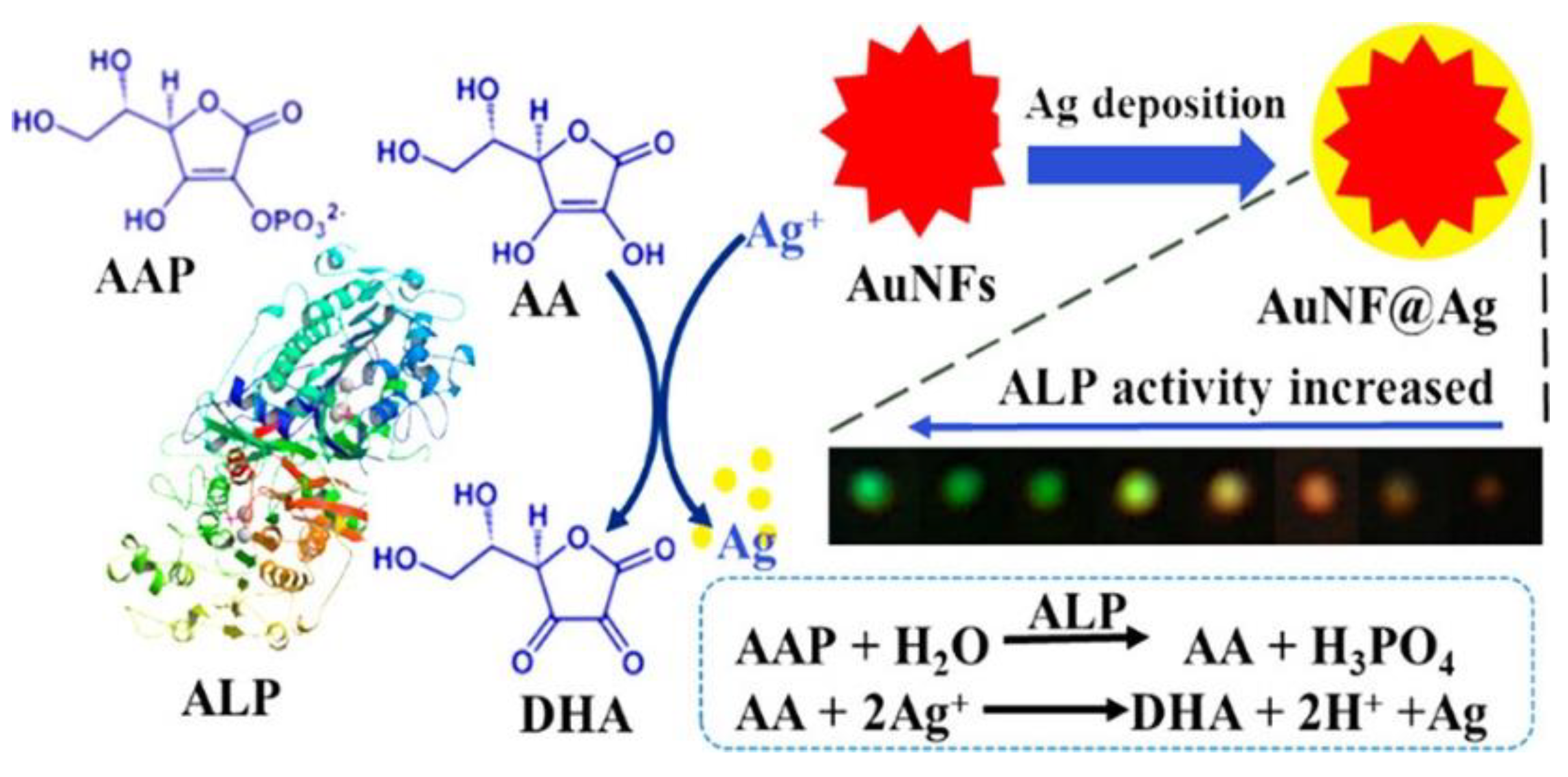
| AuNFs, Ag+ | - | ALP | 0.1−60 μU L−1 | 0.03 μU L−1 | [56] | |
| Au−Ag−HM | - | ROS | - | - | [57] | |
| Au−Ag−HM | - | caspase−3 | 0.05−20 nM | 26.7 pM | [57] | |
| AgNPs, Au3+ | - | GE | - | - | [58] | |
| AuNPs@MnO2 | - | glucose | 0.05−20 mM | 12.9 nM | [61] | |
| AuNP@FeOOH | - | ALP | 0.2−6.0 U/L | 0.06 U/L | [63] | |
| AgSHINs | - | Hg2+ | 10−10−10−4 M | - | [65] | |
| AuNR@Ag | GOx protein | glucose | 5−100 μM | 0.5 μM | [69] | |
| Au@Ag NCs | D-mannose | ConA | 10 nM−10 μM | 2 nM | [70] | |
| AuNSs, Ag+ | - | MAO-B | 0.05−1 μg mL−1 | 8.0 ng mL−1 | [71] | |
| Au@Ag CSN | - | PtCl62− | - | - | [72] | |
| AuNP@MnO2 | - | ALP | 0.06−0.48 mU/mL | 5.8 μU/mL | [60] | |
| Au/Ag NCs | oligonucleotides | miRNA−21 | - | - | [59] | |
| Au@AgI | - | S2+ | 0.1−500 nM | 33 pM | [62] | |
| Cu2O/Au | - | glucose | 0.16−5.6 mM | 4 mM | [67] | |
| AuNP@Ag | - | MnO4− | 0−6 μM | 46 nM | [73] | |
| Cu2−xSe NPs | CTAB | Hep | 0.01−0.6 μg mL−1 | 4.0 ng mL−1 | [10] | |
| ZnO QD/AuNP | - | - | - | - | [68] |
Disclaimer/Publisher’s Note: The statements, opinions and data contained in all publications are solely those of the individual author(s) and contributor(s) and not of MDPI and/or the editor(s). MDPI and/or the editor(s) disclaim responsibility for any injury to people or property resulting from any ideas, methods, instructions or products referred to in the content. |
© 2023 by the authors. Licensee MDPI, Basel, Switzerland. This article is an open access article distributed under the terms and conditions of the Creative Commons Attribution (CC BY) license (https://creativecommons.org/licenses/by/4.0/).
Share and Cite
Zhang, W.; Zi, X.; Bi, J.; Liu, G.; Cheng, H.; Bao, K.; Qin, L.; Wang, W. Plasmonic Nanomaterials in Dark Field Sensing Systems. Nanomaterials 2023, 13, 2027. https://doi.org/10.3390/nano13132027
Zhang W, Zi X, Bi J, Liu G, Cheng H, Bao K, Qin L, Wang W. Plasmonic Nanomaterials in Dark Field Sensing Systems. Nanomaterials. 2023; 13(13):2027. https://doi.org/10.3390/nano13132027
Chicago/Turabian StyleZhang, Wenjia, Xingyu Zi, Jinqiang Bi, Guohua Liu, Hongen Cheng, Kexin Bao, Liu Qin, and Wei Wang. 2023. "Plasmonic Nanomaterials in Dark Field Sensing Systems" Nanomaterials 13, no. 13: 2027. https://doi.org/10.3390/nano13132027
APA StyleZhang, W., Zi, X., Bi, J., Liu, G., Cheng, H., Bao, K., Qin, L., & Wang, W. (2023). Plasmonic Nanomaterials in Dark Field Sensing Systems. Nanomaterials, 13(13), 2027. https://doi.org/10.3390/nano13132027


