Review

Graphene Oxide Thin Films with Drug Delivery Function

by
Alexandra M. L. Oliveira
1,2,3,*,
Mónica Machado
1,2,3,
Gabriela A. Silva
2,3,
Diogo B. Bitoque
2,3,
Joana Tavares Ferreira
4,5,
Luís Abegão Pinto
4,5 and
Quirina Ferreira
1,*
1
Instituto de Telecomunicações, Avenida Rovisco Pais, 1049-001 Lisbon, Portugal
2
iNOVA4Health, CEDOC Chronic Diseases Research Centre, NOVA Medical School, Universidade Nova de Lisboa, Campo Mártires da Pátria 130, 1169-056 Lisboa, Portugal
3
NOVA Medical School, Faculdade de Ciências Médicas, Universidade Nova de Lisboa, 1169-056 Lisbon, Portugal
4
Ophthalmology Department, Centro Hospitalar Universitário de Lisboa Norte, 1649-035 Lisbon, Portugal
5
Visual Sciences Study Centre, Faculty of Medicine, Universidade de Lisboa, 1649-028 Lisbon, Portugal
*
Authors to whom correspondence should be addressed.
Nanomaterials 2022, 12(7), 1149; https://doi.org/10.3390/nano12071149
Submission received: 3 March 2022 / Revised: 24 March 2022 / Accepted: 28 March 2022 / Published: 30 March 2022
(This article belongs to the Special Issue Graphene and Related 2D Materials)

Abstract

:
Graphene oxide has been used in different fields of nanomedicine as a manager of drug delivery due to its inherent physical and chemical properties that allow its use in thin films with biomedical applications. Several studies demonstrated its efficacy in the control of the amount and the timely delivery of drugs when it is incorporated in multilayer films. It has been demonstrated that oxide graphene layers are able to work as drug delivery or just to delay consecutive drug dosage, allowing the operation of time-controlled systems. This review presents the latest research developments of biomedical applications using graphene oxide as the main component of a drug delivery system, with focus on the production and characterization of films, in vitro and in vivo assays, main applications of graphene oxide biomedical devices, and its biocompatibility properties.

Graphical Abstract

1. Introduction

Graphene-based materials have an important role in the development of drug delivery systems (DDS) due to their biocompatibility properties and the easiness with which they can be functionalized with biomolecules. Moreover, it is simple to integrate them in multilayer systems with applications in which controlled release of bioactive molecules is necessary. Graphene is a two-dimensional compound, consisting in a monolayer of aromatic carbon atoms (sp2-bound)/covalently bound, organized in a hexagonal lattice structure [1], forming sheets with the thickness of a single atom [2]. It has interesting mechanical properties, large surface area, and high electrical and thermal conductivities [3], and it can exist in other forms, such as graphene oxide (GO) and reduced graphene oxide (rGO) (see Figure 1), which are more hydrophilic, making them easier to solubilize and disperse in aqueous or polyelectrolyte solutions (such as PBS—phosphate buffered saline) and improving, therefore, their self-assembling properties [4,5]. The high content of groups with reactive oxygen in GO enhances its ability to be functionalized with various materials [1]. It is composed of carboxyl, hydroxyl, and ether groups, allowing it to absorb polar polymers or polar molecules with ease, and, therefore, it is an excellent candidate to form GO/polymer composites [4]. These active groups are ideal for molecule immobilization on the GO surface and make it hydrophilic and a powerful candidate to be used as drug carrier [6]. For instance, carboxyl groups are the main group responsible for GO colloidal stability [7,8,9]. Furthermore, GO has good water and biomedium dispersibility as well as good optical (absorption) and photothermal (conversion) properties [10]. Several functionalization strategies can be used to enhance GO’s characteristics and promote its stability and bioavailability. This functionalization can be achieved, for example, by PEGylation (PEG—polyethylene glycol) to improve biocompatibility, solubility, and stability in physiological conditions, by double oxidation of the graphene to provide electrostatic stabilization, by using a copolymer (e.g., Pluronic F127) to provide steric stabilization, by non-covalent interaction (π-π interactions) with aromatic organic molecules (such as 1-pyrenebutyrate (PB)) to improve aqueous stability, or by using synthetic peptides such as Poly L-Lysine (PLL) to improve its biological activity [11,12,13,14]. The versatility in the functionalization of GO allows its application in several devices. An example is the ability of functionalized GO to cross the blood–brain barrier (BBB), which widens its use in biomedical research, as it overcomes the inability of unstable chemicals or peptides to reach the brain [15,16]. Mendonça et al. [17] also demonstrated the ability of rGO to cause a transitory BBB breakdown and, therefore, cross this BBB into the brain.
GO is also a versatile material to be part of multilayer systems with applications in DDS and biosensors. Usually, the layer-by-layer (LbL) is the elected technique to build these systems. It is based on the alternate adsorption of oppositely charged particles (polyelectrolytes) and has been studied more each day, as these polyelectrolyte multilayer films (PEMf) have the ability to allow the fine-tuning of DDS both in time and space, thus permitting therapeutics to be delivered in situ during a course of several days or even months. In the last years, graphene has been studied as a component of PEMf and has shown promising results either as a nanocarrier or in stabilizing and/or delaying the drug release in this DDS. This article revises the recent experiments on graphene-based DDS, including the techniques usually used to develop multilayered films. Two types of uses are described:
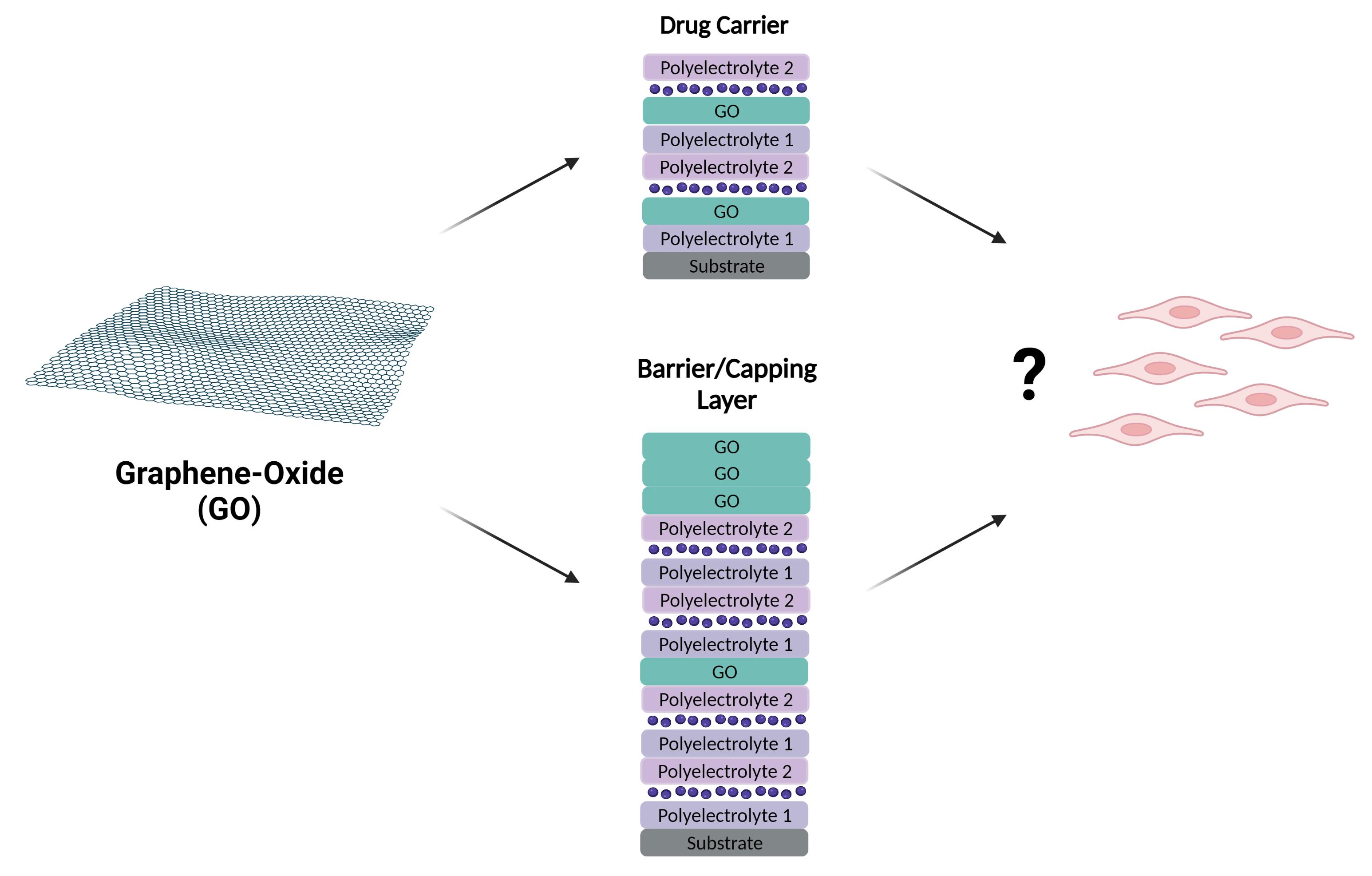
  • GO incorporated in multilayered systems in which it takes the role of carrier, helping to transport the therapeutic agents, making sure that they reach the target before being released, and helping to protect them from early degradation.
  • GO incorporated in multilayered systems in which it acts as a barrier or capping layer with the purpose of delaying and/or controlling the drug release across time, in a precise and stratified manner, with control of the release sequence.
Biocompatibility and recent applications are also addressed.

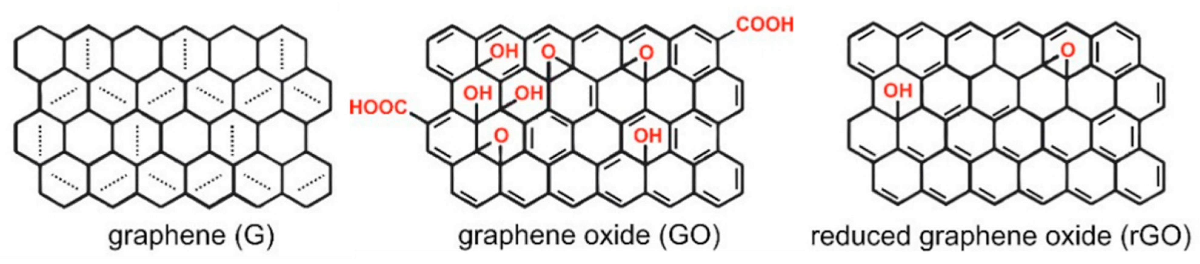
2. Graphene Oxide Multilayer Films

LbL is a versatile technique which allows the incorporation of multiple components culminating in a smart and functional system. It is a simple and cost-effective process that can be used in large-scale production and is eco-friendly, due to the fact that the solutions used are aqueous [9]. LbL is the preferred technique to develop DDS with graphene, and several methods can be used to produce thin films, such as microfluidics [19], perfusion [19], Langmuir–Blodgett assembly [20], spin coating [5], drop coating [21], spray coating [22], or dip coating [4], which is the most common one (see Figure 2).
The major driving forces in LbL multilayered films are electrostatic forces (more common), hydrogen bonding, charge transfer interaction, and covalent bonding [3,23]. LbL films can incorporate functional polymers [24], enzymes [25], small molecules [26], polysaccharides [27], carbon nanotubes (CNT) [28], nucleic acids [29,30], or inorganic nanoparticles (NPs) [31] and can be assembled in a wide range of substrates independently of their size or shape, allowing for the creation of three-dimensional objects covered in these films [3,32]. The fact that LbL films can be deposited on different substrates without altering the properties of the films is very useful, because it allows the testing of different film characteristics, which may require different substrates [9,33], for example, the quartz crystal microbalance (QCM), which requires the films to be assembled in a QCM crystal sensor/lamella [34,35], or the transmission electron microscopy, for which the films need to be deposited onto copper grids [36,37]. This also allows for their application in multiple biomedical devices without fear that their properties will change. Fox example, they can be applied in flexible substrates that can adhere to tissue and deliver different types of drugs [7] or odd-shaped substrates as coatings for biomedical implants, such as cardiovascular stents [38] [9,33]. Furthermore, LbL films containing GO can be used, for example, in tissue engineering [11,39], as carriers for vaccines [40], as antibacterials [41], in cancer therapy [42], or in biomedical imaging [43,44,45].
The incorporation of GO in a multilayered film is a chemistry game; it is necessary to have stable chemical interactions in the interfaces of the layers to avoid interdiffusion of components and to ensure a correct growth of film. Usually, to avoid aggregation or agglomerates on the surface of GO layers, bilayers of charged GO are added. GO has a natural negative charge and can act as a negatively charged component in LbL without further functionalization, and the positively charged GO can be obtained by amine functionalization [9,46,47,48,49]. As a result, there is obtained a thin and smooth bilayer of GO that can be incorporated between polyesters or polymeric thin layers. GO can interact with other molecules via hydrophobic or electrostatic interactions, hydrogen bonding, or π-π stacking. GO can be non-covalently functionalized using LbL to improve dispersibility and avoid aggregation, using, for example, chitosan (Chi)/dextran, Chi/sodium alginate (SA), or protamine sulfate/SA [6,33]. Small molecules, non-polymeric in nature, are more difficult to incorporate into LbL assemblies, because they can penetrate the graphene multilayers, and that can affect the growth of the multilayered system [6].
To start a multilayered film, it is necessary to charge the substrate to enhance the adsorption of the first layer. The most common assembly process is to start with a negatively charged substrate that is usually obtained by piranha solution (composed of sulfuric acid (H2SO4) and hydrogen peroxide (H2O2) in the proportion of 7:3, v/v) and oxygen plasma treatment [50] and, therefore, a negatively charged layer. However, it is also possible to invert the order, functionalizing the substrate with positive charges by dipping the substrate in APTES solution ((3-aminopropyl) triethoxysilane) (3 min). Furthermore, substrates should be previously cleaned to remove organic contaminants with piranha solution followed by a rinse with deionized (DI) water [46]. The cleanness and functionalization of substrate is important to ensure the homogeneity of the following layers. Usually, the films exhibit a linear growth; however, this can vary on the first few bilayers, due to interaction with the chosen substrate [9].
The pH of the dipping solutions can greatly affect the resulting film’s thickness and roughness, as it can affect the charge balance between the oppositely charged components and the degree of ionization of weak polyelectrolytes. If the pH and pKa of the dipping solution are near each other, the film’s thickness is increased [46,51]. The use of different or even cross-linked polymers in LbL films allows for the control of the porosity and rate of degradation and dissociation of the films [51].
The growth of a multilayered film can be monitored by several techniques, and it is crucial to perform a stepwise characterization to ensure the homogeneity of each layer, especially when different types of materials are included, as usually happens in a DDS in which graphene can be intercalated with polymers, NPs, drugs, or other biomolecules. Table 1 summarizes commonly used techniques to perform this characterization. Furthermore, a careful monitoring of the drug’s release profile is important. In order to access this, for the most part what happens is the incubation of the films in physiological conditions (generally PBS at 37 °C), and then the PBS solution is analyzed through various methods which might include: UV-visible/fluorescence spectroscopy to evaluate the release kinetics [34,36,52,53,54]; micro-BCA kit to evaluate loading efficiency [55]; measurement of fluorescence spectra for fluorescent molecules [56]; ELISA for release studies of some proteins [57]; and gel electrophoresis and circular dichroism (CD) spectroscopy to evaluate whether proteins released from films maintain their primary and secondary structures [58,59]. Notwithstanding, the erosion of a multilayered DDS can be followed using the same technique that was applied to follow the growth of the film to obtain a detailed interpretation of the drug release behavior.

3. Graphene Oxide as Carrier in Drug Delivery

Ideally, nanocarriers must have a size between 30 and 200 nm to be retained in blood vessels. Above this size, they are prone to aggregation in the liver or the spleen, and, below that, they can suffer blood renal filtration, being filtered from the plasma to the urine [8]. When encapsulating or bonding a drug to its respective carrier, it is also important to consider the ratio of drug:carrier, as this can affect encapsulation/bonding efficiency [71].
In recent years GO has gained a lot of attention for its potential use in biomedical applications, with several publications reporting its benefits as a drug in cancer therapy [72,73,74,75,76,77,78,79,80,81]. For example, Liu et al. [71] investigated a possible nanocarrier for oral drug administration in cancer treatment, developing a thin film with graphene as nanocarrier and pingyangmycin (PYM) as the anti-cancer drug of choice. Orally administered drugs have reduced side effects when compared with IV administration and are more easily accepted by the patients; however, they have some drawbacks, such as being easily degraded by the gastric acid and pepsin present in the stomach, low bioavailability, rapid clearance, and poor tissue distribution. The developed film was composed of poly(acrylic acid)-cysteine (PAA-cys), poly(allylamine hydrochloride) (PAH), and GO as follows: PAA-cys-PAH-GO-PYM. PAA-cys helps the DDS to adhere to the small intestine mucus layer, improving the drug bioavailability, and PAA-cys and PAH polyelectrolytes are cross-linked in the surface of the GO-PYM conjugate and help protect the nanocarrier and drug from gastric acids, allowing the safe passage of the DDS to the intestine, where it can release the drug. Drug release was higher at lower pH, further demonstrating the potential of this DDS in improving targeting to the tumor tissues (acidic environment) and reducing effects in normal tissue.
Another study reports the use of GO NPs as nanocarriers to enhance the drug bioavailability and water solubility. GO is subjected to the “piercing effect” and, therefore, is easily internalized by cells, and its sheet-like structure helps to stabilize the capsule layers, avoiding any drug escape before it reaches its target, making it an ideal choice for this kind of application. Here, they coated GO NPs with a single layer of carboxymethylcellulose (CMC) (to increase drug loading capabilities), with curcumin (Cur), a powerful anti-cancer “drug”, encapsulated into CMC. CMC was then cross-linked with poly N-vinylpyrrolidone (PVP) to produce stimuli-responsive NPs (redox-responsive disulfide linkage). The high glutathione concentrations inside the tumor induce breakage on the bridge between CMC and PVP and, therefore, cause the curcumin to be released. Further NP functionalization was obtained by conjugation of PEGylated monoclonal folic acid antibody (FA) onto the NP surface. This is an important step, as FA binds to folate receptors that are highly expressed in cancer cells, thus helping to direct the NPs to these cells and allowing them to be internalized by phagocytosis. PEGylation helps to reduce protein adsorption onto nanocarriers (preventing “protein corona” and, therefore, renal clearance), prolongs NPs blood circulation, and enhances binding efficiency of FA to the receptors. It also helps to prevent the drug from leaking early and protects it against degradation or enzymatic cleavage. The final composition of the NPs was Cur-FA-CMC/PVP GO NPs. Another important piece of information to retain is that highly charged NPs suffer less aggregation due to repulsive electrostatic forces, allowing for more stable dispersion in physiological conditions [8,61].
Associated with cancer treatment, several stimuli-responsive films which can be used in photothermal therapy are also being studied [61,82,83,84,85]. Photothermal therapy relies on the capacity of NPs to convert NIR (near-infrared radiation) to vibrational energy, generating heat and thus killing cancer cells (where the NPs accumulate). The high efficiency of NIR is mainly because it can reach the NPs without damaging the tissues in between, since biological tissues do not possess chromophores absorbing in the NIR region. GO is a great nanomaterial to use in these kinds of films, as it can generate heat sufficient to kill cancer cells when exposed to NIR, being even more effective than CNT [86]. Therefore, by using GO, it is possible to obtain a NIR-responsive capsule/film with drug loading capabilities and in which GO not only serves as a structural component but also as a NIR responsive material, avoiding the use of gold and silver NPs or addition of NIR-dyes or CNT (which were used until now to fabricate NIR-responsive capsules). This allows for a much simpler and cost-effective process [10].
A study led by Xie et al. [33] successfully developed a magnetic DDS based on GO sheets, with both magnetic and photothermal response. This kind of particle can be used in targeted therapy and has been greatly studied in cancer applications. For this work, Xie et al. [33] developed a graphene oxide-based nanocomposite loaded with doxorubicin (DOX—an antitumor antibiotic). GO has a large DOX loading capacity, making it the ideal nanocarrier. To magnetize the GO sheets, the authors used the thermal reaction method to deposit Fe3O4 onto the GO sheets. However, mGO (magnetic GO) is even more prone to aggregation than GO, and, therefore, it requires further functionalization. For that purpose, mGO sheets were coated with Chi and SA through the LbL technique. The final composite composition was mGO-Chi/SA-DOX. A ratio of mGO sheets:Chi:SA of 1:4:4 was needed in order to achieve a stable dispersion. DOX was loaded via π-π stacking and electrostatic attraction. They successfully developed a nanocarrier that not only could be directed to the target cells with magnetism, as it had strong photothermal response, causing the cancer cells to die upon NIR irradiation, but had a pH-dependent release of DOX, ideal to use with cancer cells that have a pH lower than normal cells.
Another example is GO-iron oxide (IO) PEGylated nanocomposites (GO-IONP-PEG) that can be used as a drug carrier triggered by a magnetic field, being used in photothermal therapy. GO functionalization with PEG greatly increases its stability in physiological solutions. In these cases, GO-IONP-PEG particles can be loaded, for example, with DOX by π-π stacking and then be used in cancer treatment. However, this composite can also be used as a t2-weighted magnetic resonance contrast in tumors, as demonstrated by Ma et al. [87].
Another potential application for GO-based compounds is as a carrier and/or enhancer for antibacterial or antimicrobial agents [6,76,88,89,90].
As an example, we can use the work carried out by Cao et al. [6], in which they developed a system for sustained release of an antibacterial peptide (G(IIKK)4I-NH2) using GO as a nanocarrier and assembling it into a thin film that can be used, for example, as coating for surfaces or devices. G(IIKK)4I-NH2 cannot be efficiently loaded into LbL by itself due to its low charge number, hence the need for a GO. The films were composed of G(IIKK)4I-NH2 (positively charged), poly(acrylic acid) (PAA, Polyanion), and poly(β-amino ester) (PBAE, polycation), and it was possible to confirm that G(IIKK)4I-NH2 retained its antibacterial properties even when incorporated in the films. The release speed can be tuned by varying the number of layers of PBAE.
GO’s ability to reach the brain makes it also a good candidate for neurological applications [15,91,92], as reported, for example, by Xiong et al. [91], who developed a DDS to treat Parkinson’s disease (PD) using lactoferrin (Lf)-functionalized GO as a nanocarrier for the natural drug puerarin (Pue), which presents anti-PD properties (Lf-GO-Pue). Lf was used for its ability to bind to the vascular endothelial receptor in the BBB and, therefore, promote the transport of this DDS across this barrier through a receptor-mediated transport. Their work showed promising results, with the in vivo test showing that, in PD-induced mice, there were significantly less neuronal damage and compartmental deficits.
Furthermore, it is possible to obtain hollow capsules of rGO that can be used to incorporate particles or bioactive molecules. These capsules were obtained by the sequential adsorption (due to electrostatic interactions) of positively charged (rGO-NH3+) and negatively charged (rGO-COO)) onto a sacrificial PS (polystyrene) substrate, which can then be removed by tetrahydrofuran (THF) solvent exposure (see Figure 3) [3].
Besides the already mentioned examples, there are several others that report using GO as a nanocarrier, for example, for proteins, protecting them against proteolysis and helping retain their activity [93]; as a carrier for pirfenidone (a drug used to treat subarachnoid hemorrhage) [94]; as a carrier for phytomedicines, augmenting their biocompatibility and reducing their toxicity [95]; as a carrier for famotidine (an anti-ulcerous medicine), reducing its side effects through a controlled and sustained release [96]; as a carrier for quercetin (a bioactive flavonoid with powerful antioxidant characteristics), helping circumvent its low bioavailability, extensive first passage metabolism, and instability in aqueous intestinal fluids [97]; as a carrier for transdermal delivery (hydrogel) of ondansetron, a medicine to help control vertigo that has low bioavailability and short half-life [98]; as a carrier for growth factors to enhance cell differentiation [99]; and as a carrier for pain management medicines such as flurbiprofen or buprenorphine, helping reduce its side effects and promoting a sustained release without constant need for dosing [100,101].

4. Graphene Oxide for Controlled Drug Release

In a DDS, it is of the utmost importance to control the release of the bioactive compounds. Recently, graphene-based materials, such as graphene oxide, have been gaining a lot of attention for their ability to act as a barrier or capping layer, delaying and controlling the release of biomolecules across time [21,57,102,103,104,105,106].
There are several studies using ovalbumin (OVA—the main protein found in egg white, largely used as a nutrient supplement) as model drug; some authors were able to use GO either as a nanocarrier or a capping layer to build thin films, preventing the early release of the biomacromolecule and obtaining, therefore, a long-term delivery that lasted over 70 days [35]. This mechanism was also applied in films containing OVA as the model protein/drug and PBAE as a hydrolytically degradable polymer. They were able to delay the release of OVA from less than an hour to several weeks by using GO as a capping layer in films with (PBAE/OVA)20(GO+/GO)n, or as a barrier layer in films with (PBAE/OVA-AF555)20(GO+/GO)5(PBAE/OVA-TR)20(GO+/GO)2(PBAE/OVA-FL)20 (where AF555—Alexa fluor; FL—fluorescein; and TR—Texas red were used as labels to differentiate between OVAs). When GO was used as a barrier layer, a sequential and controlled release of OVA was possible, with the possibility to control the release gaps by varying the number of GO bilayers. Furthermore, GO’s low permeability helps to reduce interlayer diffusion. This opens up a world of possibilities for DDS with multiple drugs which can be release in a sequential and ordered manner [57].
In an electro-responsive film, GO was also used as a barrier layer. OVA was used as model drug, and rGO was also incorporated in the films as an electroconductive material (because GO has poor conductivity). (PBAE/rGO/GO+/OVA/GO+/rGO)n (n stands for the number of repeated layers) showed negligible OVA release when there were no stimuli present and 50 times more OVA release upon electric stimulation, further confirming GO barrier capabilities [56].
rGO can also be used as a barrier layer in order to prevent spontaneous release of cDNA, as demonstrated by Jeong et al. [21], who developed a method to activate DNA nanodevices based on electro-responsive films containing rGO with the following composition: (PBAE/cDNA/rGO+/cDNA/rGO+/PEDOT:PSS)n/(rGO+/rGO)n, where PEDOT:PSS stands for poly(3,4-ethylenedioxythiophene) polystyrene sulfonate.
GO is also able to prevent the release of drugs in multilayered systems. A recent work related with LbL films composed of brimonidine (Brim), an alpha-2 adrenergic agonist used normally to lower intraocular pressure, encapsulated in polycyclodextrins (PolyCD+Brim) intercalated with layers of PBAE and bilayers of charged GO showed that GO can delay the drug release for several days. The study compares films with and without GO (the film (PBAE/PolyCD+Brim)6/GO+/GO/(PBAE/PolyCD+Brim)4GO+/GO/(PBAE/PolyCD+Brim)4 and the film (PBAE/PolyCD+Brim)4), and it was observed that the GO has an important role in the management of drug behavior. The number of GO bilayers is proportional to the delay time, suggesting that this system is a good model to fine-tune a DDS able to deliver a precise drug concentration at a specific period of time [34].
GO can also be used to coat a siRNA-loaded porous silicon (Si) NP in order to delay siRNA release, being able not only to slow down the release time but also the enzymatic degradation of the siRNA and the dissolution of the porous Si NP [107].

5. Graphene Oxide: Biocompatible or Cytotoxic?

Graphene biocompatibility is a controversial theme. Whereas some authors report graphene as biocompatible or with no or negligible cytotoxicity, others report it as cytotoxic with the potential to cause damage to living organisms.
Particle size can have an impact on cytotoxicity. Particles with sizes up to 100 nm are able to enter the cell, being able to enter the nucleus below 40 nm. Furthermore, particles below 35 nm are able to cross the BBB. When graphene enters the cell, there is a potential to cause cell damage; this can perhaps be minimized through functionalization [108]. In the majority of cases, coating GO with biocompatible polymers helps improve its biocompatibility and reduce its toxicity [87]. Furthermore, Hu et al. [109] propose that GO cytotoxicity is due to a physical damage to the cell membrane that occurs upon initial contact, and, therefore, it doesn’t depend on time [96,110].
GO functionalization with chitosan allows further biocompatibility, due to chitosan’s antibacterial, antifungal, mucoadhesive, and hemostatic properties and its positive charge, which allows for the functionalized GO to bind with biomolecules with negative charges [7,111].

5.1. In Vitro Assays for Graphene Oxide

The first step is to assess GO toxicity, resorting to in vitro testing in various cell lines.
Cytotoxicity is evaluated by incubating films/particles with desired cells in different ratios and then accessing cell viability, for example, through cck-8 assay [61,62], MTT assay [35,70,109], live/dead assay [61], or WST-8 assay [112], among others. Even though several groups report using MTT assay, this is not the ideal test for assessing cytotoxicity of GO, because it reacts with the MTT reagents, directly creating a false-positive cell viability regardless of the cell’s state [112].
Internalization/cellular uptake can be studied by labeling the films or nanocomposites with fluorescein isothiocyanate (FITC) and then staining the nuclei with 4′,6-diamidino-2-phenylindole (DAPI) to study the co-localization using confocal laser scanning microscopy (CLSM) [33,61].
According to Hu et al. [109], protein corona has an impact on GO cytotoxicity. In order to evaluate that, they coated GO with fetal bovine serum (FBS), performed a cell viability assay, and compared the A549 cell’s viability either with GO or FBS-coated GO and concluded that, contrary to GO, which presented a cell viability of around 60%, FBS-coated GO presented a viability of almost 100%, either at 4 or 37 °C.
Several authors demonstrated that GO or rGO-containing films/DDS show no or negligible toxicity to cells, in some cases even at high concentrations [33,34,58,87,91]. Agarwal et al. [70] demonstrated biocompatibility of rGO with rat pheochromocytoma cells (PC12), human oligodendroglia (HOG) cells, and human fetal osteoblast (hFOB) cells. Similarly, Hong et al. [57] detected GO cytotoxicity, but they decided to use one of the most sensitive primary cell types, hematopoietic stem cells (HSCs), with 20% of the cells remaining after 10 days. However, it is important to mention that, besides the high sensitivity of the cells to external perturbations, GO concentrations used were much higher than one would normally expect to use in vivo, and, therefore, this represents a promising outcome.
There are reports that state that GO has a dose-dependent cytotoxicity. Using human dermal fibroblast (HDF) cells, Wang et al. [62], demonstrated that GO in concentrations up to 20 µg/mL shows no toxicity; however, in concentrations of 50 µg/mL, the damage to the cells was obvious, with a growing decrease in cell viability and cell adhesion and with GO being able to enter the cell.
Another study that focused on the dose-dependent cytotoxicity of GO was the one performed by Liao et al. [112]. They were able to prove this dependency; however, even with doses up to 100 µg/mL, the cell viability (human skin fibroblast) remained above 80%. They also accessed GO’s hemolytic activity and verified that it was correlated not only with dosage but also with the particle’s size, being that smaller GO showed increased hemolytic activity. That being said, it is important to mention that GO sheets covered with chitosan sowed no hemolytic activity.
The effect of physical contact of GO with the cell membranes (HDF cells) was also evaluated in the past by placing the Col/GO (collagen) films on the wall of a culture well. They tested two hypotheses, one in which the GO was on the outside, Col/GO, and another one where Col was on the outside, GO/Col, and, therefore, there was no direct contact of GO with cell membrane. Their results show a decrease in cell viability with the films with GO on the outside, but it remained above 80% [35].
Taking into consideration the reports presented here, it is clear that further research regarding GO toxicity is still required, but the majority of the studies seem to suggest GO has a negligible toxicity, demonstrating that it is safe to use in biomedical applications [113].

5.2. In Vivo Assays for Graphene Oxide

GO in vivo toxicity is not well described yet, but we can nonetheless find some reports on this subject. In vivo assays (mice) of Lf-GO-Pue DDS show that after 8 days of treatment there are no differences either in the blood panel or in histological analysis of major organs between controls and treated mice [91].
Wang et al. [62] obtained results in mice somewhat similar to what they had already observed in vitro, i.e., that in concentrations up to 0.25 mg (administered by tail injection) there was no cytotoxicity, but in doses of 0.4 mg the toxic effects of GO are apparent, causing chronic toxicity, lung granuloma, and even death of some animals caused by GO agglomeration in the mouse airways leading to suffocation. The kidney’s inability to clear GO from the system, perhaps due to its sheet-like structure, may hamper its biomedical applications.
In another study, mice with 5 consecutive days of high rGO doses administered orally (300 mg/kg for small nanosheets and 60 mg/kg for large nanosheets) showed no lasting alterations in locomotory activity, neuromuscular coordination, behavior, anxiety, and memory or at a physiological level. The mice did show reduced energy and locomotor activity in the first days of treatment; these effects disappeared after 15 or 60 days. This can be due to the abnormal accumulation of rGO particles in the organs during the first days that tend to disappear with time [114].
That being said, there are several authors who report the harmful effects that GO can have in several models; namely, Fu et al. [115] reports the damaging effects to mice offspring in the lactation period; Chen et al. [116] and Dasmahapatra et al. [117] reported the toxicity GO poses to fish, especially when it is “released” into the environment, either during embryogenesis or adulthood; and Guo et al. [118] reported these effects in W1118 flies.
There are still few studies regarding GO toxicity in vivo, and further studies are required. It would be especially interesting to have reports about its toxicity in non-human primates before we can definitely conclude that it is safe. However, the reports existing so far look promising [119,120].

6. Graphene Oxide in Biomedical Applications

GO has many potential applications in the biomedical fields as, for example, part of wound-healing membranes or dressing patches [121,122,123]; as non-viral gene transfer vectors [124,125]; as part of oxygen delivery systems [126]; as part of mosquito-bite protective fabrics [127]; as a substrate to graft polymer brushes in order to obtain scaffolds that can be used in cell proliferation and tissue engineering [128]; as coating for biomedical implants [129,130,131]; as bioactive components or scaffolds in bone tissue engineering/regeneration [132,133,134]; as scaffolds to drive neuronal growth and regeneration [92]; as parts of biomimetic interfaces for monitoring cell behaviors [135]; as a therapeutic anti-angiogenic agent [136]; as a component in contact lenses to help corneal epithelial healing [137]; as part of a self-healing hydrogel capable of slow release of lubrification for artificial joints [138]; as part of free-standing films with applications in tissue engineering and wound healing [111]; or even as part of biosensors to track non-communicable diseases [139].
For example, it is possible to functionalize GO using synthetic peptides (Pep), with osteogenic or neurogenic capabilities, forming a Pep-GO conjugate which is biocompatible and electro-conductive, and which can be used either as scaffolds or coating for biomedical devices. These conjugates have the ability to promote, for example, cell adhesion and proliferation, neurogenesis, or osteogenesis. This type of functionalization (contrary, for example, to PEG, dextran, or poly(acrylic) acid functionalizations, which are inert and do not promote healing) can instruct the cells towards healing using biochemical cues. These functional graphenic materials (FGMs) are obtained by covalently functionalizing GO with bioactive moieties. For this work, Claisen graphene (CG) was used for its enhanced conductivity and biocompatibility when compared with normal GO. GC is a reduced GO with several carboxylic acids in the surface and edges of the sheets. As for peptides, polyglutamate (p(glu)) and polylysine (p(lys)) were used for their osteogenic and cellular adhesion enhancement properties, respectively [140].
Another example of a coating for biomedical devices is the work reported by Gao et al. [7] which reported a chitosan-functionalized GO (GOChi)/Heparin (Hep) coating for a biodegradable magnesium alloy stent to improve its corrosion resistance and biocompatibility. These stents can be used as interventional therapy to treat cardiovascular disease and, therefore, minimize inflammatory reactions and in-stent restenosis, which occurs with metal stents.
Microfluidic immunoassay devices are another possibility, as reported by Miyazaki et al. [141], who developed a device based on poly(methylmethacrylate) (PMMA) coated/functionalized with a thin film of poly(ethylene imine) (PEI) and GO ((PEI/GO)5). The antibody of choice was anti-rat-IgG. This coating enhanced the PMMA’s wettability (which can influence protein adsorption, blood coagulation, and platelet, cell, and bacterial adhesion), hydrophilicity, and roughness, which helps the adsorption of the antibodies. Therefore, they were able to improve the antibody binding efficiency, reducing the time of incubation from 2 h to 15 min, when compared to uncoated PMMA, to obtain a good surface coverage.
Some examples of biosensors are, for example, the work done by Kumarasamy et al. [69] and Mascagni et al. [65]. The first one developed an ultrasensitive DNA hybridization sensor using a one-step (simultaneous electro-reduction of both graphene oxide and gold chloride and co-deposition techniques) electrodeposition-assisted LbL technique to obtain it. A glassy carbon electrode (GCE) was coated with gold NPs (AuNPs) and rGO. The AuNP/rGO/AuNP/GCE composite with 3D nanoarchitecture had self-healing properties and was used to immobilize DNA onto its surface. This can be useful, for example, for cancer diagnosis. In its turn, Mascagni et al. [65] demonstrated the GO potential to be used in biosensors, as they successfully developed a film capable of sensing glucose. This potential is mainly due to the high electrical conductivity of graphene oxide and its ability to maintain the biocatalytic activity of the enzymes used to sense glucose.
Tissue engineering is also an emerging field in which GO has been widely used. Shin et al. [11], for example, presented a method based on GO-PLL particles and the LbL technique particles to construct 3D tissue structures with cardiac cells that presented spontaneous beating behavior and pumping capabilities mimicking the native heart tissue. GO showed to improved cell adhesiveness, facilitating cell separation and stacking while allowing the passage of oxygen and nutrients.
As mentioned before, stimuli-responsive devices have a huge part to play in future biomedical applications. Deng et al. [61] developed a hybrid microcapsule (h-MC) which was capable of responding to two external stimuli (magnetic field and NIR laser) when they were simultaneously applied, augmenting, therefore, the ability to control drug release (DOX, in this case). The microcapsules are formed by layer-by-layer using iron oxide-decorated graphene oxide (Fe3O4@GO) and SA, Chi, and hyaluronic acid (HA), which are deposited onto a sacrificial template (monodisperse spherical HCL-soluble melamine formaldehyde resin particles). The graphene capsules can then be loaded with DOX through pH control. Chi and Alg are biocompatible and respond to both pH and temperature stimuli. HA has a great biodistribution in cancer cells. Polysaccharides in general are able to interact with cell membranes, promoting their internalization. The authors have successfully developed a stimuli-responsive h-MC (SA/Chi/Fe3O4@GO/Chi/HA) with negligible cytotoxicity that can be used as hyperthermia therapy.
Even though the vast majority of reports related to stimuli-responsive devices are in the oncology area, there are some reports in other fields of medicine, such as Li et al. [15], which harnessed GO’s strong NIR absorption and hyperthermic effects in order to develop a conjugate which can be used to treat Alzheimer’s disease (AD). They used carboxyl-modified GO (GO-COOH) and functionalized it by covalent interactions with diaminotriethylene glycol to obtain amino-functionalized GO (AGO). After that, the AGO conjugated with activated ThS (thioflavin-S) to obtain GO-ThS. ThS can selectively bind to Aβ fibrils, and GO has the ability to pass the BBB and can effectively generate heat upon NIR irradiation and, therefore, promote the dissociation of Aβ fibrils (Figure 4). NIR spatial precision and capacity to penetrate tissues makes it a great asset in targeted therapy, because it avoids damage to surrounding tissues. The author proved that both in PC12 cells and in cerebrospinal fluid (CSF) of mice that GO-Ths, upon NIR irradiation, can effectively dissociate Aβ plaques/fibrils (which are the pathological hallmarks of AD), making it a great candidate for AD photothermal treatment. Moreover, Aβ dissociation can be monitored through changes in ThS fluorescence upon binding with these fibrils (Figure 5). Silva et al.’s [142] work is another example. They showed that nanographene oxide (GOn—average sizes of 197.6 ± 11.8 nm) can permeate the skin, with no cytotoxic effects. GOn dispersions are able to absorb NIR radiation and can, therefore, be used to apply local photothermal therapy or as DDS to treat skin conditions.
With the increasing rise in antimicrobial resistance across the globe, there is an urgency in find alternatives to the use of antibiotics, and, therefore, GO has also been studied in this context. One example is the work developed by Wu et al. [143], which demonstrated that GO alone does not show antimicrobial activity and can in fact promote bacterial growth and attachment; however, GO-based materials such as GO-polyoxyalkyleneamine (POAA) (in concentrations over 50 µg/mL) and GO-Chi can be used with success as microbial agents, as they reduced bacterial viability when added to the bacterial colonies. Another example is Katuwavila et al.’s [144] work that showed that cephalexin (CEF), a broad-spectrum antibiotic, antibacterial activity could be enhanced and more sustained when CEF was loaded into PEGylated GO (GO-PEG-CEF).
As stated throughout this chapter, there have been several studies regarding GO applications in biomedical devices [145] in the last few years, proving its great potential in this field. Its physical and chemical properties allied with the versatility of the layer-by-layer technique allow for an easy and cost-effective way to develop DDS applied to the most varied biomedical devices.

7. Conclusions

Graphene oxide is a versatile material that has been emerging in the biomedical field in the last few years, mainly due to its physical and chemical properties. Furthermore, the simple self-assembly method of LbL allows for an easy, quick, and cost-effective process that allows large-scale production. As stated throughout this review, there are numerous applications which can benefit from GO’s incorporation either as a carrier, due to the large amounts of drug it can encapsulate and transport (even across the BBB), or as a structural component maintaining the integrity of the DDS until they reach their target, preventing leakage of bioactive compounds, or as a barrier or capping layer allowing for the fine-tuning of this DDS and promoting a sequential and time-controlled release of biomolecules. However, there are several challenges ahead, and further research in this field is still necessary. Interdiffusion between layers, even when GO is used, remains a problem, and further studies are needed in order to really understand how to fine-tune the sequential release of drugs over long periods of time. The controversial results obtained in the cytotoxic studies also need further enlightenment, as there are few studies clearly stating GO toxicity in vivo, especially long-term, and this needs to be addressed for the proposed applications to be used as day-to-day tools in patient care. Furthermore, even though GO’s potential for biomedical applications still requires further evaluation, new and promising applications are being studied every day.

Author Contributions

A.M.L.O., M.M., Q.F. and G.A.S. wrote the article; A.M.L.O. was involved in the conceptualization and methodology of the article; A.M.L.O., M.M., G.A.S., D.B.B., J.T.F., L.A.P. and Q.F. were involved in the review and editing of article; G.A.S., J.T.F., L.A.P. and Q.F. were involved in the supervision of work. All authors have read and agreed to the published version of the manuscript.

Funding

Funding was obtained through the Fundação para a Ciência e Tecnologia (FCT) through the projects iNOVA4Health, Translational Medicine program—UIDB/Multi/04462/2020; UIDB/50008/2020 and PTDC/CTM-REF/2679/2020.

Informed Consent Statement

Not applicable.

Data Availability Statement

Not applicable.

Acknowledgments

All authors acknowledge funding supported by Fundação para a Ciência e Tecnologia (FCT) through the projects iNOVA4Health, Translational Medicine program—UIDB/Multi/04462/2020; UIDB/50008/2020 and PTDC/CTM-REF/2679/2020.

Conflicts of Interest

The authors declare no conflict of interest.

Abbreviations

ADAlzheimer’s Disease
AF555Alexa Fluor
AFMAtomic Force Microscopy
AGOAmino-functionalized GO
APTES((3-aminopropyl) triethoxysilane)
AuNPsGold Nanoparticles
BBBBlood Brain Barrier
BrimBrimonidine
CDCircular Dichroism
CEFCephalexin
CGClaisen Graphene
ChiChitosan
CLSMConfocal Laser Scanning Microscopy
CMCCarboxymethylcellulose
CNTCarbon Nanotubes
CSFCerebrospinal Fluid
CurCurcumin
DAPI4′,6-diamidino-2-phenylindole
DDSDrug Delivery System
DIDeionized
DLSDynamic Light Scattering
DOXDoxorubicin
FAFolic Acid Antibody
FBSFetal Bovine Serum
Fe3O4@GOIron Oxide decorated Graphene Oxide
FGMsFunctional Graphenic Materials
FITIC Fluorescein Isothiocyanate
FLFluorescein
FTIRFourier-Transform Infrared Spectroscopy
G(IIKK)4I-NH2Antibacterial peptide
GCEGlassy Carbon Electrode
GOGraphene Oxide
GOChi Chitosan functionalized Graphene Oxide
GO-IONPGraphene Oxide—Iron Oxide nanocomposites
GOnNano Graphene Oxide
HAHyaluronic Acid
HDFHuman Dermal Fibroblast
HepHeparin
hFOBHuman Fetal Osteoblast
h-MCHybrid Microcapsule
HOGHuman Oligodendroglia
HSCsHematopoietic Stem Cells
IOIron Oxide
LbLLayer-by-layer
LfLactoferrin
mGOMagnetic Graphene Oxide
NIRNear Infrared Radiation
NP’sNanoparticles
OVAOvalbumin
P(glu)Poly glutamate
P(lys)Poly lysine
PAAPoly (acrylic acid)
PAA-CysPoly(acrylic acid)-cysteine
PAHPoly(allylamine hydrochloride)
PB1-Pyrenebutyrate
PBAEPoly(β-amino ester)
PBSPhosphate Buffered Saline
PC12Pheochromocytoma Cells
PDParkinson’s Disease
PEDOT:PSSPoly(3,4-ethylenedioxythiophene) polystyrene sulfonate
PEGPolyethylene glycol
PEI Poly(ethylene imine)
PEMfPolyelectrolyte Multilayer films
PepSynthetic peptides
PLLPoly L-Lysine
PMMAPoly(methylmethacrylate)
POAAPolyoxyalkyleneamine
PolyCDPolycyclodextrins
PSPolystyrene
PuePuerarin
PVPPoly N-vinylpyrrolidone
PYMPingyangmycin
QCMQuartz Crystal Microbalance
rGOReduced Graphene Oxide
RSRaman Spectroscopy
SASodium Alginate
SEMScanning Electron Microscopy
SiSilicon
SPRSurface Plasmon Resonance
STMScanning Tunneling Microscopy
TEMTransmission Electron Microscopy
THFTetrahydrofuran
ThSThioflavin S
TRTexas Red
XPSX-ray Photoelectron Spectroscopy

References

  1. Huang, X.; Qi, X.; Boey, F.; Zhang, H. Graphene-Based Composites. Chem. Soc. Rev. 2012, 41, 666–686. [Google Scholar] [CrossRef] [PubMed]
  2. Si, Y.; Samulski, E.T. Synthesis of Water Soluble Graphene. Nano Lett. 2008, 8, 1679–1682. [Google Scholar] [CrossRef] [PubMed]
  3. Hong, J.; Char, K.; Kim, B.-S. Hollow Capsules of Reduced Graphene Oxide Nanosheets Assembled on a Sacrificial Colloidal Particle. J. Phys. Chem. Lett. 2010, 1, 3442–3445. [Google Scholar] [CrossRef]
  4. Lian, Y.; Liu, Y.; Jiang, T.; Shu, J.; Lian, H.; Cao, M. Enhanced Electromechanical Performance of Graphite Oxide-Nafion Nanocomposite Actuator. J. Phys. Chem. C 2010, 114, 9659–9663. [Google Scholar] [CrossRef]
  5. Hong, W.; Xu, Y.; Lu, G.; Li, C.; Shi, G. Transparent Graphene/PEDOT–PSS Composite Films as Counter Electrodes of Dye-sensitized Solar Cells. Electrochem. Commun. 2008, 10, 1555–1558. [Google Scholar] [CrossRef]
  6. Cao, M.; Zhao, W.; Wang, L.; Li, R.; Gong, H.; Zhang, Y.; Xu, H.; Lu, J.R. Graphene Oxide-Assisted Accumulation and Layer-by-Layer Assembly of Antibacterial Peptide for Sustained Release Applications. ACS Appl Mater. Interfaces 2018, 10, 24937–24946. [Google Scholar] [CrossRef] [Green Version]
  7. Gao, F.; Hu, Y.; Li, G.; Liu, S.; Quan, L.; Yang, Z.; Wei, Y.; Pan, C. Layer-by-layer Deposition of Bioactive Layers on Magnesium Alloy Stent Materials to Improve Corrosion Resistance and Biocompatibility. Bioact. Mater. 2020, 5, 611–623. [Google Scholar] [CrossRef]
  8. Sahne, F.; Mohammadi, M.; Najafpour, G.D. Single-Layer Assembly of Multifunctional Carboxymethylcellulose on Graphene Oxide Nanoparticles for Improving In Vivo Curcumin Delivery into Tumor Cells. ACS Biomater. Sci. Eng. 2019, 5, 2595–2609. [Google Scholar] [CrossRef]
  9. Assunção, I.C.C.; Sério, S.; Ferreira, Q.; Jones, N.C.; Hoffmann, S.V.; Ribeiro, P.A.; Raposo, M. Graphene Oxide Layer-by-Layer Films for Sensors and Devices. Nanomaterials 2021, 11, 1556. [Google Scholar] [CrossRef]
  10. Kurapati, R.; Raichur, A.M. Near-Infrared Light-Responsive Graphene Oxide Composite Multilayer Capsules: A Novel Route for Remote Controlled Drug Delivery. Chem. Commun. 2013, 49, 734–736. [Google Scholar] [CrossRef]
  11. Shin, S.R.; Aghaei-Ghareh-Bolagh, B.; Gao, X.; Nikkhah, M.; Jung, S.M.; Dolatshahi-Pirouz, A.; Kim, S.B.; Kim, S.M.; Dokmeci, M.R.; Tang, X.S.; et al. Layer-by-Layer Assembly of 3D Tissue Constructs with Functionalized Graphene. Adv. Funct. Mater. 2014, 24, 6136–6144. [Google Scholar] [CrossRef]
  12. Hong, B.J.; Compton, O.C.; An, Z.; Eryazici, I.; Nguyen, S.T. Successful Stabilization of Graphene Oxide in Electrolyte Solutions: Enhancement of Biofunctionalization and Cellular Uptake. ACS Nano 2012, 6, 63–73. [Google Scholar] [CrossRef] [Green Version]
  13. Xu, Y.; Bai, H.; Lu, G.; Li, C.; Shi, G. Flexible Graphene Films via the Filtration of Water-Soluble Noncovalent Functionalized Graphene Sheets. J. Am. Chem. Soc. 2008, 130, 5856–5857. [Google Scholar] [CrossRef]
  14. Demirel, E.; Karaca, E.; Yuksel Durmaz, Y. Effective PEGylation Method to Improve Biocompatibility of Graphene Derivatives. Eur. Polym. J. 2020, 124, 109504. [Google Scholar] [CrossRef]
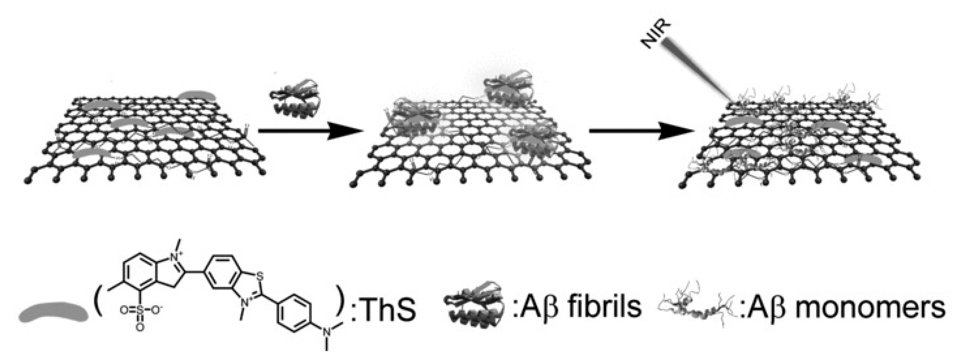
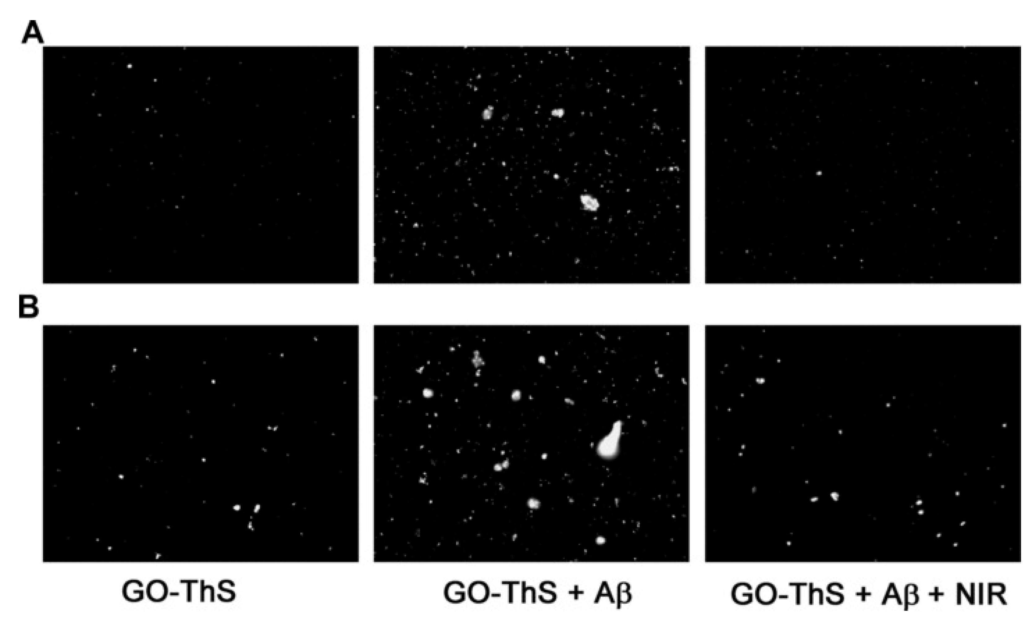
  15. Li, M.; Yang, X.; Ren, J.; Qu, K.; Qu, X. Using Graphene Oxide High Near-Infrared Absorbance for Photothermal Treatment of Alzheimer’s Disease. Adv. Mater. 2012, 24, 1722–1728. [Google Scholar] [CrossRef]
  16. Su, S.; Wang, J.; Qiu, J.; Martinez-Zaguilan, R.; Sennoune, S.R.; Wang, S. In Vitro Study of Transportation of Porphyrin Immobilized Graphene Oxide Through Blood Brain Barrier. Mater. Sci. Eng. C Mater. Biol. Appl. 2020, 107, 110313. [Google Scholar] [CrossRef]
  17. Mendonça, M.C.; Soares, E.S.; de Jesus, M.B.; Ceragioli, H.J.; Ferreira, M.S.; Catharino, R.R.; da Cruz-Höfling, M.A. Reduced Graphene Oxide Induces Transient Blood-Brain Barrier Opening: An In Vivo Study. J. Nanobiotechnol. 2015, 13, 78. [Google Scholar] [CrossRef] [Green Version]
  18. Tadyszak, K.; Wychowaniec, J.K.; Litowczenko, J. Biomedical Applications of Graphene-Based Structures. Nanomaterials 2018, 8, 944. [Google Scholar] [CrossRef] [Green Version]
  19. Costa, R.R.; Alatorre-Meda, M.; Mano, J.F. Drug Nano-Reservoirs Synthesized using Layer-by-Layer Technologies. Biotechnol. Adv. 2015, 33, 1310–1326. [Google Scholar] [CrossRef]
  20. Li, X.; Zhang, G.; Bai, X.; Sun, X.; Wang, X.; Wang, E.; Dai, H. Highly Conducting Graphene Sheets and Langmuir–Blodgett Films. Nat. Nanotechnol. 2008, 3, 538–542. [Google Scholar] [CrossRef] [Green Version]
  21. Jeong, H.; Ranallo, S.; Rossetti, M.; Heo, J.; Shin, J.; Park, K.; Ricci, F.; Hong, J. Electronic Activation of a DNA Nanodevice using a Multilayer Nanofilm. Small 2016, 12, 5572–5578. [Google Scholar] [CrossRef] [PubMed] [Green Version]
  22. Hernandez, Y.; Nicolosi, V.; Lotya, M.; Blighe, F.M.; Sun, Z.; De, S.; McGovern, I.T.; Holland, B.; Byrne, M.; Gun’Ko, Y.K.; et al. High-Yield Production of Graphene by Liquid-Phase Exfoliation of Graphite. Nat. Nanotechnol. 2008, 3, 563–568. [Google Scholar] [CrossRef] [PubMed] [Green Version]
  23. Zhang, X.; Liang, T.; Ma, Q. Layer-by-Layer Assembled Nano-Drug Delivery Systems for Cancer Treatment. Drug Deliv. 2021, 28, 655–669. [Google Scholar] [CrossRef] [PubMed]
  24. Yang, W.J.; Pranantyo, D.; Neoh, K.-G.; Kang, E.-T.; Teo, S.L.-M.; Rittschof, D. Layer-by-Layer Click Deposition of Functional Polymer Coatings for Combating Marine Biofouling. Biomacromolecules 2012, 13, 2769–2780. [Google Scholar] [CrossRef]
  25. Sieber, S.; Siegrist, S.; Schwarz, S.; Porta, F.; Schenk, S.H.; Huwyler, J. Immobilization of Enzymes on PLGA Sub-Micrometer Particles by Crosslinked Layer-by-Layer Deposition. Macromol. Biosci. 2017, 17, 1700015. [Google Scholar] [CrossRef]
  26. Smith, R.; Riollano, M.; Leung, A.; Hammond, P. Layer-by-Layer Platform Technology for Small-Molecule Delivery. Angew. Chem. Int. Ed. 2009, 48, 8974–8977. [Google Scholar] [CrossRef] [Green Version]
  27. Shutava, T.G.; Livanovich, K.S.; Sharamet, A.A. Layer-by-Layer Films of Polysaccharides Modified with Polyethylene Glycol and Dextran. Colloids Surf. B Biointerfaces 2019, 173, 412–420. [Google Scholar] [CrossRef]
  28. Silva, D.P.B.; Miyazaki, C.M.; Mascagni, D.B.T.; Ferreira, M. Layer-by-Layer Films of Gold Nanoparticles and Carbon Nanotubes for Improved Amperometric Detection of Cholesterol. J. Nanosci. Nanotechnol. 2019, 19, 5483–5488. [Google Scholar] [CrossRef]
  29. Blacklock, J.; Mao, G.; Oupický, D.; Möhwald, H. DNA Release Dynamics from Bioreducible Layer-by-Layer Films. Langmuir 2010, 26, 8597–8605. [Google Scholar] [CrossRef] [Green Version]
  30. Hong, C.A.; Son, H.Y.; Nam, Y.S. Layer-by-Layer SiRNA/Poly(L-Lysine) Multilayers on Polydopamine-Coated Surface for Efficient Cell Adhesion and Gene Silencing. Sci. Rep. 2018, 8, 7738. [Google Scholar] [CrossRef] [Green Version]
  31. Srivastava, S.; Kotov, N.A. Composite Layer-by-Layer (LBL) Assembly with Inorganic Nanoparticles and Nanowires. Acc. Chem. Res. 2008, 41, 1831–1841. [Google Scholar] [CrossRef]
  32. Smith, R.C.; Leung, A.; Kim, B.-S.; Hammond, P.T. Hydrophobic Effects in the Critical Destabilization and Release Dynamics of Degradable Multilayer Films. Chem. Mater. 2009, 21, 1108–1115. [Google Scholar] [CrossRef] [Green Version]
  33. Xie, M.; Zhang, F.; Peng, H.; Zhang, Y.; Li, Y.; Xu, Y.; Xie, J. Layer-by-Layer Modification of Magnetic Graphene Oxide by Chitosan and Sodium Alginate with Enhanced Dispersibility for Targeted Drug Delivery and Photothermal Therapy. Colloids Surf. B Biointerfaces 2019, 176, 462–470. [Google Scholar] [CrossRef]
  34. Machado, M.; Silva, G.A.; Bitoque, D.B.; Ferreira, J.; Pinto, L.A.; Morgado, J.; Ferreira, Q. Self-Assembled Multilayer Films for Time-Controlled Ocular Drug Delivery. ACS Appl. Bio Mater. 2019, 2, 4173–4180. [Google Scholar] [CrossRef]
  35. Tanum, J.; Heo, J.; Hong, J. Spontaneous Biomacromolecule Absorption and Long-Term Release by Graphene Oxide. ACS Omega 2018, 3, 5903–5909. [Google Scholar] [CrossRef]
  36. Wang, F.; Li, J.; Tang, X.; Huang, K.; Chen, L. Polyelectrolyte Three Layer Nanoparticles of Chitosan/Dextran Sulfate/Chitosan for Dual Drug Delivery. Colloids Surf. B Biointerfaces 2020, 190, 110925. [Google Scholar] [CrossRef]
  37. Guo, H.; Guo, Q.; Chu, T.; Zhang, X.; Wu, Z.; Yu, D. Glucose-Sensitive Polyelectrolyte Nanocapsules Based on Layer-by-Layer Technique for Protein Drug Delivery. J. Mater. Sci. Mater. Med. 2014, 25, 121–129. [Google Scholar] [CrossRef]
  38. Yang, M.-C.; Tsou, H.-M.; Hsiao, Y.-S.; Cheng, Y.-W.; Liu, C.-C.; Huang, L.-Y.; Peng, X.-Y.; Liu, T.-Y.; Yung, M.-C.; Hsu, C.-C. Electrochemical Polymerization of PEDOT–Graphene Oxide–Heparin Composite Coating for Anti-Fouling and Anti-Clotting of Cardiovascular Stents. Polymers 2019, 11, 1520. [Google Scholar] [CrossRef] [Green Version]
  39. Qi, W.; Xue, Z.; Yuan, W.; Wang, H. Layer-by-Layer Assembled Graphene Oxide Composite Films for Enhanced Mechanical Properties and Fibroblast Cell Affinity. J. Mater. Chem. B 2014, 2, 325–331. [Google Scholar] [CrossRef]
  40. Cao, W.; He, L.; Cao, W.; Huang, X.; Jia, K.; Dai, J. Recent Progress of Graphene Oxide as a Potential Vaccine Carrier and Adjuvant. Acta Biomater. 2020, 112, 14–28. [Google Scholar] [CrossRef]
  41. Mangadlao, J.D.; Santos, C.M.; Felipe, M.J.L.; de Leon, A.C.C.; Rodrigues, D.F.; Advincula, R.C. On the Antibacterial Mechanism of Graphene Oxide (GO) Langmuir–Blodgett Films. Chem. Commun. 2015, 51, 2886–2889. [Google Scholar] [CrossRef]
  42. Sharker, S.M.; Lee, J.E.; Kim, S.H.; Jeong, J.H.; In, I.; Lee, H.; Park, S.Y. PH Triggered in Vivo Photothermal Therapy and Fluorescence Nanoplatform of Cancer Based on Responsive Polymer-Indocyanine Green Integrated Reduced Graphene Oxide. Biomaterials 2015, 61, 229–238. [Google Scholar] [CrossRef]
  43. Gollavelli, G.; Ling, Y.-C. Multi-Functional Graphene as an In Vitro and In Vivo Imaging Probe. Biomaterials 2012, 33, 2532–2545. [Google Scholar] [CrossRef]
  44. Sheng, Z.; Song, L.; Zheng, J.; Hu, D.; He, M.; Zheng, M.; Gao, G.; Gong, P.; Zhang, P.; Ma, Y.; et al. Protein-Assisted Fabrication of Nano-Reduced Graphene Oxide for Combined In Vivo Photoacoustic Imaging and Photothermal Therapy. Biomaterials 2013, 34, 5236–5243. [Google Scholar] [CrossRef]
  45. Shi, S.; Yang, K.; Hong, H.; Valdovinos, H.F.; Nayak, T.R.; Zhang, Y.; Theuer, C.P.; Barnhart, T.E.; Liu, Z.; Cai, W. Tumor Vasculature Targeting and Imaging in Living Mice with Reduced Graphene Oxide. Biomaterials 2013, 34, 3002–3009. [Google Scholar] [CrossRef] [Green Version]
  46. Ahn, E.; Lee, T.; Gu, M.; Park, M.; Min, S.H.; Kim, B.-S. Layer-by-Layer Assembly for Graphene-Based Multilayer Nanocomposites: The Field Manual. Chem. Mater. 2017, 29, 69–79. [Google Scholar] [CrossRef]
  47. Araújo, M. Nanostructured Films for Controlled Release of Drugs for Glaucoma Treatment. Master’s Thesis, Universidade de Lisboa, Lisbon, Portugal, 2016. [Google Scholar]
  48. Morais, H. Nanostructured Films of Graphene for Controlled Ocular Drug Delivery. Master’s Thesis, Instituto Politécnico de Lisboa, Lisbon, Portugal, 2017. [Google Scholar]
  49. Pereira, T. Development of a Liquid Cell to Study the Release of Brimonidine in Real Time. Master’s Thesis, Instituto Pólitécnico de Lisboa, Lisbon, Portugal, 2019. [Google Scholar]
  50. Raposo, M.; Oliveira, O. Adsorption Mechanisms in Layer-by-Layer Films. Braz. J. Phys. 1998, 28. [Google Scholar] [CrossRef] [Green Version]
  51. Han, U.; Seo, Y.; Hong, J. Effect of pH on the Structure and Drug Release Profiles of Layer-by-Layer Assembled Films Containing Polyelectrolyte, Micelles, and Graphene Oxide. Sci. Rep. 2016, 6, 24158. [Google Scholar] [CrossRef]
  52. Saurer, E.M.; Yamanouchi, D.; Liu, B.; Lynn, D.M. Delivery of Plasmid DNA to Vascular Tissue In Vivo using Catheter Balloons Coated with Polyelectrolyte Multilayers. Biomaterials 2011, 32, 610–618. [Google Scholar] [CrossRef] [Green Version]
  53. Zhang, J.; Chen, D.; Li, Y.; Sun, J. Layer-by-Layer Assembled Highly Adhesive Microgel Films. Polymer 2013, 54, 4220–4226. [Google Scholar] [CrossRef]
  54. Wang, L.; Chen, D.; Sun, J. Layer-by-Layer Deposition of Polymeric Microgel Films on Surgical Sutures for Loading and Release of Ibuprofen. Langmuir 2009, 25, 7990–7994. [Google Scholar] [CrossRef] [PubMed]
  55. Sakr, O.S.; Jordan, O.; Borchard, G. Sustained Protein Release from Hydrogel Microparticles using Layer-by-Layer (LbL) Technology. Drug Deliv. 2016, 23, 2747–2755. [Google Scholar] [CrossRef] [PubMed] [Green Version]
  56. Choi, M.; Kim, K.-G.; Heo, J.; Jeong, H.; Kim, S.Y.; Hong, J. Multilayered Graphene Nano-Film for Controlled Protein Delivery by Desired Electro-Stimuli. Sci. Rep. 2015, 5, 17631. [Google Scholar] [CrossRef] [PubMed]
  57. Hong, J.; Shah, N.J.; Drake, A.C.; DeMuth, P.C.; Lee, J.B.; Chen, J.; Hammond, P.T. Graphene Multilayers as Gates for Multi-Week Sequential Release of Proteins from Surfaces. ACS Nano 2012, 6, 81–88. [Google Scholar] [CrossRef] [Green Version]
  58. Liu, W.; Zhang, X.; Zhou, L.; Shang, L.; Su, Z. Reduced Graphene Oxide (rGO) Hybridized Hydrogel as a Near-Infrared (NIR)/pH Dual-Responsive Platform foCr ombined Chemo-Photothermal Therapy. J. Colloid Interface Sci. 2019 536, 160–170. [CrossRef]
  59. McAloney, R.A.; Sinyor, M.; Dudnik, V.; Goh, M.C. Atomic Force Microscopy Studies of Salt Effects on Polyelectrolyte Multilayer Film Morphology. Langmuir 2001, 17, 6655–6663. [Google Scholar] [CrossRef]
  60. Raposo, M.; Ferreira, Q.; Ribeiro, P. A Guide for Atomic Force Microscopy Analysis of Soft Condensed Matter. In Modern Research and Educational Topics in Microscopy; Formatex: Badajoz, Spain, 2007; Volume 1, pp. 758–769. [Google Scholar]
  61. Deng, L.; Li, Q.; Al-Rehili, S.; Omar, H.; Almalik, A.; Alshamsan, A.; Zhang, J.; Khashab, N.M. Hybrid Iron Oxide-Graphene Oxide-Polysaccharides Microcapsule: A Micro-Matryoshka for On-Demand Drug Release and Antitumor Therapy In Vivo. ACS Appl. Mater. Interfaces 2016, 8, 6859–6868. [Google Scholar] [CrossRef] [Green Version]
  62. Wang, K.; Ruan, J.; Song, H.; Zhang, J.; Wo, Y.; Guo, S.; Cui, D. Biocompatibility of Graphene Oxide. Nanoscale Res. Lett. 2010, 6, 8. [Google Scholar] [CrossRef] [Green Version]
  63. Thierry, B.; Kujawa, P.; Tkaczyk, C.; Winnik, F.M.; Bilodeau, L.; Tabrizian, M. Delivery Platform for Hydrophobic Drugs:  Prodrug Approach Combined with Self-Assembled Multilayers. J. Am. Chem. Soc. 2005, 127, 1626–1627. [Google Scholar] [CrossRef]
  64. Chen, D.; Wu, M.; Chen, J.; Zhang, C.; Pan, T.; Zhang, B.; Tian, H.; Chen, X.; Sun, J. Robust, Flexible, and Bioadhesive Free-Standing Films for the Co-Delivery of Antibiotics and Growth Factors. Langmuir 2014, 30, 13898–13906. [Google Scholar] [CrossRef]
  65. Mascagni, D.B.T.; Miyazaki, C.M.; da Cruz, N.C.; de Moraes, M.L.; Riul, A., Jr.; Ferreira, M. Layer-by-Layer Assembly of Functionalized Reduced Graphene Oxide for Direct Electrochemistry and Glucose Detection. Mater. Sci. Eng. C Mater. Biol. Appl. 2016, 68, 739–745. [Google Scholar] [CrossRef] [Green Version]
  66. Ferreira, Q.; Delfino, C.L.; Morgado, J.; Alcácer, L. Bottom-Up Self-Assembled Supramolecular Structures Built by STM at the Solid/Liquid Interface. Materials 2019, 12, 382. [Google Scholar] [CrossRef] [Green Version]
  67. Ferreira, Q.; Bragança, A.M.; Alcácer, L.; Morgado, J. Conductance of Well-Defined Porphyrin Self-Assembled Molecular Wires up to 14 Nm in Length. J. Phys. Chem. C 2014, 118, 7229–7234. [Google Scholar] [CrossRef]
  68. Kurapati, R.; Raichur, A.M. Graphene Oxide Based Multilayer Capsules with Unique Permeability Properties: Facile Encapsulation of Multiple Drugs. Chem. Commun. 2012, 48, 6013–6015. [Google Scholar] [CrossRef]
  69. Kumarasamy, J.; Camarada, M.B.; Venkatraman, D.; Ju, H.; Dey, R.S.; Wen, Y. One-Step Coelectrodeposition-Assisted Layer-by-Layer Assembly of Gold Nanoparticles and Reduced Graphene Oxide and its Self-Healing Three-Dimensional Nanohybrid for an Ultrasensitive DNA Sensor. Nanoscale 2018, 10, 1196–1206. [Google Scholar] [CrossRef]
  70. Agarwal, S.; Zhou, X.; Ye, F.; He, Q.; Chen, G.C.K.; Soo, J.; Boey, F.; Zhang, H.; Chen, P. Interfacing Live Cells with Nanocarbon Substrates. Langmuir 2010, 26, 2244–2247. [Google Scholar] [CrossRef]
  71. Liu, Y.; Han, J.; Pan, H.; Jia, D.; Chen, L.; Yang, X. Oral Delivery of Pingyangmycin by Layer-by-Layer (LbL) Self-Assembly Polyelectrolyte-Grafted Nano Graphene Oxide. J. Nanosci. Nanotechnol. 2019, 19, 2260–2268. [Google Scholar] [CrossRef]
  72. Kavinkumar, T.; Varunkumar, K.; Ravikumar, V.; Manivannan, S. Anticancer Activity of Graphene Oxide-Reduced Graphene Oxide-Silver Nanoparticle Composites. J. Colloid Interface Sci. 2017, 505, 1125–1133. [Google Scholar] [CrossRef]
  73. Angelopoulou, A.; Voulgari, E.; Diamanti, E.K.; Gournis, D.; Avgoustakis, K. Graphene Oxide Stabilized by PLA-PEG Copolymers for the Controlled Delivery of Paclitaxel. Eur. J. Pharm. Biopharm. 2015, 93, 18–26. [Google Scholar] [CrossRef]
  74. Wang, L.H.; Liu, J.Y.; Sui, L.; Zhao, P.H.; Ma, H.D.; Wei, Z.; Wang, Y.L. Folate-Modified Graphene Oxide as the Drug Delivery System to Load Temozolomide. Curr. Pharm. Biotechnol. 2020, 21, 1088–1098. [Google Scholar] [CrossRef]
  75. Barahuie, F.; Saifullah, B.; Dorniani, D.; Fakurazi, S.; Karthivashan, G.; Hussein, M.Z.; Elfghi, F.M. Graphene Oxide as a Nanocarrier for Controlled Release and Targeted Delivery of an Anticancer Active Agent, Chlorogenic Acid. Mater. Sci. Eng. C Mater. Biol. Appl. 2017, 74, 177–185. [Google Scholar] [CrossRef] [PubMed]
  76. Hussein-Al-Ali, S.H.; Abudoleh, S.M.; Hussein, M.Z.; Bullo, S.; Palanisamy, A. Graphene Oxide-Ellagic Acid Nanocomposite as Effective Anticancer and Antimicrobial Agent. IET Nanobiotechnol. 2021, 15, 79–89. [Google Scholar] [CrossRef] [PubMed]
  77. Nasrollahi, F.; Varshosaz, J.; Khodadadi, A.A.; Lim, S.; Jahanian-Najafabadi, A. Targeted Delivery of Docetaxel by Use of Transferrin/Poly(allylamine hydrochloride)-Functionalized Graphene Oxide Nanocarrier. ACS Appl. Mater. Interfaces 2016, 8, 13282–13293. [Google Scholar] [CrossRef] [PubMed]
  78. Gong, P.; Zhao, Q.; Dai, D.; Zhang, S.; Tian, Z.; Sun, L.; Ren, J.; Liu, Z. Functionalized Ultrasmall Fluorinated Graphene with High NIR Absorbance for Controlled Delivery of Mixed Anticancer Drugs. Chem.–A Eur. J. 2017, 23, 17531–17541. [Google Scholar] [CrossRef]
  79. Wang, X.; Hao, L.; Zhang, C.; Chen, J.; Zhang, P. High Efficient Anti-Cancer Drug Delivery Systems using Tea Polyphenols Reduced and Functionalized Graphene Oxide. J. Biomater. Appl. 2017, 31, 1108–1122. [Google Scholar] [CrossRef]
  80. Zhang, B.; Yang, X.; Wang, Y.; Zhai, G. Heparin Modified Graphene Oxide for pH-Sensitive Sustained Release of Doxorubicin Hydrochloride. Mater. Sci. Eng. C 2017, 75, 198–206. [Google Scholar] [CrossRef]
  81. Farnaz, R.; Maryam, S.; Masoumeh, J.; Parvaneh, S. Colloidal HSA-Graphene Oxide Nanosheets for Sustained Release of Oxaliplatin: Preparation, Release Mechanism, Cytotoxicity and Electrochemical Approaches. Colloids Surf. B Biointerfaces 2018, 171, 10–16. [Google Scholar] [CrossRef]
  82. Han, L.; Hao, Y.-N.; Wei, X.; Chen, X.-W.; Shu, Y.; Wang, J.-H. Hollow Copper Sulfide Nanosphere–Doxorubicin/Graphene Oxide Core–Shell Nanocomposite for Photothermo-Chemotherapy. ACS Biomater. Sci. Eng. 2017, 3, 3230–3235. [Google Scholar] [CrossRef]
  83. Patil, T.V.; Patel, D.K.; Dutta, S.D.; Ganguly, K.; Lim, K.-T. Graphene Oxide-Based Stimuli-Responsive Platforms for Biomedical Applications. Molecules 2021, 26, 2797. [Google Scholar] [CrossRef]
  84. Lima-Sousa, R.; de Melo-Diogo, D.; Alves, C.G.; Cabral, C.S.D.; Miguel, S.P.; Mendonça, A.G.; Correia, I.J. Injectable In Situ Forming Thermo-Responsive Graphene Based Hydrogels for Cancer Chemo-Photothermal Therapy and NIR Light-Enhanced Antibacterial Applications. Mater. Sci. Eng. C Mater. Biol. Appl. 2020, 117, 111294. [Google Scholar] [CrossRef]
  85. Dembereldorj, U.; Choi, S.Y.; Ganbold, E.O.; Song, N.W.; Kim, D.; Choo, J.; Lee, S.Y.; Kim, S.; Joo, S.W. Gold Nanorod-Assembled PEGylated Graphene-Oxide Nanocomposites for Photothermal Cancer Therapy. Photochem. Photobiol. 2014, 90, 659–666. [Google Scholar] [CrossRef]
  86. Markovic, Z.M.; Harhaji-Trajkovic, L.M.; Todorovic-Markovic, B.M.; Kepić, D.P.; Arsikin, K.M.; Jovanović, S.P.; Pantovic, A.C.; Dramićanin, M.D.; Trajkovic, V.S. In Vitro Comparison of the Photothermal Anticancer Activity of Graphene Nanoparticles and Carbon Nanotubes. Biomaterials 2011, 32, 1121–1129. [Google Scholar] [CrossRef]
  87. Ma, X.; Tao, H.; Yang, K.; Feng, L.; Cheng, L.; Shi, X.; Li, Y.; Guo, L.; Liu, Z. A Functionalized Graphene Oxide-Iron Oxide Nanocomposite for Magnetically Targeted Drug Delivery, Photothermal Therapy, and Magnetic Resonance Imaging. Nano Res. 2012, 5, 199–212. [Google Scholar] [CrossRef]
  88. Pan, N.; Wang, Y.; Ren, X.; Huang, T.-S.; Kim, I.S. Graphene Oxide as a Polymeric N-halamine Carrier and Release Platform: Highly-Efficient, Sustained-Release Antibacterial Property and Great Storage Stability. Mater. Sci. Eng. C 2019, 103, 109877. [Google Scholar] [CrossRef]
  89. Kumar, G.; Chaudhary, K.; Mogha, N.K.; Kant, A.; Masram, D.T. Extended Release of Metronidazole Drug using Chitosan/Graphene Oxide Bionanocomposite Beads as the Drug Carrier. ACS Omega 2021, 6, 20433–20444. [Google Scholar] [CrossRef]
  90. Tran, L.; Phuong, L.; Hoang Thi, T.T.; Park, K. Graphene Oxide Immobilized Surfaces Facilitate the Sustained Release of Doxycycline for the Prevention of Implant Related Infection. Colloids Surf. B Biointerfaces 2019, 181. [Google Scholar] [CrossRef]
  91. Xiong, S.; Luo, J.; Wang, Q.; Li, Z.; Li, J.; Liu, Q.; Gao, L.; Fang, S.; Li, Y.; Pan, H.; et al. Targeted Graphene Oxide for Drug Delivery as a Therapeutic Nanoplatform Against Parkinson’s Disease. Biomater. Sci. 2021, 9, 1705–1715. [Google Scholar] [CrossRef]
  92. Bramini, M.; Alberini, G.; Colombo, E.; Chiacchiaretta, M.; DiFrancesco, M.L.; Maya-Vetencourt, J.F.; Maragliano, L.; Benfenati, F.; Cesca, F. Interfacing Graphene-Based Materials with Neural Cells. Front. Syst. Neurosci. 2018, 12. [Google Scholar] [CrossRef]
  93. Emadi, F.; Amini, A.; Gholami, A.; Ghasemi, Y. Functionalized Graphene Oxide with Chitosan for Protein Nanocarriers to Protect against Enzymatic Cleavage and Retain Collagenase Activity. Sci Rep. 2017, 7, 42258. [Google Scholar] [CrossRef] [Green Version]
  94. Yang, L.; Wang, F.; Han, H.; Yang, L.; Zhang, G.; Fan, Z. Functionalized Graphene Oxide as a Drug Carrier for Loading Pirfenidone in Treatment of Subarachnoid Hemorrhage. Colloids Surf. B Biointerfaces 2015, 129, 21–29. [Google Scholar] [CrossRef]
  95. Figueroa, T.; Aguayo, C.; Fernández, K. Design and Characterization of Chitosan-Graphene Oxide Nanocomposites for the Delivery of Proanthocyanidins. Int. J. Nanomed. 2020, 15, 1229–1238. [Google Scholar] [CrossRef] [Green Version]
  96. Mahajan, C.R.; Joshi, L.B.; Varma, U.; Naik, J.B.; Chaudhari, V.R.; Mishra, S. Sustainable Drug Delivery of Famotidine using Chitosan-Functionalized Graphene Oxide as Nanocarrier. Glob. Chall. 2019, 3, 1900002. [Google Scholar] [CrossRef] [Green Version]
  97. Rahmanian, N.; Hamishehkar, H.; Dolatabadi, J.E.; Arsalani, N. Nano Graphene Oxide: A Novel Carrier for Oral Delivery of Flavonoids. Colloids Surf. B Biointerfaces 2014, 123, 331–338. [Google Scholar] [CrossRef]
  98. Li, H.; Jia, Y.; Liu, C. Pluronic® F127 Stabilized Reduced Graphene Oxide Hydrogel for Transdermal Delivery of Ondansetron: Ex Vivo and Animal Studies. Colloids Surf. B Biointerfaces 2020, 195, 111259. [Google Scholar] [CrossRef]
  99. Zhou, M.; Lozano, N.; Wychowaniec, J.K.; Hodgkinson, T.; Richardson, S.M.; Kostarelos, K.; Hoyland, J.A. Graphene Oxide: A growth Factor Delivery Carrier to Enhance Chondrogenic Differentiation of Human Mesenchymal Stem Cells in 3D Hydrogels. Acta Biomater 2019, 96, 271–280. [Google Scholar] [CrossRef]
  100. Yang, C.; Li, T. Transdermal Delivery of Flurbiprofen from Polyoxypropylene-Polyoxyethylene Block Copolymer Stabilized Reduced Graphene Oxide to Manage Pain in Spondylitis: In Vitro and In Vivo Studies. Eur. J. Pharm. Sci. Off. J. Eur. Fed. Pharm. Sci. 2021, 165, 105929. [Google Scholar] [CrossRef]
  101. Zhang, Z.; Wang, X.; Li, P.; Bai, M.; Qi, W. Transdermal Delivery of Buprenorphine from Reduced Graphene Oxide Laden Hydrogel to Treat Osteoarthritis. J. Biomater. Sci. Polym. Ed. 2021, 32, 874–885. [Google Scholar] [CrossRef]
  102. Li, W.; Zhang, G.; Wei, X. Lidocaine-Loaded Reduced Graphene Oxide Hydrogel for Prolongation of Effects of Local Anesthesia: In Vitro and In Vivo Analyses. J. Biomater. Appl. 2021, 35, 1034–1042. [Google Scholar] [CrossRef]
  103. Luo, S.; Jin, S.; Yang, T.; Wu, B.; Xu, C.; Luo, L.; Chen, Y. Sustained Release of Tulobuterol from Graphene Oxide Laden Hydrogel to Manage Asthma. J. Biomater. Sci. Polym. Ed. 2021, 32, 524–535. [Google Scholar] [CrossRef]
  104. Choi, M.; Chung, J.-H.; Cho, Y.; Hong, B.Y.; Hong, J. Nano-Film Modification of Collagen Hydrogels for Controlled Growth Factor Release. Chem. Eng. Sci. 2015, 137, 626–630. [Google Scholar] [CrossRef]
  105. Maulvi, F.A.; Soni, P.D.; Patel, P.J.; Desai, A.R.; Desai, D.T.; Shukla, M.R.; Shah, S.A.; Shah, D.O.; Willcox, M.D.P. Controlled Bimatoprost Release from Graphene Oxide Laden Contact Lenses: In Vitro and In Vivo Studies. Colloids Surf. B Biointerfaces 2021, 208, 112096. [Google Scholar] [CrossRef] [PubMed]
  106. Laurenti, M.; Lamberti, A.; Genchi, G.G.; Roppolo, I.; Canavese, G.; Vitale-Brovarone, C.; Ciofani, G.; Cauda, V. Graphene Oxide Finely Tunes the Bioactivity and Drug Delivery of Mesoporous ZnO Scaffolds. ACS Appl. Mater. Interfaces 2019, 11, 449–456. [Google Scholar] [CrossRef] [PubMed]
  107. Joo, J.; Kwon, E.J.; Kang, J.; Skalak, M.; Anglin, E.J.; Mann, A.P.; Ruoslahti, E.; Bhatia, S.N.; Sailor, M.J. Porous Silicon–Graphene Oxide Core–Shell Nanoparticles for Targeted Delivery of siRNA to the Injured Brain. Nanoscale Horiz. 2016, 1, 407–414. [Google Scholar] [CrossRef] [PubMed]
  108. Borandeh, S.; Alimardani, V.; Abolmaali, S.S.; Seppälä, J. Graphene Family Nanomaterials in Ocular Applications: Physicochemical Properties and Toxicity. Chem. Res. Toxicol. 2021, 34, 1386–1402. [Google Scholar] [CrossRef] [PubMed]
  109. Hu, W.; Peng, C.; Lv, M.; Li, X.; Zhang, Y.; Chen, N.; Fan, C.; Huang, Q. Protein Corona-Mediated Mitigation of Cytotoxicity of Graphene Oxide. ACS Nano 2011, 5, 3693–3700. [Google Scholar] [CrossRef] [PubMed]
  110. Bianco, A. Graphene: Safe or Toxic? The Two Faces of the Medal. Angew. Chem. Int. Ed. 2013, 52, 4986–4997. [Google Scholar] [CrossRef] [PubMed]
  111. Moura, D.; Caridade, S.G.; Sousa, M.P.; Cunha, E.; Rocha, H.C.; Mano, J.F.; Paiva, M.C.; Alves, N.M. High Performance Free-Standing Films by Layer-by-Layer Assembly of Graphene Flakes and Ribbons with Natural Polymers. J. Mater. Chem. B 2016, 4, 7718–7730. [Google Scholar] [CrossRef] [Green Version]
  112. Liao, K.-H.; Lin, Y.-S.; Macosko, C.W.; Haynes, C.L. Cytotoxicity of Graphene Oxide and Graphene in Human Erythrocytes and Skin Fibroblasts. ACS Appl. Mater. Interfaces 2011, 3, 2607–2615. [Google Scholar] [CrossRef]
  113. Liao, C.; Li, Y.; Tjong, S. Graphene Nanomaterials: Synthesis, Biocompatibility, and Cytotoxicity. Int. J. Mol. Sci. 2018, 19, 3564. [Google Scholar] [CrossRef] [Green Version]
  114. Zhang, D.; Zhang, Z.; Liu, Y.; Chu, M.; Yang, C.; Li, W.; Shao, Y.; Yue, Y.; Xu, R. The Short- and Long-Term Effects of Orally Administered High-Dose Reduced Graphene Oxide Nanosheets on Mouse Behaviors. Biomaterials 2015, 68, 100–113. [Google Scholar] [CrossRef]
  115. Fu, C.; Liu, T.; Li, L.; Liu, H.; Liang, Q.; Meng, X. Effects of Graphene Oxide on the Development of Offspring Mice in Lactation Period. Biomaterials 2015, 40, 23–31. [Google Scholar] [CrossRef]
  116. Chen, Y.; Hu, X.; Sun, J.; Zhou, Q. Specific Nanotoxicity of Graphene Oxide During Zebrafish Embryogenesis. Nanotoxicology 2016, 10, 42–52. [Google Scholar] [CrossRef]
  117. Dasmahapatra, A.K.; Dasari, T.P.S.; Tchounwou, P.B. Graphene-Based Nanomaterials Toxicity in Fish. Rev. Environ. Contam. Toxicol. 2019, 247, 1–58. [Google Scholar] [CrossRef]
  118. Guo, Q.; Yang, Y.; Zhao, L.; Chen, J.; Duan, G.; Yang, Z.; Zhou, R. Graphene Oxide Toxicity in W1118 Flies. Sci. Total Environ. 2022, 805, 150302. [Google Scholar] [CrossRef]
  119. Rhazouani, A.; Gamrani, H.; El Achaby, M.; Aziz, K.; Gebrati, L.; Uddin, M.S.; Aziz, F. Synthesis and Toxicity of Graphene Oxide Nanoparticles: A Literature Review of In Vitro and In Vivo Studies. BioMed. Res. Int. 2021, 2021, 1–19. [Google Scholar] [CrossRef]
  120. Ou, L.; Song, B.; Liang, H.; Liu, J.; Feng, X.; Deng, B.; Sun, T.; Shao, L. Toxicity of Graphene-Family Nanoparticles: A General Review of the Origins and Mechanisms. Part. Fibre Toxicol. 2016, 13, 57. [Google Scholar] [CrossRef] [Green Version]
  121. Li, J.; Zhou, C.; Luo, C.; Qian, B.; Liu, S.; Zeng, Y.; Hou, J.; Deng, B.; Sun, Y.; Yang, J.; et al. N-acetyl Cysteine-Loaded Graphene Oxide-Collagen Hybrid Membrane for Scarless Wound Healing. Theranostics 2019, 9, 5839–5853. [Google Scholar] [CrossRef]
  122. Rehman, S.R.u.; Augustine, R.; Zahid, A.A.; Ahmed, R.; Hasan, A. Graphene Oxide Loaded Hydrogel for Enhanced Wound Healing in Diabetic Patients. In Proceedings of the 2019 41st Annual International Conference of the IEEE Engineering in Medicine and Biology Society (EMBC), Berlin, Germany, 23–27 July 2019; pp. 3943–3946. [Google Scholar] [CrossRef]
  123. Liang, Y.; Zhao, X.; Hu, T.; Chen, B.; Yin, Z.; Ma, P.X.; Guo, B. Adhesive Hemostatic Conducting Injectable Composite Hydrogels with Sustained Drug Release and Photothermal Antibacterial Activity to Promote Full-Thickness Skin Regeneration During Wound Healing. Small 2019, 15, 1900046. [Google Scholar] [CrossRef]
  124. Vincent, M.; de Lázaro, I.; Kostarelos, K. Graphene Materials as 2D Non-Viral Gene Transfer Vector Platforms. Gene Ther. 2017, 24, 123–132. [Google Scholar] [CrossRef] [Green Version]
  125. Hsieh, T.Y.; Huang, W.C.; Kang, Y.D.; Chu, C.Y.; Liao, W.L.; Chen, Y.Y.; Chen, S.Y. Neurotensin-Conjugated Reduced Graphene Oxide with Multi-Stage Near-Infrared-Triggered Synergic Targeted Neuron Gene Transfection In Vitro and In Vivo for Neurodegenerative Disease Therapy. Adv. Healthc. Mater. 2016, 5, 3016–3026. [Google Scholar] [CrossRef]
  126. Jalani, G.; Jeyachandran, D.; Bertram Church, R.; Cerruti, M. Graphene Oxide-Stabilized Perfluorocarbon Emulsions for Controlled Oxygen Delivery. Nanoscale 2017, 9, 10161–10166. [Google Scholar] [CrossRef]
  127. Castilho, C.J.; Li, D.; Liu, M.; Liu, Y.; Gao, H.; Hurt, R.H. Mosquito Bite Prevention through Graphene Barrier Layers. Proc. Natl. Acad. Sci. USA 2019, 116, 18304–18309. [Google Scholar] [CrossRef] [Green Version]
  128. Sha, J.; Gao, Y.; Wu, T.; Chen, X.; Cordie, T.; Zhao, H.; Xie, L.; Ma, Y.; Turng, L.-s. Biocompatible Graphene Nanosheets Grafted with Poly(2-Hydroxyethyl Methacrylate) Brushes via Surface-Initiated ARGET ATRP. RSC Adv. 2016, 6, 35641–35647. [Google Scholar] [CrossRef]
  129. Catt, K.; Li, H.; Cui, X.T. Poly (3,4-Ethylenedioxythiophene) Graphene Oxide Composite Coatings for Controlling Magnesium Implant Corrosion. Acta Biomater. 2017, 48, 530–540. [Google Scholar] [CrossRef] [Green Version]
  130. Andreeva, T.D.; Stoichev, S.; Taneva, S.G.; Krastev, R. Hybrid Graphene Oxide/Polysaccharide Nanocomposites with Controllable Surface Properties and Biocompatibility. Carbohydr. Polym. 2018, 181, 78–85. [Google Scholar] [CrossRef]
  131. Asgar, H.; Deen, K.M.; Rahman, Z.U.; Shah, U.H.; Raza, M.A.; Haider, W. Functionalized Graphene Oxide Coating on Ti6Al4V Alloy for Improved Biocompatibility and Corrosion Resistance. Mater. Sci. Eng. C 2019, 94, 920–928. [Google Scholar] [CrossRef]
  132. Raucci, M.G.; Giugliano, D.; Longo, A.; Zeppetelli, S.; Carotenuto, G.; Ambrosio, L. Comparative Facile Methods for Preparing Graphene Oxide–Hydroxyapatite for Bone Tissue Engineering. J. Tissue Eng. Regen. Med. 2017, 11, 2204–2216. [Google Scholar] [CrossRef]
  133. Guo, B.; Feng, X.; Wang, Y.; Wang, X.; He, Y. Biomimetic and Immunomodulatory Baicalin-Loaded Graphene Oxide-Demineralized Bone Matrix Scaffold for In Vivo Bone Regeneration. J. Mater. Chem. B 2021, 9, 9720–9733. [Google Scholar] [CrossRef]
  134. Arnold, A.M.; Holt, B.D.; Daneshmandi, L.; Laurencin, C.T.; Sydlik, S.A. Phosphate Graphene as an Intrinsically Osteoinductive Scaffold for Stem Cell-Driven Bone Regeneration. Proc. Natl. Acad. Sci. USA 2019, 116, 4855–4860. [Google Scholar] [CrossRef] [Green Version]
  135. Li, J.; Zheng, L.; Zeng, L.; Zhang, Y.; Jiang, L.; Song, J. RGD Peptide-Grafted Graphene Oxide as a New Biomimetic Nanointerface for Impedance-Monitoring Cell Behaviors. J. Nanomater. 2016, 2016, 1–12. [Google Scholar] [CrossRef] [Green Version]
  136. Lai, P.X.; Chen, C.W.; Wei, S.C.; Lin, T.Y.; Jian, H.J.; Lai, I.P.; Mao, J.Y.; Hsu, P.H.; Lin, H.J.; Tzou, W.S.; et al. Ultrastrong Trapping of VEGF by Graphene Oxide: Anti-Angiogenesis Application. Biomaterials 2016, 109, 12–22. [Google Scholar] [CrossRef] [PubMed]
  137. Huang, C.; Zhang, X.; Li, Y.; Yang, X. Hyaluronic Acid and Graphene Oxide Loaded Silicon Contact Lens for Corneal Epithelial Healing. J. Biomater. Sci. Polym. Ed. 2021, 32, 372–384. [Google Scholar] [CrossRef] [PubMed]
  138. Hu, F.; Lu, H.; Ye, Z.; Zhang, S.; Wang, W.; Gao, L. Slow-Release Lubrication of Artificial Joints using Self-Healing Polyvinyl Alcohol/Polyethylene Glycol/ Graphene Oxide Hydrogel. J. Mech. Behav. Biomed. Mater. 2021, 124, 104807. [Google Scholar] [CrossRef] [PubMed]
  139. Taniselass, S.; Arshad, M.K.M.; Gopinath, S.C.B. Graphene-Based Electrochemical Biosensors for Monitoring Noncommunicable Disease Biomarkers. Biosens. Bioelectron. 2019, 130, 276–292. [Google Scholar] [CrossRef] [PubMed]
  140. Eckhart, K.E.; Holt, B.D.; Laurencin, M.G.; Sydlik, S.A. Covalent Conjugation of Bioactive Peptides to Graphene Oxide for Biomedical Applications. Biomater. Sci. 2019, 7, 3876–3885. [Google Scholar] [CrossRef] [PubMed]
  141. Miyazaki, C.M.; Mishra, R.; Kinahan, D.J.; Ferreira, M.; Ducrée, J. Polyethylene Imine/Graphene Oxide Layer-by-Layer Surface Functionalization for Significantly Improved Limit of Detection and Binding Kinetics of Immunoassays on Acrylate Surfaces. Colloids Surf. B Biointerfaces 2017, 158, 167–174. [Google Scholar] [CrossRef] [PubMed]
  142. Silva, F.; Costa-Almeida, R.; Timochenco, L.; Amaral, S.I.; Pinto, S.; Gonçalves, I.C.; Fernandes, J.R.; Magalhães, F.D.; Sarmento, B.; Pinto, A.M. Graphene Oxide Topical Administration: Skin Permeability Studies. Materials 2021, 14, 2810. [Google Scholar] [CrossRef]
  143. Wu, P.-C.; Chen, H.-H.; Chen, S.-Y.; Wang, W.-L.; Yang, K.-L.; Huang, C.-H.; Kao, H.-F.; Chang, J.-C.; Hsu, C.-L.L.; Wang, J.-Y.; et al. Graphene Oxide Conjugated with Polymers: A Study of Culture Condition to Determine Whether a Bacterial Growth Stimulant or an Antimicrobial Agent? J. Nanobiotechnol. 2018, 16, 1. [Google Scholar] [CrossRef] [Green Version]
  144. Katuwavila, N.P.; Amarasekara, Y.; Jayaweera, V.; Rajaphaksha, C.; Gunasekara, C.; Perera, I.C.; Amaratunga, G.A.J.; Weerasinghe, L. Graphene Oxide-Based Nanocomposite for Sustained Release of Cephalexin. J. Pharm. Sci. 2020, 109, 1130–1135. [Google Scholar] [CrossRef]
  145. Priyadarsini, S.; Mohanty, S.; Mukherjee, S.; Basu, S.; Mishra, M. Graphene and Graphene Oxide as Nanomaterials for Medicine and Biology Application. J. Nanostruct. Chem. 2018, 8, 123–137. [Google Scholar] [CrossRef] [Green Version]
Figure 1. Graphical representation of the molecular structures of different graphene-based materials. Republished from Ref. [18].
Nanomaterials 12 01149 g001
Figure 2. Schematics of the different LbL assembling methods. (A)—Langmuir–Blodgett assembly; (B)—microfluidics; (C)—drop coating; (D)—perfusion, (E)—dip coating; (F)—spray coating; (G)—spin coating. Figure created with BioRender.com.
Nanomaterials 12 01149 g002
Figure 3. Schematic representation of rGO hollow capsules assembly. Republished with permission from Ref. [3]. Copyright 2022 American Chemical Society.
Nanomaterials 12 01149 g003
Figure 4. Schematic representation of the GO-ThS system used for AD treatment. Upon NIR irradiation, the GO-ThS system can dissolve the Aβ amyloid deposits. Republished with permission from Ref. [15]. Copyright 2022 John Wiley and Sons.
Nanomaterials 12 01149 g004
Figure 5. Fluorescence images of Aβ incubated with GO-ThS in Tris buffer (A) and mice CSF (B) (200x magnification) demonstrate that upon NIR irradiation GO-ThS can effectively dissociate Aβ amyloid deposits. Republished with permission from Ref. [15]. Copyright 2022 John Wiley and Sons.
Nanomaterials 12 01149 g005
Table 1. Compilation of different techniques used to characterize the assembly of LbL films which contain GO.
TechniquePurpose
UV-Visible/Fluorescence Spectroscopy GO structure [58]
Atomic Force Microscopy (AFM)Surface morphology and roughness [21,32,34,35,57,58,59,60]
Scanning Electron Microscopy (SEM)Surface morphology [21,35,36,54,55,56,57,58]
Transmission Electron Microscopy (TEM)Surface morphology [36,37,61]
GO structure [62]
Dynamic Light Scattering (DLS) methodZeta potential and particle size and distribution [36,37,55,61]
Quartz Crystal Microbalance (QCM)Layer adsorption [21,34,35,54,63]
ProfilometryLayer thickness [56,64]
Raman Spectroscopy (RS)Layer deposition [61]
GO structure [58]
SQUID—Field-dependent magnetization measurementMagnetism measurement [61]
Fourier Transform Infrared Spectroscopy (FTIR)GO structure [58]
Surface Plasmon Resonance (SPR) Film growth [65]
Scanning Tunneling Microscopy (STM)Characterization at molecular scale [66,67]
X-ray DiffractionGO structure [33,58,68,69]
Interlayer space [65]
X-ray Photoelectron Spectroscopy (XPS)Film chemical characteristics [69,70]
Publisher’s Note: MDPI stays neutral with regard to jurisdictional claims in published maps and institutional affiliations.

