Article

Electrochemically Fabricated Surface-Mesostructured CuNi Bimetallic Catalysts for Hydrogen Production in Alkaline Media

1
School of Materials Science and Engineering, Northeastern University, Shenyang 110819, China
2
Engineering Research Center of Continuous Extrusion, Ministry of Education, Dalian Jiaotong University, Dalian 116028, China
3
Center of Advanced Lubrication and Seal Materials, State Key Laboratory of Solidification Processing, Northwestern Polytechnical University, Xi’an 710072, China
4
Departament de Física, Facultat de Ciències, Universitat Autònoma de Barcelona, E-08193 Bellaterra, Cerdanyola del Vallès, Spain
5
Institució Catalana de Recerca i Estudis Avançats (ICREA), Pg. Lluís Companys 23, E-08010 Barcelona, Spain
*
Authors to whom correspondence should be addressed.
These authors contributed equally to this work.
Nanomaterials 2022, 12(1), 118; https://doi.org/10.3390/nano12010118
Submission received: 3 December 2021 / Revised: 22 December 2021 / Accepted: 27 December 2021 / Published: 30 December 2021
(This article belongs to the Section Energy and Catalysis)

Abstract

:
Ni-based bimetallic films with 20 at.% and 45 at.% Cu and mesostructured surfaces were prepared by electrodeposition from an aqueous solution containing micelles of P123 triblock copolymer serving as a structure-directing agent. The pH value of the electrolytic solution had a key effect on both the resulting Cu/Ni ratio and the surface topology. The catalytic activity of the CuNi films toward hydrogen evolution reaction was investigated by cyclic voltammetry (CV) in 1 M KOH electrolyte at room temperature. The Cu45Ni55 film showed the highest activity (even higher than that of a non-mesostructured pure Ni film), which was attributed to the Ni content at the utmost surface, as demonstrated by CV studies, as well as the presence of a highly corrugated surface.

1. Introduction

Bimetallic CuNi materials show significantly different properties compared to monometallic Ni [1,2]. For example, mesostructured CuNi films exhibit intrinsic magnetoelectric effects, in which a drastic reduction in coercivity can be observed under the application of voltage across an electric double layer [3]. The addition of Cu to Ni also promotes the catalytic activity and selectivity of Ni toward a variety of reactions. In carbon dioxide hydrogenation, Cu favors CO2 adsorption and CO production, which determines the CO2 conversion efficiency [4]. The addition of a small amount of Cu to Ni/Al2O3 increases the selectivity of 1,3-butadiene hydrogenation toward 1-butene [5]. An enhancement of the hydrogen evolution reaction (HER) of CuNi films with respect to their single counterparts has also been reported [6]. In general, the improvement observed in bimetallic systems tested at HER is due to a variety of reasons: preferential segregation of the most active metal to the surface, changes in the electronic properties, formation of large electrochemically active surface areas, and combinations thereof [7,8,9,10].
An increase in the electrocatalytic activity of CuNi bimetallic materials has been achieved by either modifying their structure and/or surface composition. These features, in turn, are determined by the synthesis parameters [11,12,13]. Wet chemical routes for the preparation of electrocatalysts commonly involve diffusion, adsorption, nucleation, particle growth, and phase transformation. Each step needs to be well understood and controlled to efficiently improve the performance of the resulting heterogeneous catalysts. Interestingly, Cu and Ni possess a similar crystal structure with lattice constants of 3.61 Å and 3.52 Å, respectively, but dissimilar electronic configuration. This is because Cu has a completely filled 3d band, while Ni has partially filled 3d orbitals with a high density of states at the Fermi level. As a result, Ni typically shows higher chemical reactivity than Cu. Indeed, surface enrichment in Ni has been shown to increase the catalytic performance of CuNi materials [14]. Yet, such enrichment is not straightforward because Cu has lower surface energy and slightly larger metallic radius; therefore, it is prone to occupy surface sites [15,16]. It is worth noting that the kinetics associated with material preparation plays a key role in determining the final microstructure. For example, a core–shell structure with an Ni-rich surface was obtained by solvated metal atom impregnation [17]. Electrodeposition has proven successful in synthesizing bimetallic CuNi with tunable microstructures and surface composition [18,19,20,21,22]. Concerning electrodeposition, the difference in both the equilibrium potentials and the nucleation rates for Cu and Ni is quite large, enabling the on-demand deposition of porous, multilayered, or fully dense microstructures with Ni-rich surfaces.
The electrosynthesis of films with surface roughness on the macro- and mesoscales has been intensively investigated in the past years as these materials might show increased activity toward HER by virtue of their higher density of active sites. In this context, macroporous bimetallic CuNi films with Ni contents ranging from 15% to 35% were successfully prepared using the hydrogen bubble template-assisted electrodeposition approach [18]. Surprisingly, CuNi films with the lowest Ni content showed the highest specific activity. In HER, the composition of the utmost surface in contact with the electrolyte is crucial as the surface is involved in the very first step of the interaction with water molecules and, furthermore, with the hydrogen gas desorption step [23,24]. The introduction of Cu is thought to enhance the HER activity of Ni by facilitating molecular hydrogen release from traps (by decreasing the hydrogen binding energy) [18]. In continuation to our previous work on macroporous CuNi films [18], films of the same system are herein prepared by aqueous electrodeposition using poly(ethylene oxide)-block-poly(propylene oxide)-block-poly(ethylene oxide) (PEO–PPO–PEO) triblock copolymer (P123) as a structure-directing agent [25,26,27,28]. The large surface area endowed by the resulting mesostructured surfaces makes the system a suitable candidate as an electrocatalyst for HER in alkaline conditions.
The goal of this paper is to study the HER activity of bimetallic CuNi films prepared by electrodeposition from an aqueous solution containing the P123 triblock copolymer. The electrochemical surface area (ECSA) and surface composition of the electrocatalysts were evaluated by electrochemical means. Furthermore, we demonstrate for this particular alloy that, a higher amount of Ni at the surface leads to better HER performance, thereby confirming that the electrocatalytic activity is well correlated with ECSA and surface composition.

2. Materials and Methods

2.1. Synthesis of Cu20Ni80 and Cu45Ni55 Films

Cu20Ni80 and Cu45Ni55 films with mesoscale surface roughness (denoted as ‘meso’) were electrodeposited from a solution containing 0.008 g/mL P123, 0.2 M Ni(OCOCH3)2∙4H2O, 0.02 M CuSO4∙5H2O, 0.5 M NaOCOCH3, 0.2 M boric acid, and 0.4 mg/mL saccharine. The deposition was performed galvanostatically at j = −100 mA·cm−2 for 150 s at room temperature. The pH of the solution was left at its unadjusted value for the electrodeposition of Cu20Ni80 (pH = 6.25), while it was brought to 4 with the addition of sulfuric acid for Cu45Ni55 deposition. Si/Ti (25 nm)/Au (125 nm) substrates (with a working area of 0.25 cm2) were used as the cathode. A platinum wire served as the counter electrode, and a double junction Ag|AgCl 3 M KCl electrode (E = + 0.210 V versus standard hydrogen electrode (SHE)) was utilized as the reference electrode. N2 was bubbled through the solution to get rid of oxygen before each deposition. Following deposition, the films were carefully rinsed with Milli-Q water and placed in isopropanol for 24 h in order to remove P123 remnants. All the chemicals were purchased from Sigma-Aldrich and used without further purification. CuNi films of same composition and pure Ni films, both without mesoscale surface roughness (denoted as ‘plain’ films), were prepared from different electrolytes, as reported elsewhere [29].

2.2. Electrocatalytic Activity toward HER

The electrochemical activity of the CuNi films toward HER was investigated in the same setup used for electrodeposition. The Pt counter electrode was in the same compartment as the working and reference electrodes. Both plain and surface-mesostructured films previously deposited on Si/Ti/Au substrates were used as working electrodes. Cyclic voltammetry (CV) curves were recorded in de-aerated 1 M KOH solution by cycling the potential between –1.6 V and +0.2 V at 50 mV·s−1 versus Ag|AgCl. The onset potential and current density values of Cu20Ni80 and Cu45Ni55 surface-mesostructured films were compared with those of pure Ni, Cu20Ni80, and Cu45Ni55 plain films. All the measurements were carried out at 25 °C. The measured potentials vs. Ag|AgCl were converted to the reversible hydrogen electrode (RHE) scale according to
ERHE = EAg|AgCl + 0.059 pH + E0Ag|AgCl,
where ERHE is the converted potential vs. RHE, E0Ag|AgCl is 0.210 V at 25 °C, and EAg|AgCl is the experimentally measured potential against the Ag|AgCl reference electrode. iR compensation was performed after determination of the instrumentation resistance by electrochemical impedance spectroscopy (EIS), using a potential amplitude of 10 mV and a frequency range between 100 kHz and 10 mHz. Galvanostatic long-term stability tests were performed at −10 mA·cm−2 for 24 h.
ECSA values for Cu and Ni were estimated following the procedure described in [14]. Specifically, first the peak areas for Cu2O and α-Ni(OH)2 formation were gleaned from CV, and then the mass loadings for Ni and Cu were determined. The latter were obtained from the actual mass of the deposited films using a scale with an accuracy of 0.01 mg (Mettler Toledo MS205DU) and considering the relative amounts of Cu and Ni gathered from EDX analyses. Accordingly, ECSA was estimated as
ECSA = Q r m × C ,
where Qr is the charge determined by CV, m is the Ni or Cu mass loading, and C is the charge of a full monolayer coverage of Cu2O (544 μC·cm−2) or α-Ni(OH)2 (514 μC·cm−2). This method of ECSA determination, referred to as ‘Alpha’ [30], was first devised by Machado et al. for bulk Ni [31], and variations of these method have been recently applied to Ni foams [32]. According to Baranova and coworkers [30], the Alpha method provides a precise estimation of the Ni-related ECSA unless the original electrode is made of Ni oxides. Four replicates of each type, namely, Cu20Ni80 and Cu45Ni55 surface-mesostructured films, were electrodeposited for element-selective ECSA determination. Values are given as the mean value ± standard deviation.
Finally, the turnover frequency (TOF), which gives the number of H2 molecules evolved per second per active site, was determined at −0.25 V vs. RHE following the approach described in [33]. The active sites per real surface area were obtained on the basis of crystallographic considerations of the CuNi unit cell [26].

2.3. Structural Characterization

Scanning electron microscopy (SEM) images and energy-dispersive X-ray spectroscopy (EDX) analyses were performed on a Zeiss Merlin microscope operated at 3 kV and 20 kV, respectively. θ/2θ X-ray diffraction (XRD) patterns were recorded on a Philips X’Pert diffractometer with a pixel1D detector in the 42–54° 2θ range (step size = 0.026°, total time = 1200 s) using Cu Kα radiation (λ = 0.154178 nm). Surface adsorption and desorption of N2 were studied using a Micromeritics ASAP 2000 particle analyzer. The pore size distribution was calculated by nonlocal density functional theory. Ten replicates of Cu20Ni80 and Cu45Ni55 surface-mesostructured films were synthesized and detached from the substrate in order to accumulate a sufficient amount of material. Prior to recording the adsorption-desorption isotherms, the free-standing layers were degassed at 200 °C for 4 h.

3. Results and Discussion

In the present study, Cu20Ni80 and Cu45Ni55 films with mesostructured surfaces were deposited on an Au surface from an aqueous solution containing the metal salts and the nonionic amphiphilic P123 surfactant. In contrast to the lyotropic liquid crystal (LLC) method, which requires high surfactant concentrations [25], here, a diluted surfactant solution was utilized instead [26]. This approach is referred to as micelle-assisted electrodeposition. In the plating solution, dissolved Cu and Ni ions are coordinated by water molecules forming metal–aqua complexes which adsorb onto the external ethylene oxide (EO) groups of the P123 micelles. During the electrodeposition process, the metal ion species move toward the cathode via coulombic attraction together with the micelles under the applied potential/current density. Hence, the surface of the working electrode acts as a solid–liquid interface, where the metallic films are deposited and patterned by the surfactant–inorganic aggregates. Indeed, the P123 block copolymer has the role of a structure-directing agent (Figure 1). The formation of micelles in solution is key for the development of surfaces with roughness on the mesoscale. The presence of micelles in the electrolyte was proven via the so-called Tyndall effect (Figure 1). Accordingly, the red light pointing to the volumetric flask containing the electrolyte was scattered and reflected by the individual suspension micelles, making the beam visible.
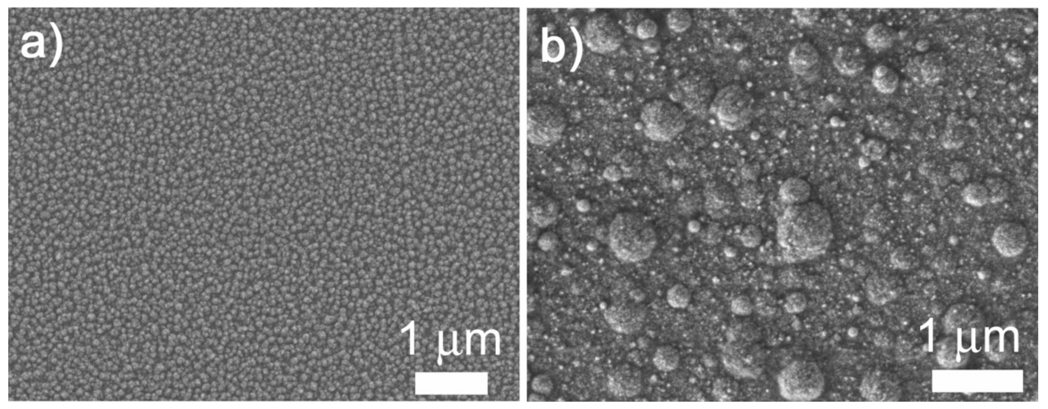
The surface-mesostructured films prepared by micelle-assisted electrodeposition were analyzed by SEM, and the corresponding images are depicted in Figure 2. When the deposition was performed from the electrolyte at pH = 6.25, the resulting CuNi deposits showed globular grains with a fluffy appearance (see Figure 2a). At a higher magnification (Figure 2b), a sort of hierarchical architecture consisting of aggregates of nanoparticles was unveiled. The surface nanovoids result from the interstices left by the aggregation of these nanoparticles (see white arrows in Figure 2b), whose size ranges from 5 to 10 nm. A distinct morphology was observed when deposition was performed at a lower bath pH. In particular, a striated pattern was seen when the pH value of the electrolyte was set to 4 (Figure 2c,d). The observed drastic change in morphology was probably due to an enhanced interaction between metal ions and P123 micelles at pH = 4. Indeed, the morphology is dependent on the type and strength of the interaction (electrostatic, hydrogen bonding, etc.) between the metal ions and surfactant assemblies [34]. In fact, attempts to produce surface-mesostructured CuNi films with lower amounts of Ni by further decreasing the pH of the electrolyte failed because mesoscale roughness did not develop as such. The corresponding EDX spectra (Figure 3) confirm that the films were composed of Cu and Ni with a mean atomic composition of Cu20Ni80 and Cu45Ni55, respectively. As shown in Figure 4, when the pH value was set at 3 or 2, although the resulting films were rough, no signs of mesostructuration were noted. The presence of Cu ions in the electrolyte was key in promoting the occurrence of surfaces with mesoscale features. Specifically, when the deposition was carried out in the absence of the Cu salt, while keeping the remaining conditions unchanged, the resulting pure Ni film showed a more compact, bumpy, fine-grained morphology (Figure 5). This observation is in good accordance with the results previously reported by our group, i.e., the presence of Cu ions in the electrolyte, and their discharge is somewhat related to the successful formation of mesostructured films, at least for the current electrolyte formulation and deposition conditions used here [26].
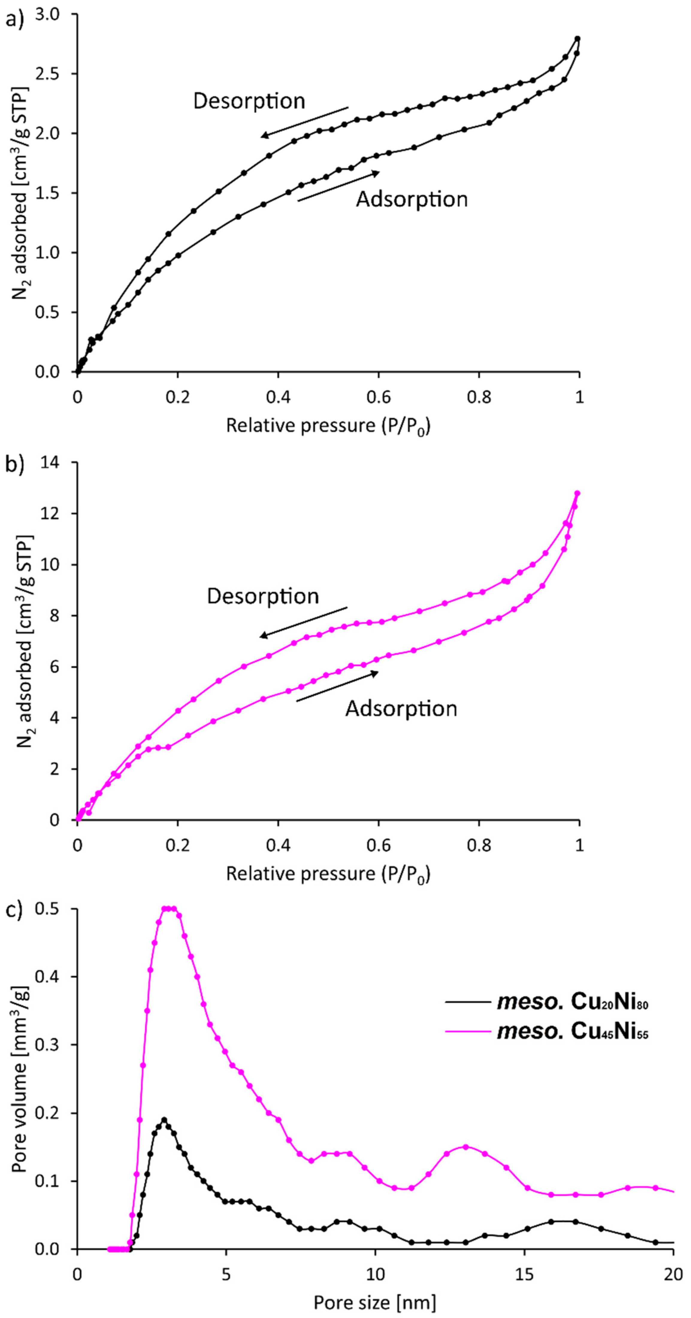
N2 adsorption–desorption curves were taken to gain further information on the roughness and porosity of the obtained films. A hysteresis was observed within the whole range of P/P0 values (Figure 6). However, the fact that the isotherm branches did not swing sharply upward near P/P0 = 0.9 indicate that mesopores were not uniformly present inside the material. Indeed, the resulting pore size distributions were ill-defined for both compositions (Figure 6c). Hence, the results point toward the formation of rough surfaces with mesoscale features, as previously seen by SEM.
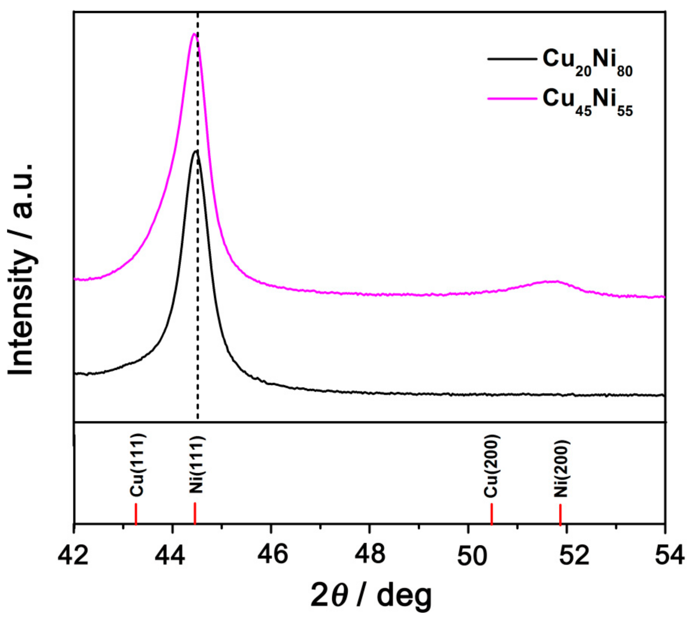
Figure 7 shows the XRD patterns of Cu20Ni80 and Cu45Ni55 surface-mesostructured films between 42° and 54°. In this region, two peaks at ca. 44° and one at 51–52° were observed, located between the (111) and (200) peaks of face-centered cubic (fcc) crystal structures of Ni and Cu. This indicates that the samples crystallized in the fcc structure (space group Fm3m). The position of the (111) reflection for Cu20Ni80 shifted toward lower angles with respect to that of Cu45Ni55, from 44.8° to 44.3° (ca. 0.5°), due to the increase in the Cu content. For both Cu45Ni55 and Cu20Ni80 samples, although peak splitting was not observed, the asymmetric broadening of the (111) peak gave a clear indication for the coexistence of Cu-rich and Ni-rich phases (i.e., two fcc solid solutions with slightly different cell parameters). Moreover, the (200) fcc peak, which should appear at around 2θ = 51–52°, was not detected for Cu20Ni80 and had a relatively low intensity for Cu45Ni55, suggesting a pronounced crystallographic texture along the (111) direction [26].
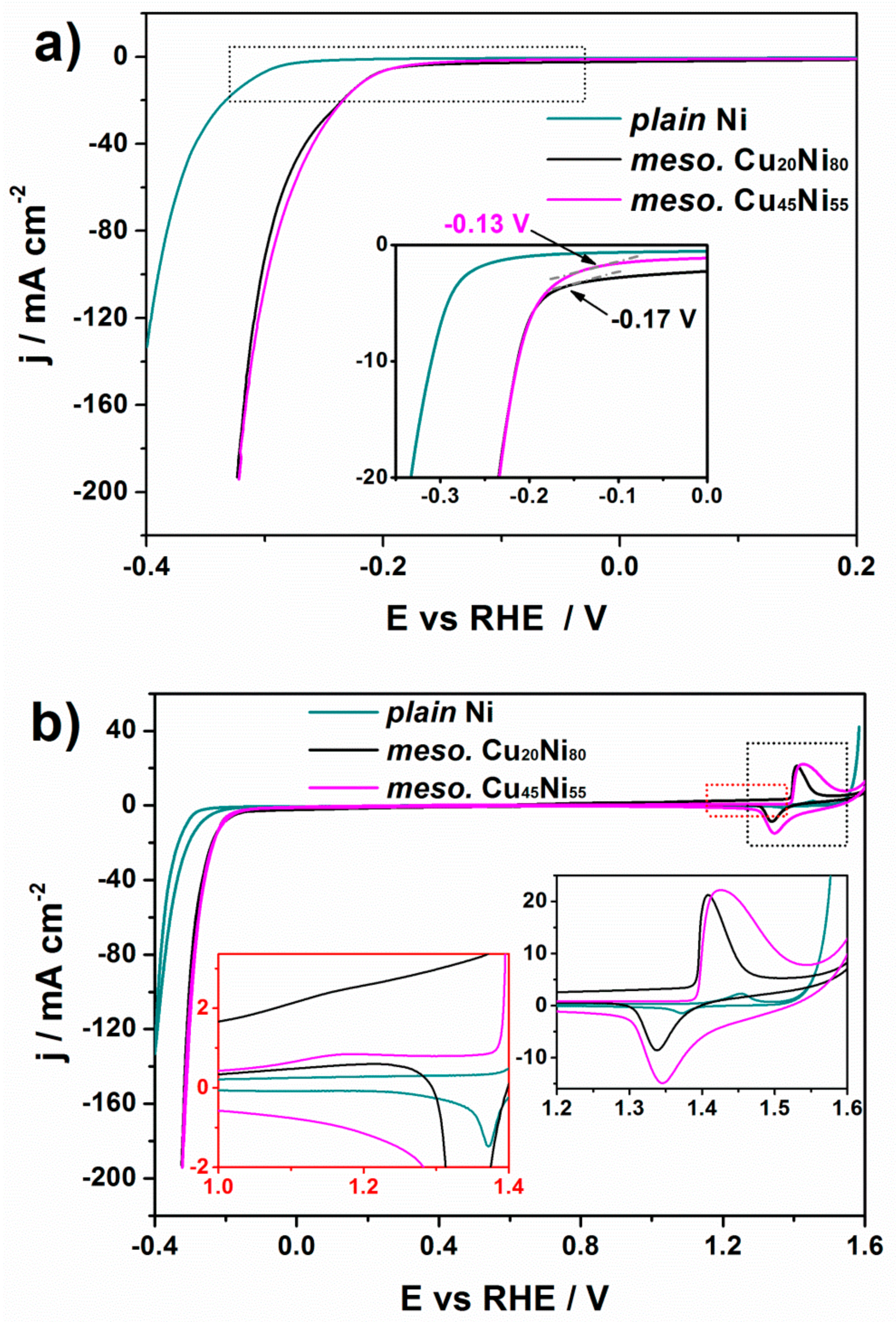
The catalytic activity toward HER of the Cu20Ni80 and Cu45Ni55 surface-mesostructured films was studied by recording polarization curves in 1 M KOH solution at a sweeping rate of 50 mV·s−1 at room temperature (Figure 8a). Note that current density is given per geometric area of the film exposed to the electrolyte. For comparison purposes, the behavior of a plain Ni film was used as a reference material. Since the onset potential can be determined from the deviation of the potential in the linear region [35], as indicated by an arrow in the inset of Figure 8a, one can conclude that the surface-mesostructured Cu45Ni55 showed lower onset potentials than those of plain Ni and surface-mesostructured Cu20Ni80 films. Polarization curves of plain Cu20Ni80 and Cu45Ni55 were also measured, as shown in Figure 9. Surface-mesostructured samples exhibited lower onset potentials, higher geometric current densities, and lower Tafel slopes than their plain counterparts (see Table 1). The enhanced HER performance of the mesostructured films was mainly due to their larger surface areas and the preferential segregation of Ni at the surface, as explained below.
In order to gain information regarding surface composition of the films, a CV study was carried out in 1 M KOH at a scan rate of 50 mV·s−1 (Figure 8b). For both Cu20Ni80 and Cu45Ni55 surface-mesostructured films, the small peak placed at approximately +1.16 V could be ascribed to the formation of Cu2O (Cu + OH → Cu(OH) + e), as shown in the left inset in Figure 8b. Likewise, from the right inset in Figure 8b, the anodic peak within the 1.39–1.57 V region could be attributed to the oxidation of Ni (0) to Ni (2+) to deliver α-Ni(OH)2. Meanwhile, the cathodic peak at around 1.29–1.44 V was due to the reduction of α-Ni(OH)2 to Ni (0). These two peaks could be detected for the three samples; in particular, they remained despite the presence of Cu in the CuNi samples. This indicates that the oxidation of Cu and Ni from surface-mesostructured CuNi films took place independently, without involving the formation of mixed oxides. Therefore, in a first approximation, the ECSA values for Ni and Cu can be derived from the charge related to the formation of α-Ni(OH)2 and Cu2O monolayers, and they are given with respect to the electrodeposited mass [14]. The charge under a monolayer formation of α-Ni(OH)2 [31,36] is 514 μC·cm2, while that of Cu2O [37] is 544 μC·cm2. Considering these, the resulting ECSA values for Ni and Cu are given in Table 1. As the Cu content increased in the film, the Cu-related ECSA increased from 0.3 m2·g−1 to 0.4 m2·g−1. More interestingly, the Ni-related ECSA decreased with the Ni content, a result which might seem counterintuitive and suggests that the number of active Ni sites at the surface can be increased by alloying Ni with Cu. The same observation was made by Jang and Kim [14], who also demonstrated that Ni segregates at the surface with addition of Cu to the material. Overall, the surface-mesostructured Cu45Ni55 film showed higher Ni-related ECSA, and, since Ni was more intrinsically active toward HER than Cu according to the Volcano plot [38], this resulted in an increased HER performance. Table 1 also lists the overpotential at −10 mA·cm−210) and the TOF values of the CuNi films with mesoscale roughness. Interestingly, the former was lower compared to their plain counterparts.
In order to examine the catalytic durability of the materials in the alkaline electrolyte within a short time scale, Cu20Ni80 and Cu45Ni55 surface-mesostructured films were subjected to 50 cycles in 1 M KOH electrolyte within the potential range from 0.2 V to −0.5 V vs. RHE. As shown in Figure 9c,d, the HER current density of the CuNi films showed the highest value at the first cycle and then decreased in subsequent cycles, indicating a faint loss of activity. The shift in the onset potential toward more negative values after 50 cycles was larger for Cu45Ni55 compared to Cu20Ni80. This was probably due to the larger Ni-related ECSA of Cu45Ni55; thus, the more vigorous hydrogen evolution required longer time to reach the equilibrium state of surface tension and buoyancy force.
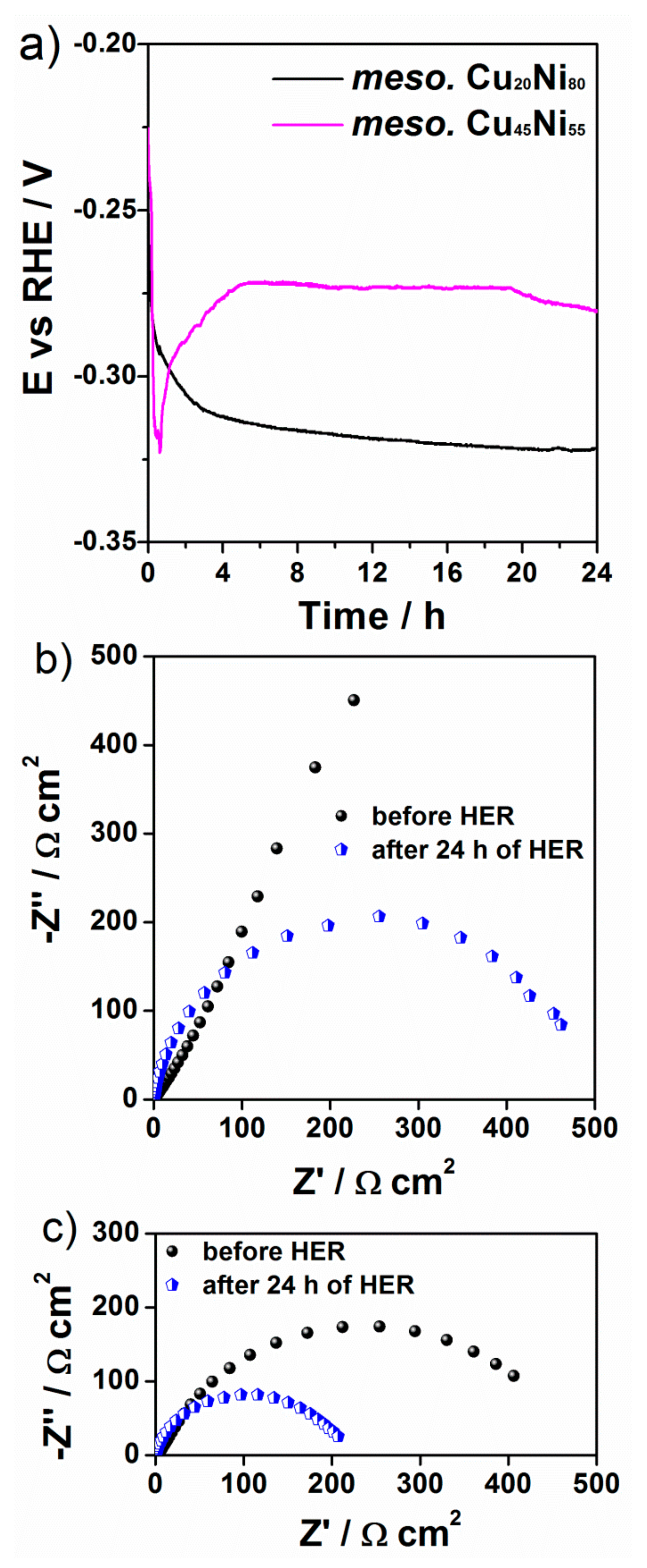
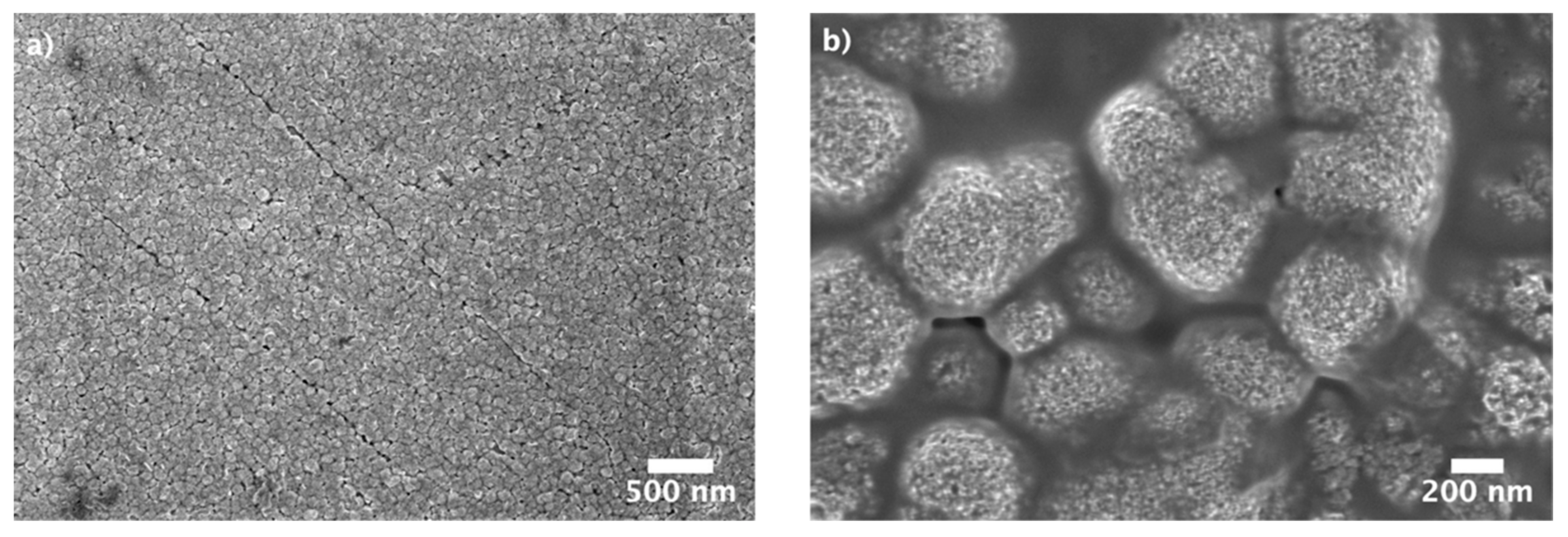
To further investigate the stability of the CuNi catalysts, the potential was recorded for 24 h at an applied geometric current density of −10 mA·cm−2 (Figure 10a). For the Cu20Ni80 film, the potential dropped from −260 mV to −320 mV during the first 2 h; then the change in potential slowed down, and, from 6 h onward, little or no variation was detected (i.e., the potential became stable). The potential also shifted toward more negative potential for the Cu45Ni55 film during the first 45 min but then it partially recovered and became stable from 5 h until the end of the experiment (E = −270 mV). These results suggest that the CuNi films experienced a decrease in the catalytic activity during the first hours, but no further decline of the activity was noticed after 5–6 h of continuous galvanostatic HER. Figure 10b,c show the Nyquist plots before and after the 24 h durability test. For both compositions of surface-mesostructured CuNi films, the impedance decreased, as indicated by smaller semicircles, hinting at an electrochemical activation of both surfaces. Finally, the morphology of the CuNi catalysts after being subject to 24 h HER experiment was investigated by SEM (Figure 11). No pronounced changes of film morphology were observed, although the surfaces seemed to be partially covered by an insulating thin layer, which could be partly attributed to remnants of KOH.
The surface-mesostructured CuNi materials were comparable to or slightly outperformed other Ni-based catalysts reported in the literature for HER in alkaline media [39], but were below the performance of noble-metal-based catalysts such as Ru and Pt with dissimilar architectures [40]. For example, Ru nanoparticles dispersed within a nitrogenated holey two-dimensional carbon structure exhibited an overpotential of 17 mV at −10 mA·cm−2 in 1.0 M KOH solution [41]. Although the performance of the here-deposited non-noble-metal-based films cannot yet compete against noble-metal catalysts, the values could be brought closer with further optimization of the catalyst surface roughness and composition.

4. Conclusions

In summary, the HER activity of electrodeposited bimetallic CuNi films was investigated in alkaline media, and the morphological and structural features of the samples were correlated with their electrocatalytic behavior. Interestingly, the surface topology and composition of the CuNi layers can be controlled by changing the pH value of the electrolyte, from Cu20Ni80 at quasi-neutral pH to Cu45Ni55 at pH = 4. The surface-mesostructured Cu45Ni55 films exhibited lower onset for HER compared to plain Ni and Cu20Ni80 films. Moreover, the surface-mesostructured Cu45Ni55 film showed higher specific activity than Cu20Ni80 in spite of its lower Ni/Cu ratio. The reason for this previously considered counterintuitive result was correlated to an increase in the surface Ni active sites, as demonstrated by cyclic voltammetry studies.

Author Contributions

Conceptualization, E.P. and J.Z.; methodology, J.Z. and K.E.; validation, E.P. and J.Z.; formal analysis, J.B.; investigation, J.B., L.F., K.E., Z.Y., M.Z. and Y.H.; resources, J.Z.; data curation, J.Z.; writing—original draft preparation, J.Z. and J.B.; writing—review and editing, K.E., J.S. and E.P.; visualization, J.Z.; supervision, J.S., E.P. and R.G.; project administration, D.Y.; funding acquisition, J.S., E.P. and J.Z. All authors have read and agreed to the published version of the manuscript.

Funding

This research was funded by National Natural Science Foundation of China (51901186) and the China Postdoctoral Science Foundation (2018M643730). K.E., E.P., and J.S. acknowledge financial support from MAT2017-86357-C3-1-R, PID2020-116844RB-C21, the associated FEDER (Spanish MINECO), the Marie Sklodowska-Curie grant agreement No 764977 (European Union’s Horizon 2020 research and innovation program), and 2017-SGR-292 (Generalitat de Catalunya).

Data Availability Statement

The data presented in this study are available on request from the corresponding author.

Acknowledgments

Financial support by National Natural Science Foundation of China (51901186) and the China Postdoctoral Science Foundation (2018M643730) is acknowledged.

Conflicts of Interest

The authors declare no conflict of interest.

References

  1. Li, P.; Liu, J.; Nag, N.; Crozier, P.A. In situ preparation of Ni–Cu/TiO2 bimetallic catalysts. J. Catal. 2009, 262, 73–82. [Google Scholar] [CrossRef]
  2. Best, R.J.; Russel, W.W. Nickel, copper and some of their alloys as catalysts for ethylene hydrogenation. J. Am. Chem. Soc. 1959, 81, 4132–4137. [Google Scholar]
  3. Quintana, A.; Zhang, J.; Isarain-Chavez, E.; Menendez, E.; Cuadrado, R.; Robles, R.; Baro, M.D.; Guerrero, M.; Pane, S.; Nelson, B.J.; et al. Voltage-induced coercivity reduction in nanoporous alloy films: A boost toward energy-efficient magnetic actuation. Adv. Funct. Mater. 2017, 27, 1701904. [Google Scholar] [CrossRef]
  4. Liu, Y.; Liu, D. Study of bimetallic Cu–Ni/γ-Al2O3 catalysts for carbon dioxide hydrogenation. Int. J. Hydrogen Energy 1999, 24, 351–354. [Google Scholar] [CrossRef]
  5. Kang, M.; Song, M.W.; Kim, T.W.; Kim, K.L. γ-alumina supported Cu-Ni bimetallic catalysts: Characterization and of 1,3-Butadiene. Can. J. Chem. Eng. 2002, 80, 63–70. [Google Scholar] [CrossRef]
  6. Solmaz, R.; Doner, A.; Kardas, G. The stability of hydrogen evolution activity and corrosion behavior of NiCu coatings with long-term electrolysis in alkaline solution. Int. J. Hydrogen Energy 2009, 34, 2089–2094. [Google Scholar] [CrossRef]
  7. Liu, Z.; Guo, L.; Chien, C.L.; Searson, P.C. Formation of a core/shell microstructure in Cu–Ni thin films. J. Electrochem. Soc. 2008, 155, D569–D574. [Google Scholar] [CrossRef]
  8. Liu, Z.; Xia, G.; Zhu, F.; Kim, S.; Markovic, N.; Chien, C.; Searson, P.C. Exploiting finite size effects in a novel core/shell microstructure. J. Appl. Phys. 2008, 103, 064313. [Google Scholar] [CrossRef] [Green Version]
  9. Wang, H.; Baker, R.T.K. Decomposition of methane over a Ni-Cu-MgO catalyst to produce hydrogen and carbon nanofibers. J. Phys. Chem. B 2004, 108, 20273–20277. [Google Scholar] [CrossRef]
  10. Bergwerff, J.A.; Visser, T.; Leliveld, B.R.; Rossenaar, B.D.; de Jong, K.P.; Weckhuysen, B.M. Envisaging the physicochemical processes during the preparation of supported catalysts:  Raman microscopy on the impregnation of Mo onto Al2O3 extrudates. J. Am. Chem. Soc. 2004, 126, 14548–14556. [Google Scholar] [CrossRef] [Green Version]
  11. Pieta, I.S.; Rathi, A.; Pieta, P.; Nowakowski, R.; Holdynski, M.; Pisarek, M.; Kaminska, A.; Gawande, M.B.; Aboril, R. Electrocatalytic methanol oxidation over Cu, Ni and bimetallic Cu-Ni nanoparticles supported on graphitic carbon nitride. Appl. Catal. B-Environ. 2019, 244, 272–283. [Google Scholar] [CrossRef]
  12. Eugenio, S.; Silva, T.M.; Carmezim, M.J. Electrodeposition and characterization of nickel–copper metallic foams for application as electrodes for supercapacitors. J. Appl. Electrochem. 2014, 44, 455–465. [Google Scholar] [CrossRef]
  13. Kear, G.; Barker, B.D.; Stokes, K.; Walsh, F.C. Electrochemical Corrosion Behaviour of 90–10 Cu–Ni Alloy in Chloride-based Electrolytes. J. Appl. Electrochem. 2004, 34, 659–669. [Google Scholar] [CrossRef]
  14. Ahn, S.H.; Park, H.Y.; Choi, I.; Yoo, S.J.; Hwang, S.J.; Kim, H.J.; Cho, E.; Yoon, C.W.; Park, H.; Son, H.; et al. Electrochemically fabricated NiCu alloy catalysts for hydrogen production in alkaline water electrolysis. Int. J. Hydrogen Energy 2013, 38, 13493–13501. [Google Scholar] [CrossRef]
  15. Poulopoulos, P.; Lindner, J.; Farle, M.; Baberschke, K. Change of magnetic anisotropy due to roughness: A quantitative scanning tunneling microscopy study on Ni/Cu (00l). Surf. Sci. 1999, 437, 277–284. [Google Scholar] [CrossRef]
  16. Sinfelt, J.H. Catalysis by alloys and bimetallic clusters. Acc. Chem. Res. 1977, 10, 28. [Google Scholar] [CrossRef]
  17. Wu, S.; Zhu, C.; Huang, W. Properties of polymer supported Ni-Cu bimetallic catalysts prepared by solvated metal atom impregnation. Chin. J. Polym. Sci. 1996, 14, 217–224. [Google Scholar]
  18. Zhang, J.; Baró, M.D.; Pellicer, E.; Sort, J. Electrodeposition of magnetic, superhydrophobic, non-stick, two-phase Cu–Ni Foam films and their enhanced performance for hydrogen evolution reaction in alkaline water media. Nanoscale 2014, 6, 12490–12499. [Google Scholar] [CrossRef]
  19. Rao, S.; Zou, X.; Wang, S.; Lu, Y.; Shi, T.; Hsu, H.; Xu, Q.; Lu, X. Electrodeposition of Ni-Cu alloy films from nickel matte in deep eutectic solvent. Mater. Chem. Phys. 2019, 232, 6–15. [Google Scholar] [CrossRef]
  20. Fu, Z.; Zhang, Z.; Meng, L.; Shu, B.; Zhu, Y.; Zhu, X. Effect of strain rate on mechanical properties of Cu/Ni multilayered composites processed by electrodeposition. Mat. Sci. Eng. A 2018, 726, 154–159. [Google Scholar] [CrossRef]
  21. Vesali, N.; Erfanifam, S.; Jamilpanah, L.; Hasheminejad, M.; Rahmani, Y.; Mohseni, S.M. Growth behavior of Cu, Ni and Cu/Ni electrodeposited microwires within porous Si. Surf. Coat. Technol. 2019, 364, 16–21. [Google Scholar] [CrossRef]
  22. Xu, W.; Du, D.; Lan, R.; Humphreys, J.; Miller, D.N.; Walker, M.; Wu, Z.; Irvine, J.T.S.; Tao, S. Electrodeposited NiCu bimetal on carbon paper as stable non-noble anode for efficient electrooxidation of ammonia. Appl. Catal. B-Environ. 2018, 237, 1101–1109. [Google Scholar] [CrossRef]
  23. Abbas, S.A.; Kim, S.H.; Iabal, M.I.; Muhammad, S.; Yoon, W.S.; Jung, K.D. Synergistic effect of nano-Pt and Ni spine for HER in alkaline solution: Hydrogen spillover from Nano-Pt to Ni spine. Sci. Rep. 2018, 8, 2986. [Google Scholar] [CrossRef] [Green Version]
  24. Rosalbino, F.; Scavino, G.; Grande, M.A. Electrocatalytic activity of Ni–Fe–M (M = Cr, Mn, Cu) sintered electrodes for hydrogen evolution reaction in alkaline solution. J. Electroanal. Chem. 2013, 694, 114–121. [Google Scholar] [CrossRef]
  25. Whitehead, A.H.; Elliott, J.M.; Owen, J.R.; Attard, G.S. Electrodeposition of Mesoporous Tin Films. Chem. Commun. 1999, 331–332. [Google Scholar] [CrossRef]
  26. Zhang, J.; Quintana, A.; Menendez, E.; Coll, M.; Pelliver, E.; Sort, J. Electrodeposited Ni-based magnetic mesoporous films as smart surfaces for atomic layer deposition: An “all-chemical” deposition approach toward 3D Nanoengineered composite layers. ACS Appl. Mater. Interfaces 2018, 10, 14877–14885. [Google Scholar] [CrossRef] [Green Version]
  27. Eiler, K.; Suriñach, S.; Sort, J.; Pellicer, E. Mesoporous Ni-rich Ni–Pt thin films: Electrodeposition, characterization and performance toward hydrogen evolution reaction in acidic media. Appl. Catal. B Environ. 2020, 265, 118597. [Google Scholar] [CrossRef]
  28. Iqbal, M.; Kaneti, Y.V.; Kashimura, K.; Yoshino, M.; Jiang, B.; Li, C.; Yuliarto, B.; Bando, Y.; Sugahara, Y.; Yamauchi, Y. Continuous mesoporous Pd films with tunable pore sizes through polymeric micelle-assisted assembly. Nanoscale Horiz. 2019, 4, 960–968. [Google Scholar] [CrossRef]
  29. Pellicer, E.; Varea, A.; Pané, S.; Nelson, B.J.; Menendez, E.; Estrader, M.; Suriñach, S.; Baró, M.D.; Nogués, J.; Sort, J. Nanocrystalline electroplated Cu–Ni: Metallic thin films with enhanced mechanical properties and tunable magnetic behavior. Adv. Funct. Mater. 2010, 20, 983–991. [Google Scholar] [CrossRef]
  30. Cossar, E.; Houache, M.S.E.; Zhang, Z.; Baranova, E.A. Comparison of electrochemical active surface area methods for various nickel nanostructures. J. Electroanal. Chem. 2020, 870, 114246. [Google Scholar] [CrossRef]
  31. Machado, S.A.S.; Avaca, L.A. The hydrogen evolution reaction on nickel surfaces stabilized by H-absorption. Electrochim. Acta 1994, 39, 1385–1391. [Google Scholar] [CrossRef]
  32. Van Drunen, J.; Kinkead, B.; Wang, M.C.P.; Sourty, E.; Gates, B.D.; Jerkiewicz, G. Comprehensive Structural, surface-chemical and electrochemical characterization of nickel-based metallic foams. ACS Appl. Mater. Interfaces 2013, 5, 6712–6722. [Google Scholar] [CrossRef] [Green Version]
  33. Wang, X.-D.; Chen, H.-Y.; Xu, Y.F.; Liao, J.-F.; Chen, B.-X.; Rao, H.-S.; Kuang, D.-B.; Su, C.-Y. Self-supported NiMoP2 nanowires on carbon cloth as an efficient and durable electrocatalyst for overall water splitting. J. Mater. Chem. A 2017, 5, 7191–7199. [Google Scholar] [CrossRef]
  34. Bender, F.; Mankelow, R.F.; Hibbert, D.B.; Gooding, J.J. Lyotropic liquid crystal templating of groups 11 and 12 metal films. Electroanalysis 2006, 16, 1558–1563. [Google Scholar] [CrossRef]
  35. Ren, B.; Li, D.; Jin, Q.; Cui, H.; Wang, C. Novel Porous Tungsten Carbide Hybrid Nanowires on Carbon Cloth for High-performance Hydrogen Evolution. J. Mater. Chem. A 2017, 5, 13196–13203. [Google Scholar] [CrossRef]
  36. Hahn, F.; Beden, B.; Croissant, M.J.; Lamy, C. In situ UV visible reflectance spectroscopic investigation of the nickel electrode-alkaline solution interface. Electrochim. Acta 1986, 31, 335–342. [Google Scholar] [CrossRef]
  37. Elhaleem, S.; Ateya, B.G. Cyclic voltammetry of copper in sodium hydroxide solutions. J. Electroanal. Chem. 1981, 117, 309–319. [Google Scholar]
  38. Quaino, P.; Juarez, F.; Santos, E.; Schmickler, W. Volcano plot in hydrogen electrocatalysis—Use and abuse. Beilstein J. Nanotechnol. 2014, 5, 846–854. [Google Scholar] [CrossRef] [Green Version]
  39. Hu, X.; Tian, X.; Lin, Y.-W.; Wang, Z. Nickel foam and stainless steel mesh as electrocatalysts for hydrogen evolution reaction, oxygen evolution reaction and overall water splitting in alkaline media. RSC Adv. 2019, 9, 31563–31571. [Google Scholar] [CrossRef] [Green Version]
  40. Mahmood, J.; Li, F.; Jung, S.M.; Okyay, M.S.; Ahmad, I.; Kim, S.J.; Park, N.; Jeong, H.Y.; Baek, J.B. An efficient and pH-universal ruthenium-based catalyst for the hydrogen evolution reaction. Nat. Nanotechnol. 2017, 12, 441. [Google Scholar] [CrossRef]
  41. Mahmood, N.; Yao, Y.; Zhang, J.-W.; Pan, L.; Zhang, X.; Zou, J.-J. Electrocatalysts for hydrogen evolution in alkaline electrolytes: Mechanisms, challenges, and prospective solutions. Adv. Sci. 2018, 5, 1700464. [Google Scholar] [CrossRef] [PubMed]
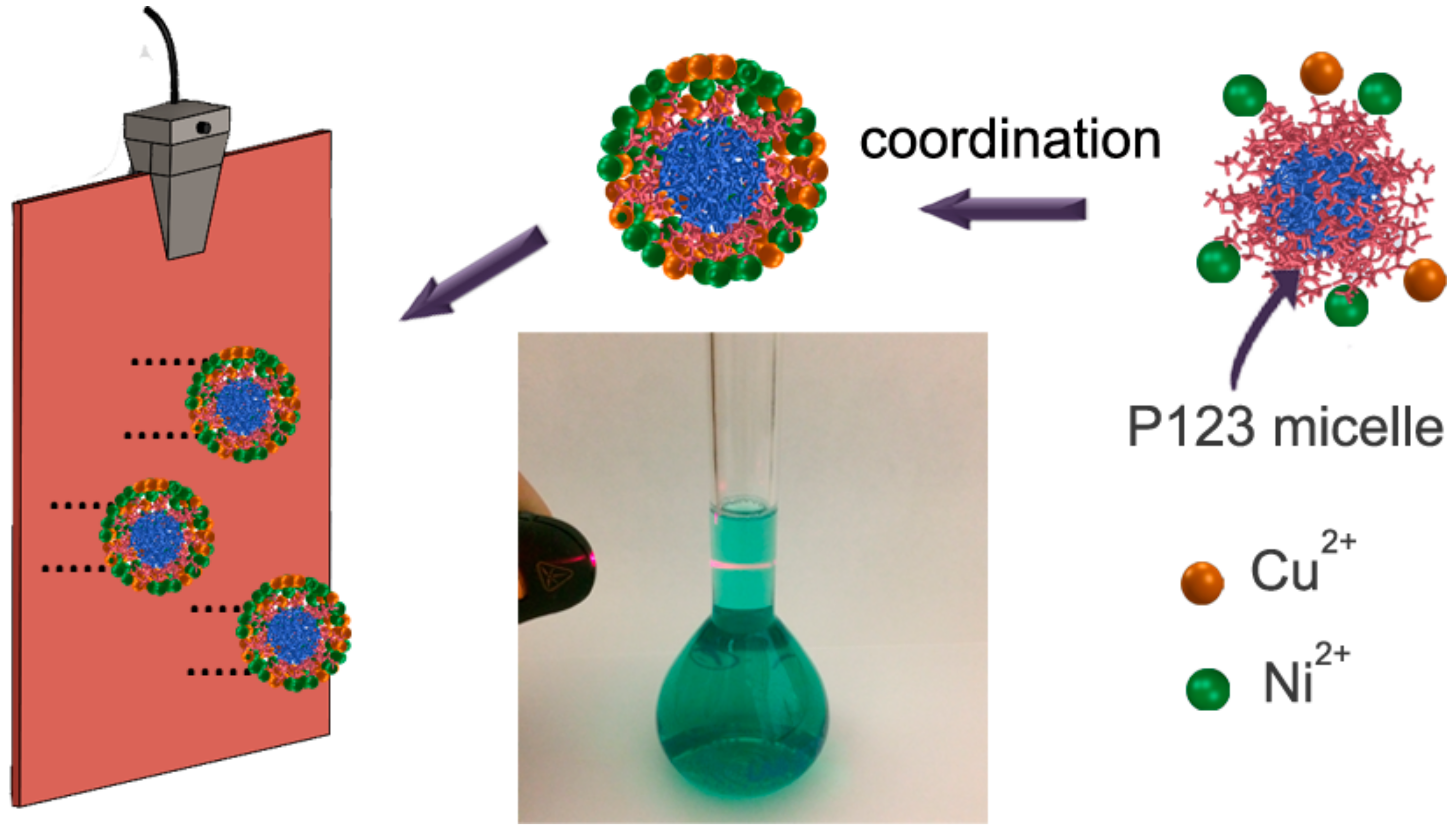
Figure 1. Schematic illustration of the preparation process. The photograph at the center of the sketch demonstrates the occurrence of the Tyndall effect.
Nanomaterials 12 00118 g001
Figure 2. Low- and high-magnification SEM images of surface-mesostructured films with composition (a,b) Cu20Ni80 and (c,d) Cu45Ni55.
Nanomaterials 12 00118 g002
Figure 3. EDX spectra of the surface-mesostructured films with composition (a) Cu20Ni80 and (b) Cu45Ni55.
Nanomaterials 12 00118 g003
Figure 4. SEM images of CuNi films deposited from the same electrolyte composition at pH values of (a) 3 and (b) 2.
Nanomaterials 12 00118 g004
Figure 5. SEM images of films electrodeposited at j = –100 mA·cm−2 and 150 s from (a) the base electrolyte and (b) the base electrolyte without Cu ions, at pH = 6.25. Note that the SEM image of (a) corresponds to a lower-magnified image of the deposit shown in Figure 2a.
Nanomaterials 12 00118 g005
Figure 6. N2 isotherms for (a) Cu20Ni80 and (b) Cu45Ni55 surface-mesostructured films; (c) resulting pore size distribution for both compositions.
Nanomaterials 12 00118 g006
Figure 7. Zoomed-in view of the XRD patterns of surface-mesostructured Cu20Ni80 and Cu45Ni55 films.
Nanomaterials 12 00118 g007
Figure 8. (a) Polarization and (b) CV curves recorded in 1 M KOH electrolyte for Cu20Ni80 and Cu45Ni55 surface-mesostructured films. The response of a plain pure Ni film is shown for comparison. The inset in (a) and right inset in (b) show zoomed-in parts of the curves enclosed within the black dotted rectangle. The left inset in (b) shows a zoomed-in view of the region enclosed in the red dotted rectangle.
Nanomaterials 12 00118 g008
Figure 9. Cathodic-going linear sweep voltammetry curves for comparison between plain and surface-mesostructured (a) Cu20Ni80 and (b) Cu45Ni55, and the variation in HER current density as a function of the number of scans for (c) Cu20Ni80 and (d) Cu45Ni55. All curves were recorded in recorded in 1 M KOH at a scan rate of 50 mV·s1 and 298 K. The insets in (a) and (b) show the Tafel slopes.
Nanomaterials 12 00118 g009
Figure 10. Potential as a function of time during galvanostatic HER at −10 mA·cm−2 for surface-mesostructured CuNi films (a). Nyquist plots before and after 24 h durability tests for surface-mesostructured films with composition (b) Cu20Ni80 and (c) Cu45Ni55.
Nanomaterials 12 00118 g010
Figure 11. SEM images of surface-mesostructured films with composition (a) Cu20Ni80 and (b) Cu45Ni55 after 24 h durability tests.
Nanomaterials 12 00118 g011
Table 1. ECSA, overpotential at −10 mA·cm210), Tafel slope (b), and TOF values for the deposited Cu20Ni80 and Cu45Ni55 surface-mesostructured films. The ECSA values are given for Cu and Ni elements separately. Values in parentheses correspond to plain films of same composition.
Cu20Ni80Cu45Ni55
ECSA (Cu)0.3 ± 0.05 m2·g10.4 ± 0.1 m2·g1
ECSA (Ni)7.4 ± 0.4 m2·g112.5 ± 0.3 m2·g1
η10210 (220) mV210 (230) mV
b65 (115) mV68 (97) mV
TOF0.5 s10.3 s1
Publisher’s Note: MDPI stays neutral with regard to jurisdictional claims in published maps and institutional affiliations.