Article

Novel Blue-Wavelength-Blocking Contact Lens with Er3+/TiO2 NPs: Manufacture and Characterization

by
Lina Mohammed Shaker
1,
Ahmed Alamiery
1,2,*,
Mohd Takriff
1,3 and
Wan Nor Roslam Wan Isahak
1
1
Department of Chemical and Process Engineering, Faculty of Engineering and Built Environment, Universiti Kebangsaan Malaysia (UKM), Bangi 43000, Selangor, Malaysia
2
Energy and Renewable Energies Technology Center, University of Technology, Baghdad 10001, Iraq
3
Chemical and Water Desalination Engineering Program, Collage of Engineering, University of Sharjah, Sharjah 26666, United Arab Emirates
*
Author to whom correspondence should be addressed.
Nanomaterials 2021, 11(9), 2190; https://doi.org/10.3390/nano11092190
Submission received: 30 July 2021 / Revised: 12 August 2021 / Accepted: 18 August 2021 / Published: 26 August 2021

Abstract

:
Thermally stable titanium dioxide nanoparticles (TiO2 NPs) doped with erbium ions (Er3+) are characterized by uniformity, low excitation energy, and high surface area. The impregnation methodology was used to enhance the optical properties of TiO2 NPs impregnated with various Er3+ ion contents. The synthesized Er3+/TiO2 samples were characterized by energy dispersive X-ray (EDX), metal mapping, UV–Visible spectrum, field emission scanning electron microscopy (FESEM), and X-ray diffraction (XRD). The Er3+ ions, per our findings, were well-distributed on the TiO2 surface of the anatase phase and there was an insignificant difference in particle size, but there was no change in the particle shapes of the Er3+/TiO2 NPs structure. The maximum band gap degradation occurred with 1.8 wt % of Er3+/TiO2, where the energy gap degraded from 3.13 to 2.63 eV for intrinsic TiO2. The synthesized Er3+/TiO2 samples possess predominantly finely dispersed erbium ion species on the surface. Er3+ ions agglomeration on the surface increased with increasing ions in each sample. We found that 0.6 wt/vol % of Er+3/TiO2 is the best optical coating and produced satisfying results in terms of blocking the transmittance of blue wavelength without reducing the image quality.

1. Introduction

Recently, various methods have been studied and applied for modifying mineral surfaces [1,2]. Such methods enhance the activity of titanium dioxide (TiO2) for different applications [3], for example, as a photocatalyst for the photocatalytic degradation (PCD) of pollutants [4], both organic and inorganic [5,6], and for sterilizing wastewater [7]. A photocatalytic antimicrobial film was coated on a polymeric contact lens surface to fabricate an anti-contamination contact lens and other applications [8]. Hybrid poly(methyl methacrylate-dioxide) systems have been widely reported, focusing mostly on PMMA-SiO2 [9], PMMA-TiO2 [10], and PMMA-ZrO2 [11]; these nanocomposites are used in the fabrication of optical and biomedical devices such as contact lenses [11]. Nanocomposite polymers have attracted significant attention in optical applications because of their enhanced optical performance. Recently, nanoparticles (NPs) such as TiO2, ZnO, ZnS, ZrO2, and so forth, have been exploited to obtain a high refractive index (n) and low dispersion nanocomposite [12,13]. Switchable wettability materials such as TiO2-based polymers are considered good photocatalytic coating candidates able to block the undesired ultraviolet (UV) spectrum [14].
The TiO2 photocatalyst is characterized by its ease of synthesis, low cost, nontoxicity, high stability, high band-gap energy (3.2 eV) [15], and applicability for PCD [16]. Despite these qualities, it is now of little use in large-scale processing operations due to its low quantum efficiency and short wavelength excitation. According to the concept of irradiation, when the particles are irradiated with near ultraviolet rays of energy greater than or equal to 350 nm [17], the electrons migrate from the valence band to reach the conduction band (ecb) leaving behind positive holes (hvb+), where they initiate oxidation reactions and reduction reactions with the absorbed species [18]. Such charges can be recombined to generate an e-hole pair that is not available to participate in the required reactions. Before this process takes place, oxidation and reduction processes occur by means of electrons and holes, which control the efficiency of the photocatalytic TiO2-catalyzed reaction [19,20].
One of the approaches that has been widely adopted to improve the photocatalytic efficiency of the TiO2 photocatalyst under UV and visible light is the doping/deposition of a minute amount of a suitable metal in TiO2, which enhances the participation of the photogenerated holes and electrons in the photocatalytic reactions [21,22,23,24,25]. It was suggested that the precipitated or doped metal in TiO2 has high Schottky barriers in the metal–TiO2 contact area and thus acts as an electronic trap, which facilitates the separation of the electron hole and enhances the electron transfer process of the interface, thus enhancing the photocatalyst’s efficiency [26]. According to the higher energy gap of TiO2 NPs, one way to make these particles useful in terms of photocatalytic activity is by doping the TiO2 NPs with rare earth ions, where these ions decrease the metal oxide band gap [27]. Visible light represents about 43% of the solar spectrum, so researchers have aimed to improve the absorption of visible light by TiO2 due to its previously mentioned properties and applications in the visible and ultraviolet frequency ranges [28,29,30].
This article attempts to explain and develop the existence of erbium ions with optimum quantities and their role in the physicochemical properties of the doped TiO2 materials, especially when used as a coating filter. The best optical characteristics were obtained by the prepared nanocomposites by designing a coating layer on a contact lens front surface to fabricate a blue-blocking contact lens. In the continuation of our work [31,32,33,34,35] on nanomaterials, herein, we report the synthesis of TiO2 NPs and Er3+/TiO2 by the impregnation method [36,37,38].

2. Materials and Methods

The chemicals erbium (III), TiO2 NPs powder, and chloroxylenol were purchased from Sigma Aldrich (St. Louis, MO, USA) and utilized without further purifications. The Er3+ contained acetylacetonate hydrate, was CAS grade, and had a purity of 97%. The TiO2 nanopowder had 99.5% purity and a 21 nm diameter according to the TEM from Philips (Eindhoven, The Netherlands), and the chloroxylenol had 98% purity. The chloroxylenol was prepared, and deionized water was used for this purpose as well as for dilution.

2.1. Er3+/TiO2 Preparation

TiO2 NPs was impregnated with Er3+ ions with 0.6, 1.2, 1.8, and 2.4 wt % using an 80 mL glass beaker. We used a 10 mL of deionized water to dissolve 60, 120, 180, and 240 mg of Er3+ acetylacetonate hydrate, which was sonicated for 10 min. Then, each solution was added dropwise to the TiO2 nanopowder, well-mixed with a glass rod, and stirred for 2 h at 1300 rpm. The drying process was carried out at 80 °C overnight and then calcination was performed at 500 °C for two hours.

2.2. Er3+/TiO2 Characterization

The estimation of Er3+/TiO2 NPs phase and size was achieved using D8 Advance Bruker AXS X-ray patterns (Bruker, Am Studio 2D, Berlin, Germany). Energy-dispersive X-ray (EDX), with higher resolution, was applied to investigate the synthesized NPs’ morphological features using field emission scanning electron microscopy (FESEM) (SuPRA 55 VP, Carl Zeiss AG, Oberkochen, Germany). The other two analysis devices were used to characterize the NP samples were an OXFORD Penta FETx3 for metal mapping (Carl Zesis AG, Oberkochen, Germany) and a Perkin Elmer UV–Vis spectrophotometer (Waltham, MA, USA). The intensity transmission curve of the designed coated contact lens was recorded by ZEMAX software.

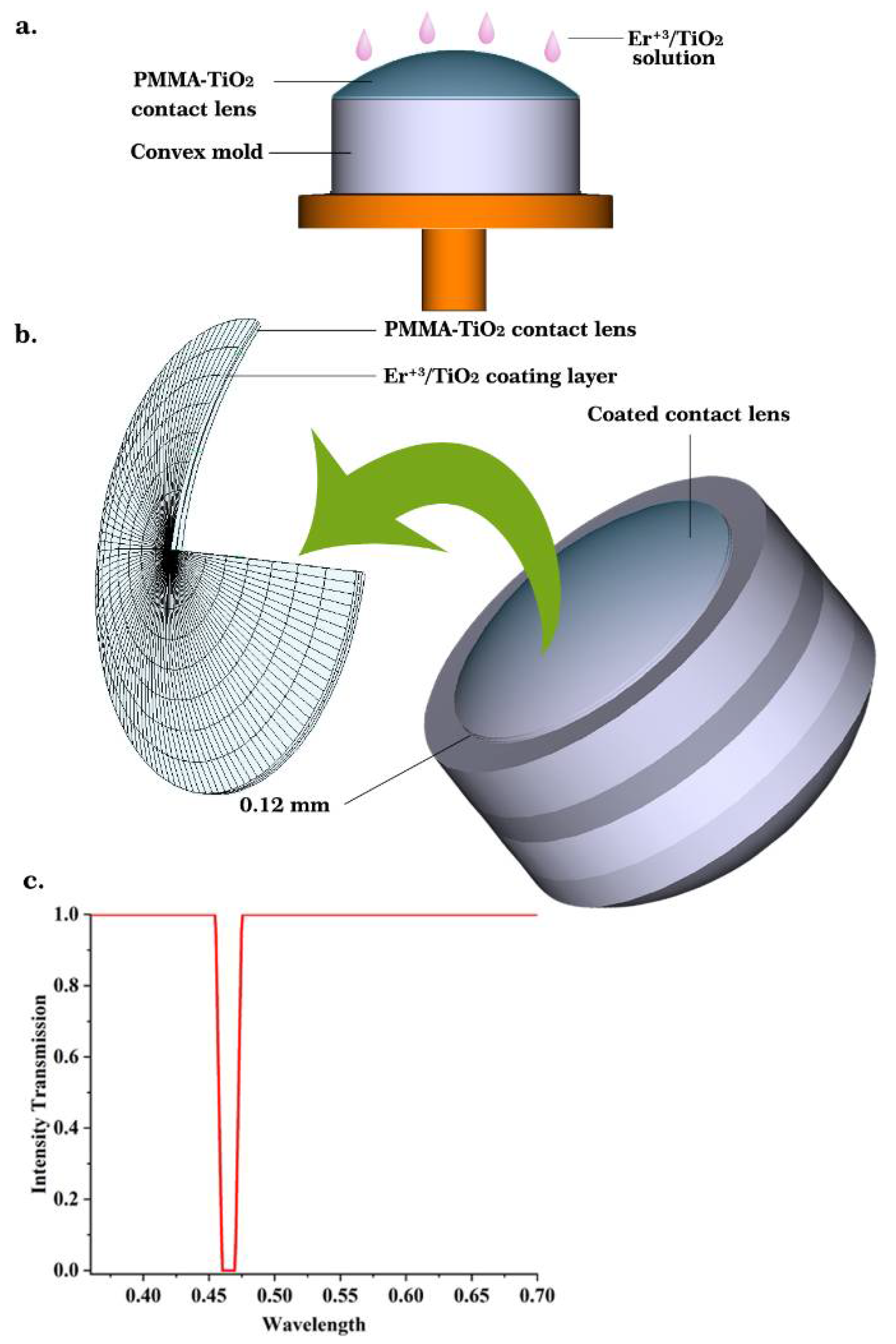
2.3. Contact Lens Modeling

To design and fabricate an anti-contamination blue wavelength blocker contact lens, the UV–Vis spectral data for all the synthesized films were compared; the best n value nanocomposite was selected for coating the contact lens. The human eye optical parameters and the coated contact lens at a 5 degrees field of view under white light illumination were entered into ZEMAX software (Version 14, serial number 34,900). The two corneal surfaces (anterior and posterior cornea) were selected as aspherical surfaces (programmatically named the standard surface). The standard surface requires two specific parameters: radius and conic constant. The pupil aperture surface was selected as the stop aperture and nasally decentered by 0.5 mm with respect to the visual axis; the crystalline lens is represented by two homogeneous gradient index shells whose n value is described by:
n = n 0 + n r 2 r 2 + n r 4 r 4 + n r 6 r 6 + n z 1 z + n z 2 z 2 + n z 3 z 3
r 2 = x 2 + y 2
Both the vitreous body of the eye and the retinal imaging surface were selected as standard surfaces. The standard surface position is centered on the optical axis and its vertex located at the Z axis. The z value (sag) of the standard surface is given by:
z = c r 2 1 + 1 ( 1 + k c 2 r 2 )
where c is the curvature (reciprocal of the radius), r is the radial coordinate in the lens unit, and k refers to the conic constant.
In this work, a coating of aspheric surface of the modeled contact lens (0.12 mm thickness, 0.035 conic, and 7.748 mm and 7.8 mm radius of front and back surfaces, respectively) was constructed by an extended polynomial surface. The aspherical surface sag is defined by:
z = c r 2 1 + 1 ( 1 + k ) c 2 r 2 + i = 1 N A i E i ( x , y )
where N represents the polynomial coefficient number in the series and Ai is the coefficient on the ith extended polynomial (E) term

3. Results

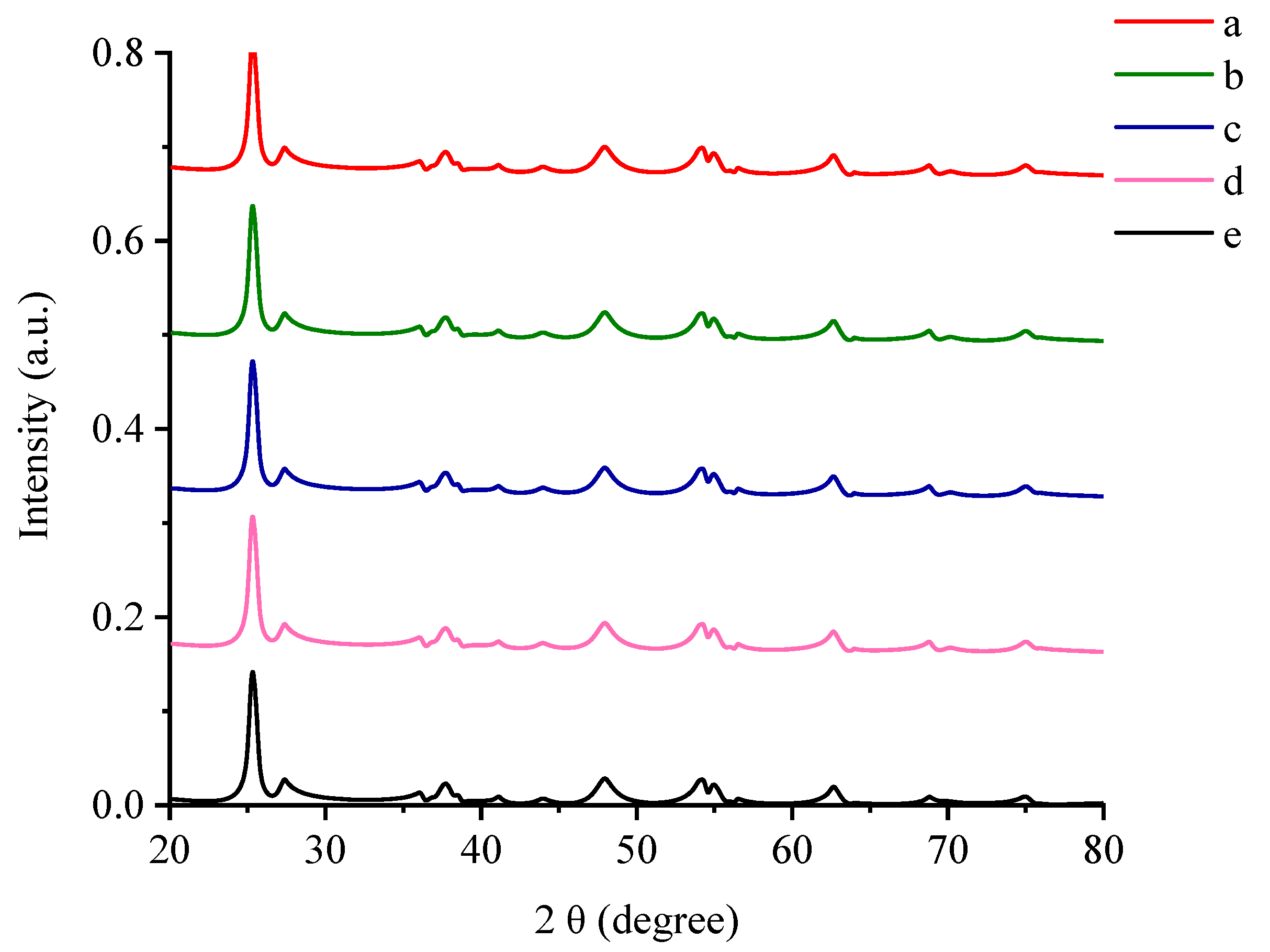
3.1. X-ray Diffraction Pattern

The synthesized specimens were analyzed by XRD in order to study the crystallinity of the erbium ions/titanium dioxide (Er3+/TiO2) NPs and to evaluate the changes, if any, that may occur through the impregnation of TiO2 NPs with Er ions. The X-ray diffraction scanning of the Er3+/TiO2 NPs is demonstrated in Figure 1. The signals of the TiO2 types, which are anatase and rutile [37,38], were clear. These patterns demonstrate that the phase composition and the structure of the TiO2 NPs did not change even after impregnation with Er ions. Additionally, the decrease in the intensity of the TiO2’s peaks indicated that the crystal structure of the titanium dioxide was impregnated with Er ions.
Table 1 demonstrates the intensity values of the signals. The diffraction spectra of the Er ions/TiO2 NPs do not support the existence of Er ions. This conclusion can most probably be ascribed to the appropriate dispersion of erbium ions in the lattice structure of TiO2 [39].
The crystallite size data are listed in Table 2 for TiO2 with Er ions in weight percent [15]. The lesser increase in the size of the particles confirmed that impregnation increased for the Er ions/TiO2 NPs sample. The Debye–Scherer equation is used to calculate the crystallite size D (Equation (5)):
D = K λ β cos θ
where the Scherer constant ( K ) is equalto 0.89, λ is the wavelength, β is the width of the peak, and θ is the angle of Bragg diffraction.

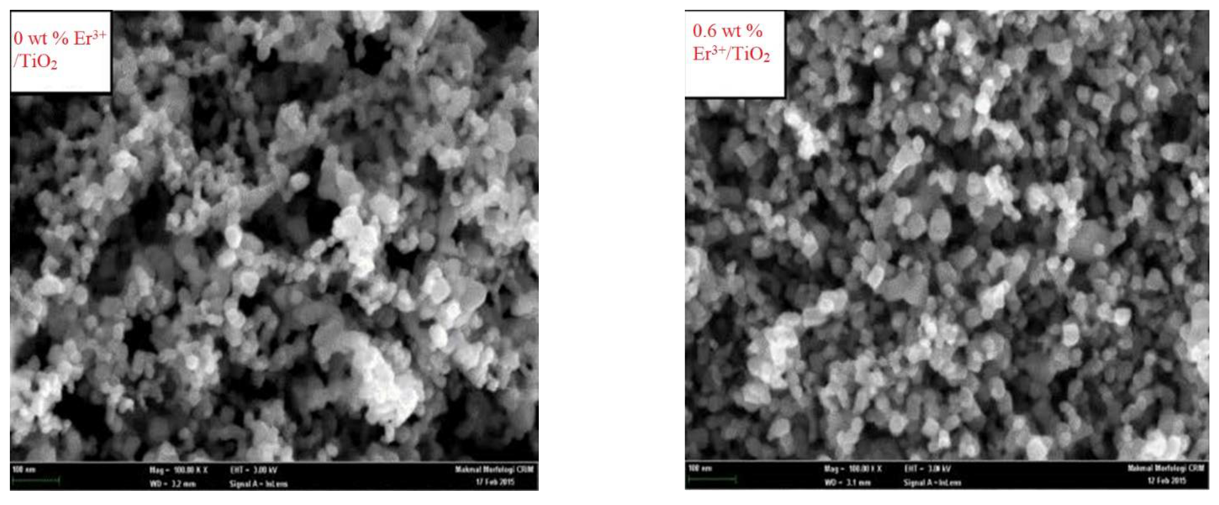
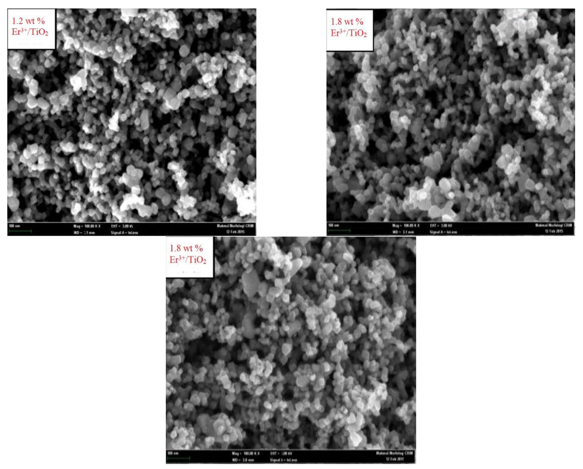
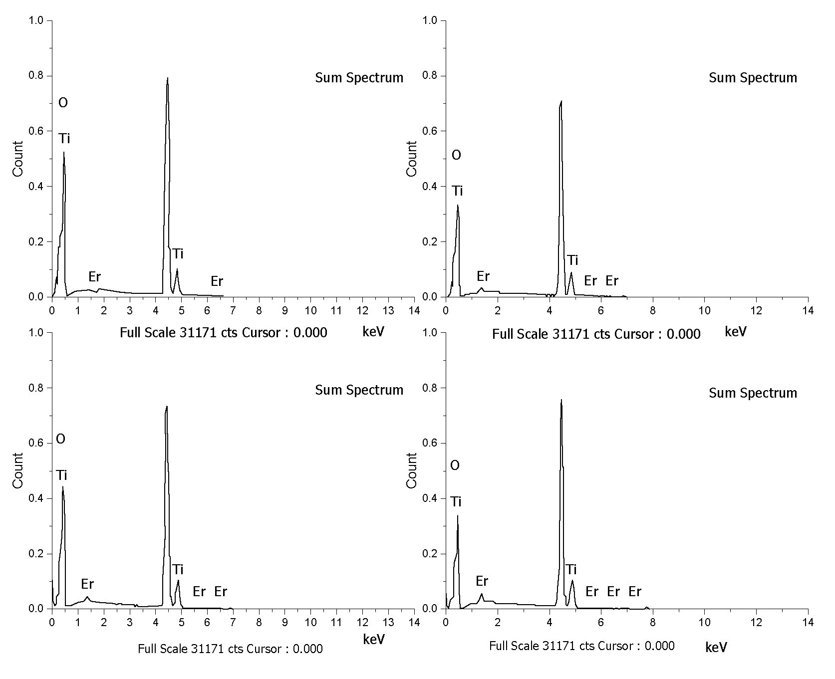
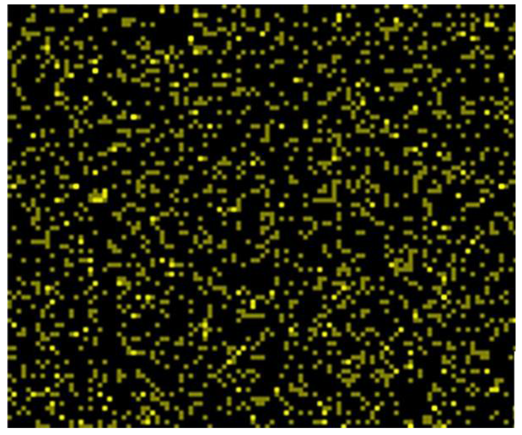
3.2. Surface Morphology
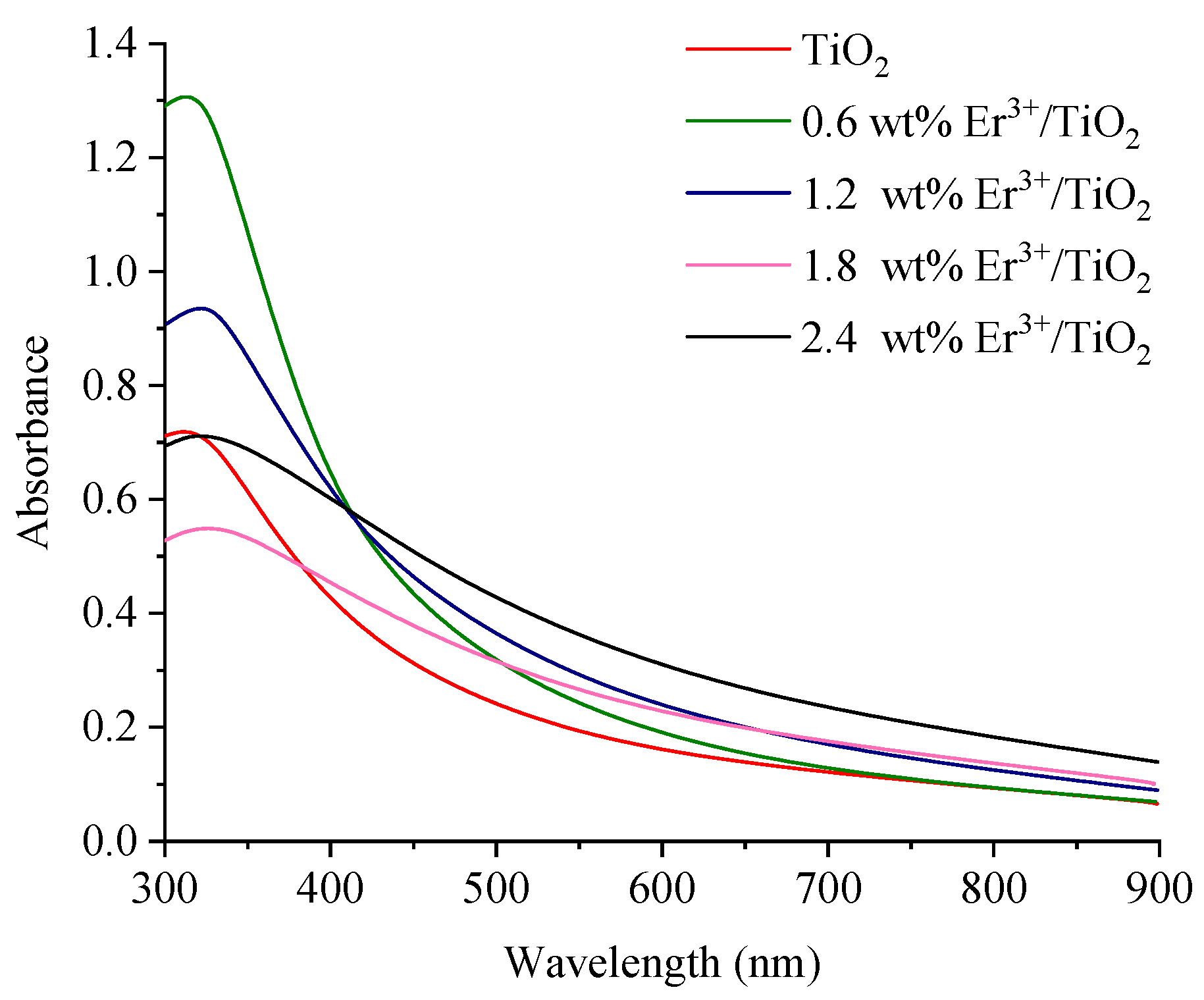
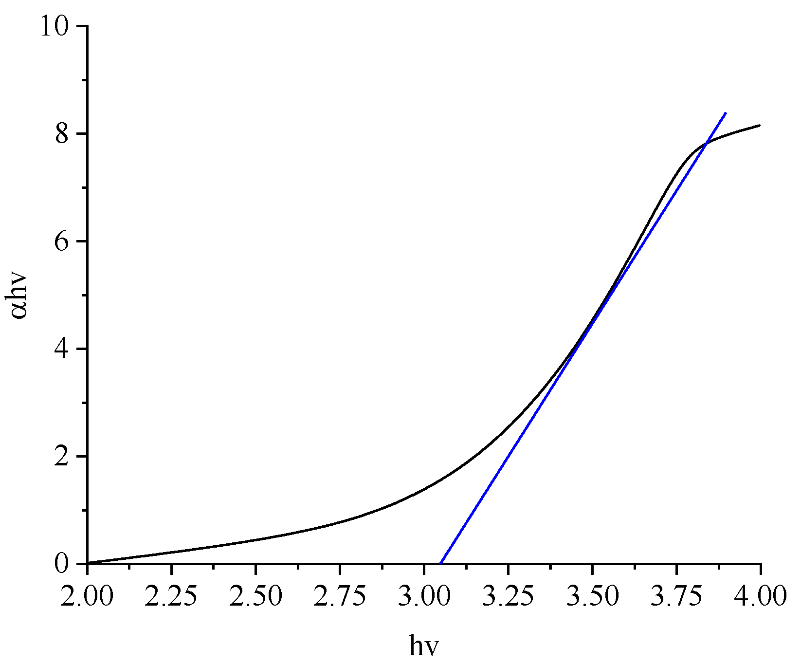
The Er3+/TiO2 NPs surface morphologies were examined utilizing FESEM and EDX in addition to metal mapping. The FESEM images are demonstrated in Figure 2. The images of the Er3+/TiO2 sample demonstrate the particles’ shapes, which showed less agglomeration and were generally spherical. EDX analysis was performed on the Er3+/TiO2 samples to study the presence of Er ions on the surfaces of the examined TiO2. The results of EDX spectra analysis are provided in Figure 3, which demonstrates that the Er ions were present in all investigated samples. In addition, the EDX analysis spectral results of the different weight percentages of the Er content in the tested samples are listed in Table 3. The theoretical and empirical Er contents show considerable agreement. Metal mapping tests were performed to study the Er ions’ dispersion on the surface of the TiO2 NPs. These data are summarized in Figure 4, which reveals a considerable distribution of Er ions on the TiO2 NPs in all synthesized samples.

3.3. Energy Gaps

The absorbance spectra of the synthesized NPs samples were obtained at room temperature and are illustrated in Figure 5. A significant absorption peak in the UV–Vis spectrum is located at 320 nm. According to Equation (6), the energy gaps for the synthesized samples of NPs were calculated:
α h ν = E d ( h ν E g ) 1 2
where α represents the optical coefficient; h refers to the Planck constant; the multiplication of hv represents the photon energy; Eg and Ed represent the energy gap and a constant, respectively [40]. (αhv) versus hv was drawn, the linear section of the curve was extrapolated, and Eg was evaluated, as shown in Figure 6. The Eg values of TiO2 NPs for different weight percentages of Er ions are given in Table 4. The lowest energy gap value obtained was 2.63 eV for Er ions/titanium dioxide NPs.

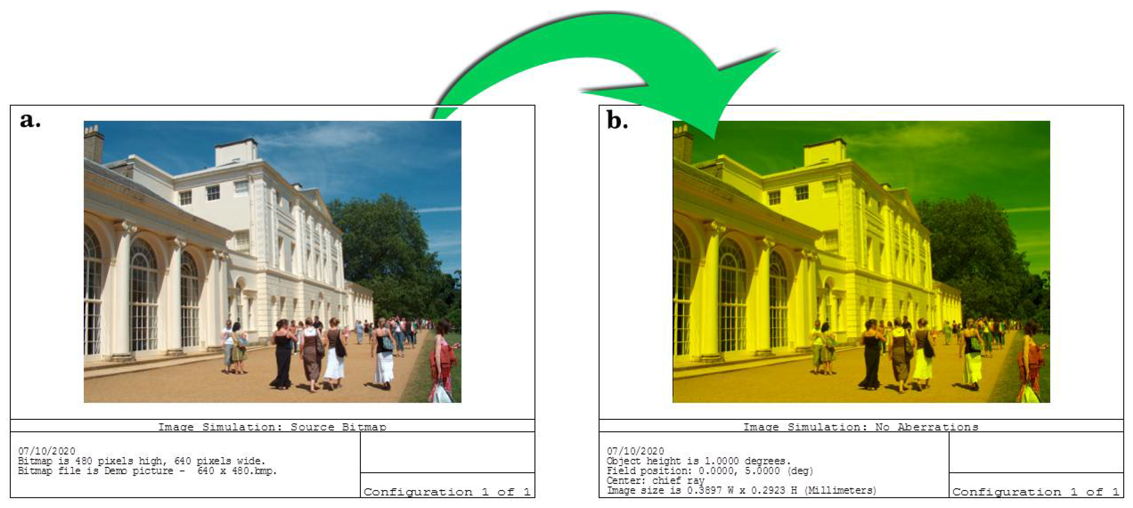
3.4. Application as a Blue-Wavelength-Blocking Contact Lens

Given most people’s modern lifestyles, they spend the majority of their waking hours staring at digital devices. According to the previous studies, 60% of people spend 6–7 h per day looking directly at digital screens [41,42]. Digital screens, sunlight, electronics, and fluorescent light sources emit a blue wavelength ranging from 300 to 500 nm. This range is harmful to the eye (damages the retinal photoreceptors and destroys the crystalline lens protein that appears as a white cloud) [43].
The human eye’s natural filters do not protect the eye’s components against these highest-energy wavelengths. The excessive exposure to blue light rays may affect the retinal tissues and cause damage. This contributes to age-related macular degradation and subsequent loss of vision. In this work, 0.6% wt/vol Er3+/TiO2 solution drops were spin-coated on a previously prepared poly(methyl methacrylate)—titanium dioxide (PMMA-TiO2) contact lens fixed on a convex glass mold (Figure 7a) [11]. The refractive index was measured using an Abbe Refractometer at wavelengths of 486.1, 587.6, and 656.3 nm as 1.509, 1.503, and 1.499, respectively. The Abbe number was calculated as 52.35 using the following equation [44]:
ν d = ( n d 1 ) ( n f n c )
where νd is the Abbe number; and nc, nd, and nf are refractive indices of the polymer films at wavelengths of 656, 589, and 486 nm, respectively.
From the results provided above, it can be found that the fabricated coated contact lens works in the acceptable physical range according to the refractive index, which is near the refractive indices of the eye tissues. The resulting contact lens was modeled using ZEMAX optical design software (version 14) to evaluate the blue-wavelength-blocking characteristics before conducting an in vivo study. The Liou and Brennan eye model was chosen in this study because it was experimentally obtained from the human eye [45]. The Er3+/TiO2 coating worked as a blue wavelength filter, where it prevented the wavelength range of 0.45–0.47 nm from being transmitted (reduced the light transmittance to 0%) through the contact lens, as illustrated in Figure 7c.
The Er3+/TiO2 coating does not degrade the retinal image’s clarity, as shown in Figure 8. To compare the retinal image before and after coating, image simulation was used for evaluation. A polychromatic light source was applied to the modeled eye, a perfect eye with a high retinal image contrast, and sharpness was obtained by the eye with the PMMA-TiO2 contact lens with no aberrations, as shown in Figure 8a. After, the resultant PMMA-TiO2 contact lens coated with Er3+/TiO2 layer was fitted on the modeled eye, and the image at the retina showed high clarity and no indication of aberration generation. This indicates that the coated lens focused the rays entering the eye in one small spot, which is the focal point. This performance was obtained due to n value of the resulting lens and the dispersion factor, which is able to maintain image clarity under polychromatic light illumination. The speed of each component of white light differs from the speed of the other wavelengths according to the concept of the refractive index (n = c/v). The blocking lens color is suitable and not too deep, so it prevents color deviation and darkness. The designed contact lens can work as a filter or polarizer, blocking the reflected blue portion of light from the object and passing the other components of the light through to the eye. From observing the sharpness and contrast of the two images before and after the application of the coating, we found the coating does not affect the visual function of the eye.

4. Conclusions

Titanium dioxide NPs were impregnated with different weight percentages of Er ions using the impregnation technique and then calcination of the product at 500 °C. The Er3+/TiO2 NPs showed an energy gap decrease with increasing Er ions weights percent up to 1.8 wt %. Agglomeration and particle size increased with increasing Er ions weight percent. Relative to the long hours people spend staring at digital displays, researchers are developing eyeglass lenses that partially filter the high-energy blue wavelength. Due to the problems with glasses such as their weight, the possibility of breaking them, amongst others, we attempted to design and manufacture a contact lens with a high n value that works as a filter to reduce the transmission of blue light intensity without affecting the clarity of the image. The fabricated spin-coated Er3+/TiO2 layer on the PMMA-TiO2 contact lens works as a filter as it absorbs the high-energy blue wavelength and passes other components of light through to the eye. A perfect polarized retinal image was achieved, and no aberration was generated.

Author Contributions

Conceptualization, A.A. and L.M.S.; methodology, A.A. and L.M.S. software, L.M.S.; validation, W.N.R.W.I. and M.T.; formal analysis, L.M.S.; investigation, A.A. and L.M.S.; resources, W.N.R.W.I. and M.T.; data curation, L.M.S.; writing—original draft preparation, A.A. and L.M.S.; writing—review and editing, A.A.; visualization, M.T.; supervision, A.A., W.N.R.W.I. and M.T.; project administration, A.A., W.N.R.W.I. and M.T.; funding acquisition, W.N.R.W.I. and M.T. All authors have read and agreed to the published version of the manuscript.

Funding

This research was funded by Universiti Kebangsaan Malaysia under grant GUP2020-012.

Institutional Review Board Statement

Not applicable.

Informed Consent Statement

Not applicable.

Data Availability Statement

The data of this study are available from the corresponding author (A.A.), upon reasonable request.

Acknowledgments

The authors thank Universiti Kebangsaan Malaysia (Malaysia) and University of Technology (Iraq) for supporting this work.

Conflicts of Interest

The authors declare no conflict of interest.

References

  1. Lindenthal, L.; Raffael, R.; Harald, S.; Thomas, R.; Janko, P.; Andreas, N.; Stefan, L.; Alexander, K.O.; Peter, B.; Christoph, R. Modifying the Surface Structure of Perovskite-Based Catalysts by Nanoparticle Exsolution. Catalysts 2020, 10, 268. [Google Scholar] [CrossRef] [Green Version]
  2. Abdel-Khalek, N.A.; Abdel-Khalek, M.A.D.; Selim, K.A.; Abdullah, S.S. Physicochemical study and application for pyrolusite separation from high manganese-iron ore in the presence of microorganisms. Physicochem. Probl. Miner. Process. 2021, 57, 273–283. [Google Scholar]
  3. Ahmad, H.; Kamarudin, S.K.; Minggu, L.J.; Hasran, U.A.; Masdar, S.; Daud, W.R.W. Enhancing methanol oxidation with a TiO2-modified semiconductor as a photo-catalyst. Int. J. Hydrog. Energy 2017, 42, 8986–8996. [Google Scholar] [CrossRef]
  4. Wasmi, B.; Al-Amiery, A.A.; Kadhum, A.A.H.; Takriff, M.S.; Mohamad, A.B. Synthesis of vanadium pentoxide nanoparticles as catalysts for the ozonation of palm oil. Ozone Sci. Eng. 2016, 38, 36–41. [Google Scholar] [CrossRef]
  5. Zheng, M.; Wang, Y.; Feng, P. Bifunctional heterometallic metal-organic frameworks for solvent-free heterogeneous cascade catalysis. Catalysts 2020, 10, 309. [Google Scholar] [CrossRef] [Green Version]
  6. Sandu, M.P.; Sidelnikov, V.S.; Geraskin, A.A.; Chernyavskii, A.V.; Kurzina, I.A. Influence of the method of preparation of the Pd-Bi/Al2O3 catalyst on catalytic properties in the reaction of liquid-phase oxidation of glucose into gluconic acid. Catalysts 2020, 10, 271. [Google Scholar] [CrossRef] [Green Version]
  7. Yu, H.; Song, L.; Hao, Y.; Lu, N.; Quan, X.; Chen, S.; Zhang, Y.; Feng, Y. Fabrication of pilot-scale photocatalytic disinfection device by installing TiO2 coated helical support into UV annular reactor for strengthening sterilization. Chem. Eng. J. 2016, 283, 1506–1513. [Google Scholar] [CrossRef]
  8. Wang, F.; Zhang, H.; Yu, B.; Wang, S.; Shen, Y.; Cong, H. Review of the research on anti-protein fouling coatings materials. Prog. Org. Coat. 2020, 147, 105860. [Google Scholar] [CrossRef]
  9. Melgoza-Ramírez, M.L.; Ramírez-Bon, R. Europium ions as a spectroscopic probe in the study of PMMA-SiO2 hybrid microstructure with variable coupling agent. J. Sol-Gel Sci. Technol. 2021, 99, 1–11. [Google Scholar]
  10. Shaker, L.M.; Al-Amiery, A.A.; Kadhum, A.A.H.; Takriff, M.S. Manufacture of Contact Lens of Nanoparticle-Doped Polymer Complemented with ZEMAX. Nanomaterials 2020, 10, 2028. [Google Scholar] [CrossRef] [PubMed]
  11. Harb, S.V.; Bassous, N.J.; de Souza, T.A.; Trentin, A.; Pulcinelli, S.H.; Santilli, C.V.; Webster, T.J.; Lobo, A.O.; Hammer, P. Hydroxyapatite and β-TCP modified PMMA-TiO2 and PMMA-ZrO2 coatings for bioactive corrosion protection of Ti6Al4V implants. Mater. Sci. Eng. C 2020, 116, 111149. [Google Scholar] [CrossRef]
  12. Savchyn, V.P.; Popov, A.I.; Aksimentyeva, O.I.; Klym, H.; Horbenko, Y.Y.; Serga, V.; Moskina, A.; Karbovnyk, I. Cathodoluminescence characterization of polystyrene-BaZrO3 hybrid composites. Low Temp. Phys. 2016, 42, 597–600. [Google Scholar] [CrossRef]
  13. Aksimentyeva, O.I.; Savchyn, V.P.; Dyakonov, V.P.; Piechota, S.; Horbenko, Y.Y.; Opainych, I.Y.; Demchenko, P.Y.; Popov, A.; Szymczak, H. Modification of polymer-magnetic nanoparticles by luminescent and conducting substances. Mol. Cryst. Liq. Cryst. 2014, 590, 35–42. [Google Scholar] [CrossRef]
  14. Li, F.; Kong, W.; Bhushan, B.; Zhao, X.; Pan, Y. Ultraviolet-driven switchable superliquiphobic/superliquiphilic coating for separation of oil-water mixtures and emulsions and water purification. J. Colloid Interface Sci. 2019, 557, 395–407. [Google Scholar] [CrossRef]
  15. Umar, A.A.; Saad, S.K.M.; Umar, M.I.A.; Abd Rahman, M.Y.; Oyama, M. Advances in porous and high-energy (001)-faceted anatase TiO2 nanostructures. Opt. Mater. 2018, 75, 390–430. [Google Scholar] [CrossRef]
  16. Li, X.Z.; Li, F.B. Study of Au/Au3+-TiO2 photocatalysts toward visible photooxidation for water and wastewater treatment. Environ. Sci. Technol. 2001, 35, 2381–2387. [Google Scholar] [CrossRef]
  17. Manassero, A.; Satuf, M.L.; Alfano, O.M. Evaluation of UV and visible light activity of TiO2 catalysts for water remediation. Chem. Eng. J. 2013, 225, 378–386. [Google Scholar] [CrossRef]
  18. Hoffmann, M.R.; Martin, S.T.; Choi, W.; Bahnemann, D.W. Environmental Applications of Semiconductor Photocatalysis. Chem. Rev. 1995, 95, 69–96. [Google Scholar] [CrossRef]
  19. Liqiang, J.; Yichun, Q.; Baiqi, W.; Shudan, L.; Baojiang, J.; Libin, Y.; Wei, F.; Honggang, F.; Jiazhong, S. Review of Photoluminescence Performance of Nano-Sized Semiconductor Materials and Its Relationships with Photocatalytic Activity. Sol. Energ. Mater. Sol. Cells 2006, 90, 1773–1787. [Google Scholar] [CrossRef]
  20. Linsebigler, A.; Lu, G.; Yates, J.T. Photocatalysis on TiO2 Surfaces: Principles, Mechanisms, and Selected Results. Chem. Rev. 1995, 95, 735–758. [Google Scholar] [CrossRef]
  21. Li, H.; Bian, Z.; Zhu, J.; Huo, Y.; Li, H.; Lu, Y. Mesoporous Au/TiO2 Nanocomposites with Enhanced Photocatalytic Activity. J. Am. Chem. Soc. 2007, 129, 4538–4539. [Google Scholar] [CrossRef] [PubMed]
  22. Zaleska, A. Doped-TiO2: A Review. Recent Pat. Eng. 2008, 2, 157–164. [Google Scholar] [CrossRef]
  23. Di Paola, A.; MarcI, G.; Palmisano, L.; Schiavello, M.; Uosaki, K.; Ikeda, S.; Ohtani, B. Preparation of Polycrystalline TiO2 Photocatalysts Impregnated with Various Transition Metal Ions: Characterization and Photocatalytic Activity for the Degradation of 4-Nitrophenol. J. Phys. Chem. B 2002, 106, 637–645. [Google Scholar] [CrossRef] [Green Version]
  24. Sakthivel, S.; Shankar, M.V.; Palanichamy, M.; Arabindoo, B.; Bahnemann, D.W.; Murugesan, V. Enhancement of Photocatalytic Activity by Metal Deposition: Characterization and Photonic Efficiency of Pt, Au and Pd Deposited on TiO2 Catalyst. Water Res. 2004, 38, 3001–3008. [Google Scholar] [CrossRef]
  25. Chen, X.; Mao, S.S. Titanium Dioxide Nanomaterials: Synthesis, Properties, Modifications, and Applications. Chem. Rev. 2007, 107, 2891–2959. [Google Scholar] [CrossRef]
  26. Ohtani, B.; Iwai, K.; Nishimoto, S.; Sato, S. Role of Platinum Deposits on Titanium(IV) Oxide Particles: Structural and Kinetic Analyses of Photocatalytic Reaction in Aqueous Alcohol and Amino Acid Solutions. J. Phys. Chem. B 1997, 101, 3349–3359. [Google Scholar] [CrossRef] [Green Version]
  27. Ibhadon, A.O.; Fitzpatrick, P. Heterogeneous photocatalysis: Recent advances and applications. Catalysts 2013, 3, 189–218. [Google Scholar] [CrossRef] [Green Version]
  28. Estrellan, C.R.; Salim, C.; Hinode, H. Photocatalytic activity of sol–gel derived TiO2 Co-doped with iron and niobium. React. Kinet. Catal. Lett. 2009, 98, 187–192. [Google Scholar] [CrossRef]
  29. Yu, C.L.; Yang, K.; Yu, J.; Peng, P.; Cao, F.; Li, X.; Zhou, X.C. Effects of rare earth Ce doping on the structure and photocatalytic performance of ZnO. Acta Phys. Chim. Sin. 2011, 27, 505–512. [Google Scholar]
  30. Minero, C.; Pelizzatti, E.; Sega, M.; Friberg, S.E.; Sjoblom, J. The role of humic substances in photocatalytic degradation of water contaminants. J. Dispers. Sci. Technol. 1999, 20, 643–661. [Google Scholar] [CrossRef]
  31. Mohammad, G.; Mohammed, W.; Marzoog, T.R.; Al-Amiery, A.; Kadhumc, A.; Mohamad, A. Green synthesis, antimicrobial and cytotoxic effects of silver nanoparticles using Eucalyptus chapmaniana leaves extract. Asian Pac. J. Trop. Biomed. 2013, 3, 58–63. [Google Scholar]
  32. Gaaz, T.S.; Sulong, A.B.; Kadhum, A.A.H.K.; Nassir, M.H.; Al-Amiery, A.A. Impact of sulfuric acid treatment of halloysite on physico-chemic property modification. Materials 2016, 9, 620. [Google Scholar] [CrossRef]
  33. Gaaz, T.S.; Sulong, A.B.; Akhtar, M.N.; Kadhum, A.A.H.; Mohamad, A.B.; Al-Amiery, A.A. Properties and applications of polyvinyl alcohol, halloysite nanotubes and their nanocomposites. Molecules 2015, 20, 22833–22847. [Google Scholar] [CrossRef] [Green Version]
  34. Obayes, H.; Al-Gebori, A.; Khazaal, S.; Jarad, A.; Alwan, G.; Al-Amiery, A. Hypothetical design of carbon nanotube materials based on [8] circulene. J. Nanoelectron. Optoelectron. 2015, 10, 711–716. [Google Scholar] [CrossRef]
  35. Wasmi, W.; Al-Amiery, A.; Kadhum, A.; Mohamad, A. Novel approach: Tungsten oxide nanoparticle as a catalyst for malonic acid ester synthesis via ozonolysis. J. Nanomater. 2014, 2014, 1–7. [Google Scholar] [CrossRef]
  36. Serga, V.; Burve, R.; Krumina, A.; Romanova, M.; Kotomin, E.A.; Popov, A.I. Extraction–Pyrolytic Method for TiO2 Polymorphs Production. Crystals 2021, 11, 431. [Google Scholar] [CrossRef]
  37. Tsebriienko, T.; Popov, A.I. Effect of Poly(Titanium Oxide) on the Viscoelastic and Thermophysical Properties of Interpenetrating Polymer Networks. Crystals 2021, 11, 794. [Google Scholar] [CrossRef]
  38. Serga, V.; Burve, R.; Krumina, A.; Pankratova, V.; Popov, A.I.; Pankratov, V. Study of phase composition, photocatalytic activity, and photoluminescence of TiO2 with Eu additive produced by the extraction-pyrolytic method. J. Mater. Res. Technol. 2021, 13, 2350–2360. [Google Scholar] [CrossRef]
  39. Dahl, M.; Liu, Y.; Yin, Y. Composite titanium dioxide nanomaterials. Chem. Rev. 2014, 114, 9853–9889. [Google Scholar] [CrossRef]
  40. Vijayalakshmi, R.; Rajendran, V. Synthesis and characterization of nano-TiO2 via different methods. Arch. Appl. Sci. Res. 2012, 4, 1183–1190. [Google Scholar]
  41. Rohatgi, S.; Hanna, T.N.; Sliker, C.W.; Abbott, R.M.; Nicola, R. After-hours radiology: Challenges and strategies for the radiologist. Am. J. Roentgenol. 2015, 205, 956–961. [Google Scholar] [CrossRef] [PubMed]
  42. Waite, S.; Kolla, S.; Jeudy, J.; Legasto, A.; Macknik, S.L.; Martinez-Conde, S.; Krupinski, E.A.; Reede, D.L. Tired in the reading room: The influence of fatigue in radiology. J. Am. Coll Radiol. 2017, 14, 191–197. [Google Scholar] [CrossRef]
  43. Michael, R.; Wegener, A. Estimation of safe exposure time from an ophthalmic operating microscope with regard to ultraviolet radiation and blue-light hazards to the eye. J. Opt. Soc. Am. A 2004, 21, 1388–1392. [Google Scholar] [CrossRef] [PubMed]
  44. Michael, A.S.; Schwiegerling, J.; Fest, E.; Hamilton, R. Visual Optics, in Molded Optics: Design and Manufacture; CRC Press: Boca Raton, FL, USA, 2016; pp. 2–58. [Google Scholar]
  45. Brennan, N.A.; Liou, H.-L. Anatomically accurate, finite model eye for optical modeling. Opt. Soc. Am. 1997, 14, 1684–1695. [Google Scholar]
Figure 1. XRD: (a) titanium dioxide; (b) 0.6 wt %; (c) 1.2 wt %; (d) 1.8 wt %; (e) 2.4 wt % erbium ions/titanium dioxide.
Nanomaterials 11 02190 g001
Figure 2. FESEM micrographs for the synthesized samples.
Nanomaterials 11 02190 g002aNanomaterials 11 02190 g002b
Figure 3. EDX micrographs of the synthesized samples.
Nanomaterials 11 02190 g003
Figure 4. Metal mapping of the synthesized Er3+/TiO2 NPs.
Nanomaterials 11 02190 g004
Figure 5. Ultraviolet–visible spectra of the synthesized NPs.
Nanomaterials 11 02190 g005
Figure 6. Calculation of the direct band gap energy obtained in our study.
Nanomaterials 11 02190 g006
Figure 7. (a) Spin-coating a contact lens with 0.6 wt/vol % Er3+/TiO2 solution; (b) The layers of the resulting contact lens; (c) intensity transmission as a function of wavelength blocking 0.45–0.47 nm from transmission.
Nanomaterials 11 02190 g007
Figure 8. (a) The retinal image resulting from the eye with a PMMA-TiO2 contact lens; (b) the retinal image resulting from the eye with a PMMA-TiO2 contact lens coated with Er3+/TiO2 NPs.
Nanomaterials 11 02190 g008
Table 1. Erbium ions/titanium dioxide intensity signals.
Intensity
TiO20.6 wt % Er3+/TiO21.2 wt % Er3+/TiO21.8 wt % Er3+/TiO22.4 wt % Er3+/TiO2
25.271221581984191118791855
27.4136515498493481474
37.30381271051009090
48.0908555536481478473
53.8206311310288262230
53.97379368351346336
55.116367340313291283
62.7142292285272249241
68.8675165149143131120
70.2128145114109108104
75.0956185164163151147
Table 2. Size of crystallite particles of all synthesized samples.
Er3+/TiO2 (wt %)Crystallite (nm)
TiO228.72
0.628.84
1.231.53
1.833.93
2.435.93
Table 3. EDX analysis of the theoretical and empirical values of the synthesized samples.
Er3+/TiO2 (wt %)Calculated Metal (wt %)Actual Metal (wt %) by EDX
0.60.60.72
1.21.21.31
1.81.82.06
2.42.42.45
Table 4. Energy gaps for the synthesized Er3+/TiO2 NP samples.
Er3+/TiO2 (wt %)Energy Gap (eV)
TiO23.13 ± 0.02
0.63.20 ± 0.02
1.23.00 ± 0.02
1.82.63 ± 0.02
2.42.70 ± 0.02
Publisher’s Note: MDPI stays neutral with regard to jurisdictional claims in published maps and institutional affiliations.

