Article

Minute-Made, High-Efficiency Nanostructured Bi2Te3 via High-Throughput Green Solution Chemical Synthesis

1
Department of Applied Physics, KTH Royal Institute of Technology, SE-106 91 Stockholm, Sweden
2
Department of Physics, University of Istanbul, Istanbul 34135, Turkey
3
Institute of Experimental Physics, University of Wroclaw, Maxa Borna 9, 50-204 Wroclaw, Poland
4
Department of Materials and Environmental Chemistry, Stockholm University, SE-106 91 Stockholm, Sweden
*
Authors to whom correspondence should be addressed.
Nanomaterials 2021, 11(8), 2053; https://doi.org/10.3390/nano11082053
Submission received: 19 July 2021 / Revised: 9 August 2021 / Accepted: 11 August 2021 / Published: 12 August 2021
(This article belongs to the Special Issue Thermoelectric Properties of Nanomaterials)

Abstract

:
Scalable synthetic strategies for high-quality and reproducible thermoelectric (TE) materials is an essential step for advancing the TE technology. We present here very rapid and effective methods for the synthesis of nanostructured bismuth telluride materials with promising TE performance. The methodology is based on an effective volume heating using microwaves, leading to highly crystalline nanostructured powders, in a reaction duration of two minutes. As the solvents, we demonstrate that water with a high dielectric constant is as good a solvent as ethylene glycol (EG) for the synthetic process, providing a greener reaction media. Crystal structure, crystallinity, morphology, microstructure and surface chemistry of these materials were evaluated using XRD, SEM/TEM, XPS and zeta potential characterization techniques. Nanostructured particles with hexagonal platelet morphology were observed in both systems. Surfaces show various degrees of oxidation, and signatures of the precursors used. Thermoelectric transport properties were evaluated using electrical conductivity, Seebeck coefficient and thermal conductivity measurements to estimate the TE figure-of-merit, ZT. Low thermal conductivity values were obtained, mainly due to the increased density of boundaries via materials nanostructuring. The estimated ZT values of 0.8–0.9 was reached in the 300–375 K temperature range for the hydrothermally synthesized sample, while 0.9–1 was reached in the 425–525 K temperature range for the polyol (EG) sample. Considering the energy and time efficiency of the synthetic processes developed in this work, these are rather promising ZT values paving the way for a wider impact of these strategic materials with a minimum environmental impact.

Graphical Abstract

1. Introduction

Our heavy dependence on non-renewable sources have led to a global energy crisis and as the consumption of these sources has increased, the prospect of their availability for a long term has reduced [1]. Certainly, to meet future energy challenges and climate change it is of paramount importance to develop renewable, and sustainable energy solutions. Thermoelectric (TE) materials have received significant attention because of their ability to convert waste heat to electrical power directly [2]. They can be used to harvest low- and high-grade waste-heat effectively, which requires the selection of proper material for the intended temperature range. As TE materials can be used for power generation, they can also be used for cooling or thermal management. TE devices have attracted significant interest because of their potential applications in energy harvesting, waste heat recovery in industries and transportation systems, chip cooling, solar cells, temperature sensing, military and space exploration [3,4,5,6,7]. Conventional TE devices are made of compact solid-state materials without any moving parts, which gives them a high operational reliability and makes them scalable [8,9]. The performance of the TE devices is strongly dependant on the efficiency of the TE materials [10], which has been formulated as a dimensionless quantity, the TE figure of merit (ZT) and is composed of electrical and thermal transport terms. Specifically, it is defined as ZT = S2σT/κ (where S is the Seebeck coefficient, σ is the electrical conductivity, T is the absolute temperature, and κ is the thermal conductivity, which is the sum of electronic (κel) and lattice (κlat) components) [11]. The desired qualities in good TEs are high σ, large S accompanied by a low κ. Nanostructuring has become an important approach in the field of TE for controlling the thermal conductivity κ independent from the electrical conductivity σ [12]. Considerable amount of research has shown that low-dimensional structures could significantly improve the TE performance due to quantum confinement and scattering of a broad range of heat carrier phonons [13,14]. This concept yielded some strategies for the enhancement of ZT by reducing κ, while maintaining σ, by taking advantage of different phonon and electron mean free paths (MFPs). The κ is more sensitive to nanostructuring than the σ due to the longer MFP of the thermal phonons than that of the charge carriers [15]. Since in nanostructures there is a large density of interface, the phonons are scattered more effectively than the electrons [16]. Thus, due to the phonon blocking effect, the κlat reduces in nanostructures while not affecting the mobility and σ significantly. The enhanced ZT in TE materials has been predicted, or experimentally demonstrated for nanostructures like quantum dots [17], thin film superlattices [18], 1D structures–nanowires [19], nanotubes [20], and 2D quantum wells [21]. Microstructure engineering through targeted nanostructure/matrix crystallographic alignment was shown to create endotaxially placed nanoscale precipitates within the matrix, leading to very low thermal conductivity values [22]. Well-controlled nanoscale-shaped interfaces and oriented nanocrystals were also demonstrated to reduce κ beyond the amorphous limit, while preserving bulk-like σ [12]. Rather promising ZT was achieved in roughened Si nanowires, which has been mainly attributed to efficient scattering throughout the phonon spectrum by the introduction of nanostructures at different length scales (diameter, roughness and point defects) [23].
The most common n-type TE material for room temperature operations is Bi2Te3. A survey on the different methods of its synthesis and the variety of morphologies has been presented in the supplementary file Table S1. Over the years, solid-state approaches like mechanochemical alloying has been used to synthesize (Bi,Sb)2Te3, however, the risk of contamination, high cost of high-energy ball milling and longer hours of ball milling, and significant batch to batch variations make this method inconvenient [24,25]. Bottom up chemical methods are commonly used because of their low cost, low investment need and possibility of scale-up of the synthesis process [26]. Different morphologies of Bi2−xSbxTe3 including nanoparticles, nanoplates, nanocrystalline films, nanorods, nanotubes, nanowires and nanoflowers have been synthesized by reverse micelle [27], metal-organic chemical vapor deposition (MOCVD) [28,29,30], vapor–liquid–solid (VLS) [31,32], refluxing [33,34,35,36], electrodeposition [37,38], chemical reduction [39,40], solvothermal [41,42,43,44,45,46,47,48,49], hydrothermal [26,50,51,52,53,54,55,56,57,58], ultrasonic-assisted [59,60,61], and microwave (MW)-assisted [62,63,64,65,66] routes, to name a few. Among all these chemical syntheses methods, solvothermal/hydrothermal routes are the preferred methods to prepare nanostructures with different morphologies. However, a major drawback in solvothermal/hydrothermal methods is the reaction time that can range from several hours to days [26,42,46,52,57,58]. This shortcoming can be resolved by introducing energy-effective MW-assisted heating.
MW technology is emerging as an alternative energy source for chemical synthesis with the aim to reduce the reaction time from hours or even days to a few minutes [64,65,67,68,69,70]. The dielectric constant of the solvent may significantly influence the outcome of the MW-assisted reaction. MW speeds up the chemical reaction by increasing the overall kinetics of the reaction as it relies on localized superheating [71]. Other advantages of using MW heating are higher yields, and since the reaction is kinetically driven it ensures a narrow-size distribution of the particles [72]. A variety of nanomaterials have been synthesized with MWs, including 0D [63,72,73,74,75,76,77,78], 1D [67,68,79,80] and 2D structures [81]. MW-assisted synthesis is also a sustainable, environmentally friendly, and economically viable heating method for bulk commercial scale production of strategic nanomaterials.
Most chemical syntheses use a large amount of conventional volatile or other organic chemicals, which create a pressing environmental concern. Therefore, the aim should be to minimize the use of toxic chemicals and follow the principles of green chemistry to synthesize nanomaterials [82]. The selection of the solvent for a reaction can dramatically affect the reaction outcome. Ethylene glycol (EG) is an abundant resource in the chemical industry, which can be produced from renewable biomass [83]. It is an odourless, non-volatile, low toxic solvent with a low dielectric constant (37 at room temperature), which has enormous applications in various industrial processes [84], including the polyol synthesis [64]. Water is the most environmentally friendly (i.e., green) solvent with a high dielectric constant (78 at room temperature), which is very effective for transferring electromagnetic energy into heat and thus driving the reduction reactions effectively [85]. Under MW heating, the superheating of water causes the hydrogen bonds to break easily, making it less polar and the self-ionization of water increases with temperature [62,86]. Due to superheating, the dielectric constant of water decreases at high temperatures (above its boiling point) and this influences the growth of the nanoparticles [86]. It has been reported that Bi2Te3 particles grow more steadily in a solvent with low dielectric constant than in a solvent with a high dielectric constant [41]. Therefore, this makes the combination of MW heating and hydrothermal methods a suitable chemical synthesis route for the growth of nanocrystals. The pH of the reaction medium is key for the synthesis of the Bi2Te3 nanocrystals. It has been reported that an alkaline medium (pH < 11–12) is required for the formation of Bi2Te3 nanostructures [26,43,52,53,57,87]. The alkaline medium helps the tellurium to disperse in the reaction medium as telluride ions (Te2−), which is beneficial for the formation of the nanocrystals [46]. A growth promoting agent, like ethylenediaminetetraacetic acid (EDTA), is required to form the bismuth-antimony complexes which self-assemble to lamellar phases leading to the formation of Bi2Te3 sheet nuclei, which facilitates the formation of nanosheets, nanoflakes and nanotubes [53,59]. The activity of EDTA is also dependent on the pH of the solution and by changing the pH, different morphologies of Bi2Te3 have been obtained [51,52,53,54,55,56,87]. Control of the reaction time and temperature are essential in determining the structure and morphology of the nanocrystals [49,57]. It can be deduced that low temperature is suitable for the reaction of nanoparticles, nanoplates and nanotubes and as the reaction temperature increases, the dimensionality changes from 0D to 1D. The effect of reaction time and temperature on the morphology, structure, and crystallinity of Bi2Te3 have been comprehensively summarized in Table S1.
We aim to provide facile and sustainable synthetic routes for large-scale production of Bi2Te3 nanomaterials with promising TE performance. The design of the experiments has been done in-line with the green chemistry perspectives considering the solvents, precursors and the energy source. Taking advantage of the unique MW dielectric heating phenomenon, rapid heating and short reaction time, hexagonal nanoplatelets of Bi2Te3 were obtained. These low dimensional structures have been effective in enhancing the charge transport properties and electrical conductivity, while reducing the thermal conductivity. Water and ethylene glycol are used as solvents making the reaction green, safe and non-hazardous, producing high purity TE nanopowders in a matter of minutes due to very effective volume heating—despite their significantly different dielectric constants. Besides yielding a large quantity of the investigated materials per batch, surface chemistry and the processing of these materials towards compacts are described, resulting in high ZT values of 1.03 and 0.88 for Bi2Te3 materials synthesized through the polyol and the hydrothermal routes, respectively.

2. Materials and Methods

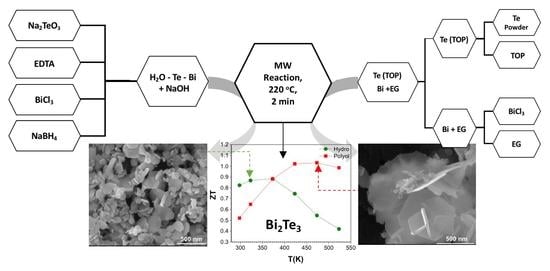
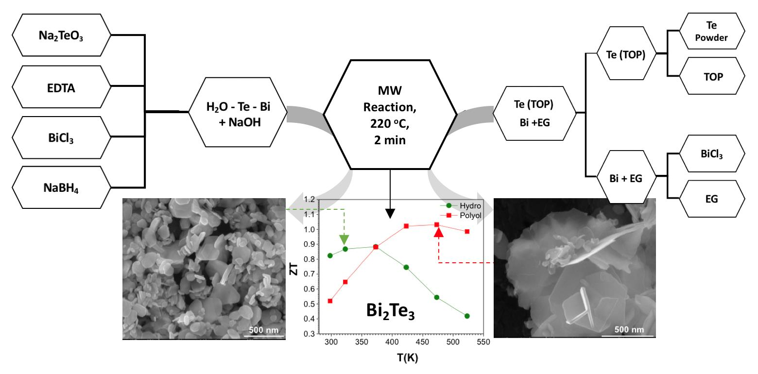
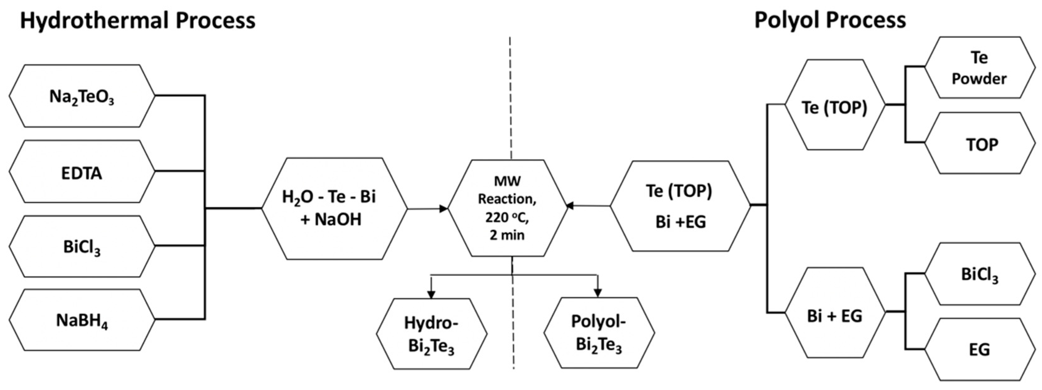
2.1. Synthesis and Processing of Thermoelectric Materials

2.1.1. Synthesis by Hydrothermal Method

Chemicals used for the synthesis were of analytical grade obtained from Sigma Aldrich (Stockholm, Sweden) and have been used as received without further purification: Bismuth chloride (BiCl3, 98% purity), sodium tellurite, (Na₂TeO₃, 99% purity), sodium hydroxide (NaOH, 97.0% purity) and sodium borohydride (NaBH4, 98% purity). BiCl3 and Na₂TeO₃ were used as the precursors for Bi and Te ions, respectively. Deionized (DI) water was used as the solvent, EDTA as the structure-directing agent, NaBH4 as the reducing agent and NaOH to control the pH of the reaction media. The synthesis was performed in a Milestone flexiWAVE MW system (MILESTONE Srl, Sorisole, Italy) (900 watt magnetrons for a total of 1800 watt). One-pot synthesis was achieved by mixing all the precursors in the same media. For the synthesis of Bi2Te3, Bi:Te molar ratio of 2:3 was used. In a 100 mL Teflon vial, BiCl3, Na₂TeO₃, EDTA, and NaBH4 were mixed where the pH was adjusted to 10.5–11 by the addition of NaOH. Two parallel reaction vessels were used in a single run. The reaction vials were kept under constant magnetic stirring for 30 min to ensure a homogeneous mixing. This was then subjected to MW heating to 220 °C with a ramp time of 4 min and dwell time of 2 min. The solution was then allowed to cool down to room temperature and a clear phase separation was observed with the product settled at the bottom, separated from water. The particles formed were easily separated from the reaction mixture by centrifugation and were then washed with isopropanol, acetone and water. Thereafter, the powder was dried in a vacuum oven at 60 °C. A schematic of the hydrothermal synthesis process is given in Figure 1. The sample is designated as hydro-Bi2Te3 throughout the manuscript.

2.1.2. Synthesis by Polyol Method

All the chemicals used for the synthesis reaction were of analytical grade obtained from Sigma Aldrich and were used as received without further purification: BiCl3 (98% purity), Te powder (99.8% purity), ethylene glycol (EG, 99% purity thioglycolic acid (TGA, 98% purity), and trioctyl phosphine (TOP, 90% purity). The synthesis was performed in the same Milestone flexiWAVE MW system, as described in our earlier work [64]. Typically, BiCl3 was dissolved in EG as a source of Bi3+ ions, by high power sonication for 2 min, then TGA was added under continuous stirring. Te precursor solution was prepared by complexing the Te powder with TOP using MW assisted heating for 90 s at 220 °C. The molar ratio of 2:3 was used for the concentration of Bi:Te respectively. Both precursors were mixed in a 100 mL Teflon vial, then loaded into the MW system heated to 220 °C with a ramp time of 4 min and dwell time of 2 min. Thereafter, the powders were washed several times with acetone and isopropanol and dried in a vacuum oven at 60 °C. A schematic of the polyol synthesis procedure is given in Figure 1. The sample is designated as polyol-Bi2Te3 throughout the manuscript.

2.2. Consolidation of the Powders via Spark Plasma Sintering

The powders were then compacted by spark plasma sintering (SPS, Dr Sinter 825, Fuji Electronic Industrial Co. Ltd., Tokyo, Japan), to obtain nanostructured bulk solid materials, in a 15 mm diameter die to form pellets for TE transport property measurements (see Figure S1). Bi2Te3 powders from both the hydrothermal and the polyol syntheses methods were subjected to the same SPS process parameters for a fair comparison (see Table 1). The sintering temperature was set to 400 °C with the ramping heat rate of 30 °C/min, and 50 MPa pressure applied with the same ramping rate, as to reach the maximum pressure when the temperature reached a maximum 400 °C. The sintered pellets were cooled down to room temperature and polished for further structural analysis and TE transport measurements. The density (dpellet) of the sintered specimens was measured with the Archimedes method and the packing density (ρ%) was calculated with reference to the bulk density (dbulk) of 7.86 g/cm3.

2.3. Structural and Morphological Characterization

The phase structures are investigated by X-ray powder diffraction (XRPD) to identify the crystal structure, crystallinity, and the lattice parameters and compare the purity of the synthesized materials. XRPD analysis has been performed by using a Philips PANalytical X’Pert Pro Powder Diffractometer (Malvern Panalytical Ltd., Malvern, UK) with Cu-Kα1 radiation (λ = 1.54059 Å). A scan speed of 0.04°/s was used in continuous scan with a rotating sample holder. Morphology and microstructure analyses of the materials were performed using scanning electron microscopy (SEM; FEI Nova 200 (FEI Company, Hillsboro, OR, USA) and FE-SEM, TESCANBRNO-Mira3 LMU (TESCAN ORSAY HOLDING, Brno, Czech Republic) equipped with a 30 kV SEM FEG column. During the operation of the instrument, the working distance was maintained at 5 mm and the accelerating voltage was 10–15 kV. Transmission electron microscopy (TEM, JEM-2100F, JEOL Ltd., Tokyo, Japan) analysis was performed by pipetting a 100 μL aliquot of the powder samples dispersed in DI water onto a 200 mesh Cu-grid and drying prior to analysis.
Zeta (ξ) potential of various samples were measured at room temperature in triplicates with Malvern Zetasizer Nano ZS90 at an incident angle of 90°. All samples were dispersed in DI H2O, and titrated with 0.01 M HCl or 0.01 M NaOH to obtain pH dependent surface charge, and eventually to determine the isoelectric point (iep). The term iep represents a specific pH value where the net surface charge of the particle is zero, which is strongly linked to the surface chemistry and functionality on the nanoparticles.

2.4. Electronic and Thermal Transport Property Measurements

Electrical conductivity (σ) and Seebeck coefficient (S) measurements were performed simultaneously by using the commercial ZEM3 ULVAC-RIKO (ULVAC, Methuen, MA, USA) system. The measurements were conducted on SPS consolidated pellets in the system for both (hydrothermal and polyol) samples. The total thermal conductivity κtot of the pellets were calculated according to the equation κtot = Cp·α·ρ, where Cp, α and ρ are specific heat capacity, thermal diffusivity, and density of mass respectively. Heat capacity measurements were performed with differential scanning calorimetry (DSC, PT1000 Linseis, Linseis Messgeraete GmbH, Selb, Germany). For measurement of the thermal diffusivity, α, 2 mm thick disk-shaped samples were used in the laser flash analysis (LFA 1000, Linseis) system.

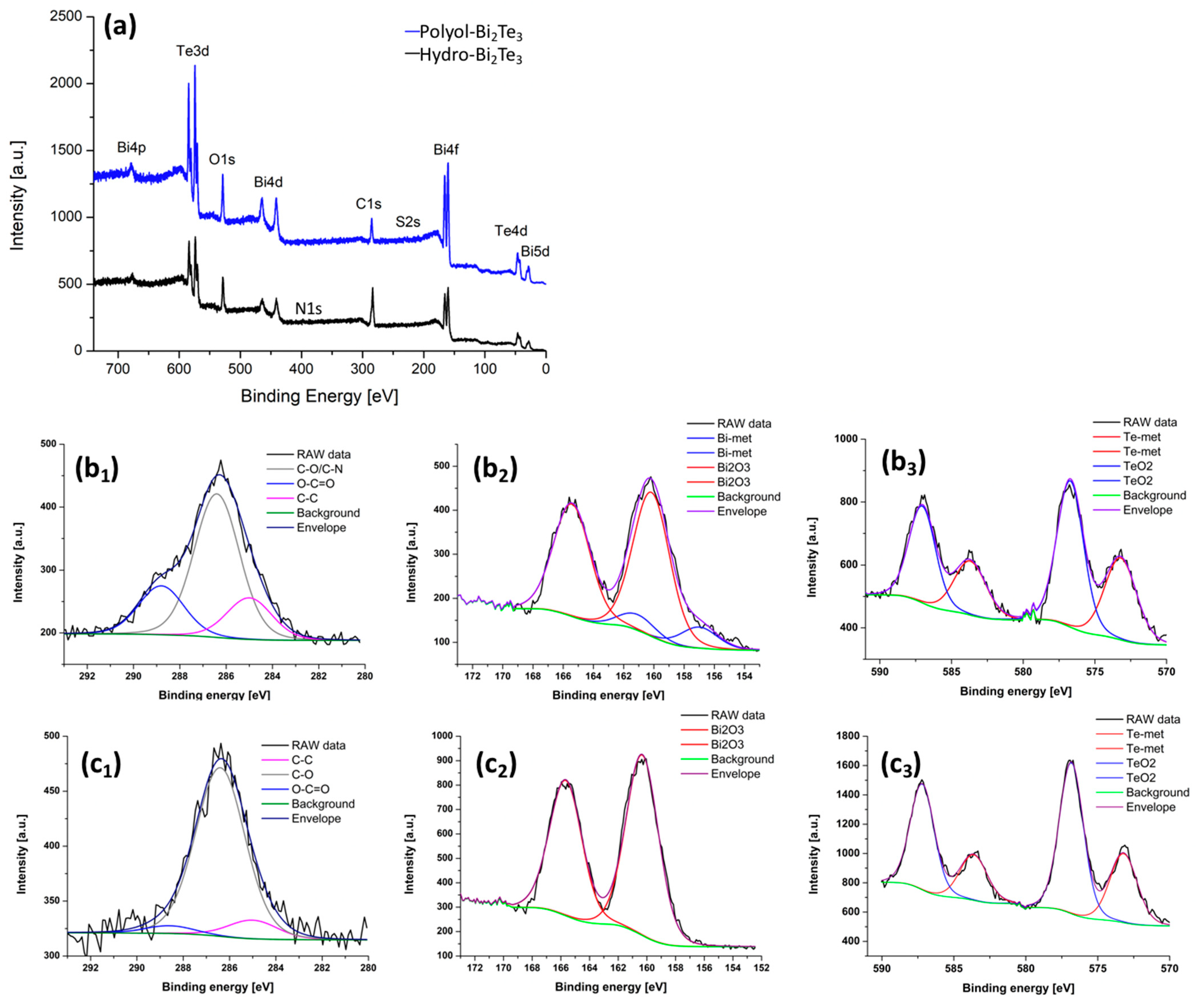
2.5. X-ray Photoelectron Spectroscopy

X-ray photoelectron spectroscopy (XPS) was used for surface analysis. The XPS spectra were acquired using the PREVAC 426 system configuration, equipped with SCIENTA R3000 hemispherical photoelectron spectrometer (Scienta Omicron, Uppsala, Sweden) and monochromatic Al source. The base pressure during measurements was better than 3 × 10⁻¹⁰ mbar. All acquired spectra were calibrated to adventitious carbon C1s at 285 eV. After subtraction of the Shirley-type background, the core-level spectra were decomposed into main components with mixed Gaussian–Lorentzian lines (70% G + 30% L for majority of photo-peaks) by a non-linear least squares curve-fitting procedure, using CasaXPS software (version 2.3.18, Casa Software Ltd., Cheshire, UK).

3. Results and Discussion

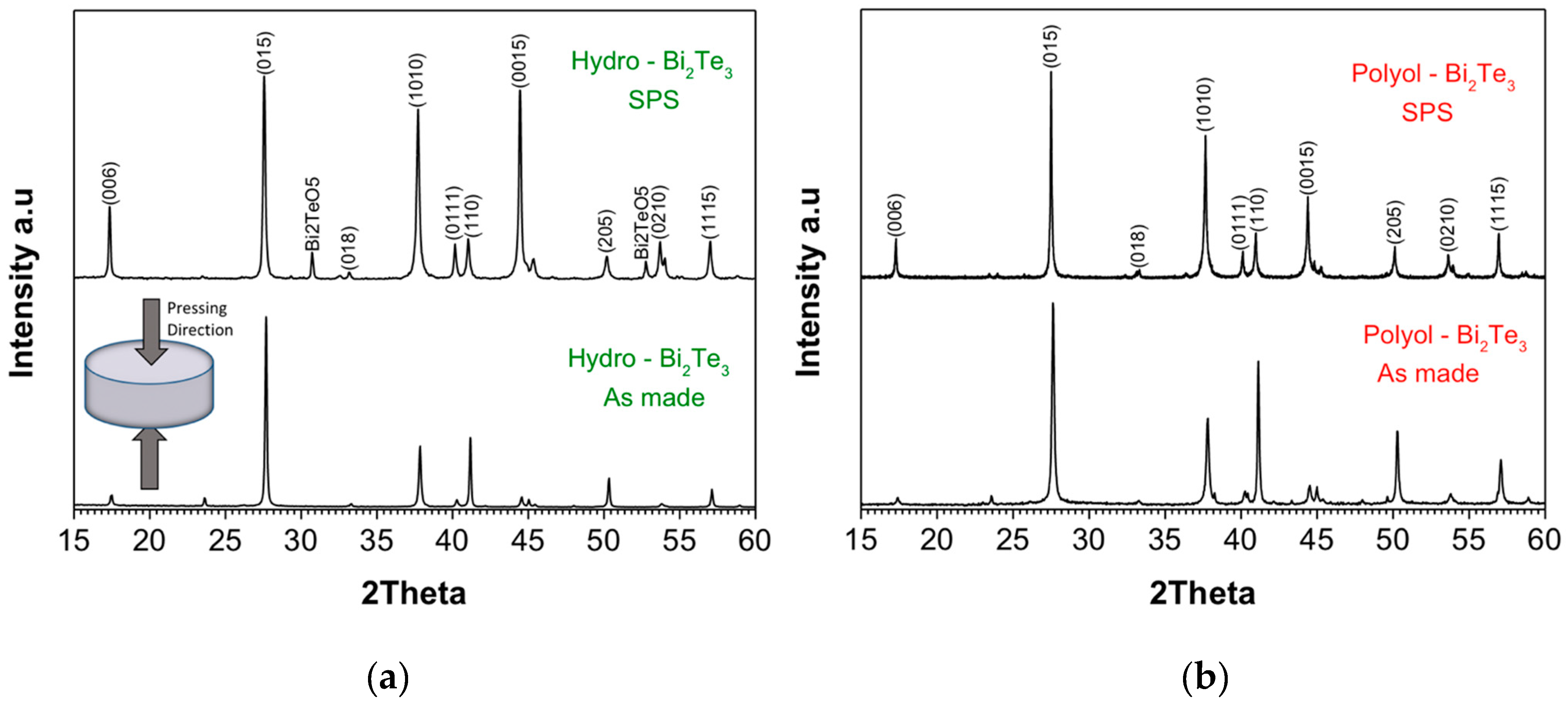
3.1. Structural Analysis

Structural analysis of as-synthesized materials through MW-assisted hydrothermal and polyol routes before and after SPS sintering are performed using XRPD. The diffraction patterns are presented in Figure 2, where the patterns are normalized for the most intense peak with Miller indices (015). As for the SPS sintered samples, XRPD was performed on the pellets’ surface perpendicular to the sintering direction (pressing axis/direction is schematically shown in the inset of Figure 2a). The crystalline phases are indexed to Bi2Te3 (ICDD: 01-089-2009) with rhombohedral crystal structure, and the corresponding indexing of the Bragg diffractions with relevant Miller indices are shown on the diffraction patterns in Figure 2. When as-made samples are compared, it can be clearly seen that polyol-Bi2Te3 sample has more intense peaks for the Bragg diffractions with the same Miller indices (e.g., (1010), (110), (205)), which reveal a higher crystallinity as compared to the hydro-Bi2Te3 sample. Upon sintering, the significant increase in the relative intensity of diffraction peaks with (001) index reveal a higher degree of texturing in the hydro-Bi2Te3 sample. The intensity ratio of (006) peak to (015) peak could be used for the quantification of the texturing. The hydro-Bi2Te3 sample has the peak intensity ratio (I006/I015) of 0.38, while it is 0.19 for the polyol-Bi2Te3 sample (see Figure S2). This indicates that after the SPS process the c-axis of the grains was preferentially oriented parallel to the pressing direction, resulting in a higher degree of texturing in the Hydro-Bi2Te3. Average crystallite size of the sintered samples was estimated from the XRD data using the Williamson–Hall model [88], as 231 nm for the polyol-Bi2Te3 sample, and 295 nm for the hydro-Bi2Te3 sample. A minor/secondary phase of Bi2TeO5 observed in the hydro-Bi2Te3 sample upon sintering. This phase was also observed in some earlier reports [89,90,91], and is estimated as 5.8% of the overall composition through the Rietveld refinement (see Figure S3). Possible reasons to observe Bi2TeO5 (ICDD: 00-024-0154) impurity phase after the SPS process may be either due to crystallization of the pre-existing secondary phase upon sintering, or to the reaction of the powder with the trace amount of oxygen in the sintering medium.

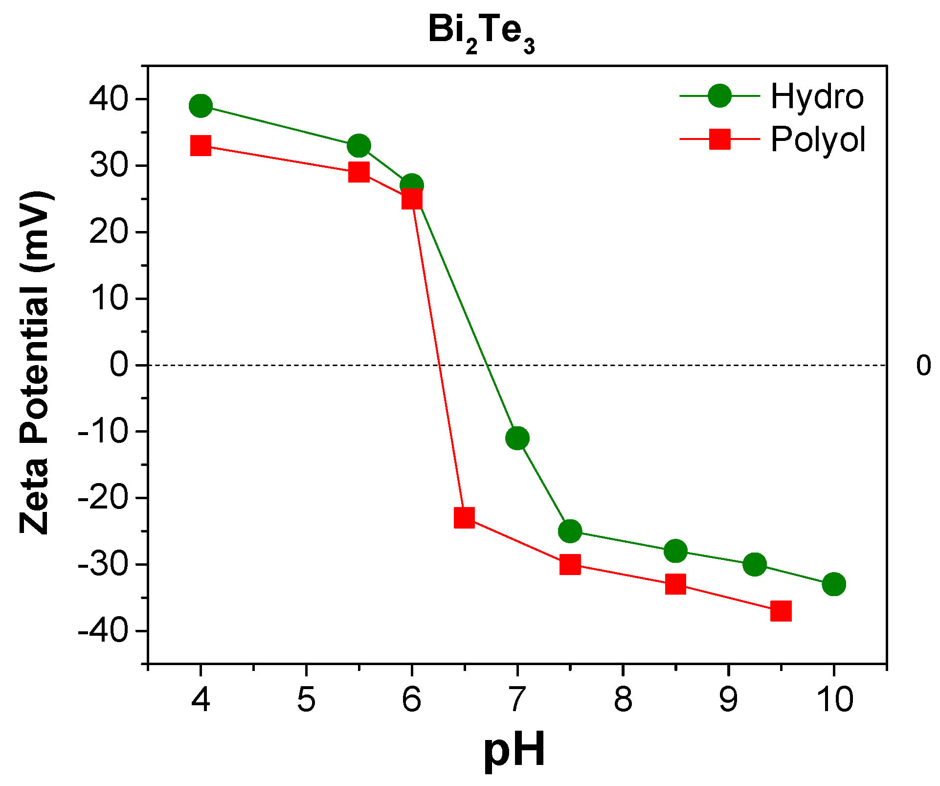
3.2. Surface Analysis

XPS analysis was performed on as made powder samples synthesized through hydrothermal and polyol routes. The data are graphically presented in Figure 3 and are summarized in Table 2. It should be noted that in both the samples tellurium (Te) exists in two states, metallic and oxide. In the case of bismuth (Bi) content, the hydro-Bi2Te3 sample consists of Bi in two states, metallic and oxide, while in the case of the polyol-Bi2Te3 sample the element Bi exists only in the oxide state. The speciation of carbon (C) is slightly different in the materials, where it is a mixture of mainly C–O, and C–C for polyol-Bi2Te3, while it consists of C–O, C–C and higher O–C=O content for the hydro-Bi2Te3 sample. The chemical speciation of C on the surface can be seen from the XPS fitting details, where a high percentage of C–O was identified in the hydro-Bi2Te3 sample, followed by C=O groups. This may be due to a strong conjugation of the terminal carboxyl groups to the nanoparticles’ surface through a bidentate or bridging configuration. In the polyol sample though, C is dominantly indexed to carboxyl, O–C=O, which is reasonable considering the conjugation of thioglycolic acid to the NP surface being through the sulfur (S) atoms. C content in the hydro-Bi2Te3 sample is higher than that of the polyol sample. This may originate from the precursors used in the synthetic process. Indeed, trace elements originating from the precursors are detected in the spectra of the respective samples. EDTA was used as a surface directing agent in the hydrothermal synthesis, which is known to be a hexadentate ligand at pH above 10. Naturally, it stays on the formed hexagonal platelets as an integral part, despite several washing-re-dispersing steps. The traces of EDTA can be seen through the nitrogen (N) signal (400.5 eV) in the XPS spectrum of the hydro-Bi2Te3 sample (Figure 3a). In the polyol process, thioglycolic acid was used as a directing agent and its traces are also visible through the sulfur (S) signal (229.3 eV) in the XPS spectrum of this particular sample (Figure 3a).
It is important to note that the relative ratio of metallic vs. oxide phases on the NPs’ surface reveals that the polyol-Bi2Te3 sample is bearing more oxide phase than the hydro-Bi2Te3 sample. In the case of Bi in particular, the spectrum for polyol-Bi2Te3 (Figure 3c2) indicates Bi2O3 as the only identified phase. It is interesting to note this as water has been generally considered a stronger oxidizing solvent.
Zeta (ξ) potential analysis was performed on both hydrothermal and polyol as-made powders dispersed in DI water. Isoelectric point (iep) is strongly dependent on the surface chemistry of the particles, and the dispersant. Determining the iep of as-synthesized Bi2Te3 materials can reveal the information about the surface charge and chemistry of the as-made nanoparticles. For the polyol synthesized sample, the iep was reached around pH 6.3, while it was about 6.8 for the hydrothermally synthesized sample (Figure 4). These values are not significantly different, and both may derive from the high C content, carbonyl and carboxyl bearing surface groups. The small difference can be attributed to the TeO2, which has an iep of <3. The higher TeO2 content of polyol-Bi2Te3 sample can explain the slightly lower iep value.

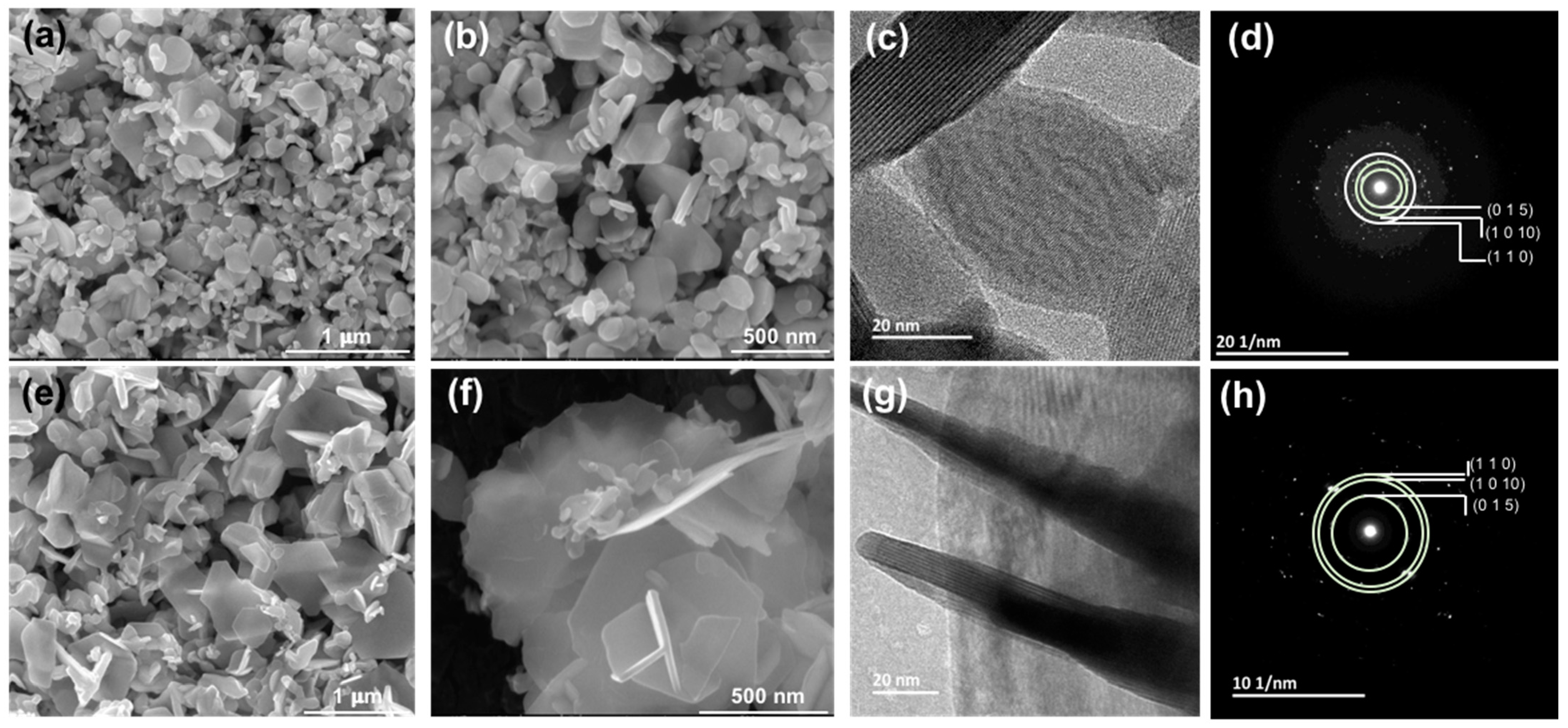
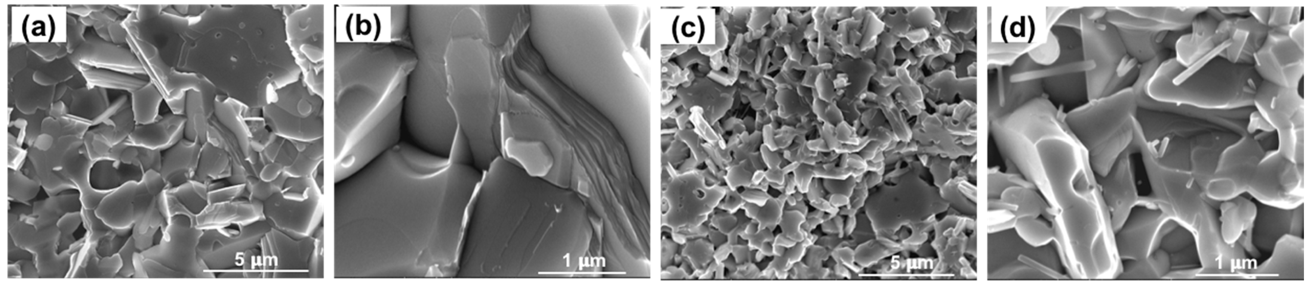
3.3. Microstructure Analysis

Morphology and microstructure analyses of as-made nanoparticles and sintered samples were performed using SEM and few micrographs for powder samples at different magnifications for hydro-Bi2Te3 and polyol-Bi2Te3 samples are given in Figure 5. Both the samples show dominantly hexagonal platelet morphology for the nanoparticles formed. The Bi2Te3 crystals grow predominantly along the ab plane direction because the strong covalent bonds between Bi–Te can be easily extended along the plane. The platelets obtained in the polyol route were observed to be thinner than those from the hydrothermal route. In general, particles obtained from the hydrothermal route are smaller than those from the polyol route. The lateral dimensions show a great variation in the range 20–250 nm for the hydrothermal and 10–1000 nm for the polyol sample, respectively. These variations are attributed to the different solvents and the stabilizing ligands used in the two syntheses process, which have had a different impact on the nucleation and growth kinetics. Looking closer to the XRPD patterns in Figure 2, it can be assessed that the diffraction peak at 41°, corresponding to the 110 Bragg diffraction, is much higher for the polyol sample, ascribed to larger lateral dimension (ab plane) of the platelets. A closer investigation was performed by TEM analysis and micrographs along with selected-area electron diffraction (SAED) patterns are shown in Figure 5c,d,g,h for hydro-Bi2Te3 and polyol-Bi2Te3 samples, respectively. Despite their large lateral dimensions, the platelets exhibit almost perfectly single crystalline character. There are some platelets aligned sideways, which reveal clearly the lamella-like structure along the c-axis, with a typical thickness of about 1 nm, as shown in Figure 5c,g. These layers are five atomic layers (Te-Bi-Te-Bi-Te) thick, which are known as the quintuple layer, held together by the van der Waals forces. SAED patterns, presented in Figure 5d,h, are indexed to typical Bragg diffractions of rhombohedral Bi2Te3 (ICDD: 01-089-2009) as was also identified in the XRPD patterns (Figure 2).
The as-synthesized nanostructured TE materials were consolidated using the SPS. The critical consolidation parameters (sintering temperature, holding time, pressure, and heating rates), and characteristics of the resultant pellets are presented in detail in Table 1. Despite both the samples being processed under identical conditions, the pellets reveal a big difference in their packing density; a packing density of about 80% for the polyol-Bi2Te3 and 90% for the hydro-Bi2Te3 sample. Densification under SPS for a given material as a function of particle size has been studied by Diouf et al. [93], where they reported a decrease in densification with increasing particle size of the specimen. In the case of the Bi2Te3 samples, particle size of the polyol-Bi2Te3 sample was assessed to be larger than the hydro-Bi2Te3 sample, which may be the cause of the observed difference in the densification, in agreement with the earlier findings [93]. Microstructure of the pellets are displayed in Figure 6. The hydro-Bi2Te3 sample shows a clear long-range stacking of hexagonal platelets, perpendicular to the c-axis, revealing a higher degree of texturing as compared to the polyol-Bi2Te3 sample. This effect was observed in XRD (Figure 2), with a significant increase in the intensity ratio (I006/I015) of the diffraction peaks for the hydro-Bi2Te3 sample (see Figure S2).

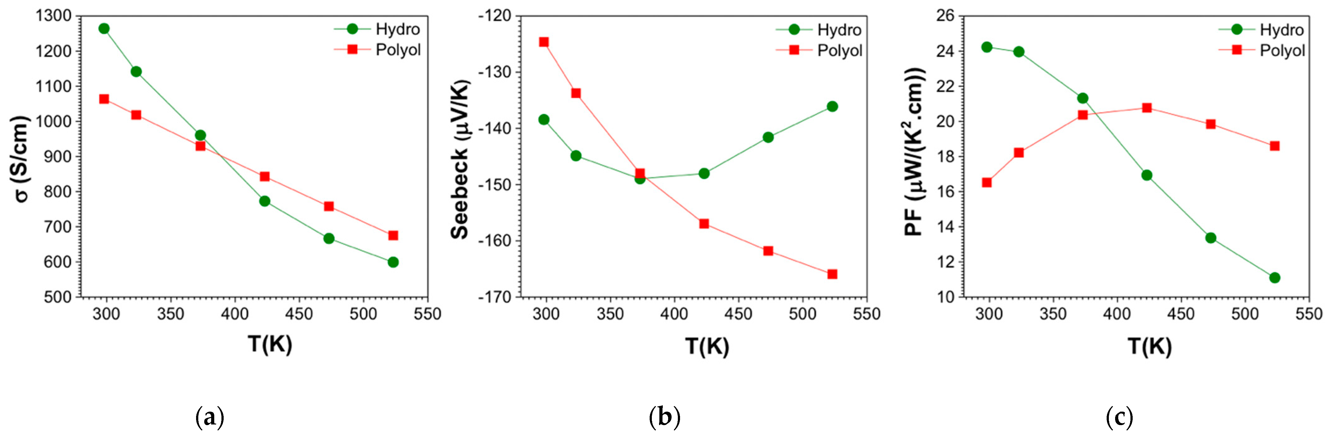
3.4. Electronic Transport Property Evaluation

The electronic transport data in the temperature range of 300–523 K are presented in Figure 7. Table 3 summarizes the transport data at room temperature and at the temperature where the maximum ZT was obtained. No adverse effects in the transport properties were observed due to the presence of surface oxides in the as-made powders. The electrical conductivity (σ) is plotted in Figure 7a, where samples show reduction of σ by increasing temperature, which is typical of heavily doped semiconductor, or metallic character. The highest electrical conductivity value at room temperature was measured for hydro-Bi2Te3. The σ for hydro-Bi2Te3 changes from about 1300 S/cm at 300 K to about 600 S/cm at 523 K. This is likely due to the higher density of the compacted materials and larger layered structure of the hydro-Bi2Te3 in comparison to the polyol-Bi2Te3 sample. The secondary phase of Bi2TeO5 in the hydro-Bi2Te3 sample is an oxide with a larger band-gap than Bi2Te3 [90,94]. Due to its low content within the main phase, it is not expected to have a percolation path to influence the electronic transport properties significantly. This can also be seen from the initial high σ of hydro-Bi2Te3. However, this oxide phase may act as phonon scattering canters due to its different anisotropic crystal structure, influencing the thermal conductivity. The σ values change between 1100 S/cm and 700 S/cm for the polyol-Bi2Te3 sample, in the same temperature range. Reduction in the electrical conductivity over 300 K is generally a signature of increasing electron-phonon scattering. There is an interesting crossover at 373 K, where the σ for the hydro-Bi2Te3 sample goes below the polyol sample. This might be due to the electron-phonon scattering process being higher than the polyol-Bi2Te3 sample. The electrical conductivity of both samples are higher than those of other solution derived samples [95,96,97] comparable with that of p-type alloy ingot (on the level of 1 × 10³ S/cm at 300 K) [98].
The Seebeck coefficient (S) is the key parameter to demonstrate the type of transport. Figure 7b shows the obtained S values for both samples, which have negative values, revealing n-type conduction. The polyol-Bi2Te3 sample starts the S from −120 μV/K at room temperature and increases in magnitude up to −165 μV/K at 525 K, while the hydro-Bi2Te3 sample does not show a strong temperature dependence, displaying values between −140 μV/K and −150 μV/K. Electronic transport properties are strongly correlated with the microstructure, as well as charge carrier density, defects, and scattering events. This correlation is clearly visible for the hydro-Bi2Te3 sample, where a decreasing σ is accompanied by an increase in the magnitude of the S. The band-gap (Eg) can be estimated from the S as it is sensitive to changes in the density of states near the Fermi level [99,100]. The Eg for the materials were calculated using the electron charge ( q ), absolute value of the maximum Seebeck, |Smax|, value and the corresponding temperature T m a x , using the following equation:
  E g = 2 q | S m a x | · T m a x
The estimated Eg from the Smax for the hydro-Bi2Te3 sample is 0.11 eV and that for the polyol-Bi2Te3 sample is 0.17 eV. The estimated Eg for the Hydro-Bi2Te3 sample is in good agreement with the calculated Eg of 0.11 eV by Mishra et al., using the density-functional theory with the spin–orbit interaction included [101]. However, the Eg for the hydro-Bi2Te3 sample is lower than the bulk Bi2Te3 value of Eg = 0.15 eV [102], while that of the polyol-Bi2Te3 sample is slightly higher. The nanocrystal size effect on the shifts of valence and conduction band edge and the band-gap expansion of semiconductor nanocrystals has been modeled by Lang et al. [103]. This model, without any adjustable parameter, predicted that the band-gap expansion (along with shifts of valence and conduction band edges) is present with dropping size of nanocrystals because of the increase of surface/volume ratio. Crystallite size of the sintered pellets has been estimated as 295 nm for the hydro-Bi2Te3 sample, and 231 nm for the polyol-Bi2Te3 sample. Therefore, the estimated Eg values agree with the trend expected from the relative size of the crystallites in the studied materials.
The power factor (PF) (S2 σ) calculated for both hydrothermal and polyol samples, as presented in Figure 7c. The highest PF at room temperature was obtained for the hydro-Bi2Te3 sample as 24 μW cm−1 K−2, followed by the polyol-Bi2Te3 sample as 16 μW cm−1 K−2. The maximum power factors we achieved are 24 μW cm−1 K−2 for the hydrothermal sample at 300 K and 21 μW cm−1 K−2 at 423 K for the polyol sample. The magnitude of PF is comparable to or much higher than other solution-derived samples—with maximum power factor 1–9 μW cm−1 K−2 [66,96].

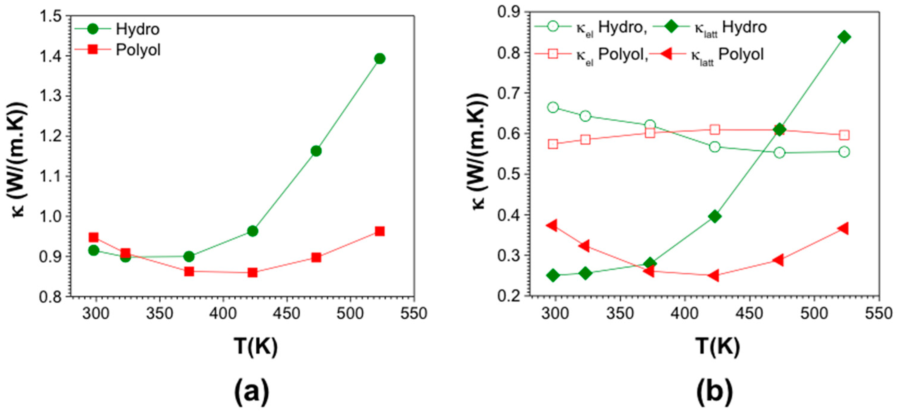
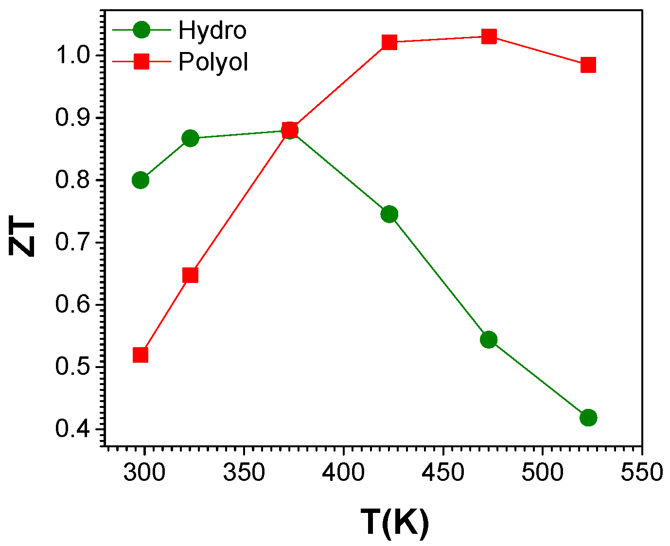
3.5. Thermal Conductivity

The total thermal conductivity values were calculated using the thermal diffusivity, heat capacity and mass density values of the samples. The total thermal conductivity of hydro-Bi2Te3 is slightly lower than polyol-Bi2Te3 at T = 293 K, as seen in Figure 8a. They both decrease first and then increase with increasing temperature, where it shows a dramatic increase for the hydro-Bi2Te3 sample after 373 K. Nanostructures at different length scales (diameter, roughness and point defects) showed efficient scattering throughout the phonon spectrum, reaching very low thermal conductivity values [23]. Looking at the microstructure of the pellets obtained, there could be a mixture of these nanostructures at different length scales, influencing the phonon propagation. The actual nanostructures include undefined structural features, such as interface defects, impurities, strain, etc., making it difficult to classify the contribution of each scattering process to phonon transport [104]. In order to make a clear assessment, we have examined lattice (κlat) and electronic thermal conductivity (κel) of both the samples by using the Wiedemann–Franz relationship, κel = LoσT where Lo, σ, and T are Lorenz number, the electrical conductivity and the absolute temperature, respectively. The κlat values were estimated by subtracting the κel from the total thermal conductivity. In order to calculate the Lorenz number (Lo), experimentally obtained Seebeck coefficient values were used and a single parabolic band model was assumed to solve the Fermi integral. Lorenz numbers for both the samples are graphically presented in Figure S4. The κlat and κe are shown in Figure 8b. Since the κel is determined by the σ, it is expected to possess a similar trend to the electrical conductivity. In both the samples the κel contribution is higher than the lattice contribution, κlat, most likely due to the electrons’ more dominant role on the thermal conductivity. The striking feature is the very low lattice contribution in these samples, strongly influenced by the increased density of grain boundaries that plays a crucial role in increased phonon scattering and thus a strong reduction in κlat. The room temperature κlatt for the hydro-Bi2Te3 and polyol-Bi2Te3 were about 0.25 W/m·K and 0.37 W/m·K, respectively. The κlat of the hydro-Bi2Te3 sample increases with temperature after 373 K. This might be due to the highly crystalline texture (long layered structure) thus longer MFP of phonons. Crystallite size of the sintered pellets has been estimated as 295 nm for the hydro-Bi2Te3 sample, and 231 nm for the polyol-Bi2Te3 sample. The difference in the Eg is the evidence of the correlation of the Eg with the average crystallite size; a smaller crystallite size leading to a larger band-gap and vice versa. Due to the smaller Eg of the hydro-Bi2Te3 sample (0.11 eV), at elevated temperatures the minority carriers’ diffusion (ambipolar effect characteristic for the electron-hole pair diffusion on the onset of the intrinsic conduction) contributes strongly to the total thermal conductivity κtot [105,106].
Regardless of the synthetic route, the total thermal conductivity of the materials is lower than that of the current best-known n-type Bi2Te2.925Se0.075tot = 1.65 W/m·K at 300 K in which the κlat was 1.27 W/m·K) [16], and are also comparable to those of ball milled and dc hot pressed n-type Bi2Te2.7Se0.3 bulk samples (κtot = 1.06 W/m·K at 300 K in which κlat was 0.7 W/m·K) [107]. This strongly suggests that the nanostructure obtained through the developed synthetic routes plays a very significant role in reducing the κlat through effective phonon scattering.
Temperature dependence of the estimated ZT values for the samples are shown in Figure 9. Room temperature ZT values of 0.8 and 0.52 were obtained for hydro-Bi2Te3 and polyol-Bi2Te3 samples, respectively. The maximum ZT values were obtained at slightly different temperatures, in the range 323–373 K for hydro-Bi2Te3, and 423–473 K for polyol-Bi2Te3. This is mainly because the samples display a maximum power factor and a minimum thermal conductivity at different temperatures. The maximum ZT value reaches about 0.9 at 373 K for hydro-Bi2Te3 while for polyol-Bi2Te3 it is about 1.03 at 473 K. These are among the highest ZT values reported for n-type Bi2Te3 in the literature. This study shows two viable synthetic strategies, which yield materials with promising efficiency at slightly shifted temperatures that can be used for guiding the synthetic route for the intended temperature range of applications.

4. Conclusions

Rapid and scalable synthetic strategies for high-quality and reproducible thermoelectric (TE) materials are demonstrated in this work, exemplified by nanostructured n-type bismuth telluride with a promising TE performance. Use of microwaves enables an effective volume heating, leading to highly crystalline TE nanopowders in a few minutes. Polyol synthesis is shown to be a reliable and scalable route to materials synthesis. Hereby we show that not only EG, but also water is an excellent reaction media, which is certainly much more benign and abundant than any other solvent. Furthermore, hydrothermal synthesis is one-pot process, where all the precursors are mixed in the same reaction medium prior to heating, which makes this process facile and therefore more attractive. Despite the significant difference in their dielectric constant, both solvents (EG and water) showed impressive outcomes for the materials investigated. Crystal structure, crystallinity, morphology, microstructure and surface chemistry of the synthesized materials were evaluated using a library of analytical techniques, including XRD, SEM/TEM, XPS and zeta potential techniques. Particles with dominantly hexagonal platelet morphology were obtained, where the polyol-Bi2Te3 generally exhibited a larger particle size than the hydro-Bi2Te3. Surface chemistry studied by XPS clearly revealed different levels of surface oxidation, based on the solvent used, and traces of the precursors used on the surface of as-synthesized materials. Surface directing agents are stably bound to the surface of the as-formed nanoparticles, thus assuring the maintenance of the hexagonal platelet morphology. The samples were then consolidated using SPS, prior to further transport measurements. The difference in the particle size of the specimens significantly influenced the densification behavior under SPS, where polyol-Bi2Te3 with larger particle size reached about 10% lower densification compared to the hydro-Bi2Te3 sample under identical conditions. Upon sintering, texturing is observed to be more predominant in the hydro-Bi2Te3 sample, as assessed from XRD and SEM analyses. Thermoelectric transport properties were evaluated using electrical conductivity and Seebeck coefficient measurements up to 525 K, and the thermal diffusivity was assessed by using the LFA system. The final estimated TE figure-of-merit values about 0.8–0.9 was reached in the temperature range of 300–375 K for the hydro-Bi2Te3 sample, while 0.9–1 was reached in the temperature range of 425–525 K for the polyol-Bi2Te3 sample. Energy and time efficiency, scalability as well as the environmental impact of the synthetic processes presented here, along with the high ZT values achieved, pave the way for wider impact of these synthetic strategies for developing promising strategic materials in the field of thermal energy harvesting.

Supplementary Materials

The following are available online at https://www.mdpi.com/article/10.3390/nano11082053/s1, Figure S1: Schematic of spark plasma sintering (SPS) process, sample geometry for thermal diffusivity, using Laser Flash technique, and power factor measurements. Figure S2: Rietveld refinement for Hydro- Bi2Te3 sample after SPS sintering, Figure S3: X-ray powder diffraction (XRPD) patterns showing a close up of the peaks with Miller indices of (006) and (015), Figure S4: Lorenz Number, Lo, estimated for Bi2Te3 samples using parabolic band model, Table S1: Survey of the synthesis methods, reaction conditions, morphology and size of Bi2Te3.

Author Contributions

B.H. and M.S.T. conceived the presented idea. B.H., M.P., N.I.K. and M.S.T. contributed to conceptualization and methodology, material synthesis, and original draft preparation. B.H., H.B. contributed to physicochemical characterization. S.B. validated the thermoelectric properties. M.K. and R.S. performed the XPS analysis. All authors discussed the results and reviewed and edited the manuscript. M.S.T., S.B. and M.J. supervised the work. All authors have read and agreed to the published version of the manuscript.

Funding

This research has received funding from the Swedish Energy Agency (43521-1) and in part by Swedish Research Agency Council (VR, 2018-03462) and the European Union’s Horizon 2020 research and innovation program under the grant agreement No 863222. SB acknowledges support by Scientific and Technological Research Council of Turkey (TUBITAK, 216M254) and Scientific Research Projects Coordination Unit of Istanbul University (BAP, 21809 and 32641).

Institutional Review Board Statement

Not applicable.

Informed Consent Statement

Not applicable.

Acknowledgments

MST acknowledges the financial support from Olle Engkvist Foundation (SOEB, 190-0315) for the establishment of MW synthesis facilities.

Conflicts of Interest

The authors declare no conflict of interest. The funders had no role in the design of the study; in the collection, analyses, or interpretation of data; in the writing of the manuscript, or in the decision to publish the results.

References

  1. Shatkin, J.A. Nanomaterials Risks and Benefits (NATO Science for Peace and Security Studies). Risk Anal. 2009, 29, 1192–1195. [Google Scholar] [CrossRef]
  2. Petsagkourakis, I.; Tybrandt, K.; Crispin, X.; Ohkubo, I.; Satoh, N.; Mori, T. Thermoelectric materials and applications for energy harvesting power generation. Sci. Technol. Adv. Mater. 2018, 19, 836–862. [Google Scholar] [CrossRef]
  3. Sajid, M.; Hassan, I.; Rahman, A. An overview of cooling of thermoelectric devices. Renew. Sustain. Energy Rev. 2017, 78, 15–22. [Google Scholar] [CrossRef]
  4. Ovik, R.; Long, B.D.; Barma, M.C.; Riaz, M.; Sabri, M.F.M.; Said, S.M.; Saidur, R. A review on nanostructures of high-temperature thermoelectric materials for waste heat recovery. Renew. Sustain. Energy Rev. 2016, 64, 635–659. [Google Scholar] [CrossRef]
  5. Huen, P.; Daoud, W.A. Advances in hybrid solar photovoltaic and thermoelectric generators. Renew. Sustain. Energy Rev. 2017, 72, 1295–1302. [Google Scholar] [CrossRef]
  6. Jaziri, N.; Boughamoura, A.; Müller, J.; Mezghani, B.; Tounsi, F.; Ismail, M. A comprehensive review of Thermoelectric Generators: Technologies and common applications. Energy Rep. 2020, 6, 264–287. [Google Scholar] [CrossRef]
  7. Yusuf, A.; Bayhan, N.; Tiryaki, H.; Hamawandi, B.; Toprak, M.S.; Ballikaya, S. Multi-objective optimization of concentrated Photovoltaic-Thermoelectric hybrid system via non-dominated sorting genetic algorithm (NSGA II). Energy Convers. Manag. 2021, 236, 114065. [Google Scholar] [CrossRef]
  8. Champier, D. Thermoelectric generators: A review of applications. Energy Convers. Manag. 2017, 140, 167–181. [Google Scholar] [CrossRef]
  9. Snyder, G.J.; Toberer, E.S. Complex TE meterials. Nat. Mater. 2008, 7, 105–114. [Google Scholar] [CrossRef]
  10. Polozine, A.; Sirotinskaya, S.; Schaeffer, L. History of development of thermoelectric materials for electric power generation and criteria of their quality. Mater. Res. 2014, 17, 1260–1267. [Google Scholar] [CrossRef] [Green Version]
  11. Ioffe, A.F. Semiconductor thermoelements and thermoelec. Phys. Today 1959, 12, 42. [Google Scholar] [CrossRef]
  12. Nakamura, Y.; Isogawa, M.; Ueda, T.; Yamasaka, S.; Matsui, H.; Kikkawa, J.; Ikeuchi, S.; Oyake, T.; Hori, T.; Shiomi, J.; et al. Anomalous reduction of thermal conductivity in coherent nanocrystal architecture for silicon thermoelectric material. Nano Energy 2015, 12, 845–851. [Google Scholar] [CrossRef] [Green Version]
  13. Rowe, D.M.; Shukla, V.S.; Savvides, N. Phonon scattering at grain boundaries in heavily doped fine-grained silicon-germanium alloys. Nature 1981, 290, 765–766. [Google Scholar] [CrossRef]
  14. Szczech, J.R.; Higgins, J.M.; Jin, S. Enhancement of the thermoelectric properties in nanoscale and nanostructured materials. J. Mater. Chem. 2011, 21, 4037–4055. [Google Scholar] [CrossRef]
  15. Nomura, M.; Kage, Y.; Müller, D.; Moser, D.; Paul, O. Electrical and thermal properties of polycrystalline Si thin films with phononic crystal nanopatterning for thermoelectric applications. Appl. Phys. Lett. 2015, 106, 1–5. [Google Scholar] [CrossRef]
  16. CRC Handbook of Thermoelectrics; CRC Press: Boca Raton, FL, USA, 2018.
  17. Harman, T.C.; Taylor, P.J.; Walsh, M.P.; LaForge, B.E. Quantum dot superlattice thermoelectric materials and devices. Science 2002, 297, 2229–2232. [Google Scholar] [CrossRef] [Green Version]
  18. Venkatasubramanian, R.; Siivola, E.; Colpitts, T.; O’Quinn, B. Thin-film thermoelectric devices with high room-temperature figures of merit. Nature 2001, 413, 597–602. [Google Scholar] [CrossRef] [PubMed]
  19. Hamawandi, B.; Noroozi, M.; Jayakumar, G.; Ergül, A.; Zahmatkesh, K.; Toprak, M.S.; Radamson, H.H. Electrical properties of sub-100 nm SiGe nanowires. J. Semicond. 2016, 37, 102001. [Google Scholar] [CrossRef]
  20. Hicks, L.; Dresselhaus, M.S. Thermoelectric figure of merit of a one-dimensional conductor. Phys. Rev. B 1993, 47, 8–11. [Google Scholar] [CrossRef] [PubMed]
  21. Harman, T.C.; Spears, D.L.; Manfra, M.J. High thermoelectric figures of merit in PbTe quantum wells. J. Electron. Mater. 1996, 25, 1121–1127. [Google Scholar] [CrossRef]
  22. Biswas, K.; He, J.; Zhang, Q.; Wang, G.; Uher, C.; Dravid, V.P.; Kanatzidis, M.G. Strained endotaxial nanostructures with high thermoelectric figure of merit. Nat. Chem. 2011, 3, 160–166. [Google Scholar] [CrossRef] [PubMed]
  23. Hochbaum, A.I.; Chen, R.; Delgado, R.D.; Liang, W.; Garnett, E.C.; Najarian, M.; Majumdar, A.; Yang, P. Enhanced thermoelectric performance of rough silicon nanowires. Nature 2008, 451, 163–167. [Google Scholar] [CrossRef]
  24. Son, J.H.; Oh, M.W.; Kim, B.S.; Park, S.D.; Min, B.K.; Kim, M.H.; Lee, H.W. Effect of ball milling time on the thermoelectric properties of p-type (Bi,Sb)2Te3. J. Alloys Compd. 2013, 566, 168–174. [Google Scholar] [CrossRef]
  25. Zheng, Y.; Liu, C.; Miao, L.; Lin, H.; Gao, J.; Wang, X.; Chen, J.; Wu, S.; Li, X.; Cai, H. Cost effective synthesis of p-type Zn-doped MgAgSb by planetary ball-milling with enhanced thermoelectric properties. RSC Adv. 2018, 8, 35353–35359. [Google Scholar] [CrossRef] [Green Version]
  26. Sun, T.; Zhao, X.B.; Zhu, T.J.; Tu, J.P. Aqueous chemical reduction synthesis of Bi2Te3 nanowires with surfactant assistance. Mater. Lett. 2006, 60, 2534–2537. [Google Scholar] [CrossRef]
  27. Foos, E.E.; Stroud, R.M.; Berry, A.D. Synthesis and Characterization of Nanocrystalline Bismuth Telluride. Nano Lett. 2001, 1, 693–695. [Google Scholar] [CrossRef]
  28. Giani, A.; Pascal-Delannoy, F.; Boyer, A.; Foucaran, A.; Gschwind, M.; Ancey, P. Elaboration of Bi2Te3 by metal organic chemical vapor deposition. Thin Solid Film. 1997, 303, 1–3. [Google Scholar] [CrossRef]
  29. Venkatasubramanian, R.; Colpitts, T.; Watko, E.; Lamvik, M.; El-Masry, N. MOCVD of Bi2Te3, Sb2Te3 and their superlattice structures for thin-film thermoelectric applications. J. Cryst. Growth 1997, 170, 817–821. [Google Scholar] [CrossRef]
  30. Longo, M.; Cecchi, S.; Selmo, S.; Fanciulli, M.; Wiemer, C.; Mdm, L. MOCVD growth and thermal analysis of Sb2Te3 thin films and nanowires. In Proceedings of the 2015 1st Workshop on Nanotechnology in Instrumentation and Measurement (NANOFIM), Lecce, Italy, 24–25 July 2015; pp. 24–25. [Google Scholar]
  31. Lee, J.S.; Brittman, S.; Yu, D.; Park, H. Supporting Information for “ Vapor-Liquid-Solid and Vapor-Solid Growth of Phase-Change Sb2Te3 Nanowires and Sb2Te3/GeTe Nanowire Heterostructures. J. Am. Chem. Soc. 2008, 130, 1–8. [Google Scholar]
  32. Park, D.; Park, S.; Jeong, K.; Jeong, H.S.; Song, J.Y.; Cho, M.H. Thermal and Electrical Conduction of Single-crystal Bi2Te3Nanostructures grown using a one step process. Sci. Rep. 2016, 6, 13–15. [Google Scholar] [CrossRef]
  33. Kim, C.; Kim, D.H.; Han, Y.S.; Chung, J.S.; Kim, H. Fabrication of antimony telluride nanoparticles using a brief chemical synthetic process under atmospheric conditions. J. Alloys Compd. 2011, 509, 609–613. [Google Scholar] [CrossRef]
  34. Gupta, S.; Neeleshwar, S.; Kumar, V.; Chen, Y.Y. Synthesis of bismuth telluride nanostructures by refluxing method. Adv. Mater. Lett. 2012, 3, 50–54. [Google Scholar] [CrossRef]
  35. Pelz, U.; Kaspar, K.; Schmidt, S.; Dold, M.; Jägle, M.; Pfaadt, A.; Hillebrecht, H. An aqueous-chemistry approach to nano-bismuth telluride and nano-antimony telluride as thermoelectric materials. J. Electron. Mater. 2012, 41, 1851–1857. [Google Scholar] [CrossRef]
  36. Yang, H.Q.; Miao, L.; Liu, C.Y.; Li, C.; Honda, S.; Iwamoto, Y.; Huang, R.; Tanemura, S. A Facile Surfactant-Assisted Reflux Method for the Synthesis of Single-Crystalline Sb2Te3 Nanostructures with Enhanced Thermoelectric Performance. ACS Appl. Mater. Interfaces 2015, 7, 14263–14271. [Google Scholar] [CrossRef]
  37. Sapp, S.A.; Lakshmi, B.B.; Martin, C.R. Template synthesis of bismuth telluride nano wires. Adv. Mater. 1999, 11, 402–404. [Google Scholar] [CrossRef]
  38. Pinisetty, D.; Gupta, M.; Karki, A.B.; Young, D.P.; Devireddy, R.V. Fabrication and characterization of electrodeposited antimony telluride crystalline nanowires and nanotubes. J. Mater. Chem. 2011, 21, 4098–4107. [Google Scholar] [CrossRef]
  39. Dhak, D.; Pramanik, P. Characterization of nanocrystalline bismuth telluride (Bi2Te3) synthesized by a novel approach through aqueous precursor method. J. Am. Ceram. Soc. 2006, 89, 534–537. [Google Scholar] [CrossRef]
  40. Saleemi, M.; Toprak, M.S.; Li, S.; Johnsson, M.; Muhammed, M. Synthesis, processing, and thermoelectric properties of bulk nanostructured bismuth telluride (Bi2Te3). J. Mater. Chem. 2012, 22, 725–730. [Google Scholar] [CrossRef]
  41. Zhao, X.B.; Ji, X.H.; Zhang, Y.H.; Lu, B.H. Effect of solvent on the microstructures of nanostructured Bi2Te3 prepared by solvothermal synthesis. J. Alloys Compd. 2004, 368, 349–352. [Google Scholar] [CrossRef]
  42. Deng, Y.; Wei, G.; Liu, J.; Zhou, X.; Wu, J.; Nan, C. Solvothermal preparation and characterization of nanocrystalline SnTe powder with different morphologies. Xiyou Jinshu Cailiao Yu Gongcheng/Rare Met. Mater. Eng. 2002, 31, 42. [Google Scholar]
  43. Deng, Y.; Nan, C.W.; Wei, G.D.; Guo, L.; Lin, Y.H. Organic-assisted growth of bismuth telluride nanocrystals. Chem. Phys. Lett. 2003, 374, 410–415. [Google Scholar] [CrossRef]
  44. Jin, R.; Liu, J.; Li, G. Facile solvothermal synthesis, growth mechanism and thermoelectric property of flower-like Bi2Te3. Cryst. Res. Technol. 2014, 49, 460–466. [Google Scholar] [CrossRef]
  45. Stavila, V.; Robinson, D.B.; Hekmaty, M.A.; Nishimoto, R.; Medlin, D.L.; Zhu, S.; Tritt, T.M.; Sharma, P.A. Wet-chemical synthesis and consolidation of stoichiometric bismuth telluride nanoparticles for improving the thermoelectric figure-of-merit. ACS Appl. Mater. Interfaces 2013, 5, 6678–6686. [Google Scholar] [CrossRef] [PubMed]
  46. Xu, Y.; Ren, Z.; Cao, G.; Ren, W.; Deng, K.; Zhong, Y. Fabrication and characterization of Bi2Te3 nanoplates via a simple solvothermal process. Phys. B Condens. Matter 2009, 404, 4029–4033. [Google Scholar] [CrossRef]
  47. Sun, S.; Peng, J.; Jin, R.; Song, S.; Zhu, P.; Xing, Y. Template-free solvothermal synthesis and enhanced thermoelectric performance of Sb2Te3 nanosheets. J. Alloys Compd. 2013, 558, 6–10. [Google Scholar] [CrossRef]
  48. Im, H.J.; Koo, B.; Kim, M.S.; Lee, J.E. Solvothermal synthesis of Sb2Te3 nanoplates under various synthetic conditions and their thermoelectric properties. Appl. Surf. Sci. 2019, 475, 510–514. [Google Scholar] [CrossRef]
  49. Wang, W.; Poudel, B.; Yang, J.; Wang, D.Z.; Ren, Z.F. High-yield synthesis of single-crystalline antimony telluride hexagonal nanoplates using a solvothermal approach. J. Am. Chem. Soc. 2005, 127, 13792–13793. [Google Scholar] [CrossRef]
  50. Kim, H.J.; Han, M.K.; Kim, H.Y.; Lee, W.; Kim, S.J. Morphology controlled synthesis of nanostructured Bi2Te3. Bull. Korean Chem. Soc. 2012, 33, 3977–3980. [Google Scholar] [CrossRef] [Green Version]
  51. Giri, L.; Mallick, G.; Jackson, A.C.; Griep, M.H.; Karna, S.P. Synthesis and characterization of high-purity, single phase hexagonal Bi2Te3 nanostructures. RSC Adv. 2015, 5, 24930–24935. [Google Scholar] [CrossRef]
  52. Xu, Y.; Ren, Z.; Ren, W.; Cao, G.; Deng, K.; Zhong, Y. Hydrothermal synthesis of single-crystalline Bi2Te3 nanoplates. Mater. Lett. 2008, 62, 4273–4276. [Google Scholar] [CrossRef]
  53. Cao, Y.Q.; Zhu, T.J.; Zhao, X.B. Thermoelectric Bi2Te3 nanotubes synthesized by low-temperature aqueous chemical method. J. Alloys Compd. 2008, 449, 109–112. [Google Scholar] [CrossRef]
  54. Shi, W.; Yu, J.; Wang, H.; Zhang, H. Hydrothermal synthesis of single-crystalline antimony telluride nanobelts. J. Am. Chem. Soc. 2006, 128, 16490–16491. [Google Scholar] [CrossRef] [PubMed]
  55. Dong, G.H.; Zhu, Y.J.; Cheng, G.F.; Ruan, Y.J. Sb2Te3 nanobelts and nanosheets: Hydrothermal synthesis, morphology evolution and thermoelectric properties. J. Alloys Compd. 2013, 550, 164–168. [Google Scholar] [CrossRef]
  56. Yuan, Q.L.; Nie, Q.L.; Huo, D.X. Preparation and characterization of the antimony telluride hexagonal nanoplates. Curr. Appl. Phys. 2009, 9, 224–226. [Google Scholar] [CrossRef]
  57. Dharmaiah, P.; Hong, S.J. Hydrothermal method for the synthesis of Sb2Te3, and Bi0.5Sb1.5Te3 nanoplates and their thermoelectric properties. Int. J. Appl. Ceram. Technol. 2018, 15, 132–139. [Google Scholar] [CrossRef]
  58. Zhang, Y.; Xu, G.; Mi, J.; Han, F.; Wang, Z.; Ge, C. Hydrothermal synthesis and thermoelectric properties of nanostructured Bi0.5Sb1.5Te3 compounds. Mater. Res. Bull. 2011, 46, 760–764. [Google Scholar] [CrossRef]
  59. Wang, Z.; Wang, F.Q.; Chen, H.; Zhu, L.; Yu, H.J.; Jian, X.Y. Synthesis and characterization of Bi2Te3 nanotubes by a hydrothermal method. J. Alloys Compd. 2010, 492, 50–53. [Google Scholar] [CrossRef]
  60. Novaconi, S.; Vlazan, P.; Malaescu, I.; Badea, I.; Grozescu, I.; Sfirloaga, P. Doped Bi2Te3 nano-structured semiconductors obtained by ultrasonically assisted hydrothermal method. Cent. Eur. J. Chem. 2013, 11, 1599–1605. [Google Scholar] [CrossRef] [Green Version]
  61. Zheng, Y.Y.; Zhu, T.J.; Zhao, X.B.; Tu, J.P.; Cao, G.S. Sonochemical synthesis of nanocrystalline Bi2Te3 thermoelectric compounds. Mater. Lett. 2005, 59, 2886–2888. [Google Scholar] [CrossRef]
  62. Zhou, B.; Zhao, Y.; Pu, L.; Zhu, J.J. Microwave-assisted synthesis of nanocrystalline Bi2Te3. Mater. Chem. Phys. 2006, 96, 192–196. [Google Scholar] [CrossRef]
  63. Dong, G.H.; Zhu, Y.J.; Chen, L.D. Microwave-assisted rapid synthesis of Sb2Te3 nanosheets and thermoelectric properties of bulk samples prepared by spark plasma sintering. J. Mater. Chem. 2010, 20, 1976–1981. [Google Scholar] [CrossRef]
  64. Hamawandi, B.; Ballikaya, S.; Batili, H.; Roosmark, V.; Orlovská, M.; Yusuf, A.; Johnsson, M.; Szukiewicz, R.; Kuchowicz, M.; Toprak, M.S. Facile solution synthesis, processing and characterization of n-and p-type binary and ternary Bi-Sb tellurides. Appl. Sci. 2020, 10, 1178. [Google Scholar] [CrossRef] [Green Version]
  65. Hamawandi, B.; Mansouri, H.; Ballikaya, S.; Demirci, Y.; Orlovská, M.; Bolghanabadi, N.; Sajjadi, S.A.; Toprak, M.S. A Comparative Study on the Thermoelectric Properties of Bismuth Chalcogenide Alloys Synthesized through Mechanochemical Alloying and Microwave-Assisted Solution Synthesis Routes. Front. Mater. 2020, 7, 1–13. [Google Scholar] [CrossRef]
  66. Pradhan, S.; Das, R.; Bhar, R.; Bandyopadhyay, R.; Pramanik, P. A simple fast microwave-assisted synthesis of thermoelectric bismuth telluride nanoparticles from homogeneous reaction-mixture. J. Nanopart. Res. 2017, 19, 69. [Google Scholar] [CrossRef]
  67. Nadagouda, M.N.; Speth, T.F.; Varma, R.S. Microwave-assisted green synthesis of silver nanostructures. Acc. Chem. Res. 2011, 44, 469–478. [Google Scholar] [CrossRef] [PubMed]
  68. Gou, L.F.; Chipara, M.; Zaleski, J.M. Convenient, rapid synthesis of Ag nanowires. Chem. Mater. 2007, 19, 4378. [Google Scholar] [CrossRef] [Green Version]
  69. Hamawandi, B.; Ballikaya, S.; Råsander, M.; Halim, J.; Vinciguerra, L.; Rosen, J.; Johnsson, M.; Toprak, M. Composition tuning of nanostructured binary copper selenides through rapid chemical synthesis and their thermoelectric property evaluation. Nanomaterials 2020, 10, 854. [Google Scholar] [CrossRef]
  70. Demirci, Y.; Yusuf, A.; Hamawandi, B.; Toprak, M.S.; Ballikaya, S. The Effect of Crystal Mismatch on the Thermoelectric Performance Enhancement of Nano Cu2Se. Front. Mater. 2021, 7, 1–9. [Google Scholar] [CrossRef]
  71. Cintas, P.; Veronesi, P.; Leonelli, C.; Keglevich, G.; Mucsi, Z.; Radoiu, M.; de la Hoz, A.; Prieto, P.; Wada, Y.; Mochizuki, D.; et al. Microwave chemistry: History, development and legacy. In Microwave Chemistry; De Gruyter: Berlin, Germany, 2017; pp. 1–17. [Google Scholar] [CrossRef]
  72. Köseoglu, Y.; Yildiz, H.; Yilgin, R. Synthesis, characterization and superparamagnetic resonance studies of ZnFe2O4 nanoparticles. J. Nanosci. Nanotechnol. 2012, 12, 2261–2269. [Google Scholar] [CrossRef] [PubMed]
  73. Kasapoǧlu, N.; Baykal, A.; Köseoǧlu, Y.; Toprak, M.S. Microwave-assisted combustion synthesis of CoFe2O4 with urea, and its magnetic characterization. Scr. Mater. 2007, 57, 441–444. [Google Scholar] [CrossRef]
  74. Nehru, L.C.; Swaminathan, V.; Sanjeeviraja, C. Rapid synthesis of nanocrystalline ZnO by a microwave-assisted combustion method. Powder Technol. 2012, 226, 29–33. [Google Scholar] [CrossRef]
  75. Qiu, G.; Dharmarathna, S.; Zhang, Y.; Opembe, N.; Huang, H.; Suib, S.L. Facile microwave-assisted hydrothermal synthesis of CuO nanomaterials and their catalytic and electrochemical properties. J. Phys. Chem. C 2012, 116, 468–477. [Google Scholar] [CrossRef]
  76. Nehru, L.C.; Sanjeeviraja, C. Rapid synthesis of nanocrystalline SnO2 by a microwave-assisted combustion method. J. Adv. Ceram. 2014, 3, 171–176. [Google Scholar] [CrossRef] [Green Version]
  77. Pires, F.I.; Joanni, E.; Savu, R.; Zaghete, M.A.; Longo, E.; Varela, J.A. Microwave-assisted hydrothermal synthesis of nanocrystalline SnO powders. Mater. Lett. 2008, 62, 239–242. [Google Scholar] [CrossRef]
  78. Gao, F.; Lu, Q.; Komarneni, S. Interface reaction for the self-assembly of silver nanocrystals under microwave-assisted solvothermal conditions. Chem. Mater. 2005, 17, 856–860. [Google Scholar] [CrossRef]
  79. Liao, X.H.; Chen, N.Y.; Xu, S.; Yang, S.B.; Zhu, J.J. A microwave assisted heating method for the preparation of copper sulfide nanorods. J. Cryst. Growth 2003, 252, 593–598. [Google Scholar] [CrossRef]
  80. Qiu, G.; Dharmarathna, S.; Genuino, H.; Zhang, Y.; Huang, H.; Suib, S.L. Facile microwave-refluxing synthesis and catalytic properties of vanadium pentoxide nanomaterials. ACS Catal. 2011, 1, 1702–1709. [Google Scholar] [CrossRef]
  81. Kolhatkar, G.; Ambriz-Vargas, F.; Thomas, R.; Ruediger, A. Microwave-Assisted Hydrothermal Synthesis of BiFexCr1−xO3 Ferroelectric Thin Films. Cryst. Growth Des. 2017, 17, 5697–5703. [Google Scholar] [CrossRef]
  82. Vaseashta, A.; Reisfeld, R.; Mihailescu, I.N. Green nanotechnologies for responsible manufacturing. Mater. Res. Soc. Symp. Proc. 2008, 1106, 15–28. [Google Scholar] [CrossRef]
  83. Huber, G.W.; Iborra, S.; Corma, A. Synthesis of transportation fuels from biomass: Chemistry, catalysts, and engineering. Chem. Rev. 2006, 106, 4044–4098. [Google Scholar] [CrossRef] [Green Version]
  84. Jiang, Y.Q.; Li, J.; Feng, Z.W.; Xu, G.Q.; Shi, X.; Ding, Q.J.; Li, W.; Ma, C.H.; Yu, B. Ethylene Glycol: A Green Solvent for Visible Light-Promoted Aerobic Transition Metal-Free Cascade Sulfonation/Cyclization Reaction. Adv. Synth. Catal. 2020, 362, 2609–2614. [Google Scholar] [CrossRef]
  85. Zeynizadeh, B.; Setamdideh, D. Water as a green solvent for fast and efficient reduction of carbonyl compounds with NaBH4 under microwave irradiation. J. Chin. Chem. Soc. 2005, 52, 1179–1184. [Google Scholar] [CrossRef]
  86. Akerlof, G.C.; Oshry, H.I. The Dielectric Constant of Water at High Temperatures and in Equilibrium with its Vapor. J. Am. Chem. Soc. 1950, 72, 2844–2847. [Google Scholar] [CrossRef]
  87. Kim, C.; Kim, D.H.; Han, Y.S.; Chung, J.S.; Park, S.H.; Kim, H. Fabrication of bismuth telluride nanoparticles using a chemical synthetic process and their thermoelectric evaluations. Powder Technol. 2011, 214, 463–468. [Google Scholar] [CrossRef]
  88. Mote, V.; Purushotham, Y.; Dole, B. Williamson-Hall analysis in estimation of lattice strain in nanometer-sized ZnO particles. J. Theor. Appl. Phys. 2012, 6, 2–9. [Google Scholar] [CrossRef] [Green Version]
  89. Wang, Y.; Liu, W.D.; Gao, H.; Wang, L.J.; Li, M.; Shi, X.L.; Hong, M.; Wang, H.; Zou, J.; Chen, Z.G. High Porosity in Nanostructured n-Type Bi2Te3 Obtaining Ultralow Lattice Thermal Conductivity. ACS Appl. Mater. Interfaces 2019, 11, 31237–31244. [Google Scholar] [CrossRef] [Green Version]
  90. Du, Y.; Cai, K.F.; Li, H.; An, B.J. The influence of sintering temperature on the microstructure and thermoelectric properties of n-Type Bi2Te3−xSex nanomaterials. J. Electron. Mater. 2011, 40, 518–522. [Google Scholar] [CrossRef]
  91. Du, Y.; Li, J.; Xu, J.; Eklund, P. Thermoelectric properties of reduced graphene oxide/Bi2Te3 nanocomposites. Energies 2019, 12, 2430. [Google Scholar] [CrossRef] [Green Version]
  92. Crist, B.V. Handbooks of Monochromatic XPS Spectra Volume 1-The Elements and Native Oxides. Handb. Elem. Nativ. Oxides 1999, 1, 1–87. [Google Scholar]
  93. Diouf, S.; Molinari, A. Densification mechanisms in spark plasma sintering: Effect of particle size and pressure. Powder Technol. 2012, 221, 220–227. [Google Scholar] [CrossRef]
  94. Domoratsky, K.V.; Kudzin, A.Y.; Sadovskaya, L.Y.; Sokolyanskii, G.C. Doping influence on the physical properties of Bi2TeO5 single crystals. Ferroelectrics 1998, 214, 191–197. [Google Scholar] [CrossRef]
  95. Zhang, C.; Peng, Z.; Li, Z.; Yu, L.; Khor, K.A.; Xiong, Q. Controlled growth of bismuth antimony telluride BixSb2−xTe3 nanoplatelets and their bulk thermoelectric nanocomposites. Nano Energy 2015, 15, 688–696. [Google Scholar] [CrossRef]
  96. Scheele, M.; Oeschler, N.; Veremchuk, I.; Reinsberg, K.G.; Kreuziger, A.M.; Kornowski, A.; Broekaert, J.; Klinke, C.; Weller, H. ZT enhancement in solution-grown Sb(2−x)BixTe 3 nanoplatelets. ACS Nano 2010, 4, 4283–4291. [Google Scholar] [CrossRef] [PubMed] [Green Version]
  97. Zhao, Y.; Dyck, J.S.; Hernandez, B.M.; Burda, C. Enhancing thermoelectric performance of ternary nanocrystals through adjusting carrier concentration. J. Am. Chem. Soc. 2010, 132, 4982–4983. [Google Scholar] [CrossRef] [PubMed]
  98. Poudel, B.; Hao, Q.; Ma, Y.; Lan, Y.; Minnich, A.; Yu, B.; Yan, X.; Wang, D.; Muto, A.; Vashaee, D.; et al. High-thermoelectric performance of nanostructured bismuth antimony telluride bulk alloys. Science 2008, 320, 634–638. [Google Scholar] [CrossRef] [Green Version]
  99. Goldsmid, H.J.; Sharp, J.W. Estimation of the thermal band gap of a semiconductor from Seebeck measurements. J. Electron. Mater. 1999, 28, 869–872. [Google Scholar] [CrossRef]
  100. Goldsmid, H.J.; Sheard, A.R.; Wright, D.A. The performance of bismuth telluride thermojunctions. Br. J. Appl. Phys. 1958, 9, 365–370. [Google Scholar] [CrossRef]
  101. Mishra, S.K.; Satpathy, S.; Jepsen, O. Electronic structure and thermoelectric properties of bismuth telluride and bismuth selenide. J. Phys. Condens. Matter 1997, 9, 461–470. [Google Scholar] [CrossRef]
  102. Goltsman, B.; Kudinov, B.A.; Smirnov, I. Thermoelectric Semiconductor Materials Based on Bi2Te3; Defense Technical Information Center: Ft. Belvoir, VA, USA, 1973.
  103. Lang, X.Y.; Zheng, W.T.; Jiang, Q. Finite-Size Effect on Band Structure and Photoluminescence of Semiconductor Nanocrystals. IEEE Trans. Nanotechnol. 2008, 7, 5–9. [Google Scholar] [CrossRef]
  104. Taniguchi, T.; Terada, T.; Komatsubara, Y.; Ishibe, T.; Konoike, K.; Sanada, A.; Naruse, N.; Mera, Y.; Nakamura, Y. Phonon transport in the nano-system of Si and SiGe films with Ge nanodots and approach to ultralow thermal conductivity. Nanoscale 2021, 13, 4971–4977. [Google Scholar] [CrossRef]
  105. Imamuddin, M.; Dupre, A. Thermoelectric properties of p-type Bi2Te3–Sb2Te3–Sb2Se3 alloys and n-type Bi2Te3–Bi2Se3 alloys in the temperature range 300 to 600 K. Phys. Status Solidi 1972, 10, 415–424. [Google Scholar] [CrossRef]
  106. Han, M.K.; Jin, Y.; Lee, D.H.; Kim, S.J. Thermoelectric properties of Bi2Te3: CuI and the effect of its doping with Pb atoms. Materials 2017, 10, 1235. [Google Scholar] [CrossRef] [PubMed] [Green Version]
  107. Yan, X.; Poudel, B.; Ma, Y.; Liu, W.S.; Joshi, G.; Wang, H.; Lan, Y.; Wang, D.; Chen, G.; Ren, Z.F. Experimental studies on anisotropic thermoelectric properties and structures of n-type Bi2Te2.7Se0.3. Nano Lett. 2010, 10, 3373–3378. [Google Scholar] [CrossRef] [PubMed]
Figure 1. Schematic of the MW-assisted hydrothermal, and polyol synthesis process.
Nanomaterials 11 02053 g001
Figure 2. X-ray powder diffraction patterns (XRPD) (normalized for the intensity of the most intense 015 peak) of as-synthesized and spark plasma sintered Bi2Te3 samples synthesized through MW-assisted heating for (a) hydro-Bi2Te3, and (b) polyol-Bi2Te3 sample. The major crystalline phases are indexed to Bi2Te3 (ICDD: 01-089-2009) with rhombohedral crystal structure (a = b = 4.3860 Å, c = 30.4970 Å; α = β = 90°, γ = 120°).
Nanomaterials 11 02053 g002
Figure 3. X-ray photoelectron spectroscopy (XPS) survey scan (a), and C 1s (b1,c1), Bi 4f (b2,c2) and Te 3d (b3,c3) spectra for Bi2Te3 samples synthesized through MW-assisted hydrothermal (b), and polyol (c) routes.
Nanomaterials 11 02053 g003
Figure 4. ξ-potential analysis of as-made Bi2Te3 samples synthesized through MW-assisted hydrothermal, and polyol routes.
Nanomaterials 11 02053 g004
Figure 5. Scanning electron microscopy (SEM) micrographs of as-made Bi2Te3 samples at different magnifications; (a,b) hydro-Bi2Te3 and (e,f) polyol-Bi2Te3. (c,g) Transmission electron microscopy (TEM) micrographs and (d,h) selected-area electron diffraction (SAED) patterns of hydro-Bi2Te3 and polyol-Bi2Te3 samples, respectively.
Nanomaterials 11 02053 g005
Figure 6. SEM micrographs of SPS sintered Bi2Te3 pellets at different magnifications for materials synthesized through MW-assisted hydrothermal (a,b) and polyol (c,d) routes.
Nanomaterials 11 02053 g006
Figure 7. Temperature-dependent electronic transport properties of SPS sintered Bi2Te3 pellets for Bi2Te3 materials synthesized through MW-assisted hydrothermal and polyol routes; (a) electronic conductivity, (b) Seebeck coefficient, and (c) power factor.
Nanomaterials 11 02053 g007
Figure 8. Total thermal conductivity (a), along with the electronic and thermal contributions to the thermal conductivity (b) for SPS compacted pellets for hydrothermal and polyol synthesized Bi2Te3.
Nanomaterials 11 02053 g008
Figure 9. Temperature dependent thermoelectric (TE) figure of merit (ZT), of SPS compacted pellets for Bi2Te3 materials synthesized through MW-assisted hydrothermal and polyol routes.
Nanomaterials 11 02053 g009
Table 1. Spark plasma sintering (SPS) parameters for Bi2Te3 samples synthesized through microwave (MW)-assisted hydrothermal (hydro-Bi2Te3) and polyol (polyol-Bi2Te3) routes: sintering temperature (Tsint), holding time (thold), sintering pressure (Psint), bulk density (dbulk), density of pellets by the Archimedes method (dpellet), and packing density (ρ) in percentage of theoretical density.
TsinttholdPsintdbulkdPelletρ (%)
(°C)(min)(MPa)(g/cm3)(g/cm3)
Hydro-Bi2Te34001507.867.05189
Polyol-Bi2Te34001507.866.1378
Table 2. XPS peak fitting results for Bi 4f, and Te 3d, O 1s, C 1s, S 2s and N 1s regions, and the measured isoelectric point (iep) values for the Bi2Te3 samples synthesized through MW-assisted heating.
SamplesFraction [at%]Assigned to [92]iep (pH)
Polyol-Bi2Te339.6CC–O, C–C,6.30
O–C=O
10.48BiBi2O3
25.86TeTe met (8.66%), TeO2 (17.2%)
22.29O
1.77S
Hydro-Bi2Te361.53CC–O/C–N, C–C,
O–C=O
6.80
5.43BiBi met (0.6%), Bi2O3 (4.8%)
13.21TeTe met (5.3%), TeO2 (7.91%)
16.24O
3.59N
Table 3. A summary of the measured TE transport properties of Bi2Te3 samples synthesized through MW-assisted hydrothermal and polyol routes: total thermal conductivity (κtot), Seebeck coefficient (S), electrical conductivity (σ), power factor (PF), and TE figure of merit (ZT) at the given temperature (T) values.
SampleTκtotSσPFZT
(K)(W/(m·K))(µV/K)(S/m)(µW/(K2·cm))
Hydro-Bi2Te32980.92−138126,42124.220.8
3730.9−14996,04821.310.88
4731.16−14266,60013.360.54
Polyol-Bi2Te32980.95−125106,35616.510.52
3730.86−14892,96420.360.88
4730.9−16275,80219.841.03
Publisher’s Note: MDPI stays neutral with regard to jurisdictional claims in published maps and institutional affiliations.

