A Review of Microwave Synthesis of Zinc Oxide Nanomaterials: Reactants, Process Parameters and Morphologies


Abstract
1. Introduction
1.1. Nanotechnology
1.2. Bulk ZnO: Properties and Application
1.3. Nano ZnO: Properties and Application
1.4. ZnO Market
- -
- pharmaceuticals,
- -
- cosmetics,
- -
- paints,
- -
- various coatings,
- -
- antibacterial products,
- -
- electronics,
- -
- and in scientific research.
1.5. Obtaining ZnO Nanomaterials
2. Microwave Heating
- -
- low microwave absorbing, where tanδ value <0.1
- -
- medium microwave absorbing, where tanδ value ranges from 0.1 to 0.5
- -
- high microwave absorbing, where tanδ value is higher than 0.5.
2.1. Comparison of Conventional Heating with Microwave Heating
- (a)
- long heating time, which depends on the thermal conduction of the material of which the reaction chamber walls are made;
- (b)
- temperature maximums occur on the reaction vessel/chamber wall surface, which is one of the direct causes of the heterogeneity of the obtained products (so-called wall effect);
- (c)
- limited reaction control caused by a high thermal inertia of the system, which results from the heating of the heating jacket and the reaction chamber walls;
- (d)
- difficulties involved in the speed of the feedstock cooling process;
- (e)
- high heat losses.
- (a)
- No direct contact of heat source with heated material (contactless method).
- (b)
- Minimisation of the “wall effect” because the wall of the vessel (reaction chamber) is not heated directly.
- (c)
- Volumetric heating of the feedstock.
- (d)
- Instantaneous and precise electronic control. Quick heating switching on and off, e.g., heating process can be controlled with the accuracy of 1 s, namely after switching off the magnetron power unit, the heat source supply is interrupted immediately.
- (e)
- Rapid heating with preservation of low thermal gradients (rapid energy transfer) [468].
- (f)
- (g)
- (h)
- (i)
- (j)
- Easy to conduct under solvent-free conditions [451].
- (k)
- Very high power densities developed in the processing zone [452].
- (l)
- Superior moisture levelling [452].
- (m)
- Energy saving [467].
- (n)
- Higher production efficiency (faster throughputs) [452].
- (o)
- Lower apparatus size (compact equipment) [452].
- (p)
- Shorter time of apparatus start-up.
- (q)
2.2. Application of Microwave Heating, Chemical Microwave Apparatus
- (a)
- (b)
- industrial application (np. drying, wood curing, rubber curing and vulcanisation, disinfection, coal pre-treatment and processing, ceramic processing, polymer processing, polymeric composites, ceramic composites, melting of glasses, melting of metallic materials, roasting of tea/coffee beans, plant extraction processes) [442,447,448,449,450,451,481,482,483],
- (c)
- waste treatment (np. medical waste, garbage, sludge) [447];
- (d)
- (e)
- (f)
- -
- random setting of the reaction vessel,
- -
- random geometry of the reaction vessel (shape and size),
- -
- impossibility to monitor the course of the process (temperature (T), pressure (P)).
3. Microwave Hydrothermal Synthesis of ZnO
- (1)
- Microwave hydrothermal synthesis of ZnO nanostructures without any additional heat treatment, where the literature review results [506,507,508,509,510,511,512,513,514,515,516,517,518,519,520,521,522,523,524,525,526,527,528,529,530,531,532,533,534,535,536,537,538,539,540,541,542,543,544,545,546,547,548,549,550,551,552,553,554,555,556,557,558,559,560,561,562,563,564,565,566,567,568,569,570,571,572,573,574,575,576,577,578,579,580,581,582,583,584,585,586,587,588,589,590,591,592,593,594,595,596,597,598,599,600,601,602,603,604,605,606,607,608,609,610,611,612,613,614,615,616,617,618,619,620,621,622,623,624,625,626,627,628,629,630,631,632,633,634,635,636,637,638,639] are summarised in Table 5.
- (2)
- (3)
- Microwave hydrothermal synthesis of ZnO nanocomposites or ZnO hybrid nanostructures without any additional heat treatment, where the literature review results [674,675,676,677,678,679,680,681,682,683,684,685,686,687,688,689,690,691,692,693,694,695,696,697,698,699,700,701,702,703,704,705,706,707,708,709,710,711,712,713,714,715,716,717,718,719,720,721,722,723] are summarised in Table 7.
- (4)
3.1. Reactants
3.2. Surfactants
- -
- Ethylenediamine (EDA, C2H8N2) for obtaining nanoneedles [525].
- -
- Hexamethylenetetramine (HMT, (C6H12N4)) for obtaining nanorods [525].
- -
- Triethyl citrate (C12H20O7) for obtaining hexagonal disks [525].
- -
- Triethanolamine (TEA, C6H15NO3) for obtaining nanosheets [521], pompon-like spheres [554], peach nut-like spheres [554], misshapen spheres [554], rugby-like nanostructures [565], raspberry-like nanostructures [566], hollow nanospheres [566], dumbbell-like [626], football-like shape [626], and spherical nanoparticles [565,566,625,645].
- -
- -
- -
- -
- Pluronic F127 (polyoxypropylene polyoxyethylene block copolymer) for obtaining heterogeneous shapes [531].
- -
- Polyethylene glycol 400 (PEG400, C2nH4n+2On+1) for obtaining nanorods [533], flowers [533], rod-like nanostructures [574], star-like nanostructures [574], particles with an irregular shape (plate and rod-like particles) [596], quasi-spherical shapes [620], flower-like structures [620], flower-like hierarchical structures [655], rod-like structures [673], and needle-like structures [673].
- -
- Acetyl acetate (ACAC, (CH3CO)2O) for obtaining rod-like structures [644].
- -
- -
- Polyvinyl alcohol 2000 (PVA2000, (C2H4O)n) for obtaining spherical nanoparticles [578].
- -
- -
- Triethyl citrate (C12H20O7) for obtaining disk- and nut-like structures [525].
- -
- Tripotassium citrate for obtaining UFOs and balls-like structures [525].
- -
- Arginine (C6H14N4O2) for obtaining rods and flowers [543].
- -
- Albumen for obtaining whisker-like and rod-like nanostructures [585].
- -
- Triton X-100 (C14H22O(C2H4O)n (n = 9–10)) for obtaining rods (400–800 nm) and flower structures [594].
3.3. Morphology
3.4. Microwave Hydrothermal Synthesis of ZnO without Any Additional Heat Treatment
3.5. Microwave Hydrothermal Synthesis of ZnO Nanostructures with Additional Heat Treatment
3.6. Types of ZnO Nanocomposites or ZnO Hybrid Nanostructures Obtained by the Microwave Hydrothermal Synthesis
3.7. ZnO Nanocomposites or ZnO Hybrid Nanostructures Obtained by the Microwave Hydrothermal Synthesis without Any Additional Heat Treatment
3.8. ZnO Nanocomposites or ZnO Hybrid Nanostructures Obtained by the Microwave Hydrothermal Synthesis with Additional Heat Treatment
4. Microwave Solvothermal Synthesis of ZnO
- (1)
- Microwave solvothermal synthesis of ZnO nanostructures without any additional heat treatment, where the literature review results [402,573,758,759,760,761,762,763,764,765,766,767,768,769,770,771,772,773,774,775,776,777,778,779,780,781,782,783,784,785,786,787,788,789,790,791,792,793,794,795,796,797] are summarised in Table 10.
- (2)
- (3)
4.1. Reactants
4.2. Surfactants
4.3. Morphology
4.4. Microwave Solvothermal Synthesis of ZnO without Any Additional Heat Treatment
4.5. Microwave Solvothermal Synthesis of ZnO from a Solution
- (1)
- Dissolution of zinc acetate in ethylene glycol (37,38), preparation of the precursor with a specified H2O content (39)–(41):
- (2)
- Formation (42)–(45) and growth of the intermediate (46):or possibly e.g.,nH2O comes from the simultaneous esterification reaction (47) or (48)
- (3)
- Achievement of equilibrium constant of the ester hydrolysis reaction for Equation (49) and at the same time of equilibrium constant of the esterification reaction (47) and decomposition of the intermediate caused by temperature (50):
- (4)
- As a result of hydrolysis of zinc acetate, water leads to the formation of acetic acid, which participates in an esterification reaction with ethylene glycol during the microwave solvothermal synthesis.
- The products of the esterification reaction are esters and water. However, the course of the reaction of obtaining and growth of the intermediate, Zn5(OH)8(CH3COO)2·xH2O, is possible only through the co-existence of the esterification reaction. Only water forming in the esterification reaction participates in reactions of obtaining/growth of the intermediate, Zn5(OH)8(CH3COO)2·xH2O. Once the equilibrium constant of the esterification reaction is reached, the intermediate rapidly decomposes into ZnO NPs, H2O and esters.
- The control of particle size arising from a change in the water content in the precursor is a consequence of the change in the quantity of formed crystalline nuclei of ZnO (NPs) relative to the remaining unconverted quantity of substrate (zinc acetate). After the decomposition of the intermediate into homogeneous nuclei of ZnO (NPs), no subsequent nuclei of ZnO (NPs) are formed as a result of further reactions. The only process that might occur is the growth of the existing nuclei of ZnO (NPs) until the still unreacted substrates are used up.
- Water fulfils the function of a catalyst in the described ZnO NPs solvothermal synthesis reaction. Water participates in the reaction with substrates and forms an unstable intermediate, Zn5(OH)8(CH3COO)2·xH2O, which at the same time is a catalyst of the esterification reaction.
4.6. Microwave Solvothermal Synthesis of ZnO from a Suspension
- -
- content of, ,
- -
- water being formed is collected physically or bound chemically,
- -
- other substances which may digest/dissolve ZnO are not formed.
4.7. Types of ZnO Nanocomposites or ZnO Hybrid Nanostructures Obtained by the Solvothermal Synthesis
- -
- -
- -
4.8. ZnO Nanocomposites or ZnO Hybrid Nanostructures Obtained by the Microwave Solvothermal Synthesis without Any Additional Heat Treatment
4.9. ZnO Nanocomposites or ZnO Hybrid Nanostructures Obtained by the Solvothermal Synthesis with Additional Heat Treatment
- -
- a change in their specific surface area from 37–39 m2/g to merely 3 m2/g,
- -
- an increase in the particle size from the range of 30–40 nm to the range of 50–2000 nm depending on the amount of Co,
- -
5. Microwave Hybrid Synthesis of ZnO
- (1)
- Microwave hybrid synthesis method of pure ZnO nano and microstructures, where the literature review results [844,845,846,847,848,849,850,851,852,853,854,855,856,857,858,859,860,861,862,863,864,865,866,867,868,869,870,871,872,873,874,875,876,877,878,879,880,881,882,883,884,885,886] are summarised in Table 13.
- (2)
- Microwave hybrid synthesis method of ZnO composites or ZnO hybrid structures, where the literature review results [887,888,889,890,891,892,893,894,895,896,897,898,899,900,901,902,903,904,905,906,907,908,909,910,911,912,913,914,915,916,917,918,919,920,921,922,923,924,925,926,927] are summarised in Table 14.
- (1)
- Ultrasonic microwave synthesis, which consists in the use of a new generation of microwave reactors, which permit the presence of an ultrasonic homogeniser’s sonotrode in the precursor mixture during the microwave heating. The ultrasonic homogeniser during its operation converts electrical energy into mechanical energy by moving the tip of the titanium sonotrode immersed in the fluid with a high frequency (19.5–40 kHz). Due to its inertia, the fluid no longer catches up with the rapid motion of the sonotrode, which results in cavitation, i.e., formation of gas bubbles that rapidly collapse, which is accompanied by sudden pressure changes, and as a consequence creates an impact wave.
- (2)
- Microwave assisted combustion synthesis, which consists in an exothermic reaction of combustion of one of the reactants of the reaction mixture in an oxygen atmosphere. Generally, a mixture composed among others of a Zn2+ salt and an organic component (fuel) is thoroughly mixed. There are several possibilities of the final state of the reaction mixture, among others, powder, pressed pastilles, gel, emulsion. The ready reaction mixture is introduced to a microwave reactor or oven, subjected to microwave radiation, which leads to a rapid increase in the sample temperature and ignition of the fuel, resulting in the formation of a ZnO powder.
- (3)
- Microwave assisted annealing, which consists in decomposition of the reaction mixture to ZnO only under the influence of its heating as a result of microwave radiation.
- (4)
- Microwave assisted sintering, which consists in microwave soaking of the earlier obtained ZnO.
- (5)
- Microwave vapour deposition, which consists in ZnO deposition from a gaseous phase, mostly at the atmospheric pressure, on the wafer (substrate) surface. For example, powdered ZnO, Zn or a Zn2+ salt is introduced to a ceramic crucible made of Al2O3, which is closed with a cover to which the substrate is attached on its inside part. Under the influence of microwave heating, a plasma arc appears in the crucible, enabling the evaporation of the Zn2+ substrate, which is deposited at the same time in the form of thin films on the whole surface of the ceramic container in the form of ZnO. Of course, there are professional microwave based plasma deposition units, which enable the application of inert carrier gases (e.g., argon, helium) or such gases (e.g., O2) that can participate in chemical reactions leading to the formation of ZnO layers.
5.1. Reactants
5.2. Morphology
5.3. Synthesis of Pure ZnO by the Microwave Hybrid Method
- -
- for the same duration (20 min) at various reaction temperatures (30, 40, 50 and 60 °C),
- -
- for the same reaction temperature (50 °C) with various durations (10, 20, 30 and 40 min).
5.4. Types of ZnO Nanocomposites or ZnO Hybrid Nanostructures Obtained by the Microwave Hybrid Synthesis Method
- -
- -
- -
- composite and hybrid materials: Ag-ZnO [887,888,889], Au-ZnO [887], Ag–ZnO–graphene [889], Al3+ doped ZnO/Sn doped In2O3 [891], Au/Fe2O3–ZnO [892], ZnO/BiOBr [898], Zn–ZnO [899,900], ZnO–ZrO2 [901], ZnAl2O4/ZnO [902,903], Cu–ZnO–Al2O3 [905], Cu–ZnO [906], Fe2O3/ZnO [909], In2O3–Ga2O3–ZnO [912,913], MgO–ZnO [916], Sb2O3–MnO–CoO–Cr2O3–ZnO [918], TixOy–ZnO [920], ZnO/ZnFe2O4 [921], ZnO/multi-walled carbon nanotube [922], ZnO—exfoliated graphene [923], ZnO–expandable graphite [927], and ZnO–reduced graphene oxide [924,925].
5.5. Synthesis of ZnO Nanocomposites or ZnO Hybrid Nanostructures by the Microwave Hybrid Method
- -
- zinc nitrate with hexamethylenetetramine was used to obtain ZnO NPs;
- -
- zinc nitrate, silver nitrate with hexamethylenetetramine was used to obtain Ag/ZnO nanocomposites;
- -
- zinc nitrate, silver nitrate with hexamethylenetetramine and an addition of graphene was used to obtain Ag/ZnO/graphene nanocomposites.
6. Conclusions
Author Contributions
Funding
Acknowledgments
Conflicts of Interest
References
- Drexler, E.; Peterson, C.; Pergamit, G. Unbounding the Future: The Nanotechnology Revolution, 1st ed.; William Morrow: New York, NY, USA, 1991. [Google Scholar]
- Lloyd’s Register Foundation. Foresight Review of Nanotechnology, the Next Industrial Revolution; Lloyd’s Register Foundation: London, UK, 2014. [Google Scholar]
- Shrivastava, S.; Dash, D. Applying nanotechnology to human health: Revolution in biomedical sciences. J. Nanotechnol. 2009, 2009, 184702. [Google Scholar] [CrossRef]
- Stirling, D.A. The Nanotechnology Revolution: A Global Bibliographic Perspective, 1st ed.; Pan Stanford Publishing: Singapore, 2018; ISBN 978-981-4774-19-2. [Google Scholar]
- Lowry, G.V.; Avellan, A.; Gilbertson, L.M. Opportunities and challenges for nanotechnology in the agri-tech revolution. Nat. Nanotechnol. 2019, 14, 517–522. [Google Scholar] [CrossRef] [PubMed]
- Durkan, C. Size Really Does Matter: The Nanotechnology Revolution, 1st ed.; World Scientific Publishing: Singapore, 2019; ISBN 978-1-78634-661-2. [Google Scholar]
- McClintock, P.V.E. Size really does matter: The nanotechnology revolution. Contemp. Phys. 2019, 60, 204–205. [Google Scholar] [CrossRef]
- Wolf, E.L.; Medikonda, M. Understanding the Nanotechnology Revolution, 1st ed.; Wiley-VCH: Weinheim, Germany, 2012; ISBN 9783527411092. [Google Scholar]
- Nasrollahzadeh, M.; Sajadi, S.M.; Sajjadi, M.; Issaabadi, Z. An introduction to nanotechnology. Interface Sci. Technol. 2019, 28, 1–27. [Google Scholar] [CrossRef]
- Wennersten, R.; Fidler, J.; Spitsyna, A. Nanotechnology: A New Technological Revolution in the 21st Century. In Handbook of Performability Engineering, 1st ed.; Misra, K.B., Ed.; Springer: London, UK, 2008; pp. 943–952. [Google Scholar]
- Vance, M.E.; Kuiken, T.; Vejerano, E.P.; McGinnis, S.P.; Hochella, M.F., Jr.; Rejeski, D.; Hull, M.S. Nanotechnology in the real world: Redeveloping the nanomaterial consumer products inventory. Beilstein J. Nanotechnol. 2015, 6, 1769–1780. [Google Scholar] [CrossRef]
- Sanders, W.C. Basic Principles of Nanotechnology, 1st ed.; CRC Press: Boca Raton, FL, USA, 2018. [Google Scholar]
- García-Martínez, J.; Wang, Z.L. Nanotechnology for the Energy Challenge, 2nd ed.; Wiley-VCH: Weinheim, Germany, 2013. [Google Scholar] [CrossRef]
- Raj, B.; Van de Voorde, M.; Mahajan, Y. Nanotechnology for Energy Sustainability, 1st ed.; John Wiley & Sons: Weinheim, Germany, 2017. [Google Scholar]
- Fakruddin, M.; Hossain, Z.; Afroz, H. Prospects and applications of nanobiotechnology: A medical perspective. J. Nanobiotechnol. 2012, 10, 31. [Google Scholar] [CrossRef]
- Ramos, A.P.; Cruz, M.A.E.; Tovani, C.B.; Ciancaglini, P. Biomedical applications of nanotechnology. Biophys. Rev. 2017, 9, 79–89. [Google Scholar] [CrossRef]
- Kumar, A.; Gupta, K.; Dixit, S.; Mishra, K.; Srivastavaet, S. A review on positive and negative impacts of nanotechnology in agriculture. Int. J. Environ. Sci. Technol. 2019, 16, 2175–2184. [Google Scholar] [CrossRef]
- Prasad, R.; Bhattacharyya, A.; Nguyen, Q.D. Nanotechnology in sustainable agriculture: Recent developments, challenges, and perspectives. Front. Microbiol. 2017, 8, 1014. [Google Scholar] [CrossRef]
- Cheraghian, G.; Hendraningrat, L. A review on applications of nanotechnology in the enhanced oil recovery part A: Effects of nanoparticles on interfacial tension. Int. Nano Lett. 2016, 6, 129–138. [Google Scholar] [CrossRef]
- Hannah, W.; Thompson, P.B. Nanotechnology, risk and the environment: A review. J. Environ. Monit. 2008, 10, 291–300. [Google Scholar] [CrossRef] [PubMed]
- Kumar, S.; Nehra, M.; Kedia, D.; Dilbaghi, N.; Tankeshwar, K.; Kim, K.-H. Nanotechnology-based biomaterials for orthopaedic applications: Recent advances and future prospects. Mater. Sci. Eng. C 2020, 106, 110154. [Google Scholar] [CrossRef] [PubMed]
- King, T.; Osmond-McLeod, M.J.; Duffy, L.L. Nanotechnology in the food sector and potential applications for the poultry industry. Trends Food Sci. Technol. 2018, 72, 62–73. [Google Scholar] [CrossRef]
- He, X.; Deng, H.; Hwang, H.M. The current application of nanotechnology in food and agriculture. J. Food Drug Anal. 2019, 27, 1–21. [Google Scholar] [CrossRef] [PubMed]
- Gothandam, K.; Ranjan, S.; Dasgupta, N.; Ramalingam, C.; Lichtfouse, E. Nanotechnology, Food Security and Water Treatment, 1st ed.; Springer: Berlin, Germany, 2018. [Google Scholar]
- AlKahtani, R.N. The implications and applications of nanotechnology in dentistry: A review. Saudi Dent. J. 2018, 30, 107–116. [Google Scholar] [CrossRef]
- Saeedi, M.; Eslamifar, M.; Khezri, K.; Dizaj, S.M. Applications of nanotechnology in drug delivery to the central nervous system. Biomed. Pharmacother. 2019, 111, 666–675. [Google Scholar] [CrossRef]
- Sanchez, F.; Sobolev, K. Nanotechnology in concrete—A review. Constr. Build. Mater. 2010, 24, 2060–2071. [Google Scholar] [CrossRef]
- Keskinbora, K.H.; Jameel, M.A. Nanotechnology applications and approaches in medicine: A review. J. Nanosci. Nanotechnol. Res. 2018, 2, 1–5. [Google Scholar]
- Krishna, V.D.; Wu, K.; Su, D.; Cheeran, M.C.J.; Wang, J.P.; Perez, A. Nanotechnology: Review of concepts and potential application of sensing platforms in food safety. Food Microbiol. 2018, 75, 47–54. [Google Scholar] [CrossRef]
- Bayford, R.; Rademacher, T.; Roitt, I.; Wang, S.X. Emerging applications of nanotechnology for diagnosis and therapy of disease: A review. Physiol. Meas. 2017, 38, R183. [Google Scholar] [CrossRef]
- Kaul, S.; Gulati, N.; Verma, D.; Mukherjee, S.; Nagaich, U. Role of nanotechnology in cosmeceuticals: A review of recent advances. J. Pharm. 2018, 2018, 3420204. [Google Scholar] [CrossRef] [PubMed]
- Schulte, J.; Dutta, J. Nanotechnology in environmental protection and pollution. Sci. Technol. Adv. Mater. 2005, 6, 219–220. [Google Scholar] [CrossRef]
- Matteucci, F.; Giannantonio, R.; Calabi, F.; Agostiano, A.; Gigli, G.; Rossi, M. Deployment and exploitation of nanotechnology nanomaterials and nanomedicine. AIP Conf. Proc. 2018, 1990, 020001. [Google Scholar] [CrossRef]
- Jaishree, V.; Gupta, P.D. Nanotechnology: A revolution in cancer diagnosis. Ind. J. Clin. Biochem. 2012, 27, 214–220. [Google Scholar] [CrossRef] [PubMed]
- Contera, S. Nano Comes to Life: How Nanotechnology is Transforming Medicine and the Future of Biology, 1st ed.; Princeton University Press: Oxford, UK, 2019. [Google Scholar]
- Kumar, A.; Pandey, A.N.; Jain, S.K. Nasal-nanotechnology: Revolution for efficient therapeutics delivery. Drug Deliv. 2016, 23, 671–683. [Google Scholar] [CrossRef] [PubMed]
- Silva, G.A. Nanotechnology applications and approaches for neuroregeneration and drug delivery to the central nervous system. Ann. N. Y. Acad. Sci. 2010, 1199, 221–230. [Google Scholar] [CrossRef]
- Agrawal, S.; Rathore, P. Nanotechnology pros and cons to agriculture: A review. Int. J. Curr. Microbiol. Appl. Sci. 2014, 3, 43–55. [Google Scholar]
- International Organisation for Standarization ISO/TS 80004-2:2015; International Organisation for Standarization: Geneva, Switzerland, 2015.
- Burda, C.; Chen, X.; Narayanan, R.; El-Sayed, M.A. Chemistry and properties of nanocrystals of different shapes. Chem. Ver. 2005, 105, 1025–1102. [Google Scholar] [CrossRef]
- Rao, C.N.R.; Kulkarni, G.U.; Thomas, P.J.; Edwards, P.P. Size-dependent chemistry: Properties of nanocrystals. Chemistry 2002, 8, 28–35. [Google Scholar] [CrossRef]
- Uglov, V.V.; Doroshevich, I.L.; Kvasov, N.T.; Remnev, G.E.; Shymanski, V.I. On physical properties of nanoparticles: Size effect and scale of nanoobjects. Phys. Status Solidi C 2016, 10, 903–907. [Google Scholar] [CrossRef]
- Guisbiers, G.; Mejía-Rosales, S.; Deepak, F.L. Nanomaterial properties: Size and shape dependencies. J. Nanomater. 2012, 2012, 180976. [Google Scholar] [CrossRef]
- Prabha, S.; Arya, G.; Chandra, R.; Ahmed, B.; Nimesh, S. Effect of size on biological properties of nanoparticles employed in gene delivery. Artif. Cells Nanomed. Biotechnol. 2016, 44, 83–91. [Google Scholar] [CrossRef] [PubMed]
- Mohanraj, V.J.; Chen, Y. Nanoparticles—A review. Trop. J. Pharm. Res. 2006, 5, 561–573. [Google Scholar] [CrossRef]
- Roduner, E. Size matters: Why nanomaterials are different. Chem. Soc. Rev. 2006, 35, 583–592. [Google Scholar] [CrossRef] [PubMed]
- Lundqvist, M.; Stigler, J.; Elia, G.; Lynch, I.; Cedervall, T.; Dawson, K.A. Nanoparticle size and surface properties determine the protein corona with possible implications for biological impacts. Proc. Natl. Acad. Sci. USA 2008, 105, 14265–14270. [Google Scholar] [CrossRef]
- Rodríguez-López, J.L.; Montejano-Carrizales, J.M.; Palomares-Báez, J.P.; Barrón-Escobar, H.; Velázquez-Salazar, J.J.; Cabrera-Trujillo, J.M.; José-Yacamán, M. Size effect and shape stability of nanoparticles. Key Eng. Mater. 2010, 444, 47–68. [Google Scholar] [CrossRef]
- Heera, P.; Shanmugam, S. Nanoparticle characterization and application: An overview. Int. J. Curr. Microbiol. Appl. Sci. 2015, 4, 379–386. [Google Scholar]
- Albanese, A.; Tang, P.S.; Chan, W.C.W. The effect of nanoparticle size, shape, and surface chemistry on biological systems. Annu. Rev. Biomed. Eng. 2012, 14, 1–16. [Google Scholar] [CrossRef]
- Klabunde, K.J. (Ed.) Nanoscale Materials in Chemistry, 1st ed.; Wiley & Sons: New York, NY, USA, 2001. [Google Scholar]
- Yaghmaee, M.S.; Shokri, B.; Rahimipour, M.R. Size dependence surface activity of metallic nanoparticles. Plasma Process. Polym. 2009, 6, S876–S882. [Google Scholar] [CrossRef]
- Ramsden, J. Nanotechnology for military applications. Nanotechnol. Percept. 2012, 8, 99–131. [Google Scholar] [CrossRef]
- Shafique, M.; Luo, X. Nanotechnology in transportation vehicles: An overview of its applications, environmental, health and safety concerns. Materials 2019, 12, 2493. [Google Scholar] [CrossRef] [PubMed]
- Foltynowicz, Z.; Czajka, B.; Maranda, A.; Wachowski, L. Aspects of nanomaterials for civil and military applications Part 1. The origin, characterization and methods of obtaining. Mater. Wysokoenergetyczne/High. Energy Mater. 2017, 9, 5–17. [Google Scholar] [CrossRef]
- Foltynowicz, Z.; Czajka, B.; Maranda, A.; Wachowski, L. Aspects of nanomaterials for civil and military applications. Part 2. The use of and concerns arising from infiltration of the natural environment. Mater. Wysokoenergetyczne/High. Energy Mater. 2017, 9, 18–39. [Google Scholar] [CrossRef]
- Abd Elkodous, M.; El-Sayyad, G.S.; Abdelrahman, I.Y.; El-Bastawisy, H.S.; Mohamed, A.E.; Mosallam, F.M.; Nassere, H.A.; Gobaraf, M.; Barakaf, A.; Elsayedf, M.A.; et al. Therapeutic and diagnostic potential of nanomaterials for enhanced biomedical applications. Colloids Surf. B 2019, 180, 411–428. [Google Scholar] [CrossRef]
- Nasrollahzadeh, M.; Sajadi, S.M.; Mohaddeseh, S.; Issaabadi, Z. Applications of nanotechnology in daily life. Interface Sci. Technol. 2019, 28, 113–143. [Google Scholar] [CrossRef]
- Zare-Zardini, H.; Ferdowsian, F.; Soltaninejad, H.; Ghorani Azam, A.; Soleymani, S.; Zare-Shehneh, M.; Mofidi, M.; Rafati, R.; Ebrahimi, L. Application of nanotechnology in biomedicine: A major focus on cancer therapy. J. Nano Res. 2016, 35, 55–66. [Google Scholar] [CrossRef]
- Jędrzak, A.; Grześkowiak, B.F.; Coy, E.; Wojnarowicz, J.; Szutkowski, K.; Jurg, S.; Jesionowski, T.; Mrówczyński, R. Dendrimer based theranostic nanostructures for combined chemo- and photothermal therapy of liver cancer cells in vitro. Colloids Surf. B 2019, 173, 698–708. [Google Scholar] [CrossRef]
- Mrówczyński, R.; Jędrzak, A.; Szutkowski, K.; Grześkowiak, B.F.; Coy, E.; Markiewicz, R.; Jesionowski, T.; Jurga, S. Cyclodextrin-based magnetic nanoparticles for cancer therapy. Nanomaterials 2018, 8, 170. [Google Scholar] [CrossRef]
- AbouAitah, K.; Hassan, H.A.; Swiderska-Sroda, A.; Gohar, L.; Shaker, O.G.; Wojnarowicz, J.; Opalinska, A.; Smalc-Koziorowska, J.; Gierlotka, S.; Lojkowski, W. Targeted nano-drug delivery of colchicine against colon cancer cells by means of mesoporous silica nanoparticles. Cancers 2020, 12, 144. [Google Scholar] [CrossRef]
- Pietrzykowska, E.; Mukhovskyi, R.; Chodara, A.A.; Wojnarowicz, J.; Koltsov, I.; Chudoba, T.; Łojkowski, W. Composites of polylactide and nano-hydroxyapatite created by cryomilling and warm isostatic pressing for bone implants applications. Mater. Lett. 2019, 236, 625–628. [Google Scholar] [CrossRef]
- Rogowska-Tylman, J.; Locs, J.; Salma, I.; Woźniak, B.; Pilmane, M.; Zalite, V.; Wojnarowicz, J.; Kędzierska-Sar, A.; Chudoba, T.; Szlązak, K.; et al. In vivo and in vitro study of a novel nanohydroxyapatite sonocoated scaffolds for enhanced bone regeneration. Mater. Sci. Eng. C 2019, 99, 669–684. [Google Scholar] [CrossRef] [PubMed]
- Pokrowiecki, R.; Zareba, T.; Mielczarek, A.; Opalińska, A.; Wojnarowicz, J.; Majkowski, M.; Lojkowski, W.; Tyski, S. Evaluation of biocidal properties of silver nanoparticles against cariogenic bacteria. Med. Dosw. Mikrobiol. 2013, 65, 197–206. [Google Scholar] [PubMed]
- Werengowska-Ciećwierz, K.; Wiśniewski, M.; Terzyk, A.P.; Furmaniak, S. The chemistry of bioconjugation in nanoparticles-based drug delivery system. Adv. Cond. Matter Phys. 2015, 2015, 98175. [Google Scholar] [CrossRef]
- Heath, J.R. Nanotechnologies for biomedical science and translational medicine. Proc. Natl. Acad. Sci. USA 2015, 112, 14436–14443. [Google Scholar] [CrossRef]
- Jeevanandam, J.; Barhoum, A.; Chan, Y.S.; Dufresne, A.; Danquah, M.K. Review on nanoparticles and nanostructured materials: History, sources, toxicity and regulations. Beilstein J. Nanotechnol. 2018, 9, 1050–1074. [Google Scholar] [CrossRef]
- Khan, I.; Saeed, K.; Khan, I. Nanoparticles: Properties, applications and toxicities. Arab. J. Chem. 2019, 12, 908–931. [Google Scholar] [CrossRef]
- Ali, A.; Phull, A.R.; Zia, M. Elemental zinc to zinc nanoparticles: Is ZnO NPs crucial for life? Synthesis, toxicological and environmental concerns. Nanotechnol. Rev. 2018, 7, 413–441. [Google Scholar] [CrossRef]
- Willander, M. Zinc Oxide Nanostructures: Advances and Applications, 1st ed.; Pan Stanford: New York, NY, USA, 2014; ISBN 9789814411332. [Google Scholar]
- Fortunato, E.; Gonçalves, A.; Pimentel, A.; Barquinha, P.; Gonçalves, G.; Pereira, L.; Ferreira, I.; Martins, R. Zinc oxide, a multifunctional material: From material to device applications. Appl. Phys. A 2009, 96, 197–205. [Google Scholar] [CrossRef]
- Xu, S.; Wang, Z.L. One-dimensional ZnO nanostructures: Solution growth and functional properties. Nano Res. 2011, 4, 1013–1098. [Google Scholar] [CrossRef]
- Morkoç, H.; Özgür, Ü. Zinc Oxide: Fundamentals, Materials and Device Technology; Wiley-VCH: Weinheim, Germany, 2009; ISBN 978-3-527-40813-9. [Google Scholar]
- Borysiewicz, M.A. ZnO as a functional material, a review. Crystals 2019, 9, 505. [Google Scholar] [CrossRef]
- Kołodziejczak-Radzimska, A.; Jesionowski, T. Zinc oxide—From synthesis to application: A review. Materials 2014, 7, 2833–2881. [Google Scholar] [CrossRef] [PubMed]
- Roberts, W.L.; Campbell, T.J.; Rapp, G.R., Jr. Encyclopedia of Minerals, 2nd ed.; Van Nostrand Reinhold: New York, NY, USA, 1990; ISBN-10 0442276818 ISBN-13. [Google Scholar]
- Ashrafi, A.; Jagadish, C. Review of zincblende ZnO: Stability of metastable ZnO phases. J. Appl. Phys. 2007, 102, 071101. [Google Scholar] [CrossRef]
- Janotti, A.; Van de Walle, C.G. Fundamentals of zinc oxide as a semiconductor. Rep. Prog. Phys. 2009, 72, 126501. [Google Scholar] [CrossRef]
- Litton, C.W.; Reynolds, D.C.; Collins, T.C. Zinc Oxide Materials for Electronic and Optoelectronic Device Applications, 1st ed.; John Wiley & Sons: Chichester, UK, 2011. [Google Scholar]
- Liu, H.; Yang, D.; Yang, H.; Zhang, H.; Zhang, W.; Fang, Y.; Liu, Z.; Tian, L.; Lin, B.; Yan, J.; et al. Comparative study of respiratory tract immune toxicity induced by three sterilization nanoparticles: Silver, zinc oxide and titanium oxide. J. Hazard. Mater. 2013, 248, 478–486. [Google Scholar] [CrossRef]
- Mirhosseini, M.; Firouzabadi, F. Antibacterial activity of zinc oxide nanoparticle suspensions on food-borne pathogens. Int. J. Dairy Technol. 2012, 65, 1–5. [Google Scholar] [CrossRef]
- Frederickson, C.J.; Koh, J.Y.; Bush, A.I. The neurobiology of zinc in health and disease. Nat. Rev. Neurosci. 2005, 6, 449–462. [Google Scholar] [CrossRef]
- Craddock, P.T.; Gurjar, L.K.; Hegde, K.T.M. Zinc production in medieval india. World Archaeol. 1983, 15, 211–217. [Google Scholar] [CrossRef]
- Halioua, B.; Ziskind, B. Medicine in the Days of the Pharaohs, 1st ed.; Belknap Press of Harvard University Press: London, UK, 2005. [Google Scholar]
- Moezzi, A.; McDonagh, A.M.; Cortie, M.B. Zinc oxide particles: Synthesis, properties and applications. Chem. Eng. J. 2012, 185, 1–22. [Google Scholar] [CrossRef]
- Schmalz, G.; Arenholt-Bindslev, D. Biocompatibility of Dental Materials, 1st ed.; Springer: Berlin, German, 2009; pp. 160–165. [Google Scholar]
- Barja-Fidalgo, F.; Moutinho-Ribeiro, M.; Oliveira, M.A.A.; Heloísa de Oliveira, B.H. A systematic review of root canal filling materials for deciduous teeth: Is there an alternative for zinc oxide-eugenol? Int. Sch. Res. Not. 2011, 2011, 367318. [Google Scholar] [CrossRef]
- Trckova, M.; Lorencova, A.; Hazova, K.; Sramkova Zajacova, Z. Prophylaxis of post-weaning diarrhoea in piglets by zinc oxide and sodium humate. Vet. Med. (Praha.) 2015, 60, 351–360. [Google Scholar] [CrossRef]
- Long, L.; Chen, J.; Zhang, Y.; Liang, X.; Ni, H.; Zhang, B.; Yin, Y. Comparison of porous and nano zinc oxide for replacing high-dose dietary regular zinc oxide in weaning piglets. PLoS ONE 2017, 12, e0182550. [Google Scholar] [CrossRef]
- Fraga, C.G. Relevance, essentiality and toxicity of trace elements in human health. Mol. Asp. Med. 2005, 26, 235–244. [Google Scholar] [CrossRef] [PubMed]
- Mońka, I.; Wiechuła, D. Importance of zinc for the human body in the aspect of zinc supplementation. Ann. Acad. Med. Siles. 2017, 71, 314–325. [Google Scholar] [CrossRef]
- Hernandezbattez, A.; Gonzalez, R.; Viesca, J.; Fernandez, J.; Diazfernandez, J.; MacHado, A.; Chou, R.; Riba, J. CuO, ZrO2 and ZnO nanoparticles as antiwear additive in oillubricants. Wear 2008, 265, 422–428. [Google Scholar] [CrossRef]
- Sturdy, L.F.; Wright, M.S.; Yee, A.; Casadio, F.; Faber, K.T.; Kenneth, R.; Shull, K.R. Effects of zinc oxide filler on the curing and mechanical response of alkyd coatings. Polymer 2020, 191, 122222. [Google Scholar] [CrossRef]
- Heideman, G.; Datta, R.N.; Noordermeer, J.W.M.; van Baarle, B. Activators in accelerated sulfur vulcanization. Rubber Chem. Technol. 2004, 77, 512–541. [Google Scholar] [CrossRef]
- Know the True Facts and Benefits of Zinc Oxide. Available online: www.uizincoxide.com/know-true-facts-benefits-zinc-oxide (accessed on 28 September 2019).
- Morkoç, H.; Özgür, Ü. General properties of ZnO. In Zinc Oxide: Fundamentals, Materials and Device Technology, 1st ed.; Morkoç, H., Özgür, U., Eds.; Wiley-VCH: Weinheim, Germany, 2009; pp. 1–76. [Google Scholar] [CrossRef]
- Umar, A.; Hahn, Y.-B. Metal Oxide Nanostructures and Their Applications, 1st ed.; American Scientific Publishers: Los Angeles, CA, USA, 2010; ISBN 1-58883-170-1. [Google Scholar]
- Pearton, S.J.; Norton, D.P.; Ip, K.; Heo, Y.W.; Steiner, T. Recent progress in processing and properties of ZnO. Superlattice Microst. 2003, 34, 3–32. [Google Scholar] [CrossRef]
- Pearton, S.J.; Norton, D.P.; Ip, K.; Heo, Y.W.; Steiner, T. Recent progress in processing and properties of ZnO. Prog. Mater. Sci. 2005, 50, 293–340. [Google Scholar] [CrossRef]
- Heo, Y.W.; Pearton, S.J.; Norton, D.P.; Ren, F. ZnO thin-film and nanowire based sensor apllications. In Semiconductor Device-Based Sensors for Gas, Chemical, and Biomedical Applications, 1st ed.; Ren, F., Pearton, S.J., Eds.; CRC Press: Boca Raton, FL, USA, 2017; pp. 149–214. [Google Scholar]
- Birnboim, A.; Gershon, D.; Calame, J.; Birman, A.; Carmel, Y.; Rodgers, J.; Levush, B.; Bykov, Y.V.; Eremeev, A.G.; Holoptsev, V.V.; et al. Comparative study of microwave sintering of zinc oxide at 2.45, 30, and 83 GHz. J. Am. Ceram. Soc. 1998, 81, 1493–1501. [Google Scholar] [CrossRef]
- Singh, S.; Thiyagarajan, P.; Mohan Kant, K.; Anita, D.; Thirupathiah, S.; Rama, N.; Tiwari, B.; Kottaisamy, M.; Ramachandra Rao, M.S. Structure, microstructure and physical properties of ZnO based materials in various forms: Bulk, thin film and nano. J. Phys. D Appl. Phys. 2007, 40, 6312. [Google Scholar] [CrossRef]
- David, R.L. (Ed.) CRC Handbook of Chemistry and Physics; CRC Press: Boca Raton, FL, USA, 2005. [Google Scholar]
- Khan, A. Synthesis, Characterization and Luminescence Properties of Zinc Oxide Nanostructures. Available online: https://search.proquest.com/docview/305294746 (accessed on 28 September 2019).
- Jagadish, C.; Pearton, S. Zinc Oxide Bulk, Thin Films and Nanostructures, Processing, Properties and Applications, 1st ed.; Elsevier: Oxford, UK, 2006; ISBN 978-0-08-044722-3. [Google Scholar]
- Ashcroft, N.W.; Mermin, N.D. Solid State Physics; Cengage Learning: Boston, MA, USA, 1976; ISBN 0030839939. [Google Scholar]
- Klingshirn, C.F.; Waag, A.; Hoffmann, A.; Geurts, J. Zinc Oxide, 1st ed.; Springer: Berlin, Germany, 2010; ISBN 978-3-642-10576-0. [Google Scholar]
- Theerthagiri, J.; Salla, S.; Senthil, R.A.; Nithyadharseni, P.; Madankumar, A.; Arunachalam, P.; Maiyalagan, T.; Kim, H.-S. A review on ZnO nanostructured materials: Energy, environmental and biological applications. Nanotechnology 2019, 30, 392001. [Google Scholar] [CrossRef] [PubMed]
- Ealias, A.E.; Saravanakumar, M.P. A review on the classification, characterisation, synthesis of nanoparticles and their application. IOP Conf. Ser. Mater. Sci. Eng. 2017, 263, 032019. [Google Scholar] [CrossRef]
- Nikam, A.V.; Prasad, B.L.V.; Kulkarni, A.A. Wet chemical synthesis of metal oxide nanoparticles: A review. CrystEngComm 2018, 20, 5091. [Google Scholar] [CrossRef]
- Mourdikoudis, S.; Pallares, R.M.; Thanh, N.T.K. Characterization techniques for nanoparticles: Comparison and complementarity upon studying nanoparticle properties. Nanoscale 2018, 10, 12871–12934. [Google Scholar] [CrossRef]
- Mansfield, E.; Kaiser, D.L.; Fujita, D.; Van de Voorde, M. Metrology and Standardization of Nanotechnology: Protocols and Industrial Innovations, 1st ed.; Wiley-VCH: Weinheim, Germany, 2017. [Google Scholar]
- Schilling, K.; Bradford, B.; Castelli, D.; Dufour, E.; Nash, J.F.; Wolfgang Pape, W.; Schulte, S.; Tooley, I.; van den Bosch, J.; Schellauf, F. Human safety review of “nano” titanium dioxide and zinc oxide. Photochem. Photobiol. Sci. 2010, 9, 495–509. [Google Scholar] [CrossRef]
- Future Markets, Inc. The Global Market for Zinc Oxide Nanoparticles; Future Markets: Rockville, MD, USA, 2015; p. 68. [Google Scholar]
- Newman, M.D.; Stotland, M.; Ellis, J.I. The safety of nanosized particles in titanium dioxide- and zinc oxide-based sunscreens. J. Am. Acad. Dermatol. 2009, 61, 685–692. [Google Scholar] [CrossRef]
- Schneider, S.L.; Lim, H.W. A review of inorganic UV filters zinc oxide and titanium dioxide. Photodermatol. Photoimmunol. Photomed. 2019, 35, 442–446. [Google Scholar] [CrossRef]
- Ajdary, M.; Moosavi, M.A.; Rahmati, M.; Falahati, M.; Mahboubi, M.; Mandegary, A.; Jangjoo, S.; Mohammadinejad, R.; Varma, R.S. Health concerns of various nanoparticles: A review of their in vitro and in vivo toxicity. Nanomaterials 2018, 8, 634. [Google Scholar] [CrossRef]
- Smijs, T.G.; Pavel, S. Titanium dioxide and zinc oxide nanoparticles in sunscreens: Focus on their safety and effectiveness. Nanotechnol. Sci. Appl. 2011, 4, 95–112. [Google Scholar] [CrossRef]
- Lewicka, Z.A.; Benedetto, A.F.; Benoit, D.N.; Yu, W.W.; Fortner, J.D.; Colvin, V.L. The structure, composition, and dimensions of TiO2 and ZnO nanomaterials in commercial sunscreens. Nanopart. Res. 2011, 13, 3607–3617. [Google Scholar] [CrossRef]
- Mohammed, Y.H.; Holmes, A.; Haridass, I.N.; Sanchez, W.Y.; Studier, H.; Grice, J.E.; Benson, H.A.E.; Roberts, M.S. Support for the safe use of zinc oxide nanoparticle sunscreens: Lack of skin penetration or cellular toxicity after repeated application in volunteers. J. Investig. Dermatol. 2019, 139, 308–315. [Google Scholar] [CrossRef] [PubMed]
- The Global Market for Metal. Oxide Nanoparticles to 2020; Future Markets: Rockville, MD, USA, 2013; p. 322.
- Listewnik, P.; Hirsch, M.; Struk, P.; Weber, M.; Bechelany, M.; Jędrzejewska-Szczerska, M. Preparation and characterization of microsphere ZnO ALD coating dedicated for the fiber-optic refractive index sensor. Nanomaterials 2019, 9, 306. [Google Scholar] [CrossRef] [PubMed]
- Barbillon, G. Fabrication and SERS performances of metal/Si and metal/ZnO nanosensors: A review. Coatings 2019, 9, 86. [Google Scholar] [CrossRef]
- Djurišić, A.B.; Ng, A.M.C.; Chen, X.Y. ZnO nanostructures for optoelectronics: Material properties and device applications. Prog. Quantum Electron. 2010, 34, 191–259. [Google Scholar] [CrossRef]
- Boscarino, S.; Filice, S.; Sciuto, A.; Libertino, S.; Scuderi, M.; Galati, C.; Scalese, S. Investigation of ZnO-decorated CNTs for UV Light Detection Applications. Nanomaterials 2019, 9, 1099. [Google Scholar] [CrossRef] [PubMed]
- Chen, C.; Zhou, P.; Wang, N.; Ma, Y.; San, H. UV-assisted photochemical synthesis of reduced graphene oxide/ZnO nanowires composite for photoresponse enhancement in UV photodetectors. Nanomaterials 2018, 8, 26. [Google Scholar] [CrossRef]
- Look, D.C. Recent advances in ZnO materials and devices. Mater. Sci. Eng. B-Adv. 2001, 80, 383–387. [Google Scholar] [CrossRef]
- Bhati, V.S.; Hojamberdiev, M.; Kumar, M. Enhanced sensing performance of ZnO nanostructures-based gas sensors: A review. Energy Rep. 2019, 6, 46–62. [Google Scholar] [CrossRef]
- Praveenkumar, S.; Manikandan, S.; Lingaraja, D.; Sugapriya, T. A review of doped and undoped ZnO nanoparticles for fabrication of gas sensor. Sens. Lett. 2018, 16, 889–900. [Google Scholar] [CrossRef]
- Zhu, L.; Zeng, W. Room-temperature gas sensing of ZnO-based gas sensor: A review. Sens. Actuators A-Phys. 2017, 267, 242–261. [Google Scholar] [CrossRef]
- Weber, M.; Kim, J.-Y.; Lee, J.-H.; Kim, J.-H.; Iatsunskyi, I.; Coy, E.; Miele, P.; Bechelany, M.; Sub Kim, S. Highly efficient hydrogen sensors based on Pd nanoparticles supported on boron nitride coated ZnO nanowires. J. Mater. Chem. A 2019, 7, 8107–8116. [Google Scholar] [CrossRef]
- Park, S.; Lee, D.; Kwak, B.; Lee, H.-S.; Lee, S.; Yoo, B. Synthesis of self-bridged ZnO nanowires and their humidity sensing properties. Sens. Actuators B-Chem. 2018, 268, 293–298. [Google Scholar] [CrossRef]
- Kumar, R.; Al-Dossary, O.; Kumar, G.; Umar, A. Zinc oxide nanostructures for NO2 gas–sensor applications: A review. Nano-Micro Lett. 2015, 7, 97–120. [Google Scholar] [CrossRef] [PubMed]
- Hjiri, M.; El Mir, L.; Leonardi, S.G.; Donato, N.; Neri, G. CO and NO2 selective monitoring by ZnO-based sensors. Nanomaterials 2013, 3, 357–369. [Google Scholar] [CrossRef] [PubMed]
- Procek, M.; Stolarczyk, A.; Pustelny, T. Impact of temperature and UV irradiation on dynamics of NO2 sensors based on ZnO nanostructures. Nanomaterials 2017, 7, 312. [Google Scholar] [CrossRef] [PubMed]
- Wu, C.-H.; Jiang, G.-J.; Chang, K.-W.; Deng, Z.-Y.; Li, Y.-N.; Chen, K.-L.; Jeng, C.-C. Analysis of the sensing properties of a highly stable and reproducible ozone gas sensor based on amorphous In-Ga-Zn-O thin film. Sensors 2018, 18, 163. [Google Scholar] [CrossRef]
- Shewale, P.S.; Yu, Y.S.; Kim, J.H.; Bobade, C.R.; Uplane, M.D. H2S gas sensitive Sn-doped ZnO thin films: Synthesis and characterization. J. Anal. Appl. Pyrol. 2015, 112, 348–356. [Google Scholar] [CrossRef]
- Gupta, S.K.; Joshi, A.; Kaur, M. Development of gas sensors using ZnO nanostructures. J. Chem. Sci. 2010, 122, 57–62. [Google Scholar] [CrossRef]
- Kanaparthi, S.; Singh, S.G. Chemiresistive sensor based on zinc oxide nanoflakes for CO2 detection. ACS Appl. Nano Mater. 2019, 2, 700–706. [Google Scholar] [CrossRef]
- Wanga, Y.; Meng, X.; Yao, M.; Sun, G.; Zhan, Z. Enhanced CH4 sensing properties of Pd modified ZnO nanosheets. Ceram. Int. 2019, 45, 13150–13157. [Google Scholar] [CrossRef]
- Shinde, V.R.; Gujar, T.P.; Lokhande, C.D.; Mane, R.S.; Han, S.-H. Development of morphological dependent chemically deposited nanocrystalline ZnO films for liquefied petroleum gas (LPG) sensor. Sens. Actuators B-Chem. 2007, 123, 882–887. [Google Scholar] [CrossRef]
- Dighavkar, C. Characterization of nanosized zinc oxide based ammonia gas sensor. Arch. Appl. Sci. Res. 2013, 5, 6–101. [Google Scholar]
- Du, H.; Li, X.; Yao, P.; Wang, J.; Sun, Y.; Dong, L. Zinc oxide coated tin oxide nanofibers for improved selective acetone sensing. Nanomaterials 2018, 8, 509. [Google Scholar] [CrossRef] [PubMed]
- Choi, H.; Kwon, S.H.; Kang, H.; Kim, J.H.; Choi, W. Zinc-oxide-deposited carbon nanowalls for acetone sensing. Thin Solid Films 2020, 700, 137887. [Google Scholar] [CrossRef]
- Zhou, Q.; Hong, C.; Yao, Y.; Ibrahim, A.M.; Xu, L.; Kumar, R.; Talballa, S.M.; Kim, S.H.; Umar, A. Fabrication and characterization of highly sensitive acetone chemical sensor based on ZnO nanoballs. Materials 2017, 10, 799. [Google Scholar] [CrossRef]
- Sholehah, A.; Faroz, D.F.; Huda, N.; Utari, L.; Septiani, N.L.W.; Yuliarto, B. Synthesis of ZnO flakes on flexible substrate and its application on ethylene sensing at room temperature. Chemosensors 2020, 8, 2. [Google Scholar] [CrossRef]
- Pronin, I.; Yakushova, N.; Averin, I.; Karmanov, A.; Moshnikov, V.; Dimitrov, D. Investigation of gas-sensitive properties of thin-film thermovoltaic sensor elements based on zinc oxide. Coatings 2019, 9, 693. [Google Scholar] [CrossRef]
- Lu, Y.; Hsieh, C.; Su, G. The Role of ALD-ZnO seed layers in the growth of ZnO nanorods for hydrogen sensing. Micromachines 2019, 10, 491. [Google Scholar] [CrossRef]
- Calestani, D. ZnO nanostructures for gas sensing applications: From tetrapods-based chemoresistive devices to carbon fiber integration. Proceedings 2019, 14, 42. [Google Scholar] [CrossRef]
- Ocola, L.E.; Wang, Y.; Divan, R.; Chen, J. Multifunctional UV and gas sensors based on vertically nanostructured zinc oxide: Volume versus surface effect. Sensors 2019, 19, 2061. [Google Scholar] [CrossRef]
- Rai, P.; Kwak, W.-K.; Yu, Y.-T. Solvothermal synthesis of ZnO nanostructures and their morphology-dependent gas-sensing properties. ACS Appl. Mater. Interfaces 2013, 5, 3026–3032. [Google Scholar] [CrossRef] [PubMed]
- Kwoka, M.; Lyson-Sypien, B.; Kulis, A.; Maslyk, M.; Borysiewicz, M.A.; Kaminska, E.; Szuber, J. Surface properties of nanostructured, porous ZnO thin films prepared by direct current reactive magnetron sputtering. Materials 2018, 11, 131. [Google Scholar] [CrossRef] [PubMed]
- Zhang, Y.H.; Mei, Z.X.; Liang, H.L.; Du, X.L. Review of flexible and transparent thin-film transistors based on zinc oxide and related materials. Chin. Phys. B 2017, 26, 047307. [Google Scholar] [CrossRef]
- Rong, P.; Ren, S.; Yu, Q. Fabrications and applications of ZnO nanomaterials in flexible functional devices—A review. Crit. Rev. Anal. Chem. 2019, 49, 336–349. [Google Scholar] [CrossRef]
- Yoshida, T.; Komatsu, D.; Shimokawa, N.; Minoura, H. Mechanism of cathodic electrodeposition of zinc oxide thin films from aqueous zinc nitrate baths. Thin Solid Films 2004, 451, 166–169. [Google Scholar] [CrossRef]
- Wang, F.; Liu, R.; Pan, A.; Cao, l.; Cheng, K.; Xue, B.; Wang, G.; Meng, Q.; Li, J.; Li, Q.; et al. The optical properties of ZnO sheets electrodeposited on ITO glass. Mater. Lett. 2007, 61, 2000–2003. [Google Scholar] [CrossRef]
- Gao, X.D.; Li, X.M.; Yu, W.D. Rapid preparation, characterization, and photoluminescence of ZnO films by a novel chemical method. Mater. Res. Bull. 2005, 40, 1104–1111. [Google Scholar] [CrossRef]
- Du, X.Y.; Fu, Y.Q.; Tan, S.C.; Luo, J.K.; Flewitt, A.J.; Milne, W.I.; Lee, D.S.; Park, N.M.; Park, J.; Choi, Y.J.; et al. ZnO film thickness effect on surface acoustic wave modes and acoustic streaming. Appl. Phys. Lett. 2008, 93, 094105. [Google Scholar] [CrossRef]
- Shinde, V.R.; Gujar, T.P.; Lokhande, C.D. Studies on growth of ZnO thin films by a novel chemical method. Sol. Energy Mater. Sol. Cells 2007, 91, 1055–1061. [Google Scholar] [CrossRef]
- Daksh, D.; Agrawal, Y.K. Rare earth-doped zinc oxide nanostructures: A review. Rev. Nanosci. Nanotechnol. 2016, 5, 1–27. [Google Scholar] [CrossRef]
- Sirelkhatim, A.; Mahmud, S.; Seeni, S.; Kaus, A.S.N.; Ann, L.C.; Bakhori, S.K.M.; Hasan, H.; Mohamad, D. Review on zinc oxide nanoparticles: Antibacterial activity and toxicity mechanism. Nano-Micro Lett. 2015, 7, 219–242. [Google Scholar] [CrossRef] [PubMed]
- Sun, Q.; Li, J.; Le, T. Zinc oxide nanoparticle as a novel class of antifungal agents: Current advances and future perspectives. J. Agric. Food Chem. 2018, 66, 43. [Google Scholar] [CrossRef] [PubMed]
- Siddiqi, K.S.; ur Rahman, A.; Tajuddin; Husen, A. Properties of zinc oxide nanoparticles and their activity against microbes. Nanoscale Res. Lett. 2018, 13, 141. [Google Scholar] [CrossRef] [PubMed]
- Oprea, O.; Andronescu, E.; Ficai, D.; Ficai, A.; Oktar, F.N.; Yetmez, M. ZnO applications and challenges. Curr. Org. Chem. 2014, 18, 192–203. [Google Scholar] [CrossRef]
- Martínez-Carmona, M.; Gun’ko, Y.; Vallet-Regí, M. ZnO nanostructures for drug delivery and theranostic applications. Nanomaterials 2018, 8, 268. [Google Scholar] [CrossRef]
- Jiang, J.; Pi, J.; Cai, J. The advancing of zinc oxide nanoparticles for biomedical applications. Bioinorg. Chem. Appl. 2018, 2018, 1062562. [Google Scholar] [CrossRef]
- Zhang, Y.; Nayak, T.R.; Hong, H.; Cai, W. Biomedical applications of zinc oxide nanomaterials. Curr. Mol. Med. 2013, 13, 1633–1645. [Google Scholar] [CrossRef]
- Jin, S.-E.; Jin, H.-E. Synthesis, characterization, and three-dimensional structure generation of zinc oxide-based nanomedicine for biomedical applications. Pharmaceutics 2019, 11, 575. [Google Scholar] [CrossRef]
- Mohd Yusof, H.; Mohamad, R.; Zaidan, U.H.; Abdul Rahman, N.A. Microbial synthesis of zinc oxide nanoparticles and their potential application as an antimicrobial agent and a feed supplement in animal industry: A review. J. Anim. Sci. Biotechnol. 2019, 10, 57. [Google Scholar] [CrossRef]
- Król, A.; Pomastowski, P.; Rafińska, K.; Railean-Plugaru, V.; Buszewski, B. Zinc oxide nanoparticles: Synthesis, antiseptic activity and toxicity mechanism. Adv. Colloid Interface Sci. 2017, 249, 37–52. [Google Scholar] [CrossRef]
- Król, A.; Railean-Plugaru, V.; Pomastowski, P.; Buszewski, B. Phytochemical investigation of Medicago sativa L. extract and its potential as a safe source for the synthesis of ZnO nanoparticles: The proposed mechanism of formation and antimicrobial activity. Phytochem. Lett. 2019, 31, 170–180. [Google Scholar] [CrossRef]
- Das, S.; Chakraborty, T. A review on green synthesis of silver nanoparticle and zinc oxide nanoparticle from different plants extract and their antibacterial activity against multi-drug resistant bacteria. J. Innov. Pharm. Biol. Sci. 2018, 5, 63–73. [Google Scholar]
- Kalpana, V.N.; Rajeswari, V.D. A review on green synthesis, biomedical applications, and toxicity studies of ZnO NPs. Bioinorg Chem. Appl. 2018, 2018, 3569758. [Google Scholar] [CrossRef] [PubMed]
- Cierech, M.; Kolenda, A.; Grudniak, A.M.; Wojnarowicz, J.; Woźniak, B.; Gołaś, M.; Swoboda-Kopeć, E.; Łojkowski, W.; Mierzwińska-Nastalska, E. Significance of polymethylmethacrylate (PMMA) modification by zinc oxide nanoparticles for fungal biofilm formation. Int. J. Pharm. 2016, 510, 323–335. [Google Scholar] [CrossRef]
- Cierech, M.; Wojnarowicz, J.; Szmigiel, D.; Bączkowski, B.; Grudniak, A.; Wolska, K.; Łojkowski, W.; Mierzwińska-Nastalska, E. Preparation and characterization of ZnO-PMMA resin nanocomposites for denture bases. Acta Bioeng. Biomech. 2016, 18, 31–41. [Google Scholar] [CrossRef]
- Cierech, M.; Osica, I.; Kolenda, A.; Wojnarowicz, J.; Szmigiel, D.; Łojkowski, W.; Kurzydłowski, K.; Ariga, K.; Mierzwińska-Nastalska, E. Mechanical and physicochemical properties of newly formed ZnO-PMMA nanocomposites for denture bases. Nanomaterials 2018, 8, 305. [Google Scholar] [CrossRef]
- Cierech, M.; Wojnarowicz, J.; Kolenda, A.; Krawczyk-Balska, A.; Prochwicz, E.; Woźniak, B.; Łojkowski, W.; Mierzwińska-Nastalska, E. Zinc oxide nanoparticles cytotoxicity and release from newly formed PMMA–ZnO nanocomposites designed for denture bases. Nanomaterials 2019, 9, 1318. [Google Scholar] [CrossRef]
- Sruthi, S.; Ashtami, J.; Mohanan, P.V. Biomedical application and hidden toxicity of zinc oxide nanoparticles. Mater. Today Chem. 2018, 10, 175–186. [Google Scholar] [CrossRef]
- Carrouel, F.; Viennot, S.; Ottolenghi, L.; Gaillard, C.; Bourgeois, D. Nanoparticles as anti-microbial, anti-inflammatory, and remineralizing agents in oral care cosmetics: A review of the current situation. Nanomaterials 2020, 10, 140. [Google Scholar] [CrossRef]
- Pokrowiecki, R.; Wojnarowicz, J.; Zareba, T.; Koltsov, I.; Lojkowski, W.; Tyski, S.; Mielczatek, A.; Zawadzki, P. Nanoparticles and human saliva: A step towards drug delivery systems for dental and craniofacial biomaterials. Int. J. Nanomed. 2019, 14, 9235–9257. [Google Scholar] [CrossRef]
- Vigneshwaran, N.; Kumar, S.; Kathe, A.A.; Varadarajan, P.V.; Prasad, V. Function finishing of cotton fabrics using zinc oxide-soluble starch nanocomposites. Nanotechnology 2006, 17, 5087–5095. [Google Scholar] [CrossRef]
- Bisht, G.; Rayamajhi, S. ZnO nanoparticles: A promising anticancer agent. Nanobiomedicine 2016, 3. [Google Scholar] [CrossRef] [PubMed]
- Ancona, A.; Dumontel, B.; Garino, N.; Demarco, B.; Chatzitheodoridou, D.; Fazzini, W.; Engelke, H.; Cauda, V. Lipid-coated zinc oxide nanoparticles as innovative ROS-generators for photodynamic therapy in cancer cells. Nanomaterials 2018, 8, 143. [Google Scholar] [CrossRef] [PubMed]
- Elsayed, E.A.; Moussa, S.A.; El-Enshasy, H.A.; Wadaan, M.A. Anticancer potentials of zinc oxide nanoparticlesagainst liver and breast cancer cell lines. J. Sci. Ind. Res. 2020, 79, 56–59. [Google Scholar]
- Kielbik, P.; Kaszewski, J.; Dominiak, B.; Damentko, M.; Serafińska, I.; Rosowska, J.; Gralak, M.A.; Krajewski, M.; Witkowski, B.S.; Gajewski, Z.; et al. Preliminary studies on biodegradable zinc oxide nanoparticles doped with fe as a potential form of iron delivery to the living organism. Nanoscale Res. Lett. 2019, 14, 373. [Google Scholar] [CrossRef] [PubMed]
- Garino, N.; Limongi, T.; Dumontel, B.; Canta, M.; Racca, L.; Laurenti, M.; Castellino, M.; Casu, A.; Falqui, A.; Cauda, V. A microwave-assisted synthesis of zinc oxide nanocrystals finely tuned for biological applications. Nanomaterials 2019, 9, 212. [Google Scholar] [CrossRef]
- Napi, M.L.M.; Sultan, S.M.; Ismail, R.; How, K.W.; Ahmad, M.K. Electrochemical-based biosensors on different zinc oxide nanostructures: A review. Materials 2019, 12, 2985. [Google Scholar] [CrossRef]
- Cao, L.; Kiely, J.; Piano, M.; Luxton, R. A copper oxide/zinc oxide composite nano-surface for use in a biosensor. Materials 2019, 12, 1126. [Google Scholar] [CrossRef]
- Hahm, J. Fundamental properties of one-dimensional zinc oxide nanomaterials and implementations in various detection modes of enhanced biosensing. Annu. Rev. Phys. Chem. 2016, 67, 691–717. [Google Scholar] [CrossRef]
- Banchero, M.; Mohamed, S.S.Y.; Leone, F.; Lopez, F.; Ronchetti, S.; Manna, L.; Onida, B. Supercritical solvent impregnation of different drugs in mesoporous nanostructured ZnO. Pharmaceutics 2019, 11, 340. [Google Scholar] [CrossRef]
- Pokrowiecki, R.; Pałka, K.; Mielczarek, A. Nanomaterials in dentistry: A cornerstone or a black box? Nanomedicine 2018, 13, 639–667. [Google Scholar] [CrossRef] [PubMed]
- Leone, F.; Cataldo, R.; Mohamed, S.S.Y.; Manna, L.; Banchero, M.; Ronchetti, S.; Mandras, N.; Tullio, V.; Cavalli, R.; Onida, B. Nanostructured ZnO as multifunctional carrier for a green antibacterial drug delivery system—A feasibility study. Nanomaterials 2019, 9, 407. [Google Scholar] [CrossRef] [PubMed]
- Viter, R.; Savchuk, M.; Riekstina, U.; Poletaev, N.; Pleiko, K.; Ramanavicius, A. Photoluminescence ZnO nanorod biosensors for medical and food safety application. In Proceedings of the 2017 IEEE 7th International Conference Nanomaterials: Application & Properties (NAP), Zatoka, Ukraine, 10–15 September 2017. [Google Scholar]
- Viter, R.; Khranovskyy, V.; Starodub, N.; Ogorodniichuk, Y.; Gevelyuk, G.; Gertnere, Z.; Poletaev, N.; Yakimova, R.; Erts, D.; Smyntyna, V.; et al. Application of room temperature photoluminescence from ZnO nanorods for salmonella detection. IEEE Sens. J. 2014, 14, 2028–2034. [Google Scholar] [CrossRef]
- Sodzel, D.; Khranovskyy, V.; Beni, V.; Turner, A.P.F.; Viter, R.; Eriksson, M.O.; Holtz, P.O.; Janot, J.M.; Bechelany, M.; Balme, S.; et al. Continuous sensing of hydrogen peroxide and glucose viaquenching of the UV and visible luminescenceof ZnO nanoparticles. Microchim. Acta 2015, 182, 1819–1826. [Google Scholar] [CrossRef]
- Tereshchenkoa, A.; Bechelany, M.; Viter, R.; Khranovskyy, V.; Smyntyna, V.; Starodub, N.; Yakimovada, R. Optical biosensors based on ZnO nanostructures: Advantages and perspectives. A review. Sens. Actuators B-Chem. 2016, 229, 664–677. [Google Scholar] [CrossRef]
- Tripathy, N.; Kim, D. Metal oxide modified ZnO nanomaterials for biosensor applications. Nano Converg. 2018, 5, 27. [Google Scholar] [CrossRef]
- Nagi, J.S.; Skorenko, K.; Bernier, W.; Jones, W.E.; Doiron, A.L. Near infrared-activated dye-linked ZnO nanoparticles release reactive oxygen species for potential use in photodynamic therapy. Materials 2020, 13, 17. [Google Scholar] [CrossRef]
- Sharma, P.; Jang, N.-Y.; Lee, J.-W.; Park, B.C.; Kim, Y.K.; Cho, N.-H. Application of ZnO-based nanocomposites for vaccines and cancer immunotherapy. Pharmaceutics 2019, 11, 493. [Google Scholar] [CrossRef]
- Bogdan, J.; Pławińska-Czarnak, J.; Zarzyńska, J. Nanoparticles of titanium and zinc oxides as novel agents in tumor treatment: A review. Nanoscale Res. Lett. 2017, 12, 225. [Google Scholar] [CrossRef]
- Nguyen, H.; Tinet, E.; Chauveau, T.; Geinguenaud, F.; Lalatonne, Y.; Michel, A.; Aid-Launais, R.; Journé, C.; Lefèbvre, C.; Simon-Yarza, T.; et al. Bimodal fucoidan-coated zinc oxide/iron oxide-based nanoparticles for the imaging of atherothrombosis. Molecules 2019, 24, 962. [Google Scholar] [CrossRef]
- Malaikozhundan, B. Pharmaceutical applications of zinc oxide nanoparticles—A review. Acta Sci. Pharm. Sci. 2018, 2, 11–12. [Google Scholar]
- TelesSouza, J.M.; de Araújo, A.R.; de Carvalho, A.M.A.; das Graças Nascimento Amorim, A.; Daboit, T.C.; de Souza de Almeida Leite, J.R.; da Silva, D.A.; Eaton, P. Sustainably produced cashew gum-capped zinc oxide nanoparticles show antifungal activity against Candida parapsilosis. J. Clean. Prod. 2020, 247, 119085. [Google Scholar] [CrossRef]
- Lee, K.S.; Song, Y.; Kim, C.H.; Kim, Y.T.; Kang, T.; Lee, S.J.; Choi, B.G.; Lee, K.G. Development of zinc oxide-based sub-micro pillar arrays for on-site capture and DNA detection of foodborne pathogen. J. Colloid Interface Sci. 2020, 563, 54–61. [Google Scholar] [CrossRef] [PubMed]
- Păunica-Panea, G.; Ficai, A.; Marin, M.M.; Marin, S.; Albu, M.G.; Constantin, V.D.; Dinu-Pîrvu, C.; Vuluga, Z.; Corobea, M.C.; Ghica, M.V. New Collagen-dextran-zinc oxide composites for wound dressing. J. Nanomater. 2016, 2016, 5805034. [Google Scholar] [CrossRef]
- Rasmussen, J.W.; Martinez, E.; Louka, P.; Wingett, D.G. Zinc oxide nanoparticles for selective destruction of tumor cells and potential for drug delivery applications. Expert. Opin. Drug. Deliv. 2010, 7, 1063–1077. [Google Scholar] [CrossRef] [PubMed]
- Jin, S.-E.; Jin, J.E.; Hwang, W.; Hong, S.W. Photocatalytic antibacterial application of zinc oxide nanoparticles and self-assembled networks under dual UV irradiation for enhanced disinfection. Int. J. Nanomed. 2019, 14, 1737–1751. [Google Scholar] [CrossRef]
- Ghaffari, H.; Tavakoli, A.; Moradi, A.; Tabarraei, A.; Bokharaei-Salim, F.; Zahmatkeshan, M.; Farahmand, M.; Javanmard, D.; Kiani, S.J.; Esghaei, M.; et al. Inhibition of H1N1 influenza virus infection by zinc oxide nanoparticles: Another emerging application of nanomedicine. J. Biomed. Sci. 2019, 26, 70. [Google Scholar] [CrossRef]
- Pérez, R.; Sanchez-Salcedo, S.; Lozano, D.; Heras, C.; Esbrit, P.; Vallet-Regí, M.; Salinas, A.J. Osteogenic effect of ZnO-mesoporous glasses loaded with osteostatin. Nanomaterials 2018, 8, 592. [Google Scholar] [CrossRef]
- Beyene, Z.; Ghosha, R. Effect of zinc oxide addition on antimicrobial and antibiofilm activity of hydroxyapatite: A potential nanocomposite for biomedical applications. Mater. Today Commun. 2019, 21, 100612. [Google Scholar] [CrossRef]
- Grenho, L.; Salgado, C.L.; Fernandes, M.H.; Monteiro, F.J.; Ferraz, F.J. Antibacterial activity and biocompatibility of three-dimensional nanostructured porous granules of hydroxyapatite and zinc oxide nanoparticles—An in vitro and in vivo study. Nanotechnology 2015, 26, 315101. [Google Scholar] [CrossRef]
- Laurenti, M.; Cauda, V. ZnO nanostructures for tissue engineering applications. Nanomaterials 2017, 7, 374. [Google Scholar] [CrossRef] [PubMed]
- Subramaniam, V.D.; Prasad, S.V.; Banerjee, A.; Gopinath, M.; Murugesan, R.; Marotta, F.; Sun, X.-F.; Pathak, S. Health hazards of nanoparticles: Understanding the toxicity mechanism of nanosized ZnO in cosmetic products. Drug Chem. Toxicol. 2019, 42, 84–93. [Google Scholar] [CrossRef] [PubMed]
- Choi, S.J.; Choy, J.H. Biokinetics of zinc oxide nanoparticles: Toxicokinetics, biological fates, and protein interaction. Int. J. Nanomed. 2014, 9, 261–269. [Google Scholar] [CrossRef]
- Hossain, F.; Perales-Perez, O.J.; Hwang, S.; Román, F. Antimicrobial nanomaterials as water disinfectant: Applications, limitations and future perspectives. Sci. Total Environ. 2014, 466, 1047–1059. [Google Scholar] [CrossRef] [PubMed]
- Lu-E Shi, L.E.; Li, Z.H.; Zheng, W.; Zhao, Y.F.; Jin, Y.F.; Tang, Z.X. Synthesis, antibacterial activity, antibacterial mechanism and food applications of ZnO nanoparticles: A review. Food Addit. Contam. Part. A 2014, 31, 173–186. [Google Scholar] [CrossRef] [PubMed]
- Długosz, O.; Szostak, K.; Staroń, A.; Pulit-Prociak, J.; Banach, M. Methods for reducing the toxicity of metal and metal oxide NPs as biomedicine. Materials 2020, 13, 279. [Google Scholar] [CrossRef]
- Singh, S. Zinc oxide nanoparticles impacts: Cytotoxicity, genotoxicity, developmental toxicity, and neurotoxicity. Toxicol. Mech. Method. 2019, 29, 300–311. [Google Scholar] [CrossRef]
- Schmidt-Mende, L.; MacManus-Driscoll, J.L. ZnO—Nanostructures, defects, and devices. Mater. Today 2007, 10, 40–48. [Google Scholar] [CrossRef]
- Willander, M.; Alnoor, H.; Savoyant, A.; Adam, R.E.; Nur, O. Optical and magneto-optical properties of zinc-oxide nanostructures grown by the low-temperature chemical route. In Oxide-Based Materials and Devices IX; SPIE: Bellingham, DC, USA, 2018; p. 105331D. [Google Scholar] [CrossRef]
- Kayani, Z.N.; Abbas, E.; Saddiqe, Z.; Riaz, S.; Naseem, S. Photocatalytic, antibacterial, optical and magnetic properties of Fe-doped ZnO nano-particles prepared by sol-gel. Mater. Sci. Semicond. Process. 2018, 88, 109–119. [Google Scholar] [CrossRef]
- Ohno, H. Making nonmagnetic semiconductors ferromagnetic. Science 1998, 281, 951–956. [Google Scholar] [CrossRef] [PubMed]
- Norris, D.J.; Efros, A.L.; Erwin, S.C. Doped nanocrystals. Science 2008, 319, 1776–1779. [Google Scholar] [CrossRef] [PubMed]
- Dietl, T.; Ohno, H.; Matsukura, M.; Cibert, J.; Ferrand, D. Zener model description of ferromagnetism in zinc-blende magnetic semiconductors. Science 2000, 287, 1019–2000. [Google Scholar] [CrossRef] [PubMed]
- Sato, K.; Katayama-Yosida, H. First principles materials design for semiconductor spintronics. Semicond. Sci. Technol. 2002, 17, 367–376. [Google Scholar] [CrossRef]
- Geetha, N.; Sivaranjani, S.; Ayeshamariam, A.; Suthan Kissinger, J.; Valan Arasu, M.; Jayachandran, M. ZnO doped oxide materials: Mini review. Fluid. Mech. Open Acc. 2016, 3, 141. [Google Scholar] [CrossRef]
- Pearton, S.J.; Norton, D.P.; Ivill, M.P.; Hebard, A.F.; Chen, W.M.; Buyanova, I.A.; Zavada, J.M. Transition metal doped ZnO for spintronics. J. Electron. Mater. 2007, 36, 462–471. [Google Scholar] [CrossRef]
- Zhang, Y.; Apostoluk, A.; Theron, C.; Cornier, T.; Canut, B.; Daniele, S.; Masenelli, B. Doping of ZnO inorganic-organic nanohybrids with metal elements. Sci. Rep. 2019, 9, 11959. [Google Scholar] [CrossRef]
- Bharat, T.C.; Shubham; Mondal, S.; Gupta, H.S.; Singh, P.K.; Das, A.K. Synthesis of doped zinc oxide nanoparticles: A review. Mater. Today Proc. 2019, 11, 767–775. [Google Scholar] [CrossRef]
- Shohany, B.G.; Zak, A.K. Doped ZnO nanostructures with selected elements - Structural, morphology and optical properties: A review. Ceram. Int. 2020, 46, 5507–5520. [Google Scholar] [CrossRef]
- Glaspell, G.; Dutta, P.; Manivannan, A. A room-temperature and microwave synthesis of M-doped ZnO (M = Co, Cr, Fe, Mn & Ni). J. Clust. Sci. 2005, 16, 523–536. [Google Scholar] [CrossRef]
- Rahman, F. Zinc oxide light-emitting diodes: A review. Opt. Eng. 2019, 58, 010901. [Google Scholar] [CrossRef]
- Ding, M.; Guo, Z.; Zhou, L.; Fang, X.; Zhang, L.; Zeng, L.; Xie, L.; Zhao, H. One-dimensional zinc oxide nanomaterials for application in high-performance advanced optoelectronic devices. Crystals 2018, 8, 223. [Google Scholar] [CrossRef]
- Jamadi, O.; Reveret, F.; Disseix, P.; Medard, F.; Leymarie, J.; Moreau, A.; Solnyshkov, D.; Deparis, C.; Leroux, M.; Cambril, E.; et al. Edge-emitting polariton laser and amplifier based on a ZnO waveguide. Light Sci. Appl. 2018, 7, 82. [Google Scholar] [CrossRef] [PubMed]
- Kennedy, J.; Fang, F.; Futter, J.; Leveneur, J.; Murmu, P.P.; Panin, G.N.; Kang, T.W.; Manikandan, E. Synthesis and enhanced field emission of zinc oxide incorporated carbon nanotubes. Diam. Relat. Mater. 2017, 71, 79–84. [Google Scholar] [CrossRef]
- Mao, D.S.; Wang, X.; Li, W.; Liu, X.H.; Li, Q.; Xu, J.F. Electron field emission from hydrogen-free amorphous carbon-coated ZnO tip array. J. Vac. Sci. Technol. B 2002, 20, 278–281. [Google Scholar] [CrossRef]
- Nunes Melo, A.H.; Andrade Macêdo, M. Permanent data storage in ZnO thin films by filamentary resistive switching. PLoS ONE 2016, 11, e0168515. [Google Scholar] [CrossRef]
- Laurenti, M.; Porro, S.; Pirri, C.F.; Ricciardi, C.; Chiolerio, A. Zinc oxide thin films for memristive devices: A review. Crit. Rev. Solid State Mater. Sci. 2017, 42, 153–172. [Google Scholar] [CrossRef]
- Jamalullail, N.; Salwani Mohamad, I.; Norizan, M.N.; Mahmed, N.; Taib, B.N. Recent improvements on TiO2 and ZnO nanostructure photoanode for dye sensitized solar cells: A brief review. Web Conf. 2017, 162, 01045. [Google Scholar] [CrossRef]
- Yu, L.; Li, Z. Synthesis of ZnxCd1-xSe@ZnO hollow spheres in different sizes for quantum dots sensitized solar cells application. Nanomaterials 2019, 9, 132. [Google Scholar] [CrossRef]
- Alhammadi, S.; Park, H.; Kim, W.K. Optimization of intrinsic ZnO thickness in Cu(In,Ga)Se2-based thin film solar cells. Materials 2019, 12, 1365. [Google Scholar] [CrossRef]
- Vittal, R.; Ho, K.-C. Zinc oxide based dye-sensitized solar cells: A review. Rene. Sust. Energ. Rev. 2017, 70, 920–935. [Google Scholar] [CrossRef]
- Anta, J.A.; Guillén, E.; Tena-Zaera, R. ZnO-based dye-sensitized solar cells. J. Phys. Chem. C 2012, 116, 11413–11425. [Google Scholar] [CrossRef]
- Giannouli, M.; Govatsi, Κ.; Syrrokostas, G.; Yannopoulos, S.N.; Leftheriotis, G. Factors affecting the power conversion efficiency in ZnO DSSCs: Nanowire vs. nanoparticles. Materials 2018, 11, 411. [Google Scholar] [CrossRef] [PubMed]
- Zhang, Q.; Li, C. TiO2 coated ZnO nanorods by mist chemical vapor deposition for application as photoanodes for dye-sensitized solar cells. Nanomaterials 2019, 9, 1339. [Google Scholar] [CrossRef]
- Omelchenko, M.M.; Wojnarowicz, J.; Salamonczyk, M.; Lojkowski, W. Lyotropic liquid crystal based on zinc oxide nanoparticles obtained by microwave solvothermal synthesis. Mater. Chem. Phys. 2017, 192, 383–391. [Google Scholar] [CrossRef]
- Salzano de Luna, M.; Galizia, M.; Wojnarowicz, J.; Rosa, R.; Lojkowski, W.; Acierno, D.; Filippone, G.; Leonelli, C. Dispersing hydrophilic nanoparticles in hydrophobic polymers: HDPE/ZnO nanocomposites by a novel template-based approach. Express Polym. Lett. 2014, 8, 362–372. [Google Scholar] [CrossRef]
- Dong, Y.; Argaiz, M.; He, B.; Tomovska, R.; Sun, T.; Martín-Fabiani, I. Zinc oxide superstructures in colloidal polymer nanocomposite films: Enhanced antibacterial activity through slow drying. ACS Appl. Polym. Mater. 2020, 2, 626–635. [Google Scholar] [CrossRef]
- Prasert, A.; Sontikaew, S.; Sriprapai, D.; Chuangchote, S. Polypropylene/ZnO nanocomposites: Mechanical properties, photocatalytic dye degradation, and antibacterial property. Materials 2020, 13, 914. [Google Scholar] [CrossRef]
- Zhu, C.; Wang, H.; Mahmood, Z.; Wang, Q.; Ma, H. Biocompatibility and biodegradability of polyacrylate/ZnO nanocomposite during the activated sludge treatment process. PLoS ONE 2018, 13, e0205990. [Google Scholar] [CrossRef]
- Abbas, M.; Buntinx, M.; Deferme, W.; Peeters, R. (Bio)polymer/ZnO nanocomposites for packaging applications: A review of gas barrier and mechanical properties. Nanomaterials 2019, 9, 1494. [Google Scholar] [CrossRef]
- Luzi, F.; Di Michele, A.; Torre, L.; Puglia, D. Active role of ZnO nanorods in thermomechanical and barrier performance of poly(vinyl alcohol-co-ethylene) formulations for flexible packaging. Polymers 2019, 11, 922. [Google Scholar] [CrossRef] [PubMed]
- Mizielińska, M.; Kowalska, U.; Jarosz, M.; Sumińska, P.; Landercy, N.; Duquesne, E. The effect of UV aging on antimicrobial and mechanical properties of PLA films with incorporated zinc oxide nanoparticles. Int. J. Environ. Res. Public Health 2018, 15, 794. [Google Scholar] [CrossRef] [PubMed]
- Roy, S.; Rhim, J.-W. Carboxymethyl cellulose-based antioxidant and antimicrobial active packaging film incorporated with curcumin and zinc oxide. Int. J. Biol. Macromol. 2020, 148, 666–676. [Google Scholar] [CrossRef] [PubMed]
- Kim, I.; Viswanathan, K.; Kasi, G.; Thanakkasaranee, S.; Sadeghi, K.; Seo, J. ZnO nanostructures in active antibacterial food packaging: Preparation methods, antimicrobial mechanisms, safety issues, future prospects, and challenges. Food Rev. Int. 2020. [Google Scholar] [CrossRef]
- Mania, S.; Cieślik, M.; Konzorski, M.; Święcikowski, P.; Nelson, A.; Banach, A.; Tylingo, R. The Synergistic Microbiological Effects of Industrial Produced Packaging Polyethylene Films Incorporated with Zinc Nanoparticles. Polymers 2020, 12, 1198. [Google Scholar] [CrossRef]
- Kim, I.; Viswanathan, K.; Kasi, G.; Sadeghi, K.; Thanakkasaranee, S.; Seo, J. Poly(Lactic Acid)/ZnO bionanocomposite films with positively charged ZnO as potential antimicrobial food packaging materials. Polymers 2019, 11, 1427. [Google Scholar] [CrossRef]
- Bolognesi, C.; Castle, L.; Cravedi, J.; Engel, K.; Franz, R.; Fowler, P.; Grob, K.; Gürtler, R.; Husøy, T.; Kärenlampi, S.; et al. Safety assessment of the substance zinc oxide, nanoparticles, for use in food contact materials. EFSA J. 2016, 14, 4408. [Google Scholar] [CrossRef]
- Sun, Y.; Chen, L.; Bao, Y.; Zhang, Y.; Wang, J.; Fu, M.; Wu, J.; Ye, D. The applications of morphology controlled ZnO in catalysis. Catalysts 2016, 6, 188. [Google Scholar] [CrossRef]
- Zhang, Z.-Y.; Xiong, H.-M. Photoluminescent ZnO nanoparticles and their biological applications. Materials 2015, 8, 3101–3127. [Google Scholar] [CrossRef]
- Lamba, N.; Gupta, R.; Modak, J.M.; Madras, G. ZnO catalyzed transesterification of Madhuca indica oil in supercritical methanol. Fuel 2019, 242, 323–333. [Google Scholar] [CrossRef]
- Ong, C.B.; Ng, L.Y.; Mohammad, A.W. A review of ZnO nanoparticles as solar photocatalysts: Synthesis, mechanisms and applications. Renew. Sustain. Energy Rev. 2018, 81, 536–551. [Google Scholar] [CrossRef]
- Chang, J.S.; Strunk, J.; Chong, M.N.; Poh, P.E.; Ocon, J.D. Multi-dimensional zinc oxide (ZnO) nanoarchitectures as efficient photocatalysts: What is the fundamental factor that determines photoactivity in ZnO? J. Hazard. Mater. 2020, 381, 120958. [Google Scholar] [CrossRef] [PubMed]
- Shen, Z.; Zhou, H.; Chen, H.; Xu, H.; Feng, C.; Zhou, X. Synthesis of nano-zinc oxide loaded on mesoporous silica by coordination effect and its photocatalytic degradation property of methyl orange. Nanomaterials 2018, 8, 317. [Google Scholar] [CrossRef] [PubMed]
- Hamid, S.B.A.; Teh, S.J.; Lai, C.W. Photocatalytic water oxidation on ZnO: A review. Catalysts 2017, 7, 93. [Google Scholar] [CrossRef]
- Tudose, I.V.; Suchea, M. ZnO for photocatalytic air purification applications. IOP Conf. Ser. Mater. Sci. Eng. 2016, 133, 012040. [Google Scholar] [CrossRef]
- Horikoshi, S.; Matsubara, A.; Takayama, S.; Sato, M.; Sakai, F.; Kajitani, M.; Abe, M.; Serpone, N. Characterization of microwave effects on metal-oxide materials: Zinc oxide and titanium dioxide. App. Catal. B 2010, 99, 490–495. [Google Scholar] [CrossRef]
- Kumari, V.; Mittal, A.; Jindal, J.; Yadav, S.; Kumar, N. S-, N- and C-doped ZnO as semiconductor photocatalysts: A review. Front. Mater. Sci. 2019, 13, 1–22. [Google Scholar] [CrossRef]
- Zhang, Y.; Ram, M.K.; Stefanakos, E.K.; Goswami, D.Y. Synthesis, characterization, and applications of ZnO nanowires. J. Nanomater. 2012, 2012, 624520. [Google Scholar] [CrossRef]
- Mirzaei, A.; Chen, Z.; Haghighat, F.; Yerushalmi, L. Removal of pharmaceuticals and endocrine disrupting compounds from water by zinc oxide-based photocatalytic degradation: A review. Sustain. Cities Soc. 2016, 27, 407–418. [Google Scholar] [CrossRef]
- Siwińska-Stefańska, K.; Kubiak, A.; Piasecki, A.; Goscianska, J.; Nowaczyk, G.; Jurga, S.; Jesionowski, T. TiO2-ZnO binary oxide systems: Comprehensive characterization and tests of photocatalytic activity. Materials 2018, 11, 841. [Google Scholar] [CrossRef]
- Kubiak, A.; Siwińska-Ciesielczyk, K.; Jesionowski, T. Titania-based hybrid materials with ZnO, ZrO2 and MoS2: A review. Materials 2018, 11, 2295. [Google Scholar] [CrossRef] [PubMed]
- Ko, H.-U.; Mun, S.; Min, S.-K.; Kim, G.-W.; Kim, J. Fabrication of cellulose ZnO hybrid nanocomposite and its strain sensing behavior. Materials 2014, 7, 7000–7009. [Google Scholar] [CrossRef] [PubMed]
- Ibrahim, A.A.; Tiwari, P.; Al-Assiri, M.S.; Al-Salami, A.E.; Umar, A.; Kumar, R.; Kim, S.H.; Ansari, Z.A.; Baskoutas, S. A highly-sensitive picric acid chemical sensor based on ZnO nanopeanuts. Materials 2017, 10, 795. [Google Scholar] [CrossRef] [PubMed]
- Chung, R.-J.; Wang, A.-N.; Liao, Q.-L.; Chuang, K.-Y. Non-enzymatic glucose sensor composed of carbon-coated nano-zinc oxide. Nanomaterials 2017, 7, 36. [Google Scholar] [CrossRef] [PubMed]
- Pavlenko, M.; Myndrul, V.; Gottardi, G.; Coy, E.; Jancelewicz, M.; Iatsunskyi, I. Porous silicon-zinc oxide nanocomposites prepared by atomic layer deposition for biophotonic applications. Materials 2020, 13, 1987. [Google Scholar] [CrossRef] [PubMed]
- Blachowicz, T.; Ehrmann, A. Recent developments in electrospun ZnO nanofibers: A short review. J. Eng. Fiber. Fabr. 2020, 15, 1–6. [Google Scholar] [CrossRef]
- Ferrone, E.; Araneo, R.; Notargiacomo, A.; Pea, M.; Rinaldi, A. ZnO nanostructures and electrospun ZnO–Polymeric hybrid nanomaterials in biomedical, health, and sustainability applications. Nanomaterials 2019, 9, 1449. [Google Scholar] [CrossRef]
- Justeau, C.; Slimani Tlemcani, T.; Poulin-Vittrant, G.; Nadaud, K.; Alquier, D. A comparative study on the effects of Au, ZnO and AZO seed layers on the performance of ZnO nanowire-based piezoelectric nanogenerators. Materials 2019, 12, 2511. [Google Scholar] [CrossRef]
- Rafique, S.; Kasi, A.K.; Kasi, J.K.; Aminullah; Bokhari, M.; Shakoor, Z. Fabrication of silver-doped zinc oxidenanorods piezoelectric nanogeneratoron cotton fabric to utilize and optimizethe charging system. Nanomater. Nanotechnol. 2020, 10, 1–12. [Google Scholar] [CrossRef]
- Wang, X.; Zhou, J.; Wang, Z.L. Nanopiezotronics and nanogenerators. In Microsystems and Nanotechnology, 1st ed.; Zhou, Z., Wang, Z., Lin, L., Eds.; Springer: Berlin, Germany, 2012; pp. 115–147. [Google Scholar]
- Wang, S.; Gao, F.; Ma, R.; Du, A.; Tan, T.; Du, M.; Zhao, X.; Fan, Y.; Wen, M. ZnO nanoparticles anchored on a N-doped graphene-coated separator for high performance lithium/sulfur batteries. Metals 2018, 8, 755. [Google Scholar] [CrossRef]
- Fernando, J.F.S.; Zhang, C.; Firestein, K.L.; Nerkarb, J.Y.; Golberg, D.V. ZnO quantum dots anchored in multilayered and flexible amorphous carbon sheets for high performance and stable lithium ion batteries. J. Mater. Chem. A 2019, 7, 8460–8471. [Google Scholar] [CrossRef]
- Movsesyan, L.; Maijenburg, A.W.; Goethals, N.; Sigle, W.; Spende, A.; Yang, F.; Kaiser, B.; Jaegermann, W.; Park, S.-Y.; Mul, G.; et al. ZnO nanowire networks as photoanode model systems for photoelectrochemical applications. Nanomaterials 2018, 8, 693. [Google Scholar] [CrossRef] [PubMed]
- Cardoza-Contreras, M.N.; Vásquez-Gallegos, A.; Vidal-Limon, A.; Romo-Herrera, J.M.; Águila, S.; Contreras, O.E. Photocatalytic and antimicrobial properties of Ga doped and Ag doped ZnO nanorods for water treatment. Catalysts 2019, 9, 165. [Google Scholar] [CrossRef]
- Dimapilis, E.A.S.; Hsu, C.S.; Mendoza, R.M.O.; Lu, M.-C. Zinc oxide nanoparticles for water disinfection. Sustain. Environ. Res. 2018, 28, 47–56. [Google Scholar] [CrossRef]
- Butt, N.M.; Chaudhary, M.F.; Rubab, F. Antibacterial effect of zinc oxide nanoparticles against water borne bacteria. TechConnect Briefs 2015, 1, 344–348. [Google Scholar]
- Azzouz, I.; Habba, Y.; Capochichi-Gnambodoe, M.; Marty, F.; Vial, J.; Leprince-Wang, Y.; Bourouina, T. Zinc oxide nano-enabled microfluidic reactor for water purification and its applicability to volatile organic compounds. Microsyst. Nanoeng. 2018, 4, 17093. [Google Scholar] [CrossRef]
- Al-Issai, L.; Elshorbagy, W.; Maraqa, M.A.; Hamouda, M.; Soliman, A.M. Use of nanoparticles for the disinfection of desalinated water. Water 2019, 11, 559. [Google Scholar] [CrossRef]
- Lee, K.M.; Lai, C.W.; Ngai, K.S.; Juan, J.C. Recent developments of zinc oxide based photocatalyst in water treatment technology: A review. Water Res. 2016, 88, 428–448. [Google Scholar] [CrossRef]
- Huang, J.; Huang, G.; An, C.; He, Y.; Yao, Y.; Zhang, P.; Shen, J. Performance of ceramic disk filter coated with nano ZnO for removing Escherichia coli from water in small rural and remote communities of developing regions. Environ. Pollut. 2018, 238, 52–62. [Google Scholar] [CrossRef]
- Kegel, J.; Povey, I.M.; Pemble, M.E. Zinc oxide for solar water splitting: A brief review of the material’s challenges and associated opportunities. Nano Energy 2018, 54, 409–428. [Google Scholar] [CrossRef]
- Lede, J.; Elorza-Ricart, E.; Ferrer, M. Solar thermal splitting of zinc oxide: A review of some of the rate controlling factors. J. Sol. Energy Eng. 2001, 123, 91–97. [Google Scholar] [CrossRef]
- Liu, M.; Nam, C.-T.; Black, C.T.; Kamcev, J.; Zhang, L. Enhancing water splitting activity and chemical stability of zinc oxide nanowire photoanodes with ultrathin titania shells. J. Phys. Chem. C 2013, 117, 13396–13402. [Google Scholar] [CrossRef]
- Al-Fori, M.; Dobretsov, S.; Myint, M.T.; Dutta, J. Antifouling properties of zinc oxide nanorod coatings. Biofouling 2014, 30, 871–882. [Google Scholar] [CrossRef] [PubMed]
- Pulit-Prociak, J.; Pszczółka, K.; Chwastowski, J.; Staroń, P.; Staroń, A.; Sikora, E.; Michałowski, S.; Banach, M. Preparation of PVA-based composites with the addition of zinc oxide nanoparticles. J. Inorg. Organomet. Polym. 2019, 29, 390–401. [Google Scholar] [CrossRef]
- Verbič, A.; Gorjanc, M.; Simončič, B. Zinc oxide for functional textile coatings: Recent advances. Coatings 2019, 9, 550. [Google Scholar] [CrossRef]
- Woźniak, B.; Dąbrowska, S.; Wojnarowicz, J.; Chudoba, T.; Łojkowski, W. Content available remote Coating synthetic materials with zinc oxide nanoparticles acting as a UV filter. Glass Ceram. 2017, 3, 15–17. [Google Scholar]
- Pulit-Prociak, J.; Chwastowski, J.; Kucharski, A.; Banach, M. Functionalization of textiles with silver and zinc oxide nanoparticles. App. Surf. Sci. 2016, 385, 543–553. [Google Scholar] [CrossRef]
- Fu, Y.; Fu, W.; Liu, Y.; Zhang, G.; Liu, Y.; Yu, H. Comparison of ZnO nanorod array coatings on wood and their UV prevention effects obtained by microwave-assisted hydrothermal and conventional hydrothermal synthesis. Holzforschung 2015, 69, 1009–1014. [Google Scholar] [CrossRef]
- Tański, T.; Matysiak, W. Optical properties of PVP/ZnO composite thin films. J. Achiev. Mater. Manuf. Eng. 2017, 82, 5–11. [Google Scholar] [CrossRef]
- Fiedot-Toboła, M.; Ciesielska, M.; Maliszewska, I.; Rac-Rumijowska, O.; Suchorska-Woźniak, P.; Teterycz, H.; Bryjak, M. Deposition of zinc oxide on different polymer textiles and their antibacterial properties. Materials 2018, 11, 707. [Google Scholar] [CrossRef]
- Mahamuni-Badiger, P.P.; Patil, P.M.; Badiger, M.V.; Patel, P.R.; Thorat-Gadgil, B.S.; Pandit, A.; Bohara, R.A. Biofilm formation to inhibition: Role of zinc oxide-based nanoparticles. Mater. Sci. Eng. C 2020, 108, 110319. [Google Scholar] [CrossRef]
- Wallenhorst, L.; Gurau, L.; Gellerich, A.; Militz, H.; Ohms, G.; Viöl, W. UV-blocking properties of Zn/ZnO coatings on wood deposited by cold plasma spraying at atmospheric pressure. Appl. Surf. Sci. 2018, 434, 1183–1192. [Google Scholar] [CrossRef]
- García-López, J.I.; Niño-Medina, G.; Olivares-Sáenz, E.; Lira-Saldivar, R.H.; Barriga-Castro, E.D.; Vázquez-Alvarado, R.; Rodríguez-Salinas, P.A.; Zavala-García, F. Foliar application of zinc oxide nanoparticles and zinc sulfate boosts the content of bioactive compounds in habanero peppers. Plants 2019, 8, 254. [Google Scholar] [CrossRef] [PubMed]
- Singh, A.; Singh, N.B.; Afzal, S.; Hussain, I. Zinc oxide nanoparticles: A review of their biological synthesis, antimicrobial activity, uptake, translocation and biotransformation in plants. J. Mater. Sci. 2018, 53, 185–201. [Google Scholar] [CrossRef]
- Chaudhary, S.; Umar, A. ZnO nanostructures and their sensing applications: A review. Nanosci. Nanotechnol. Lett. 2017, 9, 1787–1826. [Google Scholar] [CrossRef]
- Chaudhary, S.; Umar, A.; Bhasin, K.K.; Baskoutas, S. Chemical sensing applications of ZnO nanomaterials. Materials 2018, 11, 287. [Google Scholar] [CrossRef] [PubMed]
- Sabir, S.; Arshad, M.; Chaudhari, K.C. Zinc oxide nanoparticles for revolutionizing agriculture: Synthesis and applications. Sci. World J. 2014, 2014, 925494. [Google Scholar] [CrossRef] [PubMed]
- Rajput, V.D.; Minkina, T.M.; Behal, A.; Sushkova, S.N.; Mandzhieva, S.; Singh, R.; Gorovtsov, A.; Tsitsuashvili, V.S.; Purvis, W.O.; Karen, A.; et al. Effects of zinc-oxide nanoparticles on soil, plants, animals and soil organisms: A review. Environ. Nanotechnol. Monit. Manag. 2018, 9, 76–84. [Google Scholar] [CrossRef]
- Heggelund, L.H.; Diez-Ortiz, M.; Lofts, M.S.; Lahive, E.; Jurkschat, K.; Wojnarowicz, J.; Cedergreen, N.; Spurgeon, D.; Svendsen, C. Soil pH effects on the comparative toxicity of dissolved zinc, non-nano and nano ZnO to the earthworm Eisenia fetida. Nanotoxicology 2014, 8, 559–572. [Google Scholar] [CrossRef]
- Lončarić, Z.; Hackenberger, D.K.; Jug, I.; Hackenbergera, B.K. Is nano ZnO/chlorpyrifos mixture more harmful to earthworms than bulk ZnO? A multigeneration approach. Chemosphere 2020, 247, 125885. [Google Scholar] [CrossRef]
- Hooper, H.L.; Jurkschat, K.; Morgan, A.J.; Bailey, J.; Lawlor, A.J.; Spurgeon, D.J.; Svendsen, C. Comparative chronic toxicity of nanoparticulate and ionic zinc to the earthworm Eisenia veneta in a soil matrix. Environ. Int. 2011, 37, 1111–1117. [Google Scholar] [CrossRef] [PubMed]
- Laycock, A.; Diez-Ortiz, M.; Larnerm, F.; Dybowska, A.; Spurgeon, D.; Valsami-Jones, E.; Rehkämper, M.; Svendsen, C. Earthworm uptake routes and rates of ionic Zn and ZnO nanoparticles at realistic concentrations, traced using stable isotope labeling. Environ. Sci. Technol. 2016, 50, 412–419. [Google Scholar] [CrossRef] [PubMed]
- Polak, N.; Read, D.S.; Jurkschat, K.; Matzke, M.; Kelly, F.J.; Spurgeon, D.J.; Stürzenbaum, S.R. Metalloproteins and phytochelatin synthase may confer protectionagainst zinc oxide nanoparticle induced toxicity in Caenorhabditis elegans. Comp. Biochem. Phys. C 2014, 160, 75–85. [Google Scholar] [CrossRef]
- Read, D.S.; Matzke, M.; Gweon, H.S.; Newbold, L.K.; Heggelund, L.; Diez Ortiz, M.; Lahive, E.; Spurgeon, D.; Svendsen, S. Soil pH effects on the interactions between dissolved zinc, non-nano- and nano-ZnO with soil bacterial communities. Environ. Sci. Pollut. Res. 2016, 23, 4120–4128. [Google Scholar] [CrossRef] [PubMed]
- Waalewijn-Kool, P.L.; Diez Ortiz, M.; Cornelis, S.L.; van Gestel, C.A.M. The effect of pH on the toxicity of zinc oxide nanoparticles to Folsomia candida in amended field soil. Environ. Toxicol. Chem. 2013, 32, 2349–2355. [Google Scholar] [CrossRef] [PubMed]
- Waalewijn-Kool, P.L.; Diez Ortiz, M.; van Straalen, N.M.; van Gestel, C.A.M. Sorption, dissolution and pH determine the long-term equilibration and toxicity of coated and uncoated ZnO nanoparticles in soil. Environ. Pollut. 2013, 178, 59–64. [Google Scholar] [CrossRef]
- Waalewijn-Kool, P.L.; Diez Ortiz, M.; van Gestel, C.A.M. Effect of different spiking procedures on the distribution and toxicity of ZnO nanoparticles in soil. Ecotoxicology 2012, 21, 1797–1804. [Google Scholar] [CrossRef]
- Romero-Freire, A.; Lofts, S.; Martin Peinado, F.J.; van Gestel, C.A.M. Effects of aging and soil properties on zinc oxide nanoparticle availability and its ecotoxicological effects to the earthworm Eisenia Andrei. Environ. Toxicol. Chem. 2017, 36, 137–146. [Google Scholar] [CrossRef]
- Hrdá, K.; Opršal, J.; Knotek, P.; Pouzar, M.; Vlček, M. Toxicity of zinc oxide nanoparticles to the annelid Enchytraeus crypticus in agar-based exposure media. Chem. Pap. 2016, 70, 1512–1520. [Google Scholar] [CrossRef]
- Hu, C.W.; Lia, M.; Cui, Y.B.; Li, D.S.; Chen, J.; Yang, L.Y. Toxicological effects of TiO2 and ZnO nanoparticles in soilon earthworm Eisenia fetida. Soil Biol. Biochem. 2010, 42, 586–591. [Google Scholar] [CrossRef]
- van Gestel, C.A.M.; Kool, P.L.; Diez Ortiz, M. Metal-based nanoparticles in soil: New research themes should not ignore old rules and theories. Comments on the paper by Hu et al., 2010 “Toxicological effects of TiO2 and ZnO nanoparticles in soil on earthworm Eisenia fetida”. Soil Biol. Biochem. 2010, 42, 1892–1893. [Google Scholar] [CrossRef]
- Świątek, Z.M.; van Gestel, C.A.M.; Bednarska, A.J. Toxicokinetics of zinc-oxide nanoparticles and zinc ions in the earthworm Eisenia andrei. Ecotox. Environ. Saf. 2017, 143, 151–158. [Google Scholar] [CrossRef] [PubMed]
- Tourinho, P.S.; van Geste, C.A.M.; Lofts, S.; Svendsen, C.; Soares, A.M.V.M.; Loureiro, S. Metal-based nanoparticles in soil: Fate, behavior, and effects on soil invertebrates. Environ. Toxicol. Chem. 2012, 31, 1679–1692. [Google Scholar] [CrossRef] [PubMed]
- Tourinho, P.S.; van Gestel, C.A.M.; Lofts, S.; Soares, A.M.V.M.; Loureiro, S. Influence of soil pH on the toxicity of zinc oxide nanoparticles to the terrestrial isopod Porcellionides pruinosus. Environ. Toxicol. Chem. 2013, 32, 2808–2815. [Google Scholar] [CrossRef] [PubMed]
- Lopes, S.; Ribeiro, F.; Wojnarowicz, J.; Łojkowski, W.; Jurkschat, K.; Crossley, A.; Soares, A.M.; Loureiro, S. Zinc oxide nanoparticles toxicity to Daphnia magna: Size-dependent effects and dissolution. Environ. Toxicol. Chem. 2014, 33, 190–198. [Google Scholar] [CrossRef] [PubMed]
- Tomczyk-Wydrych, I.; Rabajczyk, A. Transformations of metal nanoparticles in the aquatic environment and threat to environmental safety. SFT 2019, 54, 54–68. [Google Scholar] [CrossRef]
- Maiga, D.T.; Nyoni, H.; Nkambule, T.T.; Mamba, B.B.; Makudali Msagati, T.A. Impact of zinc oxide nanoparticles in aqueous environments: Influence of concentrations, natural organic matter and ionic strength. Inorg. Nano-Met. Chem. 2020. [Google Scholar] [CrossRef]
- Garner, K.L.; Keller, A.A. Emerging patterns for engineered nanomaterials in the environment: A review of fate and toxicity studies. J. Nanopart. Res. 2014, 16, 2503. [Google Scholar] [CrossRef]
- Leareng, S.K.; Ubomba-Jaswa, E.; Musee, N. Oxicity of zinc oxide and iron oxide engineered nanoparticles to Bacillus subtilis in river water systems. Environ. Sci. Nano 2020, 7, 172–185. [Google Scholar] [CrossRef]
- Spurgeon, D.J.; Lahive, E.; Schultz, C.L. Nanomaterial Transformations in the Environment: Effects of Changing Exposure Forms on Bioaccumulation and Toxicity. Small 2020, 2000618. [Google Scholar] [CrossRef]
- Tomczyk-Wydrych, I.; Rabajczyk, A. Metal nanoparticles in surface waters—A risk to aquatic organisms. SFT 2019, 54, 70–88. [Google Scholar] [CrossRef]
- Hou, J.; Wu, Y.; Li, X.; Wei, B.; Li, S.; Wang, X. Toxic effects of different types of zinc oxide nanoparticles on algae, plants, invertebrates, vertebrates and microorganisms. Chemosphere 2018, 193, 852–860. [Google Scholar] [CrossRef] [PubMed]
- Rastogi, A.; Zivcak, M.; Sytar, O.; Kalaji, H.M.; He, X.; Mbarki, S.; Brestic, M. Impact of metal and metal oxide nanoparticles on plant: A critical review. Front. Chem. 2017, 5, 78. [Google Scholar] [CrossRef] [PubMed]
- Xu, X.; Yi, Z.; Chen, D.; Duan, X.; Zhou, Z.; Fan, X.; Jiang, M. Evaluation of photocatalytic production of active oxygen and decomposition of phenol in ZnO suspensions. Rare Metals 2011, 30, 188–191. [Google Scholar] [CrossRef]
- Vasile, O.R.; Andronescu, E.; Ghiţulică, C.; Vasile, B.S.; Vasile, E. Grain size effect on photocatalytic properties of nanocrystalline ZnO. UPB Sci. Bull. Ser. B 2014, 76, 3–12. [Google Scholar]
- Ruiz-Hitzky, E.; Aranda, P.; Akkari, M.; Khaorapapong, N.; Ogawa, M. Photoactive nanoarchitectures based on clays incorporating TiO2 and ZnO nanoparticles. Beilstein J. Nanotechnol. 2019, 10, 1140–1156. [Google Scholar] [CrossRef]
- Mclaren, A.; Valdes-Solis, T.; Li, G.; Tsang, S.C. Shape and size effects of ZnO nanocrystals on photocatalytic activity. J. Am. Chem. Soc. 2009, 131, 12540–12541. [Google Scholar] [CrossRef]
- Han, N.S.; Kim, D.; Lee, J.W.; Kim, J.; Shim, H.S.; Lee, Y.; Lee, D.; Song, J.K. Unexpected size effect observed in ZnO-Au composite photocatalysts. ACS Appl. Mater. Interfaces 2016, 8, 1067–1072. [Google Scholar] [CrossRef]
- Shahine, I.; Beydoun, N.; Gaumet, J.J.; Bendeif, E.-E.; Rinnert, H.; Magri, P.; En Naciri, A.; Miska, P.; Jradi, S.; Akil, S. Pure, size tunable ZnO nanocrystals assembled into large area PMMA layer as efficient catalyst. Catalysts 2019, 9, 162. [Google Scholar] [CrossRef]
- Li, N.; Jiao, F.; Pan, X.; Ding, Y.; Feng, J.; Bao, X. Size effects of ZnO nanoparticles in bifunctional catalysts for selective syngas conversion. ACS Catal. 2019, 9, 960–966. [Google Scholar] [CrossRef]
- Parvez Ahmada, M.D.; Venkateswara Ra, A.; Suresh Babu, K.; Narsinga Rao, G. Particle size effect on the dielectric properties of ZnO nanoparticles. Mater. Chem. Phys. 2019, 224, 79–84. [Google Scholar] [CrossRef]
- Sun, X.; Shen, Y.; Lin, P.; Kang, Z.; Zhang, Y. Property characterisation and optimisation. In ZnO Nanostructures: Fabrication and Applications, 1st ed.; Zhang, Y., Ed.; Royal Society of Chemistry: London, UK, 2017; pp. 60–123. [Google Scholar]
- Li, T.; Li, Y.T.; Qin, W.W.; Zhang, P.P.; Chen, X.Q.; Hu, X.F.; Zhang, W. Piezoelectric size effects in a zinc oxide micropillar. Nanoscale Res. Lett. 2015, 10, 394. [Google Scholar] [CrossRef] [PubMed]
- Macary, L.S.; Kahn, M.L.; Estournès, C.; Fau, P.; Trémouilles, D.; Bafleur, M.; Renaud, P.; Chaudret, B. Size effect on properties of varistors made from zinc oxide nanoparticles through low temperature spark plasma sintering. Adv. Func. Mater. 2009, 19, 1775–1783. [Google Scholar] [CrossRef]
- Zhang, L.; Yin, L.; Wang, C.; Lun, N.; Qi, Y.; Xiang, D. Origin of visible photoluminescence of ZnO quantum dots: Defect-dependent and size-dependent. J. Phys. Chem. C 2010, 114, 9651–9658. [Google Scholar] [CrossRef]
- Wang, S.; Cui, Z.; Xue, Y. Size-dependent thermodynamic properties of the reaction of nano-ZnO with benzoic acid. NANO 2015, 10, 1550104. [Google Scholar] [CrossRef]
- Herrera-Rivera, R.; Maldonado, A.; Olvera, M.L. Particle size effect on gas sensing properties of ZnO pellets. In Proceedings of the 13th International Conference on Electrical Engineering, Computing Science and Automatic Control (CCE), Aguascalientes, Mexico, 26–30 September 2016. [Google Scholar]
- Xu, J.; Pan, Q.; Shun, Y.; Tiana, Z. Grain size control and gas sensing properties of ZnO gas sensor. Sens. Actuators B-Chem. 2000, 66, 277–279. [Google Scholar] [CrossRef]
- Park, Y.; Yoo, R.; Park, S.; Lee, J.; Jung, H.; Lee, W. Size effects on sensing properties of ZnO nanoparticles for detection of isoprene. In Proceedings of the 17th International Meeting on Chemical Sensors—IMCS, Vienna, Austria, 15–18 July 2018; pp. 798–799. [Google Scholar]
- Ramya, M.; Nideep, T.K.; Nampoori, V.P.N.; Kailasnath, M. Particle size and concentration effect on thermal diffusivity of water-based ZnO nanofluid using the dual-beam thermal lens technique. Appl. Phys. B 2019, 125, 181. [Google Scholar] [CrossRef]
- Wang, X.F.; Fang, Y.L.; Li, L.L.; Wang, F.J. Size-dependence of photoluminescence property of ZnO nanoparticles. Adv. Mater. Res. 2014, 887, 143–146. [Google Scholar] [CrossRef]
- Irimpan, L.; Nampoori, V.P.N.; Radhakrishnan, P. Size-dependent enhancement of nonlinear optical properties in nanocolloids of ZnO. J. Appl. Phys. 2008, 103, 033105. [Google Scholar] [CrossRef]
- Goh, E.G.; Xu, X.; McCormick, P.G. Effect of particle size on the UV absorbance of zinc oxide nanoparticles. Scr. Mater. 2014, 78, 49–52. [Google Scholar] [CrossRef]
- Babu, P.; Subastri, E.; Suyavaran, A.; Premkumar, K.; Sujatha, V.; Aristatile, B.; Alshammari, G.M.; Dharuman, V.; Thirunavukkarasu, C. Size dependent uptake and hemolytic effect of zinc oxide nanoparticles on erythrocytes and biomedical potential of ZnO-ferulic acid conjugates. Sci. Rep. 2017, 7, 4203. [Google Scholar] [CrossRef] [PubMed]
- Pan, C.; Liu, W.; Bien, M.; Lin, I.; Hsiao, T.; Ma, C.; Lai, C.; Chen, M.; Chuang, K.; Chuang, H. Effects of size and surface of zinc oxide and aluminum-doped zinc oxide nanoparticles on cell viability inferred by proteomic analyses. Int. J. Nanomed. 2014, 9, 3631–3643. [Google Scholar] [CrossRef][Green Version]
- Kong, I.C.; Raliya, R.; Ko, K.-S.; Biswas, P. ZnO nanoparticles: Effect of size on bacterial bioluminescence, seed germination, algal growth, and gene mutation. Environ. Eng. Sci. 2018, 35, 231–239. [Google Scholar] [CrossRef]
- Sahu, D.; Kannan, G.M.; Vijayaraghavan, R. Size-dependent effect of zinc oxide on toxicity and inflammatory potential of human monocytes. J. Toxicol. Environ. Health Part A 2014, 77, 177–191. [Google Scholar] [CrossRef] [PubMed]
- Baek, M.; Kim, M.K.; Cho, H.J.; Lee, J.A.; Yu, J.; Chung, H.E.; Choi, S.J. Factors influencing the cytotoxicity of zinc oxide nanoparticles: Particle size and surface charge. J. Phys. Conf. Ser. 2011, 304, 012044. [Google Scholar] [CrossRef]
- Shalini, D.; Senthilkumar, S.; Rajaguru, P. Effect of size and shape on toxicity of zinc oxide (ZnO) nanomaterials in human peripheral blood lymphocytes. Toxicol. Mech. Methods 2018, 28, 87–94. [Google Scholar] [CrossRef]
- Kim, M.-K.; Lee, J.-A.; Jo, M.-R.; Choi, S.-J. Bioavailability of silica, titanium dioxide, and zinc oxide nanoparticles in rats. J. Nanosci. Nanotechnol. 2016, 16, 6580–6586. [Google Scholar] [CrossRef]
- Yu, J.; Kim, H.-J.; Go, M.-R.; Bae, S.-H.; Choi, S.-J. ZnO interactions with biomatrices: Effect of particle size on ZnO-protein corona. Nanomaterials 2017, 7, 377. [Google Scholar] [CrossRef]
- Zhang, J.; Wang, C.; Chowdhuryb, R.; Adhikaria, S. Size- and temperature-dependent piezoelectric properties of gallium nitride nanowires. Scr. Mater. 2013, 68, 627–630. [Google Scholar] [CrossRef]
- Umbaidilah, S.Z.; Asib, N.A.M.; Afaah, A.N.; Rusop, M.; Khusaimi, Z. A review on the effect of metal doped ZnO nanostructures on ultraviolet photoconductive sensor performance. AIP Conf. Proc. 2019, 2151, 020038. [Google Scholar] [CrossRef]
- Hirohata, A.; Takanashi, K. Applied physics topical review future perspectives for spintronic devices. J. Phys. D Appl. Phys. 2014, 47, 193001. [Google Scholar] [CrossRef]
- Joshi, V.K. Spintronics: A contemporary review of emerging electronics devices. Eng. Sci. Technol. Int. J. 2016, 19, 1503–1513. [Google Scholar] [CrossRef]
- Teulon, J.-M.; Godon, C.; Chantalat, L.; Moriscot, C.; Cambedouzou, J.; Odorico, M.; Ravaux, J.; Podor, R.; Gerdil, A.; Habert, A.; et al. On the operational aspects of measuring nanoparticle sizes. Nanomaterials 2019, 9, 18. [Google Scholar] [CrossRef] [PubMed]
- Żelechowska, K. Methods of ZnO nanoparticles synthesis. BioTechnologia 2014, 95, 150–159. [Google Scholar] [CrossRef]
- Mirzaei, H.; Darroudi, M. Zinc oxide nanoparticles: Biological synthesis and biomedical applications. Cer. Int. 2017, 43, 907–914. [Google Scholar] [CrossRef]
- Agarwal, H.; Venkat Kumar, S.; Rajeshkumar, S. A review on green synthesis of zinc oxide nanoparticles—An eco-friendly approach. Resour.-Effic. Technol. 2017, 3, 406–413. [Google Scholar] [CrossRef]
- Manjunatha, R.L.; Usharani, K.V.; Naik, D. Synthesis and characterization of ZnO nanoparticles: A review. J. Pharmacogn. Phytochem. 2019, 8, 1095–1101. [Google Scholar]
- Umamaheswari, A.; Lakshmana Prabu, S.; Puratchikody, A. Biosynthesis of zinc oxide nanoparticle: A review on greener approach. MOJ Bioequiv. Availab. 2018, 5, 151–154. [Google Scholar] [CrossRef]
- Parihar, V.; Raja, M.; Paulose, R. A brief review of structural, electrical and electrochemical properties of zinc oxide nanoparticles. Rev. Adv. Mater. Sci. 2018, 53, 119–130. [Google Scholar] [CrossRef]
- Raut, S.B.; Thorat, P.V. A review on preparation, characterization and application of zinc oxide (ZnO) nanoparticles by green synthesis method. Int. J. Emerg. Technol. Adv. Eng. 2015, 5, 521–524. [Google Scholar]
- Kumar, P.; Kumar Walia, Y. Synthesis and structural properties of zinc oxide nano particles (ZnO NPs): A review. Asian J. Adv. Basic Sci. 2014, 2, 39–49. [Google Scholar]
- Ul Haq, A.N.; Nadhman, A.; Ullah, I.; Mustafa, G.; Yasinzai, M.; Khan, I. Synthesis approaches of zinc oxide nanoparticles: The dilemma of ecotoxicity. J. Nanomater. 2017, 2017, 8510342. [Google Scholar] [CrossRef]
- Wang, Z.L. Zinc oxide nanostructures: Growth, properties and applications. J. Phys. Condens. Matter 2004, 16, R829–R858. [Google Scholar] [CrossRef]
- Lakshmi Ranganatha, V.; Nithin, K.S.; Khanum, S.A.; Nagaraju, G.; Mallikarjunaswamy, C. Zinc oxide nanoparticles: A significant review on synthetic strategies, characterization and applications. AIP Conf. Proc. 2019, 2162, 020089. [Google Scholar] [CrossRef]
- Bandeira, M.; Giovanela, M.; Roesch-Ely, M.; Devine, D.M.; da Silva Crespo, J. Green synthesis of zinc oxide nanoparticles: A review of the synthesis methodology and mechanism of formation. Sustain. Chem. Pharm. 2020, 15, 100223. [Google Scholar] [CrossRef]
- Güllüce, M.; Karaday, M.; Demir, A.Y.; Işık, C.; Alaylar, B.; İspirli, N.H. Genotoxic potentials of biosynthesized zinc oxide nanoparticles. Pol. J. Environ. Stud. 2020, 29, 111–119. [Google Scholar] [CrossRef]
- Basnet, P.; Chanu, T.I.; Samanta, D.; Chatterjee, S. A review on bio-synthesized zinc oxide nanoparticles using plant extracts as reductants and stabilizing agents. J. Photoch. Photobiol. B 2018, 183, 201–221. [Google Scholar] [CrossRef]
- Lakshmi, S.J.; Bai, R.S.; Sharanagouda, H.; Ramachandra, C.; Nidoni, U. A review study of zinc oxide nanoparticles synthesis from plant extracts. Green Chem. Technol. Lett. 2017, 3, 26–37. [Google Scholar] [CrossRef]
- Tynell, T.; Karppinen, M. Atomic layer deposition of ZnO: A review. Semicond. Sci. Technol. 2014, 29, 043001. [Google Scholar] [CrossRef]
- Rodrigues, J.; Fernandes, A.J.S.; Monteiro, T.; Costa, F.M. A review on the laser-assisted flow deposition method: Growth of ZnO micro and nanostructures. CrystEngComm 2019, 21, 1071–1090. [Google Scholar] [CrossRef]
- Znaidi, L. Sol–gel-deposited ZnO thin films: A review. Mater. Sci. Eng. B 2010, 174, 18–30. [Google Scholar] [CrossRef]
- Taghizadeh, S.-M.; Lal, N.; Ebrahiminezhad, A.; Moeini, F.; Seifan, M.; Ghasemi, Y.; Berenjian, A. Green and economic fabrication of zinc oxide (ZnO) nanorods as a broadband UV blocker and antimicrobial agent. Nanomaterials 2020, 10, 530. [Google Scholar] [CrossRef] [PubMed]
- Biron, D.D.S.; Santos, V.D.; dos Bergmann, C.P. Synthesis and Characterization of Zinc Oxide Obtained by Combining Zinc Nitrate with Sodium Hydroxide in Polyol Medium. Mater. Res. 2020, 23, e20200080. [Google Scholar] [CrossRef]
- Salah, N.; Habib, S.S.; Khan, Z.H.; Memic, A.; Azam, A.; Alarfaj, E.; Zahed, N.; Alhamedi, S. High-energy ball milling technique for ZnO nanoparticles as antibacterial material. Int. J. Nanomed. 2011, 6, 863–869. [Google Scholar] [CrossRef]
- Thanh, N.T.K.; Maclean, N.; Mahiddine, S. Mechanisms of nucleation and growth of nanoparticles in solution. Chem. Rev. 2014, 114, 7610–7630. [Google Scholar] [CrossRef]
- Niederbergera, M.; Cölfena, H. Oriented attachment and mesocrystals: Non-classical crystallization mechanisms based on nanoparticleassembly. Phys. Chem. Chem. Phys. 2006, 8, 3271–3287. [Google Scholar] [CrossRef]
- Inubushi, Y.; Takami, R.; Iwasaki, M.; Tada, H.; Ito, S. Mechanism of formation of nanocrystalline ZnO particles through the reaction of [Zn(Acac)2] with NaOH in EtOH. J. Colloid Interface Sci. 1998, 200, 220–227. [Google Scholar] [CrossRef]
- Du, H.; Yuan, F.; Huang, S.; Li, J.; Zhu, Y. A new reaction to ZnO nanoparticles. Chem. Lett. 2004, 33, 770–771. [Google Scholar] [CrossRef]
- Rodríguez-Paéz, J.E.; Caballero, A.C.; Villegas, M.; Moure, C.; Durán, P.; Fernández, J.F. Controlled precipitation methods: Formation mechanism of ZnO nanoparticles. J. Eur. Ceram. Soc. 2001, 21, 925–930. [Google Scholar] [CrossRef]
- Tonto, P.; Mekasuwandumrong, O.; Phatanasri, S.; Pavarajarn, V.; Praserthdam, P. Preparation of ZnO nanorod by solvothermal reaction of zinc acetate in various alcohols. Ceram. Int. 2008, 1, 57–62. [Google Scholar] [CrossRef]
- Šarića, A.; Despotović, I.; Štefanić, G. Alcoholic solvent influence on ZnO synthesis: A joint experimental and theoretical study. J. Phys. Chem. C 2019, 123, 29394–29407. [Google Scholar] [CrossRef]
- Šarić, A.; Gotić, M.; Štefanić, G.; Dražić, G. Synthesis of ZnO particles using water molecules generated in esterification reaction. J. Mol. Struct. 2017, 1140, 12–18. [Google Scholar] [CrossRef]
- Lee, W.; Leem, J.Y. Size control of ZnO nanorods using the hydrothermal method in conjunction with substrate rotation. J. Nanosci. Nanotechnol. 2017, 17, 7952–7956. [Google Scholar] [CrossRef]
- Niederberger, M.; Pinna, N. Metal Oxide Nanoparticles in Organic Solvents, 1st ed.; Springer: London, UK, 2009; pp. 97–126. [Google Scholar]
- Duraimurugan, J.; Kumar, G.S.; Venkatesh, M.; Maadeswaran, P.; Girija, E.K. Morphology and size controlled synthesis of zinc oxide nanostructures and their optical properties. J. Mater. Sci. Mater. Electron. 2018, 29, 9339–9346. [Google Scholar] [CrossRef]
- Wojnarowicz, J.; Chudoba, T.; Koltsov, I.; Gierlotka, S.; Dworakowska, S.; Lojkowski, W. Size control mechanism of ZnO nanoparticles obtained in microwave solvothermal synthesis. Nanotechnology 2018, 29, 065601. [Google Scholar] [CrossRef] [PubMed]
- Konduracka, E. A link between environmental pollution and civilization disorders: A mini review. Rev. Environ. Health 2019, 34, 227–233. [Google Scholar] [CrossRef] [PubMed]
- Ali, S.H.; Puppim de Oliveira, J.A. Pollution and economic development: An empirical research review. Environ. Res. Lett. 2018, 13, 123003. [Google Scholar] [CrossRef]
- Weiand, L.; Schmitz, S.; Becker, S.; Niehoff, N.; Schwartzbach, F.; von Schneidemesser, E. Climate change and air pollution: The connection between traffic intervention policies and public acceptance in a local context. Environ. Res. Lett. 2019, 14, 085008. [Google Scholar] [CrossRef]
- Tsuji, M. Microwave-assisted synthesis of metallic nanomaterials in liquid phase. ChemistrySelect 2017, 2, 805–819. [Google Scholar] [CrossRef]
- Zhu, Y.J.; Chen, F. Microwave-assisted preparation of inorganic nanostructures in liquid phase. Chem. Rev. 2014, 114, 6462–6555. [Google Scholar] [CrossRef]
- Saloga, P.E.J.; Kästner, C.; Thünemann, A.F. High-speed but not magic: Microwave-assisted synthesis of ultra-small silver nanoparticles. Langmuir 2018, 34, 147–153. [Google Scholar] [CrossRef] [PubMed]
- Kusnieruk, S.; Wojnarowicz, S.; Chodara, A.; Chudoba, T.; Gierlotka, S.; Lojkowski, W. Influence of hydrothermal synthesis parameters on the properties of hydroxyapatite nanoparticles. Beilstein J. Nanotechnol. 2016, 7, 1586–1601. [Google Scholar] [CrossRef] [PubMed]
- Szałaj, U.; Świderska-Środa, A.; Chodara, A.; Gierlotka, S.; Łojkowski, W. Nanoparticle size effect on water vapour adsorption by hydroxyapatite. Nanomaterials 2019, 9, 1005. [Google Scholar] [CrossRef] [PubMed]
- Opalinska, A.; Malka, I.; Dzwolak, W.; Chudoba, T.; Presz, A.; Lojkowski, W. Size-dependent density of zirconia nanoparticles. Beilstein J. Nanotechnol. 2015, 6, 27–35. [Google Scholar] [CrossRef] [PubMed]
- Opalinska, A.; Leonelli, C.; Lojkowski, W.; Pielaszek, R.; Grzanka, E.; Chudoba, T.; Matysiak, H.; Wejrzanowski, T.; Kurzydlowski, T. Effect of pressure on synthesis of Pr-doped zirconia powders produced by microwave-driven hydrothermal reaction. J. Nanomater. 2006, 2006, 98769. [Google Scholar] [CrossRef]
- Falk, G.S.; Borlaf, M.; López-Muñoz, M.J.; Fariñas, J.C.; Rodrigues Neto, J.B.; Moreno, R. Microwave-assisted synthesis of TiO2 nanoparticles: Photocatalytic activity of powders and thin films. J. Nanopart. Res. 2018, 20, 23. [Google Scholar] [CrossRef]
- Bonamartini Corradia, A.; Bondiolia, F.; Ferrarib, A.M.; Fochera, B.; Leonelli, C. Synthesis of silica nanoparticles in a continuous-flow microwave reactor. Powder Technol. 2006, 167, 45–48. [Google Scholar] [CrossRef]
- He, D.; Wan, G.; Hao, H.; Chen, D.; Lu, J.; Zhang, L.; Liu, F.; Zhong, L.; He, S.; Luo, Y. Microwave-assisted rapid synthesis of CeO2 nanoparticles and its desulfurization processes for CH3SH catalytic decomposition. Chem. Eng. J. 2016, 289, 161–169. [Google Scholar] [CrossRef]
- Krishnakumar, T.; Pinna, N.; Prasanna Kumari, K.; Perumal, K.; Jayaprakash, R. Microwave-assisted synthesis and characterization of tin oxide nanoparticles. Mater. Lett. 2008, 62, 3437–3440. [Google Scholar] [CrossRef]
- Koltsov, I.; Przesniak-Welenc, M.; Wojnarowicz, J.; Rogowska, A.; Mizeracki, J.; Malysa, M.; Kimmel, G. Thermal and physical properties of ZrO2-AlO(OH) nanopowders synthesised by microwave hydrothermal method. J. Therm. Anal. Calorim. 2017, 13, 2273–2284. [Google Scholar] [CrossRef]
- Koltsov, I.; Wojnarowicz, J.; Nyga, P.; Smalc-Koziorowska, J.; Stelmakh, S.; Babyszko, A.; Morawski, A.W.; Lojkowski, W. Novel photocatalytic nanocomposite made of polymeric carbon nitride and metal oxide nanoparticles. Molecules 2019, 24, 874. [Google Scholar] [CrossRef] [PubMed]
- Zeng, G.; Huang, L.; Huang, Q.; Liu, M.; Xu, D.; Huang, H.; Yang, Z.; Deng, F.; Zhang, X.; Wei, Y. Rapid synthesis of MoS2-PDA-Ag nanocomposites as heterogeneous catalysts and antimicrobial agents via microwave irradiation. Appl. Surf. Sci. 2018, 459, 588–595. [Google Scholar] [CrossRef]
- Hitchcock, R.T. Radio-Frequency and Microwave Radiation, 3rd ed.; American Industrial Hygiene Association: Fairfax, VA, USA, 2004; ISBN 978-1931504553. [Google Scholar]
- Vollmer, M. Physics of the microwave oven. Phys. Educ. 2004, 39, 74–81. [Google Scholar] [CrossRef]
- International Telecommunication Union. Available online: www.itu.int/en/Pages/default.aspx (accessed on 11 January 2020).
- ITU-R V.li-8 (08/2015) Nomenclature of the Frequency and Wavelength Bands Used in Telecommunications. Available online: www.itu.int/rec/R-REC-V.431-8-201508-I/en (accessed on 11 January 2020).
- IEC. Part 713: Radiocommunications: Transmitters, receivers, networks and operation. In International Electrotechnical Vocabulary (IEV); IEC: Geneva, Switzerland, 1988. [Google Scholar]
- Official Conference Website “World Radiocommunication Conferences (WRC)”. Available online: www.itu.int/en/ITU-R/conferences/wrc/Pages/default.aspx (accessed on 11 January 2020).
- Official Website of the Ministry of Infrastructure (Poland). Available online: www.gov.pl/web/infrastruktura (accessed on 11 January 2020).
- Torgovnikov, G.I. Dielectric Properties of Wood and Wood-Based Materials, 1st ed.; Springer: Berlin, Germany, 1993; ISBN 978-3-642-77455-3. [Google Scholar]
- Rana, K.; Rana, S. Microwave reactors: A brief review on its fundamental aspects and applications. Open Access Libr. J. 2014, 1, 1–20. [Google Scholar] [CrossRef]
- Mohammadi, E.; Aliofkhazraei, M.; Hasanpoor, M.; Chipara, M. Hierarchical and complex ZnO nanostructures by microwave-assisted synthesis: Morphologies, growth mechanism and classification. Crit. Rev. Solid State Mater. Sci. 2018, 43, 475–541. [Google Scholar] [CrossRef]
- Bilecka, I.; Niederberger, M. Microwave chemistry for inorganic nanomaterials synthesis. Nanoscale 2010, 2, 1358–1374. [Google Scholar] [CrossRef]
- Falciglia, P.P.; Roccaro, P.; Bonanno, L.; De Guidi, G.; Vagliasi, F.G.A.; Romano, S. A review on the microwave heating as a sustainable technique for environmental remediation/detoxification applications. Renew. Sustain. Energy Rev. 2018, 95, 147–170. [Google Scholar] [CrossRef]
- Gaba, M.; Dhingra, N. Microwave chemistry: General features and applications. Indian J. Pharm. Educ. Res. 2011, 45, 175–183. [Google Scholar]
- Leadbeater, N.E. Microwave Heating as a Tool for Sustainable Chemistry, 1st ed.; CRC Press: Boca Raton, FL, USA, 2011. [Google Scholar] [CrossRef]
- Leadbeater, N.E.; McGowan, C.B. Laboratory Experiments Using Microwave Heating, 1st ed.; CRC Press: Boca Raton, FL, USA, 2013. [Google Scholar] [CrossRef]
- Kappe, C.O. Controlled microwave heating in modern organic synthesis. Angew. Chem. 2004, 43, 625–6284. [Google Scholar] [CrossRef]
- Kappe, C.O.; Stadler, A.; Dallinger, D. Microwaves in Organic and Medicinal Chemistry, 2nd ed.; Wiley-VCH: Weinheim, Germany, 2012. [Google Scholar]
- Cravotto, G.; Carnaroglio, D. Microwave Chemistry, 1st ed.; De Gruyter: Boston, MA, USA, 2017; ISBN 9783110479935. [Google Scholar]
- Horikoshi, S.; Serpone, N. Microwaves in Nanoparticle Synthesis: Fundamentals and Applications, 1st ed.; Wiley-VCH: Weinheim, Germany, 2013; ISBN 9783527331970. [Google Scholar]
- Yang, G.; Park, S.-J. Conventional and microwave hydrothermal synthesis and application of functional materials: A review. Materials 2019, 12, 1177. [Google Scholar] [CrossRef]
- Yang, G.; Park, S.-J. Author response to comment on: Conventional and microwave hydrothermal synthesis and application of functional materials: A review. Materials 2019, 12, 3640. [Google Scholar] [CrossRef] [PubMed]
- Jalouli, B.; Abbasi, A.; Musavi Khoei, S.M. A comment on: “Conventional and microwave hydrothermal synthesis and application of functional materials: A review”. Materials 2019, 12, 3631. [Google Scholar] [CrossRef] [PubMed]
- Horikoshi, S.; Schiffmann, R.F.; Fukushima, J.; Serpone, N. Microwave Chemical and Materials Processing, 1st ed.; Springer: Singapore, 2018; ISBN 978-981-10-6465-4. [Google Scholar]
- Lidstrom, P.; Tierney, J.; Wathey, B.; Westman, J. Microwave assisted organic synthesis—A review. Tetrahedron 2001, 57, 9225–9283. [Google Scholar] [CrossRef]
- Feliczak-Guzik, A.; Górzyńska, M.; Zielińska, A.; Wawrzyńczak, A. Promieniowanie mikrofalowe w laboratorium. Laborant 2014, 7, 52–60. [Google Scholar]
- Chandrasekaran, S.; Ramanathan, S.; Basak, T. Microwave food processing—A review. Food Res. Int. 2013, 52, 243–261. [Google Scholar] [CrossRef]
- Feng, H.; Yin, Y.; Tang, J. Microwave drying of food and agricultural materials: Basics and heat and mass transfer modeling. Food Eng. Rev. 2012, 4, 89–106. [Google Scholar] [CrossRef]
- Vasudev, H.; Singh, G.; Bansal, A.; Vardhan, S.; Thakur, L. Microwave heating and its applications in surface engineering: A review. Mater. Res. Express 2019, 6, 102001. [Google Scholar] [CrossRef]
- Agrawal, D. Microwave sintering of ceramics, composites and metallic materials, and melting of glasses. Trans. Indian Ceram. Soc. 2006, 65, 129–144. [Google Scholar] [CrossRef]
- Oghbaei, M.; Mirzaee, O. Microwave versus conventional sintering: A review of fundamentals, advantages and applications. J. Alloys Compd. 2010, 494, 175–189. [Google Scholar] [CrossRef]
- Mishra, R.R.; Sharma, A.K. A review of research trends in microwave processing of metal-based materials and opportunities in microwave metal casting. Crit. Rev. Solid State Mater. Sci. 2016, 41, 217–255. [Google Scholar] [CrossRef]
- Singh, S.; Gupta, D.; Jain, V.; Sharma, A.K. Microwave processing of materials and applications in manufacturing industries: A review. Mater. Manuf. Process. 2015, 30, 1–29. [Google Scholar] [CrossRef]
- Leonelli, C.; Mason, T.J. Microwave and ultrasonic processing: Now a realistic option for industry. Chem. Eng. Process. 2010, 49, 885–900. [Google Scholar] [CrossRef]
- Sun, J.; Wang, W.; Yue, Q. Review on microwave-matter interaction fundamentals and efficient microwave-associated heating strategies. Materials 2016, 9, 231. [Google Scholar] [CrossRef] [PubMed]
- Chavan, R.S.; Chavan, S.R. Microwave baking in food industry: A review. Int. J. Dairy Sci. 2010, 5, 113–127. [Google Scholar] [CrossRef][Green Version]
- Bogdal, D. Microwave-Assisted Organic Synthesis. One Hundred Reaction Procedures, 1st ed.; Elsevier: Oxford, UK, 2005. [Google Scholar]
- Kim, T.; Lee, J.; Lee, K.-H. Microwave heating of carbon-based solid materials. Carbon Lett. 2014, 15, 15–24. [Google Scholar] [CrossRef]
- Jones, D.A.; Lelyveld, T.P.; Mavrofidis, S.D.; Kingman, S.W.; Mile, N.J. Microwave heating applications in environmental engineering—A review. Resour Conserv. Recycl. 2002, 34, 75–90. [Google Scholar] [CrossRef]
- Hayes, B.L. Microwave Synthesis: Chemistry at the Speed of Light, 1st ed.; CEM Publishing: Durham, UK, 2002. [Google Scholar]
- Microwave-Assisted Synthesis. Available online: wiki.anton-paar.com/en/microwave-assisted-synthesis/ (accessed on 11 January 2020).
- Sweygers, N.; Alewaters, N.; Dewil, R.; Appels, L. Microwave effects in the dilute acid hydrolysis of cellulose to 5-hydroxymethylfurfural. Sci. Rep. 2018, 8, 7719. [Google Scholar] [CrossRef]
- Majcher, A.; Wiejak, J.; Przybylski, J.; Chudoba, T.; Wojnarowicz, J. A novel reactor for microwave hydrothermal scale-up nanopowder synthesis. Int. J. Chem. React. Eng. 2013, 11, 361–368. [Google Scholar] [CrossRef]
- Metaxas, A.C. Foundations of Electroheat: A Unified Approach, 1st ed.; John Wiley and Sons: New York, NY, USA, 1996; ISBN 978-0-471-95644-0. [Google Scholar]
- General on Microwave Absorption—Examples of Penetration Depth. Available online: www.por.se/references/dieldata.html (accessed on 11 January 2020).
- Horikoshi, S.; Serpone, N. Microwave frequency effects in organic synthesis. In Microwaves in Organic Synthesis, 3rd ed.; de la Hoz, A., Loupy, A., Eds.; Wiley-VCH: Weinheim, Germany, 2013. [Google Scholar]
- Afolabi, O.O.D.; Sohail, M. Microwaving human faecal sludge as a viable sanitation technology option for treatment and value recovery—A critical review. J. Environ. Manag. 2017, 187, 401–415. [Google Scholar] [CrossRef]
- Fang, Z.; Smith, R.L., Jr.; Qi, X. (Eds.) Production of Biofuels and Chemicals with Microwave, Biofuels and Biorefineries; Springer: Dodlek, The Netherlands, 2015; ISBN 978-94-017-9612-5. [Google Scholar]
- Lojkowski, W.; Leonelli, C.; Chudoba, T.; Wojnarowicz, J.; Majcher, A.; Mazurkiewicz, A. High-energy-low-temperature technologies for the synthesis of nanoparticles: Microwaves and high pressure. Inorganics 2014, 2, 606–619. [Google Scholar] [CrossRef]
- Schanche, J.S. Microwave synthesis solutions from personal chemistry. Mol. Divers. 2003, 7, 291–298. [Google Scholar] [CrossRef] [PubMed]
- Leonelli, C.; Lojkowski, W. Main development directions in the application of microwave irradiation to the synthesis of nanopowders. Chem. Today 2007, 25, 34–38. [Google Scholar]
- Wojnarowicz, J.; Chudoba, T.; Smoleń, D.; Łojkowski, W.; Majcher, A. Mazurkiewicz, examples of the nanoparticles produced by microwave solvothermal synthesis (MSS) route. A. Glass Ceram. 2014, 6, 8–11. [Google Scholar]
- Shah, J.J.; Mohanraj, K. Comparison of conventional and microwave-assisted synthesis of benzotriazole derivatives. Indian J. Pharm. Sci. 2014, 76, 46–53. [Google Scholar] [PubMed]
- Zhu, Y.J.; Wang, W.W.; Qi, R.J.; Hu, X.L. Microwave- assisted synthesis of single-crystalline tellurium nanorods and nanowires in ionic liquids. Angew. Chem. Int. Ed. 2004, 43, 1410–1414. [Google Scholar] [CrossRef] [PubMed]
- Li, Q.; Cao, W.; Lei, J.; Zhao, X.; Hou, T.; Fan, B.; Chen, D.; Zhang, L.; Wang, H.; Xu, H.; et al. Synthesis and growth mechanism of ZnO rod-like nanostructures by a microwave-assisted low-temperature aqueous solution route. Cryst. Res. Technol. 2014, 49, 298–302. [Google Scholar] [CrossRef]
- Rizzuti, A.; Leonelli, C. Crystallization of aragonite particles from solution under microwave irradiation. Powder Technol. 2008, 186, 255–262. [Google Scholar] [CrossRef]
- Stefanidis, G.; Stankiewicz, A. Alternative Energy Sources for Green Chemistry, 1st ed.; Royal Society of Chemistry: Cambridge, UK, 2016. [Google Scholar] [CrossRef]
- Ameta, S.C.; Punjabi, P.B.; Ameta, R.; Ameta, C. Microwave-Assisted Organic Synthesis A Green Chemical Approach, 1st ed.; CRC Press: Boca Raton, FL, USA, 2015. [Google Scholar] [CrossRef]
- Bhusnure, O.G.; Gholve, S.B.; Giram, P.S.; Warad, T.A.; Pangave, V.S.; Sangshetti, J.N. Green approaches for the industrial production of active pharmaceutical ingredients. World J. Pharm. Res. 2015, 4, 629–648. [Google Scholar]
- Bose, A.K.; Manhas, M.S.; Ganguly, S.N.; Sharma, A.H.; Banik, B.K. More chemistry for less pollution: Applications for process development. Synthesis 2002, 11, 1578–1591. [Google Scholar] [CrossRef]
- Kumar, A.; Kuang, Y.; Liang, Z.; Sun, X. Microwave chemistry, recent advancements and eco-friendly microwave-assisted synthesis of nanoarchitectures and their applications: A review. Mater. Today Nano 2020, 100076. [Google Scholar] [CrossRef]
- Pal, A. Microwave assisted synthesis: A green chemistry approach. Int. J. Eng. Sci. Invent. 2018, 7, 51–53. [Google Scholar]
- Huang, J.; Xu, G.; Liang, Y.; Hu, G.; Changa, P. Improving coal permeability using microwave heating technology—A review. Fuel 2020, 266, 117022. [Google Scholar] [CrossRef]
- Leonelli, C.; Veronesi, P.; Denti, L.; Gatto, A.; Iuliano, L. Microwave assisted sintering of green metal parts. J. Mater. Process. Technol. 2008, 205, 489–496. [Google Scholar] [CrossRef]
- Das, S.; Mukhopadhyay, A.K.; Datta, S.; Basu, D. Prospects of microwave processing: An overview. Bull. Mater. Sci. 2009, 32, 1–13. [Google Scholar] [CrossRef]
- Irving, J.; Cepeda, M.F.J.; Valdés, F.; Guerrero, G. Microwave ablation: State-of-the-art review. OncoTargets Ther. 2015, 8, 1627–1632. [Google Scholar] [CrossRef] [PubMed]
- Giberson, R.T.; Demaree, R.S., Jr. Microwave Techniques and Protocols, 1st ed.; Human Press INC.: Totowa, NJ, USA, 2001. [Google Scholar]
- Dworakowska, S.; Bogdal, D.; Prociak, A. Microwave-assisted synthesis of polyols from rapeseed oil and properties of flexible polyurethane foams. Polymers 2012, 4, 1462–1477. [Google Scholar] [CrossRef]
- Dworakowska, S.; Bogdał, D.; Zaccheria, F.; Ravasio, N. The role of catalysis in the synthesis of polyurethane foams based on renewable raw materials. Catal. Today 2014, 223, 148–156. [Google Scholar] [CrossRef]
- Bogdał, D.; Prociak, A. Microwave-Enhanced Polymer Chemistry and Technology, 1st ed.; Blackwell Publishing Professional: Ames, IA, USA, 2007. [Google Scholar]
- Kempe, K.; Becer, C.R.; Schubert, U.S. Microwave-assisted polymerizations: Recent status and future perspectives. Macromolecules 2011, 44, 5825–5842. [Google Scholar] [CrossRef]
- Adam, D. Microwave chemistry: Out of the kitchen. Nature 2003, 421, 571–572. [Google Scholar] [CrossRef]
- Kappe, C.O.; Dallinger, D. The impact of microwave synthesis on drug discovery. Nat. Rev. Drug Discov. 2006, 5, 51–63. [Google Scholar] [CrossRef]
- Nagariya, A.K.; Meena, A.K.; Kiran, K.; Yadav, A.K.; Niranjan, U.S.; Pathak, A.K.; Singh, B.; Rao, M.M. Microwave assisted organic reaction as new tool in organic synthesis. J. Pharm. Res. 2010, 3, 575–580. [Google Scholar]
- Jignasa, K.S.; Ketan, T.S.; Bhumika, S.P.; Anuradha, K.G. Microwave assisted organic synthesis: An alternative synthetic strategy. Der Pharma Chem. 2010, 2, 342–353. [Google Scholar]
- Kłosowski, G.; Mikulski, D.; Menka, A. Microwave-Assisted One-Step Conversion of Wood Wastes into Levulinic Acid. Catalysts 2019, 9, 753. [Google Scholar] [CrossRef]
- Díaz-Ortiz, A.; Prieto, P.; de la Hoz, A. A critical overview on the effect of microwave irradiation in organic synthesis. Chem. Rec. 2019, 19, 85–97. [Google Scholar] [CrossRef]
- Dąbrowska, S.; Chudoba, T.; Wojnarowicz, J.; Łojkowski, W. Current trends in the development of microwave reactors for the synthesis of nanomaterials in laboratories and industries: A review. Crystals 2018, 8, 379. [Google Scholar] [CrossRef]
- Buttress, A.J.; Hargreaves, G.; Ilchev, A.; Monti, T.; Sklavounou, A.; Katrib, J.; Martin-Tanchereau, P.; Unthank, M.G.; Irvine, D.J.; Dodds, C.D. Design and optimisation of a microwave reactor for kilo-scale polymer synthesis. Chem. Eng. Sci. X 2019, 2, 100022. [Google Scholar] [CrossRef]
- Priecel, P.; Lopez-Sanchez, J.A. Advantages and limitations of microwave reactors: From chemical synthesis to the catalytic valorization of biobased chemicals. ACS Sustain. Chem. Eng. 2019, 7, 3–21. [Google Scholar] [CrossRef]
- Mitani, T.; Hasegawa, N.; Nakajima, R.; Shinohara, N.; Nozaki, Y.; Chikata, T.; Watanabe, T. Development of a wideband microwave reactor with a coaxial cable structure. Chem. Eng. J. 2016, 299, 209–216. [Google Scholar] [CrossRef]
- Leonelli, C.; Veronesi, P. Microwave reactors for chemical synthesis and biofuels preparation. In Production of Biofuels and Chemicals with Microwave, 1st ed.; Fang, Z., Smith, R.L., Jr., Qi, X., Eds.; Springer: Dordrecht, The Netherlands, 2015; pp. 17–40. [Google Scholar]
- Kappe, C.O. My twenty years in microwave chemistry: From kitchen ovens to microwaves that aren’t microwaves. Chem. Rec. 2019, 19, 15–39. [Google Scholar] [CrossRef]
- Wojnarowicz, J.; Chudoba, T.; Gierlotka, S.; Lojkowski, W. Effect of microwave radiation power on the size of aggregates of ZnO NPs prepared using microwave solvothermal synthesis. Nanomaterials 2018, 8, 343. [Google Scholar] [CrossRef]
- Dąbrowska, S.; Chudoba, T.; Wojnarowicz, J.; Łojkowski, W. Problems of exploitations of microwave reactors for nanoparticles synthesis. J. Mach. Constr. Maint. 2018, 110, 41–47. [Google Scholar]
- Byrappaa, K.; Adschirib, T. Hydrothermal technology for nanotechnology. Prog. Cryst. Growth Ch. Mater. 2017, 53, 117–166. [Google Scholar] [CrossRef]
- Byrappa, K.; Yoshimura, M. Handbook of Hydrothermal Technology, 1st ed.; Noyes Publications: Norwich, NJ, USA, 2001. [Google Scholar]
- Komarneni, S.; Bruno, M.; Mariani, E. Synthesis of ZnO with and without microwaves. Mater. Res. Bull. 2000, 35, 1843–1847. [Google Scholar] [CrossRef]
- Pulit-Prociak, J.; Banach, M. Effect of process parameters on the size and shape of nano- and micrometric zinc oxide. Acta Chim. Slov. 2016, 63, 317–322. [Google Scholar] [CrossRef]
- Strachowski, T.; Grzanka, E.; Palosz, B.F.; Presz, A.; Ślusarski, L.; Łojkowski, Ł. Microwave driven hydrothermal synthesis of zinc oxide nanopowders. Solid State Phenom. 2003, 94, 189–192. [Google Scholar] [CrossRef]
- Hu, Z.-L.; Zhu, Y.-J.; Wang, S.-W. Sonochemical and microwave-assisted synthesis of linked single-crystalline ZnO rods. Mater. Chem. Phys. 2004, 88, 421–426. [Google Scholar] [CrossRef]
- Wang, W.-W.; Zhu, Y.-J. Shape-controlled synthesis of zinc oxide by microwave heating using an imidazolium salt. Inorg. Chem. Commun. 2004, 7, 1003–1005. [Google Scholar] [CrossRef]
- Abhulimen, I.U.; Das, K.; Rangari, V.K.; Bergman, L.; Chen, X.-B.; Morrison, J.L. Sonochemical and microwave synthesis and characterization of ZnO nanoparticles. In Proceedings of the Thirty-Seventh Southeastern Symposium on System Theory; IEEE: Piscataway, NJ, USA, 2005; pp. 391–395. [Google Scholar] [CrossRef]
- Lu, C.-H.; Hwang, W.-J.; Godbole, S.V. Microwave-hydrothermal synthesis and photoluminescence characteristics of zinc oxide powders. J. Mater. Res. 2005, 20, 464–471. [Google Scholar] [CrossRef]
- Phuruangrat, A.; Thongtem, T.; Thongtem, S. Controlling morphologies and growth mechanism of hexagonal prisms with planar and pyramid tips of ZnO microflowers by microwave radiation. Ceram. Int. 2014, 40, 9069–9076. [Google Scholar] [CrossRef]
- Ivanov, V.K.; Shaporev, A.S.; Sharikov, F.Y.; Baranchikov, A.Y. Hydrothermal and microwave-assisted synthesis of nanocrystalline ZnO photocatalysts. Superlattices Microst. 2007, 42, 421–424. [Google Scholar] [CrossRef]
- Kong, X.; Duan, Y.; Peng, P.; Qiu, C.; Wu, L.; Liu, L.; Zheng, W. A novel route to prepare ZnO nanotubes by using microwave irradiation method. Chem. Lett. 2007, 36, 428–429. [Google Scholar] [CrossRef]
- Shaporev, S.; Ivanov, V.K.; Baranchikov, A.E.; Tret’yakov, Y.D. Microwave-assisted hydrothermal synthesis and photocatalytic activity of ZnO. Inorg. Mater. 2007, 43, 35–39. [Google Scholar] [CrossRef]
- Bhat, D. Facile synthesis of ZnO nanorods by microwave irradiation of zinc–hydrazine hydrate complex. Nanoscale Res. Lett. 2008, 3, 31–35. [Google Scholar] [CrossRef][Green Version]
- Huang, J.; Xia, C.; Cao, L.; Zeng, X. Facile microwave hydrothermal synthesis of zinc oxide one-dimensional nanostructure with three-dimensional morphology. Mater. Sci. Eng. B 2008, 150, 187–193. [Google Scholar] [CrossRef]
- Sadhukhan, P.; Kundu, M.; Rana, S.; Kumar, R.; Das, J.; Sil, P.C. Microwave induced synthesis of ZnO nanorods and their efficacy as a drug carrier with profound anticancer and antibacterial properties. Toxicol. Rep. 2019, 6, 176–185. [Google Scholar] [CrossRef]
- Marzouqi, F.; Al Adawi, H.; Qi, K.; Liu, S.Y.; Kim, Y.; Selvaraj, R. A green approach to the microwave-assisted synthesis of flower-like ZnO nanostructures for reduction of Cr(VI). Toxicol. Environ. Chem. 2019, 101, 1–21. [Google Scholar] [CrossRef]
- Ma, M.G.; Zhu, Y.J.; Cheng, G.F.; Huang, Y.H. Microwave synthesis and characterization of ZnO with various morphologies. Mater. Lett. 2008, 62, 507–510. [Google Scholar] [CrossRef]
- Goharshadi, E.K.; Ding, Y.; Nancarrow, P. Green synthesis of ZnO nanoparticles in a room-temperature ionic liquid 1-ethyl-3-methylimidazolium bis(trifluoromethylsulfonyl)imide. J. Phys. Chem. Solids 2008, 69, 2057–2060. [Google Scholar] [CrossRef]
- Unalan, H.E.; Hiralal, P.; Rupesinghe, N.; Dalal, S.; Milne, W.I.; Amaratunga, G.A.J. Rapid synthesis of aligned zinc oxide nanowires. Nanotechnology 2008, 19, 25. [Google Scholar] [CrossRef]
- Bayrami, A.; Ghorbani, E.; Pouran, S.R.; Habibi-Yangjeh, A.; Khataee, A.; Bayramia, M. Enriched zinc oxide nanoparticles by Nasturtium officinale leaf extract: Joint ultrasound-microwave-facilitated synthesis, characterization, and implementation for diabetes control and bacterial inhibition. Ultrason. Sonochem. 2019, 58, 104613. [Google Scholar] [CrossRef]
- Cho, S.; Jung, S.H.; Lee, K.H. Morphology-controlled growth of ZnO nanostructures using microwave irradiation: From basic to complex structures. J. Phys. Chem. C 2008, 112, 12769–12776. [Google Scholar] [CrossRef]
- Manavalan, S.; Veerakumar, P.; Chen, S.-M.; Lin, K.-C. Three-dimensional zinc oxide nanostars anchored on graphene oxide for voltammetric determination of methyl parathion. Microchim. Acta 2020, 187, 17. [Google Scholar] [CrossRef] [PubMed]
- Chaudhuri, T.K.; Kothari, A. Microwave-assisted chemical bath deposition of nanostructured ZnO particles. J. Nanosci. Nanotechnol. 2009, 9, 5578–5585. [Google Scholar] [CrossRef] [PubMed]
- Sakata, K.; Macounová, K.M.; Nebel, R.; Krtil, P. pH dependent ZnO nanostructures synthesized by hydrothermal approach and surface sensitivity of their photoelectrochemical behavior. SN Appl. Sci. 2020, 2, 203. [Google Scholar] [CrossRef]
- Su, X.; Zhao, H.; Xiao, F.; Jian, J.; Wang, J. Synthesis of flower-like 3D ZnO microstructures and their size-dependent ethanol sensing properties. Ceram. Int. 2012, 38, 1643–1647. [Google Scholar] [CrossRef]
- Chen, L.; Xie, H. Synthesis of ZnO microcrystals with controllable morphology by microwave reaction. In 2009 Symposium on Photonics and Optoelectronics; IEEE: Piscataway, NJ, USA, 2009; pp. 1–3. [Google Scholar]
- Marković, S.; Stojković Simatović, I.; Sanita Ahmetović, S.; Veselinović, L.; Stojadinović, S.; Rac, V.; Škapin, S.D.; Bogdanović, D.B.; Častvang, I.J.; Uskoković, D. Surfactant-assisted microwave processing of ZnO particles: A simple way for designing the surface-to-bulk defect ratio and improving photo(electro)catalytic properties. RSC Adv. 2019, 9, 17165. [Google Scholar] [CrossRef]
- Wannapop, S.; Somdee, A.; Thongtem, T.; Thongtem, S. Synthesis of ZnO nanostructures by microwave irradiation for energy conversion material in for dye sensitized solar cells and materials for photocatalytic dye degradation applications. J. Ceram. Soc. Jap. 2019, 127, 428–434. [Google Scholar] [CrossRef]
- Erten-Ela, S.; Cogal, S.; Icli, S. Conventional and microwave-assisted synthesis of ZnO nanorods and effects of PEG400 as a surfactant on the morphology. Inorg. Chim. Acta 2009, 362, 1855–1858. [Google Scholar] [CrossRef]
- Rocha, L.S.R.; Foschini, C.R.; Silva, C.C.; Longo, E.; Simões, A.Z. Novel ozone gas sensor based on ZnO nanostructures grown by the microwave-assisted hydrothermal route. Ceram. Int. 2016, 42, 4539–4545. [Google Scholar] [CrossRef]
- Krishnakumar, T.; Jayaprakash, R.; Pinna, N.; Donato, N.; Bonavita, A.; Micali, G.; Neri, G. CO gas sensing of ZnO nanostructures synthesized by an assisted microwave wet chemical route. Sens. Actuators B-Chem. 2009, 143, 198–204. [Google Scholar] [CrossRef]
- Pauzi, N.; Zain, N.M.; Amira, N.; Yusof, A. Gum arabic as natural stabilizing agent in green synthesis of ZnO nanofluids for antibacterial application. J. Environ. Chem. Eng. 2019, 103331. [Google Scholar] [CrossRef]
- Pauzi, N.; Zain, N.M.; Amira, N.; Yusof, A. Microwave-assisted synthesis of ZnO nanoparticles stabilized with gum arabic: Effect of microwave irradiation time on ZnO nanoparticles size and morphology. Bull. Chem. React. Eng. Catal. 2019, 14, 182–188. [Google Scholar] [CrossRef]
- Krishnakumar, T.; Jayaprakash, R.; Pinna, N.; Singh, V.N.; Mehta, B.R.; Phani, A.R. Microwave-assisted synthesis and characterization of flower shaped zinc oxide nanostructures. Mater. Lett. 2009, 63, 242–245. [Google Scholar] [CrossRef]
- Min, C.; Shen, X.; Sheng, W. Microwave-assisted aqueous synthesis of ultralong ZnO nanowires: Photoluminescence and photovoltaic performance for dye-sensitized solar cell. Appl. Phys. A 2009, 96, 799–803. [Google Scholar] [CrossRef]
- Padmanabhan, S.C.; Ledwith, D.; Pillai, S.C.; McCormack, D.E.; Kelly, J.M. Microwave-assisted synthesis of ZnO micro-javelins. J. Mater. Chem. 2009, 19, 9250–9259. [Google Scholar] [CrossRef]
- Phuruangrat, A.; Thongtem, T.; Thongtem, S. Microwave-assisted synthesis of ZnO nanostructure flowers. Mater. Lett. 2009, 63, 1224–1226. [Google Scholar] [CrossRef]
- Zhu, J.Y.; Zhang, J.X.; Zhou, H.F.; Qin, W.Q.; Chai, L.Y.; Hu, Y.H. Microwave-assisted synthesis and characterization of ZnO-nanorod arrays. Trans. Nonferrous Met. Soc. China 2009, 19, 1578–1582. [Google Scholar] [CrossRef]
- Wu, S.S.; Jia, Q.M.; Sun, Y.L.; Shan, S.Y.; Jiang, L.H.; Wang, Y.M. Microwave-hydrothermal preparation of flower-like ZnO microstructure and its photocatalytic activity. Trans. Nonferrous Met. Soc. China 2012, 22, 2465–2470. [Google Scholar] [CrossRef]
- Boudjadar, S.; Achour, S.; Boukhenoufa, N.; Guerbous, L. Microwave hydrothermal synthesis and characterization of ZnO nanosheets. Int. J. Nanosci. 2010, 9, 585–590. [Google Scholar] [CrossRef]
- Kajbafvala, A.; Zanganeh, S.; Kajbafvalac, K.; Zargarc, H.R.; Bayatic, M.R.; Sadrnezhaad, S.K. Microwave-assisted synthesis of narcis-like zinc oxide nanostructures. J. Alloys Compd. 2010, 497, 325–329. [Google Scholar] [CrossRef]
- Lee, Y.C.; Yang, C.S.; Huang, H.J.; Hu, S.Y.; Lee, J.W.; Cheng, C.F.; Huang, C.C.; Tsai, M.K.; Kuang, H.C. Structural and optical properties of ZnO nanopowder prepared by microwave-assisted synthesis. J. Lumin. 2010, 130, 1756–1759. [Google Scholar] [CrossRef]
- De Moura, A.P.; Lima, R.C.; Moreira, M.L.; Volanti, D.P.; Espinosa, J.W.M.; Orlandi, M.O.; Pizani, P.S.; Varela, J.A.; Longo, E. ZnO architectures synthesized by a microwave-assisted hydrothermal method and their photoluminescence properties. Solid State Ion. 2010, 181, 775–780. [Google Scholar] [CrossRef]
- Shojaee, N.; Ebadzadeh, T.; Aghaei, A. Effect of concentration and heating conditions on microwave-assisted hydrothermal synthesis of ZnO nanorods. Mater. Charact. 2010, 61, 1418–1423. [Google Scholar] [CrossRef]
- Shojaee, N.; Ebadzadeh, T.; Aghaei, A. Microwave assisted hydrothermal synthesize of ZnO nanorods and their characterization. Int. J. Mod. Phys. Conf. Ser. 2012, 148, 72–78. [Google Scholar] [CrossRef]
- Thongtem, T.; Phuruangrat, A.; Thongtem, S. Characterization of nanostructured ZnO produced by microwave irradiation. Ceram. Int. 2010, 36, 257–262. [Google Scholar] [CrossRef]
- Al-Gaashani, R.; Radiman, S.; Tabet, N.; Razak Daud, A. Effect of microwave power on the morphology and optical property of zinc oxide nano-structures prepared via a microwave-assisted aqueous solution method. Mater. Chem. Phys. 2011, 125, 846–852. [Google Scholar] [CrossRef]
- Chen, Y.C.; Lo, S.L. Effects of operational conditions of microwave-assisted synthesis on morphology and photocatalytic capability of zinc oxide. Chem. Eng. J. 2011, 170, 411–418. [Google Scholar] [CrossRef]
- Kathalingam, A.; Chae, Y.S.; Rhee, J.K. Synthesis of multi-linked ZnO rods by microwave heating. Cryst. Res. Technol. 2011, 46, 517–522. [Google Scholar] [CrossRef]
- Deng, C.H.; Hu, H.M.; Han, C.L.; Yang, B.H.; Shao, G.Q. Shape-controlled synthesis of zinc oxide spherical structures by microwave-assisted chemical aqueous refluxing process. Asian J. Chem. 2011, 23, 2309–2313. [Google Scholar]
- Hamedani, N.F.; Mahjoub, A.R.; Khodadadi, A.A.; Mortazavi, Y.; Farzaneh, F. Microwave assisted fast synthesis of flower-like ZnO based guanidinium template for photodegradation of azo dye congo red. Int. J. Chem. Mol. Nucl. Mater. Met. Eng. 2011, 5, 314–316. [Google Scholar]
- Kondawar, S.B.; Acharya, S.A.; Dhakate, S.R. Microwave assisted hydrothermally synthesized nanostructure zinc oxide reinforced polyaniline nanocomposites. Adv. Mater. Lett. 2011, 2, 362–367. [Google Scholar] [CrossRef]
- Kothari, A.; Chaudhuri, T.K. One-minute microwave-assisted chemical bath deposition of nanostructured ZnO rod-arrays. Mater. Lett. 2011, 65, 847–849. [Google Scholar] [CrossRef]
- Krishnakumar, T.; Jayaprakash, R.; Sathya Raj, D.; Pinna, N.P.; Singh, V.N.; Phani, A.R.; Neri, G. Microwave-assisted synthesis, characterization and ammonia sensing properties of polymer-capped star-shaped zinc oxide nanostructures. J. Nanopart. Res. 2011, 13, 3327–3334. [Google Scholar] [CrossRef]
- Liu, H.; Zhu, F.; Yang, D.; Sun, H.J. Microwave assisted hydrothermal synthesis of hierarchical structured ZnO nanorods. Mater. Technol. 2011, 26, 62–66. [Google Scholar] [CrossRef]
- Sharma, D.; Sharma, S.; Kaith, B.S.; Rajputa, J.; Kaur, M. Synthesis of ZnO nanoparticles using surfactant free in-air and microwave method. Appl. Surf. Sci. 2011, 257, 9661–9672. [Google Scholar] [CrossRef]
- Jugong, Z.; Ting, Y. Microwave assisted synthesis of ZnO Calthrop-like Nanostructures in Ionic-Liquid. Adv. Mater. Res. 2011, 295, 1397–1401. [Google Scholar] [CrossRef]
- Zhu, Z.; Yang, D.; Liu, H. Microwave-assisted hydrothermal synthesis of ZnO rod-assembled microspheres and their photocatalytic performances. Adv. Powder Technol. 2011, 22, 493–497. [Google Scholar] [CrossRef]
- Baghbanzadeh, M.; Skapin, S.D.; Orel, Z.C.; Kappe, C.O. A critical assessment of the specific role of microwave irradiation in the synthesis of ZnO micro- and nanostructured materials. Chem. Eur. J. 2012, 18, 5724–5731. [Google Scholar] [CrossRef]
- Chen, W.; Yu, D.; Ruan, H.; Li, D.; Hu, Y.; Chen, Y.; He, Y.; Fu, X.; Shao, Y. Microwave-assisted rapid synthesis of zno hexagonal quasi-hourglasses. J. Am. Ceram. Soc. 2012, 95, 2322–2329. [Google Scholar] [CrossRef]
- Guo, L.T.; Wu, J.; Guo, L.Z.; Zhu, Y.B.; Xu, C.; Qiang, Y.H. Microwave hydrothermal synthesis and characterization of ZnO nanostructures in aqueous solution. J. Shanghai Jiaotong Univ. (Sci.) 2012, 17, 734–737. [Google Scholar] [CrossRef]
- Mohajerani, M.S.; Mazloumi, M.; Lak, A.; Kajbafvala, A.; Zanganeh, S.; Sadrnezhaad, S.K. Self-assembled zinc oxide nanostructures via a rapid microwave-assisted route. J. Cryst. Growth 2008, 310, 3621–3625. [Google Scholar] [CrossRef]
- Hassana, J.J.; Mahdi, M.A.; Chin, C.W.; Hassan, Z.; Abu-Hassan, H. Microwave assisted chemical bath deposition of vertically aligned ZnO nanorods on a variety of substrates seeded by PVA–Zn(OH)2 nanocomposites. Appl. Surf. Sci. 2012, 258, 4467–4472. [Google Scholar] [CrossRef]
- Hassan, J.J.; Hassan, Z.; Abu-Hassan, H. High-quality vertically aligned ZnO nanorods synthesized by microwave-assisted CBD with ZnO–PVA complex seed layer on Si substrates. J. Alloys Compd. 2011, 509, 6711–6719. [Google Scholar] [CrossRef]
- Hassan, J.J.; Mahdi, M.A.; Chin, C.W.; Abu-Hassan, H.; Hassan, Z. Room temperature hydrogen gas sensor based on ZnO nanorod arrays grown on a SiO2/Si substrate via a microwave-assisted chemical solution method. J. Alloys Compd. 2013, 546, 107–111. [Google Scholar] [CrossRef]
- Baruah, S.; Mahmood, M.A.; Myint, M.T.Z.; Bora, T.; Dutta, J. Enhanced visible light photocatalysis through fast crystallization of zinc oxide nanorods. Beilstein J. Nanotechnol. 2010, 1, 14–20. [Google Scholar] [CrossRef]
- Jiang, Q.; Liu, Y.; Kan, H.; Yuan, B.; Zhao, H. Microwave-assisted synthesis of hexagonal structure ZnO micro-tubes. Mater. Lett. 2012, 81, 198–201. [Google Scholar] [CrossRef]
- Byzynski, G.; Pereira, A.P.; Volanti, D.P.; Ribeiro, C.; Longo, E. High-performance ultraviolet-visible driven ZnO morphologies photocatalyst obtained by microwave-assisted hydrothermal method. J. Photoch. Photobiol. A-Chem. 2018, 353, 358–367. [Google Scholar] [CrossRef]
- Kajbafvala, A.; Ghorbani, H.; Paravar, A.; Joshua, P.; Samberg, J.P.; Kajbafvala, E.; Sadrnezhaad, S.K. Effects of morphology on photocatalytic performance of Zinc oxide nanostructures synthesized by rapid microwave irradiation methods. Superlattice. Microst. 2012, 51, 512–522. [Google Scholar] [CrossRef]
- Klofac, J.; Munster, L.; Bazant, P.; Sedlak, J.; Kuritka, I. Preparation of Flower-Like ZnO Microparticles by Microwave Assisted Synthesis; NanoCon: Brno, Czech Republic, 2012. [Google Scholar]
- Liu, X.; Lv, T.; Pan, L.; Sun, Z.; Sun, C.Q. Microwave-assisted synthesis of ZnO for photocatalytic reduction of Cr(VI) in aqueous solution. Desalin. Water Treat. 2012, 42, 216–221. [Google Scholar] [CrossRef]
- Rai, P.; Song, H.M.; Kim, Y.S.; Song, M.K.; Oh, P.R.; Yoon, J.M.; Yu, Y.T. Microwave assisted hydrothermal synthesis of single crystalline ZnO nanorods for gas sensor application. Mater. Lett. 2012, 68, 90–93. [Google Scholar] [CrossRef]
- Sahoo, S.; Barik, S.K.; Gaur, A.P.S.; Correa, M.; Singh, G.; Katiyar, R.K.; Puli, V.S.; Liriano, J.; Katiyar, R.S. Microwave assisted synthesis of ZnO nano-sheets and their application in UV-detector. ECS J. Solid State Sci. Technol. 2012, 1, 140–143. [Google Scholar] [CrossRef]
- Sapnar, K.B.; Ghule, L.A.; Bankar, A.; Zinjarde, S.; Bhoraskar, V.N.; Garadkar, K.M.; Dhole, S.D. Antimicrobial activity of 6.5 MeV electron-irradiated ZnO nanoparticles synthesized by microwave-assisted method. Int. J. Green Nanotechnol. 2012, 4, 477–483. [Google Scholar] [CrossRef]
- Xavier, C.S.; de Moura, A.P.; Li, M.S.; Varela, J.A.; Longo, E.; Cava, S. Microwave-assisted hydrothermal synthesis of ZnO powders with different reagents. TechConnect Briefs 2012, 1, 405–408. [Google Scholar]
- Tseng, C.C.; Chou, Y.H.; Liu, C.M.; Liu, Y.M.; Ger, M.D.; Shu, Y.Y. Microwave-assisted hydrothermal synthesis of zinc oxide particles starting from chloride precursor. Mater. Res. Bull. 2012, 47, 96–100. [Google Scholar] [CrossRef]
- Barreto, G.P.; Morales, G.; Quintanilla Ma, L.L. Microwave assisted synthesis of ZnO nanoparticles: Effect of precursor reagents, temperature, irradiation time, and additives on nano-ZnO morphology development. J. Mater. 2013, 2013, 478681. [Google Scholar] [CrossRef]
- Liu, D. Technical study on microwave method for the preparation of flower-like ZnO. Adv. Mater. Res. 2013, 750, 344–347. [Google Scholar] [CrossRef]
- Majithia, R.; Speich, J.; Meissner, K.E. Mechanism of generation of ZnO microstructures by microwave-assisted hydrothermal approach. Materials 2013, 6, 2497–2507. [Google Scholar] [CrossRef]
- Ogata, K.; Koike, K.; Sasa, S.; Inoue, M.; Yano, M. ZnO nanorod growth from aqueous solution via microwave heating on paper substrates. Phys. Status Solidi C 2013, 10, 1314–1316. [Google Scholar] [CrossRef]
- Prakash, T.; Jayaprakash, R.; Neri, G.; Kumar, S. Synthesis of ZnO nanostructures by microwave irradiation using albumen as a template. J. Nanopart. Res. 2013, 2013, 274894. [Google Scholar] [CrossRef]
- Ridha, N.J.; Umar, A.A.; Alosfur, F.; Jumali, M.H.; Salleh, M.M. Microwave assisted hydrothermal method for porous zinc oxide nanostructured-films. J. Manosci. Nanotechnol. 2013, 13, 2667–2674. [Google Scholar] [CrossRef]
- Ridha, N.J.; Jumali, M.H.H.; Umar, A.A.; Mohama, F.K. Ethanol sensor based on ZnO nanostructures prepared via microwave oven. In Proceedings of the 2013 Seventh International Conference on Sensing Technology (ICST), Wellington, New Zealand, 3–5 December 2013; pp. 121–126. [Google Scholar]
- Tong, L.; Liu, Y.; Rong, H.; Gong, L. Microwave-assisted synthesis of hierarchical ZnO nanostructures. Mater. Lett. 2013, 112, 5–7. [Google Scholar] [CrossRef]
- Tan, S.T.; Umar, A.A.; Yahaya, M.; Yap, C.C.; Salleh, M.M. Ultrafast formation of ZnO nanorods via seed-mediated microwave assisted hydrolysis process. J. Phys. Conf. Ser. 2013, 431, 012001. [Google Scholar] [CrossRef]
- Tan, S.T.; Tan, C.H.; Chong, Y.; Yap, C.C.; Umar, A.A.; Ginting, R.T.; Lee, H.B.; Lim, K.S.; Yahayaa, M.; Salleh, M.M. Microwave-assisted hydrolysis preparation of highly crystalline ZnO nanorod array for room temperature photoluminescence-based CO gas sensor. Sens. Actuators B-Chem. 2016, 227, 304–312. [Google Scholar] [CrossRef]
- Yahaya, M.; Tan, S.T.; Umar, A.A.; Yap, C.C.; Salleh, M.M. Synthesis of ZnO nanorod arrays by chemical solution and microwave method for sensor application. Key Eng. Mater. 2014, 605, 585–588. [Google Scholar] [CrossRef]
- Ridha, N.J.; Alosfur, F.K.M.; Hafizuddin Haji Jumali, M.; Radiman, S. Effect of Al thickness on the structural and ethanol vapor sensing performance of ZnO porous nanostructures prepared by microwave-assisted hydrothermal method. Nanotechnology 2020, 31, 145502. [Google Scholar] [CrossRef]
- Liang, S.; Zhu, L.; Gai, G.; Yao, Y.; Huang, J.; Ji, X.; Zhou, X.; Zhang, D.; Zhang, P. Synthesis of morphology-controlled ZnO microstructures via a microwave-assisted hydrothermal method and their gas-sensing property. Ultrason. Sonochem. 2014, 21, 1335–1342. [Google Scholar] [CrossRef]
- Pimentel, A.; Nunes, D.; Duarte, P.; Rodrigues, J.; Costa, F.M.; Monteiro, T.; Martins, R.; Fortunato, E. Synthesis of long ZnO nanorods under microwave irradiation or conventional heating. J. Phys. Chem. C 2014, 118, 14629–14639. [Google Scholar] [CrossRef]
- Singh, N.P.; Shivashankar, S.A.; Pratap, R. In Defect driven emission from ZnO nano rods synthesized by fast microwave irradiation method for optoelectronic applications. Mater. Res. Soc. Symp. Proc. 2014, 1663, 75–80. [Google Scholar] [CrossRef]
- Sujaridworakun, P.; Natrchalayuth, K. Photocatalytic performance of ZnO nanoparticles synthesized by microwave-assisted process using zinc-dust waste as a starting material. Key Eng. Mater. 2014, 608, 200–205. [Google Scholar] [CrossRef]
- Yu, H.; Fan, H.; Wang, X.; Wang, J. Synthesis and characterization of ZnO microstructures via microwaveassisted hydrothermal synthesis process. Optik 2014, 125, 1461–1464. [Google Scholar] [CrossRef]
- Barreto, G.; Morales, G.; Cañizo, A.; Eyler, N. Microwave assisted synthesis of ZnO tridimensional nanostructures. Procedia Mater. Sci. 2015, 8, 535–540. [Google Scholar] [CrossRef]
- Hasanpoora, M.; Aliofkhazraeia, M.; Delavaria, H. Microwave-assisted synthesis of zinc oxide nanoparticles. Procedia Mater. Sci. 2015, 11, 320–325. [Google Scholar] [CrossRef]
- Ocakoglu, K.; Mansour, S.A.; Yildirimcan, S.; Al-Ghamdi, A.A.; El-Tantawy, F.; Yakuphanoglu, F. Microwave- assisted hydrothermal synthesis and characterization of ZnO nanorods. Spectrochim. Acta-Part A Mol. Biomol. Spectrosc. 2015, 148, 362–368. [Google Scholar] [CrossRef] [PubMed]
- Promnopas, W.; Thongtem, T.; Thongtem, S. Effect of microwave power on energy gap of ZnO nanoparticles synthesized by microwaving through aqueous solutions. Superlattices Microstruct. 2015, 78, 71–78. [Google Scholar] [CrossRef]
- Witkowski, S.; Wachnicki, L.; Gierałtowska, S.; Dluzewski, P.; Szczepanska, A.; Kaszewski, J.; Godlewski, M. Ultra-fast growth of the monocrystalline zinc oxide nanorods from the aqueous solution. Int. J. Nanotechnol. 2014, 11, 758–772. [Google Scholar] [CrossRef]
- Witkowski, S.B.; Dluzewski, P.; Kaszewski, J.; Wachnicki, L.; Gieraltowska, S.; Kurowska, B.; Godlewski, M. Ultra-fast epitaxial growth of ZnO nano/microrods on a GaN substrate, using the microwave-assisted hydrothermal method. Mater. Chem. Phys. 2018, 205, 16–22. [Google Scholar] [CrossRef]
- Tang, J.; Chai, J.; Huang, J.; Deng, L.; Nguyen, X.S.; Sun, L.; Venkatesan, T.; Shen, Z.; Tay, C.B.; Chua, S.J. ZnO nanorods with low intrinsic defects and high optical performance grown by facile microwave-assisted solution method. ACS Appl. Mater. Interfaces 2015, 7, 4737–4743. [Google Scholar] [CrossRef]
- Husham, M.; Hamidon, M.N.; Paiman, S.; Abuelsamen, A.A.; Farhat, O.F.; Al-Dulaimi, A.A. Synthesis of ZnO nanorods by microwave-assisted chemical-bath deposition for highly sensitive self-powered UV detection application. Sens. Actuators A-Phys. 2017, 263, 166–173. [Google Scholar] [CrossRef]
- Sun, F.; Zhao, Z.; Qiao, X.; Tan, F.; Wang, W. Microwave synthesis and photocatalytic activities of ZnO bipods with different aspect ratios. Mater. Res. Bull. 2016, 74, 367–373. [Google Scholar] [CrossRef]
- Matias, M.L.; Nunes, D.; Pimentel, A.; Ferreira, S.H.; d’Agua, R.B.; Duarte, M.P.; Fortunato, E.; Martins, R. Paper-based nanoplatforms for multifunctional applications. J. Nanomater. 2019, 2019, 6501923. [Google Scholar] [CrossRef]
- Gray, R.J.; Jaafar, A.H.; Verrelli, E.; Kemp, N.T. Method to reduce the formation of crystallites in ZnO nanorod thin-films grown via ultra-fast microwave heating. Thin Solid Films 2018, 662, 116–122. [Google Scholar] [CrossRef]
- Ridwan, M.; Fauzia, V.; Roza, L. Synthesis and characterization of ZnO nanorods prepared using microwave-assisted hydrothermal method. IOP Conf. Ser. Mater. Sci. Eng. 2019, 496, 012018. [Google Scholar] [CrossRef]
- Shinde, V.R.; Gujar, T.P.; Noda, T.; Fujita, D.; Vinu, A.; Grandcolas, M.; Ye, J. Growth of shape- and size-selective zinc oxide nanorods by a microwave- assisted chemical bath deposition method: Effect on photocatalysis properties. Chem. Eur. J. 2010, 16, 10569–10575. [Google Scholar] [CrossRef] [PubMed]
- Fang, M.; Liu, Z.W. Controllable size and photoluminescence of ZnO nanorod arrays on Si substrate prepared by microwave-assisted hydrothermal method. Ceram. Int. 2017, 43, 6955–6962. [Google Scholar] [CrossRef]
- Mosayebi, E.; Azizian, S.; Hajian, A. Synthesis of nanostructured and microstructured ZnO and Zn(OH)2 on activated carbon cloth by hydrothermal and microwave-assisted chemical bath deposition methods. Superlattice Microst. 2015, 81, 226–232. [Google Scholar] [CrossRef]
- Thamima, T.; Karuppuchamy, S. Microwave assisted synthesis of zinc oxide nanoparticles. Int. J. Chem. Tech. Res. 2015, 8, 250–256. [Google Scholar]
- Khaghani, S.; Ghanbari, B. Microwave synthesis of Fe2O3 and ZnO nanoparticles and evaluation its application on grain iron and zinc concentrations of wheat (triticum aestivum L.) and their relationships to grain yield. J. Nanostruct. 2016, 6, 149–155. [Google Scholar] [CrossRef]
- Ou, M.; Ma, L.; Xu, L.; Li, X.; Yang, Z.; Lan, Z. Microwave-assisted synthesis of hierarchical ZnO nanostructures and their photocatalytic properties. MATEC Web Conf. 2016, 67, 02005. [Google Scholar] [CrossRef]
- Mostafa, N.Y.; Heiba, Z.K.; Ibrahim, M.M. Structure and optical properties of ZnO produced from microwave hydrothermal hydrolysis of tris(ethylenediamine)zinc nitrate complex. J. Mol. Struct. 2015, 1079, 480–485. [Google Scholar] [CrossRef]
- Pimentel, A.; Ferreira, S.H.; Nunes, D.; Calmeiro, T.; Martins, R.; Fortunato, E. Microwave synthesized ZnO nanorod arrays for UV sensors: A seed layer annealing temperature study. Materials 2016, 9, 299. [Google Scholar] [CrossRef] [PubMed]
- Soleimanzadeh, R.; Mousavi, M.S.S.; Mehrfar, A.; Esfahanic, Z.K.; Kolahdouz, M.; Zhang, K. Sequential microwave-assisted ultra-fast ZnO nanorod growth on optimized sol–gel seedlayers. J. Cryst. Growth 2015, 426, 228–233. [Google Scholar] [CrossRef]
- Thi, V.H.T.; Lee, B.K. Great improvement on tetracycline removal using ZnO rod-activated carbon fiber composite prepared with a facile microwave method. J. Hazard. Mater. 2017, 324, 329–339. [Google Scholar] [CrossRef]
- Quirino, M.R.; Oliveira, M.J.L.; Keyson, D.; Lucena, G.L.; Oliveira, J.B.L.; Gama, L. Synthesis of zinc oxide by microwave hydrothermal method for application to transesterification of soybean oil (biodiesel). Mater. Chem. Phys. 2017, 185, 24–30. [Google Scholar] [CrossRef]
- Rana, A.U.; Kang, M.; Kim, H.S. Microwave-assisted facile and ultrafast growth of ZnO nanostructures and proposition of alternative microwave-assisted methods to address growth stoppage. Sci. Rep. 2016, 6, 24870. [Google Scholar] [CrossRef]
- Saravanan, K.; Rajathi, K. Microwave-assisted synthesis of ZnO nanoparticles from 1, 2, 3 tri substituted ionic liquid and their antimicrobial and biofilm activities. Int. J. Chem. Sci. 2016, 14, 2877–2885. [Google Scholar]
- Bayrami, A.; Parvinroo, S.; Habibi-Yangjeh, A.; Rahim Pouran, S. Bio-extract-mediated ZnO nanoparticles: Microwave-assisted synthesis, characterization and antidiabetic activity evaluation. Artif. Cells Nanomed. Biotechnol. 2018, 46, 730–739. [Google Scholar] [CrossRef]
- Pimentel, A.; Samouco, A.; Nunes, D.; Araújo, A.; Martins, R.; Fortunato, E. Ultra-fast microwave synthesis of ZnO nanorods on cellulose substrates for UV sensor applications. Materials 2017, 10, 1308. [Google Scholar] [CrossRef]
- Song, H.; Zhu, K.; Liu, Y.; Zhai, X. Microwave-assisted synthesis of ZnO and its photocatalytic activity in degradation of CTAB. Russ. J. Phys. Chem. A 2017, 91, 59–62. [Google Scholar] [CrossRef]
- Sun, H.; Sun, L.; Sugiura, T.; White, M.S.; Stadler, P.; Sariciftci, N.S.; Masuhara, A.; Yoshida, T. Microwave-assisted hydrothermal synthesis of struture-controlled ZnO nanocrystals and their properties in dye-sensitized solar cells. Electrochemistry 2017, 85, 253–261. [Google Scholar] [CrossRef]
- Chauhan, D.S.; Gopal, C.S.A.; Kumar, D.; Mahato, N.; Quraishi, M.A.; Chod, M.H. Microwave induced facile synthesis and characterization of ZnO nanoparticles as efficient antibacterial agents. Mater. Discov. 2018, 11, 19–25. [Google Scholar] [CrossRef]
- Goswami, S.R.; Singh, M. Microwave-mediated synthesis of zinc oxide nanoparticles: A therapeutic approach against Malassezia species. IET Nanobiotechnol. 2018, 12, 903–908. [Google Scholar] [CrossRef] [PubMed]
- Hirai, Y.; Furukawa, K.; Sun, H.; Matsushima, Y.; Shito, K.; Masuhara, A.; Ono, R.; Shimbori, Y.; Shiroishi, H.; Schuette White, M.; et al. Microwave-assisted hydrothermal synthesis of ZnO and Zn-terephthalate hybrid nanoparticles employing benzene dicarboxylic acids. Microsyst. Technol. 2018, 24, 699–708. [Google Scholar] [CrossRef]
- Giridhar, M.; Naik, H.S.B.; Sudhamani, C.N.; Prabakara, M.C.; Kenchappa, R.; Venugopal, N.; Patil, S. Microwave-assisted synthesis of water-soluble styrylpyridine dye-capped zinc oxide nanoparticles for antibacterial applications. J. Chin. Chem. Soc. 2019, 67, 316–323. [Google Scholar] [CrossRef]
- Rana, A.U.H.S.; Chang, S.B.; Kim, H.S. NH₄OH-Oriented and pH-Dependent growth of ZnO nanostructures via microwave-assisted growth method. J. Nanosci. Nanotechnol. 2018, 18, 2125–2127. [Google Scholar] [CrossRef] [PubMed]
- Chae, H.U.; Rana, A.U.H.S.; Park, Y.J.; Kim, H.S. High-speed growth of ZnO nanorods in preheating condition using microwave-assisted growth method. J. Nanosci. Nanotechnol. 2018, 18, 2041–2044. [Google Scholar] [CrossRef]
- Ahmed, F.; Arshi, N.; Anwar, M.S.; Danish, R.; Koo, B.H. Facile growth of ZnO nanorod arrays by a microwave-assisted solution method for oxygen gas sensing. Thin Solid Films 2013, 547, 168–172. [Google Scholar] [CrossRef]
- Sutradhar, P.; Debbarma, M.; Saha, M. Microwave synthesis of zinc oxide nanoparticles using coffee powder extract and its application for solar cell. Syn. React. Inorg. Metal.-Org. Nano-Met. Chem. 2016, 46, 1622–1627. [Google Scholar] [CrossRef]
- Sutradhar, P.; Saha, M. Green synthesis of zinc oxide nanoparticles using tomato (Lycopersicon esculentum) extract and its photovoltaic application. J. Exp. Nanosci. 2016, 11, 314–327. [Google Scholar] [CrossRef]
- Sutradhar, P.; Saha, M. Synthesis of zinc oxide nanoparticles using tea leaf extract and its application for solar cell. Bull. Mater. Sci. 2015, 38, 653–657. [Google Scholar] [CrossRef]
- Chankaew, C.; Tapala, W.; Grudpan, K.; Rujiwatra, A. Microwave synthesis of ZnO nanoparticles using longan seeds biowaste and their efficiencies in photocatalytic decolorization of organic dyes. Environ. Sci. Poll. Res. 2019, 26, 17548–17554. [Google Scholar] [CrossRef] [PubMed]
- Rakkesh, R.A.; Durgalakshmi, D.; Karthe, P.; Balakumar, S. Anisotropic growth and strain-induced tunable optical properties of Ag-ZnO hierarchical nanostructures by a microwave synthesis method. Mater. Chem. Phys. 2020, 244, 122720. [Google Scholar] [CrossRef]
- Saberon, S.I.; Maguyon-Detras, M.C.; Migo, M.V.P.; Marvin, U.; Herrera, M.U.; Manalo, R.D. Microwave-assisted synthesis of zinc oxide nanoparticles on paper. Key Eng. Mater. 2018, 775, 163–168. [Google Scholar] [CrossRef]
- Moloto, N.; Mpelane, S.; Sikhwivhilu, L.M.; Ray, S.S. Optical and morphological properties of ZnO- and TiO2-derived nanostructures synthesized via a microwave-assisted hydrothermal method. Int. J. Photoenergy 2012, 2012, 189069. [Google Scholar] [CrossRef]
- Yanga, L.Y.; Donga, S.Y.; Suna, J.H.; Fenga, J.L.; Wua, Q.H.; Sun, S.P. Microwave-assisted preparation, characterization and photocatalytic properties of a dumbbell-shaped ZnO photocatalyst. J. Hazard. Mater. 2010, 179, 438–443. [Google Scholar] [CrossRef]
- Ahmed, F.; Kumar, S.; Arshi, N.; Anwar, M.S.; Koo, B.H.; Lee, C.G. Rapid and cost effective synthesis of ZnO nanorods using microwave irradiation technique. Funct. Mater. Lett. 2011, 4, 1–5. [Google Scholar] [CrossRef]
- Ahmed, F.; Kumar, S.; Arshi, N.; Anwar, P.R. Growth and characterization of ZnO nanorods by microwave-assisted route: Green chemistry approach. Adv. Mater. Lett. 2011, 2, 183–187. [Google Scholar] [CrossRef]
- Hamedani, N.F.; Mahjoub, A.R.; Khodadadi, A.A.; Mortazavi, Y. Microwave assisted fast synthesis of various ZnO morphologies for selective detection of CO, CH4 and ethanol. Sens. Actuators B-Chem. 2011, 156, 737–742. [Google Scholar] [CrossRef]
- Hamedani, N.F.; Farzaneh, F. Synthesis of ZnO nanocrystals with hexagonal (wurtzite) structure in water using microwave irradiation. J. Sci. I. R. Iran. 2006, 17, 231–234. [Google Scholar]
- Mahpeykar, S.M.; Koohsorkhi, J.; Ghafoori-fard, H. Ultra-fast microwave-assisted hydrothermal synthesis of long vertically aligned ZnO nanowires for dye-sensitized solar cell application. Nanotechnology 2012, 23, 165602. [Google Scholar] [CrossRef]
- Vijayalakshmi, K.; Karthick, K. Influence of annealing on the photoluminescence of nanocrystalline ZnO synthesized by microwave processing. Philos. Mag. Lett. 2012, 92, 710–717. [Google Scholar] [CrossRef]
- Jaafar, N.F.; Najman, A.M.M.; Marfur, A.; Jusoh, N.W.C. Strategies for the formation of oxygen vacancies in zinc oxide nanoparticles used for photocatalytic degradation of phenol under visible light irradiation. J. Photoch. Photobiol. A-Chem. 2020, 388, 112202. [Google Scholar] [CrossRef]
- Al-Gaashani, R.; Radiman, S.; Daud, A.R.; Tabet, N.; Al-Dourid, Y. XPS and optical studies of different morphologies of ZnO nanostructures prepared by microwave methods. Ceram. Int. 2013, 39, 2283–2292. [Google Scholar] [CrossRef]
- Al-Hazmi, F.; Abdel Aal, N.; Al-Ghamdi, A.A.; Alnowaiser, F.; Gafer, Z.H.; Al-Sehemi, A.G.; El-Tantawy, F.; Yakuphanoglu, F. Facile green synthesis, optical and photocatalytic properties of zinc oxide nanosheets via microwave assisted hydrothermal technique. J. Electroceram. 2013, 31, 324–330. [Google Scholar] [CrossRef]
- Papadaki, D.; Foteinis, S.; Mhlongo, G.H.; Nkosi, S.S.; Motaung, D.E.; Ray, S.S.; Tsoutsos, T.; Kiriakidis, G. Life cycle assessment of facile microwave-assisted zinc oxide (ZnO) nanostructures. Sci. Total Environ. 2017, 586, 566–575. [Google Scholar] [CrossRef]
- Li, X.; Sun, P.; Yang, T.; Zhao, J.; Wang, Z.; Wang, W.; Liu, Y.; Lu, G.; Du, Y. Template-free microwave-assisted synthesis of ZnO hollow microspheres and their application in gas sensing. CrystEngComm 2013, 15, 2949–2955. [Google Scholar] [CrossRef]
- Sangari, N.U.; Devi, S.C. Synthesis and characterization of nano ZnO rods via microwave assisted chemical precipitation method. J. Solid State Chem. 2013, 197, 483–488. [Google Scholar] [CrossRef]
- Gu, F.; You, D.; Wang, Z.; Han, D.; Guo, G. Improvement of gas-sensing property by defect engineering in microwave-assisted synthesized 3D ZnO nanostructures. Sens. Actuators B-Chem. 2014, 204, 342–350. [Google Scholar] [CrossRef]
- Li, X.; Wang, C.; Zhou, X.; Liu, J.; Sun, P.; Lu, G. Gas sensing properties of flower-like ZnO prepared by a microwave-assisted technique. RSC Adv. 2016, 6, 80455. [Google Scholar] [CrossRef]
- Li, X.; Zhou, X.; Liu, Y.; Sun, P.; Shimanoe, K.; Yamazoe, N.; Lu, G. Microwave hydrothermal synthesis and gas sensing application of porous ZnO core–shell microstructures. RSC Adv. 2014, 4, 32538. [Google Scholar] [CrossRef]
- Shindea, V.V.; Dalavi, D.S.; Malic, S.S.; Hong, C.K.; Kim, J.H.; Patil, P.S. Surfactant free microwave assisted synthesis of ZnO microspheres: Study of their antibacterial activity. Appl. Surf. Sci. 2014, 307, 495–502. [Google Scholar] [CrossRef]
- Caglar, Y.; Gorgun, K.; Aksoy, S. Effect of deposition parameters on the structural properties of ZnO nanopowders prepared by microwave-assisted hydrothermal synthesis. Spectrochim. Acta A 2015, 138, 617–622. [Google Scholar] [CrossRef] [PubMed]
- Al-Sabahi, J.; Bora, T.; Al-Abri, M.; Dutta, J. Controlled defects of zinc oxide nanorods for efficient visible light photocatalytic degradation of phenol. Materials 2016, 9, 238. [Google Scholar] [CrossRef] [PubMed]
- Salah, N.; AL-Shawafi, W.M.; Alshahrie, A.; Baghdadi, N.; Soliman, Y.M.; Memic, A. Size controlled, antimicrobial ZnO nanostructures produced by the microwave assisted route. Mater. Sci. Eng. C 2019, 99, 1164–1173. [Google Scholar] [CrossRef] [PubMed]
- Shingange, K.; Mhlongo, G.H.; Motaung, D.E.; Ntwaeaborwa, O.M. Tailoring the sensing properties of microwave-assisted grown ZnO nanorods: Effect of irradiation time on luminescence and magnetic behavior. J. Alloys Compd. 2016, 657, 917–926. [Google Scholar] [CrossRef]
- Kumar, P.N.; Sakthivel, K.; Balasubramanian, V. Microwave assisted biosynthesis of rice shaped ZnO nanoparticles using Amorphophallus konjac tuber extract and its application in dye sensitized solar cells. Mater. Sci.-Pol. 2017, 35, 111–119. [Google Scholar] [CrossRef]
- Tong, L.; Li, W.; Shen, O.; Rong, H.; Gong, L. Microwave-assisted the facile synthesis and photocatalytic properties of rhombic ZnO microstructures. Mater. Lett. 2018, 218, 1–4. [Google Scholar] [CrossRef]
- Nandi, A.; Nag, P.; Saha, H.; Majumdar, S. Precursor dependent morphologies of microwave assisted ZnO nanostructures and their VOC detection properties. Mater. Today Proc. 2018, 5, 9831–9838. [Google Scholar] [CrossRef]
- Wolska, E.; Sibera, D.; Witkowski, B.S.; Yatsunenko, S.A.; Pelech, I.; Narkiewicz, U.; Godlewski, M. Photoluminescence and chromaticity properties of ZnO nanopowders made by a microwave hydrothermal method. Acta Phys. Pol. A 2011, 120, 908–910. [Google Scholar] [CrossRef]
- Fu, L.H.; Dong, Y.Y.; Ma, M.G.; Li, S.M.; Sun, S.L.; Sun, R.C. Zn5(OH)8Cl2·H2O sheets formed using cellulose as matrix via microwave-assisted method and its transformation to ZnO. Mater. Lett. 2013, 92, 136–138. [Google Scholar] [CrossRef]
- Somsri, S.; Sonwaew, W.; Rujiwatra, A. Psidium guajava Linn extract mediated microwave synthesis and photocatalytic activities of ZnO nanoparticles. Mater. Lett. 2016, 177, 124–127. [Google Scholar] [CrossRef]
- Vahidi, A.; Vaghari, H.; Najian, Y.; Najian, M.N.; Jafarizadeh-Malmiri, H. Evaluation of three different green fabrication methods for the synthesis of crystalline ZnO nanoparticles using Pelargonium zonale leaf extract. Green Process. Synth. 2018, 8, 302–308. [Google Scholar] [CrossRef]
- Dutta, S.; Jaiswal, K.K.; Verma, R.; Basavaraju, D.M.; Ramaswamy, A.P. Green synthesis of zinc oxide catalyst under microwave irradiation using banana (Musa spp.) corm (rhizome) extract for biodiesel synthesis from fish waste lipid. Biocatal. Agric. Biotechnol. 2019, 22, 101390. [Google Scholar] [CrossRef]
- Thankachan, R.M.; Joy, N.; Abrahama, J.; Kalarikkal, K.; Thomas, S.; Oluwafemi, O.S. Enhanced photocatalytic performance of ZnO nanostructures produced via a quick microwave assisted route for the degradation of rhodamine in aqueous solution. Mater. Res. Bull. 2017, 85, 131–139. [Google Scholar] [CrossRef]
- Zhu, L.; Zheng, Y.; Hao, T.; Shi, X.; Chen, Y.; Ou-Yang, J. Synthesis of hierarchical ZnO nanobelts via Zn(OH)F intermediate using ionic liquid-assistant microwave irradiation method. Mater. Lett. 2009, 63, 2405–2408. [Google Scholar] [CrossRef]
- Malik, L.A.; Bashir, A.; Manzoor, T.; Pandith, A.H. Microwave-assisted synthesis of glutathionecoated hollow zinc oxide for the removal of heavy metal ions from aqueous systems. RSC Adv. 2019, 9, 15976. [Google Scholar] [CrossRef]
- Sooksaen, P.; Chuankrerkkul, N. Morphology-design and semiconducting characteristics of zinc oxide nanostructures under microwave irradiation. Integr. Ferroelectr. 2016, 177, 90–102. [Google Scholar] [CrossRef]
- Singh, A.K. Microwave synthesis, optical, structural and magnetic characterization of ZnO/Mn doped ZnO nanoparticles. J. Optoelectron. Adv. Mater. 2010, 12, 2255–2260. [Google Scholar]
- Hadzic, B.; Romcevic, N.; Romcevic, N.; Kuryliszyn-Kudelska, I.; Dobrowolski, W.; Narkiewicz, U.; Sibera, D. Raman study of surface optical phonons in hydrothermally obtained ZnO(Mn) nanoparticles. Opt. Mater. 2016, 58, 317–322. [Google Scholar] [CrossRef]
- Konicki, W.; Sibera, D.; Narkiewicz, U. Adsorptive removal of cationic dye from aqueous solutions by ZnO/ZnMn2O4 nanocomposite. Separ. Sci. Technol. 2018, 53, 1295–1306. [Google Scholar] [CrossRef]
- Konicki, W.; Sibera, D.; Narkiewicz, U. Adsorption of acid red 88 anionic dye from aqueous solution onto ZnO/Mn2O4 nanocomposite: Equilibrium, kinetics, and thermodynamics. Pol. J. Environ. Stud. 2017, 26, 2585–2593. [Google Scholar] [CrossRef]
- Limaye, M.V.; Singh, S.B.; Das, R.; Poddar, P.; Kulkarni, S.K. Room temperature ferromagnetism in undoped and Fe doped ZnO nanorods: Microwave-assisted synthesis. J. Solid State Chem. 2011, 184, 391–400. [Google Scholar] [CrossRef]
- Kwong, T.L.; Yung, K.F. Surfactant-free microwave-assisted synthesis of Fe-doped ZnO nanostars as photocatalyst for degradation of tropaeolin O in water under visible light. J. Nanomater. 2015, 2015, 190747. [Google Scholar] [CrossRef]
- Konicki, W.; Sibera, D.; Narkiewicz, U. Removal of Rhodamine B from aqueous solution by ZnFe2O4 nanocomposite with magnetic separation performance. Pol. J. Chem. Technol. 2017, 19, 65–74. [Google Scholar] [CrossRef]
- Sibera, D.; Jedrzejewski, R.; Mizeracki, J.; Presz, A.; Narkiewicz, U.; Lojkowski, W. Synthesis and characterization of ZnO doped with Fe2O3—Hydrothermal synthesis and calcination process. Acta Phys. Pol. A 2009, 116, 133–135. [Google Scholar] [CrossRef]
- Guskos, N.; Typek, J.; Zolnierkiewicz, G.; Wardal, K.; Sibera, D.; Narkiewicz, U. Magnetic resonance study of nanocrystalline ZnO nanopowders doped with Fe2O3 obtained by hydrothermal synthesis. Rev. Adv. Mater. Sci. 2011, 29, 142–149. [Google Scholar]
- Typek, J.; Wardal, K.; Zolnierkiewicz, G.; Guskos, N.; Narkiewicz, U. Magnetic properties of Fe2O3/ZnO nanocomposites. In Nanotechnology in the Security Systems, 1st ed.; Bonca, J., Kruchinin, S., Eds.; Springer: Dordrecht, The Netherlands, 2015; pp. 93–109. [Google Scholar]
- Kuryliszyn-Kudelska, I.; Hadzic, B.; Sibera, D.; Kilanski, L.; Romcevic, N.; Romcevic, M.; Narkiewicz, U.; Dobrowolski, W. Dynamic magnetic properties of ZnO nanocrystals incorporating Fe. J. Alloys Compd. 2011, 509, 3756–3759. [Google Scholar] [CrossRef]
- Konicki, W.; Sibera, D.; Mijowska, E.; Lendzion-Bieluń, Z.; Narkiewicz, U. Equilibrium and kinetic studies on acid dye Acid Red 88 adsorption by magnetic ZnFe2O4 spinel ferrite nanoparticles. J. Colloid Interface Sci. 2013, 398, 152–160. [Google Scholar] [CrossRef]
- Rezaei, M.; Habibi-Yangjeh, A. Microwave-assisted preparation of Ce-doped ZnO nanostructures as an efficient photocatalyst. Mater. Lett. 2013, 110, 53–56. [Google Scholar] [CrossRef]
- Shulga, A.; Butusov, L.A.; Chudinova, G.K.; Sheshko, T.F. Microwave-assisted synthesis of cerium doped ZnO nanostructures and its optical properties. J. Phys. Conf. Ser. 2020, 1461, 012162. [Google Scholar] [CrossRef]
- Hadžić, B.; Romčević, N.; Romčević, M.; Kuryliszyn-Kudelska, I.; Dobrowolski, W.; Trajić, J.; Timotijević, D.; Narkiewicz, U.; Sibera, D. Surface optical phonons in ZnO(Co) nanoparticles: Raman study. J. Alloys Compd. 2012, 540, 49–56. [Google Scholar] [CrossRef]
- Kuryliszyn-Kudelska, I.; Hadžić, B.; Sibera, D.; Romčević, M.; Romčević, N.; Narkiewicz, U.; Łojkowski, W.; Arciszewska, M.; Dobrowolski, W. Magnetic properties of ZnO(Co) nanocrystals. J. Alloys Compd. 2013, 561, 247–251. [Google Scholar] [CrossRef]
- Kuryliszyn-Kudelska, I.; Dobrowolski, W.; Arciszewska, M.; Romčević, N.; Romčević, M.; Hadžić, B.; Sibera, D.; Narkiewicz, U.; Lojkowski, W. Transition metals in ZnO nanocrystals: Magnetic and structural properties. Sci. Sinter. 2013, 45, 31–48. [Google Scholar] [CrossRef]
- Typek, J.; Guskos, N.; Zolnierkiewicz, G.; Sibera, D.; Narkiewicz, U. Magnetic resonance study of Co-doped ZnO nanomaterials: A case of high doping. Rev. Adv. Mater. Sci. 2017, 50, 76–87. [Google Scholar]
- Li, D.; Huang, J.F.; Cao, L.Y.; Ou Yang, H.B.; Li, J.Y.; Yao, C.Y. Microwave hydrothermal synthesis of K+ doped ZnO nanoparticles with enhanced photocatalytic properties under visible-light. Mater. Lett. 2014, 118, 17–20. [Google Scholar] [CrossRef]
- Eliasa, M.; Amin, M.K.; Firoz, S.H.; Hossain, M.A.; Akter, S.; Hossain, M.A.; Uddin, M.N.; Siddiquey, I.A. Microwave-assisted synthesis of Ce-doped ZnO/CNT composite with enhanced photo-catalytic activity. Ceram. Int. 2017, 43, 84–91. [Google Scholar] [CrossRef]
- Rosowska, J.; Kaszewski, J.; Witkowski, B.; Wachnicki, Ł.; Godlewski, M. The effect of synthesis pressure on properties of Eu-doped ZnO nanopowders prepared by microwave hydrothermal method. Acta Phys. Pol. A 2016, 130, 1205–1208. [Google Scholar] [CrossRef]
- Fang, M.; Tang, C.M.; Liu, Z.W. Microwave-assisted hydrothermal synthesis of Cu-doped ZnO single crystal nanoparticles with modified photoluminescence and confirmed ferromagnetism. J. Electron. Mater. 2018, 47, 1390–1396. [Google Scholar] [CrossRef]
- Rana, A.H.S.; Shahid, A.; Lee, J.Y.; Kim, H.S. High-power microwave-assisted Ga doping, an effective method to tailor n-ZnO/p-Si heterostructure optoelectronic characteristics. Phys. Status Solidi A 2018, 215, 1700763. [Google Scholar] [CrossRef]
- Bernardo, M.S.; Villanueva, P.G.; Jardiel, T.; Calatayud, D.G.; Peiteado, M.; Caballero, A.C. Ga-doped ZnO self-assembled nanostructures obtained by microwave-assisted hydrothermal synthesis: Effect on morphology and optical properties. J. Alloys Compd. 2017, 722, 920–927. [Google Scholar] [CrossRef]
- Azqhandi, M.H.A.; Vasheghani, F.B.; Rajabi, F.H.; Keramati, M. Synthesis of Cd doped ZnO/CNT nanocomposite by using microwave method: Photocatalytic behavior, adsorption and kinetic study. Results Phys. 2017, 7, 1106–1114. [Google Scholar] [CrossRef]
- Li, D.; Huangn, J.F.; Cao, L.Y.; Li, J.Y.; Yang, H.B.O.; Yao, C.Y. Microwave hydrothermal synthesis of Sr2+ doped ZnO crystallites with enhanced photocatalytic properties. Ceram. Int. 2014, 40, 2647–2653. [Google Scholar] [CrossRef]
- Hu, Y.; Qian, H.; Liu, Y.; Du, G.; Zhang, F.; Wang, L.; Hu, X. A microwave-assisted rapid route to synthesize ZnO/ZnS core–shell nanostructures via controllable surface sulfidation of ZnO nanorods. CrystEngComm 2011, 13, 3438–3443. [Google Scholar] [CrossRef]
- Ruiz Peralta, M.D.L.; Pal, U.; Sánchez Zeferino, R. Photoluminescence (PL) quenching and enhanced photocatalytic activity of Au-decorated ZnO nanorods fabricated through microwave-assisted chemical synthesis. ACS Appl. Mater. Interfaces 2012, 4, 4807–4816. [Google Scholar] [CrossRef]
- Bazant, P.; Kuritka, I.; Hudecek, O.; Machovsky, M.; Mrlik, M.; Sedlacek, T. Microwave-assisted synthesis of Ag/ZnO hybrid filler, preparation, and characterization of antibacterial poly(vinyl chloride) composites made from the same. Polym. Compos. 2013, 35, 19–26. [Google Scholar] [CrossRef]
- Bazant, P.; Klofac, J.; Munster, L.; Kuritka, I. Antibacterial powders for medical application prepared by microwave hydrothermal assisted synthesis. Nanosci. Nanotechnol. 2016, 6, 88–91. [Google Scholar] [CrossRef]
- Liu, H.; Liu, H.; Yang, J.; Zhai, H.; Liu, X.; Jia, H. Microwave-assisted one-pot synthesis of Ag decorated flower-like ZnO composites photocatalysts for dye degradation and NO removal. Ceram. Int. 2019, 45, 20133–20140. [Google Scholar] [CrossRef]
- Saoud, K.; Rola Alsoubaihi, R.; Bensalah, N.; Bora, T.; Bertino, M.; Dutta, J. Synthesis of supported silver nano-spheres on zinc oxide nanorods for visible light photocatalytic applications. Mater. Res. Bull. 2015, 63, 134–140. [Google Scholar] [CrossRef]
- Motshekga, S.C.; Ray, S.S.; Onyango, M.S.; Momba, M.N.B. Microwave-assisted synthesis, characterization and antibacterial activity of Ag/ZnO nanoparticles supported bentonite clay. J. Hazard. Mater. 2013, 262, 439–446. [Google Scholar] [CrossRef]
- Liu, X.; Pan, L.; Lv, T.; Lu, T.; Zhu, G.; Sun, Z.; Sun, C. Microwave-assisted synthesis of ZnO–graphene composite for photocatalytic reduction of Cr(VI). Catal. Sci. Technol. 2011, 1, 1189–1193. [Google Scholar] [CrossRef]
- Lu, T.; Pan, L.; Li, H.; Zhu, G.; Lv, T.; Liu, X.; Sun, Z.; Chen, T.; Chua, D.H.C. Microwave-assisted synthesis of graphene–ZnO nanocomposite for electrochemical supercapacitors. J. Alloys Compd. 2011, 509, 5488–5492. [Google Scholar] [CrossRef]
- Lv, T.; Pan, L.; Liu, X.; Lu, T.; Zhu, G.; Sun, Z. Enhanced photocatalytic degradation of methylene blue by ZnO-reduced graphene oxide composite synthesized via microwave-assisted reaction. J. Alloys Compd. 2011, 509, 10086–10091. [Google Scholar] [CrossRef]
- Omar, F.S.; Ming, H.N.; Hafiz, S.M.; Ngee, L.H. Microwave synthesis of zinc oxide/reduced graphene oxide hybrid for adsorption-photocatalysis application. Int. J. Photoenergy 2014, 2014, 176835. [Google Scholar] [CrossRef]
- Lellala, K.; Namratha, K.; Byrappa, K. Microwave assisted synthesis and characterization of nanostructure zinc oxide-graphene oxide and photo degradation of brilliant blue. Mater. Today Proc. 2016, 3, 74–83. [Google Scholar] [CrossRef]
- Sreejesh, M.; Dhanush, S.; Rossignol, F.; Nagaraja, H.S. Microwave assisted synthesis of rGO/ZnO composites for non-enzymatic glucose sensing and supercapacitor applications. Ceram. Int. 2017, 43, 4895–4903. [Google Scholar] [CrossRef]
- Romeiro, F.C.; Rodrigues, M.A.; Silva, L.A.J.; Catto, A.C.; Luis, F.; Silva, L.F.; Longo, E.; Nossol, E.; Lima, R.C. rGO-ZnO nanocomposites for high electrocatalytic effect on water oxidation obtained by microwave-hydrothermal method. Appl. Surf. Sci. 2017, 423, 743–751. [Google Scholar] [CrossRef]
- Liu, X.; Pan, L.; Lv, T.; Sun, Z. Investigation of photocatalytic activities over ZnO–TiO2–reduced graphene oxide composites synthesized via microwave-assisted reaction. J. Colloid Interface Sci. 2013, 394, 441–444. [Google Scholar] [CrossRef]
- Zhu, G.; Li, X.; Wang, H.; Zhang, L. Microwave assisted synthesis of reduced graphene oxide incorporated MOF-derived ZnO composites for photocatalytic application. Catal. Commun. 2017, 88, 5–8. [Google Scholar] [CrossRef]
- Długosz, O.; Szostak, K.; Banach, M. Photocatalytic properties of zirconium oxide–zinc oxide nanoparticles synthesised using microwave irradiation. Appl. Nanosci. 2020, 10, 941–954. [Google Scholar] [CrossRef]
- Obaidullah, M.; Takeshi Furusawa, T.; Siddiquey, I.A.; Bahadur, N.M.; Sato, M.; Suzuki, N. A fast and facile microwave irradiation method for the synthesis of ZnO@ZrO2 core-shell nanocomposites and the investigation of their optical properties. Adv. Powder Technol. 2018, 29, 1804–1811. [Google Scholar] [CrossRef]
- Yusof, N.A.A.; Zain, N.M.; Pauzi, N. Synthesis of chitosan/zinc oxide nanoparticles stabilized by chitosan via microwave heating. Bull. Chem. React. Eng. Catal. 2019, 14, 450–458. [Google Scholar] [CrossRef]
- Hoseinzadeha, A.; Habibi-Yangjeha, A.; Davari, M. Antifungal activity of magnetically separable Fe3O4/ZnO/AgBr nanocomposites prepared by a facile microwave-assisted method. Prog. Nat. Sci. Mater. Int. 2016, 26, 334–340. [Google Scholar] [CrossRef]
- Krithika, S.; Balavijayalakshmi, J. Synthesis of molybdenum disulfide doped zinc oxide nanocomposites by microwave assisted method. Mater. Res. Express 2019, 6, 105023. [Google Scholar] [CrossRef]
- Ahmada, N.; Umar, A.; Kumar, R.; Alam, M. Microwave-assisted synthesis of ZnO doped CeO2 nanoparticles as potential scaffold for highly sensitive nitroaniline chemical sensor. Ceram. Int. 2016, 42, 11562–11567. [Google Scholar] [CrossRef]
- Babitha, K.B.; Linsha, V.; Anas, S.; Mohamed, A.P.; Kiran, M.; Ananthakumar, S. Microwave assisted aqueous synthesis of organosilane treated mesoporous Si@ZnO nano architectures as dual-functional, photocatalysts. J. Environ. Chem. Eng. 2015, 3, 1337–1345. [Google Scholar] [CrossRef]
- Cho, S.; Kim, S.; Oh, E.; Seung-Ho Jung, S.-H.; Lee, K.-H. Synthesis of hierarchical hexagonal zinc oxide/zinc aluminium hydroxide heterostructures through epitaxial growth using microwave irradiation. CrystEngComm 2009, 11, 1650–1657. [Google Scholar] [CrossRef][Green Version]
- Arin, J.; Thongtem, S.; Thongtem, T. Single-step synthesis of ZnO/TiO2 nanocomposites by microwave radiation and their photocatalytic activitie. Mater. Lett. 2013, 96, 78–81. [Google Scholar] [CrossRef]
- Ashok, C.H.; Venkateswara Rao, K. ZnO/TiO2 nanocomposite rods synthesized by microwave-assisted method for humidity sensor application. Superlattice Microst. 2014, 76, 46–54. [Google Scholar] [CrossRef]
- Vijayalakshmi, K.; Karthick, K. High quality ZnO/CuO nanocomposites synthesized by microwave assisted reaction. J. Mater. Sci. Mater. Electron. 2014, 25, 832–836. [Google Scholar] [CrossRef]
- Vijayalakshmi, K.; Sivaraj, D. Enhanced antibacterial activity of Cr doped ZnO nanorods synthesized using microwave processing. RSC Adv. 2015, 5, 68461. [Google Scholar] [CrossRef]
- Bhattia, S.; Surveb, S.; Shukla, V.N. Structure and Magnetic Characteristics of Co & Cr-doped ZnO nanoparticles, synthesized by using Microwave method. Mater. Today Proc. 2017, 4, 3825–3831. [Google Scholar] [CrossRef]
- Badhusha, M.S.M. Microwave assisted synthesis of ZnO and Co doped ZnO nanoparticles and their antibacterial activity. Der Pharma. Chem. 2016, 8, 78–84. [Google Scholar]
- Karthik, K.; Dhanuskodi, S.; Gobinath, C.; Sivaramakrishnan, S. Microwave-assisted synthesis of CdO-ZnO nanocomposite and its antibacterial activity against human pathogens. Spectrochim. Acta A 2015, 139, 7–12. [Google Scholar] [CrossRef] [PubMed]
- Wolska-Kornio, E.; Kaszewski, J.; Witkowski, B.S.; Wachnicki, Ł.; Godlewski, M. The effect of annealing on properties of europium doped ZnO nanopowders obtained by a microwave hydrothermal method. Opt. Mater. 2016, 59, 103–106. [Google Scholar] [CrossRef]
- Zhao, J.; Ge, S.; Pan, D.; Pan, Y.; Murugadoss, V.; Li, R.; Xie, W.; Lu, Y.; Wu, T.; Wujcik, E.K.; et al. Microwave hydrothermal synthesis of In2O3-ZnO nanocomposites and their enhanced photoelectrochemical properties. J. Electrochem. Soc. 2019, 166, H3074–H3083. [Google Scholar] [CrossRef]
- Prakash, T.; Jayaprakash, R.; Espro, C.; Neri, G.; Ranjith, E. Kumar Effect of Sn doping on microstructural and optical properties of ZnO nanoparticles synthesized by microwave irradiation method. J. Mater. Sci. 2014, 49, 1776–1784. [Google Scholar] [CrossRef]
- Kashinath, L.; Namratha, K.; Byrappa, K. Microwave assisted facile hydrothermal synthesis and characterization of zinc oxide flower grown on graphene oxide sheets for enhanced photodegradation of dyes. Appl. Surf. Sci. 2015, 357, 1849–1856. [Google Scholar] [CrossRef]
- Karunakaran, C.; Rajeswari, V.; Gomathisankar, P. Optical, electrical, photocatalytic, and bactericidal properties of microwave synthesized nanocrystalline Ag-ZnO and ZnO. Solid State Sci. 2011, 13, 923–928. [Google Scholar] [CrossRef]
- Cao, W.; Chen, L.; Qi, Z. Microwave-assisted synthesis of Ag/Ag2SO4/ZnO nanostructures for efficient visible-light-driven photocatalysis. J. Mol. Catal. A-Chem. 2015, 401, 81–89. [Google Scholar] [CrossRef]
- Duan, L.; Wang, P.; Wei, F.; Yu, X.; Fan, J.; Xia, H.; Zhu, P.; Tian, Y. Microwave-assisted fabrication and characterization of p-ZnO:(Ag,N) nanorods/n-Si photodetector. J. Alloys Compd. 2014, 602, 290–293. [Google Scholar] [CrossRef]
- Shingange, K.; Tshabalala, Z.P.; Ntwaeaborwa, O.M.; Motaung, D.E.; Mhlongo, G.H. Highly selective NH3 gas sensor based on Au loaded ZnO nanostructuresprepared using microwave-assisted method. J. Colloid Interface Sci. 2016, 479, 127–138. [Google Scholar] [CrossRef] [PubMed]
- Zhang, H.; Chen, W.-G.; Li, Y.-Q.; Song, Z.-H. Gas sensing performances of ZnO hierarchical structures for detecting dissolved gases in transformer oil: A mini review. Front. Chem. 2018, 6, 508. [Google Scholar] [CrossRef]
- Bakshi, M.S. How surfactants control crystal growth of nanomaterials. Cryst. Growth Des. 2016, 16, 1104–1133. [Google Scholar] [CrossRef]
- Morsy, S.M.I. Role of surfactants in nanotechnology and their applications. Int. J. Curr. Microbiol. Appl. Sci 2014, 3, 237–260. [Google Scholar]
- Quintanilla-Carvajal, M.X.; Matiacevich, S. Role of surfactants and their applications in structured nanosized systems. In Food Nanoscience and Nanotechnology, 1st ed.; Hernández-Sánchez, H., Gutiérrez-López, G.F., Eds.; Springer: New York, NY, USA, 2015; pp. 177–186. [Google Scholar]
- Heinz, H.; Pramanik, C.; Heinz, O.; Ding, Y.; Mishra, R.K.; Marchon, D.; Flatt, R.J.; Estrela-Lopis, I.; Llop, J.; Moya, S.; et al. Nanoparticle decoration with surfactants: Molecular interactions, assembly, and applications. Surf. Sci. Rep. 2017, 72, 1–58. [Google Scholar] [CrossRef]
- Chen, Y.; Renner, P.; Liang, H. Dispersion of Nanoparticles in Lubricating Oil: A Critical Review. Lubricants 2019, 7, 7. [Google Scholar] [CrossRef]
- Watt, J.; Cheong, S.; Tilley, R.D. How to control the shape of metal nanostructures in organic solution phase synthesis for plasmonics and catalysis. Nano Today 2013, 8, 198–215. [Google Scholar] [CrossRef]
- Wang, J.; Chen, R.; Xiang, L.; Komarneni, S. Synthesis, properties and applications of ZnO nanomaterials with oxygen vacancies: A review. Ceram. Int. 2018, 44, 7357–7377. [Google Scholar] [CrossRef]
- Demazeau, G. Solvothermal reactions: An opening-up on the synthesis of novel materials or the development of new processes. High. Press. Res. 2007, 27, 173–177. [Google Scholar] [CrossRef]
- Demazeau, G. Solvothermal processes: Definition, key factors governing the involved chemical reactions and new trends. Z. Nat. 2010, 65, 999–1006. [Google Scholar] [CrossRef]
- Demazeau, G. Solvothermal reactions: An original route for the synthesis of novel materials. J. Mater. Sci. 2008, 43, 2104–2114. [Google Scholar] [CrossRef]
- Demazeau, G. Solvothermal processes: New trends in materials chemistry. J. Phys. Conf. Ser. 2008, 121, 082003. [Google Scholar] [CrossRef]
- Inoue, M. Solvothermal synthesis of metal oxides. In Handbook of Advanced Ceramics, 2nd ed.; Somiya, S., Ed.; Academic Press: Oxford, UK, 2013; pp. 927–948. [Google Scholar]
- Lai, J.; Niu, W.; Luque, R.; Xu, G. Solvothermal synthesis of metal nanocrystals and their applications. Nano Today 2015, 10, 240–267. [Google Scholar] [CrossRef]
- Anžlovar, A.; Kogej, K.; Orel, Z.C.; Žigon, M. Impact of inorganic hydroxides on ZnO nanoparticle formation and morphology. Cryst. Growth Des. 2014, 14, 4262–4269. [Google Scholar] [CrossRef]
- Garnweitner, G.; Niederberger, M. Organic chemistry in inorganic nanomaterials synthesis. J. Mater. Chem. 2008, 18, 1171–1182. [Google Scholar] [CrossRef]
- Šarić, A.; Štefanić, G.; Dražić, G.; Gotić, M. Solvothermal synthesis of zinc oxide microspheres. J. Alloys Compd. 2015, 652, 91–99. [Google Scholar] [CrossRef]
- Šarić, A.; Despotović, I.; Štefanić, G.; Dražić, G. The influence of ethanolamines on the solvothermal synthesis of zinc oxide: A combined experimental and theoretical study. ChemistrySelect 2017, 2, 10038–10049. [Google Scholar] [CrossRef]
- Šarić, A.; Despotović, I.; Štefanić, G. Solvothermal synthesis of zinc oxide nanoparticles: A combined experimental and theoretical study. J. Mol. Struct. 2019, 1178, 251–260. [Google Scholar] [CrossRef]
- Wojnarowicz, J.; Opalinska, A.; Chudoba, T.; Gierlotka, S.; Mukhovskyi, R.; Pietrzykowska, E.; Sobczak, K.; Lojkowski, W. Effect of water content in ethylene glycol solvent on the size of ZnO nanoparticles prepared using microwave solvothermal synthesis. J. Nanomater. 2016, 2016, 2789871. [Google Scholar] [CrossRef]
- Hu, X.; Gong, J.; Zhang, L.; Yu, J.C. Continuous size tuning of monodisperse ZnO colloidal nanocrystal clusters by a microwave-polyol process and their application for humidity sensing. Adv. Mater. 2008, 20, 4845–4850. [Google Scholar] [CrossRef]
- Jamatia, T.; Skoda, D.; Urbanek, P.; Munster, L.; Sevcik, J.; Kuritka, I. Microwave-assisted particle size-controlled synthesis of ZnO nanoparticles and its application in fabrication of PLED device. J. Phys. Conf. Ser. 2019, 1310, 012012. [Google Scholar] [CrossRef]
- Zhu, P.; Zhang, J.; Wu, Z.; Zhang, Z. Microwave-assisted synthesis of various ZnO hierarchical nanostructures: Effects of heating parameters of microwave oven. Cryst. Growth Des. 2008, 8, 3148–3153. [Google Scholar] [CrossRef]
- Bilecka, I.; Elser, P.; Niederberger, M. Kinetic and thermodynamic aspects in the microwave-assisted synthesis of ZnO nanoparticles in benzyl alcohol. ACS Nano 2009, 3, 467–477. [Google Scholar] [CrossRef] [PubMed]
- Bilecka, I.; Djerdj, I.; Niederberger, M. One-minute synthesis of crystalline binary and ternary metal oxide nanoparticles. Chem. Commun. 2008, 7, 886–888. [Google Scholar] [CrossRef] [PubMed]
- Canh, T.D.; Tuyen, N.V.; Long, N.N. Influence of solvents on the growth of zinc oxide nanoparticles fabricated by microwave irradiation. VNUJ. Sci. Math. Phys. 2009, 25, 71–76. [Google Scholar]
- Kajbafvala, A.; Shayegh, M.R.; Mazloumi, M.; Zanganeh, S.; Lak, A.; Mohajerani, M.S.; Sadrnezhaad, S.K. Nanostructure sword-like ZnO wires: Rapid synthesis and characterization through a microwave-assisted route. J. Alloys Compd. 2009, 469, 293–297. [Google Scholar] [CrossRef]
- Garces, H.F.; Espinal, A.E.; Suib, S.L. Tunable shape microwave synthesis of zinc oxide nanospheres and their desulfurization performance compared with nanorods and platelet-like morphologies for the removal of hydrogen sulfide. J. Phys. Chem. C 2012, 116, 8465–8474. [Google Scholar] [CrossRef]
- Zhang, L.; Zhu, Y.J. ZnO micro- and nano-structures: Microwave-assisted solvothermal synthesis, morphology control and photocatalytic properties. Appl. Phys. A 2009, 97, 847–852. [Google Scholar] [CrossRef]
- Liu, J.-S.; Cao, J.-M.; Li, Z.-Q.; Ji, G.-B.; Zheng, M.-B. A simple microwave-assisted decomposing route for synthesis of ZnO nanorods in the presence of PEG400. Mater. Lett. 2007, 61, 4409–4411. [Google Scholar] [CrossRef]
- Bhatte, K.D.; Tambade, P.; Fujita, S.I.; Arai, M.; Bhanage, B.M. Microwave-assisted additive free synthesis of nanocrystalline zinc oxide. Powder Technol. 2010, 203, 415–418. [Google Scholar] [CrossRef]
- Brahma, S.; Jagannatha, R.K.; Shivashankar, S. Rapid growth of nanotubes and nanorods of würtzite ZnO through microwave-irradiation of a metalorganic complex of zinc and a surfactant in solution. Bull. Mater. Sci. 2010, 33, 89–95. [Google Scholar] [CrossRef]
- Brahma, S.; Shivashankar, S.A. Microwave irradiation assisted rapid growth of ZnO nanorods over metal coated/electrically conducting substrate. Mater. Lett. 2020, 264, 127370. [Google Scholar] [CrossRef]
- Schneider, J.J.; Hoffmann, R.C.; Engstler, J.; Klyszcz, A.; Erdem, E.; Jakes, P.; Eichel, R.A.; Pitta-Bauermann, L.; Bill, J. Synthesis, characterization, defect chemistry, and FET properties of microwave-derived nanoscaled zinc oxide. Chem. Mater. 2010, 22, 2203–2212. [Google Scholar] [CrossRef]
- Ambrozic, G.; Orel, Z.C.; Zigon, M. Mcirowave-assisted non-aqueous synthesis of ZnO Nanoparticles. Mater. Technol. 2011, 45, 173–177. [Google Scholar]
- Kuo, K.T.; Lin, J.C. Synthesis of zinc oxide nanoparticles in microwave reactor for TFT fabrication. SID 2011, 42, 1406–1407. [Google Scholar] [CrossRef]
- Rai, P.; Kim, S.G.; Yu, Y.T. Microwave assisted synthesis of flower-like ZnO and effect of annealing atmosphere on its photoluminescence property. J. Mater. Sci. Mater. Electron. 2012, 23, 344–348. [Google Scholar] [CrossRef]
- Bhatte, K.D.; Sawant, N.D.; Watile, R.A.; Bhanage, B.M. A rapid, one step microwave assisted synthesis of nanosize zinc oxide. Mater. Lett. 2012, 69, 66–68. [Google Scholar] [CrossRef]
- Kajbafvala, A.; Samberg, J.P.; Ghorbani, H.; Kajbafvala, E.; Sadrnezhaad, S.K. Effects of initial precursor and microwave irradiation on step-by-step synthesis of zinc oxide nano-architectures. Mater. Lett. 2012, 67, 342–345. [Google Scholar] [CrossRef]
- Khoza, P.B.; Moloto, M.J.; Sikhwivhilu, L.M. The effect of solvents, acetone, water, and ethanol, on the morphological and optical properties of ZnO nanoparticles prepared by microwave. J. Nanotechnol. 2012, 2012, 195106. [Google Scholar] [CrossRef]
- Shen, X.; Sun, J.; Zhu, G.; Ji, Z.; Chen, Z.; Li, N. Morphological syntheses of ZnO nanostructures under microwave irradiation. J. Mater. Sci. 2013, 48, 2358–2364. [Google Scholar] [CrossRef]
- Zhao, X.; Qi, L. Rapid microwave-assisted synthesis of hierarchical ZnO hollow spheres and their application in Cr(VI) removal. Nanotechnology 2012, 23, 235604. [Google Scholar] [CrossRef]
- Mahmood, M.A.; Baruah, S.; Dutta, J. Enhanced visible light photocatalysis by manganese doping or rapid crystallization with ZnO nanoparticles. Mater. Chem. Phys. 2011, 130, 531–535. [Google Scholar] [CrossRef]
- Baruah, S.; Rafique, R.F.; Dutta, J. Visible light photocatalysis by tailoring crystal defects in zinc oxide nanostructures. Nano Brief. Rep. Rev. 2008, 3, 399–407. [Google Scholar] [CrossRef]
- Bu, I.Y.Y. Rapid synthesis of ZnO nanostructures through microwave heating process. Ceram. Int. 2013, 39, 1189–1194. [Google Scholar] [CrossRef]
- Garza, M.D.L.; Lópeza, I.; Aviñaa, F.; Gomez, I. Microwave-assisted solvothermal synthesis of porous zinc oxide nanostructures. J. Ovonic. Res. 2013, 9, 89–94. [Google Scholar]
- Ma, J.; Liu, J.; Bao, Y.; Zhu, Z.; Liu, H. Morphology-photocatalytic properties-growth mechanism for ZnO nanostructures via microwave-assisted hydrothermal synthesis. Cryst. Res. Technol. 2013, 48, 251–260. [Google Scholar] [CrossRef]
- Ma, J.; Liu, J.; Bao, Y.; Zhu, Z.; Wang, X.; Zhang, J. Synthesis of largescale uniform mulberry-like ZnO particles with microwave hydrothermal method and its antibacterial property. Ceram. Int. 2013, 39, 2803–2810. [Google Scholar] [CrossRef]
- Krishnapriya, R.; Praneetha, S.; Murugan, A.V. Energy-efficient, microwave-assisted hydro/solvothermal synthesis of hierarchical flowers and rice grain-like ZnO nanocrystals as photoanodes for high performance dye-sensitized solar cells. CrystEngComm 2015, 17, 8353–8367. [Google Scholar] [CrossRef]
- Pimentel, A.; Rodrigues, J.; Duarte, P.; Nunes, D.; Costa, F.M.; Monteiro, T.; Martins, R.; Fortunato, E. Effect of solvents on ZnO nanostructures synthesized by solvothermal method assisted by microwave radiation: A photocatalytic study. J. Mater. Sci. 2015, 50, 5777–5787. [Google Scholar] [CrossRef]
- Hoffmann, R.C.; Sanctis, S.; Erdem, E.; Weber, S.; Schneider, J.J. Zinc diketonates as single source precursors for ZnO nanoparticles: Microwave-assisted synthesis, electrophoretic deposition and field-effect transistor device properties. J. Mater. Chem. C 2016, 4, 7345–7452. [Google Scholar] [CrossRef]
- Sekhar, M.C.; Ramana, M.V. Instant synthesis of ZnO nanoparticles by microwave hydrothermal method. Int. J. Nanosci. Nanotechnol. 2017, 8, 17–23. [Google Scholar]
- Karthikeyan, L.; Akshaya, M.V.; Basu, P.K. Microwave assisted synthesis of ZnO and Pd-ZnO nanospheres for UV photodetector. Sens. Actuators A-Phys. 2017, 264, 90–95. [Google Scholar] [CrossRef]
- Iwamura, T.; Goto, S.; Sakaguchi, M.; Chujo, Y. Synthesis of submicrometer zinc oxide particles and zinc oxide nanowires using microwave irradiation. Chem. Lett. 2016, 45, 508–510. [Google Scholar] [CrossRef]
- Skoda, D.; Urbanek, P.; Sevcik, J.; Munster, L.; Antos, J.; Kuritka, I. Microwave-assisted synthesis of colloidal ZnO nanocrystals and their utilization in improving polymer light emitting diodes efficiency. Mater. Sci. Eng. B 2018, 232, 22–32. [Google Scholar] [CrossRef]
- Bhatta, L.K.G.; Bhatta, U.M.; Venkatesh, K. Facile microwave-assisted synthesis of zinc oxide and characterization. J. Sci. Ind. Res. 2019, 78, 173–176. [Google Scholar]
- Saloga, P.E.J.; Thünemann, A.F. Microwave-assisted synthesis of ultrasmall zinc oxide nanoparticles. Langmuir 2019, 35, 12469–12482. [Google Scholar] [CrossRef]
- Sedlák, J.; Kuřitka, I.; Machovský, M.; Šuly, P.; Bažant, P.; Sedláček, T. Zinc oxide nanoparticles with surface modified by degradation of capping polymers in situ during microwave synthesis. Adv. Powder Technol. 2015, 26, 1064–1071. [Google Scholar] [CrossRef]
- De Peres, M.L.; de Avila Delucis, R.; Amico, S.C.; Gatto, D.A. Zinc oxide nanoparticles from microwave-assisted solvothermal process: Photocatalytic performance and use for wood protection against xylophagous fungus. Nanomater. Nanotechnol. 2019, 9, 1–8. [Google Scholar] [CrossRef]
- Wojnarowicz, J.; Kusnieruk, S.; Chudoba, T.; Gierlotka, S.; Lojkowski, W.; Knoff, W.; Lukasiewicz, M.I.; Witkowski, B.S.; Wolska, A.; Klepka, M.T.; et al. Paramagnetism of cobalt-doped ZnO nanoparticles obtained by microwave solvothermal synthesis. Beilstein J. Nanotechnol. 2015, 6, 1957–1969. [Google Scholar] [CrossRef]
- Wojnarowicz, J.; Kuśnieruk, S.; Chudoba, T.; Mizeracki, J.; Łojkowski, W. Microwave solvothermal synthesis of Co-doped ZnO nanoparticles. Glass Ceram. 2015, 3, 8–13. [Google Scholar]
- Wojnarowicz, J.; Chudoba, T.; Gierlotka, S.; Sobczak, K.; Lojkowski, W. Size control of cobalt-doped ZnO nanoparticles obtained in microwave solvothermal synthesis. Crystals 2018, 8, 179. [Google Scholar] [CrossRef]
- Skoda, D.; Urbanek, P.; Sevcik, J.; Munster, L.; Nadazdy, V.; Cullen, D.A.; Bazant, P.; Antos, J.; Kuritka, I. Colloidal cobalt-doped ZnO nanoparticles by microwave-assisted synthesis and their utilization in thin composite layers with MEH-PPV as an electroluminescent material for polymer light emitting diodes. Org. Electron. 2018, 59, 337–348. [Google Scholar] [CrossRef]
- Wojnarowicz, J.; Mukhovskyi, R.; Pietrzykowska, E.; Kusnieruk, S.; Mizeracki, J.; Lojkowski, W. Microwave solvothermal synthesis and characterization of manganese-doped ZnO nanoparticles. Beilstein J. Nanotechnol. 2016, 7, 721–732. [Google Scholar] [CrossRef] [PubMed]
- Wojnarowicz, J.; Omelchenko, M.; Szczytko, J.; Chudoba, T.; Gierlotka, S.; Majhofer, A.; Twardowski, A.; Lojkowski, W. Structural and magnetic properties of Co-Mn codoped ZnO nanoparticles obtained by microwave solvothermal synthesis. Crystals 2018, 8, 410. [Google Scholar] [CrossRef]
- Tomaszewska-Grzeda, A.; Opalinska, A.; Grzanka, E.; Lojkowski, W.; Gedanken, A.; Godlewski, M.; Yatsunenko, S.; Osinin, V.; Story, T. Magnetic properties of ZnMnO nanopowders solvothermally grown at low temperature from zinc and manganese acetate. Appl. Phys. Lett. 2006, 89, 242102. [Google Scholar] [CrossRef]
- Bhattacharyya, S.; Gedanken, A. Microwave-assisted insertion of silver nanoparticles into 3-D mesoporous zinc oxide nanocomposites and nanorods. J. Phys. Chem. C 2008, 112, 659–665. [Google Scholar] [CrossRef]
- Wang, L.; Hu, Q.; Li, Z.; Guo, J.; Li, Y. Microwave-assisted synthesis and photocatalytic performance of Ag-doped hierarchical ZnO architectures. Mater. Lett. 2012, 79, 277–280. [Google Scholar] [CrossRef]
- Herring, N.P.; AbouZeid, K.; Mohamed, M.B.; Pinsk, J.; El-Shall, M.S. Formation mechanisms of gold zinc oxide hexagonal nanopyramids by heterogeneous nucleation using microwave synthesis. Langmuir 2011, 27, 15146–15154. [Google Scholar] [CrossRef]
- Fidelus, J.; Piticescu, R.R.; Piticescu, R.M.; Lojkowski, W.; Giurgiu, L. Solvothermal synthesis of Co-doped ZnO nanopowders. Z. Naturforsch. 2008, 63b, 725–729. [Google Scholar] [CrossRef]
- Lojkowski, W.; Gedanken, A.; Grzanka, E.; Opalinska, A.; Strachowski, T.; Pielaszek, R.; Tomaszewska-Grzeda, A.; Yatsunenko, S.; Godlewski, M.; Matysiak, H.; et al. Solvothermal synthesis of nanocrystalline zinc oxide doped with Mn2+, Ni2+, Co2+ and Cr3+ ions. J. Nanopart. Res. 2009, 11, 1991–2002. [Google Scholar] [CrossRef]
- Bilecka, I.; Luo, L.; Djerdj, I.; Rossel, M.D.; Jagodic, M.; Jaglicic, Z.; Masubuchi, Y.; Kikkawa, S.; Niederberger, M. Microwave-assisted nonaqueous Sol-Gel chemistry for highly concentrated ZnO-based magnetic semiconductor nanocrystals. J. Phys. Chem. C 2011, 115, 1484–1495. [Google Scholar] [CrossRef]
- Hammarberg, E.; Prodi-Schwab, A.; Feldmann, C. Microwave-assisted polyol synthesis of aluminium- and indium-doped ZnO nanocrystals. J. Colloid Interface Sci. 2009, 334, 29–36. [Google Scholar] [CrossRef] [PubMed]
- Tuyen, N.V.; Canh, T.D.; Long, N.N.; Nghia, N.X.; Trinh, B.N.Q.; Shen, Z.J. Synthesis of undoped and M-doped ZnO (M = Co, Mn) nanopowder in water using microwave irradiation. Phys. Conf. Ser. 2009, 187, 012020. [Google Scholar] [CrossRef]
- Tuyen, N.V.; Long, N.N.; Canh, T.D. Synthesis and characteristics of single-crystal Ni-doped ZnO nanorods prepared by a microwave irradiation method. Surf. Sci. Nanotechnol. 2011, 9, 472–476. [Google Scholar] [CrossRef]
- Liu, Q.; Zhu, P.; Li, G.; Guo, Q.; Shuai, X.; Sun, R.; Wong, C. A microwave-assisted solvothermal process to synthesize Al-doped ZnO powders and its optical and electrical properties. In Proceedings of the 17th International Conference on Electronic Packaging Technology (ICEPT), Wuhan, China, 16–19 August 2016. [Google Scholar]
- Jayathilake, D.S.Y.; Nirmal Peiris, T.A.; Sagu, J.S.; Potter, D.B.; Wijayantha, K.G.U.; Carmalt, C.J.; Southee, D.J. Microwave-assisted synthesis and processing of Al-doped, Ga-doped, and Al, Ga codoped ZnO for the pursuit of optimal conductivity for transparent conducting film fabrication. ACS Sustain. Chem. Eng. 2017, 5, 4820–4829. [Google Scholar] [CrossRef]
- Obaidullah, M.; Furusawa, T.; Siddiquey, I.S.; Sato, M.; Suzuki, N. Synthesis of ZnO-Al2O3 core-shell nanocomposite materials by fast and facile microwave irradiation method and investigation of their optical properties. Adv. Powder Technol. 2017, 28, 2678–2686. [Google Scholar] [CrossRef]
- Manoharan, S.S.; Arora, S. Photoluminescent properties of Mg doped ZnO by microwave combustion and microwave polyol method. Mater. Sci. Eng. B 2009, 162, 68–73. [Google Scholar] [CrossRef]
- Liu, K.; Qin, Y.; Muhammad, Y.; Zhu, Y.; Tang, R.; Chen, N.; Shi, H.; Zhang, H.; Tong, Z.; Yu, B. Effect of Fe3O4 content and microwave reaction time on the properties of Fe3O4/ZnO magnetic nanoparticles. J. Alloys Compd. 2019, 781, 790–799. [Google Scholar] [CrossRef]
- Zhou, M.; Hu, Y.; Liu, Y.; Yang, W.; Qian, H. Microwave-assisted route to fabricate coaxial ZnO/C/CdS nanocables with enhanced visible light-driven photocatalytic activity. CrystEngComm 2012, 14, 7686–7693. [Google Scholar] [CrossRef]
- Liu, Y.; Hu, Y.; Zhou, M.; Qian, H.; Hu, X. Microwave-assisted non-aqueous route to deposit well-dispersed ZnO nanocrystals on reduced graphene oxide sheets with improved photoactivity for the decolorization of dyes under visible light. Appl. Catal. B 2012, 125, 425–431. [Google Scholar] [CrossRef]
- Hsueh, Y.-H.; Hsieh, C.-T.; Chiu, S.-T.; Tsai, P.-H.; Liu, C.-Y.; Ke, W.-J. Antibacterial property of composites of reduced graphene oxide with nano-silver and zinc oxide nanoparticles synthesized using a microwave-assisted approach. Int. J. Mol. Sci. 2019, 20, 5394. [Google Scholar] [CrossRef] [PubMed]
- Guo, Y.; Wang, H.; He, C.; Qiu, L.; Cao, X. Uniform carbon-coated ZnO nanorods: Microwave-assisted preparation, cytotoxicity, and photocatalytic activity. Langmuir 2009, 25, 4678–4684. [Google Scholar] [CrossRef] [PubMed]
- Moghaddam, F.M.; Saeidian, H. Controlled microwave-assisted synthesis of ZnO nanopowder and its catalytic activity for O-acylation of alcohol and phenol. Mater. Sci. Eng. B 2007, 139, 265–269. [Google Scholar] [CrossRef]
- Rabieh, S.; Bagheri, M.; Heydari, M.; Badiei, E. Microwave assisted synthesis of ZnO nanoparticles in ionic liquid [Bmim]cl and their photocatalytic investigation. Mater. Sci. Semicon. Proc. 2014, 26, 244–250. [Google Scholar] [CrossRef]
- Brahma, S.; Shivashankar, S.A. Microwave irradiation-assisted method for the deposition of adherent oxide films on semiconducting and dielectric substrates. Thin Solid Films 2010, 518, 5905–5911. [Google Scholar] [CrossRef]
- Brahma, S.; Shivashankar, S.A. Surfactant free, non-aqueous method, for the deposition of ZnO nanoparticle thin films on Si(100) substrate with tunable ultraviolet (UV) emission. Curr. Nanosci. 2013, 9, 346–350. [Google Scholar] [CrossRef]
- Brahma, S.; Huang, J.-L.; Liu, C.-P.; Kukreja, L.M.; Shivashankar, S.A. Low temperature and rapid deposition of ZnO nanorods on Si(100) substrate with tunable optical emissions. Mater. Chem. Phys. 2013, 140, 634–642. [Google Scholar] [CrossRef]
- Brahma, S.; Shivashankar, S.A. Preparation of zinc oxide coatings by using newly designed metal–organic complexes of Zn: Effect of molecular structure of the precursor and surfactant over the crystallization, growth and luminescence. J. Alloys Compd. 2014, 584, 331–338. [Google Scholar] [CrossRef]
- Brahma, S.; Lo, K.-Y.; Shivashankar, S.A. Facile, low temperature (≤100 °C) growth of vertically aligned ZnO nanorods over an amorphous or disordered surface. Mater. Lett. 2015, 140, 177–179. [Google Scholar] [CrossRef]
- Brahma, S.; Jaiswal, P.; Suresh, K.S.; Lo, K.-Y.; Suwas, S.; Shivashankar, S.A. Effect of substrates and surfactants over the evolution of crystallographic texture of nanostructured ZnO thin films deposited through microwave irradiation. Thin Solid Films 2015, 593, 81–90. [Google Scholar] [CrossRef][Green Version]
- Kumar, V.; Gohain, M.; Som, S.; Kumar, V.; Bezuindenhoudt, B.C.B.; Swart, H.C. Microwave assisted synthesis of ZnO nanoparticles for lighting and dye removal application. Phys. B Condens. Matter. 2016, 480, 36–41. [Google Scholar] [CrossRef]
- Sun, L.; Shao, Q.; Zhang, Y.; Jiang, H.; Ge, S.; Lou, S.; Lin, J.; Zhang, J.; Wu, S.; Dong, M.; et al. N self-doped ZnO derived from microwave hydrothermal synthesized zeolitic imidazolate framework-8 toward enhanced photocatalytic degradation of methylene blue. J. Colloid Interface Sci. 2020, 565, 142–155. [Google Scholar] [CrossRef] [PubMed]
- Varadhaseshan, R.; Meenakshi Sundar, S.; Prema, C. On the preparation, structural and magnetic properties of ZnO: Co nanoparticles. Eur. Phys. J. Appl. Phys. 2014, 66, 10602. [Google Scholar] [CrossRef]
- Varadhaseshan, R.; Meenakshi Sundar, S.; Prema, C. Effect of pH and particle size on the magnetic behavior of cobalt doped ZnO nanocrystals. Phys. Procedia 2014, 54, 55–61. [Google Scholar] [CrossRef][Green Version]
- Guruvammal, D.; Selvaraj, S.; Meenakshi Sundar, S. Structural, optical and magnetic properties of Co doped ZnO DMS nanoparticles by microwave irradiation method. J. Magn. Magn. Mater. 2018, 452, 335–342. [Google Scholar] [CrossRef]
- Neupanea, G.R.; Kaphle, A.; Hari, P. Microwave-assisted Fe-doped ZnO nanoparticles for enhancement of silicon solar cell efficiency. Sol. Energy Mater. Sol. Cells 2019, 201, 110073. [Google Scholar] [CrossRef]
- Varadhaseshan, R.; Ravi, S.; Meenakshi Sundar, S. Synthesis, structural and magnetic characterization of Mn2+ doped ZnO nanopowders. Int. J. Creat. Res. Thoughts 2018, 6, 117–124. [Google Scholar]
- Xin, Z.; Li, L.; Zhang, X.; Zhang, W. Microwave-assisted hydrothermal synthesis of chrysanthemum-like Ag/ZnO prismatic nanorods and their photocatalytic properties with multiple modes for dye degradation and hydrogen production. RSC Adv. 2018, 8, 6027–6038. [Google Scholar] [CrossRef]
- Wojnarowicz, J.; Chudoba, T.; Majcher, A.; Łojkowski, W. Microwaves applied to hydrothermal synthesis of nanoparticles. In Microwave Chemistry, 1st ed.; Cravotto, G., Carnaroglio, D., Eds.; De Gruyter: Berlin, Germany, 2017; pp. 205–224. ISBN 9783110479935. [Google Scholar]
- Yiamsawas, D.; Boonpavanitchakul, K.; Kangwansupamonkon, W. Preparation of ZnO nanostructures by solvothermal method. J. Microsc Soc. Thail. 2009, 23, 75–78. [Google Scholar]
- Yuan, F.; Hu, P.; Yu, L.; Li, S.; Ke, J. Deposition of ZnO on the surface of Al metal particles by esterification reaction under solvothermal conditions. J. Mater. Sci. 2008, 43, 2397–2401. [Google Scholar] [CrossRef]
- Demir, M.M.; Muñoz-Espi, R.; Lieberwirtha, I.; Wegner, G. Precipitation of monodisperse ZnO nanocrystalsvia acid-catalyzed esterification of zinc acetate. J. Mater. Chem. 2006, 16, 2940–2947. [Google Scholar] [CrossRef]
- Gotic, M.; Music, S. Synthesis of nanocrystalline iron oxide particles in the Iron(III) acetate/alcohol/acetic acid system. Eur. J. Inorg. Chem. 2008, 6, 966–973. [Google Scholar] [CrossRef]
- Li, Q.; Li, H.; Wang, R.; Li, G.; Yang, H.; Chen, R. Controllable microwave and ultrasonic wave combined synthesis of ZnO micro-/nanostructures in HEPES solution and their shape-dependent photocatalytic activities. J. Alloys Compd. 2013, 567, 1–9. [Google Scholar] [CrossRef]
- Li, D.; Wang, J.; Wu, X.; Feng, C.; Li, X. Ultraviolet-assisted synthesis of hourglass-like ZnO microstructure through an ultrasonic and microwave combined technology. Ultrason. Sonochem. 2013, 20, 133–136. [Google Scholar] [CrossRef]
- Lagashetty, A.; Havanoor, V.; Basavaraja, S.; Balaji, S.D.; Venkataraman, A. Microwave-assisted route for synthesis of nanosized metal oxides. Sci. Technol. Adv. Mater. 2007, 8, 484–493. [Google Scholar] [CrossRef]
- Cao, Y.; Liu, B.; Huang, R.; Xia, Z.; Ge, S. Flash synthesis of flower-like ZnO nanostructures by microwave-induced combustion process. Mater. Lett. 2011, 65, 160–163. [Google Scholar] [CrossRef]
- Nehru, L.C.; Swaminathan, V.; Sanjeeviraja, C. Rapid synthesis of nanocrystalline ZnO by a microwave-assisted combustion method. Powder Technol. 2012, 226, 29–33. [Google Scholar] [CrossRef]
- Köseoğlu, Y. A simple microwave-assisted combustion synthesis and structural, optical and magnetic characterization of ZnO nanoplatelets. Ceram. Int. 2014, 40, 4673–4679. [Google Scholar] [CrossRef]
- Chandore, V.; Carpenter, G.; Sen, R.; Gupta, N. Synthesis of nanocrystalline ZnO by Microwave Assisted Combustion method: An eco friendly and solvent free route. Int. J. Environ. Sci. Dev. Monit. 2013, 4, 45–47. [Google Scholar]
- Kooti, M.; Naghdi Sedeh, A. Microwave-assisted combustion synthesis of ZnO nanoparticles. J. Chem. 2013, 2013, 562028. [Google Scholar] [CrossRef]
- Assi, N.; Mohammadi, A.; Manuchehri, Q.S.; Walker, R.B. Synthesis and characterization of ZnO nanoparticle synthesized by a microwave-assisted combustion method and catalytic activity for the removal of ortho-nitrophenol. Desalin. Water Treat. 2014, 54, 1939–1948. [Google Scholar] [CrossRef]
- Manikandan, A.; Vijaya, J.J.; Ragupathi, C.; Kennedy, L.J. Optical properties and dye-sensitized solar cell applications of ZnO nanostructures prepared by microwave combustion synthesis. J. Nanosci. Nanotechnol. 2014, 14, 2584–2590. [Google Scholar] [CrossRef] [PubMed]
- Safeera, T.A.; Anila, E.I. Synthesis and characterization of ZnO nanophosphor by microwave combustion technique. Int. J. Recent Innov. Eng. Res. 2017, 2, 21–25. [Google Scholar]
- Mallikarjunaswamy, C.; Lakshmi Ranganatha, V.; Ramu, R.; Nagaraju, U.G. Facile microwave-assisted green synthesis of ZnO nanoparticles: Application to photodegradation, antibacterial and antioxidant. J. Mater. Sci. Mater. Electron. 2020, 31, 1004–1021. [Google Scholar] [CrossRef]
- Jun, T.; Song, K.; Jung, Y.; Jeong, S.; Moon, J. High-performance low-temperature solution-processable ZnO thin film transistors by microwave-assisted annealing. J. Mater. Chem. 2011, 21, 1102–1108. [Google Scholar] [CrossRef]
- Huang, H.; Lv, L.; Hu, Y.; Lou, Z.; Hou, Y.; Teng, F. Enhanced performance in inverted polymer solar cells employing microwave-annealed sol-gel ZnO as electron transport layers. Org. Electron. 2017, 42, 107–114. [Google Scholar] [CrossRef]
- Peiris, T.A.N.; Sagu, J.S.; Yusof, Y.H.; Wijayantha, K.G.U. Microwave-assisted low temperature fabrication of ZnO thin film electrodes for solar energy harvesting. Thin Solid Films 2015, 590, 293–298. [Google Scholar] [CrossRef]
- Azizi, S.; Mohamad, R.; Mahdavi Shahri, M. Green microwave-assisted combustion synthesis of zinc oxide nanoparticles with citrullus colocynthis (L.) schrad: Characterization and biomedical applications. Molecules 2017, 22, 301. [Google Scholar] [CrossRef]
- Anas, S.; Rahul, S.; Babitha, K.B.; Mangalaraja, R.V.; Ananthakumar, S. Microwave accelerated synthesis of zinc oxide nanoplates and theirenhanced photocatalytic activity under UV and solar illuminations. Appl. Surf. Sci. 2015, 355, 98–103. [Google Scholar] [CrossRef]
- Chua, X.; Chena, T.; Zhanga, W.; Zhenga, B.; Shui, H. Investigation on formaldehyde gas sensor with ZnO thick film prepared through microwave heating method. Sens. Actuators B-Chem. 2009, 142, 49–54. [Google Scholar] [CrossRef]
- Alnarabiji, M.S.; Yahya, N.; Hamid, S.B.A.; Azizli, K.A.; Shafie, A.; Solemani, H. Microwave synthesis of ZnO nanoparticles for enhanced oil recovery. Adv. Mater. Res. 2014, 1024, 83–86. [Google Scholar] [CrossRef]
- Assi, N.; Azar, P.A.; Tehrani, M.S.; Husain, S.W.; Darwish, M.; Pourmand, S. Synthesis of ZnO-nanoparticles by microwave assisted sol-gel method and its role in photocatalytic degradation of food dye Tartrazine (Acid Yellow 23). Int. J. Nano Dimens. 2017, 8, 241–249. [Google Scholar]
- Xu, W.G.; Ma, C.X.; Lu, S.X.; Dai, L.; Zhang, H.F. Microwave-assisted synthesis and photocatalytic activity of quantum-sized ZnO. Adv. Mater. Res. 2011, 284, 866–870. [Google Scholar] [CrossRef]
- Bai, Z.; Xie, C.; Zhang, S.; Xu, W.; Xu, J. Microwave sintering of ZnO nanopowders and characterization for gas sensing. Mater. Sci. Eng. B 2011, 176, 181–186. [Google Scholar] [CrossRef]
- Chaiyo, P.; Ngiwlai, A.; Thumthan, O.; Nutariya, J.; Sumran, S.; Pukird, S. Electrical and gas sensing properties of ZnO nanoclusters by microwaves and carbon assisted. Mater. Today Proc. 2018, 5, 14204–14208. [Google Scholar] [CrossRef]
- Solis-Pomar, F.; Jaramillo, A.; Lopez-Villareal, J.; Medina, C.; Rojas, D.; Mera, A.C.; Meléndrez, M.F.; Pérez-Tijerina, E. Rapid synthesis and photocatalytic activity of ZnO nanowires obtained through microwave-assisted thermal decomposition. Ceram. Int. 2016, 42, 18045–18052. [Google Scholar] [CrossRef]
- Zhou, J.; Wang, Z.; Wang, L.; Wu, M.; Ouyang, S.; Gu, E. Synthesis of ZnO hexagonal tubes by a microwave heating method. Superlattice Microst. 2006, 39, 314–318. [Google Scholar] [CrossRef]
- Al-Naser, Q.A.H.; Zhou, J.; Wang, H.; Liu, G.; Wang, L. Synthesis, growth and characterization of ZnO microtubes using a traveling-wave mode microwave system. Mater. Res. Bull. 2015, 66, 65–70. [Google Scholar] [CrossRef]
- Al-Naser, Q.A.H.; Zhou, J.; Liu, G.; Wang, L. ZnO single crystal microtubes: Synthesis, growth mechanism, and geometric structure using direct microwave irradiation. Ceram. Int. 2016, 42, 828–833. [Google Scholar] [CrossRef]
- Takahashi, N. Simple and rapid synthesis of ZnO nano-fiber by means of a domestic microwave oven. Mater. Lett. 2008, 62, 1652–1654. [Google Scholar] [CrossRef]
- Sakamato, N.; Ishizuka, S.; Wakiya, N.; Suzuki, H. Shape controlled ZnO nanoparticle prepared by microwave irradiation method. J. Ceram. Soc. Jpn. 2009, 117, 961–963. [Google Scholar] [CrossRef]
- Ansari, A.R.; Hussain, S.; Imran, M.; Al-Ghamdi, A.A.; Chandan, M.R. Optical investigations of microwave induced synthesis of zinc oxide thin-film. Mater. Sci.-Pol. 2018, 36, 304–309. [Google Scholar] [CrossRef]
- Qurashi, A.; Tabet, N.; Faiz, M.; Yamzaki, T. Ultra-fast microwave synthesis of ZnO nanowires and their dynamic response toward hydrogen gas. Nanoscale Res. Lett. 2009, 4, 948–954. [Google Scholar] [CrossRef] [PubMed]
- Cheng, J.; Guo, R.; Wang, Q.-M. Zinc oxide single-crystal microtubes. Appl. Phys. Lett. 2004, 85, 5140–5142. [Google Scholar] [CrossRef]
- Cheng, H.; Cheng, J.; Zhang, Y.; Wang, Q.-M. Large-scale fabrication of ZnO micro-and nano-structures by microwave thermal evaporation deposition. J. Cryst. Growth 2007, 299, 34–40. [Google Scholar] [CrossRef]
- Wei, G.; Qin, W.; Ning, L.; Kim, R.; Wang, G.; Zhang, D.; Zhu, P.; Zheng, K.; Wang, L. Synthesis of ZnO nanosheets by microwave thermal vapor method. J. Nanosci. Nanotechnol. 2010, 10, 2065–2069. [Google Scholar] [CrossRef] [PubMed]
- Meléndrez, M.F.; Solis-Pomar, F.; Gutierrez-Lazos, C.D.; Flores, P.; Jaramillo, A.F.; Fundora, A.; Pérez-Tijerina, E. A new synthesis route of ZnO nanonails via microwave plasma-assisted chemical vapor deposition. Ceram. Int. 2016, 42, 1160–1168. [Google Scholar] [CrossRef]
- Raj, R.T.; Pajeevkumar, K. Synthesis of ZnO nanostructures using domestic microwave oven based remote plasma deposition system. Nanosci. Nanotechnol. 2012, 2, 66–70. [Google Scholar] [CrossRef]
- Subannajui, K. Super-fast synthesis of ZnO nanowires by microwave air-plasma. Chem. Commun. 2016, 52, 3195–3198. [Google Scholar] [CrossRef]
- Hattoria, Y.; Mukasa, S.; Toyota, H.; Inoue, T.; Nomura, S. Continuous synthesis of magnesium-hydroxide, zinc-oxide, and silver nanoparticles by microwave plasma in water. Mater. Chem. Phys. 2011, 131, 425–430. [Google Scholar] [CrossRef]
- Irzh, A.; Genish, I.; Klein, L.; Solovyov, L.A.; Gedanken, A. Synthesis of ZnO and Zn nanoparticles in microwave plasma and their deposition on glass slides. Langmuir 2010, 26, 5976–5984. [Google Scholar] [CrossRef] [PubMed]
- Lee, B.-J.; Jo, S.-I.; Jeong, G.-H. Synthesis of ZnO nanomaterials using low-cost compressed air as microwave plasma gas at atmospheric pressure. Nanomaterials 2019, 9, 942. [Google Scholar] [CrossRef] [PubMed]
- Yonezawa, T.; Hyono, A.; Sato, S.; Ariyada, O. Preparation of Zinc Oxide nanoparticles by using microwave-induced plasma in liquid. Chem. Lett. 2010, 39, 783–785. [Google Scholar] [CrossRef]
- Chen, D.; Ai, S.; Liang, Z.; Wei, F. Preparation and photocatalytic properties of zinc oxide nanoparticles by microwave-assisted ball milling. Ceram. Int. 2016, 42, 3692–3696. [Google Scholar] [CrossRef]
- Cho, S.; Shim, D.S.; Jung, S.H.; Oh, E.; Lee, B.R.; Lee, K.H. Fabrication of ZnO nanoneedle arrays by direct microwave irradiation. Mater. Lett. 2009, 63, 739–741. [Google Scholar] [CrossRef]
- Li, H.; Liu, E.T.; Chan, F.Y.F.; Lu, Z.; Chen, R. Fabrication of ordered flower-like ZnO nanostructures by a microwave and ultrasonic combined technique and their enhanced photocatalytic activity. Mater. Lett. 2011, 65, 3440–3443. [Google Scholar] [CrossRef]
- Liu, Q.; Liu, E.; Li, J.; Qiu, Y.; Chen, R. Rapid ultrasonic-microwave assisted synthesis of spindle-like Ag/ZnO nanostructures and their enhanced visible-light photocatalytic and antibacterial activities. Catal. Today 2020, 339, 391–402. [Google Scholar] [CrossRef]
- Dou, P.; Tan, F.; Wang, W.; Sarreshteh, A.; Qiao, X.; Qiu, X.; Chen, J. One-step microwave-assisted synthesis of Ag/ZnO/graphene nanocomposites with enhanced photocatalytic activity. J. Photoch. Photobiol. A 2015, 302, 17–22. [Google Scholar] [CrossRef]
- Selvakumari, D.; Abraham, C.S.; Subhashini, P.; Lakshminarayan, N. Microwave synthesis of aluminium doped ZnO nanostructures and characterization. Funct. Nanostruct. 2016, 67–72. [Google Scholar] [CrossRef]
- Chang, S.C.; Li, T.H.; Liao, Y.Z.; Luo, Y.K.; Shen, Y.L.; Syu, W.S.; Chan, H.T. Influence of applied power in microwave hydrogen plasma annealing on aluminum doped zinc oxide/tin doped indium oxide bilayer films for low emissivity application. In Proceedings of the 13th International Microsystems, Packaging, Assembly and Circuits Technology Conference (IMPACT), Taipei, Taiwan, 24–26 October 2018; pp. 309–312. [Google Scholar]
- Li, Y.; Chen, L.L.; Zhao, F.X. Highly selective acetone sensor based on ternary Au/Fe2O3−ZnO synthesized via co-precipitation and microwave irradiation. Trans. Nonferrous Met. Soc. China 2018, 28, 137–144. [Google Scholar] [CrossRef]
- N’Konou, K.; Haris, M.; Lare, Y.; Baneto, M.; Napo, K.; Torchio, P. Effect of barium doping on structural and optical properties of zinc oxide nanoparticles synthesized by microwave hydrothermal method. Phys. Status Solidi B 2016, 253, 260–266. [Google Scholar] [CrossRef]
- Mary, J.A.; Vijaya, J.J.; Kennedy, L.J.; Bououdina, M. Microwave-assisted synthesis, characterization and antibacterial properties of Ce-Cu dual doped ZnO nanostructures. Optik 2016, 127, 2360–2365. [Google Scholar] [CrossRef]
- Rasouli, S.; Valefi, M.; Moeen, S.J.; Arabi, A.M. Microwave-assisted gel combustion synthesis of ZnO-Co nano-pigments. J. Ceram. Process. Res. 2011, 12, 450–455. [Google Scholar]
- Hamdelou, S.; Guergouri, K. Microstructure and electrical properties of Co-Doped ZnO varistors. J. Ceram. Sci. Tech. 2016, 7, 357–364. [Google Scholar] [CrossRef]
- Yathisha, R.O.; Nayaka, Y.A.; Vidyasagar, C.C. Microwave combustion synthesis of hexagonal prism shaped ZnO nanoparticles and effect of Cr on structural, optical and electrical properties of ZnO nanoparticles. Mater. Chem. Phys. 2016, 181, 167–175. [Google Scholar] [CrossRef]
- Yang, H.; Zhang, Q.; Chen, Y.; Huang, Y.; Yang, F.; Lu, Z. Ultrasonic-microwave synthesis of ZnO/BiOBr functionalized cotton fabrics with antibacterial and photocatalytic properties. Carbohydr. Polym. 2018, 201, 162–171. [Google Scholar] [CrossRef]
- Zhang, X.H.; Xie, S.Y.; Jiang, Z.Y.; Zhang, X.; Tian, Z.Q.; Xie, Z.X.; Huang, R.B.; Zheng, L.S. Rational design and fabrication of ZnO nanotubes from nanowire templates in a microwave plasma system. J. Phys. Chem. B 2003, 107, 10114–10118. [Google Scholar] [CrossRef]
- Tinacba, E.J.C.; Nuñez, J.A.; Tumlos, R.B.; Ramos, H.J. ZnO/Zn and ZnO filmdeposited viamicrowave atmospheric plasma jet as photo-catalyst for Rhodamine 6G dye degradation. Thin Solid Films 2017, 624, 197–200. [Google Scholar] [CrossRef]
- Sherlya, E.D.; Vijaya, J.J.; Selvam, N.C.S.; Kennedy, L.J. Microwave assisted combustion synthesis of coupled ZnO–ZrO2 nanoparticles and their role in the photocatalytic degradation of 2,4-dichlorophenol. Ceram. Int. 2014, 40, 5681–5691. [Google Scholar] [CrossRef]
- Shahmirzaee, M.; Afarani, M.S.; Arabi, A.M.; Nejhad, A.I. In situ crystallization of ZnAl2O4/ZnO nanocomposite on alumina granule for photocatalytic purification of wastewater. Res. Chem. Intermed. 2017, 43, 321–340. [Google Scholar] [CrossRef]
- Shahmirzaee, M.; Afarani, M.S.; Nejhad, A.I.; Arabi, A.M. Microwave-assisted combustion synthesis of ZnAl2O4 and ZnO nanostructure particles for photocatalytic wastewater treatment. Particul. Sci. Technol. 2019, 37, 110–117. [Google Scholar] [CrossRef]
- Vidyasagara, C.C.; Hosamani, G.; Kariyajjanavar, P.; Yathishad, R.O.; Nayaka, Y.A. One-pot microwave synthesis and effect of Cu2+ ions on structural properties of Cu-ZnO nano crystals. Mater. Today Proc. 2018, 10, 22171–22180. [Google Scholar] [CrossRef]
- Ajamein, H.; Haghighi, M. Influence of ambient gas on microwave-assisted combustion synthesis of CuO–ZnO–Al2O3 nanocatalyst used in fuel cell grade hydrogen production via methanol steam reforming. Ceram. Int. 2016, 42, 17978–17989. [Google Scholar] [CrossRef]
- Prasad, V.; Simiyon, G.G.; Mammen, A.E.; Jayaprakash, N. Microwave assisted synthesis, characterization and photo-catalytic study of Cu/ZnO nanocomposite. Rasayan J. Chem. 2019, 12, 860–865. [Google Scholar] [CrossRef]
- Korake, P.V.; Kadam, A.N.; Garadkar, K.M. Photocatalytic activity of Eu3+-doped ZnO nanorods synthesized via microwave assisted technique. J. Rare Earth 2014, 32, 306–313. [Google Scholar] [CrossRef]
- Rasouli, S.; Arabi, A.-M.; Naeimi, A.; Hashemi, S.-M. Microwave-assisted combustion synthesis of ZnO: Eu nanoparticles: Effect of fuel types. J. Fluoresc. 2018, 28, 167–172. [Google Scholar] [CrossRef]
- Nagao, D.; Fukushiman, J.; Hayashi, Y.; Takizawa, H. Synthesis of homologous compounds Fe2O3(ZnO)m (m = 6, 8, 34) by various selective microwave heating conditions. Ceram. Int. 2015, 41, 14021–14028. [Google Scholar] [CrossRef]
- Hata, S.; Taguchi, K.; Oshima, K.; Du, Y.; Shiraishi, Y.; Toshima, N. Preparation of Ga-ZnO nanoparticles using microwave and ultrasonic irradiation, and the application of poly (3,4-ethylenedioxythiophene)-poly(styrenesulfonate) hybrid thermoelectric films. ChemistrySelect 2019, 4, 6800–6804. [Google Scholar] [CrossRef]
- Jantrasee, S.; Moontragoon, P.; Pinitsoontorn, S.; Ruttanapun, C. Enhancing thermoelectric properties of nanostructure Ga-doped ZnO prepared by microwave-hydrothermal synthesizing with comparing to calculation results. Mater. Res. Express 2019, 6, 045047. [Google Scholar] [CrossRef]
- Jun, J.-H.; Cho, W.-J. Multi- and single-step in-situ microwave annealing as low-thermal-budget techniques for solutionprocessed indium–gallium–zinc oxide thin films. Semicond. Sci. Technol. 2019, 34, 015006. [Google Scholar] [CrossRef]
- Wu, C.H.; Kuo, S.N.; Chang, K.M.; Chen, Y.M.; Zhang, Y.X.; Xu, N.; Liu, W.Y.; Chin, A. Reliability of atmosphere pressure-plasma enhanced chemical vapor deposition deposited indium gallium zinc oxide resistive random access memory device with microwave annealing. J. Nanosci. Nanotechnol. 2020, 20, 4057–4060. [Google Scholar] [CrossRef] [PubMed]
- Köseoğlu, Y. Fuel aided rapid synthesis and room temperature ferromagnetism of M0.1Co0.1Zn0.8O (M = Mn, Ni, Fe and Cu) DMS nanoparticles. Ceram. Int. 2016, 42, 9190–9195. [Google Scholar] [CrossRef]
- Angel Ezhilarasi, A.; Judith Vijaya, J.; John Kennedy, L.; Vasanth, M. Green synthesis of Mg doped zinc oxide nanoparticles using aloe vera plant extract and its characterization. J. Chem. Pharm. Sci. 2016, 9, 1450–1453. [Google Scholar]
- Al-Naser, Q.A.H.; Zhou, J.; Wang, H.; Liu, G.; Wang, L. Synthesis and optical properties of MgO-doped ZnO microtubes using microwave heating. Opt. Mater. 2015, 46, 22–27. [Google Scholar] [CrossRef]
- Abdullahi, S.S.; Köseog, Y.; Güner, S.; Kazan, S.; Kocaman, B.; Ndikilar, C.E. Synthesis and characterization of Mn and Co codoped ZnO nanoparticles. Superlattices Microstruct. 2015, 83, 342–352. [Google Scholar] [CrossRef]
- Gunnewiek, R.F.K.; Kiminami, R.H.G.A. Two-step microwave sintering of nanostructured ZnO-based varistors. Ceram. Int. 2017, 43, 847–853. [Google Scholar] [CrossRef]
- Dastafkan, K.; Kiani, A.; Obeydavi, A.; Rahimi, M. Crystallization and solid solution attainment of samarium doped ZnO nanorods via a combined ultrasonic-microwave irradiation approach. Ultrason. Sonochem. 2018, 42, 97–111. [Google Scholar] [CrossRef] [PubMed]
- Liang, B.; Feng, Y.; Zhang, W. ZnO whiskers synthesized by rapid microwave reduction reaction. Int. J. Mater. Res. 2017, 108, 611–614. [Google Scholar] [CrossRef]
- Feng, J.; Wang, Y.; Zou, L.; Li, B.; He, X.; Ren, Y.; Lv, Y.; Fan, Z. Synthesis of magnetic ZnO/ZnFe2O4 by a microwave combustion method, and its high rate of adsorption of methylene blue. J. Colloid Interface Sci. 2015, 438, 318–322. [Google Scholar] [CrossRef]
- Potirak, P.; Pecharapa, W.; Techitdheera, W. Microwave-assisted synthesis of ZnO/MWCNT hybrid nanocomposites and their alcohol-sensing properties. J. Exp. Nanosci. 2014, 9, 96–105. [Google Scholar] [CrossRef]
- Kumar, R.; Kumar Singh, R.; Vaz, A.R.; Moshkalev, S.A. Microwave-assisted synthesis and deposition of a thin ZnO layer on microwave-exfoliated graphene: Optical and electrochemical evaluations. RSC Adv. 2015, 5, 67988–67995. [Google Scholar] [CrossRef]
- Guo, Y.; Chang, B.; Wen, B.; Zhao, C.; Yin, C.; Zhou, Y.; Wang, Y.; Yang, B.; Zhang, S. One-pot synthesis of graphene/zinc oxide by microwave irradiation with enhanced supercapacitor performance. RSC Adv. 2016, 6, 19394–19403. [Google Scholar] [CrossRef]
- Kumar, R.; Kumar Singh, R.; Singh, D.P.; Savu, R.; Moshkalev, S.A. Microwave heating time dependent synthesis of various dimensional graphene oxide supported hierarchical ZnO nanostructures and its photoluminescence studies. Mater. Des. 2016, 111, 291–300. [Google Scholar] [CrossRef]
- Varma, H.K.; Ananthakumar, S.; Warrier, K.G.K.; Damodaran, A.D. Synthesis of zinc oxide varistors through microwave-derived precursor. Ceram. Int. 1996, 22, 53–56. [Google Scholar] [CrossRef]
- Kim, H.W.; Kwon, Y.J.; Mirzaei, A.; Kang, S.Y.; Choi, M.S.; Bang, J.H.; Kim, S.S. Synthesis of zinc oxide semiconductors-graphene nanocomposites by microwave irradiation for application to gas sensors. Sens. Actuators B-Chem. 2017, 249, 590–601. [Google Scholar] [CrossRef]


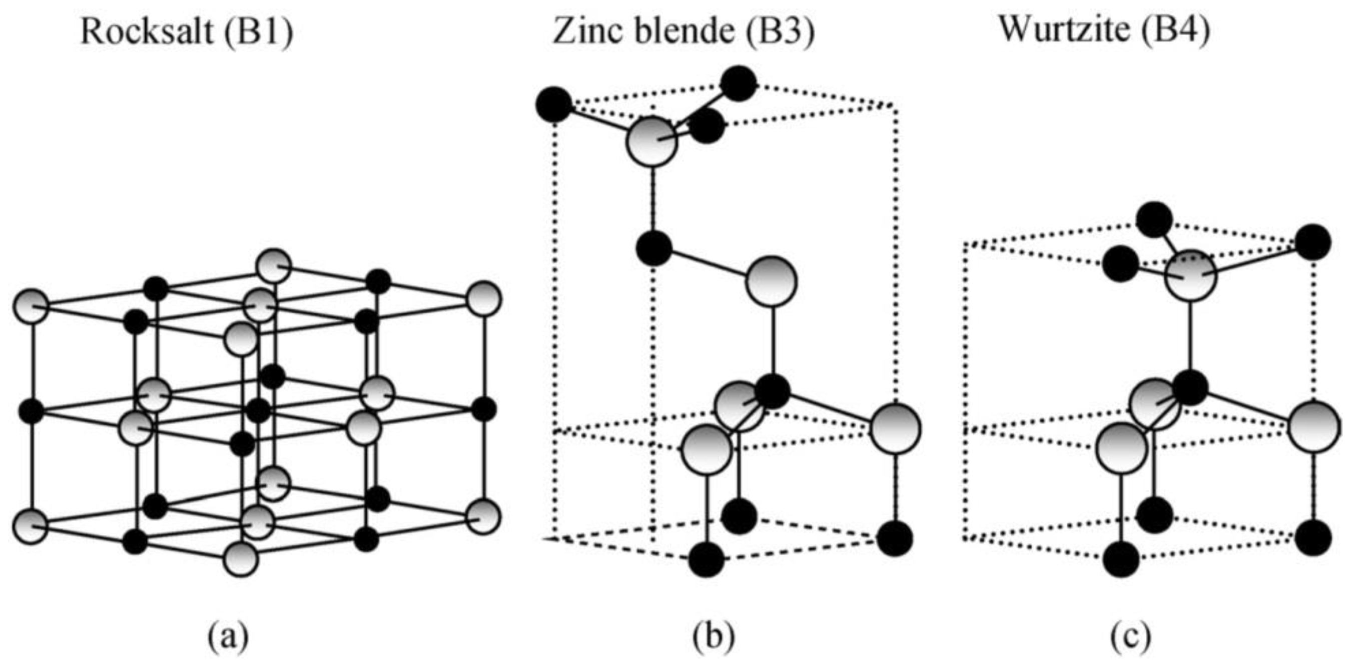
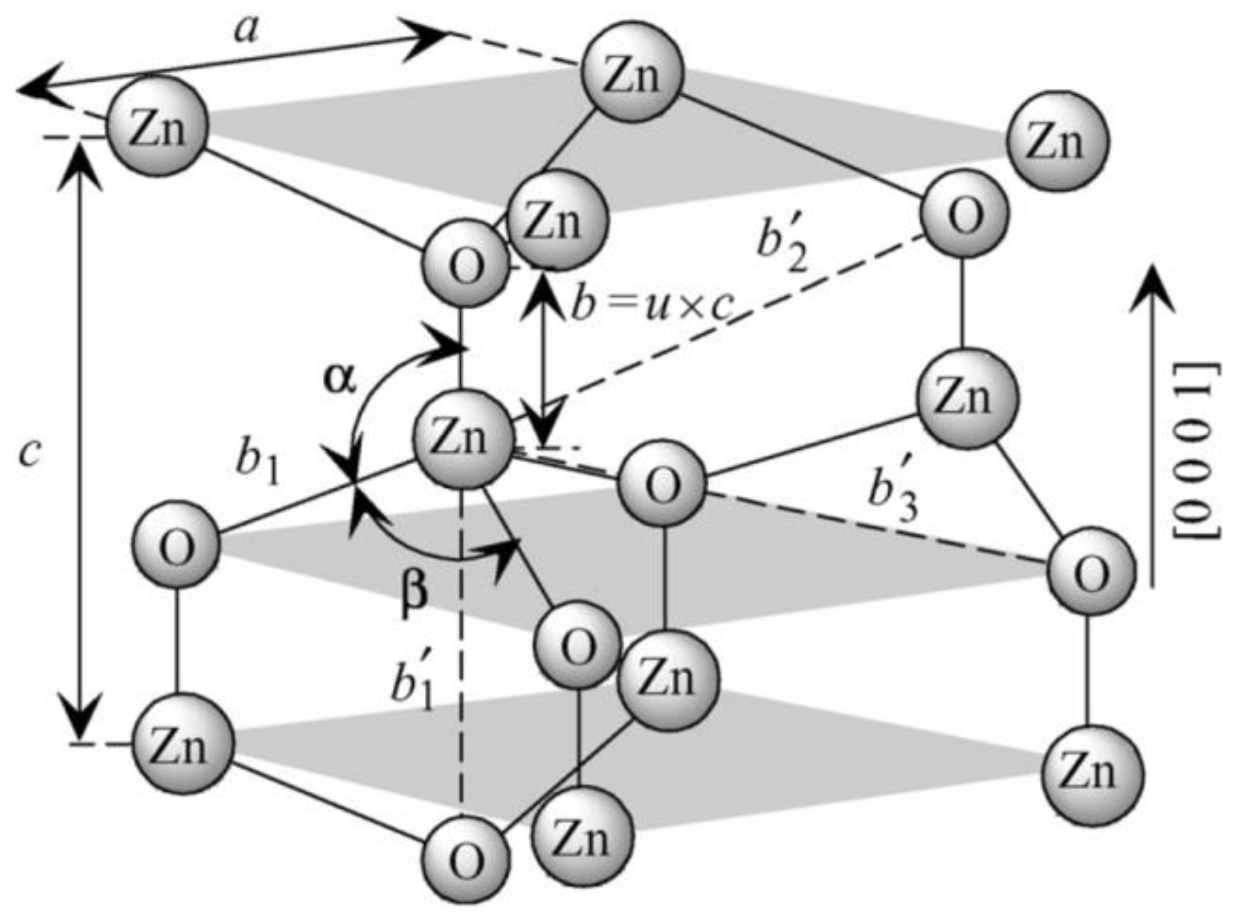
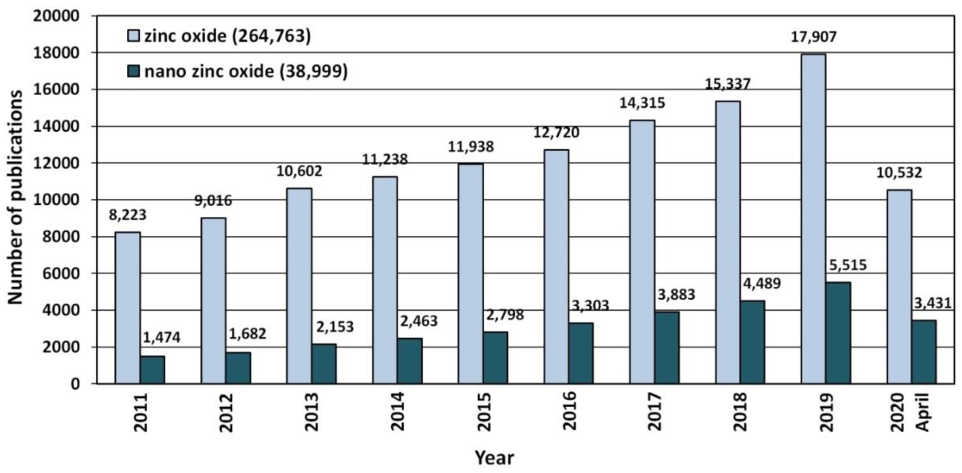
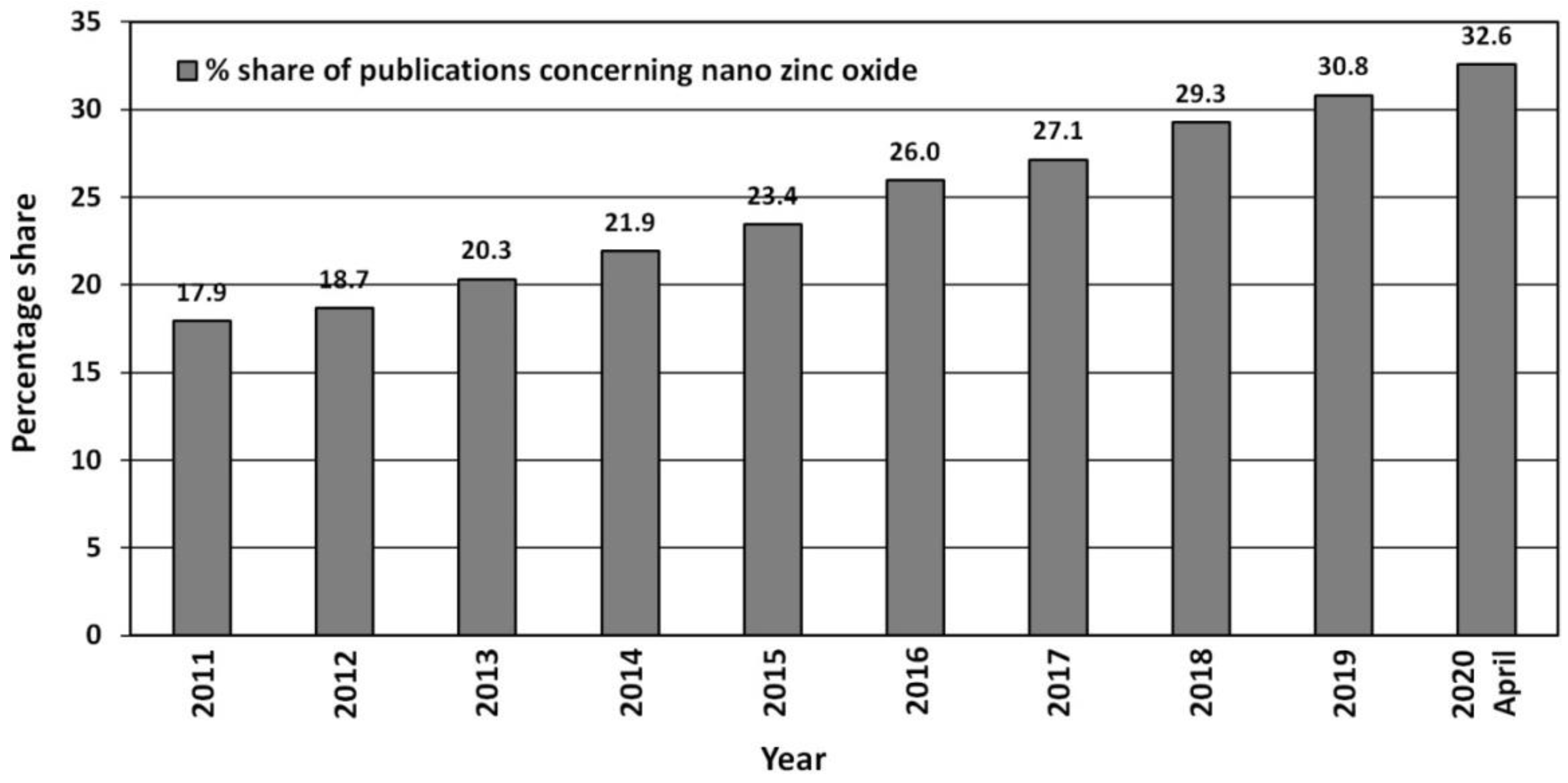
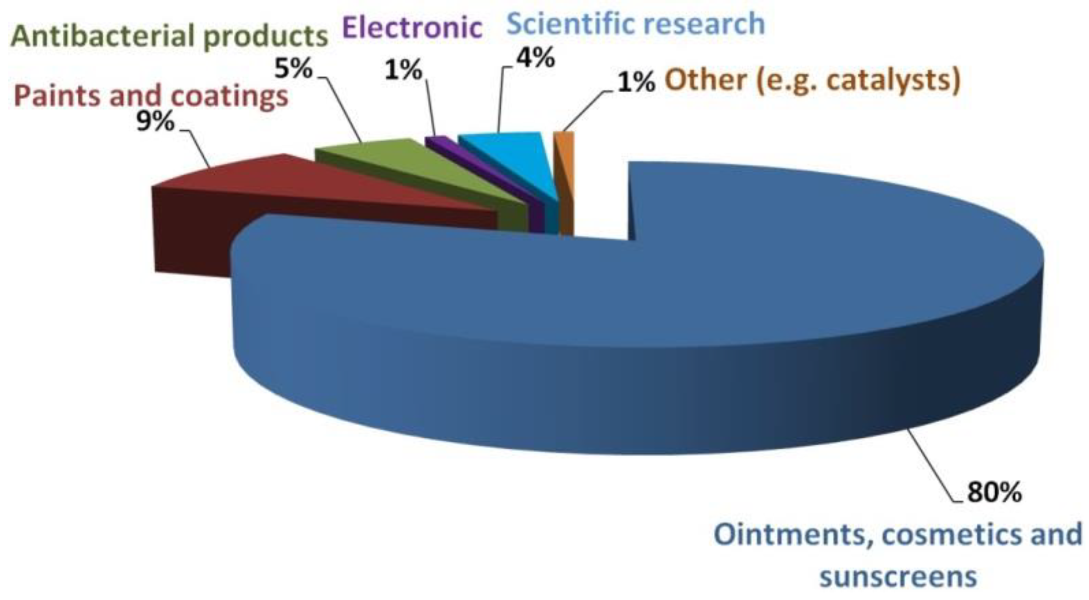
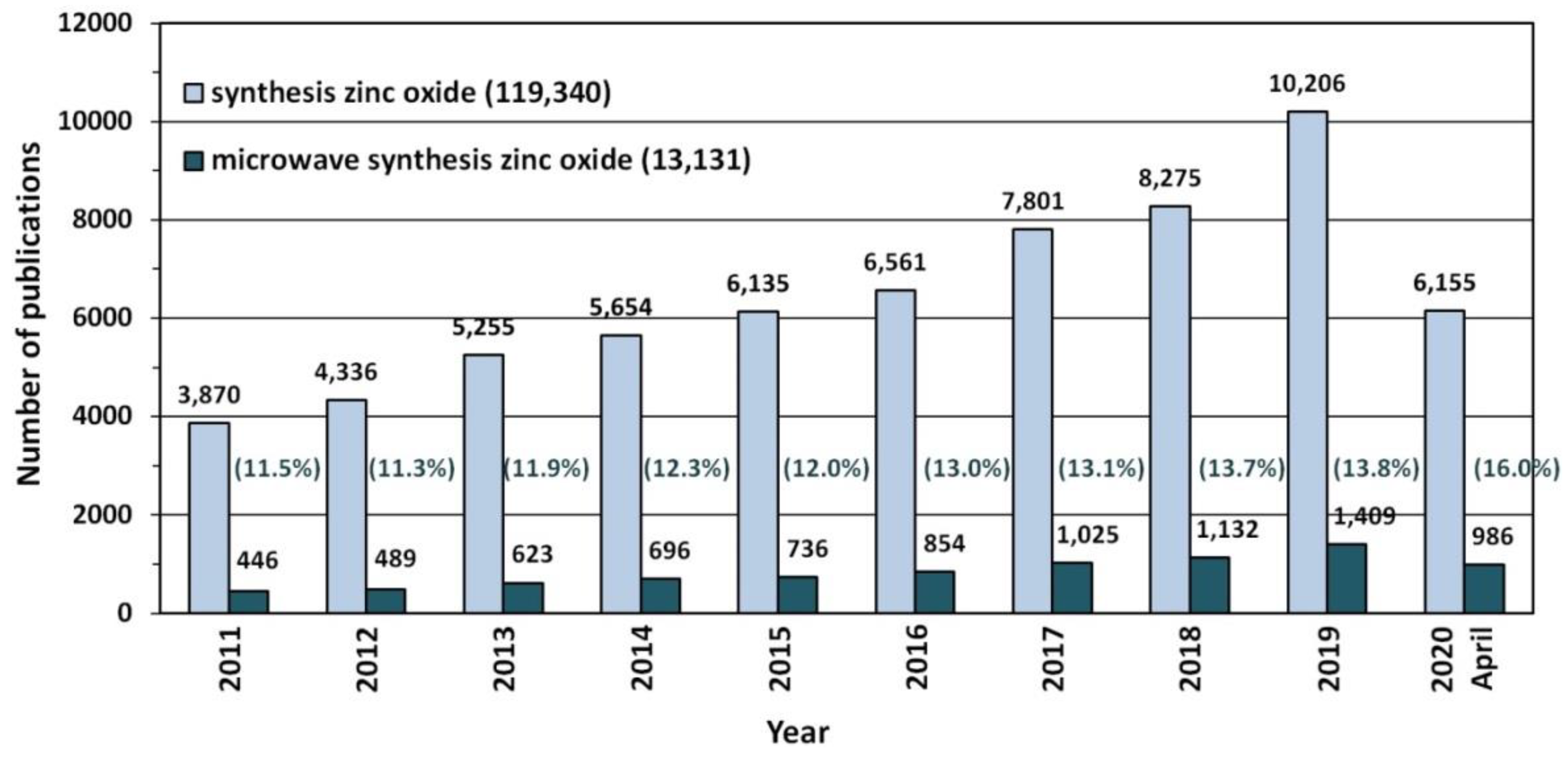
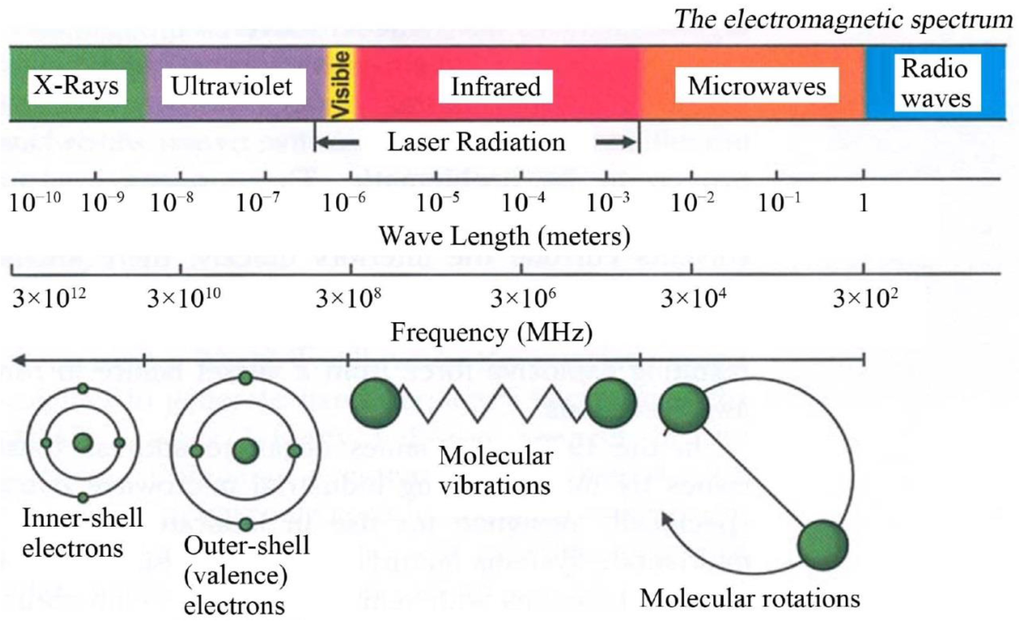
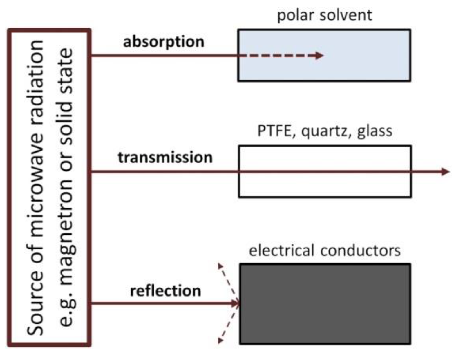
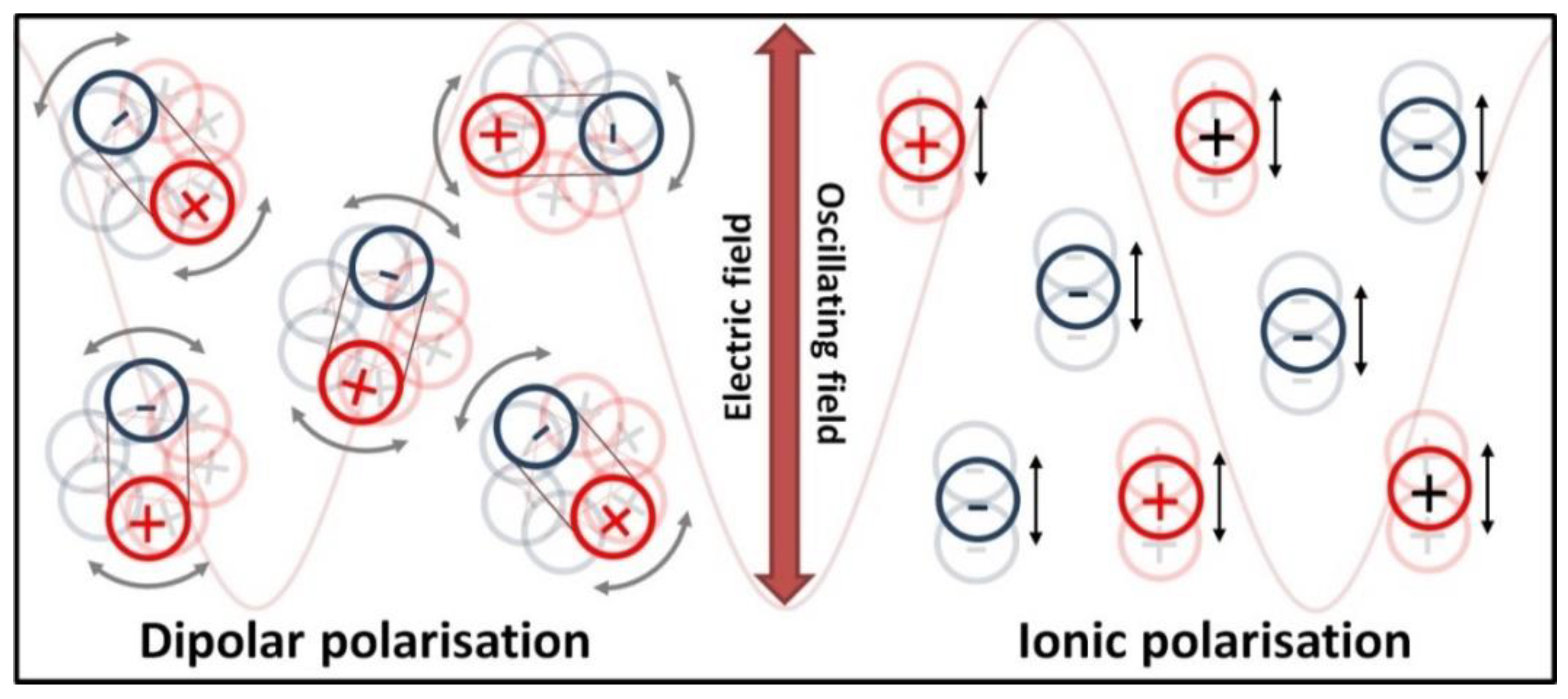
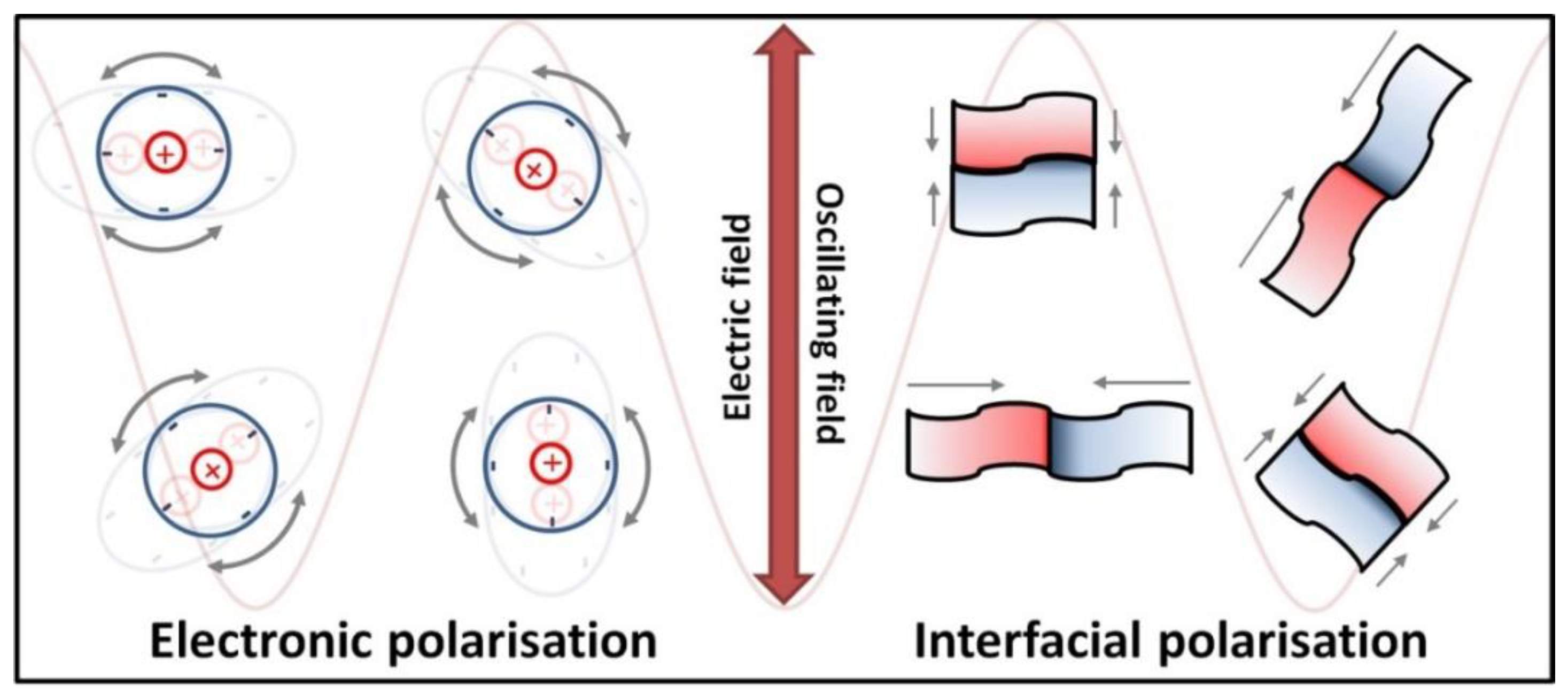
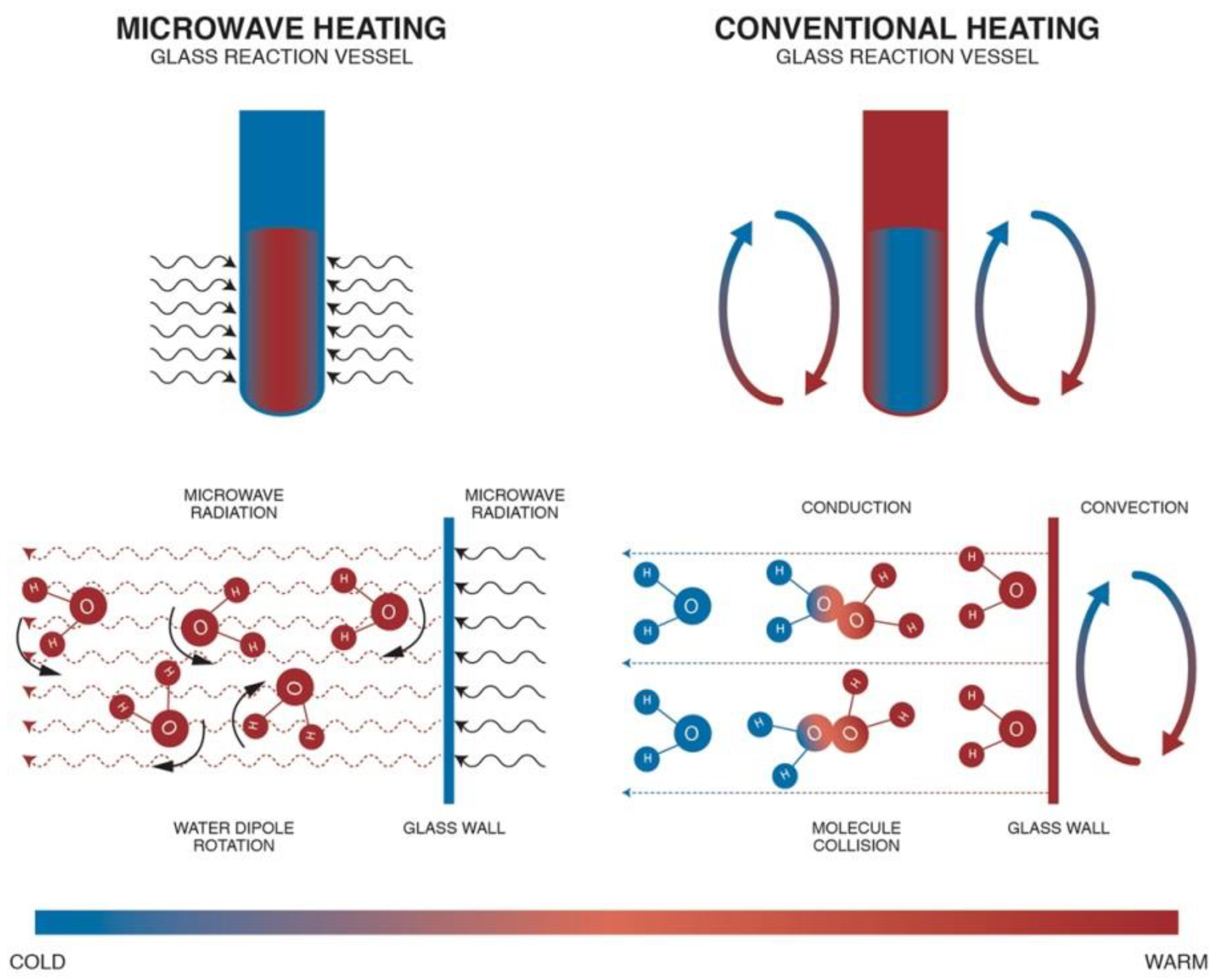
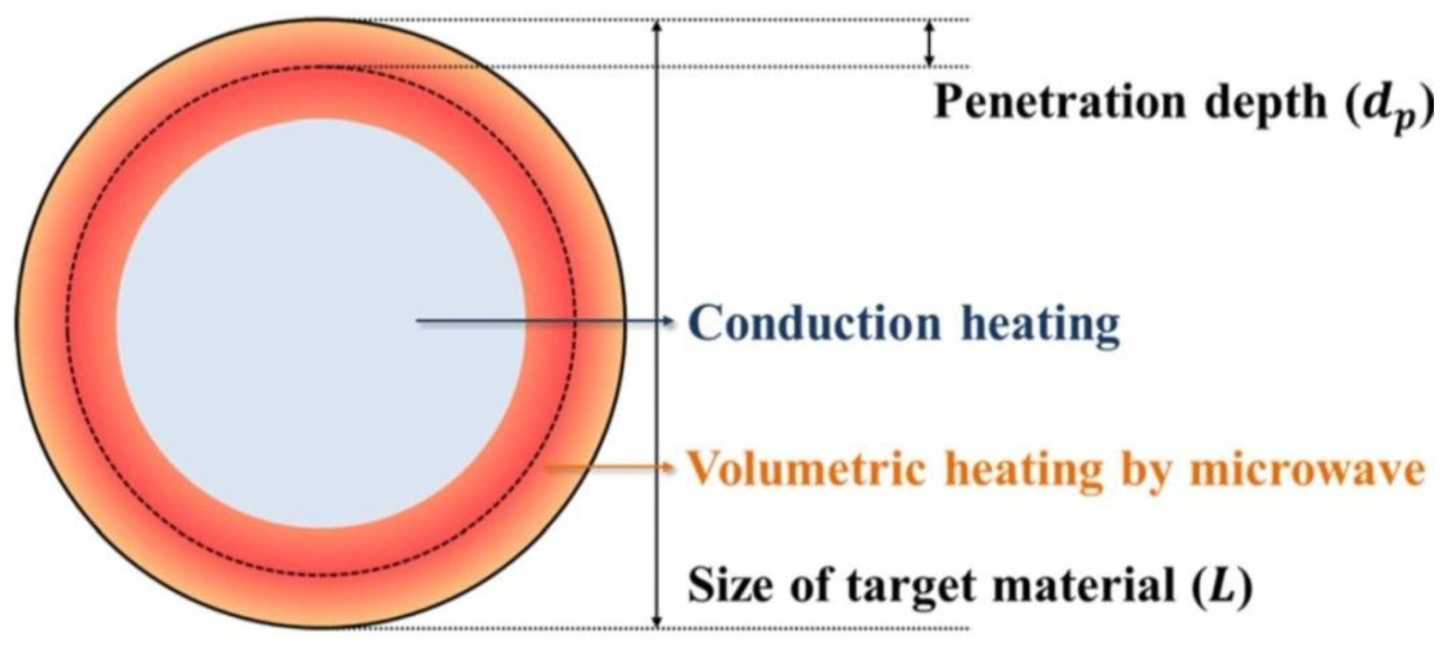
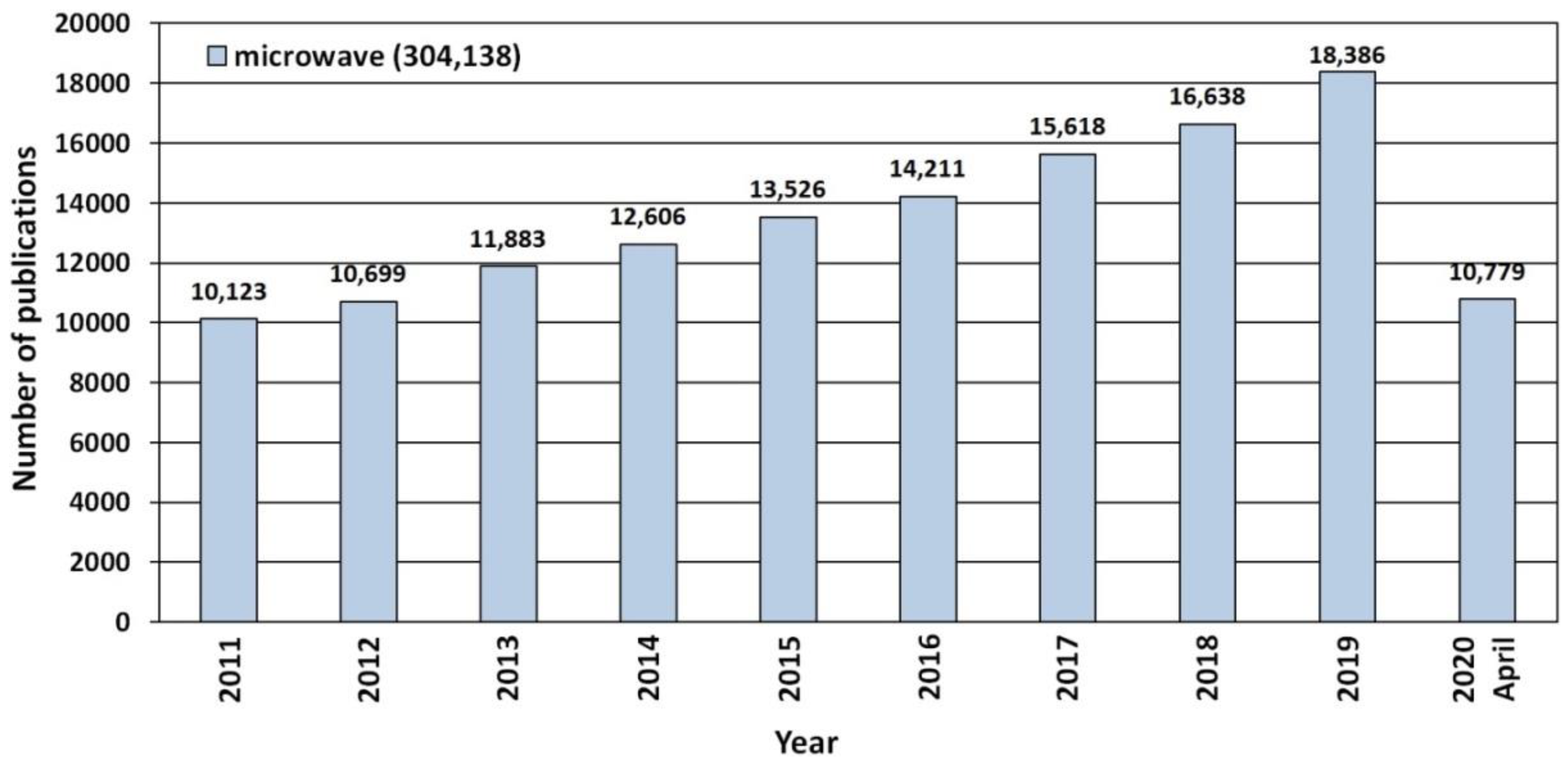
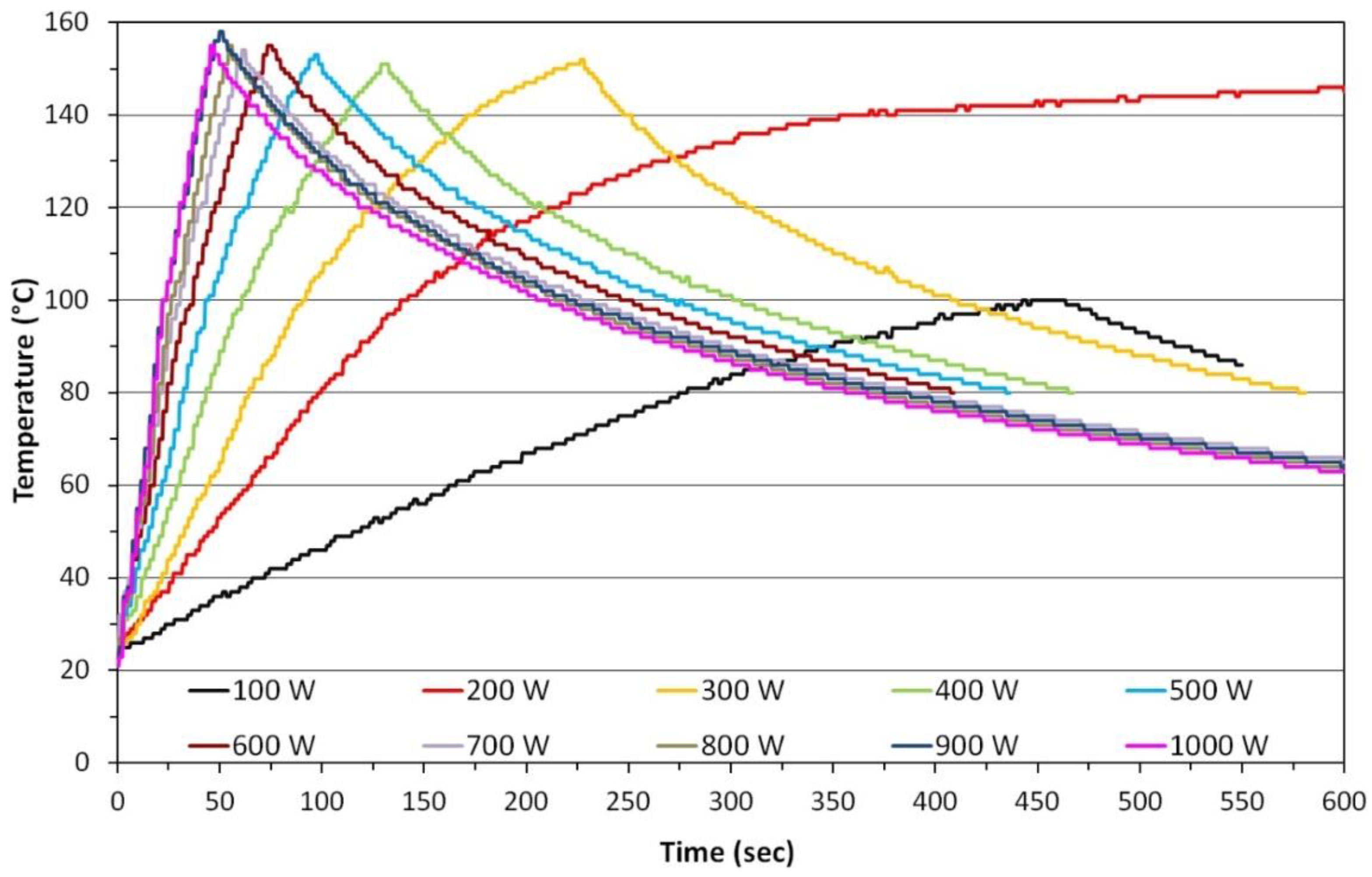
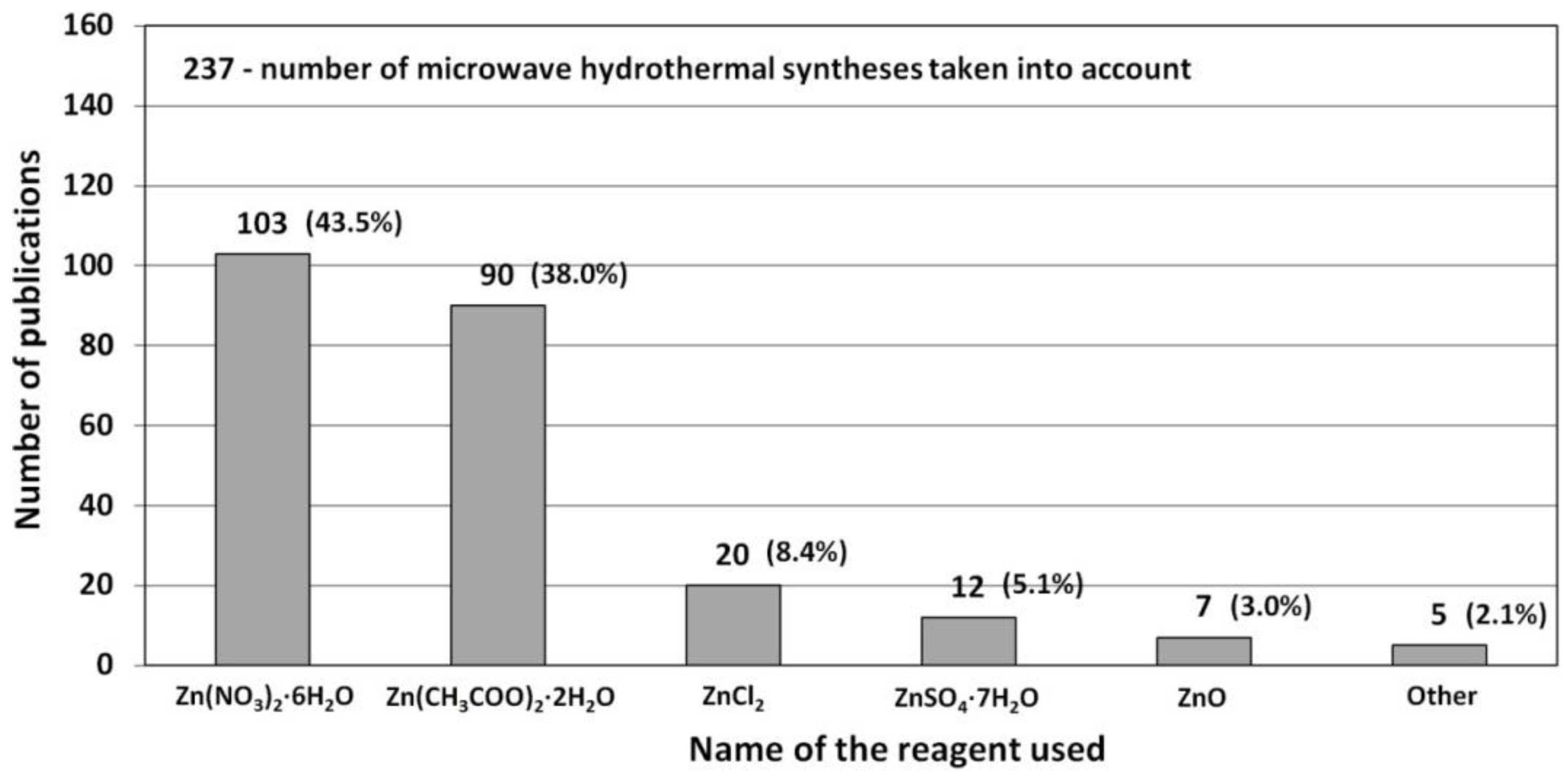
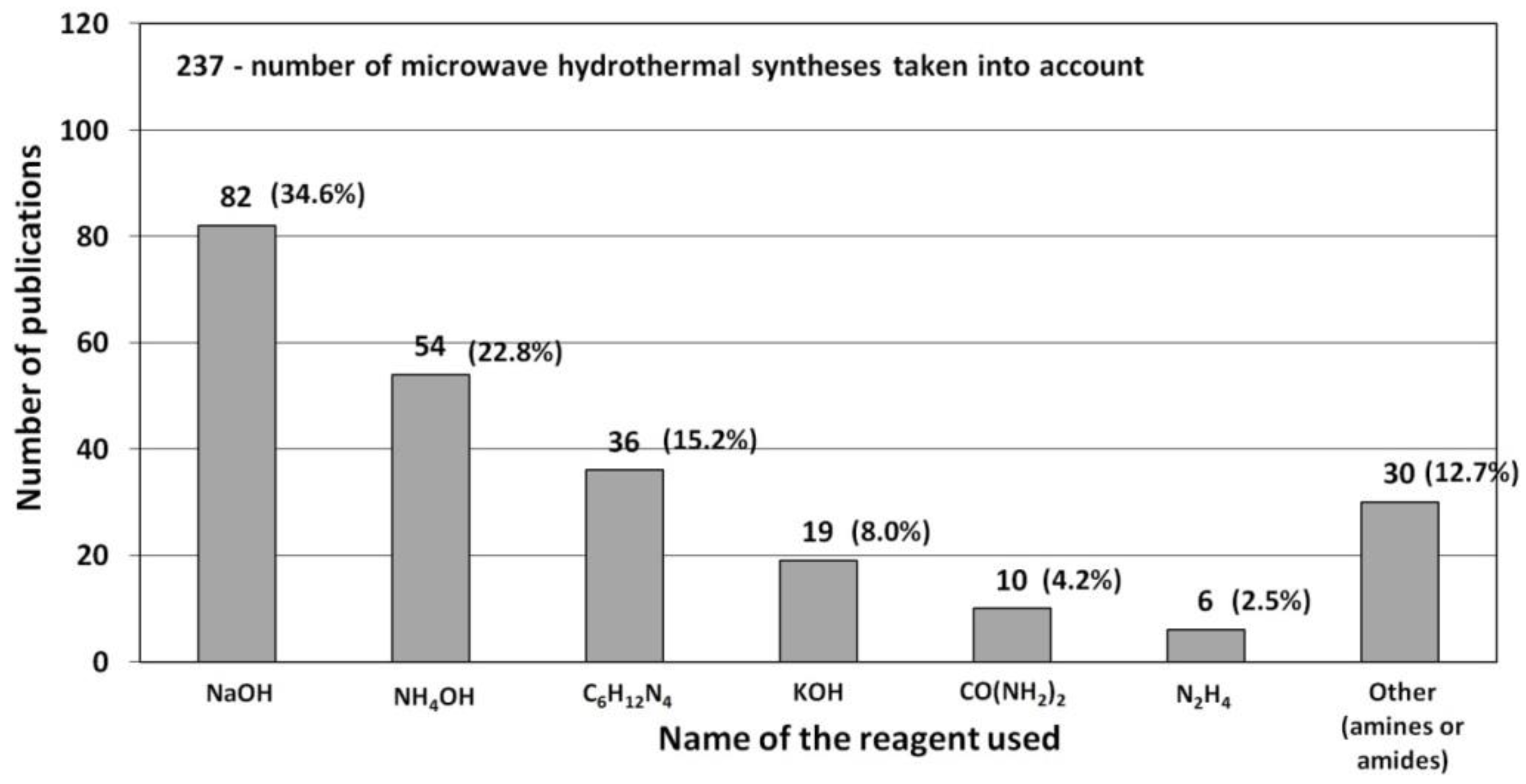
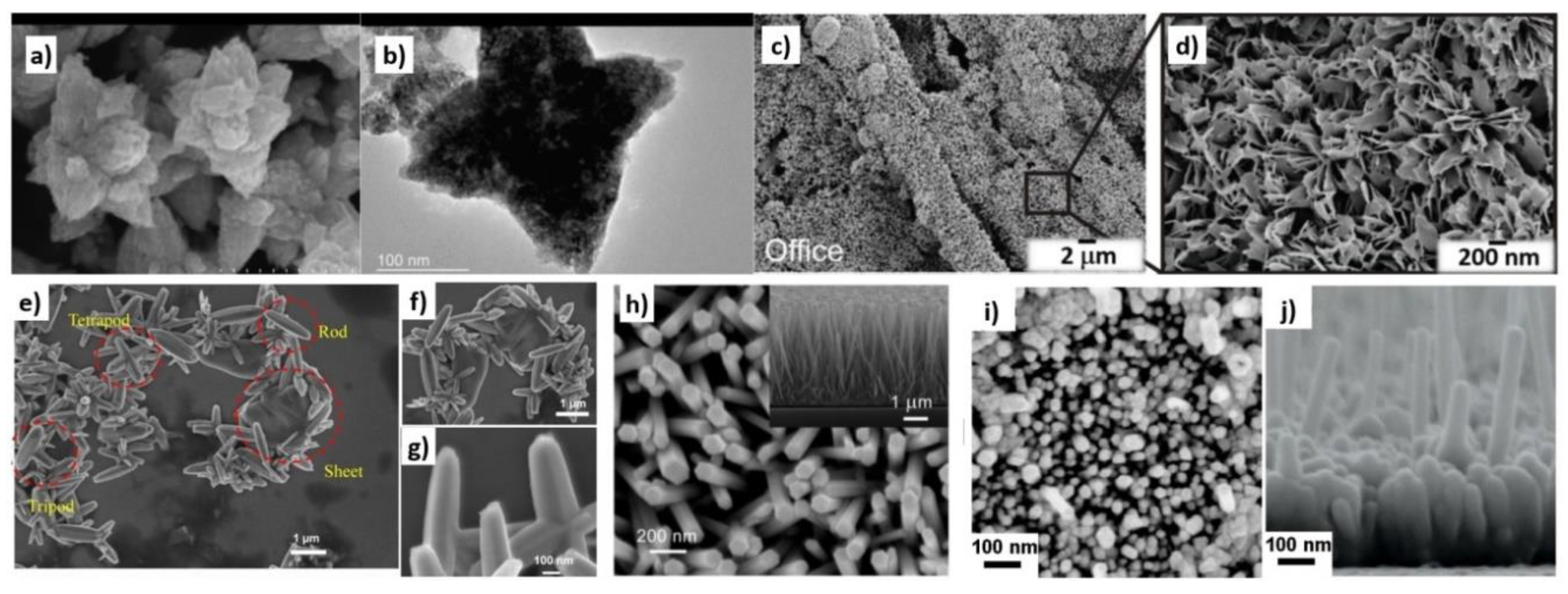
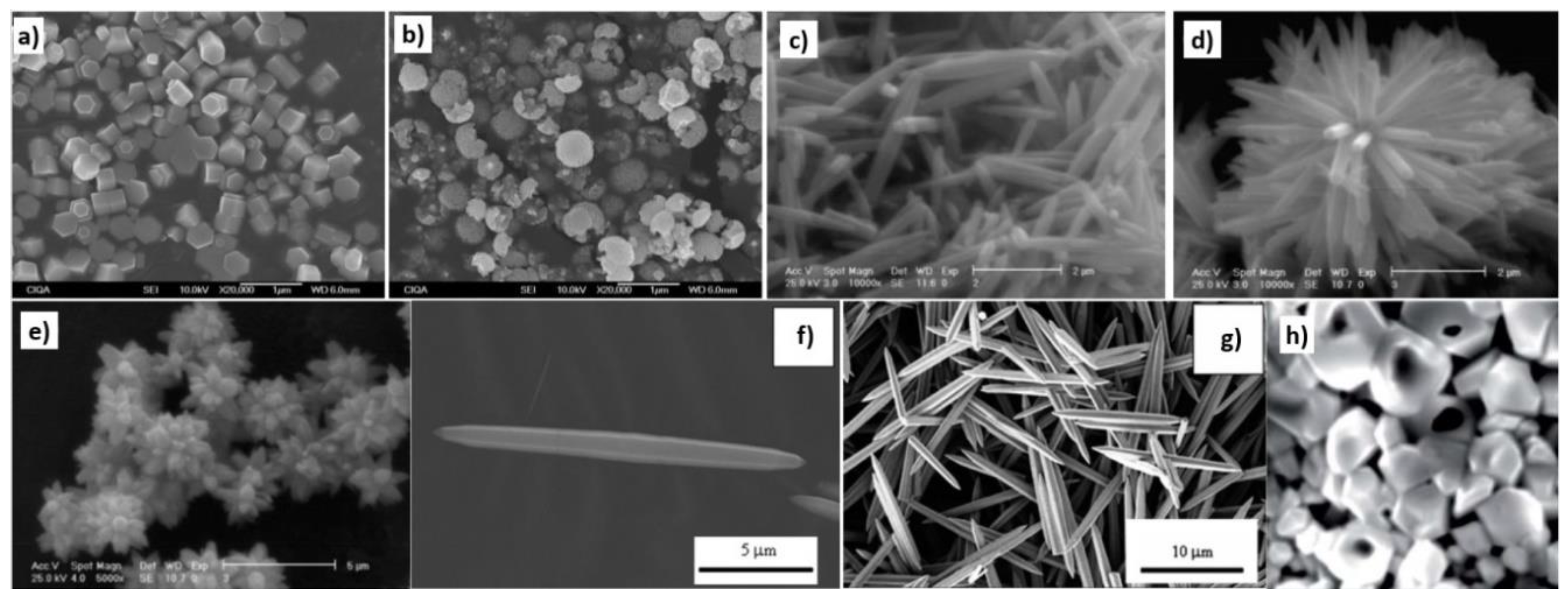
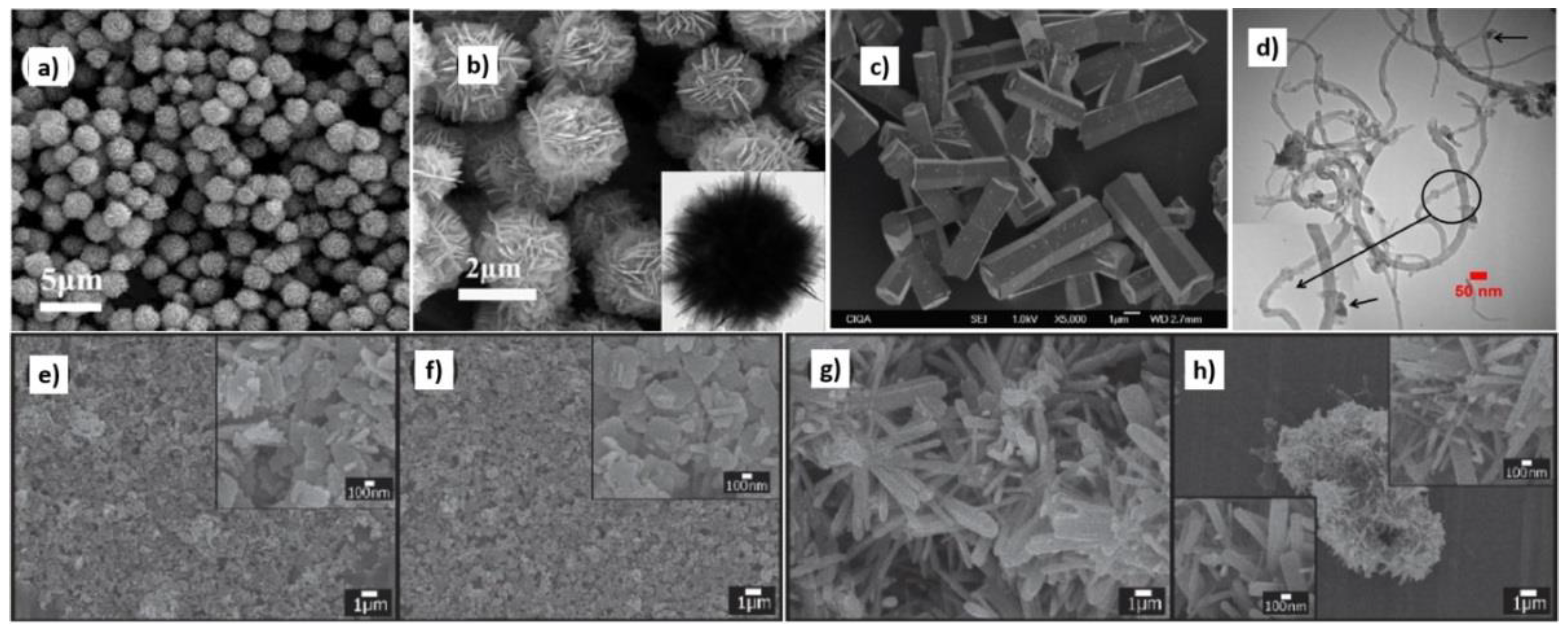
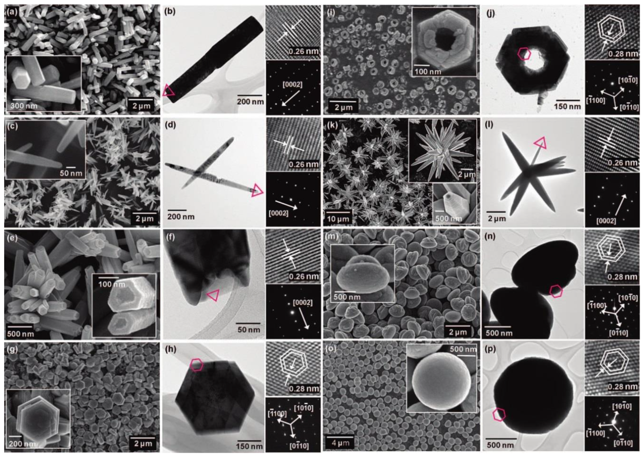
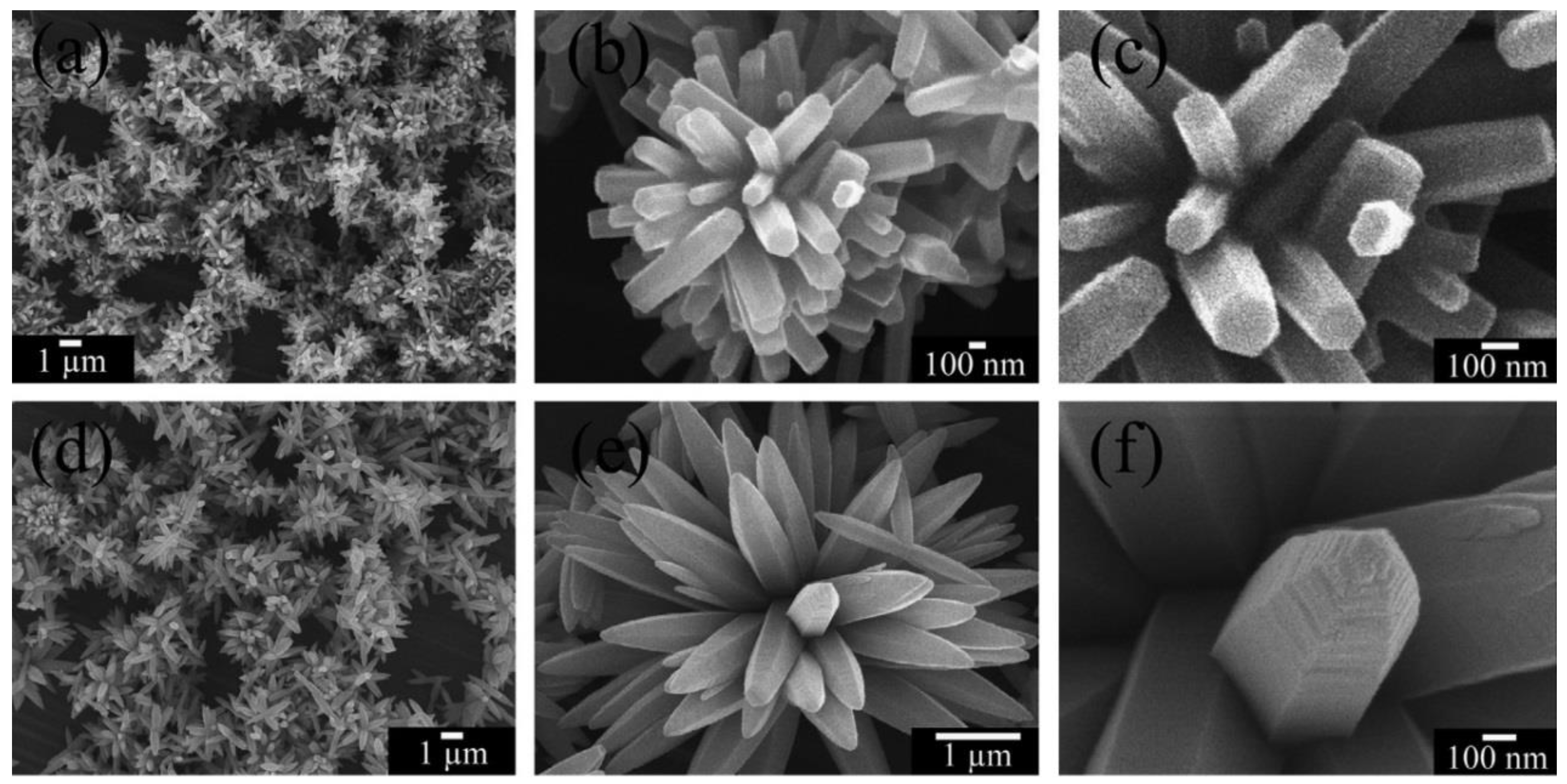
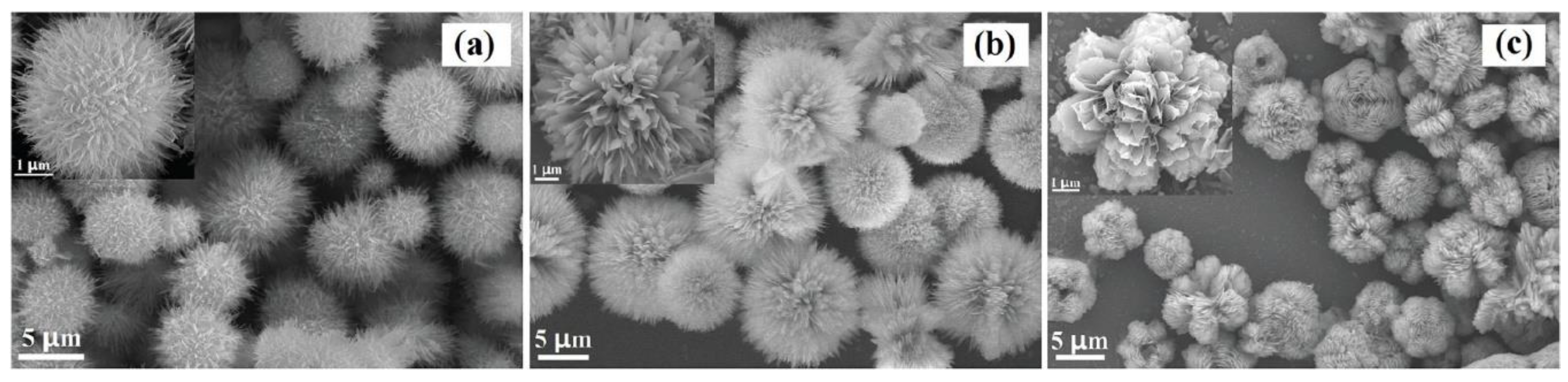
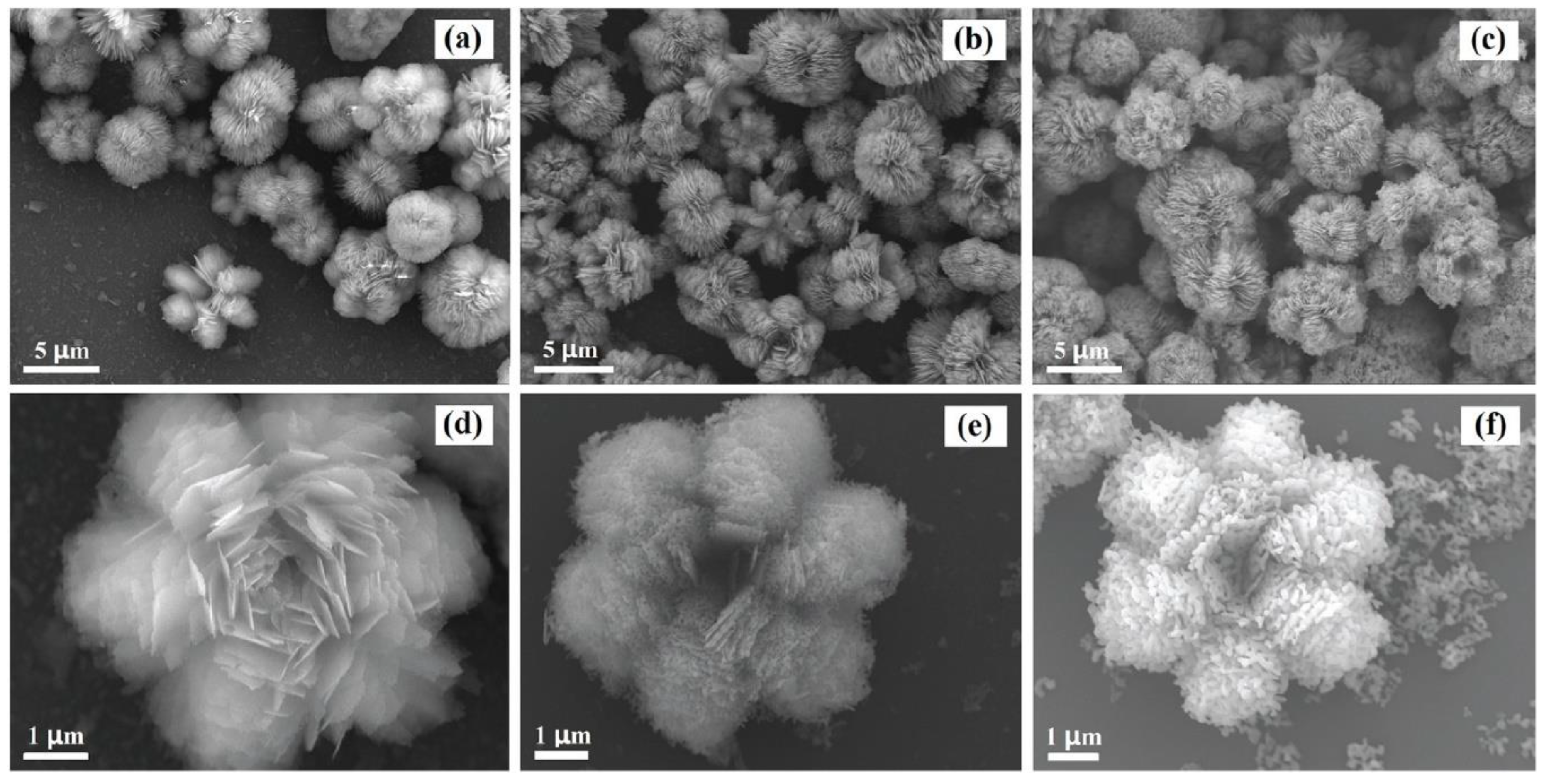
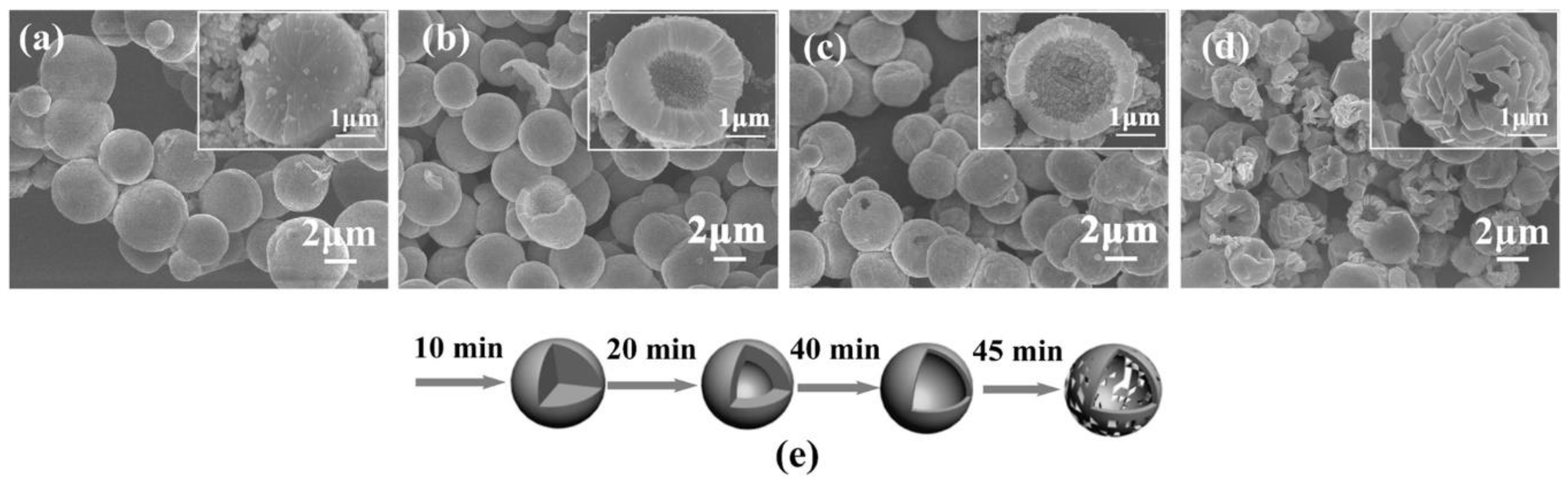
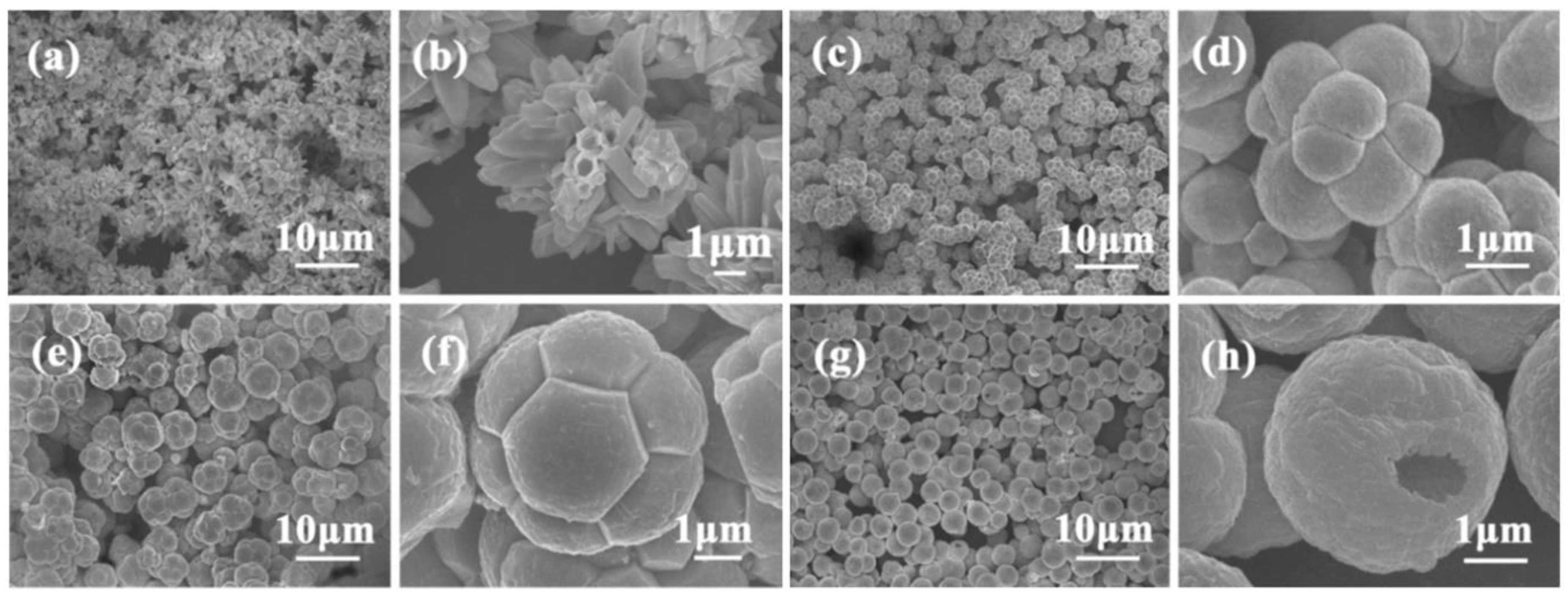
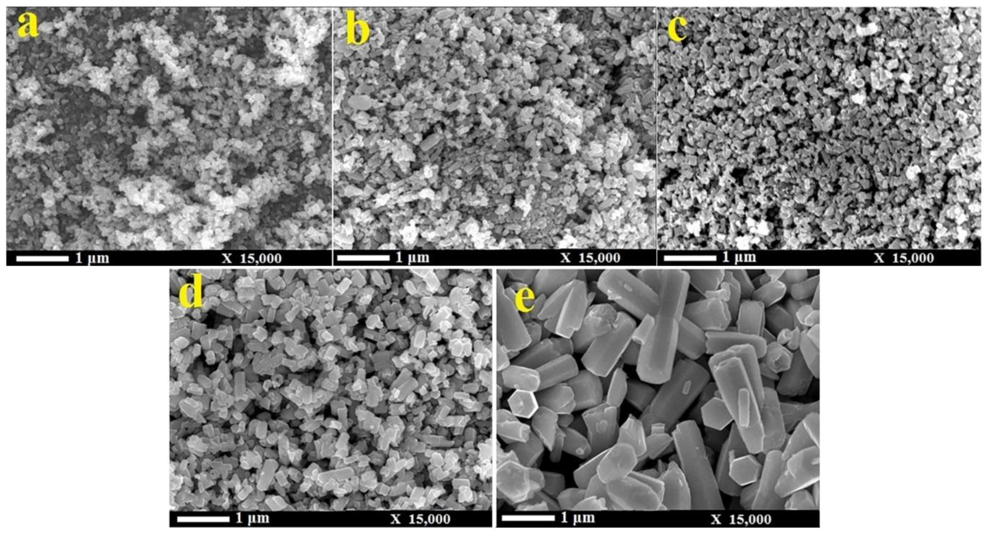
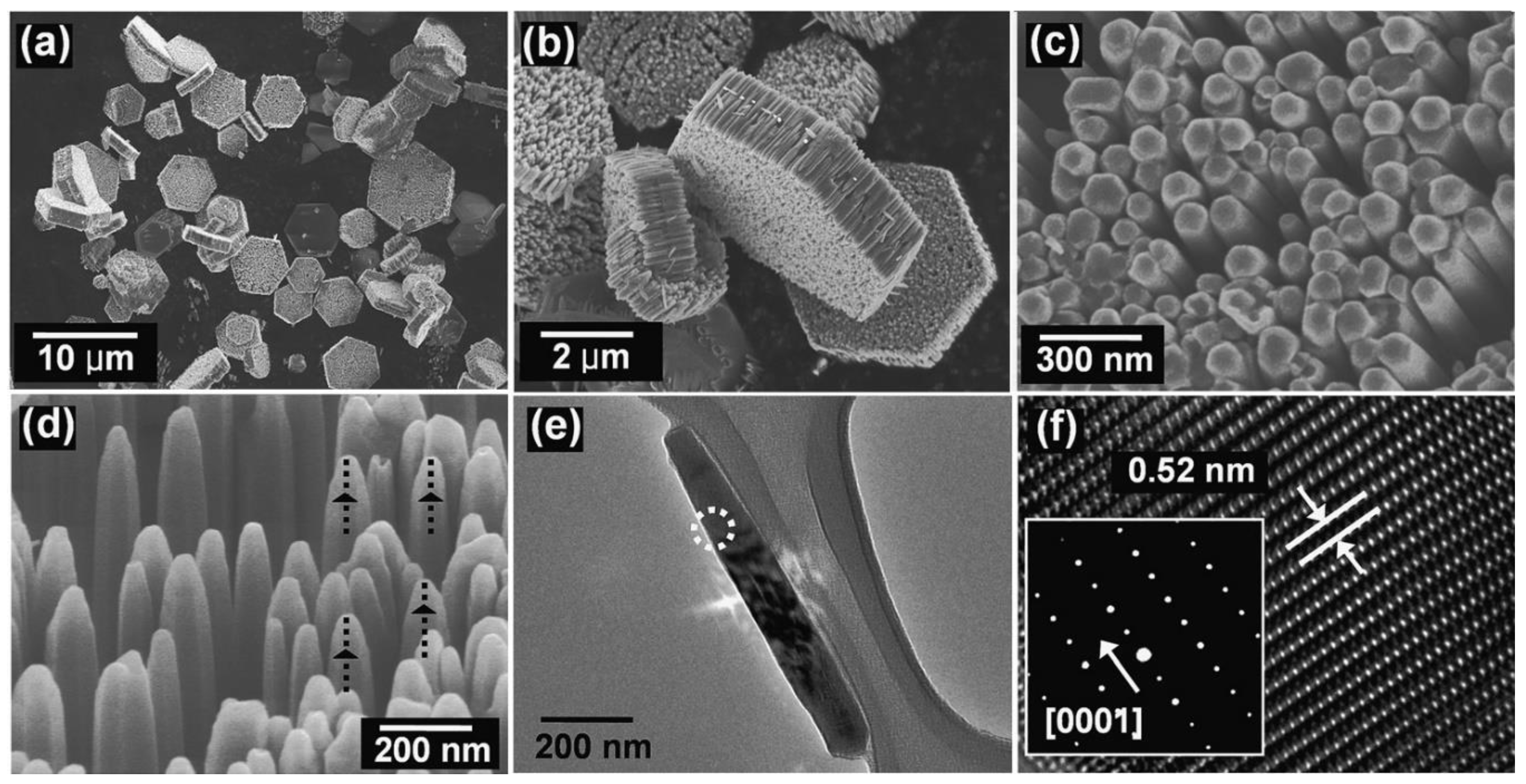
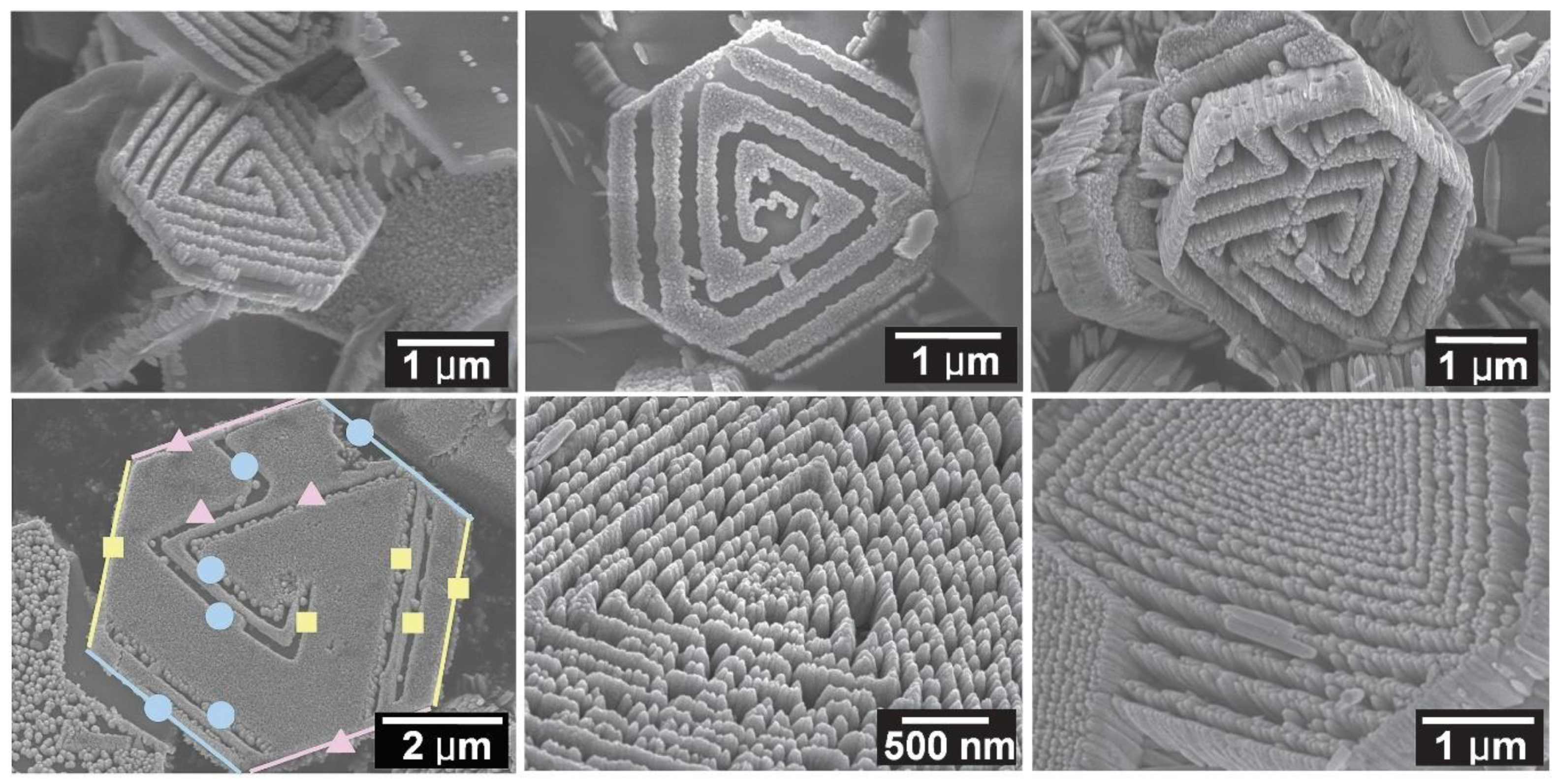
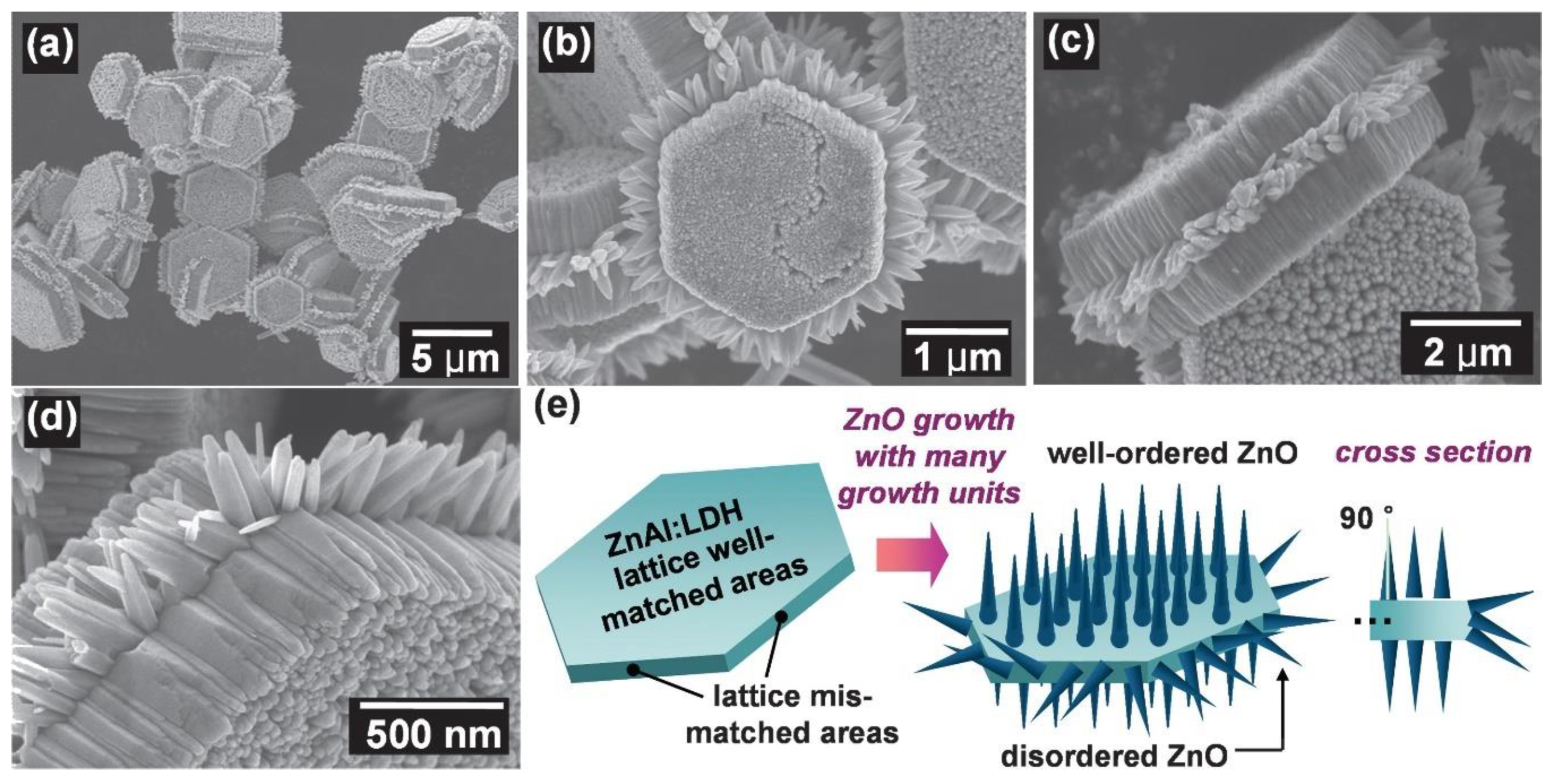
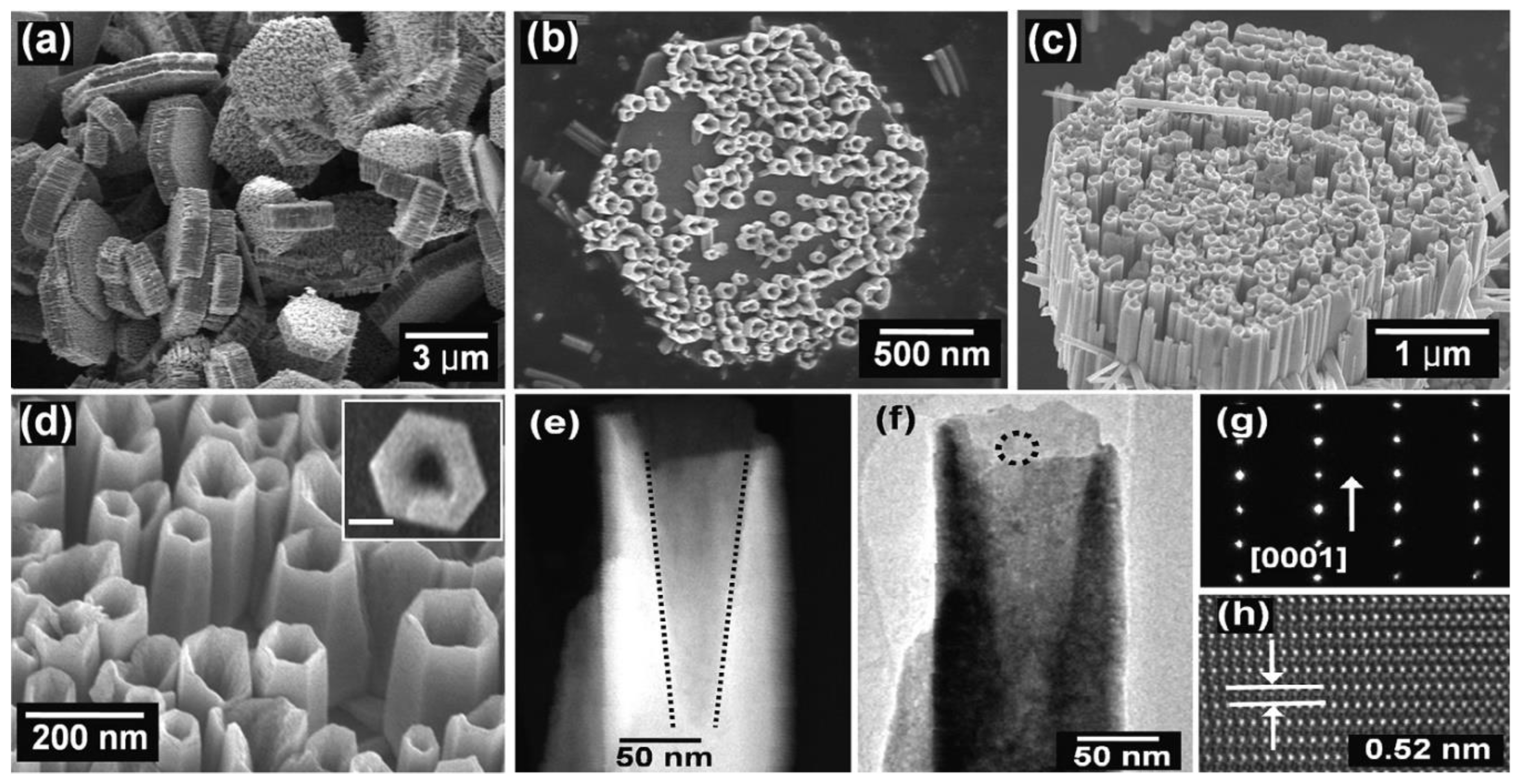
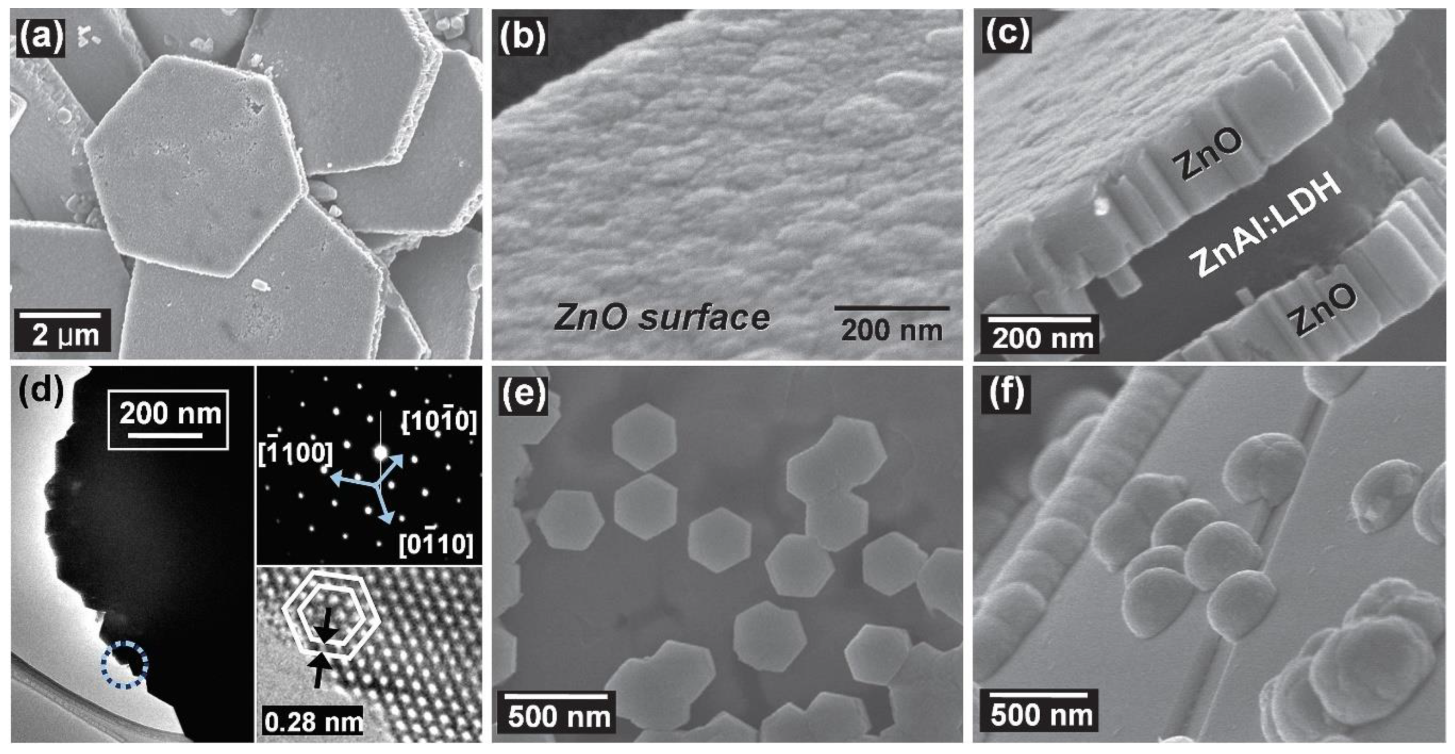
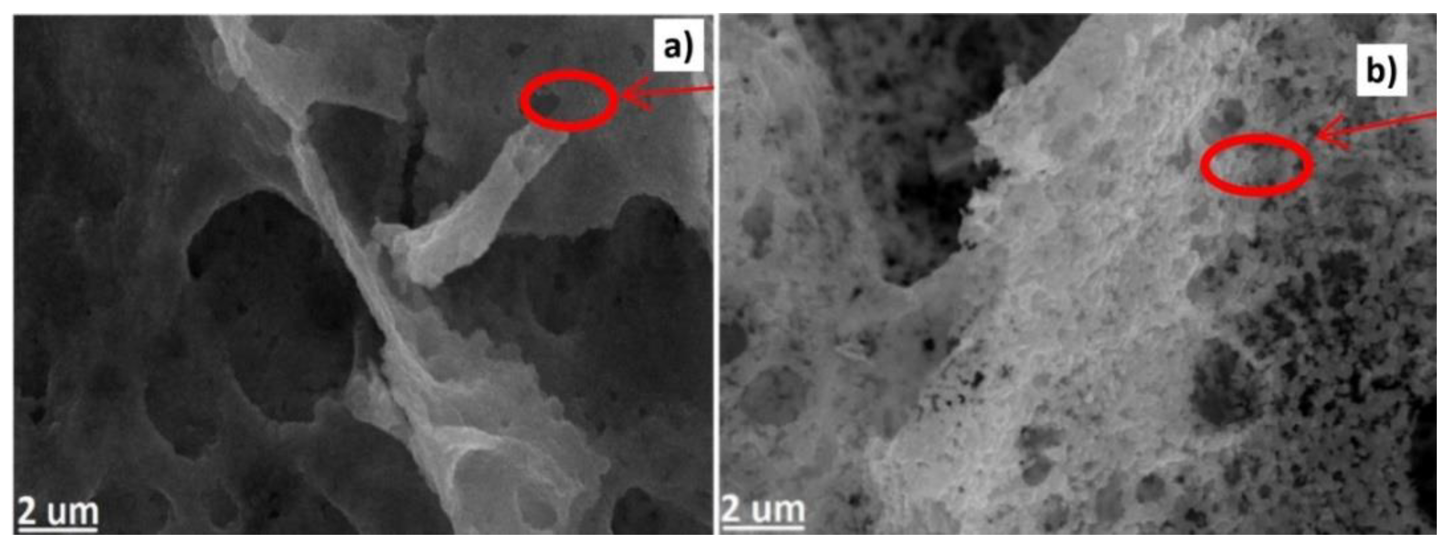
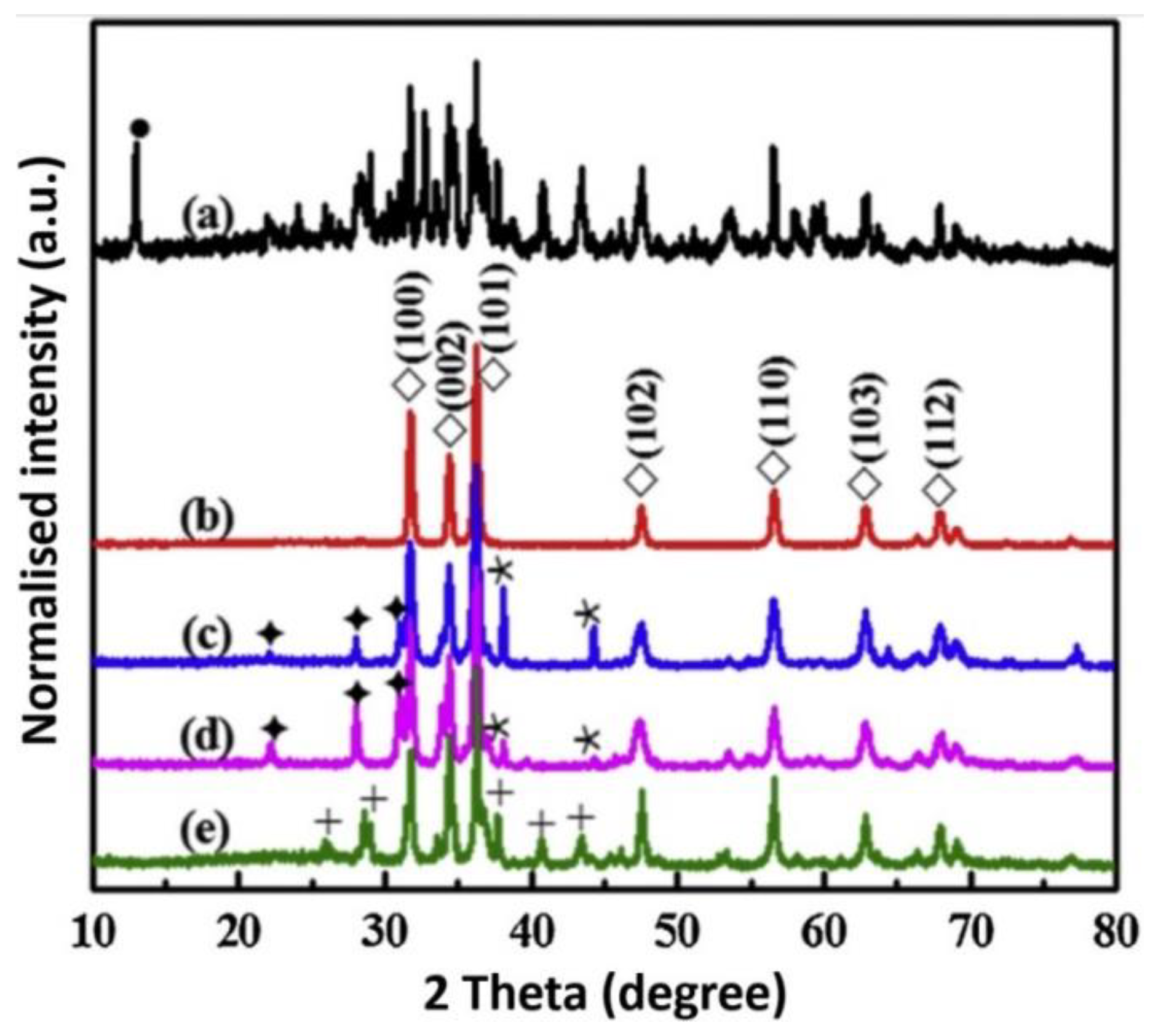
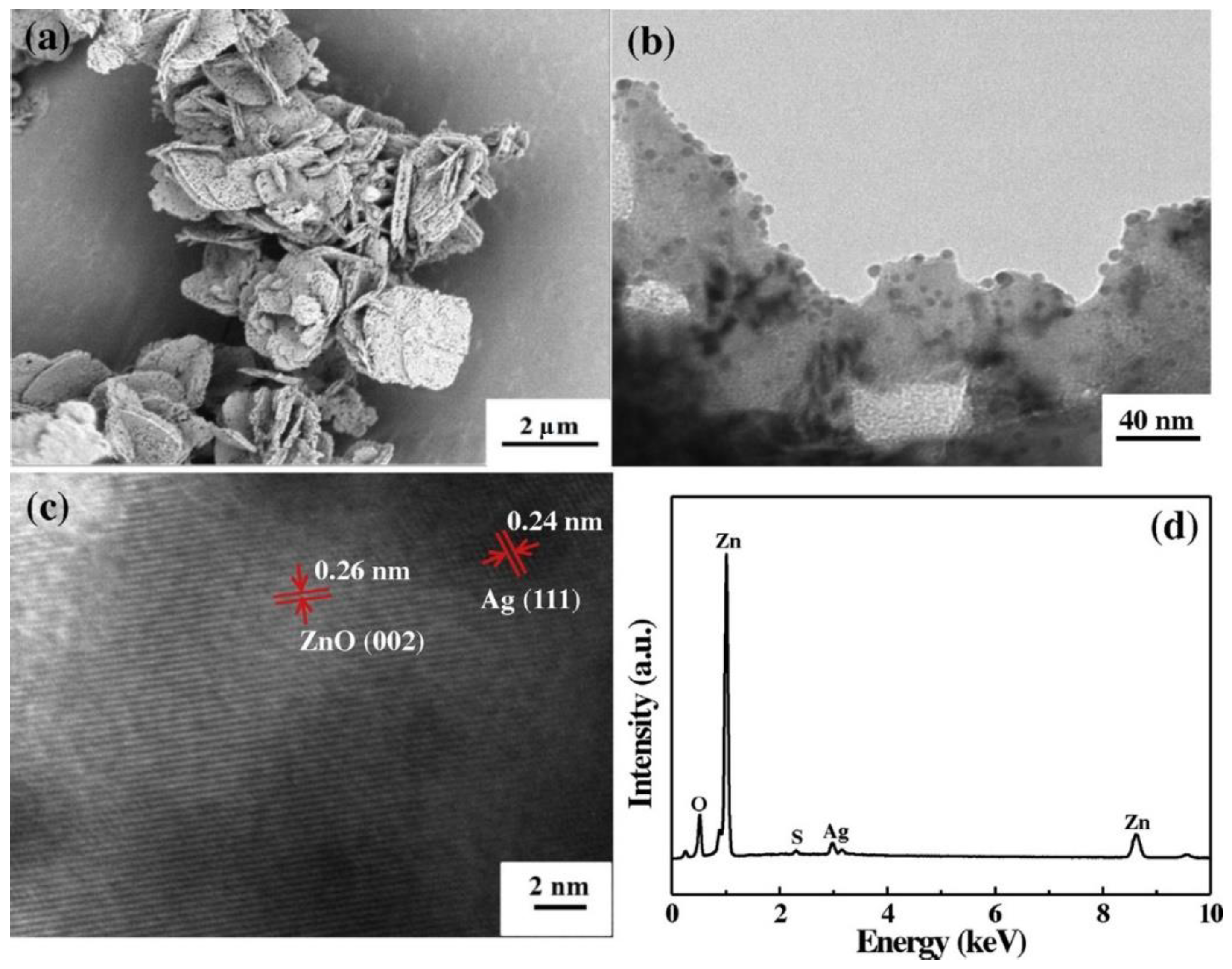
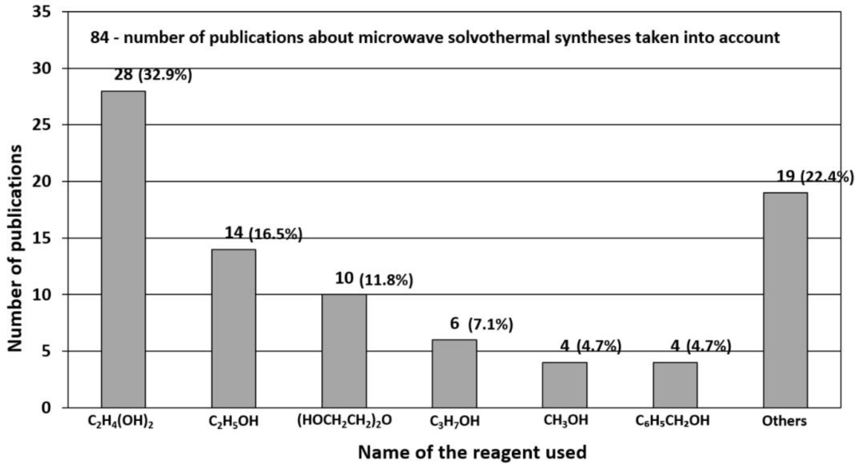
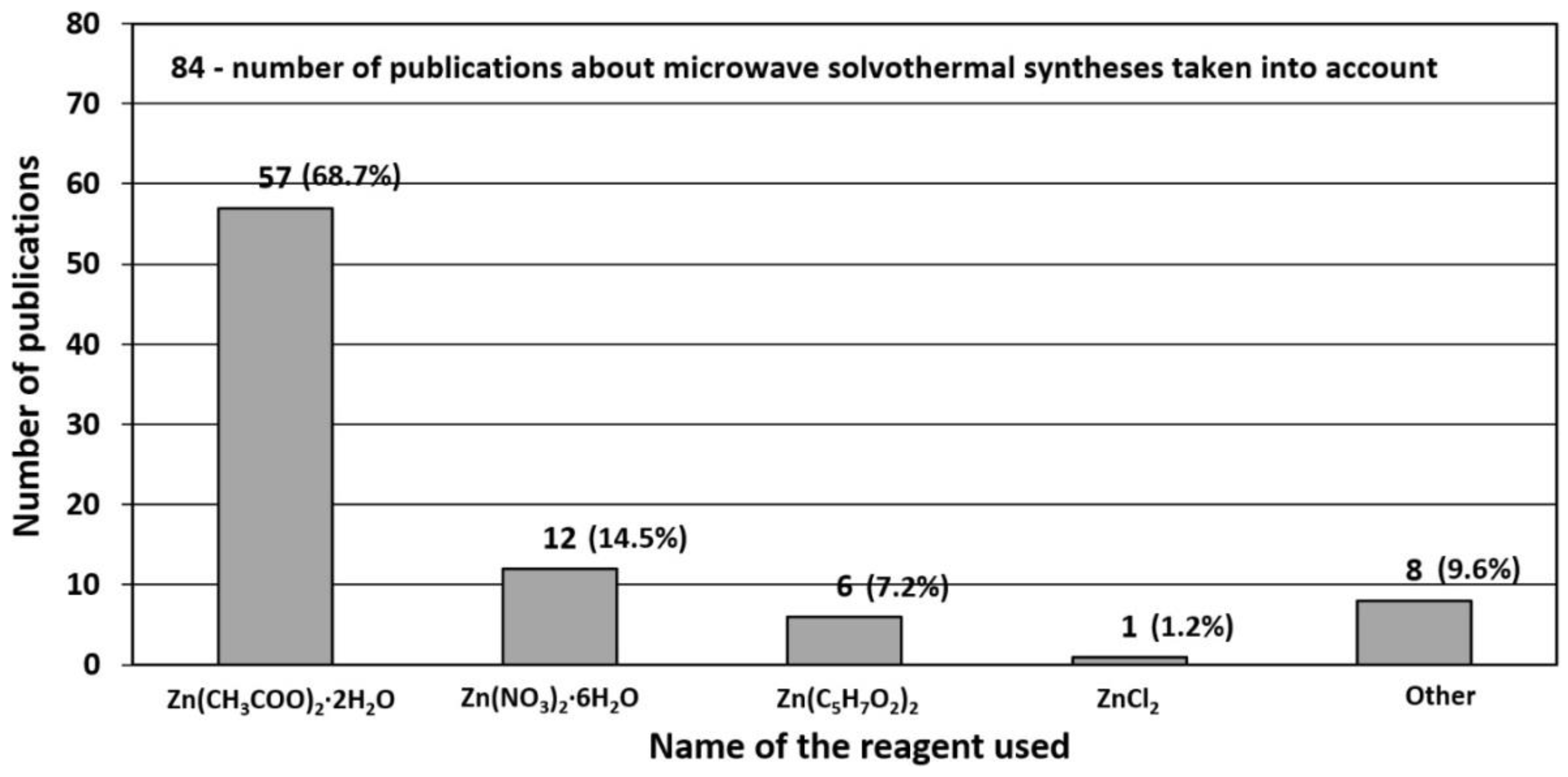
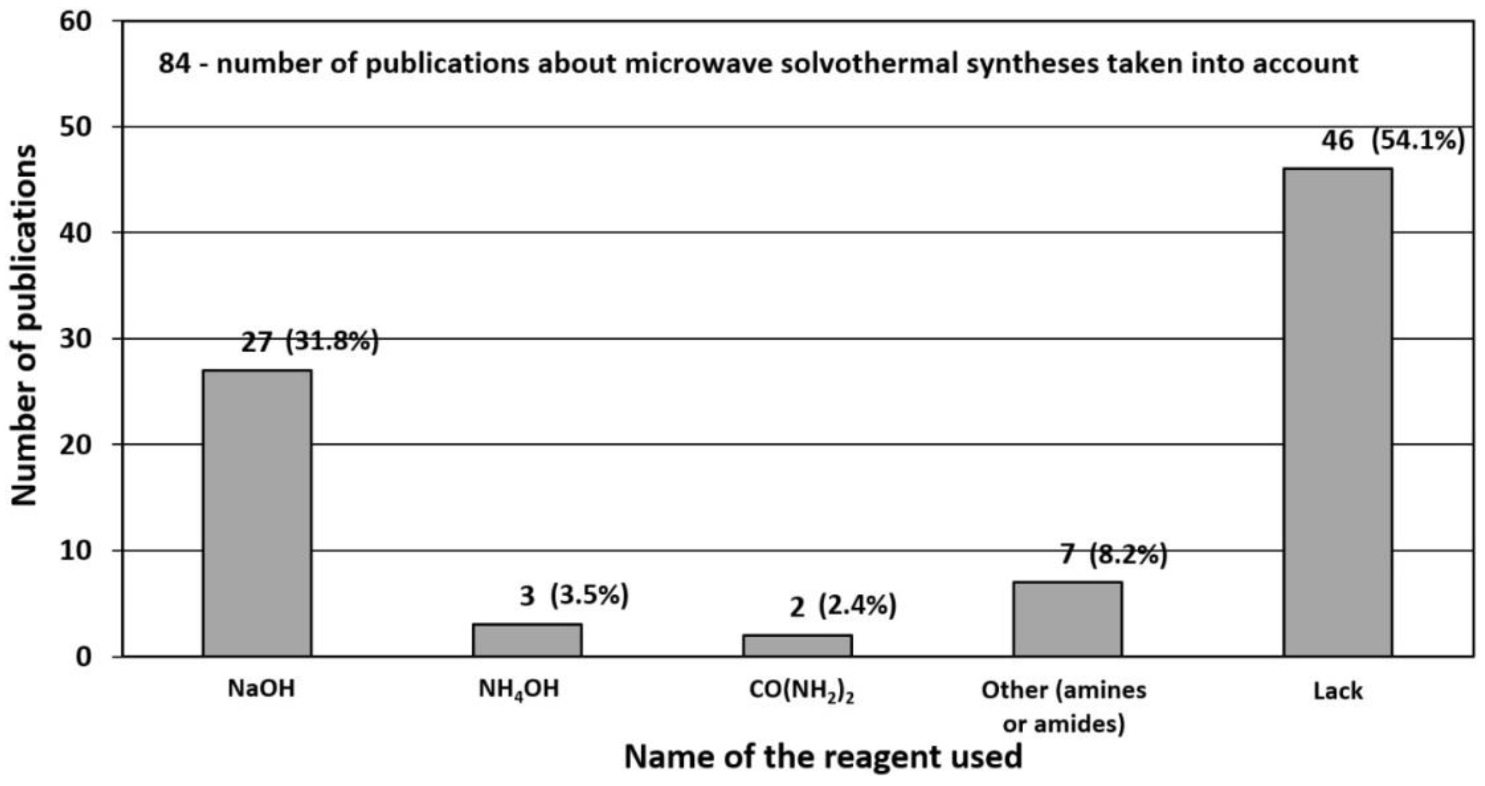
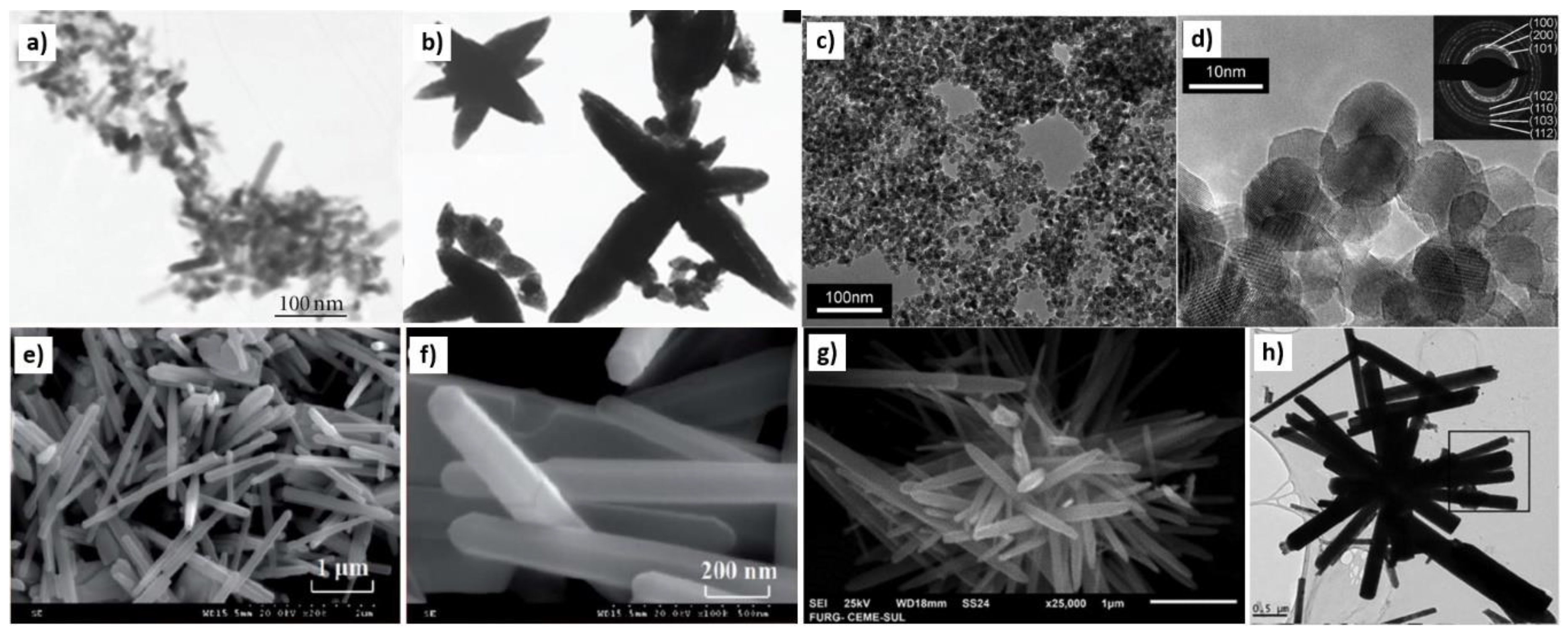
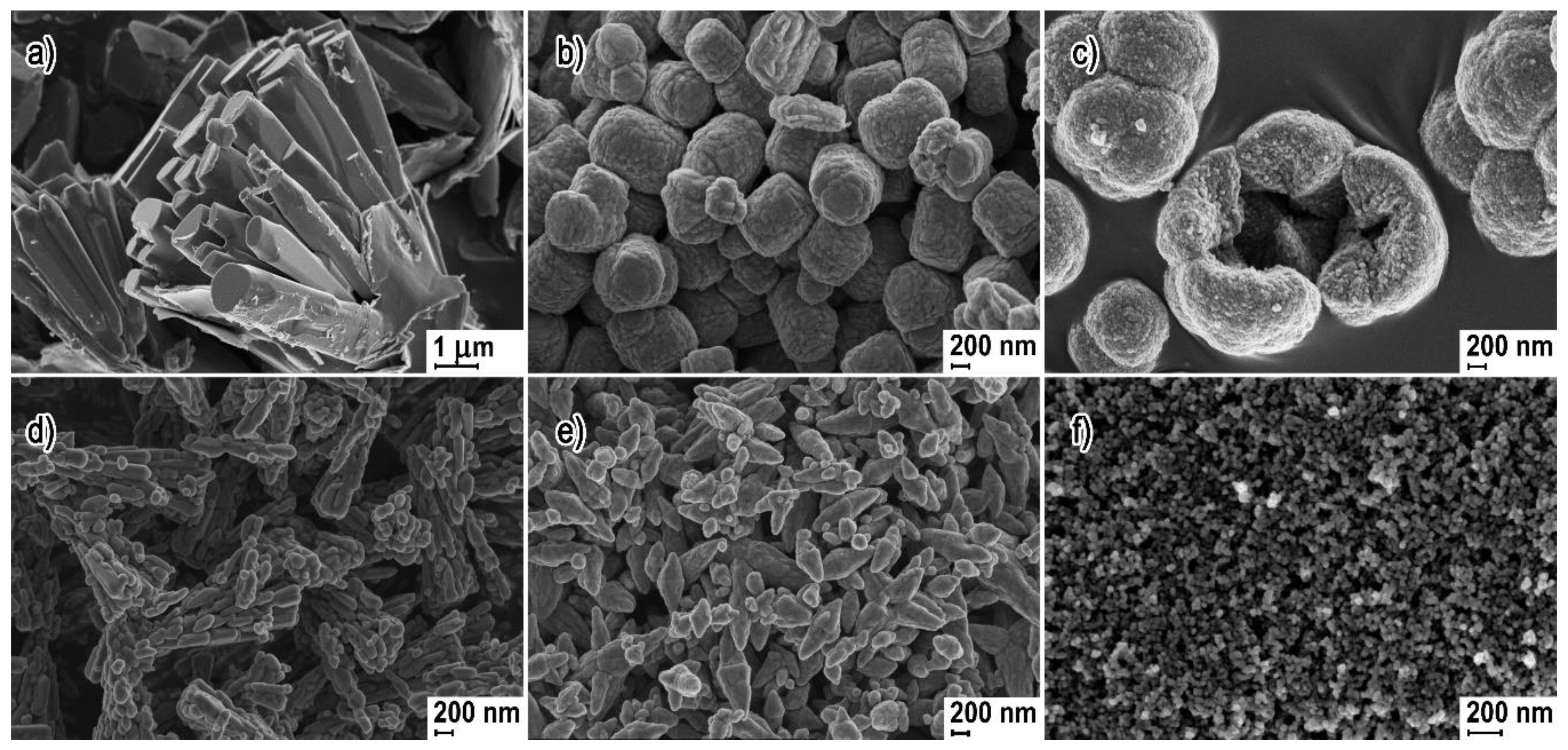
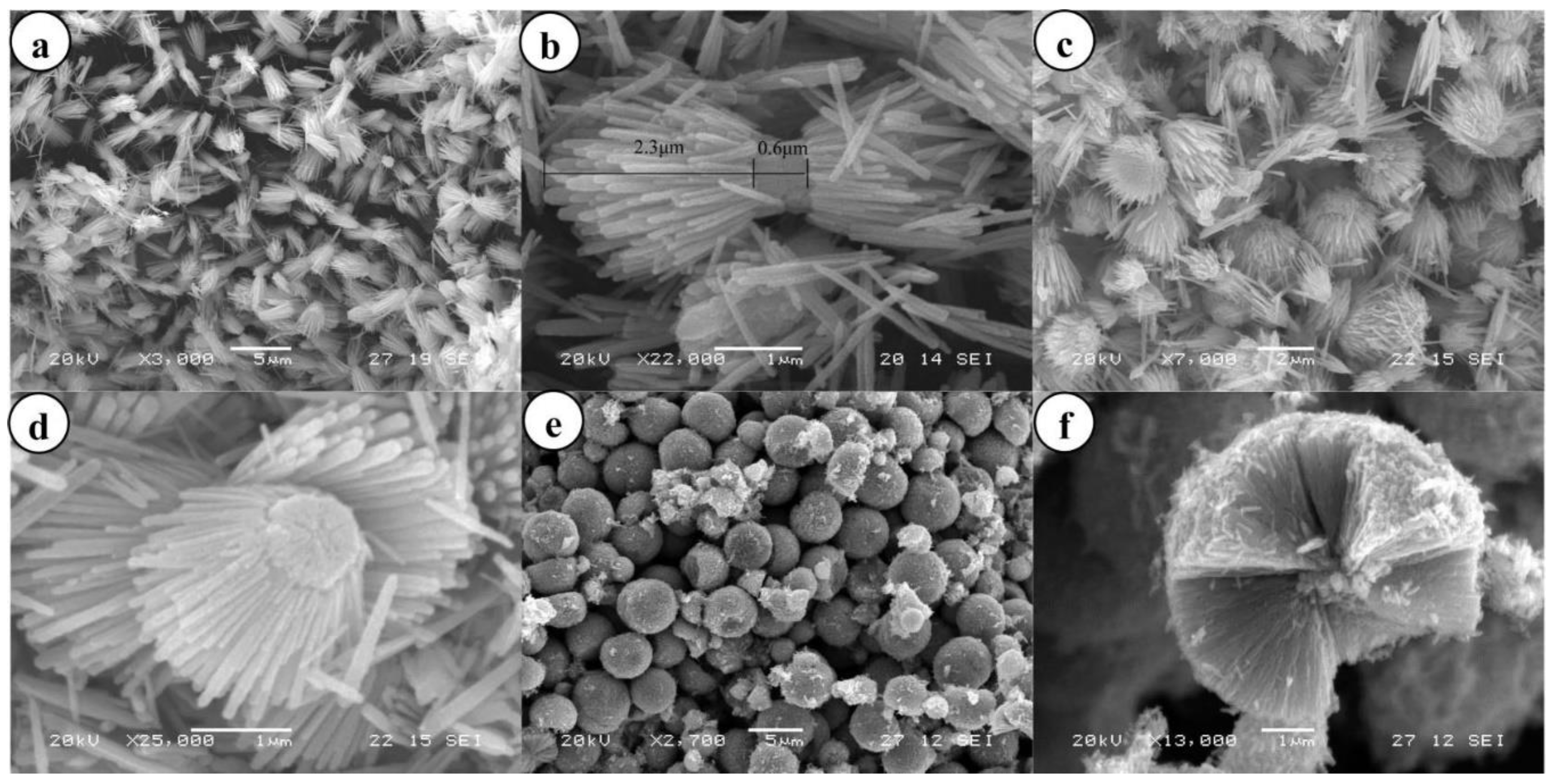
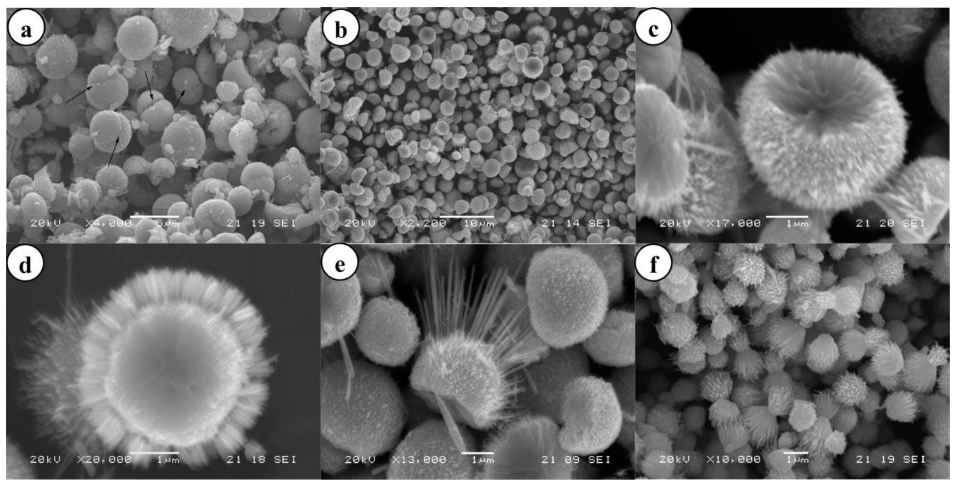
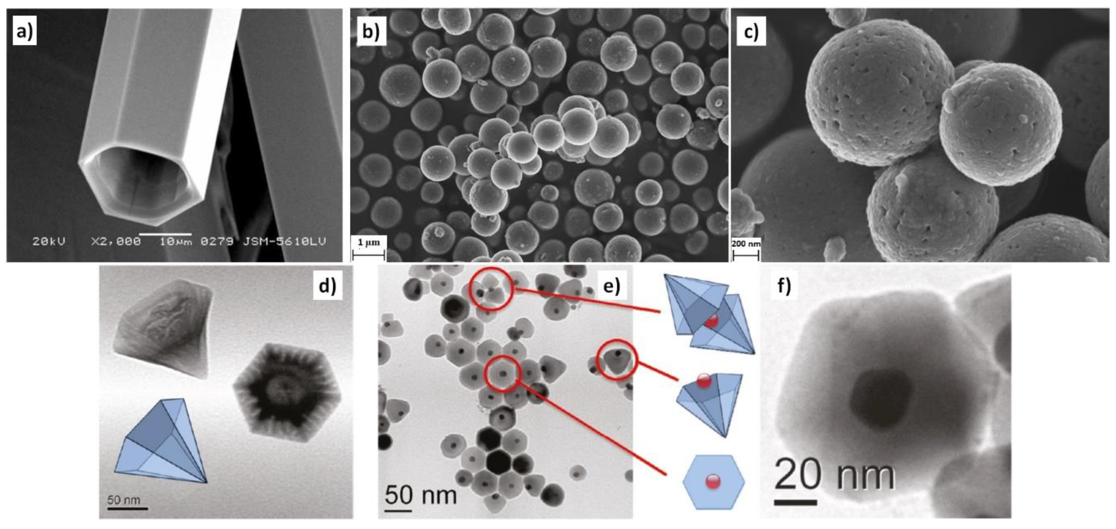
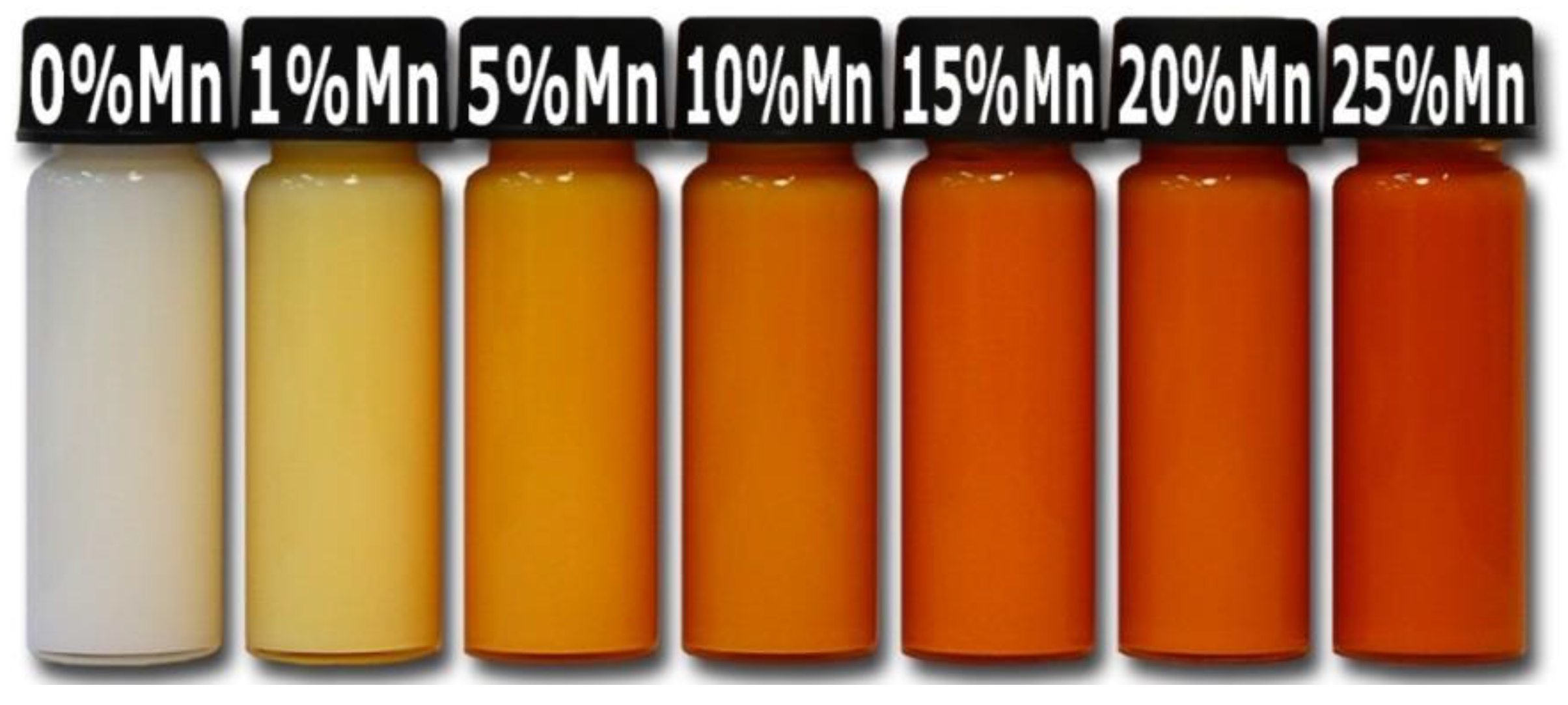
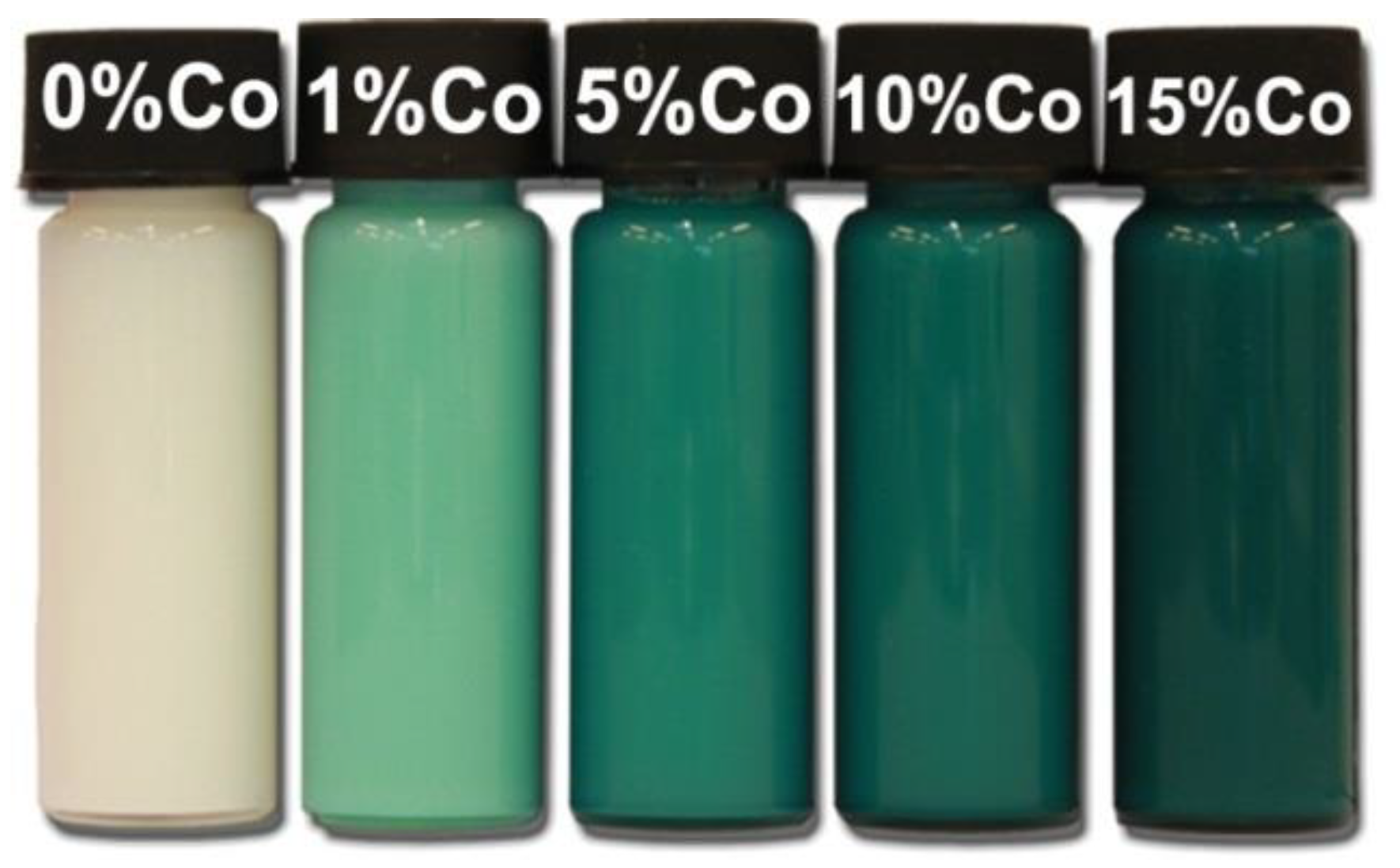
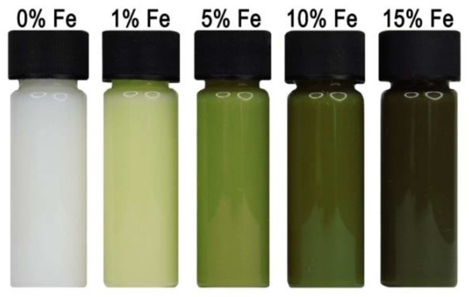
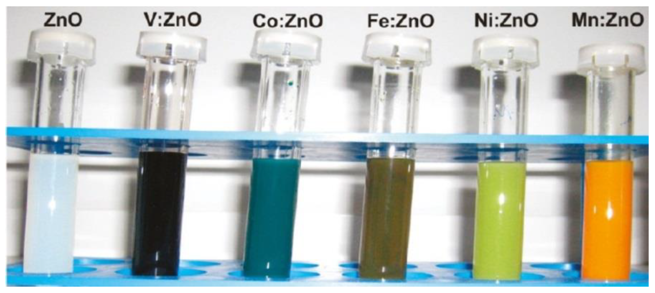
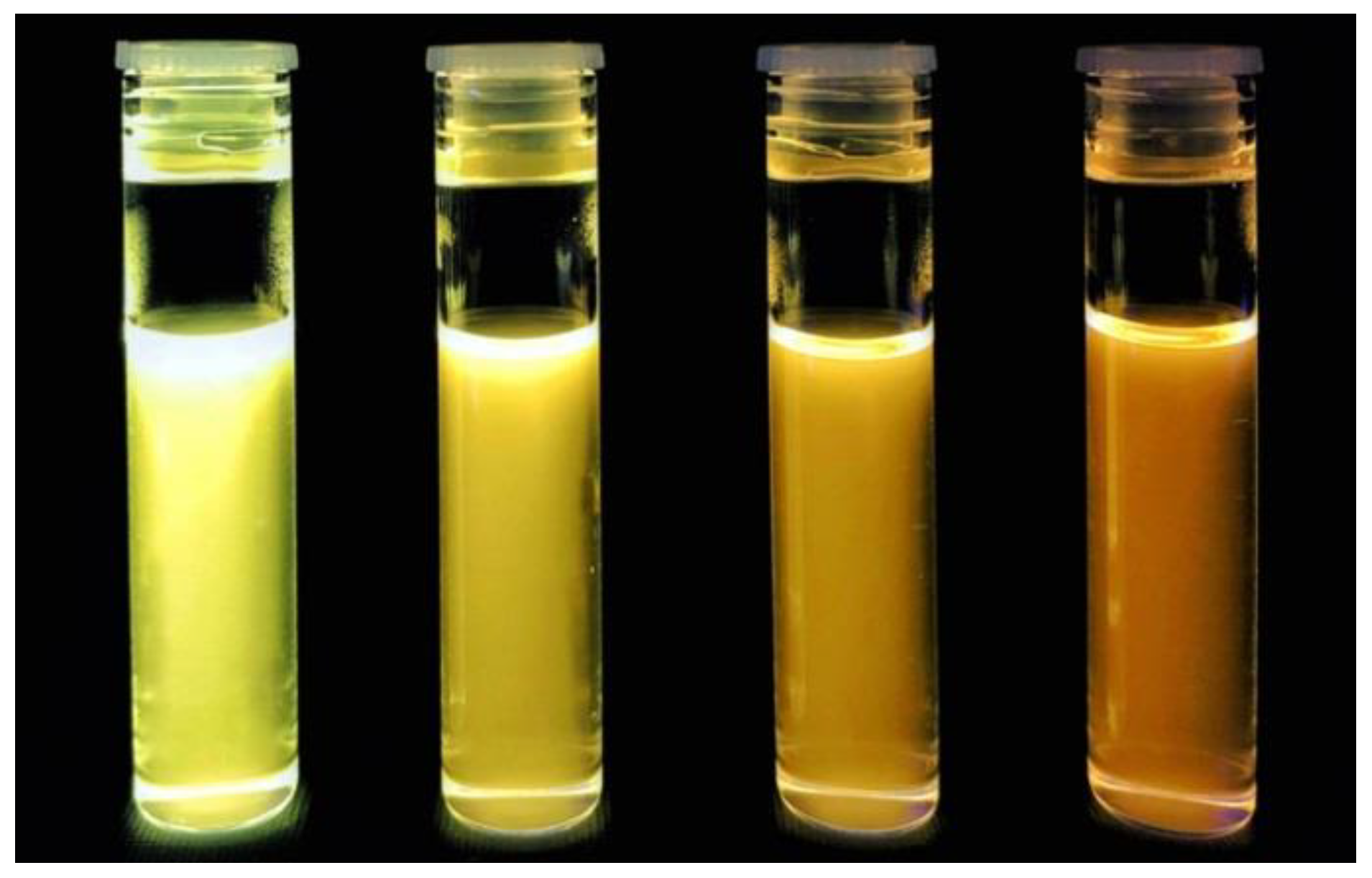
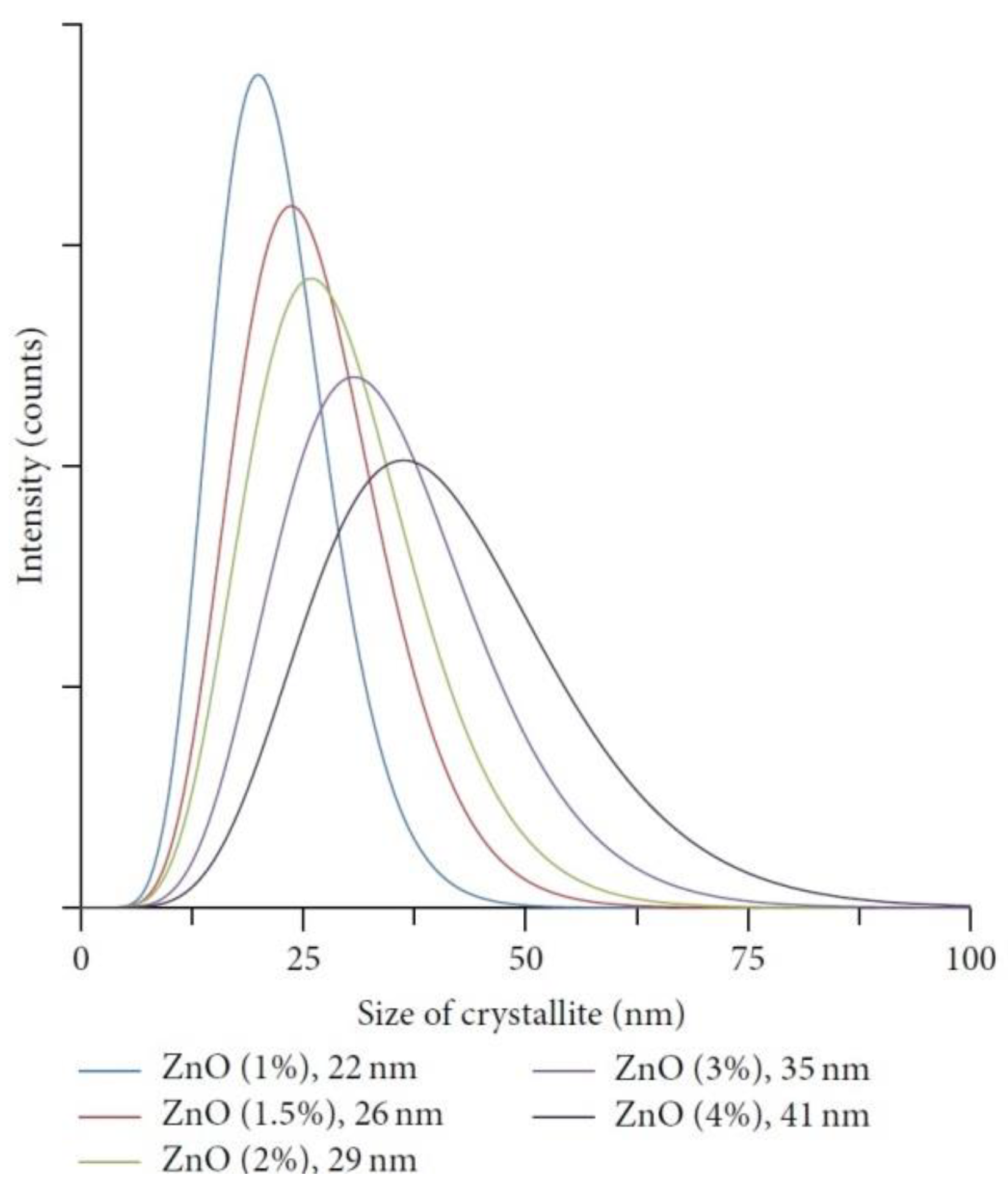
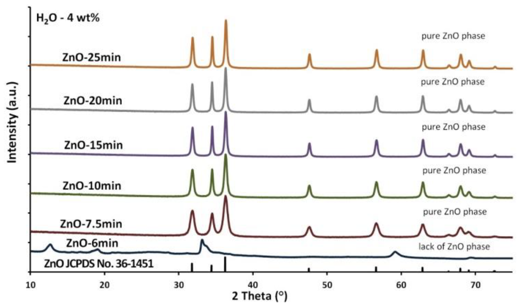
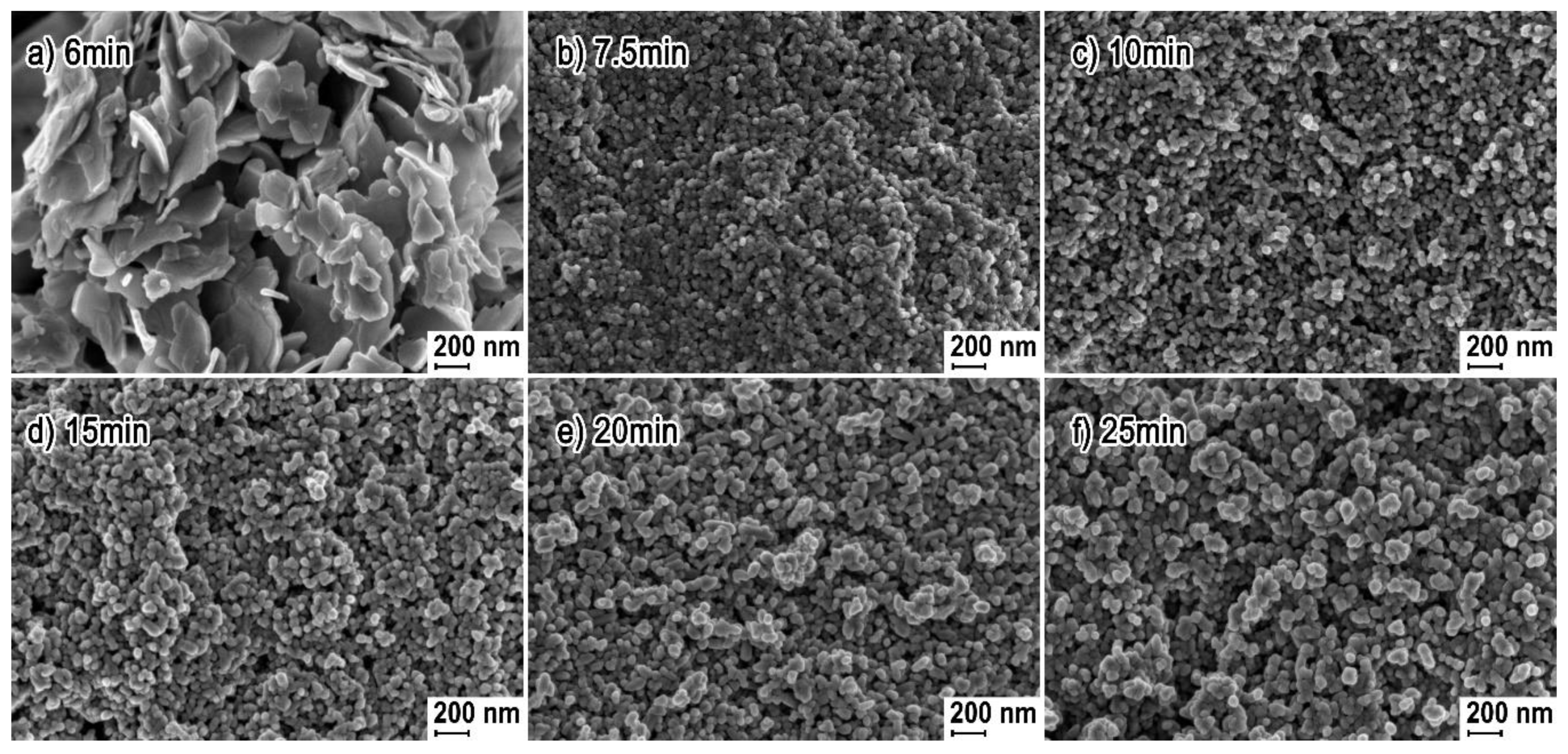
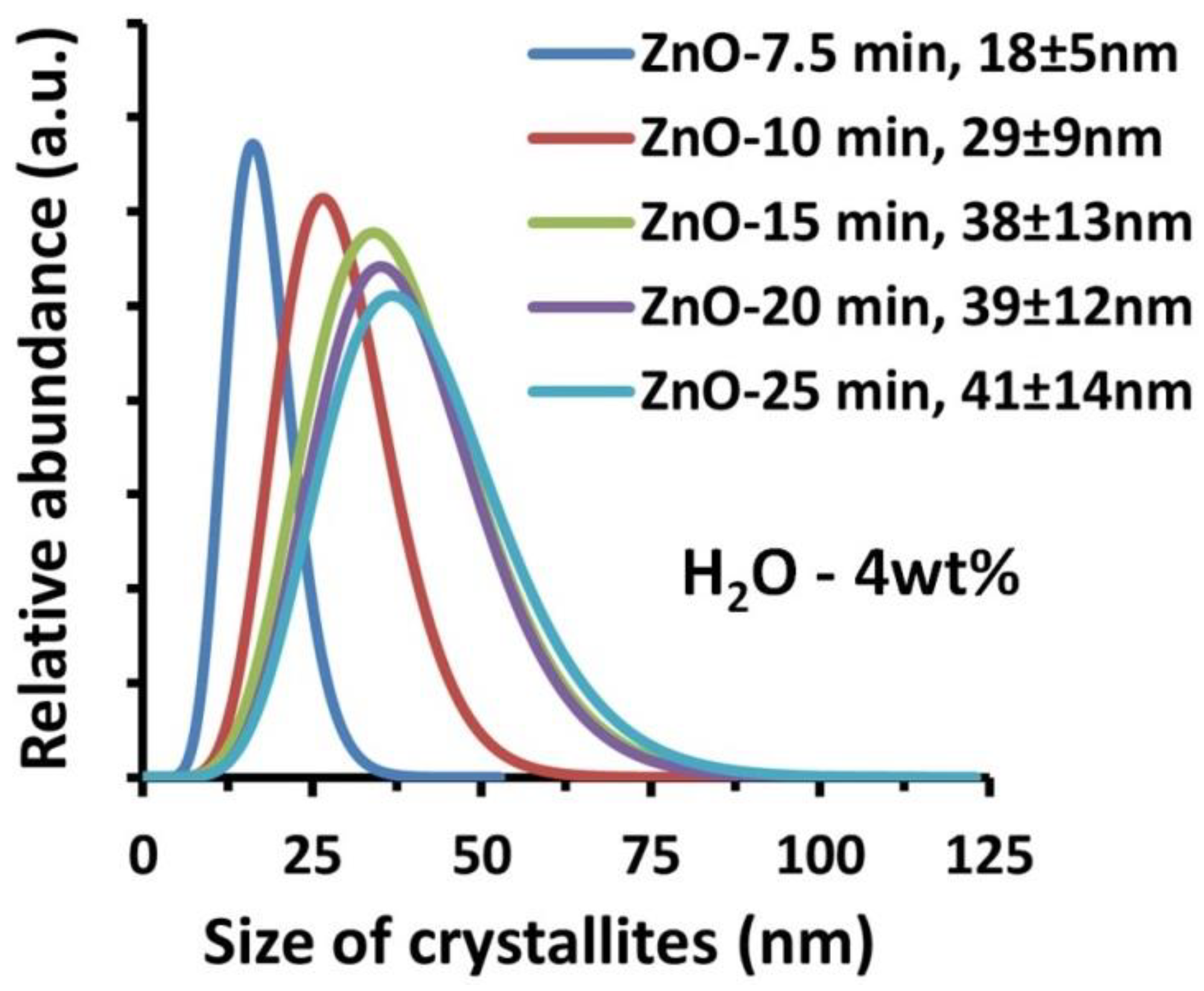
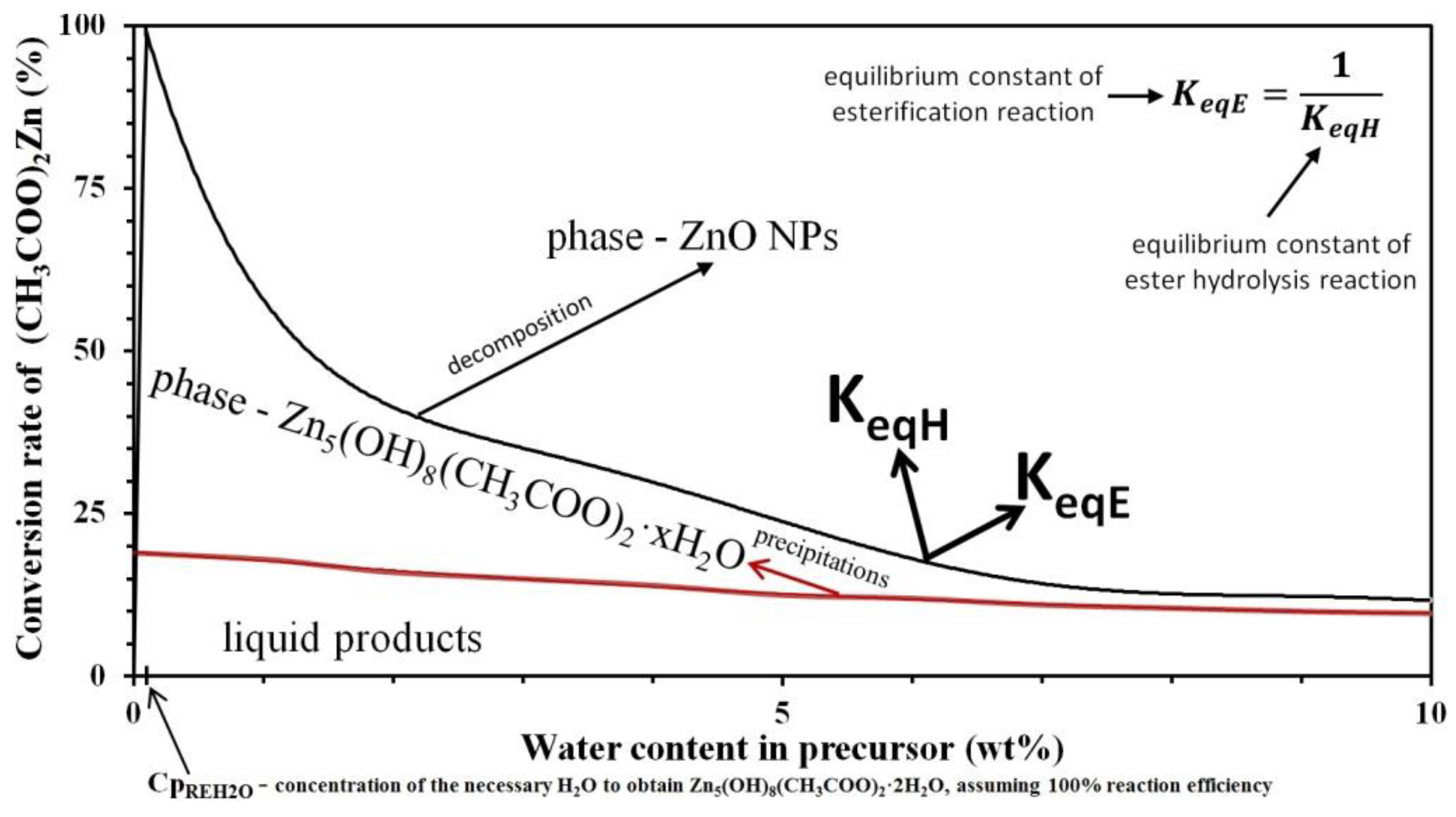
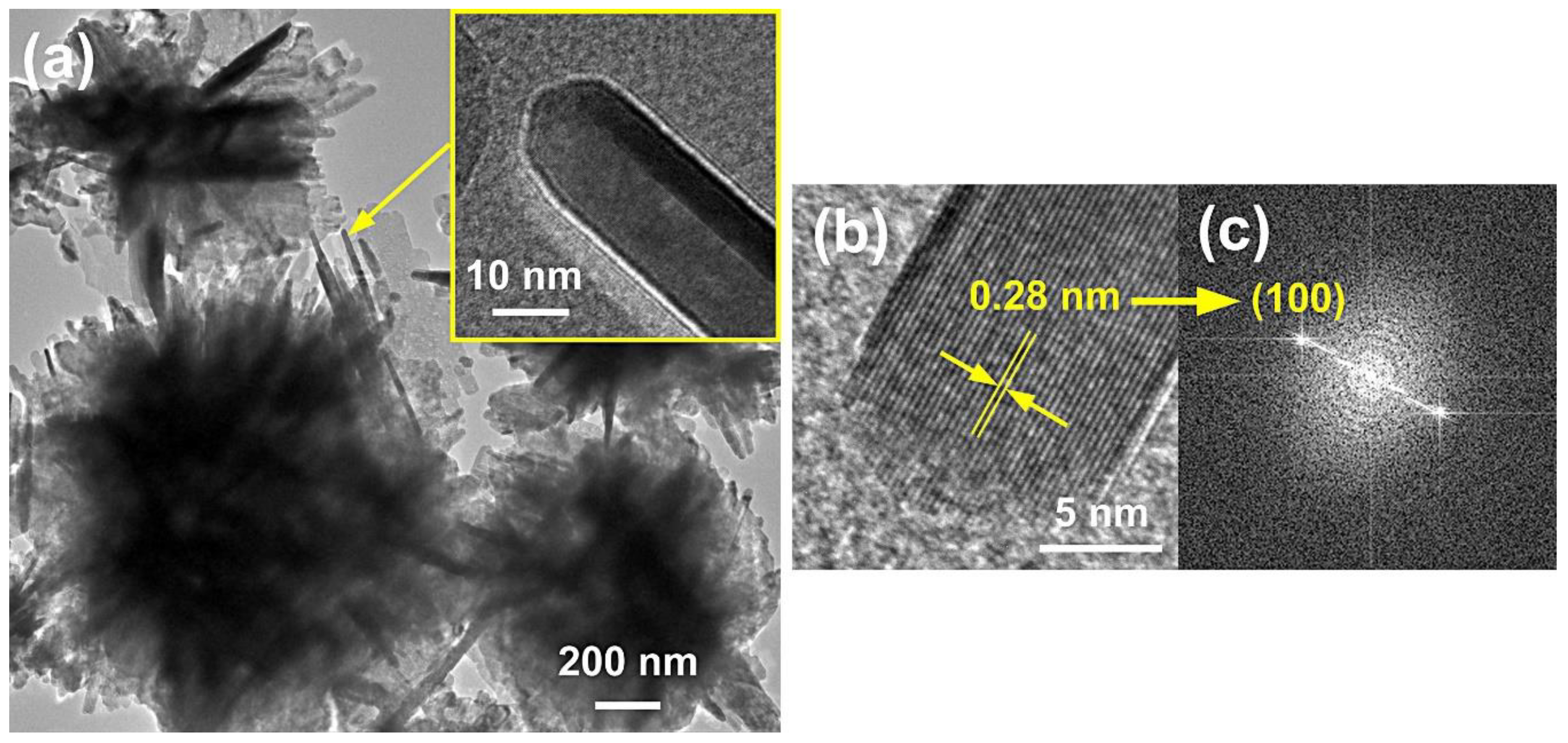
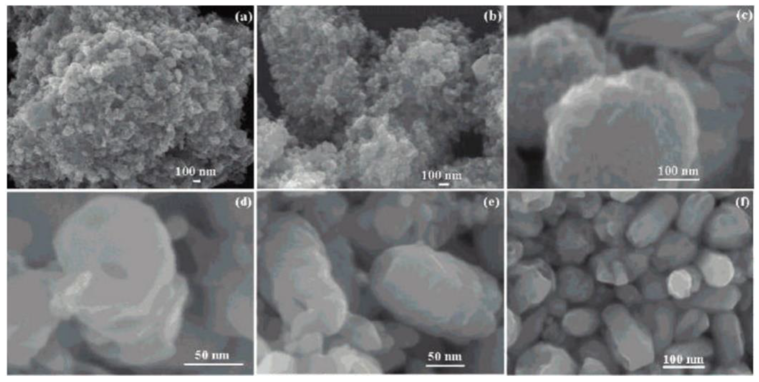
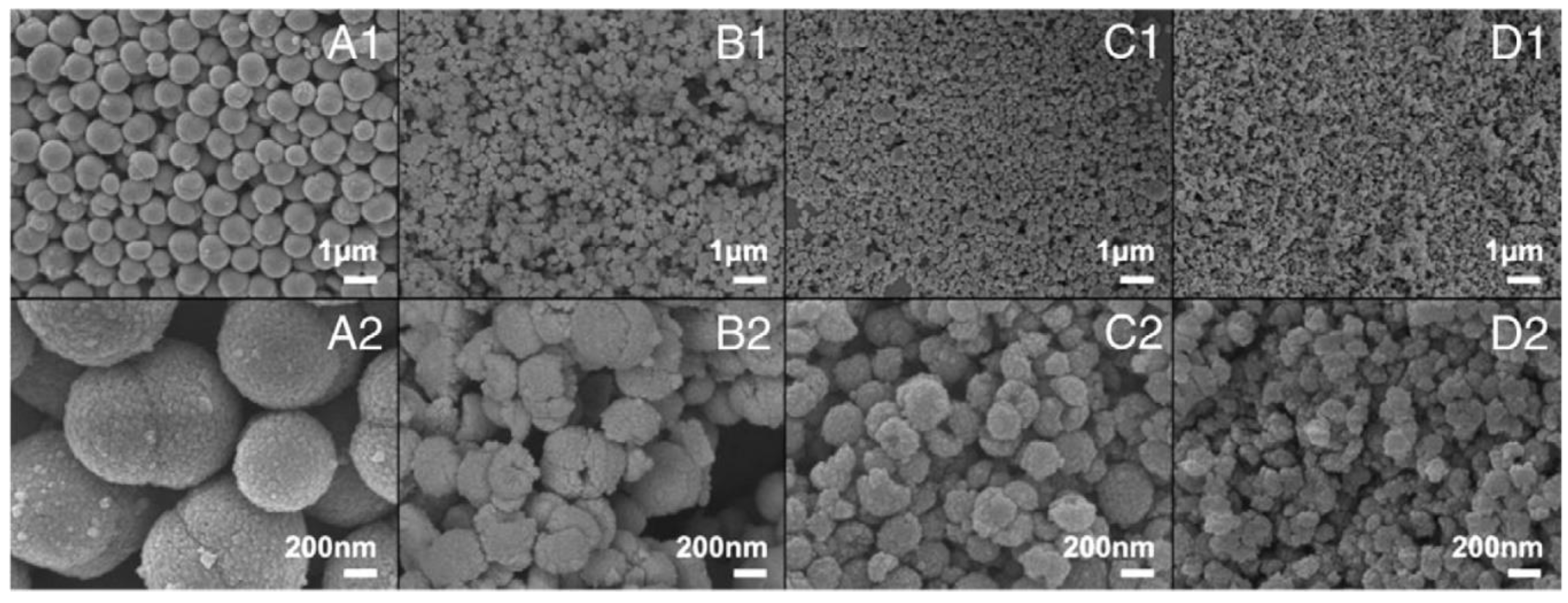
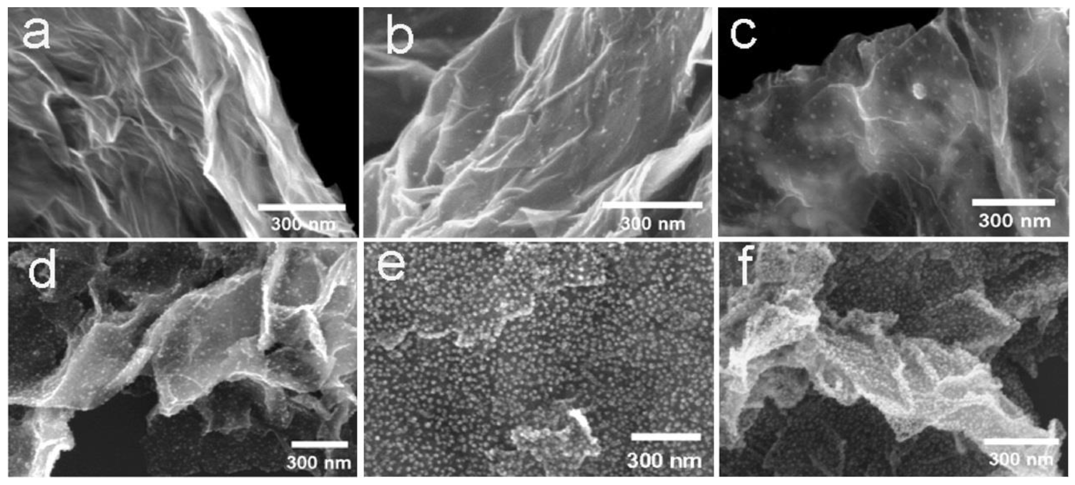
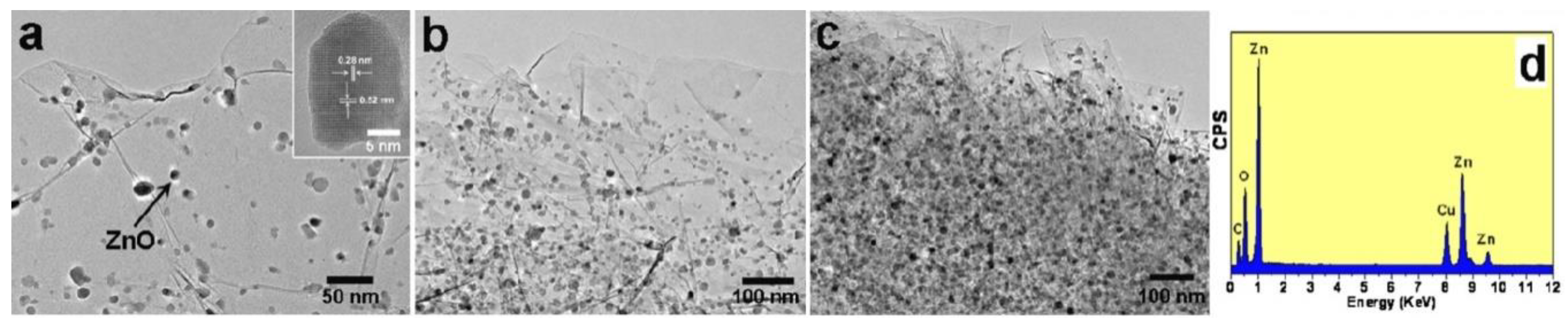
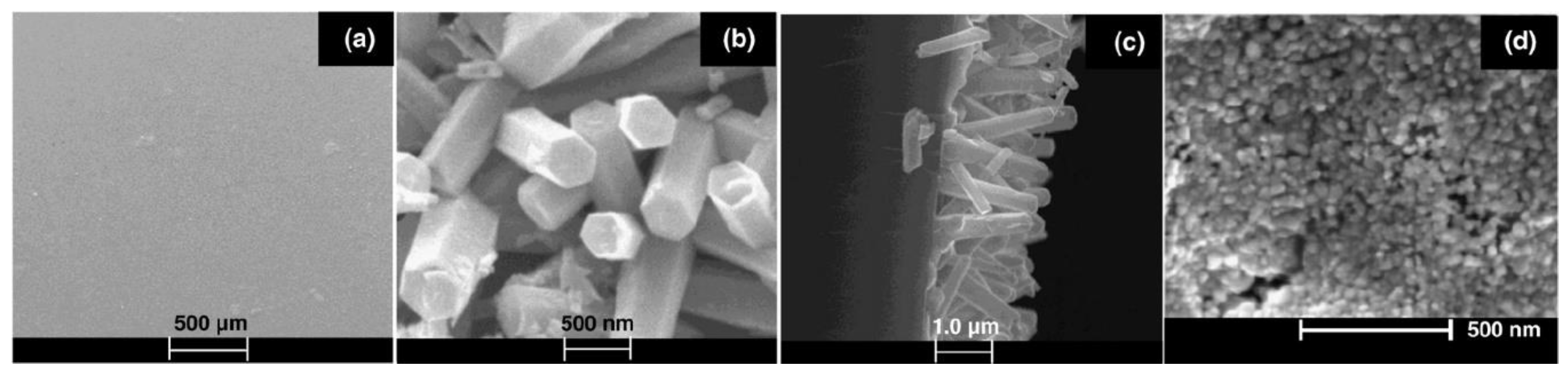
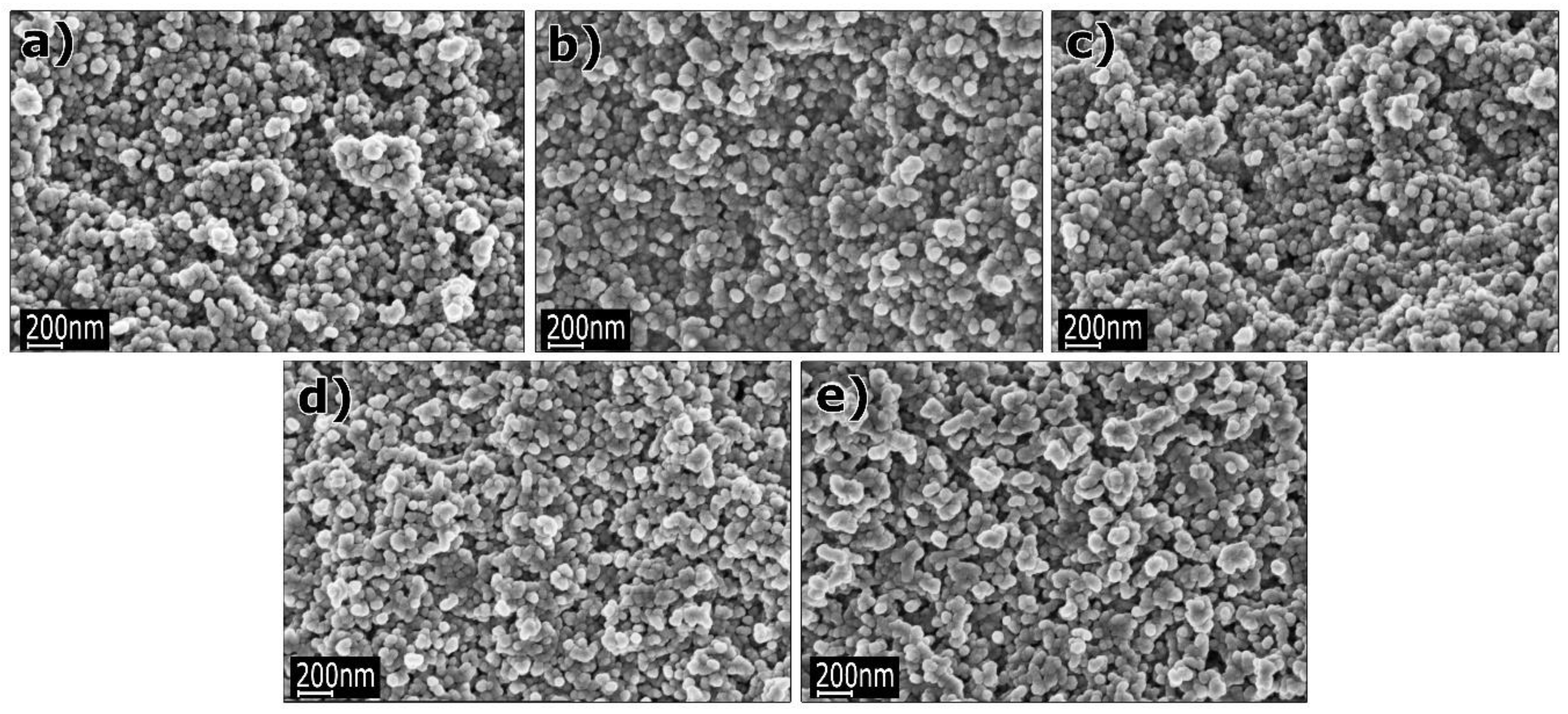
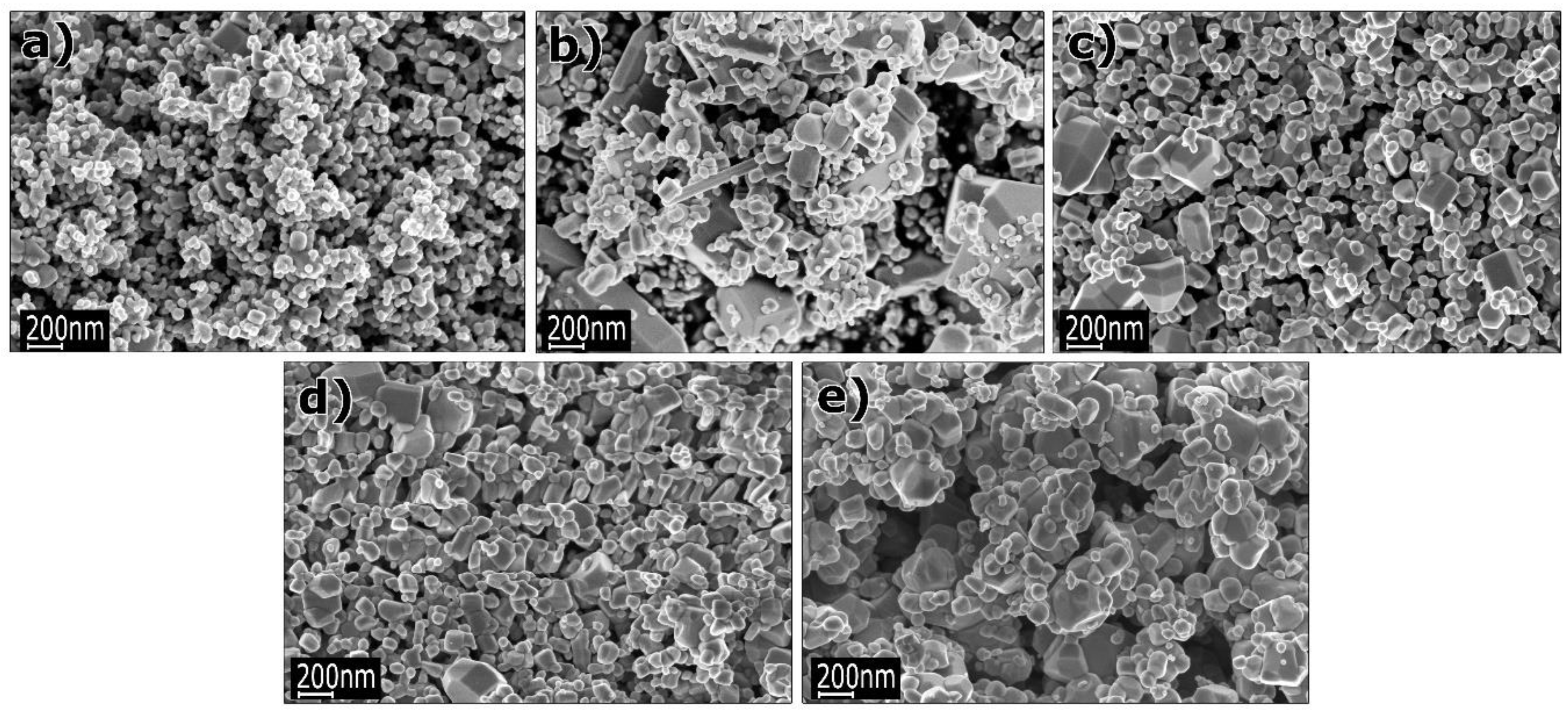
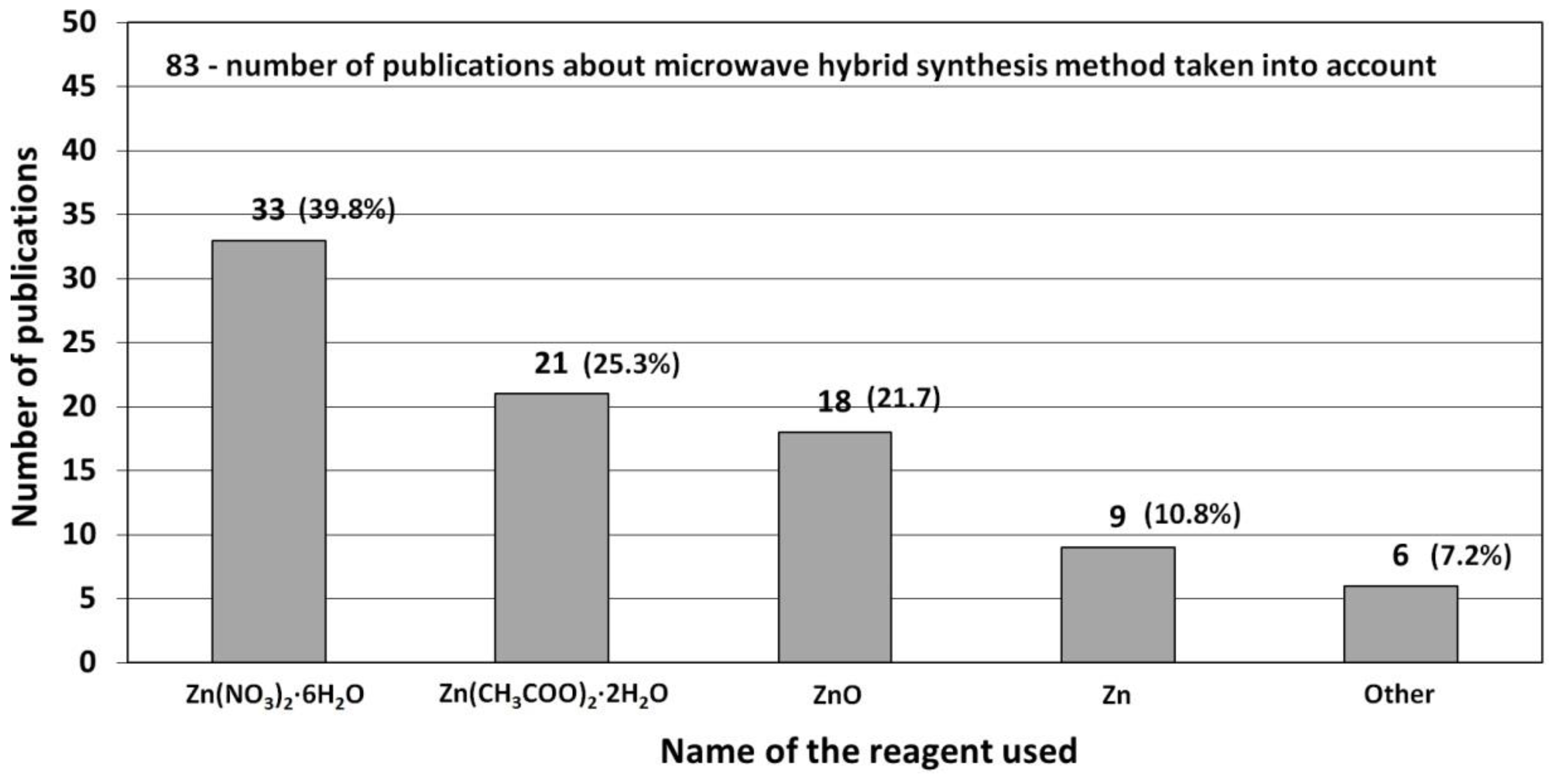
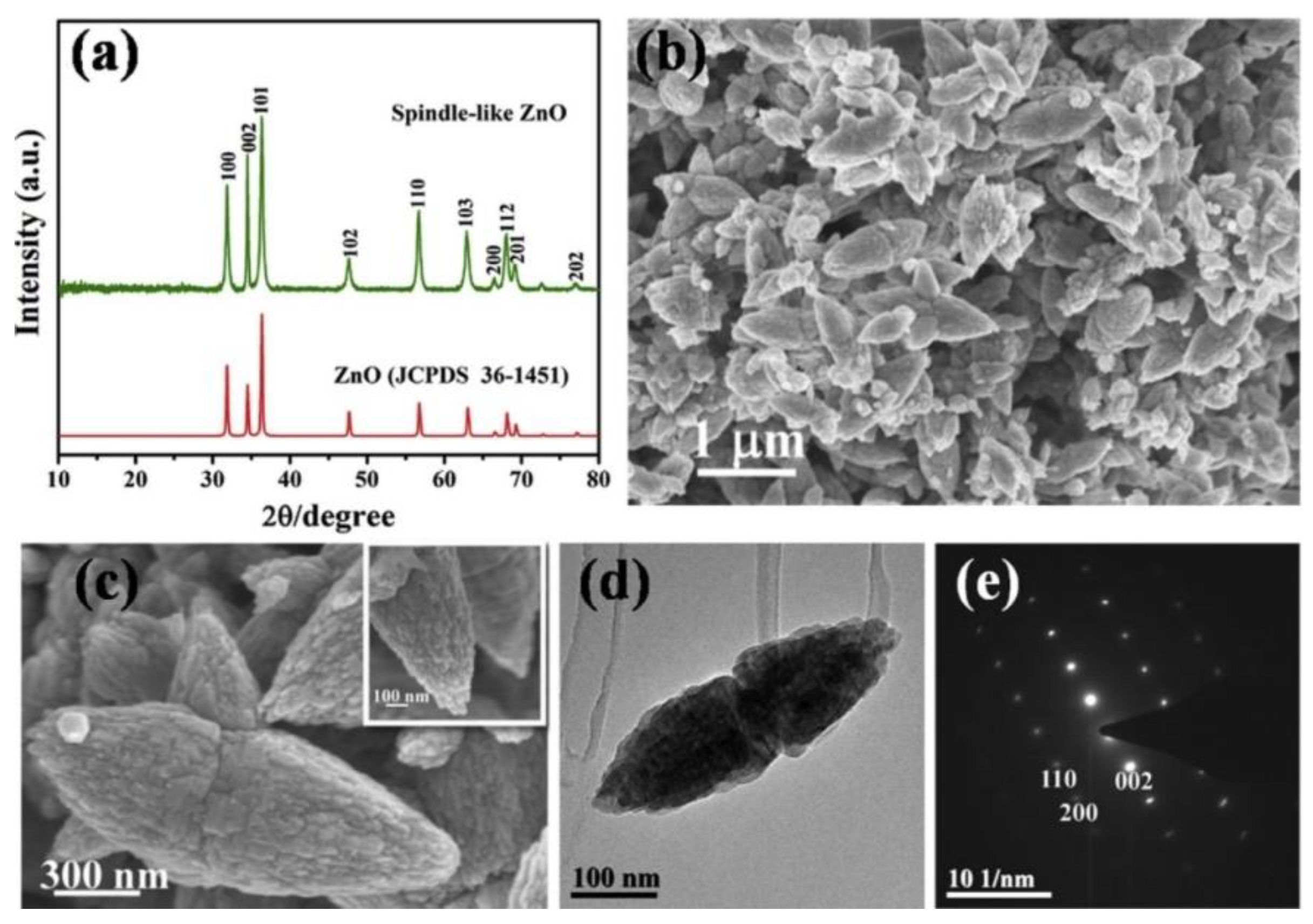
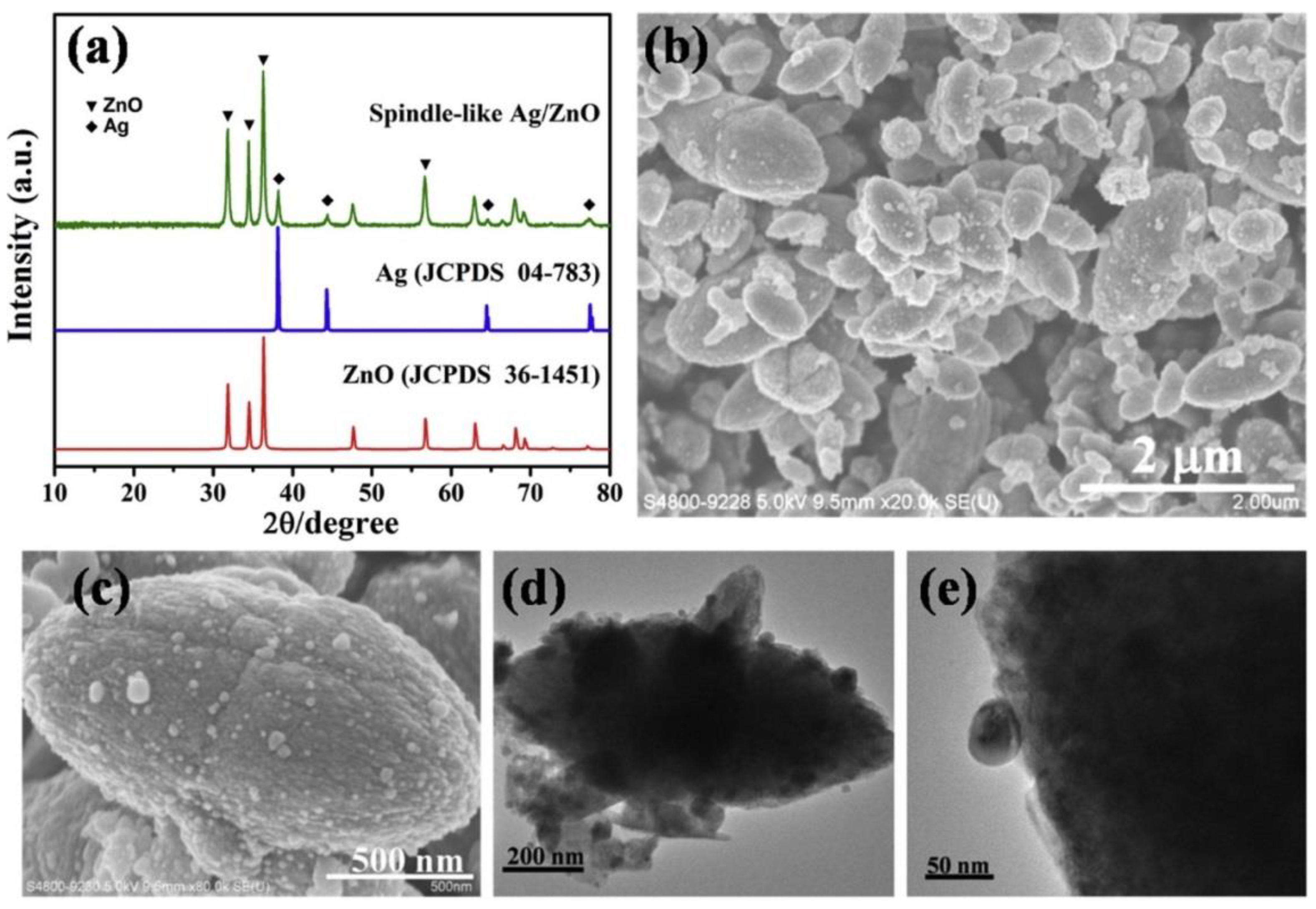
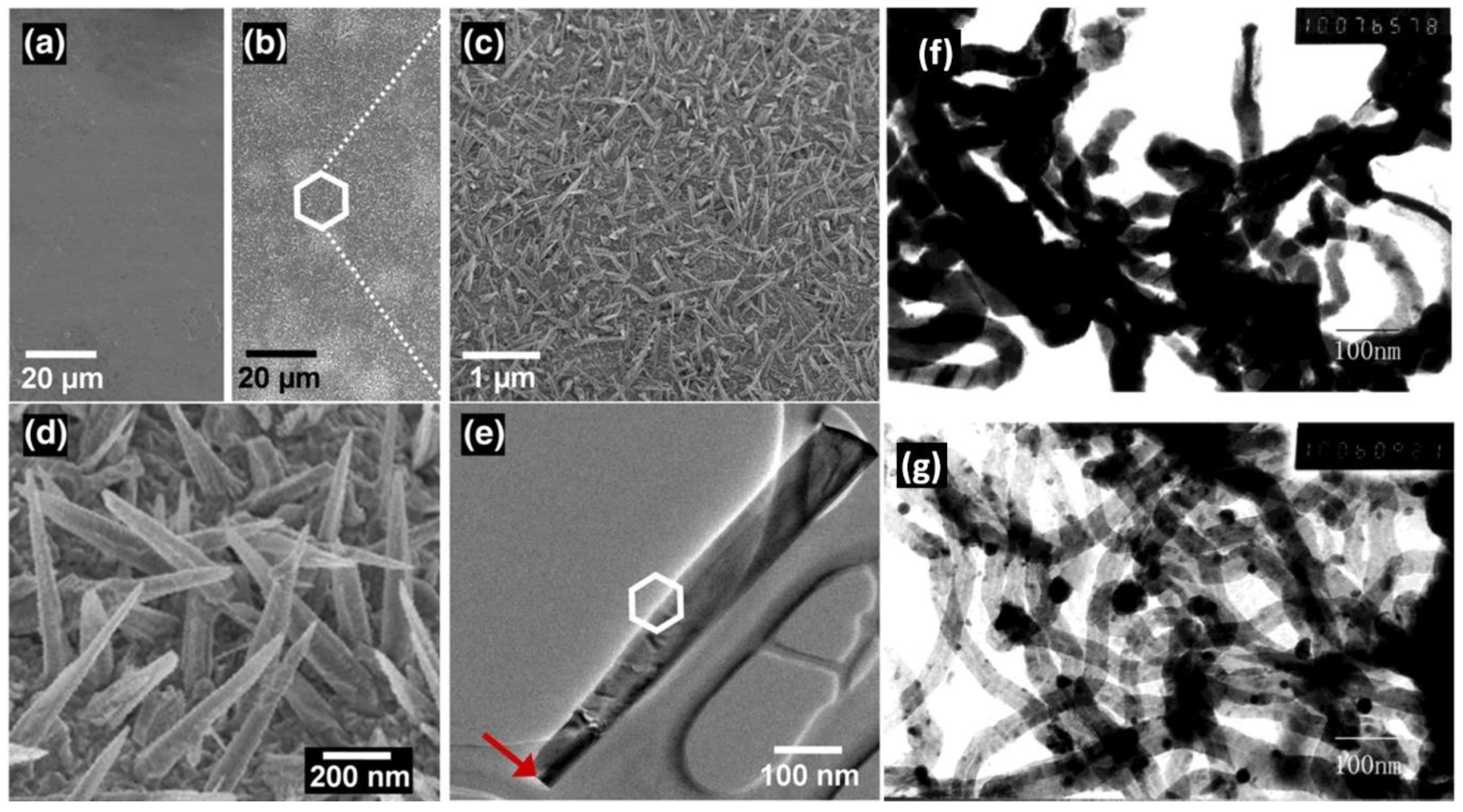
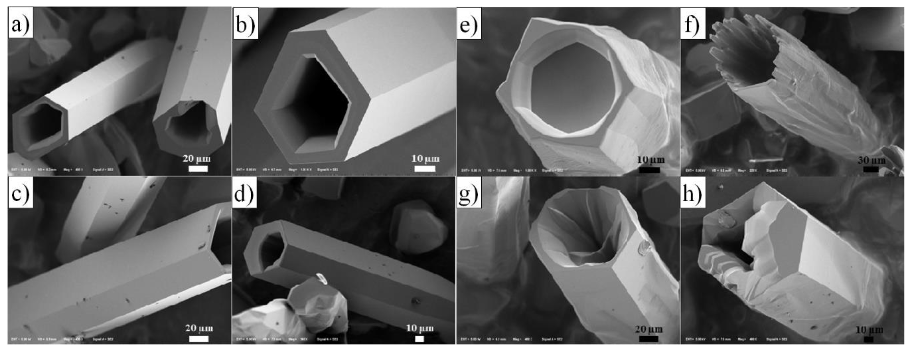
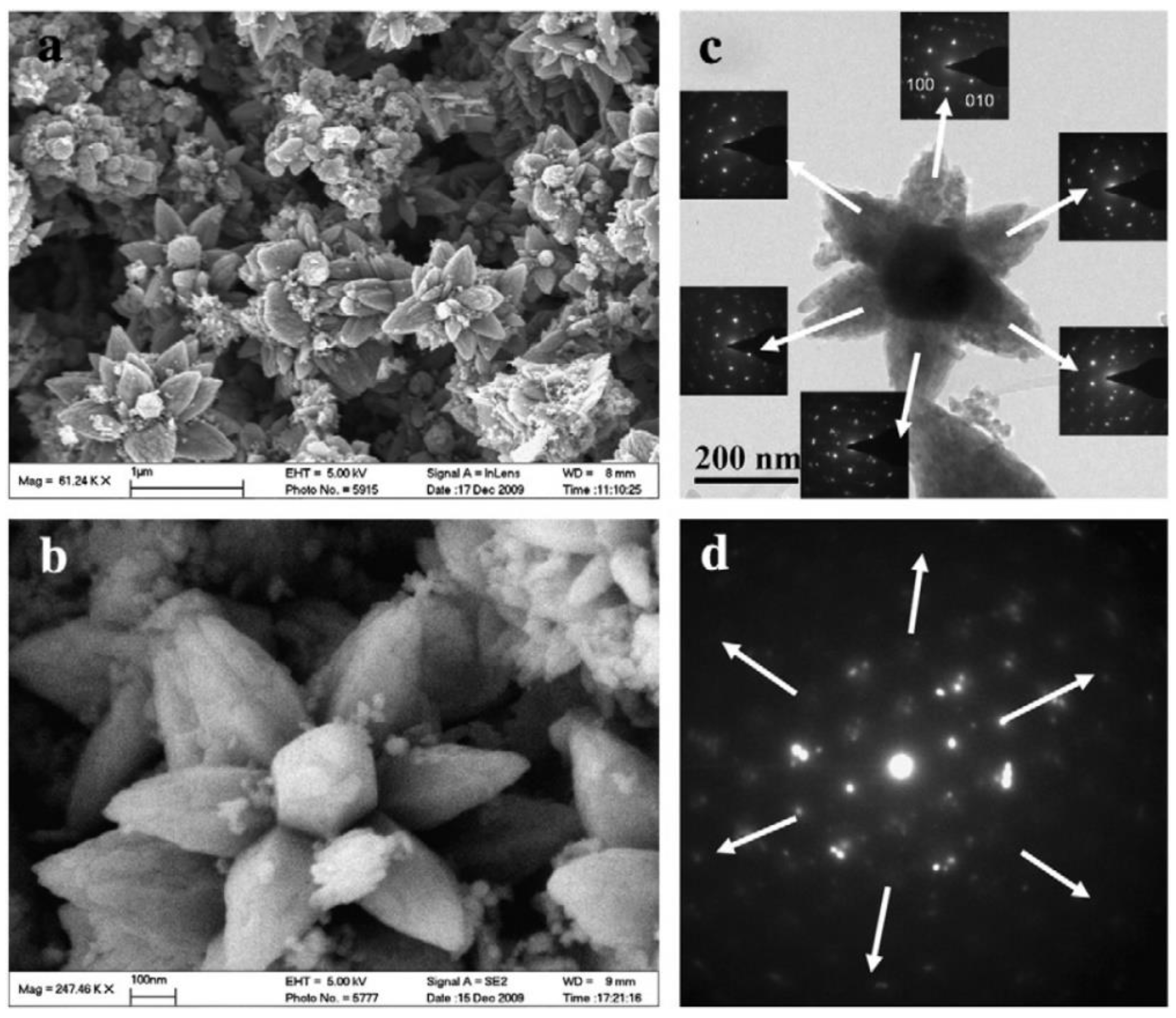
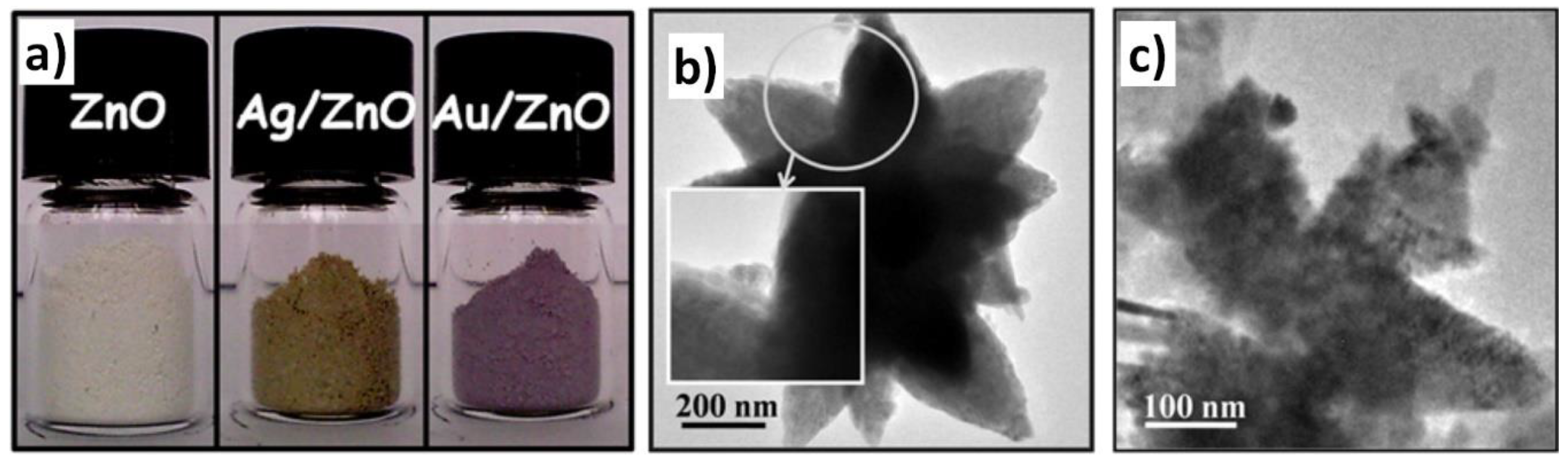
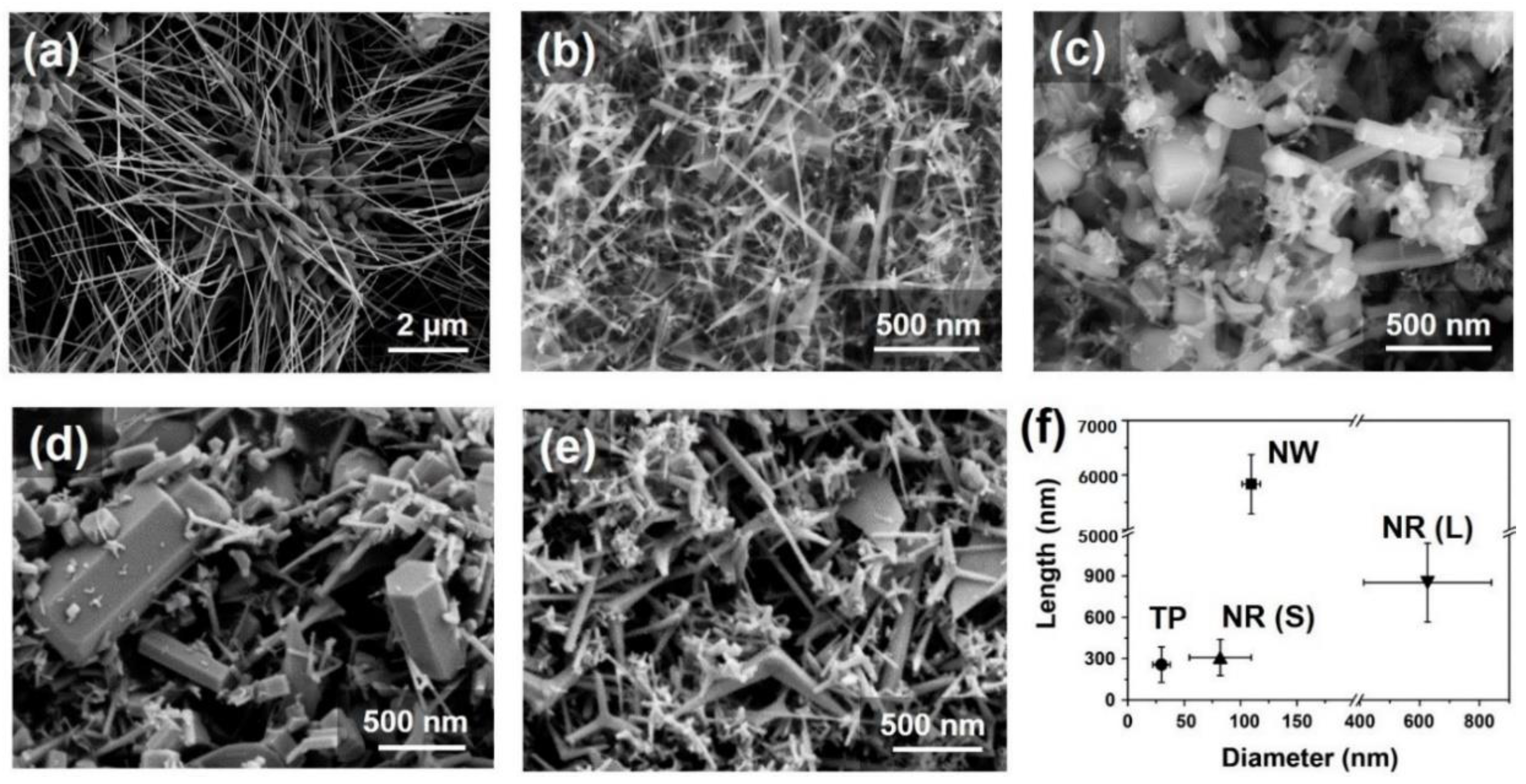
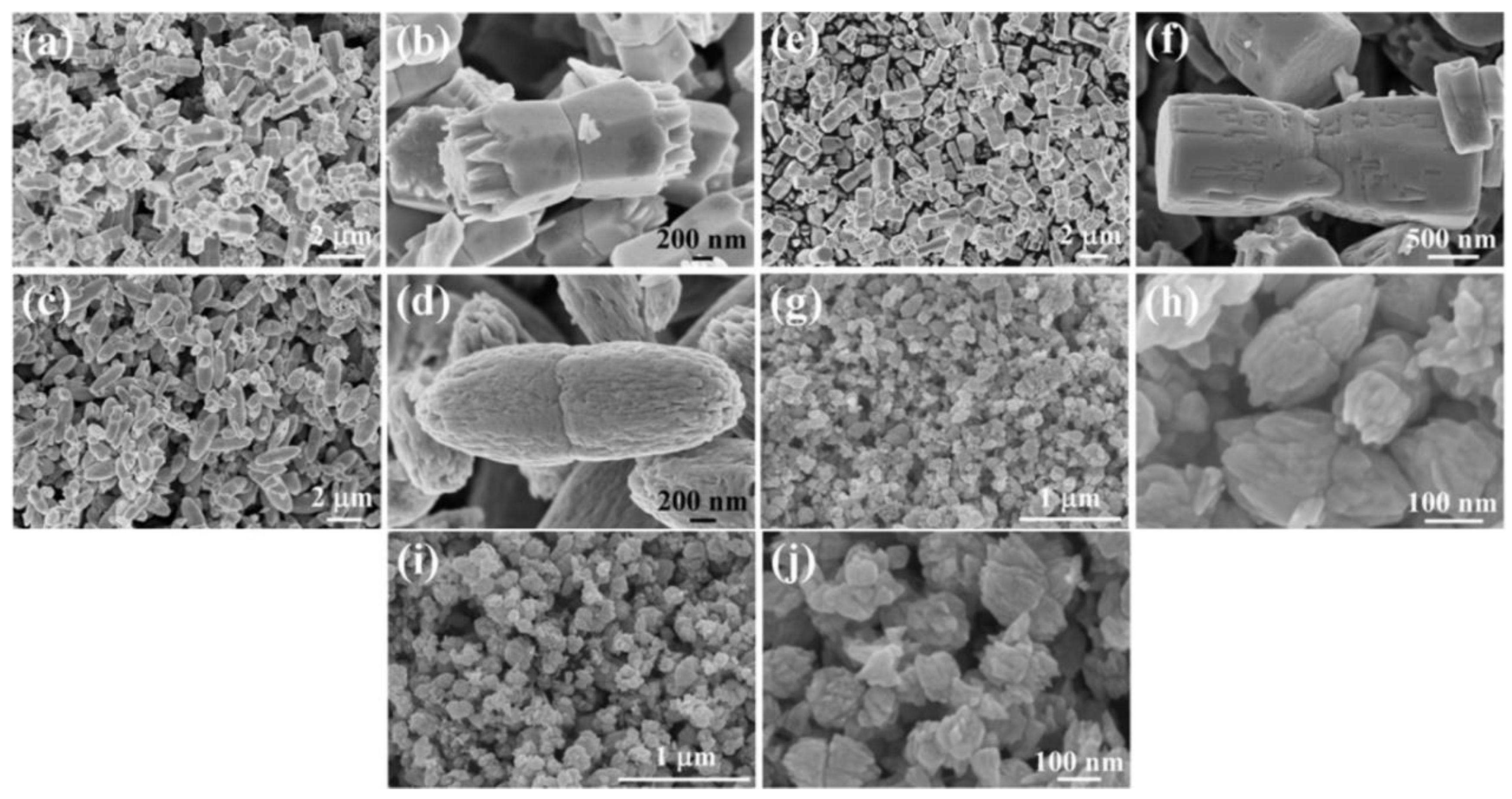
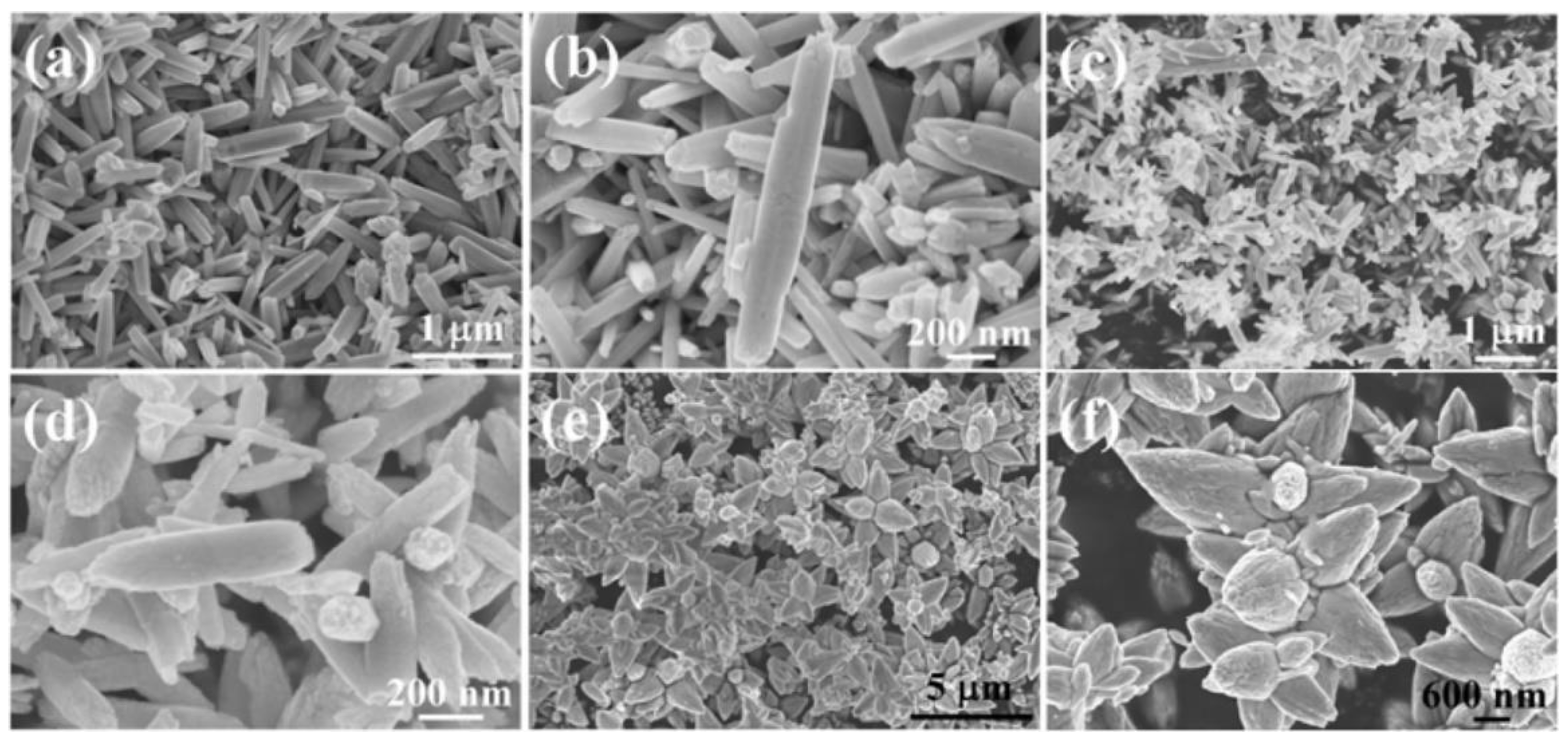
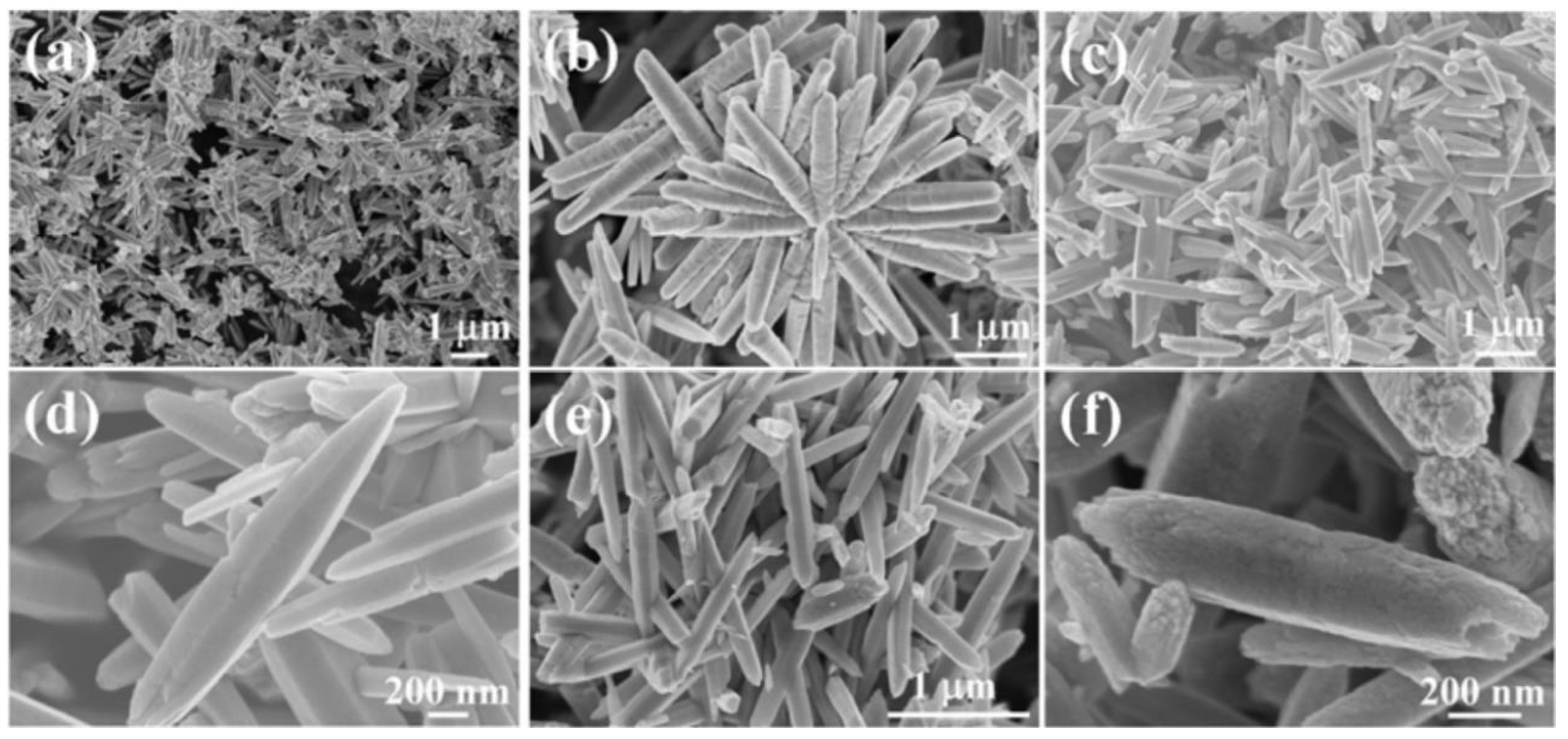
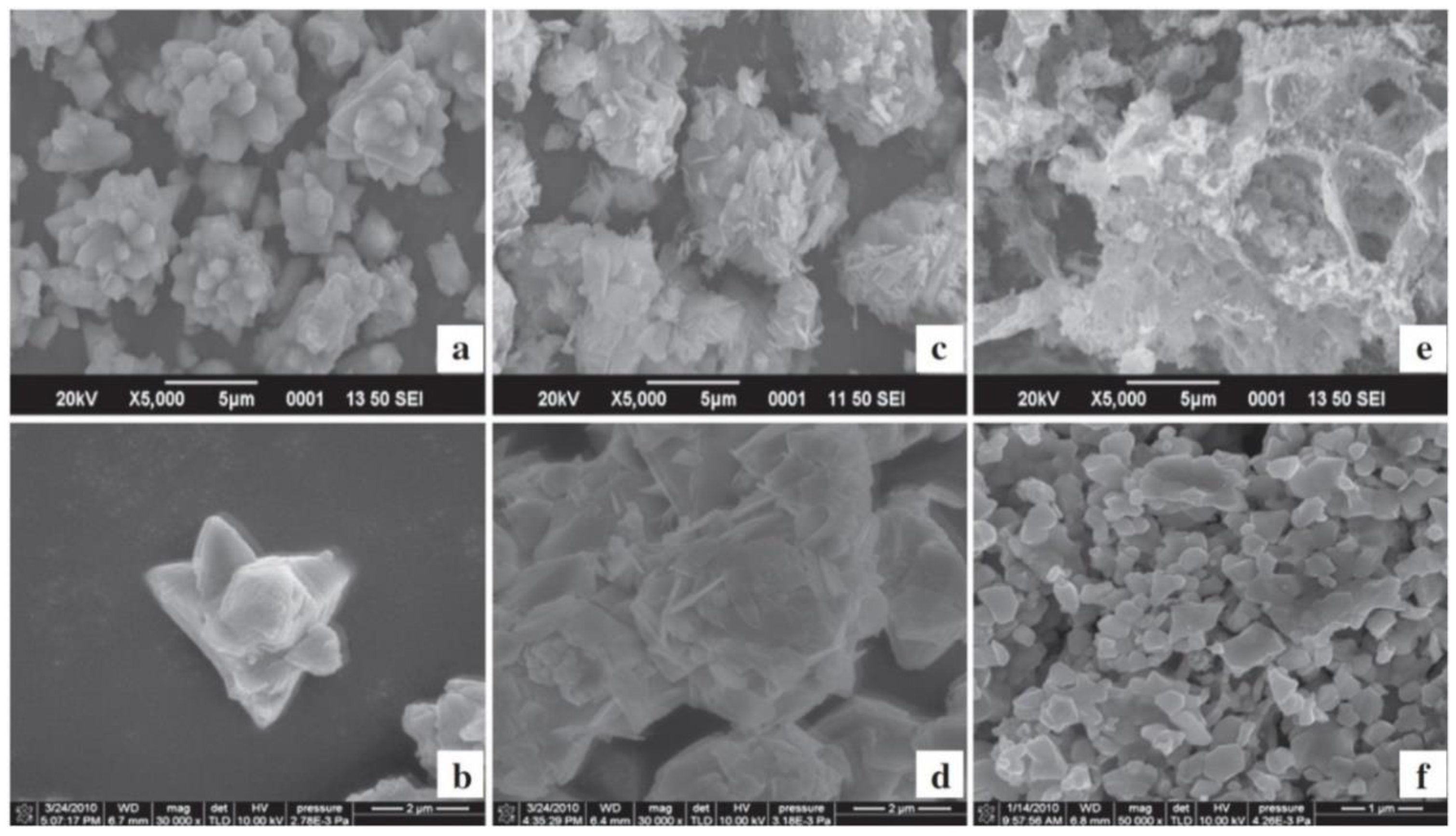
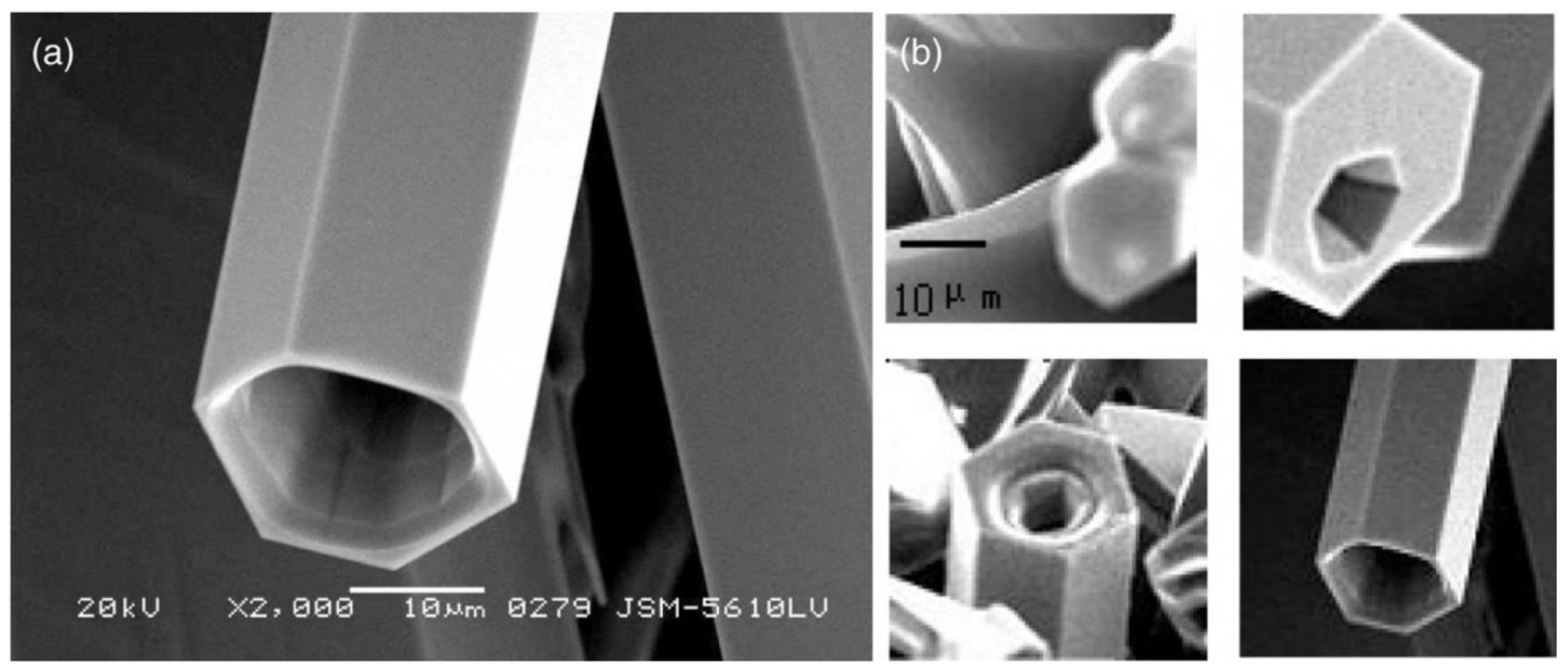
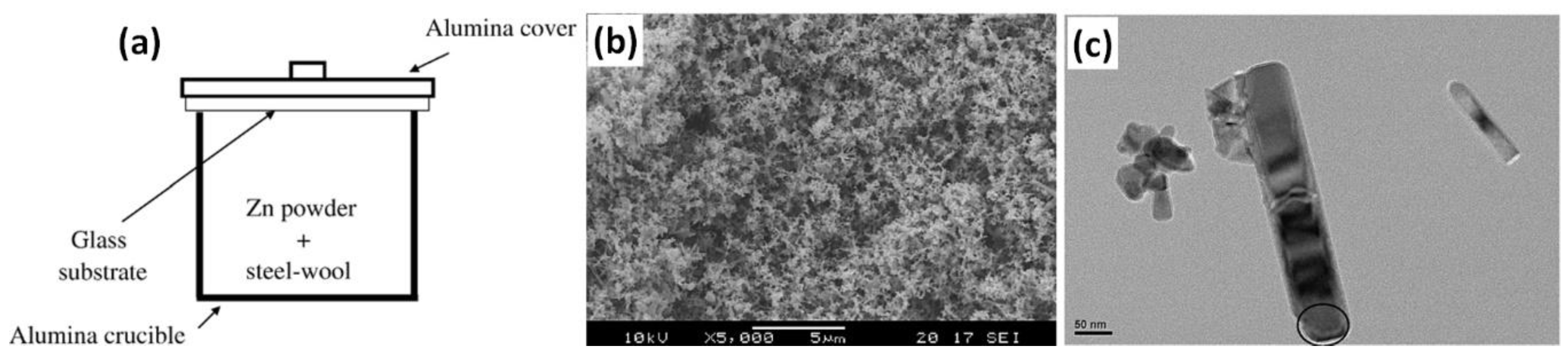
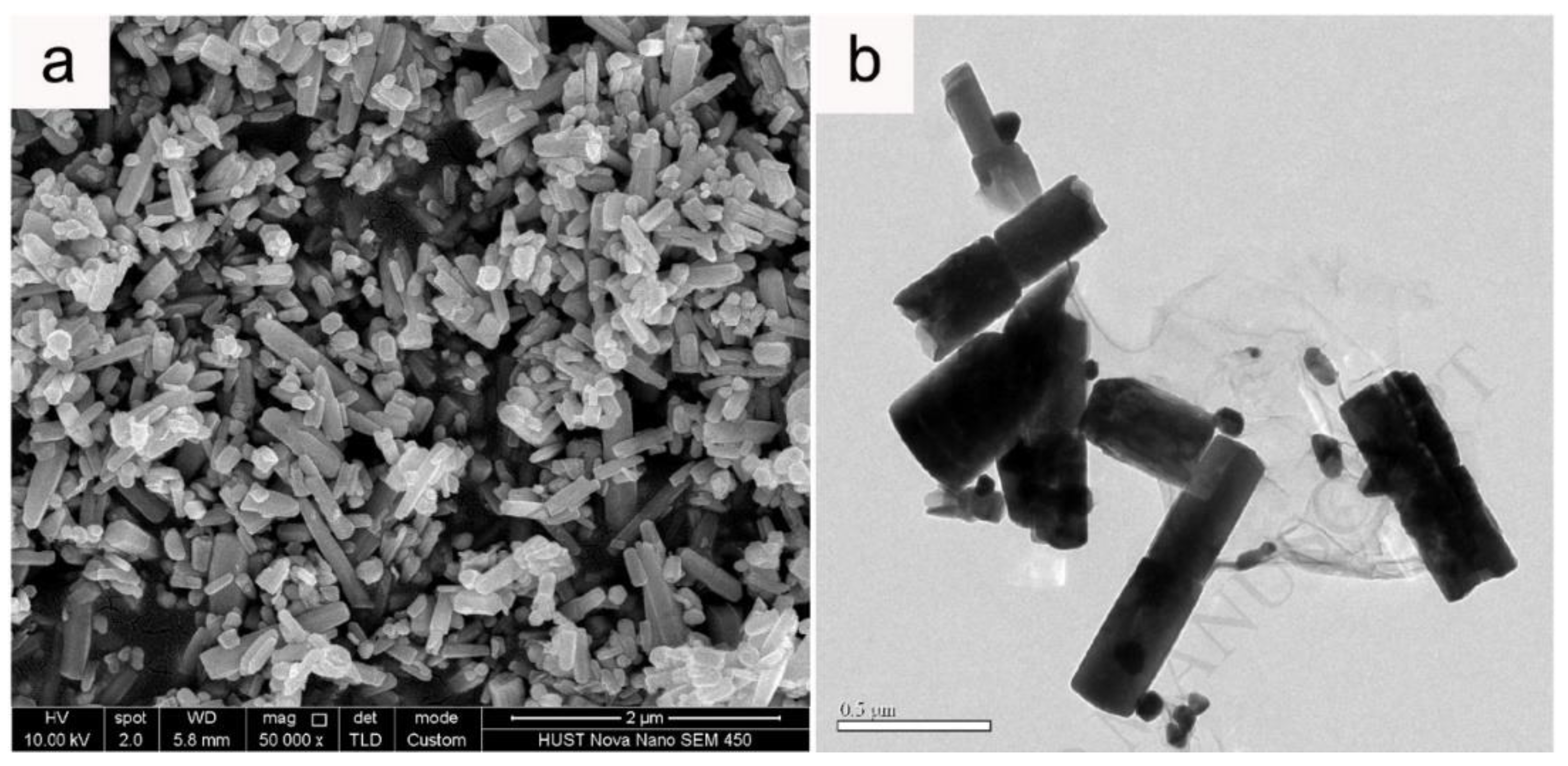
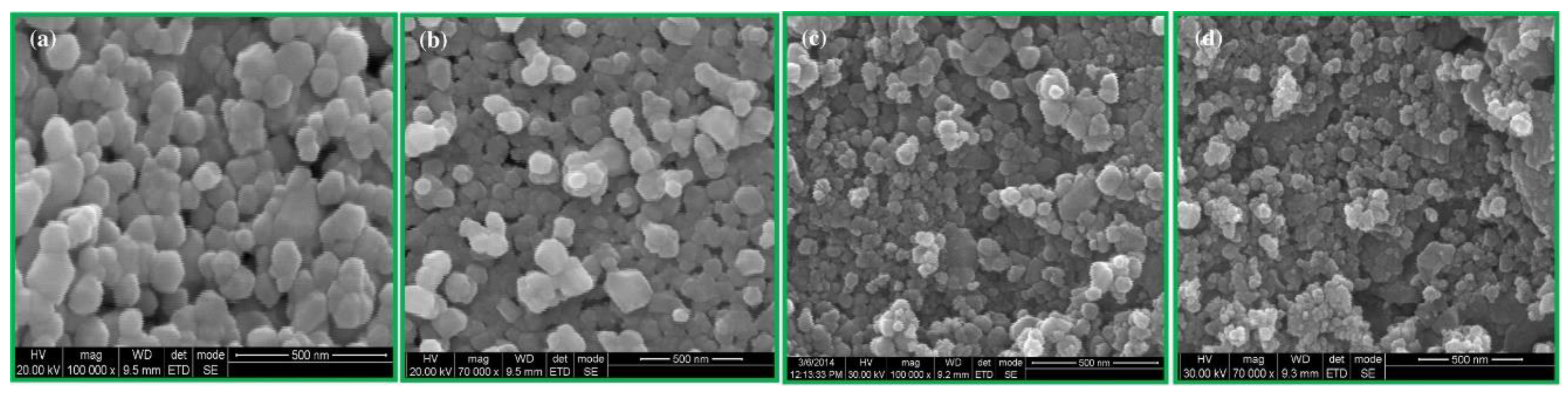
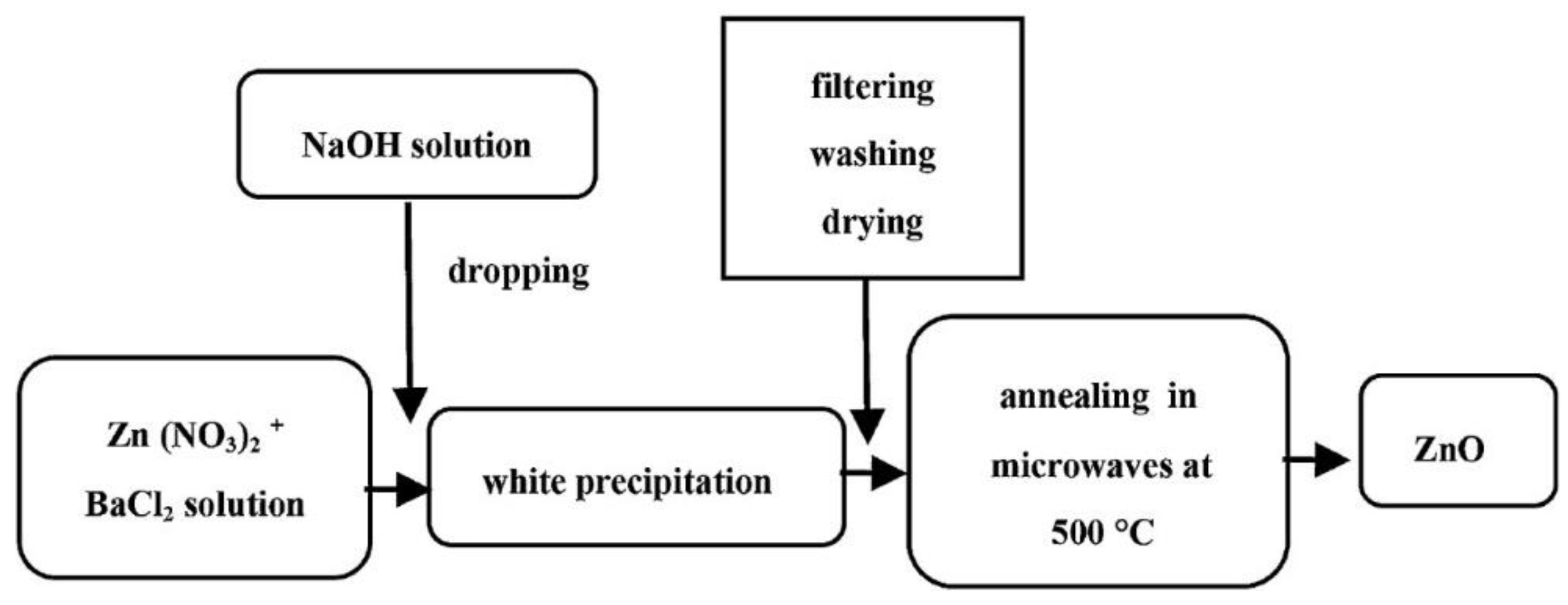
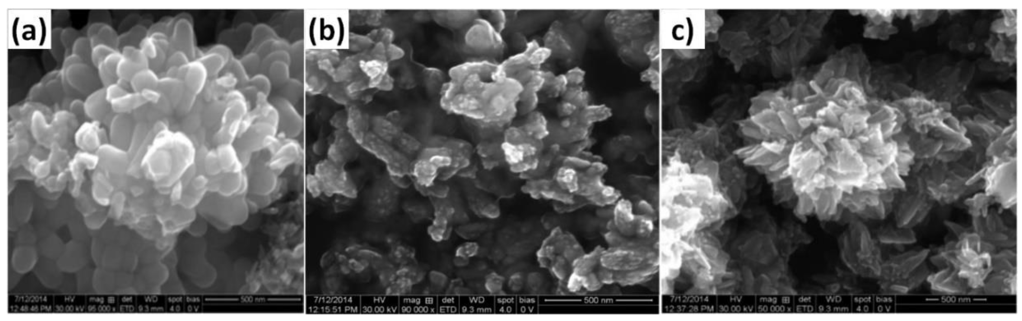
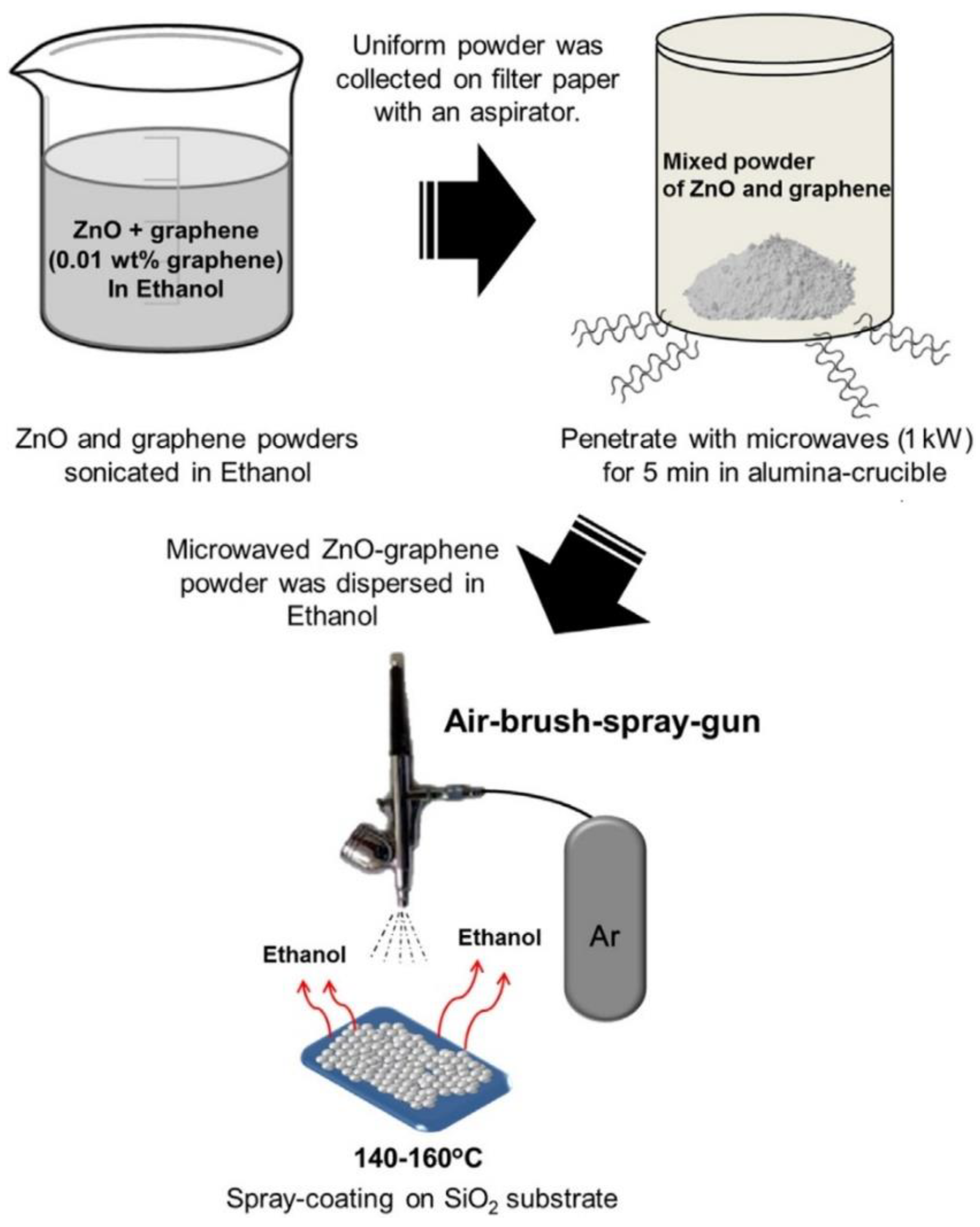
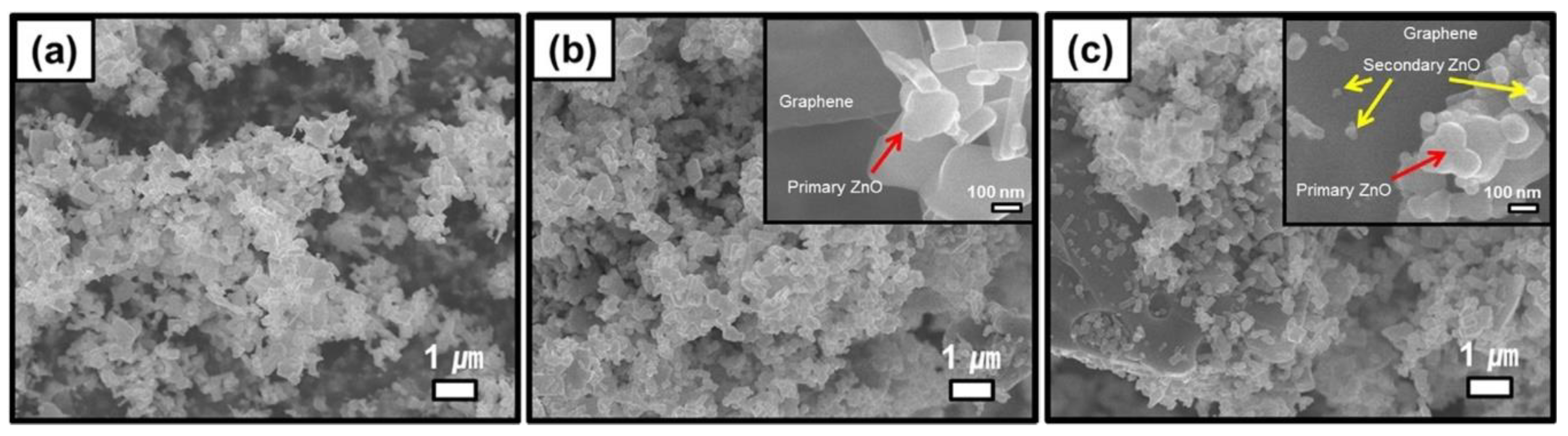
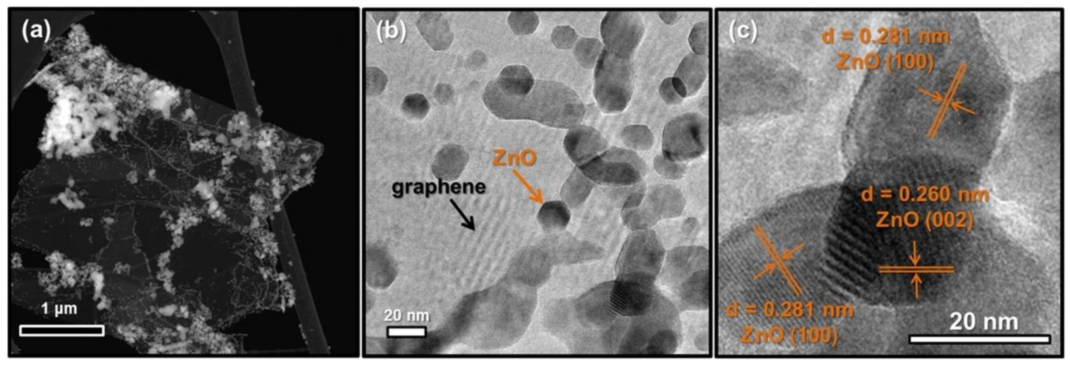
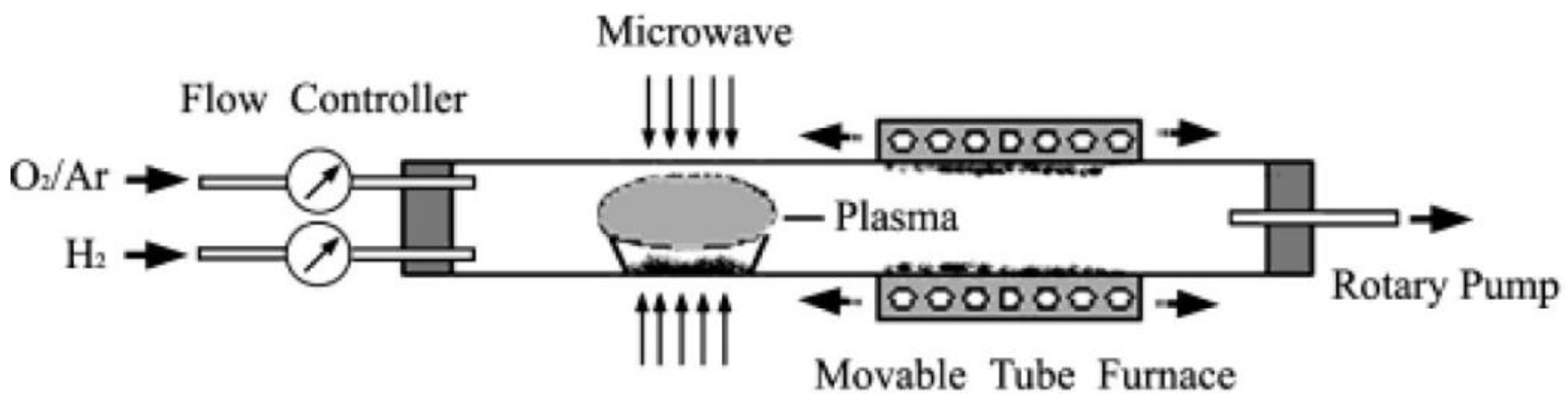
| Properties | Value |
|---|---|
| Molecular formula | ZnO |
| State (colour, form) | white powder |
| CAS Reg No. | 314-13-2 |
| Molar mass | 81.39 g/mol |
| Density at room temperature | 5.606 g/cm3 (crystal theoretical density 5.61 g/cm3) |
| Solubility in water (25 °C) | 1.6 mg/L |
| Melting point | 1975 °C |
| Boiling point | 2360 °C |
| Stable phase at room temperature | wurtzite |
| Structure | Hexagonal, where a0 = b0 ≠ c0 |
| Space group symmetries | C4 6v (P63mc) |
| Bulk effective piezoelectric constant | 9.9 pm/V |
| Hardness | 5.0 ± 0.1 GPa |
| Lattice parameters at 300 K | |
| a0 | 3.2495 Å |
| c0 | 5.2069 Å |
| c0/a0 | 1.602 (ideal hexagonal structure shows 1.633) |
| U | 0.345 |
| Thermal conductivity | 0.6, 1–1.2 W·cm−1·K−1 |
| Specific heat | 0.125 cal/gm·°C |
| Linear expansion coefficient | a0: 6.5 cm 3 × 10⁻⁶ K |
| c0: 3.0 cm 3 × 10⁻⁶ K | |
| Static dielectric constant | 8.656 ε(0), ε(∞) |
| Thermoelectric constant at 573 K | 1200 mV/K |
| Refractive index | 2.008–2.029 |
| Band gap | at RT: 3.370 eV |
| at 4 K: 3.437 eV | |
| Exciton binding energy | 60 meV |
| Intrinsic carrier concentration | <106 cm3 |
| Electron effective mass | 0.24 m0 |
| Hole effective mass | 0.59 m0 |
| Electron Hall mobility at 300 K | 200 cm2/V·s |
| Hole Hall mobility at 300 K | 5–50 cm2/V·s |
| Ionicity | 62% |
| High (>0.5) | Medium (0.1–0.5) | Low (<1) | |||
|---|---|---|---|---|---|
| Solvent | tanδ | Solvent | tanδ | Solvent | tanδ |
| Ethylene glycol | 1.350 | 2-Butanol | 0.447 | Chloroform | 0.091 |
| Ethanol | 0.941 | Dichlorobenzene | 0.280 | Acetonitrile | 0.062 |
| DMSO | 0.825 | NMP | 0.275 | Ethyl acetate | 0.059 |
| 2-Propanol | 0.799 | Acetic acid | 0.174 | Acetone | 0.054 |
| Formic acid | 0.722 | DMF | 0.161 | THF | 0.047 |
| Methanol | 0.659 | Dichloroethane | 0.127 | Dichloromethane | 0.042 |
| Nitrobenzene | 0.589 | Water | 0.123 | Toluene | 0.040 |
| 1-Butanol | 0.571 | Chlorobenzene | 0.101 | Hexane | 0.020 |
| Material | Temperature (°C) | Penetration Depth (cm) | Ref. |
|---|---|---|---|
| Water (distilled) | 20 | 1.6 | [463] |
| Water (distilled) | 25 | 2.88 | [456] |
| Water (distilled) | 100 | 80 | [463] |
| 0.125 M NaCl solution of salt water | 25 | 0.88 | [464] |
| 0.5 M NaCl solution of salt water | 25 | 0.45 | [456] |
| 2 M NaCl solution of salt water | 25 | 0.14 | [464] |
| Water (ice) | −12 | 1100 | [465] |
| Ethylene glycol | 25 | 0.46 | [464] |
| Methanol | 25 | 0.68 | [464] |
| Ethanol | 25 | 0.93 | [464] |
| 1-propanol | 25 | 1.39 | [464] |
| Acetone | 25 | 7.07 | [464] |
| Ethyl acetate | 25 | 11.05 | [464] |
| Xylene | 25 | 28.32 | [464] |
| Rubber, styrene-butadiene (SBR), vulc. | - | 19 | [463] |
| Nitrile rubber, natural | - | 65 | [463] |
| Aluminium oxide (Al2O3) ceram, for MW use | - | 3000 | [463] |
| Polyethylene | 25 | 4000 | [463] |
| Polyethylene | - | 5907.1 | [456] |
| Polystyrene | - | 7619.3 | [456] |
| PTFE (Teflon®) | - | 9000 | [463] |
| Quartz, pure | - | 20,000 | [463] |
| Silver | - | 0.33 × 10⁻⁴ | [463] |
| Zinc, pure (Zn) | - | 1.24·× 10−4 | [463] |
| Copper (Cu) | - | 1.3 × 10⁻⁴ | [456] |
| Aluminium 100% (Al) | - | 0.86 × 10⁻⁴ | [463] |
| Aluminium (Al) | - | 1.7 × 10⁻⁴ | [456] |
| Nickel (Ni) | - | 2.7 × 10⁻⁴ | [456] |
| Iron (Fe) | - | 3.2 × 10⁻⁴ | [456] |
| Titanium, pure (Ti) | - | 3.3 × 10⁻⁴ | [463] |
| Stainless steel (304) | - | 4.3 × 10⁻⁴ | [463] |
| Microwave Power | Heating Time after Which the Temperature Was Reached (s) | |
|---|---|---|
| 100 °C | 140 °C | |
| 100 W | 443 | - |
| 200 W | 139 | 353 |
| 300 W | 106 | 171 |
| 400 W | 61 | 112 |
| 500 W | 43 | 141 |
| 600 W | 37 | 61 |
| 700 W | 31 | 52 |
| 800 W | 27 | 45 |
| 900 W | 22 | 41 |
| 1000 W | 22 | 39 |
| Substrates | Conditions during Preparation | Properties | Ref. |
|---|---|---|---|
| Zn(NO3)2·6H2O (0.1, 0.5 and 2 M), NaOH, H2O | pH: 8–12; T: 100–190 °C; P: 1–13 bar, duration: 2 min–2 h; microwave reactor | SSA: 4.7–18.1 m2/g; particles, submicrometre grains and star-like morphology | [506] |
| Zn(NO3)2·6H2O (0.1 M), NaOH (2 M), H2O | P: 9–39 bar, duration: 3–7 min; power: 70–100%; microwave reactor (750 W) | heterogeneous nano- and microstructures; particles size: 10–300 nm | [507] |
| ZnCl2·2H2O (0.1 M), KOH or urea, H2O | pH: 12; P: 10–40 bar; duration: 3–15 min; microwave reactor | SSA: 8.6–102.2 m2/g; particles size: 37–114 nm, flower-like morphology | [508] |
| Zn(NO3)2·6H2O (0.005 M), hexamethylenetetramine (C6H12N4) (0.005 M), H2O | T: 90 °C; duration: 2 min; microwave reactor | ZnO rods (e.g., bipods, tripods, tetrapods and multipods); diameter: 160–220 nm; length: 1.25–1.3 µm | [509] |
| Zn(NO3)2·6H2O (0.13 M), NaOH (1.3 M), 1-n-butyl-3-methyl imidazolium tetrafluoroborate, H2O | T: 90–125 °C; duration: 2–10 min; microwave reactor | morphology: flower-like + needle-like, from 60 to 450 nm and lengths up to several micrometres | [510] |
| Zn(CH3COO)2·2H2O, N,N dimethylformamide, H2O | duration: 23 min; power: 50%; microwave oven | spherical particles ~160 nm and nanoplatelets and nanorods ~2 nm in diameter and ~80 nm in length | [511] |
| ZnO was dissolved in NH4(OH) and Zn2+ 0.08 M, H2O, NH4(OH) (0.5, 1, 5, 10 and 14.8 M) was obtained | T: 90–150 °C; P: 0.7–4.8 bar; power: 1000 W; microwave reactor | flower-like agglomeration | [512] |
| Zn(NO3)2·6H2O (0.005 M), hexamethylenetetramine (C6H12N4) (0.010 M), NaOH (3 M) | pH: 9 and 13; T: 96 °C; duration: 60 min; | flower-like ZnO microstructures (2–3 µm) of hexagonal prisms (length: 1–2 μm, diameter: 50–130 nm) with planar and hexagonal pyramid tips (length: 1.5 μm, diameter: 300 nm) | [513] |
| Zn(NO3)2·6H2O (0.43 M), NaOH (0.43 M), H2O, NaCl, wet mechanical mixtures obtained | T: 75–135 °C; power: 650 W; microwave oven | SSA: 9–13 m2/g; microtubes | [514] |
| Zn(NO3)2·6H2O (0.05 M), urea (0.05 M), H2O | duration: 40 min; power: 180 W; microwave oven | nanotubes have regular polyhedral shapes, hollow cores with diameters of 100–200 nm, lengths of 1–3 mm and wall thicknesses of 10–40 nm. | [515] |
| Zn(NO3)2·6H2O (0.43 M), NaOH (0.43 M), H2O | duration: 15 min; T: 75–170 °C; power: 40–450 W; Teflon cell in microwave oven; pulsed mode | SSA: 9–19 m2/g, crystallite size: 30–45 nm | [516] |
| Zn(CH3COO)2·2H2O and hydrazine (N2H4·H2O) in a molar ratio of 1:4 in H2O | duration: 10 min; power: 150 W; microwave oven | nanorods; diameter about 25–75 nm and length in the range of 500–1500 nm | [517] |
| Zn(NO3)2·6H2O, NaOH, H2O | duration: 20 min; T: 100–180 °C; power: 0–1000 W; microwave reactor | nanorods; nanowires; nanothruster vanes; nanodandelions; nanospindles | [518] |
| Zn(NO3)2·6H2O (1.6 M), NaOH (3.2 M), H2O | pH: 8.3; duration: 1–5 min; microwave oven | nanorods (diameter: 100–200 nm) and flower structures | [519] |
| Zn(NO3)2·6H2O, NaOH (different concentrations), H2O | duration: 1 h; T: 110 °C; microwave oven | submicron starshaped structures, chrysanthemum flower structures, nanoflakes | [520] |
| Zn(NO3)2·6H2O, pyridine (C5H5N), aniline (C6H5NH2) and triethanolamine (TEA, C6H15NO3) (different concentrations), H2O | duration: 10 min; T: 90 °C; microwave reactor | various morphologies: linear linked needles, regularly hexagonal cross section of a needle, hollow structures, hexagonal nanorings, hexagonal columns, nanosheets | [521] |
| Zn(CH3COO)2·2H2O, NaOH, 1-ethyl-3- methylimidazolium bis(trifluoromethylsulfonyl)imid, H2O | duration: 2–7 min; T: power: 255 W; microwave oven (850 W) | nanoparticles, length less than 100 nm | [522] |
| Zn(NO3)2·6H2O (0.025 M); hexamethylenetetramine (C6H12N4) (0.025 M), H2O | duration: 1–30 min; T: 100–180 °C; power: 120–700 W; microwave oven | nanowires | [523] |
| Zn(NO3)2·6H2O, NaOH (5 M) nasturtium officinale leaf extract, H2O | pH: 10; duration: 10 min; microwave oven (1000 W) | heterogeneous aggregates of NPs | [524] |
| Zn(NO3)2·6H2O (0.025 M), Zn(CH3COO)2·2H2O, NH4(OH) (0.16 M), H2O, ethylenediamine (EDA, C2H8N2), hexamethylenetetramine (C6H12N4), triethyl citrate (C12H20O7), tripotassium citrate monohydrate (C₆H₅K₃O₇·H₂O) | duration: 15 min; T: 90 °C; microwave reactor | nanorods, nanoneedles, nanocandles, nanodisks and nanonuts | [525] |
| Zn(NO3)2·6H2O, hexamethylenetetramine (HMTA), polyvinylpyrrolidone (PVP), H2O | duration: 60 min; T: 100 °C; power: 300 W; microwave reactor | nanostars, average size: ≈625 nm, crystallite size ≈550 nm, SSA = 20.6 m2/g | [526] |
| Zn(CH3COO)2·2H2O (different concentrations), NH4(OH), H2O | duration: 85 s; T: 90 °C; power: 800 W; microwave oven | flower-like shapes with diameter of 3 to 5 µm, flowers with rod-like nanostructures, spherical particles in 2–4 µm diameter; 2–3 µm structured balls with occasional large 10 µm lumps | [527] |
| Zn(CH3COO)2·2H2O, NH4(OH) (different concentrations), H2O | pH: 7.0–11.1; duration: 2 h; T: 150 °C; microwave reactor | hexagonally shaped prismatic (width ≈ 1 μm, length ≈ 5 μm); flower-like structures formed by a micron sized crystals; heterogeneous particles (size from ~50 nm to 300 nm) | [528] |
| Zn(CH3COO)2·2H2O, NaOH, (o- and m- and p)-nitrobenzoic acid, H2O | duration: 10 min; microwave reactor | flower-like products consist of sword-like ZnO nanorods, which were 60–100 nm in width and several micrometres in length. | [529] |
| ZnCl2·2H2O, NaOH, H2O, bis(dodecyldimethyl ammonium bromide) (C26H56BrN) | duration: 5–10 h; T: 100–140 °C; power: 300–400 W; microwave reactor | flower-like with mean r 0.5–1.5 µm, sphere-like with mean diameter of 0.5 µm | [530] |
| ZnCl2·2H2O, NaOH, H2O, cetyltrimethylammonium bromide (CTAB, C19H42BrN), Pluronic F127 | duration: 5 min; power: 130 W; microwave oven | SSA: 15.5–24.8 m2/g, diameter: 58–93 nm, heterogeneous shape | [531] |
| Zn(CH3COO)2·2H2O, Na(OH) (different concentrations), H2O | duration: 5 min; power: 450 W; microwave oven | nanoplates flowers | [532] |
| Zn(CH3COO)2·2H2O, NaOH, H2O, polyethylene glycol, ethanol | duration: 30 min; T: 140 °C; power: 700 W; microwave reactor (multimode) | nanorods, flowers | [533] |
| Zn(CH3COO)2·2H2O (0.5 M), KOH (2 M), H2O | pH: 8; T: 120 and 140 °C; duration: 8 min; microwave oven (800 W) | multiwires with a flower-like shape of 50–400 nm in width and length | [534] |
| Zn(CH3COO)2·2H2O (0.1 M), Zn(NO3)2·6H2O (0.1 M), NH4OH, polyvinilpirrolidone, hydrazine hydrate solution (N2H4 in H2O), H2O | pH: 7.5–8; duration: 5–10 min; microwave oven (1000 W) | spherical nanoparticles, stars, flowers | [535] |
| Zn(NO3)2·6H2O, NaOH, gum arabic (stabilising agent), NaOH, H2O | pH: 10; duration: 5 min; power: 450 W | stars (diameter: 1020 nm), spherical particles (diameter: 240 nm) | [536] |
| Zn(NO3)2·6H2O, NaOH, gum arabic (stabilising agent), NaOH, H2O | pH: 10; duration: 2–10 min; power: 350 W | aggregates of NPs (20–40 nm), size of aggregates: 150–200 nm | [537] |
| Zn(NO3)2·6H2O (0.1 M), NH4(OH), hydrazine hydrate (N2H4·H2O), H2O | pH: 8; duration: 10 min; microwave oven | flower morphology consists of sharp nanorods which look like petals | [538] |
| Zn(NO3)2·6H2O (0.005 M), KOH (4 M), H2O | pH: 12; T: 120 °C; duration: 4 h; microwave oven | nanowires with diameter of 80 nm and lengths of up to 10 µm. | [539] |
| Zn(NO3)2·6H2O (0.01 M), urea (0.1 M), H2O | T: 120 °C; duration: 10–24 min; power: 150 W; microwave oven | javelins, length: 14–17 µm, width: 0.9–1.4 µm | [540] |
| Zn(CH3COO)2·2H2O, KOH | duration: 20 min; power: 180 W; microwave oven | flowerlike structures composed of hexagonal ZnO spear-shaped nanorods with diameters and lengths of 50 nm and 2–4 μm, | [541] |
| ZnCl2·2H2O (different concentrations), NH4(OH), H2O | duration: 10–40 min; power: 10–50%; microwave oven (800 W) | nanorods | [542] |
| ZnCl2·2H2O, arginine (C6H14N4O2), H2O | T: 120–180 °C; duration: 3–10 min; microwave reactor | rods and flowers | [543] |
| Zn(CH3COO)2·2H2O (different concentrations), NaOH, H2O | pH: 12; T: 120–140 °C; duration: 15–60 min; power: 0–100%; microwave oven (900 W) | nanosheets | [544] |
| Zn(CH3COO)2·2H2O, NH4(OH), H2O | pH: 9; duration: 90 sec; microwave oven (900 W) | narcissus-like nanostructures with crystallite sizes of 10–15 nm and average diameter of 1–2.5 µm | [545] |
| Zn(CH3COO)2·2H2O, NaHCO3, H2O | duration: 15 min; power: 200 W; microwave oven | amorphous material | [546] |
| Zn(CH3COO)2·2H2O (0.0026 M), NaOH (1 M), H2O, cetyltrimethylammonium bromide (CTAB, C19H42BrN) | T: 130 °C; duration: 30–180 min; Teflon autoclave in microwave oven (800 W) | nanowires and microwires of about 50–400 nm in width and several micrometres in length | [547] |
| Zn(NO3)2·6H2O (different concentrations), hexamethylenetetramine (C6H12N4), H2O | duration: 2–3 min; power: 600–900 W; Teflon bottle in microwave oven (1000 W) | nanorods diameters from 117 to 156 nm | [548,549] |
| Zn(NO3)2·6H2O (different concentrations), NaOH, H2O | pH: 7–13.1; duration: 20; power: 180 W; microwave oven | nanoparticles in clusters, nanoplates in flower-like clusters, and spear-shaped particles in flower-like clusters | [550] |
| ZnCl2·2H2O (0.066 M), NaOH (1.75 M), H2O | pH: 13.75; duration: 5 min; power: 150–1000 W; microwave oven (1000 W) | nanoparticles, nanoneedles, nanosheets (leaf-like) | [551] |
| Zn(CH3COO)2·2H2O (0.1 M), NaOH (4 M), CH3(CH2)11OSO3Na (0.1 M), C12H25C6H4SO3Na (0.025 M), H2O | T: 75–130 °C; duration: 1–5 h; power: 400 and 700 W; microwave reactor | SSA: 33.1–419.7 m2/g; square shaped sheets | [552] |
| Zn(NO3)2·6H2O, hexamethylenetetramine (C6H12N4) | duration: 30–45 sec; power: 700 W, microwave oven | multiple linked rods such as bipods, tripods (T- shaped), tetrapods (+ and X-shaped), tassel brush and flower shaped and individual rods | [553] |
| Zn(CH3COO)2·2H2O (different concentrations), triethanolamine (TEA, C6H15NO3), H2O | T: 80–100 °C; duration: 10–30 min; microwave reactor (1000 W) | pompon-like spheres, peach nut-like spheres, misshapen spheres | [554] |
| Zn(CH3COO)2·2H2O, NaOH, bis(triaminomethyl) carbonate (C3H12N6O3), H2O | pH: 12; duration: 2 min; power: 600 W; microwave oven | flower-like structure (2 µm) composed of petals with average size of about 600–700 nm in length, 300–400 nm in width, and 50–70 nm in tip | [555] |
| Zn(NO3)2·6H2O, NaOH, H2O | duration: 15–50 min; power: 120–420 W; microwave reactor (700 W) | nanostructures consisted of flower-like, sword-like, needle-like and rods-like structures | [556] |
| Zn(CH3COO)2·2H2O (different concentrations), NH4(OH), H2O | pH: 10.2; duration: 50–70 s; microwave oven (800 W) | rod-arrays film on glass | [557] |
| Zn(NO3)2·6H2O, polyvinyl pyrrolidone, NH4(OH), H2O | pH: 10.2; duration: 10 min; microwave oven (1000 W) | star-shaped nanostructures | [558] |
| Zn(NO3)2·6H2O (0.06 M), NaOH (0.06 M), polyethylene glycols (PEG)-2000, H2O | T: 180 °C; duration: 10, 20, 30, 60 min; microwave reactor | hierarchical structured nanorods | [559] |
| ZnSO4·7H2O (0.1 M), NaOH (0.4 M) | duration: 2 min; microwave oven | nanoparticles (10–15 nm) | [560] |
| Zn(CH3COO)2·2H2O, NaOH, 1-butyl-3-ethyl imidazolium tetrafluoroborate (C8H15BF4N2), H2O | T: 120–140 °C; duration: 5 min; microwave reactor (800 W) | calthrop-like framework | [561] |
| Zn(NO3)2·6H2O (0.03 M), NaOH (0.06 M), H2O, polyethylene glycol (PEG) 2000, H2O | T: 180 °C; duration: 30 min; microwave reactor | rods with the diameter of 300 nm and length of 1 µm | [562] |
| Zn(NO3)2·6H2O, Zn(C5H7O2)2·xH2O, urea, C2H4(OH)2 (different concentrations), H2O | T: 150 °C; duration: 1–30 min; microwave reactor | microrods with width of 200–300 nm and length of up to 4 µm | [563] |
| Zn(NO3)2·6H2O, dodecylamine (C12H27N), H2O | T: 80–130 °C; duration: 1–50 min; power: 150 W; microwave oven | hexagonal quasi-hourglasses (tripods, tetrapods, pentapods, multipods) | [564] |
| Zn(CH3COO)2·2H2O, triethanolamine (TEA, C6H15NO3), NH4(OH), H2O | pH: 6-12; T: 80–160 °C; duration: 10–60 min; power: 150 W; microwave reactor | spherical nanoparticles 60–90 nm, rugby-like nanostructures with diameter of 450 nm and length of about 700 nm | [565] |
| Zn(CH3COO)2·2H2O, triethanolamine (TEA, C6H15NO3, (different concentrations), NaOH (different concentrations), H2O | pH: 9.0–12; duration: 90 s; power: 900 W; microwave oven | nanospheres with the crystallite size of 57 nm; raspberry-like nanostructures with the crystallite size of 62 nm; hollow nanospheres with the crystallite size of 78 nm; nanoparticles with the crystallite size of 24 nm | [566] |
| Zn(NO3)2·6H2O (0.1 M), C6H12N4 (0.1 M) | T: 90 °C; duration: 2 h; microwave oven | nanorods on the surface of GaN 80–170 nm, nanorods on the surface of glass 40–100 nm | [567,568,569] |
| Zn(NO3)2·6H2O, C6H12N4 | duration: 5 h; microwave oven | nanorods on the surface of glass, average width of nanorod: 20–1000 nm, average length of nanorod: 150–5000 nm | [570] |
| Zn(CH3COO)2·2H2O, KOH, | T: 90 °C; duration: 10–30 min; microwave oven | micro-tube structure 200–400 nm in diameter, flower-like structure composed of spear-shaped nanorods with diameters and lengths of 70 nm and 1–5 μm, | [571] |
| Zn(CH3COO)2·2H2O (different concentrations), NaOH, C2H5OH | pH: 10; T: 100 °C duration: 45–60 min; power: 800 W; microwave reactor | plates (SSA: 10.7 m2/g), rounded plates (SSA: 9.18 m2/g), brush-like (SSA: 9.5 m2/g), flower-like (SSA: 8.5 m2/g) | [572] |
| Zn(CH3COO)2·2H2O, NH4(OH), H2O | pH: 8; duration: 180 s; microwave oven (900 W) | SSA: 22.9 m2/g; uniform flower-like nanostructures composed of petals attached in the centre with lengths in the range of 700–950 nm and a width in the range of 130–230 nm; each single petal is composed of nanoparticles with lengths of 45–95 nm | [573] |
| Zn(CH3COO)2·2H2O, NH4(OH), C25N3H30Cl, Polyethylene glycol (PEG) 400, C19H42BrN, H2O | duration: 10 min; microwave reactor | rod-like nanostructures, star-like nanostructures | [574] |
| ZnSO4·7H2O, NaOH, H2O | pH: 9; duration: 5–25 min; microwave reactor | sheet nanostructures | [575] |
| Zn(NO3)2·6H2O (0.1 M), NH4(OH), H2O, cetyltrimethylammonium bromide (CTAB, C19H42BrN) | pH: 7; T: 150 °C; duration: 1 h; microwave reactor | nanorods, length: 1–2 μm and width: 100–150 nm | [576] |
| Zn(CH3COO)2·2H2O, NH4(OH), H2O | duration: 8 min; power: 900 W; microwave oven | nanoparticles (15 nm) which were self-assembled to form a sheet-like structure | [577] |
| Zn(NO3)2·6H2O, NaOH, polyvinyl alcohol, H2O | duration: 10 min; power: 700 W; microwave oven | nanoparticles (40 nm) | [578] |
| Zn(NO3)2·6H2O (0.005 M), Zn(CH3COO)2·2H2O (0.005 M), ZnSO4·7H2O (0.005 M), KOH (2 M), H2O | pH: 12; T: 130 °C; duration: 1 h; microwave reactor (800 W) | flower-like structures, plates | [579] |
| ZnCl2·2H2O (0.5 M), urea, H2O | duration: 5 min; power: 800 W microwave reactor | sponge-like nanostructure | [580] |
| Zn(CH3COO)2·2H2O, Zn(NO3)2·6H2O, ZnCl2·2H2O, NaOH, KOH, NH4(OH), sodium di-2-ethylhexyl-sulfosuccinate (C20H36Na2O7S), H2O | T: 80–140 °C; duration: 5–20 min; power: 300–1200 W; microwave reactor | hexagonal rods (3–4 μm long and 1 μm wide), hexagonal prismatic, bihexagonal rod-like structure (6 μm long and 2 μm wide), hexagonal prismatic particles (60–80 nm in diameter and length between 90 and 110 nm) | [581] |
| ZnCl2·2H2O, NH4(OH), H2O | T: 80–140 °C; duration: 20 min; power: 240 W; microwave oven | flower-shaped ZnO microcrystals (about 5 μm) | [582] |
| Zn(NO3)2·6H2O, hexamethylenetetramine (C6H12N4), H2O | T: 170 °C; duration: 2–20 min; microwave reactor | irregular sheet-like structures and rods, tripods | [583] |
| Zn(NO3)2·6H2O, hexamethylenetetramine (C6H12N4), H2O | T: 90 °C; duration: 3 h; microwave reactor | nanorods on paper | [584] |
| Zn(NO3)2·6H2O (0.1 M), NH4(OH), albumen, H2O | pH: 8; duration: 5 min; microwave oven | sheet-like and spherical-like nanostructures (13–50 nm), nanowhiskers and nanorods (10–57 nm) | [585] |
| Zn(NO3)2·6H2O (0.03 M), hexamethylenetetramine (C6H12N4), H2O | duration: 2–30 min; power: 10–100%; microwave oven (1100 W) | nanostructured-films (networked-nanoflakes morphology) | [586,587] |
| Zn(CH3COO)2·2H2O, NaOH, glutamic tetrofluoroborate (different concentrations), H2O | T: 80 °C; duration: 10 min; power: 1000 W; microwave reactor | clew-like hierarchical nanosheet spheres, nanoneedle-like structures | [588] |
| Zn(NO3)2·6H2O (different concentrations), hexamethylenetetramine (C6H12N4), H2O | duration: 5–20 s; power: 180-1100 W; microwave oven | growth of nanorods, diameters: 50–80 nm | [589,590,591] |
| Zn(NO3)2·6H2O (0.03 M), hexamethylenetetramine (C6H12N4), H2O | duration: 30 min; power: 110 W; modified microwave oven (1100 W) | porous nanostructures grown on Al-Si substrate (Al layer thickness form 0 to 150 nm); Al-free Si substrate: nanorods were formed (length: 350 nm, diameter: 50 nm); Al on Si substrate: nanoflake (height ~380 nm ) with pores sizes ranging from 50 nm to several hundreds of nanometres. | [592] |
| Zn(NO3)2·6H2O (different concentrations), hexamethylenetetramine (C6H12N4, different concentrations), H2O | T: 80 °C; duration: 10 min; power: 30–50%; microwave oven | ZnO nanoarray (rod-like structures) on glass, size control achieved by regulating the parameters | [473] |
| Zn(NO3)2·6H2O (different concentrations), NH4(OH), H2O | duration: 8 min; power: 800 W; microwave oven | flower-like and rod-like structures | [593] |
| Zn(CH3COO)2·2H2O (0.45 M), NaOH (8 M), Triton X-100 | duration: 1–6 min; power: 100–600 W; microwave oven | rods (400–800 nm), flower structures | [594] |
| Zn(CH3COO)2·2H2O, KOH, H2O | duration: 3 min; power: 800 W; microwave oven | hexagonal nanorods (length from ~1.5 μm to 3 μm and in diameter from ~30 nm to 80 nm) | [595] |
| Zn-dust, HNO3, NaOH, polyethylene glycol (PEG, MW 2000), H2O | duration: 10–20 min; microwave oven | SSA: 14.4-21.8 m2/g; particles with irregular shape (plate and rod-like particles), crystallite size: 34–42 nm | [596] |
| Zn(CH3COO)2·2H2O, NaOH, cetyltrimethylammonium bromide (CTAB, C19H42BrN, different concentrations), H2O | T: 130°; duration: 15–60 min; microwave oven | wire-like architecture with a width in the range of 60–80 nm, flower-like microstructures composed of nanorods, rod has a width of 300–400 nm and a length of 3–4 µm | [597] |
| Zn(CH3COO)2·2H2O, Zn(NO3)2·6H2O, NaOH, NH4(OH), di-2-ethylhexyl sodium sulfosuccinate (C20H36Na2O7S), H2O | T: 80–140 °C; duration: 5–20 min; power: 300–1200 W; microwave reactor | cauliflower-like structures, hexagonal prismatic type particles (200–300 nm) | [598] |
| Zn(CH3COO)2·2H2O, Zn(NO3)2·6H2O, NH4(OH), hydrazine hydrate (N2H4·H2O), H2O | pH: 11.5; duration: 10–25 min; power: 510–680 W; microwave oven | nanoparticles, nanorods, flowers | [599] |
| Zn(CH3COO)2·2H2O, NaOH, H2O | T: 140 °C; duration: 45 min; power: 400 W; microwave reactor (1600 W) | nanorods, diameter ranging from 60 to 80 nm with average length of about 250 nm | [600] |
| Zn(CH3COO)2·2H2O (0.005 M), NaOH (0.025 M), H2O | duration: 6 min; power: 400–600 W; microwave oven | mixture of nanorods and nanoplates | [601] |
| Zn(CH3COO)2·2H2O (0.18 M), NaOH (different concentrations), H2O | pH: 7–9.5; T: 50 °C; duration: 3 min; microwave reactor (600 W) | nanorods (width: 80–300 nm, height: 150–1000 nm) grown on Si, GaAs and GaN substrate | [602] |
| Zn(CH3COO)2·2H2O (0.18 M), NaOH (different concentrations), H2O | pH: 6.75–7.75; T: 50 °C; duration: 2 min; microwave reactor (600 W) | rods (width: 0.5–2.5, height: 1.5–2.2 µm) grown on GaN substrate | [603] |
| Zn(CH3COO)2·2H2O, NH4(OH), H2O | pH: 10.1–10.9; T: 90 °C; duration: 20 min; power: 100 W; microwave oven | nanorods grown on Si substrate | [604] |
| Zn(NO3)2·6H2O, hexamethylenetetramine (C6H12N4 ), H2O | T: 90 °C; duration: 2 h (switched on and off automatically); microwave oven | nanorods grown on Si substrate (thickness: ~1 µm) | [605] |
| Zn(NO3)2·6H2O, hexamethylenetetramine (C6H12N4), H2O | duration: 10–30 min; power: 120 W; microwave oven | rods, bipods (length: 0.46–1 µm, width: 0.1–0.13 µm) | [606] |
| Zn(NO3)2·6H2O, hexamethylenetetramine (C6H12N4), H2O, oxalic acid dihydrate (C2H2O4·2H2O) | P: 20.68 bar; duration: 15 min; microwave reactor (300 W) | nanorods or flower grown on paper | [607] |
| Zn(NO3)2·6H2O, hexamethylene tetramine (C6H12N4), H2O | T: 80 °C; duration: 5–20 min; power: 100–1600 W; microwave reactor (1600 W) | nanorods grown on glass substrate | [608] |
| Zn(NO3)2·6H2O, hexamethylene tetramine (C6H12N4, different concentrations), H2O | duration: 10 min; power: 240 W; microwave oven | nanorods (diameter: 89–216 nm) grown on glass substrate | [609] |
| Zn(NO3)2·6H2O, NH4(OH), H2O | pH: 10.0–12.0; T: 90–120 °C; duration: 1 h min; microwave reactor | nanorods grown on glass substrate, control of size of diameter of rods within the size range between circa 125 and 770 nm | [610] |
| Zn(CH3COO)2·2H2O (different concentrations), hexamethylenetetramine (C6H12N4), H2O | T: 60–110 °C; duration: 5–40 min; microwave reactor | nanorods (diameter from ~30 to ~300 nm, length from ~60 nm to ~520 nm) grown on Si substrate | [611] |
| Zn(CH3COO)2·2H2O, NH4(OH), NaOH, CH3COOH, H2O | pH: 9.8 or 10.8; duration: five steps (each step included 30 s of irradiation and 10 s off); microwave oven | dandelion-like nanostructures (needles: 50–200 nm, height ~2 µm) or a flower-like microstructures grown on activated carbon cloth | [612] |
| Zn(CH3COO)2·2H2O, NH4(OH), palmitic acid (CH3(CH2)14COOH), H2O | pH: 4–5; duration: 10–30 min; microwave oven | rod shaped structures | [613] |
| ZnSO4·7H2O, NH4(OH), H2O | pH: 10; duration: 2 min; power: 600 W; microwave oven | nanoparticles (~50 nm) | [614] |
| Zn(NO3)2·6H2O, potassium sodium citrate, NaOH, H2O | T: 90 °C; duration: 2 min; power: 600 W; microwave oven (650 W) with a refluxing apparatus | sphere-like particles (~2.32 μm) | [615] |
| tris(ethylenediamine)zinc nitrate ([Zn(en)3](NO3)2), NaOH, H2O | pH: 7–12; T: 180 °C; duration: 20 min; power: 400 W; microwave reactor (1600 W) | nanorods, diameter: from 40 nm (pH 12) to 600 nm (pH 7) | [616] |
| Zn(NO3)2·6H2O, hexamethylenetetramine (C6H12N4), H2O | T: 100 °C; duration: 60 min; power: 100 W; microwave reactor | nanorods (180–350 nm) grown on glass | [617] |
| Zn(NO3)2·6H2O, hexamethylenetetramine (C6H12N4), polyethylenimine, NH4OH, H2O | T: power on and off in order to control the solution temperature; duration: 6–10 × (30–60 s “on” and 5 min “off”); microwave oven (350 W) | nanorods (180–350 nm) grown on glass | [618] |
| Zn(CH3COO)2·2H2O, NH4(OH), carbon fibre, H2O | duration: 3 × (30 s irradiation and 30 s stop); microwave oven (1120 W) | rods (diameter: 0.3–0.5 µm, length: 1.0–1.5 µm) grown on carbon fibre | [619] |
| Zn(NO3)2·6H2O, NaOH, NH4(OH), polyethylene glycol (PEG, MW 400), H2O | T: 100 °C, duration: 5 min; microwave oven (800 W) | quasi-spherical shape and dimensions of less than 5 μm, flower-like structures (>5 μm), | [620] |
| Zn(NO3)2·6H2O, C6H12N4, polyethylenimine, NH4(OH), H2O | duration: 5–15 min; microwave oven (800 W) | nanoflowers and nanowalls grown on P–Si | [621] |
| Zn(CH3COO)2·2H2O, NaOH, 1-hexyl-2-ethyl-3-methylimidazoliumtetrafluoroborate (C6H11BF4N2), H2O | duration: 2–9 min; power: 30%; microwave oven | flakes-shaped particles, flower-like shaped particles | [622] |
| Zn(NO3)2·6H2O, NaOH, plant extract, H2O | pH: 10; duration: 15 min; microwave oven | nanoparticles | [623] |
| Zn(NO3)2·6H2O, (C6H12N4)2, H2O | T: 70–130 °C; duration: 10 min; microwave reactor | nanorods on paper, length 120–480 nm, thickness 55–75 nm | [624] |
| Zn(NO3)2·6H2O, triethanolamine (TEA, C6H15NO3), H2O | duration: 10 min; power: 640 W; microwave oven | nanoparticles | [625] |
| Zn(CH3COO)2·2H2O, KOH, triethanolamine (TEA, C6H15NO3), 1,2,4,5-benzenetetracarboxylic acid, H2O | pH: 8–12; T: 150 °C; duration: 30 min; power: 800 W; microwave reactor | dumbbell-like structures, football-like structures, hexagonal bi-pyramidal structures, SSA: 7–24 m2/g; size 50 nm–10 μm | [626] |
| Zn(CH3COO)2·2H2O, NaOH, H2O | pH: 8–10; duration: 6 min; microwave oven (700 W) | sheet-like structures and uniform microstructures | [627] |
| Zn(CH3COO)2·2H2O, Tris (hydroxymethyl) aminomethane (C4H11NO3), H2O | duration: 3 min; microwave oven (300 W) | spherical nanoparticles | [628] |
| Zn(CH3COO)2·2H2O, KOH, benzene-1,2-dicarboxylic acid, benzene-1,3-dicarboxylic acid, benzene-1,4-dicarboxylic acid, H2O | pH: 7–12; T: 150 °C; duration: 30 min; power: 800 W; microwave reactor | rod-like structures, needle-like structures, platelet-like structures, hexagonal columnar shape of the particles, rice-grain shape structures | [629] |
| Zn(CH3COO)2·2H2O (0.1 M), NaOH (0.1 M), {4-[(E)-2-(furan-2-yl)ethenyl]pyridin-1-ium-1-yl} acetate (1 wt% and 3 wt%), CH3OH, H2O | duration: 20 min; microwave oven | heterogeneous shape of nanoparticles, size: 200–800 nm, average crystallite size: 21–23 nm | [630] |
| Zn(NO3)2·6H2O, hexamethylenetetramine (C6H12N4), NH4(OH), H2O | pH, 6.8–13, duration: 10–15 min; pulsed microwave heating in microwave oven (850 W) | rod, flower, star, tetrapod | [631] |
| Zn(NO3)2·6H2O (different concentrations), hexamethylenetetramine (C6H12N4), H2O | T: 105 °C; duration: 10–30 min; microwave oven (850 W) | growth of nanorods on P-type silicon wafer, diameters: from 26–32 nm to 35–40 nm, lengths: from 330 nm–607 nm | [632] |
| Zn(NO3)2·6H2O, hexamethylenetetramine (C6H12N4), H2O | duration: 20 min; power: 750 W; microwave oven | growth of nanorods on silicon substrate, diameters ~80 nm, lengths ~500 nm | [633] |
| Zn(NO3)2·6H2O, coffee powder extract, H2O | duration: 5 min; power: 540 W; microwave oven | spherical nanoparticles (80–120 nm) | [634] |
| Zn(NO3)2·6H2O, tomato extract, H2O | duration: 5 min; power: 180–540 W; microwave oven | spherical nanoparticles (40–100 nm) | [635] |
| Zn(NO3)2·6H2O, tea leaf extract, H2O | duration: 7 min; power: 540 W; microwave oven | spherical nanoparticles (26 nm) | [636] |
| Zn(CH3COO)2·2H2O, Longan fruits extract, H2O | duration: 1 min on & 1 min off irradiation cycle (1–30 cycles); power: 450–800 W; microwave oven | SSA: 35 m2/g, diameter: 10–100 nm, heterogeneous shape | [637] |
| Zn(CH3COO)2·2H2O (0.22 M), carbinol, H2O | duration: 5–15 min; power: 900 W; microwave oven | spherical nanoparticles, diameters: 30 nm–50 nm; hexagonal facetted nanostructures, average size: 400–450 nm | [638] |
| Zn(NO3)2·6H2O, NH4(OH) (28%), H2O | pH: 12; duration: 5–25 min; power: 180–540 W; microwave oven (1200 W) | spherical and flower-like particles on paper; non-uniform size | [639] |
| ZnO powder, hydrogen peroxide (H2O2, 30%) | P: 30 bar; duration: 15 min; power: 1200 W; microwave oven (1200 W) | rod-like nanostructures, average size: 36 nm | [640] |
| Substrates | Conditions during Preparation | Parameters of Additional Heat Treatment | Properties | Ref. |
|---|---|---|---|---|
| Zn(CH3COO)2·2H2O, NH4OH, H2O | duration: 5–10 min; power: 240–400 W; microwave oven | 500 °C in air for 1 h | dumbbell-shaped structures built of particles sized ~100 nm | [641] |
| Zn(CH3COO)2·2H2O, KOH, H2O | duration: 15 min (irradiation 12 s, stop 10 s); power: 180 W; microwave oven | 400 °C in air for 1 h | nanorods assembled in flower shaped, rods: diameter 150–190 nm (tip diameter ~15 nm), length 2 μm, with an aspect ratio of 20–22 | [642,643] |
| Zn(CH3COO)2·2H2O, NaOH, guanidinium carbonate, acetyl acetone (ACAC), H2O | pH: 8–12; duration: 2 min; power level: 75%; microwave oven | without and 600 °C in air for 2 h | petals: length 600–700 nm, width 300–400 nm, tip 50–70 nm; rod-like nanostructures diameters 60–90 nm maximal length 1.5 µm; spherical-like nanostructures: diameter 50 nm | [644] |
| Zn(CH3COO)2·2H2O (0.2 M), NaOH (0.4 M), H2O, triethanolamine (TEA, C6H15NO3) | duration: 20 min; power: 20%; microwave oven | 900 °C in air for 1 h | spherical particles ~50 nm | [645] |
| Zn(NO3)2·6H2O, hexamethylenetetramine (C6H12N4), H2O | duration: 2 h; microwave oven | 250–550 °C for 1 h in oxygen flow (5 cm3/min) | nanorods (diameter from 50–300 nm) grown on surface of silicon substrates | [568] |
| Zn(NO3)2·6H2O, hexamethylenetetramine (C6H12N4), polyethylenimine, NH4OH, H2O | duration: 4–80 min; power: 180–850 W; microwave oven | 350 °C for 20 min in air | nanowire grown on an ITO-coated glass substrate | [646] |
| Zn(CH3COO)2·2H2O, NaOH, H2O | duration: 10 min; power: 200 W; microwave oven | 400–800 °C for 1 h in air | circular- and hexagonal-shaped particles | [647] |
| Zn(CH3COO)2·2H2O, sodium dodecyl, NH4OH, (CH3)2CHOH (2-propanol) H2O | duration: 2 h; power: 100–800 W; microwave oven | 500 °C for 3 h in air | flakes-like structures, spherical-like, crystallite size: 31–39 nm (morphology dependent on the microwave power) | [648] |
| Zn(CH3COO)2·2H2O, ZnCl2·2H2O, Zn(NO3)2·6H2O, pyridine (C5H5N), H2O | pH: 13.75; duration: 2–5 min; power: 1000 W; microwave oven | 300–500 °C for 2 h in air | nanoparticles, nanoflowers, nanorods | [649] |
| Zn(CH3COO)2·2H2O, urea, H2O | T: 220 °C; duration: 15 min; microwave oven | 400 °C for 90 min in air | nanosheets | [650] |
| Zn(NO3)2·6H2O, hexamethylenetetramine (C6H12N4), NaOH, H2O | pH: 13; T: 90–220 °C; duration: 15 min; power: 110–710 W; microwave reactor (1400 W) | 200 °C for 2 h in air | nano-platelets | [651] |
| Zn(CH3COO)2·2H2O, trisodium citrate dihydrate (Na3C6H5O7·2H2O) (different amounts), NH4OH, H2O | T: 90 °C; duration: 10–45 min; power: 300 W; microwave reactor | 400 °C for 4 h in air | hollow microspheres average dimensions ~4 μm; thickness 400–600 nm. | [652] |
| Zn(NO3)2·6H2O, NH4OH, albumen, H2O | pH: 8; duration: 5 min; microwave oven (1000 W) | 130 °C for 5 h in air | whisker-like and rod-like nanostructures: thickness 10–57 nm | [585] |
| Zn(NO3)2·6H2O, KOH, H2O | pH: 9–13; duration: 30 min; power: 180 W microwave oven | 200 °C for 2 h in vacuum | nanorods | [653] |
| Zn(NO3)2·6H2O, urea (different concentrations), H2O | T: 90 °C; duration: 40 min; power: 400 W microwave oven | 400–600 °C for 3 h in different atmospheres (O2, N2, H2 and air), | various flower-like nano and microstructures, urchin-like structures | [654] |
| Zn(CH3COO)2·2H2O, NH4OH, hexamethylenetetramine ((CH2)6N4), hexadecyl trimethyl ammonium bromide, polyethylene glycol (PEG400), H2O | duration: 10 min; power: 300 W; microwave reactor | 500 °C for 2 h in air | needle-assembled structures with flower-like morphology; bundle-like microstructures assembled by nanorods; flower-like hierarchical structures composed of some tight aggregations | [655] |
| Zn(CH3COO)2·2H2O, trisodium citrate dihydrate (Na3C6H5O7·2H2O), urea, H2O | T: 140 °C; duration: 20 min; microwave reactor | 500 °C for 2 h in air | porous core–shell microstructures | [656] |
| Zn(NO3)2·6H2O, NaOH, H2O, | pH: 13; duration: 1–5 min with 30 s on–off cycling mode; power: 900 W; microwave oven | 400 °C for 1 h in air | microspheres | [657] |
| Zn(CH3COO)2·2H2O, NaOH, H2O, | pH: 12.66–13; duration: 10–30 min; power: 300 W; microwave reactor | 500 °C for 2 h in air | nanorods, nanoplates | [658] |
| Zn(NO3)2·6H2O, hexamethylenetetramine (C6H12N4), H2O | duration: 45 min; power: 1800 W; microwave oven | 350 °C for 1 h in air | nanorods, length: ~4.3 ± 0.2 µm, diameter: 100 ± 10 nm | [659] |
| Zn(NO3)2·6H2O, hexamethylenetetramine (C6H12N4) (different concentrations), H2O | duration: 10 min; power: 750 W; microwave reactor | 400 °C for 1 h in air | shape from random spherical to highly conserved hexagonal shaped rods, size: from ~25 nm to µm/sub µm | [660] |
| Zn(NO3)2·6H2O, hexamethylenetetramine (C6H12N4), hydrazine hydrate (N2H4), H2O | pH: 10; duration: 10–30 min; microwave reactor (1000 W) | 500 °C for 2 h in air | nanorods, length: 579–909 nm, diameter: 116–240 nm | [661] |
| Zn(CH3COO)2·2H2O, Tuber (Amorphophallus konjac) extract, H2O | duration: 5 min; microwave oven | 400 °C for 1 h in air | rice shaped nanoparticles, length: 237 nm, diameter: 76 nm | [662] |
| ZnCl2, NaOH, H2O | pH: 6.1–13.7; T: 80 °C; duration: 5–20 min; microwave reactor (900 W) | 150 °C for 3 h in air | hexagonal flake, velvet flower-like, rough globular, needle bunch-like, cauliflower-like, clew-like, nanorods, and rhombic microstructures | [663] |
| Zn(CH3COO)2·2H2O, ZnCl2·3H2O, Zn(NO3)2·6H2O, ZnSO4·7H2O, NH4OH, H2O | duration: 10 min; microwave reactor | 600 °C for 2 h in air | nanoflakes, nanorods, hexagonal tubular, pseudo-spherical | [664] |
| Zn(NO3)2·6H2O, NaOH, H2O (solvent) and C2H5OH (solvent) | P: 20–40 bars; duration: 15 min; microwave reactor | 750 °C for 1 h in air | particles, irregular shape | [665] |
| ZnCl2·3H2O, H2O, microcrystalline cellulose | T: 100 °C; duration: 10–60 min; microwave oven | 600 °C for 3 h in air | heterogeneous nanostructures and microstructures | [666] |
| Zn(CH3COO)2·2H2O, ZnCl2·3H2O, ZnSO4·7H2O, ZnCO3, Psidium guajava Linn. Extract, H2O | T: 100 °C; duration: varying cycles of 3 min-on and 1 min-off min; power: 720 W; microwave oven | 900 °C for 1.5 h in air | diameter: 60–180 nm | [667] |
| Zn(NO3)2·6H2O, pelargonium leaf extract, H2O | duration: 3 min; power: 800 W; microwave oven | 400 °C for 2 h in air | heterogeneous particles | [668] |
| ZnSO4·7H2O (different concentrations), banana corm extract, H2O | duration: 15 min; power: 540 W; microwave oven | 400 °C for 3 h in air | microparticles | [669] |
| Zn(CH3COO)2·2H2O, KOH, H2O | duration: 0.5–2 min; microwave oven (1000 W) | 400 °C for 1 h in air | nanorods, tetrapods (length: 255 nm), flowers (petal length: 387 nm) | [670] |
| Zn5(OH)8(NO3)2·2H2O, 1,2,3- trimethyl-imidazole tetrafluoroborate, H2O | duration: 2 h; power: 180 W; microwave oven (900 W) | 400 °C for 2 h in air | nanobelts, width: 500–800 nm; length: several micrometres, thickness: 100 nm | [671] |
| Zn(CH3COO)2·2H2O, NH4OH, C2H5OH, H2O | T: 120 °C; duration: 10 min; power: 180 W; microwave reactor | 527 °C in air | heterogeneous hollow NPs, SSA: 17.1 m2/g | [672] |
| Zn(NO3)2·6H2O, NaOH, polyethylene glycols (MW= 1500 and MW= 4000), sugar, cassava starch, H2O | duration: 10 min (with 5 s/15 s on/off step); power: 320 and 480 W; microwave oven | 450 °C for 1 h in air | rod-like structures and needle-like structures, length: 300–3000 nm | [673] |
| Type of Composite | Substrates | Conditions during Preparation | Properties | Ref. |
|---|---|---|---|---|
| Mn (5%) doped ZnO | Zn(CH3COO)2·2H2O, Mn(CH3COO)2·4H2O, NaOH, polyvinylpyrrolidone | T: 60 °C, power: 700 W; microwave oven | nanoparticles sized 10–59 nm | [674] |
| Mn (5–70% wt%) doped ZnO | Zn(NO3)2·6H2O, Mn(NO3)2·4H2O, KOH, H2O | P: 38 bars; duration: 15 min; microwave reactor | nanoparticles, irregular spherical shape, ZnO and ZnMn2O4 phases, size: 33–99 nm | [675] |
| ZnO/ZnMn2O | Zn(NO3)2·6H2O, Mn(NO3)2·4H2O, KOH, H2O | P: 38 bars; duration: 15 min; microwave reactor | ZnO (30 wt%)-MnO (70 wt%); nearly spherical in shape and agglomerated to the form of irregular clusters; crystallite size of ZnO and ZnMn2O4 was 99 and 27 nm, respectively; SSA = 25 m2/g | [676,677] |
| Fe (0.18, 1.70 and 3.05 at%) doped ZnO | ZnSO4·7H2O, FeSO4·7H2O, NaOH, H2O | power: 140 W; microwave oven | nanorods length ~1 µm and diameter in the range of ~50 nm | [678] |
| Fe doped ZnO | Zn(CH3COO)2·2H2O, Fe(NO3)3⋅9H2O, NH4(OH), H2O | T: 80 °C; power: 400 W; duration: 40 min; microwave oven | star-like structure (~433 nm) | [679] |
| ZnO/ZnFe2O4, Fe (5–95%) doped ZnO | Zn(NO3)2·6H2O, Fe(NO3)2·4H2O, KOH, H2O | P: 39 bars; duration: 15 min; microwave reactor | particles, irregular spherical shape, diameter 4–13 nm | [680,681,682,683,684,685] |
| Ce (0–0.15 mol) doped ZnO | Zn(CH3COO)2·2H2O, Ce(SO4)2, NaOH, H2O | pH: 13; duration: 10 min; power: 55%; microwave oven (1000 W) | nanosheets | [686] |
| Ce doped ZnO | Zn(CH3COO)2·2H2O, Ce(CH3COO)3·xH2O, NH3·H2O, H2O | pH: 9.5–11; duration: 5–15 min (10/20 s power on/off); power: 200 W; microwave oven | heterogeneous particles (2% of dopant) | [687] |
| CoO doped ZnO | Zn(NO3)2·6H2O, Co(NO3)2·4H2O, KOH, H2O | duration: 15 min; pressure: 38 bar; microwave reactor | CoO content from 5 to 50%, particles >100 nm | [688,689,690,691] |
| K (0–5 mol%) doped ZnO | Zn(NO3)2·6H2O, KNO3, H2O | T: 160 °C; duration: 30 min; microwave reactor | lamellar-like and granule-like structures (100–300 nm) | [692] |
| Ce (5 wt%) doped ZnO, Ce (5 wt%) doped carbon nanotube/ZnO | Zn(CH3COO)2·2H2O, Ce(SO4)2, NaOH, H2O, commercial multi-walled carbon nanotubes (length 5–9 μm, diameter 110–170 nm) | pH: 11; duration: 5 min; power: 450 W; microwave oven | nanorods on carbon nanotubes | [693] |
| Eu doped ZnO | Zn(NO3)2·5H2O, Eu(NO3)3·5H2O, NH4(OH), H2O | pH: 10; pressure: 1–100 bar; duration: 20 min; microwave reactor | nanoplate-like structures, elongated hexagonal prisms | [694] |
| Cu doped ZnO | Zn(CH3COO)2·2H2O, Cu(CH3COO)2·2H2O, NaOH, H2O | T: 100 °C; duration: 20 min; microwave reactor | Zn1−xCuxO (x = 0.00, 0.01, 0.02, 0.03, and 0.04), particles | [695] |
| Ga doped ZnO | Zn(CH3COO)2·2H2O, Ga(NO3)3·xH2O, NH4(OH), H2O | pH: 10; duration: 15–20 min; microwave oven (850 W) | Zn(1−x)GaxO (x = 1, 2, and 5 mol%), nanorods are grown on p-Si substrates | [696] |
| Ga doped ZnO | Zn(CH3COO)2·2H2O, Ga(NO3)3·xH2O, KOH, H2O | T: 180 °C; duration: 15 min; power 250 W; microwave reactor (1900 W) | Zn(1-x)GaxO (x=0, 0.05, 1.00, 3.00 mol%), micron-sized rods (slightly above 1 mm in length) | [697] |
| Cd doped ZnO/ carbon nanotube | commercial carbon nanotube diameter 10–20 nm and length 30 μm (modified HNO3), Zn(CH3COO)2·2H2O, Cd(NO3)2·5H2O, NH4(OH), H2O | pH: 10; duration: 6 min; power: 490 W; microwave reactor (1000 W) | growing Cd doped ZnO nanoparticles over the surface of carbon nanotube | [698] |
| Sr doped ZnO | Zn(NO3)2·6H2O, Sr(NO3)2·6H2O (different concentrations), NaOH, H2O | T: 160 °C, duration: 30 min; microwave reactor | Zn1−xSrxO (x = 0.00, 0.001, 0.002, and 0.003), ZnO - lamellar structures, size: 200–300 nm; Zn1−xSrxO - heterogeneous granule nano and microstructures | [699] |
| ZnO/ZnS | ZnCl2·2H2O, NH4(OH), thioacetamide, H2O | duration: 30 min; power: 400 W; microwave refluxing system | core–shell nanorods | [700] |
| Au-decorated ZnO | Zn(NO3)2·6H2O, hexamethylenetetramine, NaOH, HAuCl4·3H2O, sodium citrate dihydrate, H2O | duration: 10 min; power: 475 W; microwave oven | growing Au NPs (~20 nm) over the surface of ZnO nanorods (diameter of 162 nm and an average length of 1.27 μm) | [701] |
| Au-decorated ZnO | Zn(CH3COO)2·2H2O, Ag(NO3)·6H2O, C6H12N4, H2O | duration: 5 min; microwave oven | ZnO particles sized up to 2 μm and spherical particles are sized up to 200 nm | [702] |
| Ag-decorated ZnO | ZnO, Ag(NO3), cetyltrimethylammonium bromide (CTAB, C19H42BrN), H2O | duration: 20 min; power: 900 W; microwave oven | spike-like nanostructures, length: few microns, diameter: 50–100 nm | [638] |
| Ag-ZnO | Zn(NO3)2·6H2O, Ag(NO3), C6H12N4, H2O | T: 120 °C, duration: 30 min; power: 400 W; microwave reactor | star-like structures (up to 2 μm), rod-like structures (~0.5 μm) | [703] |
| Ag-ZnO | Zn(CH3COO)2·2H2O, AgNO3, C2H4(OH)2 (ethylene glycol, EG), Na2O2, H2O | duration: 5 min; power: 400 W, microwave oven | flower-like structures Ag-ZnO with a molar ratio of 0:100; 2:98; 4:96; 6:94; 8:92; 10:90 | [704] |
| Ag-ZnO | ZnO (nanorods 10–20 nm), AgNO3, glucose, H2O | pH: 7; duration: 10 min; microwave oven | Ag/ZnO nanoparticles (ZnO nanorods 10–20 nm; Ag nanoparticles ~60 nm) | [705] |
| Ag-ZnO-clay composite, ZnO-clay composite | chemically activated bentonite clay, AgNO3, ZnO NPs, H2O | T: 80 °C, duration: 20 min; power: 500 W; microwave reactor | nanoparticles in clay, ZnO NPs size 15–70 nm, Ag NPs size 9–30 nm, SSA(ZnO-clay) = 33 m2/g, SSA(Ag-ZnO-clay) = 25 m2/g | [706] |
| ZnO-reduced graphene oxide | graphite (modified Hummers method), ZnSO4·7H2O, NaOH, H2O | pH: 8; T: 150 °C, duration: 10 min; microwave reactor | graphene nanosheets are decorated densely by ZnO nanosheets (20–30 nm), | [707] |
| ZnO-reduced graphene oxide | graphite (modified Hummers method), ZnSO4·7H2O, NaOH, H2O | pH: 9; T: 150 °C, duration: 30 min; microwave reactor | graphene sheets packed by nanosized and irregularly shaped ZnO nanoparticles | [708] |
| ZnO-reduced graphene oxide | graphite (modified Hummers method), Zn(NO3)2·6H2O, NaOH, H2O | pH: 9–11; T: 100 °C, duration: 30 min; microwave reactor | reduced graphene oxide sheets with wrinkles and folds are decorated densely by the ZnO nanorods | [709] |
| ZnO-reduced graphene oxide | graphite (modified Hummers method), Zn(CH3COO)2·2H2O, HN(CH2CH2NH2)2, NaOH, H2O | pH: 13; duration: 30 min; microwave oven | reduced graphene oxide sheets with ZnO rod-like (diameter ~100 nm and length ~1 μm) | [710] |
| ZnO-reduced graphene oxide | graphite (modified Hummers method), ZnCl2·2H2O, NaOH, H2O | duration: 5 min; power: 450 W; microwave oven | nanowires of ZnO were decorated/anchored on the surface of graphene oxide | [711] |
| ZnO-reduced graphene oxide | graphite (modified Hummers method), Zn(CH3COO)2·2H2O, NaOH, H2O | duration: 20 min; power: 450 W; microwave oven | reduced graphene oxide sheets with ZnO nanoparticles (10–20 nm) | [712] |
| ZnO-reduced graphene oxide | graphite (modified Hummers method), Zn(CH3COO)2·2H2O, KOH, H2O | T: 100 °C; duration: 8 min; power: 450 W; microwave reactor | reduced graphene oxide sheets with ZnO nanoparticles (irregular elongated shapes and agglomerates with particle size of 122 nm) | [713] |
| ZnO, ZnO-reduced graphene oxide | graphite (modified Hummers method), Zn(NO3)2·6H2O, hexamethylenetetramine (HMTA), polyvinylpyrrolidone (PVP), H2O | T: 100 °C; duration: 8 min; power: 450 W; microwave reactor | reduced graphene oxide nanosheets with ZnO nanostars, SSA = 34.3 m2/g, size of ZnO nanostars: ~625 nm | [526] |
| ZnO-TiO2-reduced graphene oxide | graphite (modified Hummers method), commercial TiO2, ZnSO4·7H2O, NaOH, H2O | pH: 9; T: 150 °C, duration: 10 min; power: 150 W; microwave reactor | reduced graphene oxide nanosheets are decorated by ZnO nanosheets and TiO2 nanoparticles | [714] |
| ZnO-MOF-reduced graphene oxide | zeolitic imidazolate framework-8, graphite (modified Hummers method), ZnO, H2O2, H2O | T: 150 °C, duration: 10 min; power: 150 W; microwave reactor | ZnO-MOF-reduced graphene oxide with 0, 0.5, 1.0, 1.5 and 2 wt% reduced graphene oxide | [715] |
| ZrO2-ZnO | ZnSO4·5H2O (1 M), Na2CO3 (2 M), ZrOCl2·8H2O (1 M), H2O | T: 180 °C, duration: 10 min; microwave reactor | needle-shaped micro- and nanoparticles, mass concentrations of ZrO2: 1%, 5%, 10%, 20% | [716] |
| ZrO2 coated ZnO | ZnO NPs (diameter: 12–25 nm, 38.4 m2/g), (Zr(SO4)2·4H2O), NH4OH | T: 70 °C, duration: 5 min; microwave reactor (500 W) | core-shell nanocomposites | [717] |
| ZnO chitosan/ZnO | Zn(NO3)2·2H2O, NaOH, chitosan, H2O | duration: 4–8 min; power: 400–800 W; microwave oven | nanoparticles, diameter: 32–82 nm | [718] |
| Fe3O4/ZnO/AgBr | Zn(NO3)2·2H2O, FeCl3, AgNO3, NaBr, NH4OH, NaOH, C2H5OH, malt extract agar (MEA), H2O | duration: 10 min; power: 550 W (55%); microwave oven (1000 W) | composition (weight ratio): Fe3O4/ZnO (1:8), Fe3O4/ZnO/AgBr (1:2; 1:4; 1:6; 1:8; 1:10); in homogeneous oval particles | [719] |
| MoS2/ZnO, ZnO | ZnCl2·2H2O, Na2MoO4·2H2O, C2H5NS, H2O | T: 60 °C; duration: 20 min; power: 140 W | heterogeneous nano- and microstructures | [720] |
| ZnO doped CeO2 | ZnCl2·2H2O, Ce(NO3)3·6H2O; NH4OH, H2O | pH: 8; T: 200 °C, duration: 30 min; microwave reactor (1500 W) | homogeneous spherical particles (diameter ≈ 5–40 nm) | [721] |
| mesoporous Si@ZnO | Zn(NO3)2·6H2O, 3-aminopropyl trimethoxy silane (APTMS, (CH3O)3-Si-(CH2)3NH2), LiOH, H2O | T: 80 °C; duration: 30 min; power: 300 W; microwave reactor (1000 W) | APTMS/ZnO molar ratios: 0.15, 0.2, 0.3 and 0.4.; agglomerate size ≈ 200 nm | [722] |
| ZnO/zinc aluminium hydroxide | Zn(CH3COO)2·2H2O, AlCl3, NH4OH, tripotassium citrate monohydrate (HOC(COOK)(CH2COOK)2·H2O, H2O | T: 95 °C; duration: 20 min; microwave reactor | ZnO nanorod on zinc aluminium hydroxide heterostructures; sunflower-like ZnO nanorods on zinc aluminium hydroxide heterostructures; ZnO nanotubes on zinc aluminium hydroxide heterostructures; ZnO film on zinc aluminium hydroxide heterostructures | [723] |
| Type of Composite | Substrates | Conditions during Preparation | Parameters of Additional Heat Treatment | Properties | Ref. |
|---|---|---|---|---|---|
| ZnO/TiO2 | Zn(NO3)2·6H2O, C4K2O9Ti·2H2O, NH4(OH), H2O | pH: 10; duration: 40 min; power: 180 W; microwave oven | without and T: 500–700 °C; duration: 3 h | without calcination: nanoparticles <10 nm; after calcination: nanoparticles 25 nm and 80–100 nm | [724] |
| ZnO/TiO2 | Zn(CH3COO)2·2H2O, titanium isopropoxide, NaOH, H2O | T: 180 °C; duration: 5 min; microwave reactor | T: 500–800 °C | rods: diameters: 150–250 nm, length: 1000–2000 nm | [725] |
| ZnO/CuO | Zn(CH3COO)2·2H2O, Cu(CH3COO)2·2H2O NaOH, H2O | duration: 15 min; power: 450 W; microwave oven | T: 800 °C; duration: 7 h | nanorod like structures | [726] |
| Cr doped ZnO | Zn(CH3COO)2·2H2O, (Cr(CH3COO)2)2·2H2O, NaOH, H2O | duration: 5 min; power: 400 W; microwave oven (1350 W) | T: 700 °C; duration: 2 h | particles (~100 nm) | [727] |
| Cr doped ZnO | Zn(NO3)2·6H2O, Cr(NO3)3·9H2O, citric acid (C6H8O7), H2O | microwave oven (650 W) | 250 °C for 1 h in air | doped concentration (5%, 10%, and 15%), porous structures | [728] |
| Co doped ZnO | Zn(NO3)2·6H2O, Co(NO3)2·6H2O, citric acid (C6H8O7), H2O | duration: until reaction was completed; microwave oven (650 W) | 250 °C for 1 h in air | doped concentration (5%, 10%, and 15%), porous structures | [728] |
| Co doped ZnO | Zn(CH3COO)2·2H2O, Co(CH3COO)2·4H2O, polyethylene glycol, NaOH, H2O | pH: 9; duration: 30 min; power: 300 W; microwave oven | T: 400 °C; duration: 1 h | Zn1−xCoxO (x = 0.00, 0.01, 0.03 and 0.05), needle shaped microstructures, nanospheres | [729] |
| CdO-ZnO | ZnCl2·2H2O, CdCl2·2H2O, NH4OH | pH: 8; duration: 10–20 min; microwave oven (1000 W) | 500 °C for 4 h in air | heterogeneous nanostructures and microstructures | [730] |
| Eu doped ZnO | Zn(NO3)2·5H2O, Eu(NO3)3·5H2O, NaOH, H2O | pressure: 60 bar; duration: 15 min; microwave reactor (600 W) | 750 °C for 1 h in air | 10 mol% of Eu, heterogeneous size and shape of NPs | [731] |
| In2O3-ZnO | Zn(CH3COO)2·2H2O, In(NO3)3·4.5H2O, CO(NH2)2, H2O | T: 120 °C; duration: 30 min; power: 800 W; microwave reactor | 550 °C for 2 h in air | n(In):n(Zn) (0.03:1, 0.05:1, 0.07:1), rods (long: 200–300 nm; wide 75 nm) and flowers | [732] |
| Sn doped ZnO | Zn(NO3)2·6H2O, SnCl4, H2O | duration: 5 min; microwave oven | 400 °C for 3 h in air | 0, 5, 10 and 15 wt% Sn doped ZnO; needle-like structures for the pure ZnO (30–70 nm); agglomerated spherical crystallites in all Sn doped ZnO samples | [733] |
| ZnO-reduced graphene oxide | graphite (modified Hummers method), ZnCl2·2H2O, NaOH, H2O | duration: 2 min; power: 450 W; microwave oven | stainless steel (SS-316) Teflon lined autoclaves were kept at 150 °C in hot air oven for 24 h | SSA: 140–182 m2/g; flower-like ZnO nanoparticles well decked on graphene/graphene oxide sheet | [734] |
| Ag-ZnO | Zn(NO3)2·6H2O, AgNO3, citric acid (C6H8O7) | duration: 15–20 cycles (cycling mode: on for 30 s and off for 30 s); power: 800 W; microwave oven | 500 °C for 2 h in air | nanoparticles; SSA: 61 m2/g; size: ~17 nm | [735] |
| Ag/Ag2SO4/ZnO | Zn(CH3COO)2·2H2O, AgNO3 (different concentrations), urea, thiourea (different concentrations), | duration: 30 min; power: 400 W; microwave reactor | 500 °C for 4 h in air | plate-like aggregates, diameter: 1.5–2 µm, thickness: 100–200 nm | [736] |
| Ag-N co-doped ZnO | Zn(NO3)2·6H2O, hexamethylenetetramine (HMT; C6H12N4), CH3COONH4, AgNO3 | duration: 40 min; power: 500 W; microwave oven | 800 °C for 1 h in oxygen | Ag-N co-doped ZnO nanorods (diameter: 50–200 nm) were vertically grown on n-type Si substrate | [737] |
| Au-ZnO | Zn(NO3)2·6H2O, hexamethylenetetramine (HMT; C6H12N4), hydrazine hydrate (N2H4), HAuCl4, H2O | pH: 10; microwave oven (1000 W) | 500 °C in oxygen | Au NPs on the surface of ZnO nanorods (width: 140 nm, length: 626 nm) Au size of NPs: 11–36 nm | [738] |
| Solvent | Boiling Point (°C) |
|---|---|
| Acetone | 56.2 |
| Methanol | 64.6 |
| Tetrahydrofuran | 65–66 |
| Ethanol | 78.5 |
| 2-Propanol | 82.4 |
| 1-Propanol | 97.0 |
| Water | 100.0 |
| 1-Butanol | 117.6 |
| 1-Hexanol | 156.5 |
| Ethylene glycol | 197.6 |
| 1,3-Butanediol | 207.0 |
| Benzyl alcohol | 203–205 |
| 1,4-Butanediol | 228.0 |
| Diethylene glycol | 244.9 |
| Triethylene glycol | 288.0 |
| Glycerine | 290.0 |
| Tetraethylene glycol | ~327 |
| Substrates | Conditions during Preparation | Properties | Ref. |
|---|---|---|---|
| Zn(CH3COO)2·2H2O, C2H4(OH)2 (solvent), H2O (different concentrations) | T: 190–220 °C, duration: 25 min; power: 100%; microwave reactor (600 W) | control of size of particles within the size range between circa 15 and 120 nm | [402,758] |
| Zn(CH3COO)2·2H2O, diethylene glycol (solvent), triethylene glycol (solvent), tetraethylene glycol (solvent), H2O (constant concentration) | T: 190 °C, duration: 25 min; power: 100%; microwave reactor (600 W) | spherical nanoparticles and rod-like shape nanoparticles; diameters: 32–47 nm | [402] |
| Zn(CH3COO)2·2H2O, C2H4(OH)2 (solvent), H2O (constant concentration) | pressure: 4 bar; duration: 12 min; power: 1000–3000 W; microwave reactor (3000 W) | nanoparticles (27 nm) aggregates precise size-control ranging from about 60 to 120 nm | [502] |
| Zn(CH3COO)2·2H2O (different concentrations), diethylene glycol (solvent) | T: 180 °C, duration: 5 min; microwave reactor (300 W) | nanoparticles (6–10 nm) clusters precise size-control ranging from about 57 to 274 nm | [759] |
| Zn(CH3COO)2·2H2O (different concentrations), oleic acid, diethylene glycol (solvent) | T: 250 °C, duration: 15 min; power: 100%; microwave reactor | nanoparticles (4–14 nm) | [760] |
| Zn(CH3COO)2·2H2O, C2H4(OH)2 (ethylene glycol - solvent), H2O | different irradiation cycling modes, duration: 3–60 min; power: 200–600 W; microwave oven (750 W) | straw-bundle-like, wide chrysanthemum-like, nanorod-based microspheres, microspheres (irregular), nanorod-based microspheres, mixture of straw-bundle-like, wide chrysanthemum-like oat-arista-like | [761] |
| Zn(CH3COO)2·2H2O, C6H5CH2OH (anhydrous benzyl alcohol, solvent) | T: 120–180 °C duration: 30 s–35 min; power: 300 W; microwave reactor | nanoparticles (4–8 nm) | [762] |
| Zn(CH3COO)2, Zn(C5H7O2)2, C6H5CH2OH (benzyl alcohol, solvent) | T: 120–180 °C duration: 30 s–35 min; power: 300 W; microwave reactor | nanoparticles (20 nm for Zn(C5H7O2)2; 25–30 nm for Zn(CH3COO)2) | [763] |
| Zn(CH3COO)2·2H2O, C2H5OH (solvent); C3H7OH (solvent), H2O (solvent), NaOH | duration: 5 min; power: 150 W; microwave oven | water (solvent): ZnO nanoparticles had elliptical shape and large size with size of larger axis of about 100 nm and size of the other axis of about 40 nm; ethanol (solvent): rod nanostructures form with length of ~45 nm and radius of ~20 nm; isopropanol (solvent): spherical particles with radius of 10–12 nm. | [764] |
| Zn(CH3COO)2·2H2O, CH3OH (solvent), N(CH2CH2OH)3, NaOH, H2O | pH: 9.5; duration: 150 s; microwave oven (900 W) | sword-like wires with diameters of about 80–250 nm and the length of ∼1–4 μm | [765] |
| Zn(C5H7O2)2·xH2O, CH3OH+C6H13OH (1-hexanol) (solvent) | T: 160 °C; duration: 1 h; microwave reactor | spherical aggregates of NPs, SSA: 52 m2/g | [766] |
| Zn(CH3COO)2·2H2O, C2H4(OH)2 (solvent), H2O (solvent), various volume ratio H2O:C2H4(OH)2 | T: 200 °C; duration; 30 min; power: 1000 W; microwave reactor | rods, hexagonal prisms, peanut-like, butterfly-like, spheres | [767] |
| Zn(CH3COO)2·2H2O, Na2CO3, polyethylene glycol (PEG400) (solvent) | duration: 10 min; microwave oven (700 W) | nanorods, diameters: 10–25 nm, length: 60–200 nm | [768] |
| Zn(CH3COO)2·2H2O, C4H8(OH)2 (1,4- butanediol, solvent) | duration: 2 min with on–off mode with a duration interval of 30 sec; power: 200 W; microwave oven (800 W) | nanoparticles 59 ± 16 nm | [769] |
| Zn(C5H7O2)2 (zinc acetylacetonate), C2H5OH (solvent), polyvinylpyrrolidone (various concentrations) | duration: 15 min; power: 800 W; microwave oven | irregularly shaped nanoparticles, nanorods and nanotubes | [770] |
| Zn(C5H7O2)2 (zinc acetylacetonate), C2H5OH + H2O (solvent), cetyltrimethylammonium bromide (CTAB) | duration: 15 min; power: 100 W; microwave reactor | nanorods coatings deposited on Cr/Si, Al/Si, Au/Si, ITO/glass; nanorod (length > 1 μm, width ~140–180 nm) | [771] |
| Zn(C5H7O2)2·H2O (zinc acetylacetonate monohydrate), alkoxyethanol (solvent), methoxyethanol (solvent), ethoxyethanol (solvent), n-butoxyethanol (solvent), | duration: 4 min; power: 800 W; microwave oven | SSA: 10–70 m2/g, nanoparticles 30–200 nm | [772] |
| Zn(C5H7O2)2·xH2O (zinc acetylacetonate hydrate, various concentrations), C4H9OH (1-butanol, solvent) | T: 120 °C; duration: 30 min; power: 250 W; pulsed microwave irradiation in microwave reactor | spherical-shaped (diameter 10–60 nm) and rod-shaped structures (length 100 nm) | [773] |
| Zn(CH3COO)2·2H2O, NaOH, (CH3)2CHOH (isopropanol, solvent) | duration: 5 min; power: 150 W; microwave reactor | spherical nanoparticles (3–5 nm) | [774] |
| commercial ZnO was dissolved in 5 M NaOH, C2H8N2, C2H5OH (solvent) | T: 150 °C; duration: 30 min; microwave reactor | flower-like microstructures, rod-like microstructures (length: 5 μm, diameter: 1 μm) | [775] |
| Zn(CH3COO)2·2H2O, C3H6(OH)2 (1,3-propanediol, solvent) | duration: 3 min with on–off mode having duration interval of 30 s; power: 360 W; microwave oven (800 W) | nanoparticles, diameter: 10–50 nm | [776] |
| Zn(CH3COO)2·2H2O, N(CH2CH2OH)3), CH3OH (solvent), H2O (solvent), NaOH | pH: 10.6–11.0; duration: 120–180 s; microwave oven (900 W) | uniform flower-like ZnO nanostructures (water), lengths: 700–950 nm, width: 130–230 nm; spheres (methanol): 250–400 nm | [573] |
| Zn(CH3COO)2·2H2O, N(CH2CH2OH)3, NaOH, C4H9OH (butanol, solvent) | pH: 8–10; T: 110 °C; duration: 40–60 s; microwave oven (900 W) | aggregated particles, average diameters: 550 nm; semi-spherical particles along with rice-like particles, average diameters ~600 nm; nanospheres average diameter 250 nm | [777] |
| Zn(NO3)2·6H2O, CH3(CH2)15NH2 (hexadecylamine), NaOH, C2H5OH (solvent), C3H6O (acetone, solvent), H2O (solvent) | T: 120 °C; duration: 15 min; microwave reactor | ethanol: spherical particles (20–60 nm) and rods with aspect ratio of 8–60; acetone: spherical particles (45–100 nm) and rods with aspect ratio which ranges from 37 to 94. water: star-shaped nanoparticles | [778] |
| Zn(C5H7O2)2·xH2O (zinc acetylacetonate hydrate), ethylenediamine (solvent), water (solvent), polyvinyl alcohol (PVA), polyethylene glycol 400 (PEG-400), polyvinylpyrrolidone (PVP), cetyltrimethylammonium bromide (CTAB) | T: 100–110 °C; duration: 10 min, microwave oven | ethylenediamine: flower with rod-like petals, nanorod, nanoflake, flower with rod-like petals, flower with rod-like petals; water: flower cluster with spindle-like petals | [779] |
| Zn(CH3COO)2·2H2O, C2H5OH (anhydrous solvent), C6H5CH2OH (benzyl alcohol, solvent), H2O (solvent) | T: 200 °C; duration: 5–30 min; microwave reactor | SSA: 32–34 m2/g; ethanol: nanoparticles (~20 nm), hollow spheres consisting of nanoparticles (20–30 nm) ranging from 200 to 700 nm in diameter; benzyl alcohol: nanoparticles (10–80 nm) water: nanorods, length: 1–2 µm | [780] |
| Zn(CH3COO)2·2H2O, NaOH, C2H5OH (solvent) | T: 60 °C; duration: 5–6 min; microwave oven | spherical nanoparticles (4–8 nm) | [781,782] |
| Zn(CH3COO)2·2H2O, (CH3)2CHOH (isopropanol, solvent) | duration: 180 s; power: 140 W; microwave oven | nanowires | [783] |
| Zn(CH3COO)2·2H2O, (CH3)2CHOH (isopropanol) + H2O (solvent, various concentrations) | duration: 1–3 min; microwave oven (1450 W) | spheroidal nanostructures | [784] |
| Zn(NO3)2·6H2O, NaOH (different concentrations), sodium dodecyl sulfate (SDS), C2H5OH + H2O (solvent) | T: 80–180 °C duration: 5–60 min; microwave reactor | rod-like, sheet-like, needle-like and flower-like nanostructures | [785] |
| Zn(NO3)2·6H2O, NaOH, C2H5OH + H2O (solvent), hexamethylene tetramine (C6H12N4), triethanolamine ((TEA, C6H15NO3, various concentrations) | T: 180 °C duration: 15 min; microwave reactor (600 W) | mulberry-like structures (~150 nm) was constructed by many nanoparticles (~5 nm); flower-like structures; hexagonal structure | [786] |
| Zn(CH3COO)2·2H2O, Zn(NO3)2·6H2O, zinc metal powder, NaOH, NH4(OH), C2H5OH (solvent), C2H4(OH)2 (solvent) | pH: 10–14; duration: 5–10 min: power: 300–900 W: reactor microwave | marigold-flower like, multipod jasmine- flower like, urchin-rod-flower like, calendula-flower like and rice-grain-shape like | [787] |
| Zn(CH3COO)2·2H2O, NaOH, H2O (solvent), C2H4(OH)2 (solvent), 2-ethoxyethanol (solvent), Triton X-100 | duration: 3 min; power: 300 W; reactor microwave | nanorods with a pencil-like tip, nanorods with hexagonal flat tops, flower-like nanostructures | [788] |
| bis(acetylacetonato)zinc monohydrate, bis(methylacetato)zinc, bis(dimethylmalonato)zinc, C2H3N (acetonitrile, solvent) | T: 160–220 °C; duration: 30 min; power: 300 W; microwave reactor | spherical nanoparticles (3–16 nm) | [789] |
| Zn(CH3COO)2·2H2O, NaOH, C2H5OH (solvent), H2O | T: 90 °C; duration: 20 min; microwave reactor | spherical nanoparticles (20–25 nm) | [790] |
| Zn(CH3COO)2·2H2O, (CH3)2CHOH (isopropanol, solvent), (CH2CH2OH)2NH (diethanolamine) | T: 150–200 °C; microwave reactor | nanospheres | [791] |
| Zn(CH3COO)2·2H2O, C4H9NO (N,N-dimethylacetamide, DMAc, solvent), H2O | duration: 1.5–6 min; microwave oven (800 W) | particles (300–510 nm) and nanowires | [792] |
| Zn(CH3COO)2·2H2O, oleic acid, diethylene glycol (solvent) | T: 220–230 °C; duration: 10–15 min; microwave reactor | nanoparticles; diameters: 5–9 nm | [793] |
| Zn(NO3)2·6H2O, CO(NH2)2, H2O, C2H4(OH)2 (ethylene glycol, solvent) | T: 150 °C; duration: 15 min; microwave reactor (850 W) | flowers | [794] |
| ZnCl2·2H2O, sodium oleate, tetrabutylammonium hydroxide, tetrahydrofuran (C4H8O, THF, solvent) | T: 125–200 °C; duration: 5 min; microwave reactor | spherical nanoparticles, size: 2.6–3.8 nm | [795] |
| Zn(CH3COO)2·2H2O, Na2CO3, polyvinyl alcohol (PVA), polyethylene glycol (PEG), diethylene glycol (DEG, solvent), | duration: 1.5–5 min; microwave oven (1150 W) | nanoparticles, SSA: 35–86 m2/g | [796] |
| Zn(NO3)2·6H2O, NaOH, polyethylene glycol, C2H5OH (solvent) | T: 140 °C; duration: 10 min; microwave oven | rods, diameter: 0.167 ± 0.05 µm, length: 1.63 ± 0.33 µm | [797] |
| Zn(NO3)2·6H2O, NaOH, C2H5OH (solvent) | duration: 10–15 min; power 33%; microwave reactor (800 W) | nanorods, diameters: 10–20 nm | [705] |
| Type of Composite | Substrates | Conditions during Preparation | Properties | Ref. |
|---|---|---|---|---|
| Co doped ZnO | Zn(CH3COO)2·2H2O, Co(CH3COO)2·4H2O, C2H4(OH)2 (solvent) | T: 220 °C, duration: 25 min; power: 100%; microwave reactor (600 W) | Zn1−xCoxO (x = 0, 0.01, 0.05, 0.10 and 0.15); spherical nanoparticles: SSA: 37–39 m2/g, diameter: 30–32 nm, paramagnetic behaviour | [798,799] |
| Co doped ZnO | Zn(CH3COO)2·2H2O, Co(CH3COO)2·4H2O, C2H4(OH)2 (solvent), H2O (different concentrations) | T: 190 °C, duration: 25 min; power: 100%; microwave reactor (600 W) | Zn0.90Co0.10O, control of size of particles within the size range between circa 20 and 53 nm, SSA: 43–21 m2/g | [800] |
| Co doped ZnO | Zn(CH3COO)2·2H2O, Co(CH3COO)2·4H2O, oleic acid, (HOC2H4)2O (diethylene glycol, DEG, solvent) | T: 250 °C, duration: 15 min; power: 100%; microwave reactor | Zn1−xCoxO (x = 0, 0.01, 0.05, 0.10), spherical nanoparticles: diameter: 5–40 nm | [801] |
| Mn doped ZnO | Zn(CH3COO)2·2H2O, Mn(CH3COO)2·4H2O, C2H4(OH)2 (solvent) | T: 200 °C, duration: 25 min; power: 100%; microwave reactor (600 W) | Zn1−xMnxO (x = 0, 0.01, 0.05, 0.10 0.15, 0.20, 0.25); spherical nanoparticles, diameter: 19–30 nm, SSA: 40–63 m2/g | [802] |
| Co-Mn co-doped ZnO | Zn(CH3COO)2·2H2O, Co(CH3COO)2·4H2O Mn(CH3COO)2·4H2O, C2H4(OH)2 (solvent) | T: 190 °C, duration: 25 min; power: 100%; microwave reactor (600 W) | Zn(1−x−y)MnxCoyO NPs was x = y = 0.00, 0.01, 0.05, 0.10, 0.15 (the amount of both ions was equal), spherical nanoparticles, diameter: 19–30 nm, SSA: 40–56 m2/g, paramagnetic and ferromagnetic behaviour | [803] |
| M doped ZnO (M = Co, Cr, Fe, Mn, Ni) | Zn(NO3)2·6H2O, Co(NO3)2·6H2O, Cr(NO3)3·9H2O, Cr(NO3)3·9H2O, Mn(NO3)2·4H2O, Ni(NO3)2·6H2O, NaOH, C2H5(OH) (solvent), polyethylene glycol MW ≈ 2000 | T: 280 °C; pressure: 20 bar; microwave reactor (300 W) | nanoparticles, paramagnetic behaviour | [232] |
| Mn doped ZnO | Zn(CH3COO)2·2H2O, Mn(CH3COO)2·4H2O, C2H4(OH)2 (solvent) | duration: 30 s cycles (on for 10 s, off for 20 s) for 10 min; power: 33%; microwave reactor (650 W) | Zn1−xMnxO (x = 0, 0.05, 0.10, 0.20), nanoparticles | [804] |
| Ag-ZnO | Zn(CH3COO)2·2H2O, Ag(CH3COO), C2H4(OH)2 (solvent) | duration: 12 min; power: 1 kW (33%); microwave reactor (3 kW) | ZnO spherical nanoparticles (30–35 nm); Ag spherical nanoparticles (35–39 nm) | [181] |
| Ag-ZnO | Zn(CH3COO)2·2H2O, AgNO3, C2H4(OH)2 (solvent), H2O | duration: 30 s cycles (on for 21 s, off for 9 s) for 10 min; power: 900 W; microwave oven | spherical nanoparticles (13–30 nm); hexagonal disks (14–165 nm); nanorods diameter: 104 nm, aspect ratio of 2.8; 3.5 nm Ag particles were inserted into the pores of ZnO; SSA: 25–51 m2/g | [805] |
| Ag-ZnO | Zn(CH3COO)2·2H2O, AgNO3, C2H4(OH)2 (solvent), H2O | T: 170 °C; duration: 30 min; microwave oven | hierarchical architectures constructed by nanoparticles (50 nm) | [806] |
| Au-ZnO | Zn(CH3COO)2, HAuCl4, oleic acid + oleylamine (solvent) | duration: 10 s–15 min; power: 100–1000 W; microwave oven | nanopyramids, height: 100–130 nm, diameter of the hexagonal basal plane of the final Au-ZnO nanopyramid is about 50–60 nm | [807] |
| Co doped ZnO | Zn(CH3COO)2·2H2O, Co(CH3COO)2·2H2O, C2H4(OH)2 (solvent) | T: 280 °C; pressure: 20 bar; duration 20 min; microwave reactor (300 W) | Zn1−xCoxO (x = 0, 0.001, 0.01; 0.05, 0.10, 0.15), nanoparticles | [808] |
| M doped ZnO (M = Mn, Ni, Co, Cr) | Zn(CH3COO)2·2H2O, Mn(CH3COO)2·4H2O, Ni(CH3COO)2·4H2O, Co(CH3COO)2·2H2O, Cr(CH3COO)3, C2H4(OH)2 (solvent) | T: 280 °C; pressure: 20 bar; duration 40 min; microwave reactor (300 W) | Zn1−xMxO (x = 0, 0.05, 0.10, 0.15), nanoparticles (20–30 nm) | [809] |
| M doped ZnO (M = V, Co, Fe, Ni, Mn) | Zn(CH3COO)2, Co(CH3COO)2, Fe(CH3COO)2, Ni(CH3COO)2·4H2O, Mn(CH3COO)2, C6H5CH2OH (benzyl alcohol, solvent) | T: 160 °C; duration: 3 min; microwave reactor | Zn1−xMxO (x = 0–0.3), nanoparticles (10–20 nm), Fe doped samples showed room temperature ferromagnetism | [810] |
| In doped ZnO, Al doped ZnO | Zn(CH3COO)2·2H2O, InCl3·4H2O, AlCl3·6H2O, diethylene glycol (solvent), H2O | T: 200 °C; duration 30 min; laboratory microwave oven (1200 W), 1 h at 400 °C in H2/N2 = 10/90% | nanoparticles (10–15 nm) | [811] |
| Co doped ZnO, Mn doped ZnO | Zn(NO3)2, Co(NO3)2, Mn(NO3)2, NaOH, C2H5(OH) (solvent), H2O | pH: 12; duration 5 min; power: 150 W; microwave oven | concentration of the dopant was 5%; spherical nanoparticles (10–15 nm), paramagnetic | [812] |
| Ni doped ZnO | Zn(CH3COO)2·2H2O, Ni(CH3COO)2·2H2O, NaOH, polyvinylpyrrolidone, (CH3)2CHOH (isopropanol, solvent) | duration 5 min; power: 150 W; microwave oven | ZnO:Ni nanorods with diameter: 8–10 nm and length: 35-45 nm | [813] |
| Al doped ZnO | Zn(CH3COO)2·2H2O, Al(NO3)3, NaOH, C2H5(OH) (solvent) | T: 80 °C; duration 60 min; power: 400 W; microwave oven | Al doping levels: 0, 1.0, 2.0, 3.0, 4.0 at%; spherical-like structures, crystallite size: 11–15 nm | [814] |
| Al. doped ZnO, Ga doped ZnO, Al, Ga co-doped ZnO | Zn(CH3COO)2·2H2O, Al(NO3)3·9H2O, Ga(NO3)3·xH2O, diethylene glycol (solvent), H2O | T: 200 °C; duration 30 min; microwave reactor (1500 W) | Al and Ga dopant levels were from 0.5 to 2.5 at%; doped and co-doped powders exhibited a broad size distribution with particles around 100–200 nm | [815] |
| Al2O3 coated ZnO | ZnO NPs (diameter: 12–25 nm, 38.4 m2/g), aluminium triisopropoxide (Al(O-i-Pr)3), NH4OH, C2H5(OH) (solvent) | pH: 12; T: 70 °C; duration 5 min; microwave reactor (500 W) | core-shell structures | [816] |
| Mg doped ZnO | Zn(NO3)2·6H2O, Mg(NO3)2·6H2O, CO(NH2)2, urea, C2H4(OH)2 (solvent), H2O | T: 130 °C; duration 4 h; power: 150–200 W; microwave reactor | Zn1−xMgxO (x = 0, 0.2, 0.4, 0.6, 0.8), nano- and sub-micron particle size | [817] |
| Fe3O4@SiO2/ZnO | Fe3O4@SiO2 (different quantities), Zn(CH3COO)2·2H2O, diethylene glycol (DEG, solvent) | T: 160 °C; duration 15–60 h; mechanical stirring | Fe3O4 content: 10–30 wt%; spherical shape, size 250–850 nm | [818] |
| coaxial ZnO/C/CdS nanocables | ZnO/C core-shell nanocables (80 nm in diameter and a range of 0.5–2 mm in length), CdCl2·2H2O, C2H5(OH) (solvent), thioacetamide | duration 10 min; power: 280 W; microwave refluxing system | CdS nanoparticles (5.5 nm) uniformly deposited on the surface of nanocables | [819] |
| ZnO/reduced graphene oxide | graphite (modified Hummers method and Fan’s method), Zn(CH3COO)2·2H2O (various concentrations), diethylene glycol (solvent) | duration 10 min; power: 300 W; microwave refluxing system | ZnO nanocrystals (10–14 nm) anchored onto reduced graphene oxide sheets | [820] |
| ZnO/reduced graphene oxide, Ag/ZnO/reduced graphene oxide | graphite, Zn(CH3COO)2·2H2O (various concentrations), AgNO3, NaOH, C2H4(OH)2 | duration: 4 cycles (heated 1 min, stirred for 3 min); microwave oven | ZnO NPs and Ag NPs anchored onto reduced graphene oxide sheets | [821] |
| C/ZnO | ZnO nanorods grafted by glucose, glycerol | T: 100 °C; duration 30 min; microwave reactor | carbon-coated ZnO nanorods | [822] |
| Type of Composite | Substrates | Conditions during Preparation | Calcination Parameters | Properties | Ref. |
|---|---|---|---|---|---|
| undoped ZnO | Zn(CH3COO)2·2H2O, NaOH, isopropanol (solvent) | duration 5 min; microwave oven | 600 °C in air for 1 h | spherical nanoparticles (30 nm) | [823] |
| undoped ZnO | Zn(CH3COO)2·2H2O, NaOH, 1-butyl-3-methylimidazolium chloride (solvent) | duration 1 min; power: 400–1000 W; microwave oven | 500 °C in air for 3 h | nanoparticles; size from 15–25 to 50–70 nm | [824] |
| undoped ZnO | Zn(C5H7O2)2·xH2O, C2H5(OH) (solvent), CH3OH (solvent), polyvinylpyrrolidone (PVP), cetyltrimethylammonium bromide (CTAB), and Triton X100, H2O | duration: 20 s–5 min; power: 160–800 W; microwave oven (800 W) with a water-cooled condenser (reflux system) | 500 °C in air for 2–5 min | films on Si, Ge, Cr/Si, glass, indium tin oxide coated glass and polymer substrates, (spherical nanoparticles (diameter: ∼15) nm or nanorods (length: 1–3 μm)) | [825,826,827,828,829,830] |
| undoped ZnO | Zn(CH3COO)2·2H2O, NaOH, dimethylformamide (DMF) | duration 60 min; power: 300 W; microwave oven | 500 °C in air for 3 h | spherical nanoparticles (as-synthesised 24–26 nm, annealed 33–34 nm) | [831] |
| undoped ZnO | Zn(NO3)2·6H2O, 2-methylimidazole, HCOONa, CH3OH (solvent) | T: 120 °C; duration: 2 h; microwave reactor | 550 °C in air for 2 h | uniform particle approximately rhombic dodecahedron facets, size: ~97 nm | [832] |
| Co doped ZnO | Zn(CH3COO)2·2H2O, Co(CH3COO)2·2H2O, urea, C2H4(OH)2 (solvent) | duration 30 min; power: 500 W; microwave oven | 400 °C in air | Zn1−xCoxO (x = 0.1, 0.2, 0.3, 0.4), nanoparticles (~24 nm), paramagnetic behaviour | [833] |
| Co doped ZnO | Zn(CH3COO)2·2H2O, Co(CH3COO)2·4H2O, C2H4(OH)2 (solvent) | T: 220 °C, duration: 25 min; power: 100%; microwave reactor (600 W) | 800 °C in nitrogen for 0.5 h, 800 °C in synthetic air for 0.5 h | Zn1−xCoxO (x = 0, 0.01, 0.05, 0.10, 0.15); spherical nanoparticles: SSA: 3 m2/g, diameter: 300–400 nm, paramagnetic behaviour | [798] |
| Co doped ZnO | Zn(CH3COO)2·2H2O, Co(CH3COO)2·2H2O, NaOH, HCl, C2H4(OH)2 (solvent) | pH: 6–12; duration: until microwave heating solvents were evaporated; power: 500 W; microwave oven | 400 °C in air | Zn0.94Co0.06O; nanoparticles (7–23 nm), paramagnetic behaviour | [834] |
| Co doped ZnO | Zn(CH3COO)2·2H2O, Co(CH3COO)2·2H2O, urea, C2H4(OH)2 (solvent) | until microwave heating solvents were evaporated; microwave oven | 300 °C in air for 1 h | Zn1−xCoxO (x = 0.001-0.004), average crystallite size 18–28 nm; super paramagnetic nature and ferromagnetic behaviour | [835] |
| Fe doped ZnO | Zn(CH3COO)2·2H2O, Fe(NO3)2·9H2O, NaOH, polyvinylpyrrolidone (PVP), C2H5OH (solvent) | pH: 11; duration 2 min; power: 140 W; microwave oven | 200 °C in air for 1 h | nanoparticles (11–17 nm), Fe content: 0–20% | [836] |
| Mn doped ZnO | Zn(CH3COO)2·2H2O, Mn(CH3COO)2·4H2O, C2H4(OH)2 (solvent) | duration: 30 s cycles for 20 min in total; power: 650 W; microwave oven | 400 °C in air | Zn1−xMnxO (x = 0.1–0.4), spherical nanoparticles | [837] |
| Ag-ZnO | Zn(CH3COO)2·2H2O, AgNO3, Na2O2, isopropanol (solvent), cetyltrimethylammonium bromide (CTAB) | T: 200 °C; duration 2–6 h; microwave reactor | 300 °C in air for 2 h | chrysanthemum-like prismatic nanorods | [838] |
| Type of Method | Substrates | Conditions during Preparation | Calcination Parameters | Properties | Ref. |
|---|---|---|---|---|---|
| Ultrasonic microwave synthesis | Zn(CH3COO)2·2H2O, ZnCl2·2H2O, 2-[4-(2-hydroxyethyl)-1-piperazinyl]ethanesulfonic acid (HEPES) (different concentrations), H2O | pH: 5.4–9.4; T: 110 °C; duration: 17 min (discontinuous ultrasonic irradiation (1 s sonication and 2 s interruption); power: 500 W; microwave and ultrasonic wave combined reactor | - | half-backed grenade-like ZnO microstructures, uniform spindle-like ZnO microstructures; spindle-like to double-prism-like structures | [844] |
| Ultraviolet assisted - ultrasonic microwave synthesis | Zn(CH3COO)2·2H2O, C16H33(CH3)3NBr (cetyltrimethylammonium bromide, CTAB), (CH2)6N4 (hexamethylenetetramine, HMT), H2O | T: 98 °C; duration: 10–25 min; power: 150 W; UV- microwave and ultrasonic wave combined reactor | - | hourglass-like ZnO microstructures (diameter: 1 µm, length: 2 µm) | [845] |
| Microwave induced combustion synthesis | ZnSO4, oxalic acid, polyvinyl alcohol (fuel) | duration: 0–30 min; power level: 0–90%; microwave oven | - | nanoparticles: crystallite size: ~33 nm | [846] |
| Microwave induced combustion process | Zn(NO3)2·6H2O, urea (fuel), H2O | power: 170–680 W; microwave oven | - | flower-like microstructures (2–5 μm) and irregular block-shaped particles (100–300 nm) | [847] |
| Microwave induced combustion process | Zn(NO3)2·6H2O, urea (fuel), H2O | duration: 10 min; microwave oven (700 W) | spherical nanoparticles (10–100 nm) | [848] | |
| Microwave induced combustion process | Zn(NO3)2·6H2O, urea (fuel), H2O | duration: 20 min; microwave oven (800 W) | nanoplatelets | [849] | |
| Microwave induced combustion process | Zn(NO3)2·6H2O, C2H4(OH)2 (fuel) | microwave oven (800 W) | - | foamy and porous structures | [850] |
| Microwave induced combustion process | Zn(NO3)2·6H2O, glycine (fuel) | duration: 2 min; power: 80%; microwave oven | - | nanoparticles (20–25 nm) | [851] |
| Microwave induced combustion process | Zn(NO3)2·6H2O, using citric acid (C6H8O7) as a fuel, NH4OH, H2O | pH: 4; duration: 2 min; power: 80%; microwave oven | 800 °C for 2 h in air | rods | [852] |
| Microwave induced combustion process | Zn(NO3)2·6H2O, glycine (fuel), H2O | duration: 10 min; microwave oven (750 W) | - | nanoflakes | [853] |
| Microwave induced combustion process | Zn(CH3COO)2·2H2O, H2O | duration: 6 min; power: 800 W; microwave oven | - | powder | [854] |
| Microwave induced combustion process | Zn(NO3)2·6H2O, Indian bael (Aegle marmelos) juice - different volumes (fuel) | duration: 10 min; microwave oven | 500 °C for 1 h in air | heterogeneous particles, sheet like structures, | [855] |
| Microwave assisted annealing | Zn(OH)2, NH4(OH) | T: 140–320 °C, duration: 30 min; microwave reactor | - | layers on SiO2 | [856] |
| Sol-gel -microwave assisted annealing | Zn(CH3COO)2·2H2, 2-methoxyethanol (ME), monoethanolamine (MEA), | T: 140–320 °C, duration: 3–12 min; power: 65 W; microwave oven (750 W) | - | layers (40 nm) on indium-tin oxide cathodes | [857] |
| Microwave assisted annealing | Zn(NO3)2·6H2O, H2O | T: 250 °C, duration: 3 min; microwave oven (3 kW) | - | layers on SnO2 | [858] |
| Microwave assisted annealing | Zn(NO3)2·6H2O, extract (fruit, seed or pulp), H2O | duration: 8 min; power: 340 W; microwave oven | - | flower-like, hexagonal, and block-shaped nanostructures, size: 27–85 | [859] |
| Microwave assisted annealing | Zn(NO3)2·6H2O, cetyltrimethylammonium bromide (CTAB), hexamethylenetetramine – (HMTA) (different concentrations), H2O | duration: 30, 60 min; microwave oven | - | nanopowders | [860] |
| Microwave assisted annealing | Zn(NO3)2·6H2O, PEG400 (polyethylene glycol, M = 400), NaOH, H2O, C2H5(OH) | duration: 10 × 10 min and 4 × 10 min with on–off mode with a duration interval of 1 min; power: 136–800 W; microwave oven (800 W) | 500 °C in air for 0.5 h | sphere particles (10–50 nm) and rods (lengths: 50–200 nm, diameters: 15–50 nm) | [861] |
| Sol-gel – microwave assisted annealing | Zn(NO3)2·6H2O, HNO3 (65%), H2O | duration: 30–40 min; microwave oven | - | SSA: 0.12–0.2 m2/g, corn-like microstructures | [862] |
| Sol-gel - microwave assisted annealing | Zn(CH3COO)2·2H2O, C2H4(OH)2 (solvent) | pH: 6; duration: 10 min; power: 600 W; microwave oven | 450 °C in air for 2 h | nanoparticles (average crystallite size 24 nm) | [863] |
| Sol-gel - microwave assisted annealing | Zn(CH3COO)2·2H2O, LiOH·H2O, C2H5OH (solvent) | T: 30–60 °C; duration: 10–40 min; microwave reactor (600 W) | 80 °C in air for 2 h | quantum-sized particles 4–7 nm | [864] |
| Microwave assisted sintering | ZnO, organic compounds | duration: 10–30 min; microwave oven (900 W) | - | thick films on alumina substrate, heterogeneous microstructures | [865] |
| Microwave assisted sintering | ZnO powder, carbon charcoal powder | duration: 20–60 min; microwave oven (1000 W) | - | ZnO nanoclusters, size from 8 μm to 10 μm | [866] |
| Microwave sintering | ZnO powder, graphite, O2 | duration: 3 min; power: 100–1200 W; microwave oven; oxygen flow | - | nanowires, diameters: 2–70 nm, length: 5–15 µm | [867] |
| Microwave sintering | ZnO nano and micropowders, acetone | T: 1100–1350 °C; duration: 40 min; power 5 kW; | - | hexagonal tubes, diameter: 10 µm, length: 100 µm, wall thickness: 0.5–1 µm | [868] |
| Microwave vapour deposition | ZnO nanoparticles (20–30 nm; 48.9 m2/g) ) | T: 1350–1400 °C; duration: 15 min; power: 3850 W; traveling-wave mode microwave system (5000 W) | - | microtubes (hexagonal hollow), average diameter: 60 µm, length: 250 µm; wall thickness: 3–5 µm | [869,870] |
| Microwave vapour deposition | Zn powder, steel-wool | duration: 30 s; microwave oven (1000 W) | - | nanofiber: diameters 50 nm, lengths 0.5–1 μm | [871] |
| Microwave vapour deposition | Zn powder, steel-wool, O2/N2 = 20/80, 40/60, 60/40, 80/20 | microwave oven (1000 W) | - | nanoparticles with controlled morphologies | [872] |
| Microwave vapour deposition | Zn(CH3COO)2·2H2O, C2H5OH, vacuum of 5 kPa, O2 | power: 400–1200 W; microwave based plasma deposition unit (2000 W) | - | films (NPs diameter: 10–18 nm) deposited on glass substrates | [873] |
| Microwave vapour deposition | metallic Zn flakes (2–3 mm) | duration: <5 min; microwave oven (800 W) | - | nanowires: diameter 70–80 nm | [874] |
| Microwave vapour deposition | ZnO microtubes (wall thickness less than 0.5–1) | T: 800–1450 °C; microwave system (3000 W) | - | single-crystal microtubes rods (above 1300 °C); crystal rods (below 1150 °C, diameters from 50 nm to a few μm) | [875] |
| Microwave vapour deposition | ZnO, Zn, graphite, N2 (carrier gas, 25 cm3) | T: 1100 °C; duration: 3–5 min microwave system (3000 W) | - | nanowires, nanobelts and microrods on different substrates materials (sapphire, silicon carbide, polycrystalline alumina); micro/nanotubes up 150 mm, wall thickness 0.3–1 mm, length up to 2–4 mm | [876] |
| Microwave vapour deposition | ZnO, graphite | duration: 2 min; microwave oven (800 W) | - | nanosheets: widths several to tens of µm thickness 20–80 nm | [877] |
| Microwave plasma assisted chemical vapour deposition | ZnO powder, graphite powder, He (carrier gas) | duration: 5 min.; microwave oven (1000 W) | - | nanonails: size of caps: 25–150 nm, necks: 30–200 nm, shank: 250–450 nm | [878] |
| Microwave vapour deposition | Zn(CH3COO)2·2H2O, C2H5(OH) | duration: 70 s; carrier gas: air and O2; microwave plasma system (700 W) | - | film like, worm-like, flower like and dot-like structures on glass, silicon wafer and Al2O3/Si wafer substrates | [879] |
| Microwave vapour deposition | Zn, air | duration: 4 s; carrier gas: air; microwave plasma system | - | nanowires deposited on aluminium foil substrate, glass, paper, microfibre, polycarbonate film, paraffin wax | [880] |
| Microwave vapour deposition | Zn, H2O | P: 20 kPa; power: 135 W; microwave plasma system | - | flower-like structures composed of nanorods (diameter 50 nm, lengths 150–200 nm) | [881] |
| Microwave vapour deposition | Zn(CH3COO)2·2H2O, Zn(NO3)2·6H2O, ZnCl2·2H2O, ethanol | duration: 10–120 s; protective gas and carrier gas: pure Ar (300 cm3/g); microwave plasma system | - | glass quadratic slides were coated by ZnO and/or Zn particles, whose sizes ranged from a few micrometres to ∼20 nm | [882] |
| Microwave plasma | Zn powder (10 µm) | protective gas and carrier gas: air, O2, O2/N2 (20/80 vol%); flow rate 10 l/m; microwave plasma system | - | wires (diameter: 111 nm, length: 5835 nm), tetrapods (diameter: 30 nm, length: 257 nm), rods (diameter: 82–627 nm, length: 309–853 nm), tetrapods (diameter: 30 nm, length: 257 nm) | [883] |
| Microwave vapour deposition | Zn(CH3COO)2·2H2O, NaOH, H2O, atmospheric pressure | pH: 11; power: 800 W; home-made microwave induced plasma in liquid system (1.5 kW) | - | NPs; diameter: 23 nm | [884] |
| Microwave assisted ball milling | Zn(CH3COO)2·2H2O | duration: 4–20 h; power: 800 W; microwave reactor | - | nanoparticles, diameters: 15 nm | [885] |
| - | 1 cm × 1 cm × 125 μm Zn sheet, gas mixture of O2 (20 sccm) and Ar (80 sccm) | duration: 3 s; microwave oven (1000 W) | - | growth of nanoneedles on the Zn sheet; length ~500 nm, tip ~40, pillar: ~100 | [886] |
| Type of Product | Type of Method | Substrates | Conditions during Preparation | Calcination Parameters | Properties | Ref. |
|---|---|---|---|---|---|---|
| Ag/ZnO, Au/ZnO, ZnO | ultrasonic microwave synthesis - UV irradiation | Zn(CH3COO)2·2H2O, NaOH, H2O, HAuCl4 - C2H5(OH) (solvent) AgNO3 - C2H5(OH) (solvent) | duration: 30 min (combined discontinuous ultrasonic irradiation (1 s sonication and 2 s interruption); power: 500 W; microwave and ultrasonic wave combined reactor | - | flower-like ZnO nanostructures (~800 nm) | [887] |
| Ag/ZnO, ZnO | ultrasonic microwave synthesis and deposition-precipitation | Zn(CH3COO)2·2H2O (0.5 M), urea (0.001 M), cetyltrimethylammonium bromide (CTAB_ (0.00035 M), C2H4(OH)2 (solvent) AgNO3 (10 M), NaOH (1 M) | duration: 20 min (combined discontinuous ultrasonic irradiation (1 s sonication and 2 s interruption); power: 500 W; microwave and ultrasonic wave combined reactor | - | spindle-like micro-and nanostructures | [888] |
| Ag/ZnO, Ag/ZnO/graphene, ZnO | ultrasonic microwave synthesis | graphite oxide (modified Hummers method), Zn(NO3)2·6H2O, AgNO3, hexamethylenetetramine [HMT, (CH2)6N4], H2O | T: 90 °C, duration: 2 h, ultrasonic microwave reaction system (700 W) | - | ZnO: rods, Ag/ZnO: rods with nanoparticles Ag/ZnO/graphene: rods with nanoparticles and sheets | [889] |
| Al doped ZnO | microwave induced combustion process | Zn(NO3)2·6H2O, Al(NO3)3·9H2O, C2H4(OH)2 (fuel) | duration: 10 min with on/off cycles of 30/10 s; power: 300 W; microwave oven | - | 0 to 7% atomic weight percentage Al doped ZnO, hexagonal shaped, diameter: 20–53 nm | [890] |
| Al doped ZnO/Sn doped In2O3 | microwave plasma assisted chemical vapour deposition | In2O3:SnO2 = 90:10 wt%, ZnO:Al2O3 = 98:2 wt%, gas pressure: 25 torr, hydrogen flow rate: 100 sccm | duration: 5 min; power: 400–800 W; microwave plasma system | - | films on glass substrates | [891] |
| Au/Fe2O3-ZnO | microwave assisted annealing | ZnCl2·5H2O, HAuCl4, FeSO3∙7H2O, NaOH, H2O | power: 700 W | 400 °C for 4 h in air | nanostructures | [892] |
| Ba doped ZnO | microwave assisted annealing | Zn(NO3)2·6H2O, BaCl2, NaOH, H2O | T: 500 °C, duration: 30 min; microwave oven | - | doping range of 0 to 2 at%; nanoparticles (crystallite sizes: 20–22 nm) | [893] |
| Ce–Cu co-doped ZnO | microwave induced combustion process | Zn(NO3)2·6H2O, Ce(NO3)2·6H2O, Cu(NO3)2·6H2O, H2O, using urea as a fuel | duration: 20 min; power: 800 W; microwave oven | 600 °C in air for 2 h | Zn1−2xCexCuxO (x = 0.00, 0.01, 0.02, 0.03, 0.04 and 0.05), crystallite size: 35–46 nm, heterogeneous shape | [894] |
| Co doped ZnO | microwave induced combustion process | Zn(NO3)2·6H2O, Co(NO3)2·6H2O, using citric acid (C6H8O7) as a fuel (solution obtained was heated up to 80-90 °C until the excess water was removed and a highly viscous precursor gel was gained) | T: 900 and 1000 °C; duration: 50 s: power level: 100%; microwave oven (900 W) | - | Zn0.90Co0.10O, micron particle size | [895] |
| Co doped ZnO | microwave sintered | Co doped ZnO from (Zn(CH3COO)2·2H2O, Co(CH3COO)2·4H2O, C2H5OH) | T: 900–1075 °C duration: 15 min microwave furnace (850 W) | - | doping range of 0 to 7 mol%, powders, crystallite size: 36–210 nm | [896] |
| Cr doped ZnO | microwave induced combustion process | Zn(NO3)2·6H2O, Cr2(SO4)3·6H2O (various concentrations), C2H4(OH)2 (fuel) | duration: 8 min; power 210 W; microwave oven (420 W) | 500 °C in air | Cr doped ZnO (0.00≤ x ≤0.15), heterogeneous nano/microstructures | [897] |
| ZnO/BiOBr ZnO | ultrasonic microwave synthesis | Bi(NO3)3·5H2O, KBr, Zn(CH3COO)2·2H2O, NH4OH, C2H5OH | pH: 8–9; T: 85 °C; duration: 35 min (discontinuous ultrasonic irradiation (2 s sonication and 1 s interruption); power: 500 W; microwave and ultrasonic wave combined reactor | - | ZnO/BiOBr grown on cotton fabric | [898] |
| Zn–ZnO, ZnO | microwave plasma | Zn powder and bulk ZnO, H2, Ar, O2 | duration: 15–30 min; protective gas and carrier gas: pure H2; microwave plasma system (400 W) | - | nanowire-nanocable-nanotube route has been designed to fabricate ZnO nanotubes with desired dimensions | [899] |
| Zn–ZnO ZnO | microwave plasma | Zn(CH3COO)2·2H2O, H2O, air (0.3 L/min) | gases: Ar (flow rate 10 L/m) N2 (flow rate 5 L/m); power: 650 W; microwave plasma system | 400 °C for 1 h in air | ZnO/Zn films on glass substrate | [900] |
| ZnO–ZrO2, ZnO | microwave induced combustion process | Zn(NO3)2·6H2O, ZrO(NO3)2·xH2O, urea (fuel), H2O | duration: 7 min; power: 850 W; microwave oven | - | heterogeneous particles, ZnO-ZrO2 M ratio: 1-1, 2-1, 1-2 | [901] |
| ZnAl2O4/ZnO, ZnO | microwave induced combustion process | Zn(NO3)2·6H2O, Al(NO3)3·9H2O, urea (fuel), H2O | duration: 1–2 min; power: 900 W; microwave oven | 1000 °C for 1 h in air | ZnAl2O4/ZnO nanocomposites with different ZnO (20, 30, and 40 mol%); flakes and plates structures of fine particles (ZnAl2O4 diameter 55 nm; ZnO diameter 38 nm) | [902,903] |
| Cu doped ZnO | microwave induced combustion process | Zn(NO3)2·6H2O, Cu(CH3COO)2·2H2O, C2H4(OH)2 | duration: 10 min; power: 320 W; microwave oven (800 W) | 450–750 °C | ZnO micro/nanostructures, wt% of Cu: 0, 0.8, 1.6, 2.5 and 5% | [904] |
| Cu–ZnO–Al2O3 | microwave induced combustion process | Zn(NO3)2·6H2O, Cu(NO3)2·3H2O, Al(NO3)3·9H2O, H2O, using C2H4(OH)2 as a fuel(Cu, Zn and Al nitrates with 3/6/1 molar ratio) | microwave oven | - | nanoparticles | [905] |
| Cu–ZnO, ZnO | microwave assisted annealing | ZnSO4·7H2O (0.1 M), CuSO4·5H2O (0.1 M) NaOH (0.2 M), ascorbic acid, H2O | microwave oven | - | heterogeneous structures | [906] |
| Eu doped ZnO | microwave assisted annealing | Zn(CH3COO)2·2H2O, Eu(NO3)3·5H2O, NaOH, polyvinyl alcohol, H2O | pH: 10; duration: 5 min with on-off cycle (20 s on - 40 s off); microwave oven (600 W) | 200 °C for 3 h in air | doping range of 0 to 0.5 mol%, nanorods | [907] |
| Eu doped ZnO | microwave induced gel combustion process | Zn(NO3)2·6H2O, Eu2O3, HNO3, using citric acid (C6H8O7) and glycine (NH2CH2COOH) as a fuel (different fuel mixtures); (solution obtained was heated up to 80 °C for 1 h until the excess water was removed and a highly viscous precursor gel was gained) | duration: 50 s, microwave oven | 900 °C for 1 h in air | Zn0.99Eu0.01O nanoparticles; diameter: 30 nm | [908] |
| Fe2O3/ZnO | microwave sintering (solid state reaction) | ZnO powders (1 µm), γ-Fe2O3 (20–40 nm) | T: 1000–1400 °C; duration: different; power: 3 kW and 4 kW; microwave system (2.45 GHz and 28 GHz) | - | Fe2O3(ZnO)x (x = 6, 8, 34); homogeneous micro- and nanostructures | [909] |
| Ga doped ZnO | ultrasonic microwave synthesis | Zn(NO3)2·6H2O, Ga(NO3)3·xH2O, NaOH | T: 140 °C, duration: 30 min, ultrasonic microwave reaction system (150 W) | - | nanoparticles, Ga mol% content: 1, 2, 3, 5 and 10%) | [910] |
| Ga doped ZnO | microwave assisted annealing | Zn(CH3COO)2·2H2O, Ga(NO3)3·xH2O, NaOH, H2O | T: 150 °C, duration: 5 min, microwave reactor (4 × 800 W) | - | Zn1−xGaxO (x = 0, 0.01, 0.02, 0.03), heterogeneous nanoparticles | [911] |
| In2O3-Ga2O3-ZnO | microwave assisted annealing | Zn(CH3COO)2·2H2O, Ga(NO3)3·xH2O, In(NO3)3·xH2O, mono-ethanolamine (C2H7NO), methoxyethanol (solvent) (various reactant concentrations) | duration: 2 min; power: 150–1400 W; microwave annealing system | 600 °C for 0.5 h in air | thin films | [912] |
| In2O3-Ga2O3-ZnO | post treatment process microwave assisted annealing | In2O3-Ga2O3-ZnO (layer is deposited by atmospheric pressure plasma enhanced chemical vapour deposition (AP-PECVD)) | duration: 50/100 s; power: 150/300 W; microwave oven | - | thin films on silicon wafer | [913] |
| M and Co co-doped ZnO | microwave induced combustion process | Mn(NO3)2·4H2O, Co(NO3)2·6H2O, Zn(NO3)2·6H2O, Fe(NO3)3·9H2O, Ni(NO3)2·6H2O, water, using urea as a fuel | duration: 20 min microwave oven (1000 W) | M0.1Co0.1Zn0.8O (M = Cu, Fe, Mn, Ni); nanoparticles (25–34 nm) | [914] | |
| Mg doped ZnO | microwave induced combustion process | Zn(NO3)2·6H2O, Mg(NO3)2·6H2O (various concentrations), vera plant extract (fuel), H2O | duration: 10 min; microwave oven (800 W) | - | 0 and 1.5 wt% Mg doped ZnO | [915] |
| MgO doped ZnO | microwave sintered | ZnO, MgO powders used with the same purity and particle size of (99.9%, 20–30 nm) | T: 1400–1470 °C; duration: 20 min; power: 1300–1500 W; microwave reactor (5000 W) | - | microtubes, lengths of up to several micrometres in the ranges between 150 and 200 µm and an average diameter of 70 µm | [916] |
| Mn and Co co-doped ZnO | microwave induced combustion process | Mn(NO3)2·4H2O, Co(NO3)2·6H2O, Zn(NO3)2·6H2O, water, using urea as a fuel | duration: 15 min microwave oven (800 W) | - | MnxCo0.1Zn0.9−xO (x = 0.0, 0.05, 0.1, 0.15, and 0.2); nanoparticles (24–33 nm) | [917] |
| Sb2O3-MnO-CoO-Cr2O3-ZnO | microwave sintering | ZnO nanopowder (35 nm), Sb2O3, MnO, CoO, Cr2O3 | T: 1000–1150 °C; duration: 1–60 min; microwave oven (2000 W) | - | average particle size: 2.1 µm | [918] |
| Sm doped ZnO | sol-gel-ultrasonic microwave sintering | Zn(NO3)2·6H2O, Sm(NO3)3·6H2O, polyvinylpyrrolidone (PVP, MW ≈ 40,000), diethylene glycol (DEG), triethylenetetramine (TETA), NaOH, H2O | pH: 9; duration: 7 min; power: 700 W; microwave reactor | 400 °C for 2 h in air | nanorods, lengths: 200–400 nm, widths: 50–90 nm | [919] |
| TixOy–ZnO | microwave sintering(solid state reaction) | ZnO powder (1 µm), Ti powder (53 µm) | T: 550–1100 °C; duration: 1 min; microwave sintering furnace | - | rods (lengths: 1.5–3 µm, diameters: 0.2–0.5 µm) and coarser and irregularly shaped ZnO whiskers | [920] |
| ZnO/ZnFe2O4 | microwave induced combustion process | Zn(NO3)2·6H2O, Fe(NO3)2·9H2O, NaCH3COO (different concentrations) using polyethylene glycol as a fuel | duration: 5 and 10 min; power: 120 and 700 W; microwave oven | - | nanoparticles, size in range from 23–54 nm (SSA = 94.5 m2/g) to 102–209 nm (SSA = 59.5 m2/g) | [921] |
| ZnO/multi-walled carbon nanotube | microwave assisted annealing | Zn(CH3COO)2·2H2O, multi-walled carbon nanotubes, (diameter about 20–50 nm), H2O | power: 300–700 W; microwave oven | - | nanocomposites | [922] |
| ZnO–exfoliated graphene | microwave assisted annealing | graphite (modified Staudenmaiers method), Zn(NO3)2·6H2O, NH4OH, H2O | duration: 5 min; power: 700 W; microwave oven | - | wrinkles and a fluff-like structure | [923] |
| ZnO–reduced graphene oxide | microwave assisted annealing | graphite (modified Hummers method), Zn(CH3COO)2·2H2O, H2O | duration: 5 min; power: 1000 W; microwave reactor | - | reduced graphene oxide sheets with ZnO nanoparticles (10–20 nm), SSA = 109.5 m2/g; ZnO NPs diameter range of 50 to 100 nm | [924] |
| ZnO–graphene oxide | microwave assisted annealing | graphite (modified Staudenmaiers method), Zn(NO3)2·6H2O, NaOH, C2H5OH, H2O | duration: 3–7.5 min; power: 800 W; microwave oven | - | ZnO microcubes-graphene oxide; ZnO nanoflakes-graphene oxide; ZnO nanoneedles-graphene oxide | [925] |
| ZnO–Pr2O3–CoO–Cr2O3–K2O | microwave induced combustion process | ZnO (96.7 mol%), Pr2O3 (2 mol%), CoO (0.5 mol%), Cr2O3 (0.5 mol%), K2O (0.3 mol%); agate mortar in presence of n-hexane as a fuel | duration: 8 min; power: 600 W; microwave oven | 1200, 1250 and 1350 °C for 2 h in air | Agglomerated particles, size 4–6 µm | [926] |
| expandable graphite–ZnO | microwave sintered | exfoliated graphite, ZnO nanopowder | duration: 5 min; power: 1000 W; microwave oven | - | graphite-ZnO nanocomposite powders | [927] |
© 2020 by the authors. Licensee MDPI, Basel, Switzerland. This article is an open access article distributed under the terms and conditions of the Creative Commons Attribution (CC BY) license (http://creativecommons.org/licenses/by/4.0/).
Share and Cite
Wojnarowicz, J.; Chudoba, T.; Lojkowski, W. A Review of Microwave Synthesis of Zinc Oxide Nanomaterials: Reactants, Process Parameters and Morphologies. Nanomaterials 2020, 10, 1086. https://doi.org/10.3390/nano10061086
Wojnarowicz J, Chudoba T, Lojkowski W. A Review of Microwave Synthesis of Zinc Oxide Nanomaterials: Reactants, Process Parameters and Morphologies. Nanomaterials. 2020; 10(6):1086. https://doi.org/10.3390/nano10061086
Chicago/Turabian StyleWojnarowicz, Jacek, Tadeusz Chudoba, and Witold Lojkowski. 2020. "A Review of Microwave Synthesis of Zinc Oxide Nanomaterials: Reactants, Process Parameters and Morphologies" Nanomaterials 10, no. 6: 1086. https://doi.org/10.3390/nano10061086
APA StyleWojnarowicz, J., Chudoba, T., & Lojkowski, W. (2020). A Review of Microwave Synthesis of Zinc Oxide Nanomaterials: Reactants, Process Parameters and Morphologies. Nanomaterials, 10(6), 1086. https://doi.org/10.3390/nano10061086