Article

Platinum and Iridium Oxide Co-modified TiO2 Nanotubes Array Based Photoelectrochemical Sensors for Glutathione

1
College of Chemistry, The Key Laboratory of Advanced Energy Materials Chemistry (Ministry of Education), Tianjin Key Lab of Metal and Molecule-based Material Chemistry, Nankai University, Tianjin 300071, China
2
Department of Microelectronics, Nankai University, Tianjin 300350, China
3
National Demonstration Center for Experimental Chemistry Education (Nankai University), Tianjin 300071, China
*
Authors to whom correspondence should be addressed.
Nanomaterials 2020, 10(3), 522; https://doi.org/10.3390/nano10030522
Submission received: 15 February 2020 / Revised: 5 March 2020 / Accepted: 10 March 2020 / Published: 13 March 2020
(This article belongs to the Special Issue Nano-Fabrication Technology and Applications)

Abstract

:
Oriented TiO2 nanotubes, which are fabricated by anodic oxidation method, are prospective in photoelectrochemical analysis and sensors. In this work, Pt and IrO2 co-modified TiO2 nanotubes array was prepared via a two-step deposition process involving the photoreductive deposition of Pt and chemical deposition of IrO2 on the oriented TiO2 nanotubes. Due to the improved separation of photo-generated electrons and holes, Pt-IrO2 co-modified TiO2 nanotubes presented significantly higher PEC activity than pure TiO2 nanotubes or mono-modified TiO2 nanotubes. The PEC sensitivity of Pt-IrO2 co-modified TiO2 nanotubes for glutathione was also monitored and good sensitivity was observed.

1. Introduction

Photoelectrochemical (PEC) detection is a rapidly developing technique, due to its characteristics of high sensitivity, fast response speed, and simple instrumentation [1,2,3,4,5,6], and attracts a lot of research attention for detection of biomolecules, such as NADH, glucose, and glutathione, which are closely related to many serious diseases [7]. In PEC biosensors, photocurrent is produced by the physical and chemical interactions between biomolecules; photoactive species are identified as detection signal. The relationship between the biomolecule concentrations and photocurrent provides the foundation for PEC sensors. This promising analytical technology relies intensively on photoelectrodes. Hence, the selection of a proper photoelectrode is critical.
Diverse nanomaterials, such as Au [8], ZnO [9,10], CdS [11,12], TiO2 [13,14], and porphyrin [15,16], have been exploited as electrodes. Among them, TiO2 nanomaterials have been widely investigated due to their high PEC activity, stable performance, good biological compatibility, and obvious surface effect [17,18,19]. TiO2 nanotube arrays can be grown directly on Ti substrates (TiO2NTs/Ti) by anodic oxidation, which are good candidates for PEC electrodes. For such electrodes, Ti substrates have superior performance of electronic conduction, stability, and exhibit excellent compatibility, which enable them to be excellent implantable devices [20]. In addition, the unique one-dimensional nanostructure of TiO2 nanotube arrays accelerates electron transport and shortens the transfer distance of photogenerated carriers [21]. The tubular structure of TiO2NTs can also facilitate the high dispersion of modified components and adsorption of aimed biomolecules. However, the application of TiO2 in PEC detection is limited by its wide band gap and fast recombination of photogenerated charges, which can lower the photoenergy conversion efficiency and sensitivity of PEC sensors. A typical method to solve these problems is modification the TiO2 photoelectrode with noble metals [22,23,24] or metal oxides [25,26]. The presence of noble metal results in the formation of a Schottky barrier at the metal-semiconductor interface, which can reduce the recombination of photogenerated charges and promote the separation of photogenerated charge carriers [27,28,29]. For instance, He’s group prepared Pt particles modified TiO2 film, and the photocurrent of 3 wt% Pt modified TiO2 film under UV irradiation was 1.5 times higher than that of bare TiO2 film [30]. In contrast to Pt, IrO2 can intercept photogenerated holes from semiconductors and mediate the hole transfer process [31,32]. The synergistic effect of Pt and IrO2 has been reported by Yuan’s group [33]. Pt functioned as an electron collector while IrO2 functioned as a hole capture, which may have led to significant charge separation and increased photocatalytic activity.
Glutathione (γ-glutamyl-cysteinyl-glycine, GSH), an important tripeptide, plays a major role in many biological functions such as gene expression regulation, cell protection, immune regulation, enzyme activity, and metabolic regulation, etc. [34,35]. Cellular concentration of GSH is related to a variety of human diseases [34,36], and the detection of GSH is urgent because of its importance in physiological circumstances. In this paper, Pt and IrO2 nanoparticles were loaded on TiO2NTs/Ti electrodes to form a Pt-IrO2/TiO2NTs/Ti electrode as shown in Scheme 1, leading to a novel PEC biosensing platform, which was then applied for the biodetection of GSH. This biosensor showed good sensing performance of GSH with a rapid response. The functions of Pt and IrO2 in this system were also explored.

2. Experimental Section

2.1. Reagent and Apparatus

All chemical reagents were analytical grade and used without further purification. Reduced glutathione (GSH), PBS (0.1 M phosphate buffer, pH = 7.4 at 25 °C), H2IrCl6·xH2O and Ti foil were purchased from Sigma (Merck Life Science (Shanghai) Co., Ltd. Shanghai, China). H2PtCl6·4H2O (Keruisi Chemical Reagent Co., Ltd. Tianjin, China) was used as platinum precursor. All aqueous solutions were prepared with 18 MΩ ultra purified water.
The chemical state of the elements on the photoelectrode was determined by Axis Ultra DLD multi-technique X-ray photoelectron spectrometer (Kratos Analytical Ltd., Manchester, UK). The phase structure of the catalysts was characterized by Rigaku D/Max-2500 X-ray diffractometer with Cu Kα radiation (Rigaku, Tokyo, Japan). Transmission electron microscopy (TEM) was performed using a Talos F200X G2 transmission electron microscope (FEI, Waltham, MA, USA). Scanning electron microscopy (SEM) was performed using a JSM-7500F scanning electron microscope (JEOL, Tokyo, Japan).

2.2. Preparation of TiO2NTs/Ti

According to the reported anodic oxidation method [37,38], TiO2 nanotubes arrays were prepared on Ti foil (30 mm × 10 mm × 0.127 mm). First, Ti foils were cleaned by sonicating in acetone, alcohol, and deionized water for 15 min, respectively. Then they were etched in mixture liquor HF: HNO3: H2O = 1:4:5 for 30 s to remove the oxide layer. The prepared Ti foils were anodized by anodic inoxidation at 30 V for 6 h in an electrolytic solution (0.3 wt% NH4F +2 vol% H2O in ethylene glycol) under magnetic stirring at room temperature. The samples were sonicated for 15 min in ethanol and then dried in air. Finally, the as-prepared samples were calcinated at 400 °C for 2 h in a muffle furnace to obtain TiO2NTs/Ti.

2.3. Preparation of Pt-IrO2/TiO2NTs/Ti

Pt-IrO2/TiO2NTs/Ti was prepared via a two-step deposition process involving the photoreductive deposition of Pt and chemical deposition of IrO2 on TiO2NTs/Ti. First, TiO2NTs/Ti was dipped into a 0.05 mM H2PtCl6 solution (ethanol and deionized water with volume ratio 1:1) under the irradiation of 300 W UV light for 3 h to get the Pt modified TiO2 nanotube array electrode (Pt/TiO2NTs/Ti). Then, the Pt/TiO2NTs/Ti was dipped into a 0.5 mM H2IrCl6 aqueous solution for 10 min, and dried in the oven at 150 °C for 10 min. After repeating the immersion process 5 times under the same conditions, the as-obtained sample was further annealed at 400 °C for 2 h to obtain Pt-IrO2/TiO2NTs/Ti. For comparation, IrO2 modified TiO2NTs (IrO2/TiO2NTs/Ti) were prepared by using TiO2NTs/Ti as support.

2.4. Photoelectrochemical Measurement System

The photoelectrochemical experiments were performed with a CHI 604D electrochemical analyzer (CH Instruments, USA) using a three-electrode system. In the system, Ag/AgCl in 3 M KCl, platinum wire, and prepared electrodes were used as the reference electrode, counter electrode, and working electrode, respectively. An LED light source (M365L2: 365 nm, 90 mW) was fixed 30 cm above the working electrode and modulated by Transistor-Transistor Logic (TTL) out from a SR830 lock-in amplifier. PEC detection was carried out in PBS (pH = 7.4) containing different concentrations of GSH at room temperature. The switching on and off of the light source was controlled manually.

3. Results and Discussions

3.1. Microstructure Analysis

The XRD patterns of Ti foil, bare TiO2NTs, and the modified TiO2NTs/Ti are shown in Figure 1. The characteristic peaks of Ti (JCPDS# 44-1294) emerged in all patterns, because Ti foils were substrates. The peaks appearing at 25.28°, 37.80°, 48.05°, 53.89°, 55.06°, 62.69° observed in Figure 1b–e correspond to the diffraction of (101), (004), (200), (105), (211), (204) crystal planes of anatase TiO2 (JCPDS# 21-1272), respectively. However, diffraction peaks of Pt or IrO2 were not observed in the modified electrodes, which should be attributed to their high dispersion and low content.
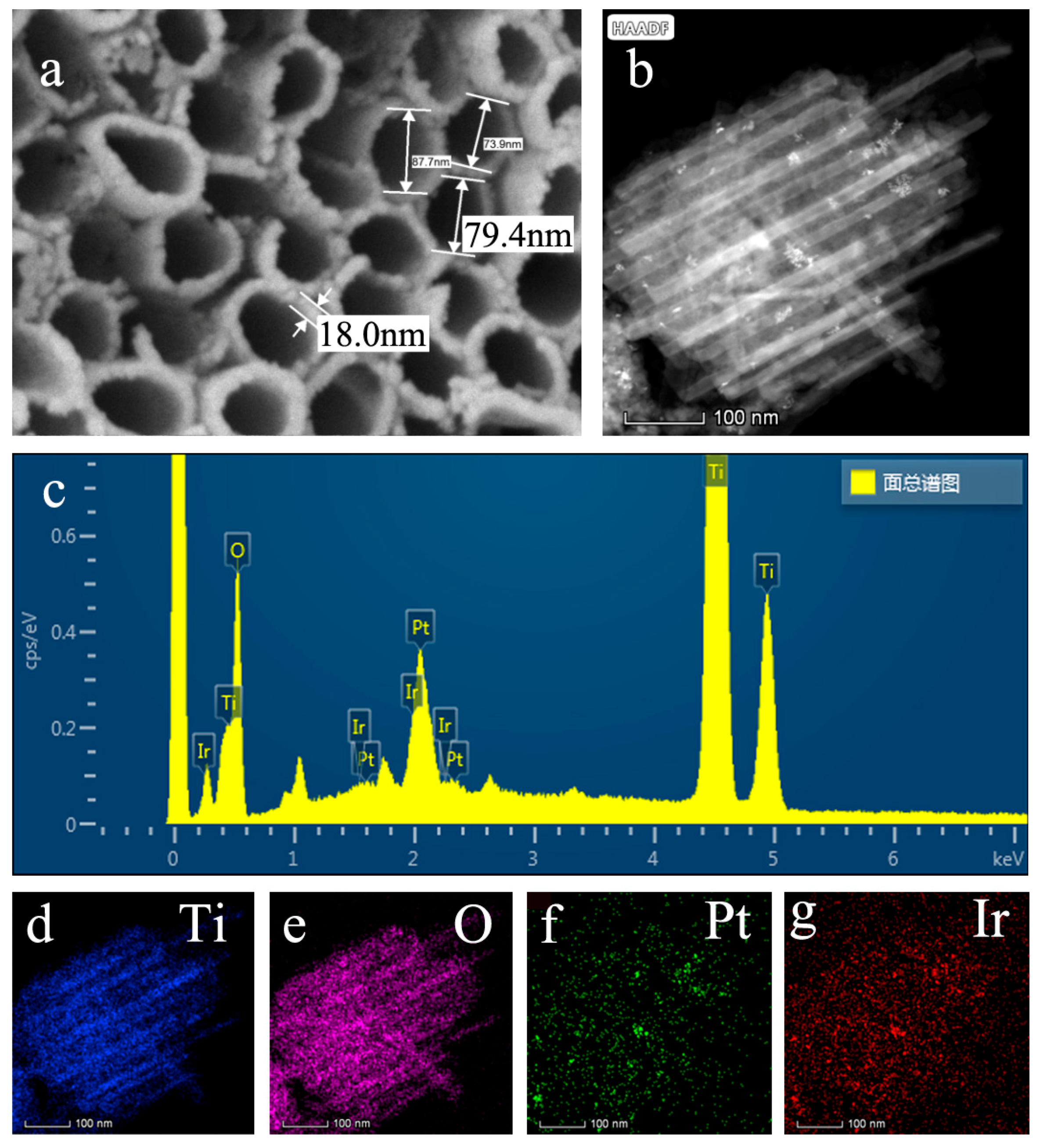
The morphology and microstructure of the as-prepared TiO2NTs/Ti was characterized by SEM and the Pt-IrO2/TiO2NTs/Ti was characterized by TEM. The top view SEM image (Figure 2a) shows that the bare TiO2 nanotubes were highly ordered, with an average diameter of 80 nm and wall thickness of 18 nm. It is also clearly observed that the tubes were open on the top.
Figure 2b shows the microstructure of TiO2NTs modified with Pt and IrO2. To confirm the presence of Pt and IrO2 on the TiO2NTs, EDS and elemental mapping were carried out. The EDS (Figure 2c) showed that atomic percentages of Pt and Ir in Pt-IrO2/TiO2NTs/Ti were 0.70% and 0.41%, respectively. The elemental mappings present the existence of Pt and IrO2 visually, as shown in Figure 2d–g. It can observe that the distributed Pt and Ir elements were homogeneous.
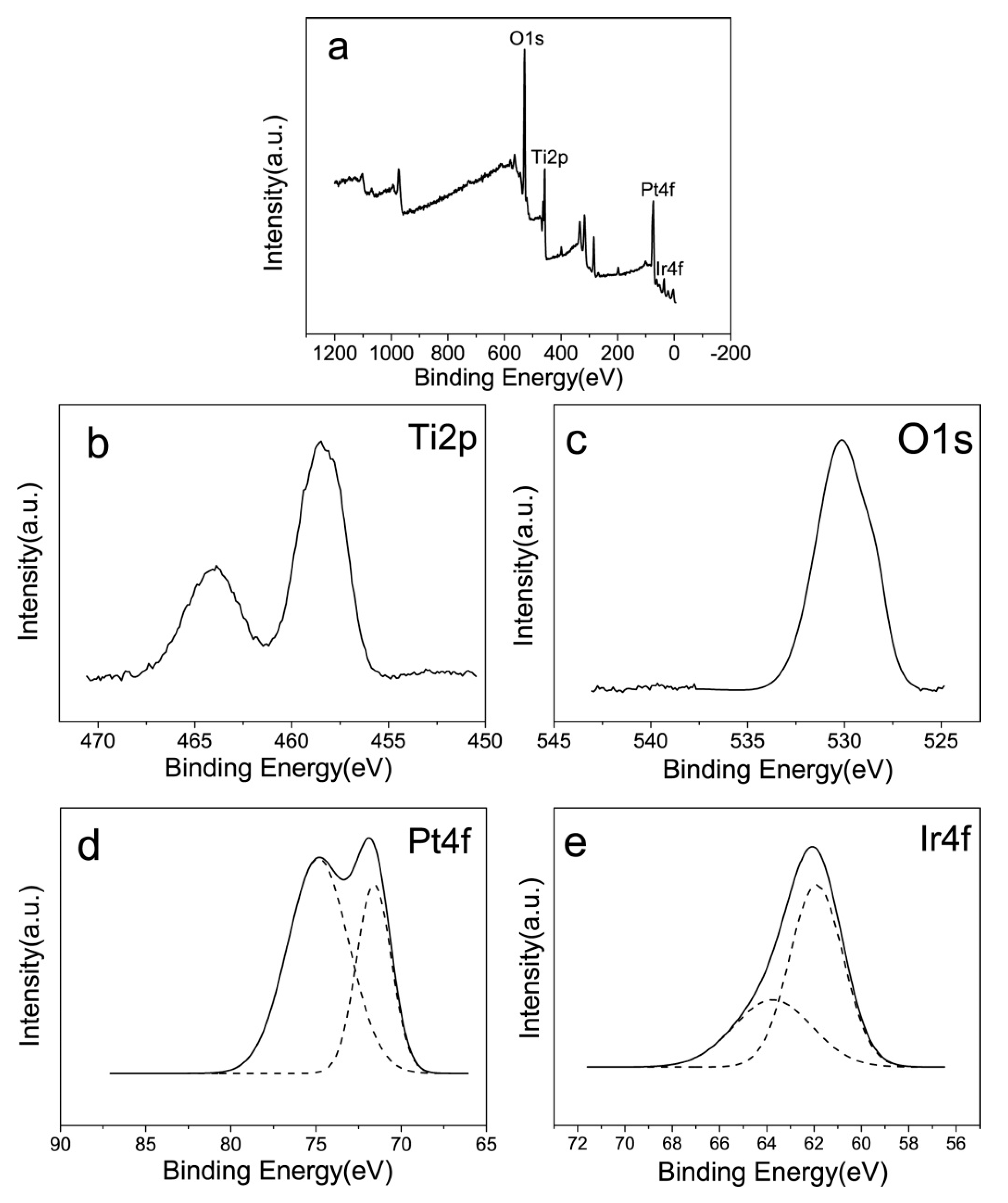
To further investigate the composition and elemental valences of the modified electrode, XPS analysis was performed. Figure 3 shows the XPS spectrum of Pt-IrO2/TiO2NTs/Ti, and corresponding high-resolution spectra of Ti2p, O1s, Pt4f, and Ir4f (Figure 3b–e). Figure 3a shows that the sample contained Ti, O, Pt and Ir, indicating successful deposition of Pt and IrO2 on the TiO2NTs, consistent with the EDS results. In Figure 3b, two peaks at BE of 464.0 eV and 458.5 eV in Pt-IrO2/TiO2NTs/Ti were assigned to Ti2p1/2 and Ti2p3/2 respectively, indicating the existence of Ti4+ in TiO2 [14,39,40]. In Figure 3c, the peak at BE of 529.5 eV was assigned to Ti-O [41].
As displayed in Figure 3d, the satellite peaks at 75.0 eV and 71.6 eV are strong evidence for Pt4f5/2 and Pt4f7/2, which confirms the formation of metallic Pt after the photoreduction deposition process [42,43]. After deconvolution, the high-resolution Ir4f (Figure 3d) showed the peak at 64.9 eV for Ir4f5/2 and 62.0 eV for Ir4f7/2, which is the characteristic of Ir4+ in IrO2 [41,44,45]. Consequently, Pt and IrO2 were successfully deposited on the TiO2NTs/Ti electrode, and Pt-IrO2 co-modified electrode was fabricated.

3.2. Photoelectrochemical Performance

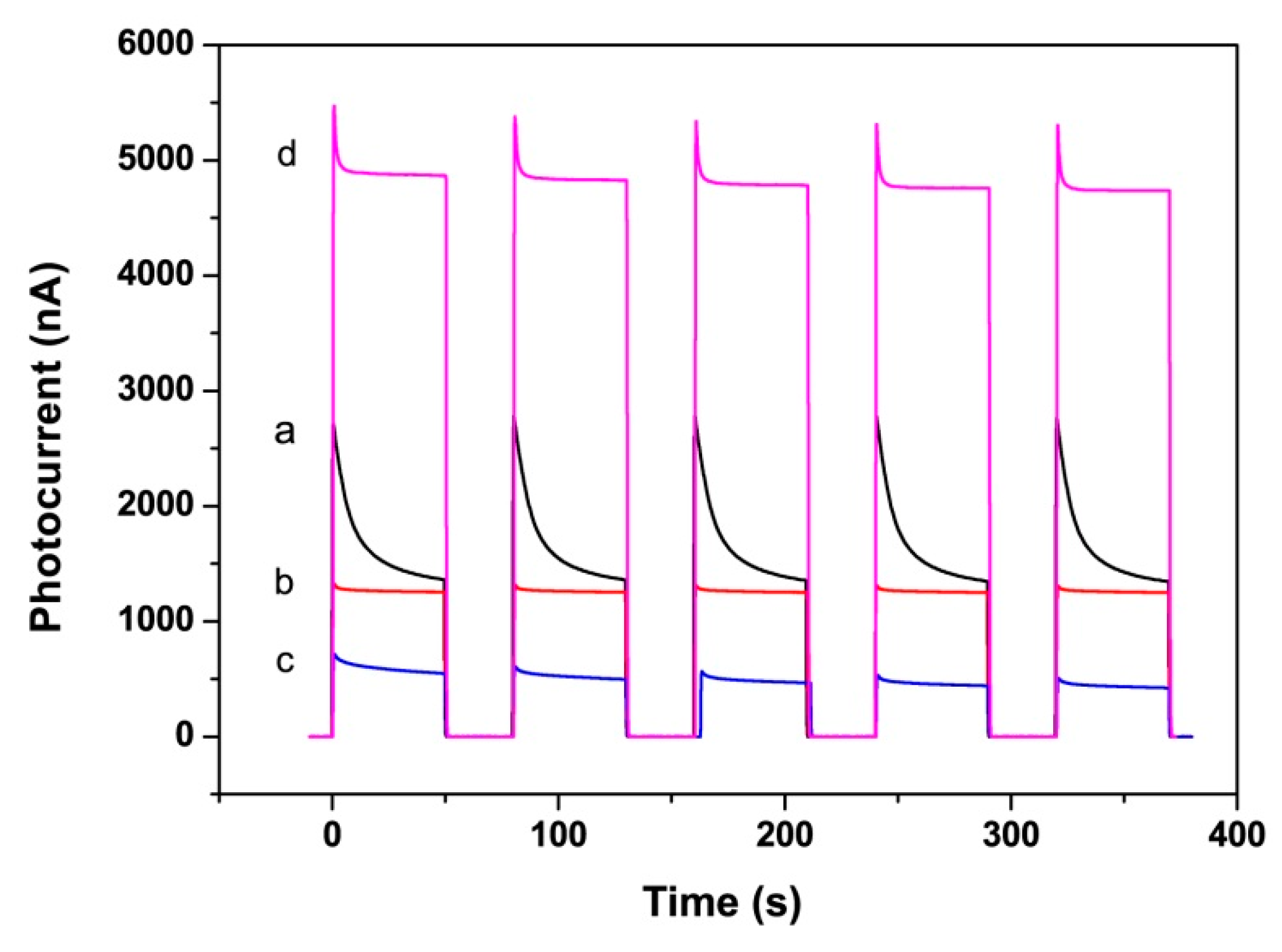
A photocurrent-time curve can be used to characterize the separation effect of photogenerated carriers, and the increase of photocurrent indicates the low recombination probability of photogenerated electron-holes. Figure 4 shows the photocurrent-time curves of different electrodes in 0.1 M PBS solution under 365 nm LED UV light irradiation. From Figure 4a, it can be seen that the photocurrent curve of the bare TiO2NTs electrode had an anodic photocurrent spike at the initial time of irradiation, and then continuously decreased until a constant current was reached. However, the decorated TiO2NTs electrodes could generate stable photocurrent more rapidly under the same condition, as shown in Figure 4b–d.
Under illumination, TiO2 could absorb UV light and generate electron-hole pairs, and electrons from the TiO2 electrode could transfer to Pt. So the surface of Pt nanoparticles appeared negatively charged and TiO2 appeared positively charged. The Schottky barrier prevents electrons from recombining with holes, and promotes the migration of charges, thus improves the stability of the photocurrent (Figure 4b). For IrO2/TiO2NTs/Ti, the photoinduced holes were taken by IrO2, effectively preventing the combination of electron-hole. So the photocurrent of IrO2/TiO2NTs/Ti electrode also could quickly stabilize, as shown in Figure 4c. In the Pt-IrO2 modified TiO2 system, due to the synergistic effect of Pt and IrO2, the recombination of photogenerated electrons and holes could be effectively blocked. The Pt-IrO2/TiO2NTs/Ti electrode in Figure 4d exhibited an apparent and stable photocurrent signal compared with the TiO2NTs/Ti.
However, the mono Pt- or IrO2-modified TiO2NTs/Ti exhibited lower photocurrent responses than bare TiO2NTs/Ti. The reason may be that Pt- and IrO2-loading on TiO2NTs shelter the TiO2NTs from the light illumination, which could reduce the efficiency of photogenerated carriers. Another reason may be that when Pt or IrO2 nanoparticles appeared alone on the TiO2, both photogenerated electrons and holes may transfer to Pt or IrO2 nanoparticles, making them act as recombination centers [46,47]. However, if Pt and IrO2 nanoparticles co-exist, electrons would move to Pt, while holes move to IrO2. The electron-hole recombination would be suppressed, leading to a higher photocurrent.

3.3. PEC biosensing Application for GSH

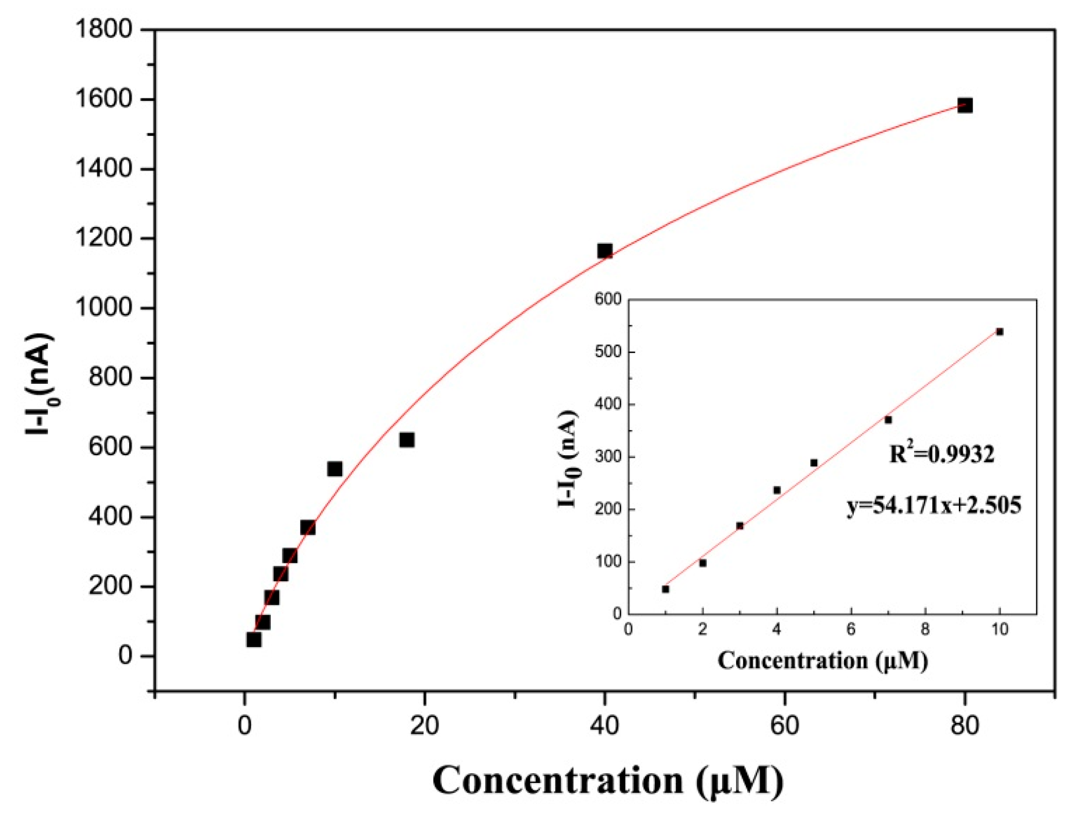
On the basis of photoelectrochemical performance, the application of the fabricated Pt-IrO2/TiO2NTs/Ti for detection of GSH was investigated. The electrode was kept in PBS at +0.3 V bias voltage under UV illumination for 5 min as pretreatment, and then the positive photocurrent was detected in the presence of GSH in PBS. As shown in Figure 5, the photocurrent increased with the addition of GSH under illumination, fitted with the Langmuir curve. The inset presented in Figure 5 shows that the photocurrent response of the Pt-IrO2/TiO2NTs/Ti biosensor was proportional to GSH concentration in the range of 1~10 μM with the regression equation of I-I0(μA) = 2.505 + 54.171CGSH(uM) (R2 = 0.9932) (I represents the photocurrent obtained in the presence of GSH, and I0 is the blank photocurrent). The detection limit (LOD) of the sensor can be obtained from the formula LOD = 3 Sb/S (Sb = standard deviation of blank signal, S = sensitivity), and the calculated value was 0.8 μM.
Scheme 2 illustrates the PEC process for GSH detection by Pt-IrO2/TiO2NTs/Ti biosensor. Under the illumination of UV light, electron-hole pairs were generated in TiO2. Due to the Schottky barrier at the Pt/TiO2 interface, electrons moved from TiO2 electrode to Pt. Simultaneously, the photoinduced holes were taken by IrO2, and then oxidized GSH to GSSG.
Table 1 summarizes the comparison of analytical performance of various GSH biosensors. The Pt-IrO2/TiO2NTs/Ti biosensor had a relatively reasonable linear range in 1-10 μM. Compared with other non-enzymatic sensors, Pt-IrO2/TiO2NTs/Ti was sensitive and could be used at relatively low GSH concentrations. Though enzymatic sensors offer high sensitivity, non-enzymatic sensors are widely recognized for their good advantages of simple operation, lack of need for expensive equipment, and high stability. The stability of the Pt-IrO2/TiO2NTs biosensor was tested by measuring photocurrent response after 30 days (see Supplementary Figure S1). The photocurrent did not decrease significantly, indicating that the electrode’s good stability for GSH detection. This should contribute to the good stability of Pt and IrO2 on TiO2NTs/Ti electrodes.
In summary, a PEC sensor for GSH was designed using Pt-IrO2/TiO2NTs/Ti as an electrode. Pt and IrO2 were evenly distributed on TiO2NTs/Ti. Due to the synergistic effects of Pt and IrO2, the Pt-IrO2/TiO2NTs/Ti electrode exhibited an apparent and stable photocurrent signal compared with the TiO2NTs/Ti or the mono-modified ones. The photocurrent signals of the Pt-IrO2/TiO2NT/Ti biosensor was linear to GSH concentration in the range of 1~10 μM. Other kinds of modified TiO2NTs/Ti electrodes which may exhibit high PEC sensitivity under visible light are anticipated.

Supplementary Materials

The following are available online at https://www.mdpi.com/2079-4991/10/3/522/s1, Figure S1: The curve of Pt-IrO2/TiO2NTs/Ti (after 30 days) for the detection of different concentrations for GSH.

Author Contributions

All authors have participated actively in the development of this work. J.T. and B.Z. conceived and designed the experiments; J.T. performed the experiments; J.T., Z.Y., S.Z. (Shoumin Zhang), W.H., and B.Z. analyzed the data; P.Z. contributed analysis tools; S.Z. (Shasha Zhang), G.H., and Z.S. contributed reagents; J.T. wrote the paper. All authors have read and agreed to the published version of the manuscript.

Funding

This research was funded by the National Natural Science Foundation of China (Nos. 61471207 and 61871240).

Conflicts of Interest

The authors declare no conflict of interest.

References

  1. Zhang, J.; Tu, L.; Zhao, S.; Liu, G.; Wang, Y.; Wang, Y.; Yue, Z. Fluorescent gold nanoclusters based photoelectrochemical sensors for detection of H2O2 and glucose. Biosens. Bioelectron. 2015, 67, 296–302. [Google Scholar] [CrossRef] [PubMed]
  2. Zhang, N.; Ruan, Y.F.; Zhang, L.B.; Zhao, W.W.; Xu, J.J.; Chen, H.Y. Nanochannels photoelectrochemical biosensor. Anal. Chem. 2018, 90, 2341–2347. [Google Scholar] [CrossRef] [PubMed]
  3. Yu, L.M.; Zhu, Y.C.; Liu, Y.L.; Qu, P.; Xu, M.T.; Shen, Q.; Zhao, W.W. Ferroelectric perovskite oxide@TiO2 nanorod heterostructures: Preparation, characterization, and application as a platform for photoelectrochemical bioanalysis. Anal. Chem. 2018, 90, 10803–10811. [Google Scholar] [CrossRef] [PubMed]
  4. Ruan, Y.F.; Zhang, N.; Zhu, Y.C.; Zhao, W.W.; Xu, J.J.; Chen, H.Y. Photoelectrochemical bioanalysis platform of gold nanoparticles equipped perovskite Bi4NbO8Cl. Anal. Chem. 2017, 89, 7869–7875. [Google Scholar] [CrossRef]
  5. Li, Y.; Zhang, N.; Zhao, W.W.; Jiang, D.C.; Xu, J.J.; Chen, H.Y. Polymer dots for photoelectrochemical bioanalysis. Anal. Chem. 2017, 89, 4945–4950. [Google Scholar] [CrossRef]
  6. Tu, L.; Liu, G.; Zhang, W.; Qin, J.; Yue, Z. Doped QDs based photoelectrochemical sensors for detection of H2O2 and Glucose. IEEE J. Sel. Top. Quantum. 2014, 20, 175–183. [Google Scholar] [CrossRef]
  7. Wu, S.; Song, H.; Song, J.; He, C.; Ni, J.; Zhao, Y.; Wang, X. Development of triphenylamine functional dye for selective photoelectrochemical sensing of cysteine. Anal. Chem. 2014, 86, 5922–5928. [Google Scholar] [CrossRef]
  8. Tee, S.Y.; Ye, E.; Pan, P.H.; Lee, C.J.; Hui, H.K.; Zhang, S.Y.; Koh, L.D.; Dong, Z.; Han, M.Y. Fabrication of bimetallic Cu/Au nanotubes and their sensitive, selective, reproducible and reusable electrochemical sensing of glucose. Nanoscale 2015, 7, 11190–11198. [Google Scholar] [CrossRef]
  9. Li, Y.; Zhang, X.; Jiang, S.; Dai, H.; Sun, X.; Li, Y. Improved photoelectrochemical property of a nanocomposite NiO/CdS@ZnO photoanode for water splitting. Sol. Energy Mater. Sol. Cells 2015, 132, 40–46. [Google Scholar] [CrossRef]
  10. Wang, T.; Jin, B.; Jiao, Z.; Lu, G.; Ye, J.; Bi, Y. Electric field-directed growth and photoelectrochemical properties of cross-linked Au-ZnO hetero-nanowire arrays. Chem. Commun. 2015, 51, 2103–2106. [Google Scholar] [CrossRef]
  11. Okoth, O.K.; Yan, K.; Feng, J.; Zhang, J. Label-free photoelectrochemical aptasensing of diclofenac based on gold nanoparticles and graphene-doped CdS. Sens. Actuators B 2018, 256, 334–341. [Google Scholar] [CrossRef]
  12. Zhang, J.; Wang, L.; Liu, X.; Li, X.A.; Huang, W. High-performance CdS–ZnS core–shell nanorod array photoelectrode for photoelectrochemical hydrogen generation. J. Mater. Chem. A 2015, 3, 535–541. [Google Scholar] [CrossRef]
  13. Zhu, J.; Huo, X.; Liu, X.; Ju, H. Gold nanoparticles deposited polyaniline-TiO2 nanotube for surface plasmon resonance enhanced photoelectrochemical biosensing. ACS Appl. Mater. Interfaces 2016, 8, 341–349. [Google Scholar] [CrossRef] [PubMed]
  14. Tao, J.; Gong, Z.; Yao, G.; Cheng, Y.; Zhang, M.; Lv, J.; Shi, S.; He, G.; Chen, X.; Sun, Z. Enhanced photocatalytic and photoelectrochemical properties of TiO2 nanorod arrays sensitized with CdS nanoplates. Ceram. Int. 2016, 42, 11716–11723. [Google Scholar] [CrossRef]
  15. Zhou, Y.; Shi, Y.; Wang, F.B.; Xia, X.H. Oriented self-assembled monolayer of Zn(II)-tetraphenylporphyrin on TiO2 electrode for photoelectrochemical analysis. Anal. Chem. 2019, 91, 2759–2767. [Google Scholar] [CrossRef]
  16. Tu, W.; Dong, Y.; Lei, J.; Ju, H. Low-potential photoelectrochemical biosensing using porphyrin-functionalized TiO2 nanoparticles. Anal. Chem. 2010, 82, 8711–8716. [Google Scholar] [CrossRef]
  17. Tang, J.; Kong, B.; Wang, Y.; Xu, M.; Wang, Y.; Wu, H.; Zheng, G. Photoelectrochemical detection of glutathione by IrO2-hemin-TiO2 nanowire arrays. Nano Lett. 2013, 13, 5350–5354. [Google Scholar] [CrossRef]
  18. Chen, D.; Zhang, H.; Li, X.; Li, J. Biofunctional titania nanotubes for visible-light-activated photoelectrochemical biosensing. Anal. Chem. 2010, 82, 2253–2261. [Google Scholar] [CrossRef]
  19. Singh, S.C.; Swarnkar, R.K.; Gopal, R. Synthesis of Titanium Dioxide Nanomaterial by Pulsed Laser Ablation in Water. J. Nanosci. Nanotechnol. 2009, 9, 5367–5371. [Google Scholar] [CrossRef]
  20. Prasad, K.; Bazaka, O.; Chua, M.; Rochford, M.; Fedrick, L.; Spoor, J.; Symes, R.; Tieppo, M.; Collins, C.; Cao, A.; et al. Metallic biomaterials: Current challenges and opportunities. Materials 2017, 10, 884. [Google Scholar] [CrossRef]
  21. Wang, L.; Han, J.; Feng, J.; Wang, X.; Su, D.; Hou, X.; Hou, J.; Liang, J.; Dou, S.X. Simultaneously efficient light absorption and charge transport of CdS/TiO2 nanotube array toward improved photoelectrochemical performance. Int. J. Hydrogen Energy 2019, 44, 30899–30909. [Google Scholar] [CrossRef]
  22. Subramanian, V.; Wolf, E.; Kamat, P.V. Semiconductor-Metal composite nanostructures. To what extent do metal nanoparticles improve the photocatalytic activity of TiO2 films? J. Phys. Chem. B 2001, 105, 11439–11446. [Google Scholar] [CrossRef]
  23. Zhang, Z.; Wang, Z.; Cao, S.-W.; Xue, C. Au/Pt nanoparticle-decorated TiO2 nanofibers with plasmon-enhanced photocatalytic activities for solar-to-fuel conversion. J. Phys. Chem. C 2013, 117, 25939–25947. [Google Scholar] [CrossRef]
  24. Chandrasekharan, N.; Kamat, P.V. Improving the photoelectrochemical performance of nanostructured TiO2 films by adsorption of gold nanoparticles. J. Phys. Chem. B 2000, 104, 10851–10857. [Google Scholar] [CrossRef]
  25. Akita, A.; Kobayashi, H.; Tada, H. Ultrathin Silicon Oxide film-induced enhancement of charge separation and transport of nanostructured Titanium(IV) Oxide photoelectrode. Chemphyschem 2019, 20, 2054–2059. [Google Scholar] [CrossRef]
  26. Wang, M.; Sun, L.; Lin, Z.; Cai, J.; Xie, K.; Lin, C. p–n Heterojunction photoelectrodes composed of Cu2O-loaded TiO2 nanotube arrays with enhanced photoelectrochemical and photoelectrocatalytic activities. Energy Environ. Sci. 2013, 6, 1211–1220. [Google Scholar] [CrossRef]
  27. Ji, L.; Spanu, D.; Denisov, N.; Recchia, S.; Schmuki, P.; Altomare, M. A dewetted-dealloyed nanoporous Pt co-catalyst formed on TiO2 nanotube arrays leads to strongly enhanced photocatalytic H2 production. Chem. Asian J. 2019. [Google Scholar] [CrossRef] [Green Version]
  28. Linsebigler, A.L.; Lu, G.; Yates, J.T. Photocatalysis on TiO2 surfaces: Principles, mechanisms, and selected results. Chem. Rev. 1995, 95, 735–758. [Google Scholar] [CrossRef]
  29. Li, X.Z.; Li, F.B. Study of Au/Au3+-TiO2 photocatalysts toward visible photooxidation for water and waste water treatment. Environ. Sci. Technol. 2001, 35, 2381–2387. [Google Scholar] [CrossRef]
  30. Guo, Y.; He, J.; Wu, S.; Wang, T.; Li, G.; Hu, Y.; Xue, H.; Sun, X.; Tang, J.; Liu, M. Effects of platinum on photo-assisted electrocatalytic activity of fringe-shaped highly ordered mesoporous titanium dioxide film. J. Power Sources 2012, 208, 58–66. [Google Scholar] [CrossRef]
  31. Meekins, B.H.; Kamat, P.V. Role of water oxidation catalyst IrO2 in shuttling photogenerated holes across TiO2 interface. J. Phys. Chem. Lett. 2011, 2, 2304–2310. [Google Scholar] [CrossRef]
  32. Kamat, P.V. Manipulation of charge transfer across semiconductor interface. A criterion that cannot be ignored in photocatalyst design. J. Phys. Chem. Lett. 2012, 3, 663–672. [Google Scholar] [CrossRef] [PubMed]
  33. Wang, D.; Wang, W.; Wang, Q.; Guo, Z.; Yuan, W. Spatial separation of Pt and IrO2 cocatalysts on SiC surface for enhanced photocatalysis. Mater. Lett. 2017, 201, 114–117. [Google Scholar] [CrossRef]
  34. Li, X.; Xu, Y.; Chen, Y.; Wang, C.; Jiang, J.; Dong, J.; Yan, H.; Du, X. Dual enhanced electrochemiluminescence of aminated Au@SiO2/CdS quantum dot superstructures: Electromagnetic field enhancement and chemical enhancement. ACS Appl. Mater. Interfaces 2019, 11, 4488–4499. [Google Scholar] [CrossRef] [PubMed]
  35. Sui, M.; Zhao, Y.; Ni, Z.; Gu, X. Photoelectrochemical performance and biosensor application for glutathione (GSH) of W-doped BiVO4 thin films. J. Mater. Sci. Mater. Electron 2018, 29, 10109–10116. [Google Scholar] [CrossRef]
  36. Li, Z.; Zhang, J.; Li, Y.; Zhao, S.; Zhang, P.; Zhang, Y.; Bi, J.; Liu, G.; Yue, Z. Carbon dots based photoelectrochemical sensors for ultrasensitive detection of glutathione and its applications in probing of myocardial infarction. Biosens. Bioelectron. 2018, 99, 251–258. [Google Scholar] [CrossRef]
  37. Prakasam, H.E.; Shankar, K.; Paulose, M.; Varghese, O.K.; Grimes, C.A. A new benchmark for TiO2 nanotube array growth by anodization. J. Phys. Chem. C 2007, 111, 7235–7241. [Google Scholar] [CrossRef]
  38. Li, W.; Kuang, D.; Gu, P.; Xiang, M. Partially disordered TiO2 nanotube photonic crystal: Randomness characterization and tuning of reflected scattering light. Nanotechnology 2020, 31, 025711. [Google Scholar] [CrossRef]
  39. Zhou, H.; Zhang, Y. Electrochemically self-doped TiO2 nanotube arrays for supercapacitors. J. Phys. Chem. C 2014, 118, 5626–5636. [Google Scholar] [CrossRef]
  40. Zhang, R.; Ren, X.; Shi, X.; Xie, F.; Zheng, B.; Guo, X.; Sun, X. Enabling Effective Electrocatalytic N2 Conversion to NH3 by the TiO2 Nanosheets Array under Ambient Conditions. ACS Appl. Mater. Interfaces 2018, 10, 28251–28255. [Google Scholar] [CrossRef]
  41. Abb, M.J.S.; Weber, T.; Glatthaar, L.; Over, H. Growth of ultrathin single-crystalline IrO2(110) films on a TiO2(110) single crystal. Langmuir 2019, 35, 7720–7726. [Google Scholar] [CrossRef] [PubMed]
  42. Vovk, E.I.; Kalinkin, A.V.; Smirnov, M.Y.; Klembovskii, I.O.; Bukhtiyarov, V.I. XPS study of stability and reactivity of oxidized Pt nanoparticles supported on TiO2. J. Phys. Chem. C 2017, 121, 17297–17304. [Google Scholar] [CrossRef]
  43. Liang, Y.; Ding, M.; Yang, Y.; Xu, K.; Luo, X.; Yu, T.; Zhang, W.; Liu, W.; Yuan, C. Highly dispersed Pt nanoparticles on hierarchical titania nanoflowers with {010} facets for gas sensing and photocatalysis. J. Mater. Sci. 2019, 54, 6826–6840. [Google Scholar] [CrossRef]
  44. Chung, W.-H.; Tsai, D.-S.; Fan, L.-J.; Yang, Y.-W.; Huang, Y.-S. Surface oxides of Ir(111) prepared by gas-phase oxygen atoms. Surf. Sci. 2012, 606, 1965–1971. [Google Scholar] [CrossRef]
  45. Chakrapani, K.; Sampath, S. The dual role of borohydride depending on reaction temperature: Synthesis of iridium and iridium oxide. Chem. Commun. 2015, 51, 9690–9693. [Google Scholar] [CrossRef]
  46. Wang, W.N.; An, W.J.; Ramalingam, B.; Mukherjee, S.; Niedzwiedzki, D.M.; Gangopadhyay, S.; Biswas, P. Size and structure matter: Enhanced CO2 photoreduction efficiency by size-resolved ultrafine Pt nanoparticles on TiO2 single crystals. J. Am. Chem. Soc. 2012, 134, 11276–11281. [Google Scholar] [CrossRef]
  47. Swierk, J.R.; McCool, N.S.; Saunders, T.P.; Barber, G.D.; Strayer, M.E.; Vargas-Barbosa, N.M.; Mallouk, T.E. Photovoltage Effects of Sintered IrO2 Nanoparticle Catalysts in Water-Splitting Dye-Sensitized Photoelectrochemical Cells. J. Phys. Chem. C 2014, 118, 17046–17053. [Google Scholar] [CrossRef]
  48. Li, J.; Li, H.; Xue, Y.; Fang, H.; Wang, W. Facile electrodeposition of environment-friendly Cu2O/ZnO heterojunction for robust photoelectrochemical biosensing. Sens. Actuators B 2014, 191, 619–624. [Google Scholar] [CrossRef]
  49. Zhao, X.; Zhou, S.; Shen, Q.; Jiang, L.P.; Zhu, J.J. Fabrication of glutathione photoelectrochemical biosensor using graphene-CdS nanocomposites. Analyst 2012, 137, 3697–3703. [Google Scholar] [CrossRef] [PubMed]
  50. Kang, Z.; Gu, Y.; Yan, X.; Bai, Z.; Liu, Y.; Liu, S.; Zhang, X.; Zhang, Z.; Zhang, X.; Zhang, Y. Enhanced photoelectrochemical property of ZnO nanorods array synthesized on reduced graphene oxide for self-powered biosensing application. Biosens. Bioelectron. 2015, 64, 499–504. [Google Scholar] [CrossRef] [PubMed]
Scheme 1. Schematic representation of preparation process of Pt-IrO2/TiO2NTs/Ti electrode.
Nanomaterials 10 00522 sch001
Figure 1. XRD patterns of (a) Ti foil; (b) TiO2NTs/Ti; (c) Pt/TiO2NTs/Ti; (d) IrO2/TiO2NTs/Ti; (e) Pt-IrO2/TiO2NTs/Ti.
Nanomaterials 10 00522 g001
Figure 2. (a) Top-view SEM of TiO2NTs; (b) The TEM image of Pt-IrO2/TiO2NTs/Ti; (c) The EDS of Pt-IrO2/TiO2NTs/Ti; (dg) The EDS mappings of Pt-IrO2/TiO2NTs/Ti.
Nanomaterials 10 00522 g002
Figure 3. (a) XPS spectra of Pt-IrO2/TiO2NTs/Ti, (b) Ti2p, (c) O1s, (d) Pt4f; (e) Ir4f.
Nanomaterials 10 00522 g003
Figure 4. Photocurrent responses of different electrodes: (a) TiO2NTs/Ti, (b) Pt/TiO2NTs/Ti, (c) IrO2/TiO2NTs/Ti, (d) Pt-IrO2/TiO2NTs/Ti electrodes in 0.1M PBS solution under 365 nm LED irradiation.
Nanomaterials 10 00522 g004
Scheme 2. Schematic illustration of the PEC process for GSH detection by Pt-IrO2/TiO2NTs/Ti biosensor.
Nanomaterials 10 00522 sch002
Figure 5. The curve of Pt-IrO2/TiO2NTs/Ti for the detection of different concentrations for GSH.
Nanomaterials 10 00522 g005
Table 1. Comparison of various GSH sensors.
GSH BiosensorLinear Range (μM)Detection Limit (μM)Ref.
Non-enzymatic sensorPt-IrO2/TiO2NTs/Ti1–100.8This work
Cu2O/ZnO 1–10 and 20–1000.8[48]
GR-CdS/ITO 10–15003[49]
rGO/ZnO10–2002.17[50]
Porphyrin-Functionalized TiO2-ITO50–240030[16]
Enzymatic sensorIrO2-Hemin-TiO2 nanowire arrays0.01–100.01[17]