Article

Antimicrobial Effect of Fluoride-Generating Nonthermal Atmospheric Plasmas on In Vitro Triple-Species Oral Biofilms


1
Department of Internal Medicine, University of Missouri, Columbia, MO 65211, USA
2
Department of Public Health Sciences, Texas A&M College of Dentistry, Dallas, TX 75246, USA
3
Department of Mechanical and Aerospace Engineering, University of Missouri, Columbia, MO 65211, USA
*
Authors to whom correspondence should be addressed.
J. Funct. Biomater. 2026, 17(9), 477; https://doi.org/10.3390/jfb17090477 (registering DOI)
Submission received: 15 August 2026 / Revised: 8 September 2026 / Accepted: 15 September 2026 / Published: 19 September 2026
(This article belongs to the Section Dental Biomaterials)

Abstract

This study investigates the feasibility of fluoride delivery via fluoride-generating nonthermal atmospheric plasma (FNTAP) and its antimicrobial effects on in vitro triple-species oral biofilms. FNTAPs were generated by adding 5 or 10 sccm (standard cubic centimeters per min) 1,1,1,2-tetrafluoroethane (TFE) to atmospheric argon (3000 sccm) nonthermal plasma (ArNTAP). Reactive plasma species were measured in plasma-treated phosphate-buffered saline (PBS). Twenty-four-hour biofilms were constructed from Streptococcus mutans, Streptococcus sanguinis, and Candida albicans for assessing the antimicrobial effects of FNTAPs via viability assays and colony-forming unit (CFU) assays. FNTAP treatments significantly increased the concentrations of F and NO2 in PBS compared to ArNTAP treatments (p < 0.01), and decreased the concentrations of H2O2 and NO3 in PBS (p < 0.001, p < 0.05). 5 sccm 2 min FNTAP treatment reduced biofilm viability by 94.12 ± 1.03% (p < 0.001), compared to a 69.32 ± 2.05% (p < 0.001) reduction with ArNTAP. CFU assays showed that FNTAPs were especially efficacious against C. albicans. 5 sccm 3 min FNTAP treatment reduced fungal log10 CFUs by 4.28 ± 0.36 (p < 0.001). Fungal CFUs were below detectable levels for the 10 sccm 2 and 3 min groups, except for two data points in each group. In conclusion, FNTAP effectively produced measurable fluoride in PBS and significantly enhanced antimicrobial efficacy against triple-species biofilms, highlighting its potential as a multifunctional tool for caries control.

1. Introduction

Dental caries remains one of the most prevalent chronic diseases globally, affecting billions of people, despite being largely preventable [1,2]. Cariogenic bacteria (e.g., Streptococcus mutans) form dental plaque biofilms on tooth surfaces, producing acids that demineralize tooth hard tissues, eventually leading to dental caries. S. mutans is considered the major etiological agent disrupting enamel mineral equilibrium and has consistently shown a correlation with cases of active caries [3]. The dysbiosis of supragingival plaque with overrepresentation of S. mutans is considered an environment conducive to the formation of dental caries [4]. Streptococcus sanguinis is an early colonizer of the oral cavity, contributing to the development of complex microbial communities on dental surfaces. Dental biofilms are formed by initial bacterial colonization from 0 to 18 h by aerobic bacteria such as S. sanguinis and other mitis group streptococci. S. sanguinis has been consistently recovered from individuals with high incidences of dental caries [5]. This biofilm matures from 18 h to 4 days with the addition of co-aggregator bacteria such as S. mutans. Obligate anaerobes invade the gum margins from 4 to 7 days, with gradual hardening of the extracellular polymeric substance (EPS) [6]. The extracellular matrix in these oral biofilms enhances the resistance of encased bacteria to both host defenses and antimicrobial agents [7]. This resilience of established dental biofilms necessitates frequent mechanical removal to manage the plaque. However, the anatomy of human dentition limits the efficacy of daily brushing at home in removing dental plaque. Even with diligent oral hygiene and professional cleanings, bacteria can rapidly recolonize and restore the biomass of biofilms within days, underscoring the difficulty in maintaining long-term biofilm control with conventional means alone [7]. Oral biofilms are linked to cardiovascular disease through direct effects of oral bacteria and inflammatory responses to oral disease [8]. S. sanguinis has shown the potential to impair clotting processes, further solidifying the connection between oral biofilms and systemic disease [9]. Thus, maintaining oral health and preventing dental caries are important public health concerns [10].
Candida species are the most prevalent fungal genera found in the oral cavity [11]. Specifically, C. albicans may transition from being a commensal to a pathogen in multi-species oral biofilms [12]. Candida albicans has been frequently detected in the oral cavity of children with severe early childhood caries [12]. As an opportunistic fungal organism, Candida albicans, in conjunction with S. gordonii, S. oralis, and S. sanguinis, enhances bacterial colonization and biofilm formation [13,14]. S. mutans interacts with C. albicans via cell surface glucans, while mitis group streptococci such as S. sanguinis bind to C. albicans hyphae via cell-to-cell interactions to form cariogenic cross-kingdom biofilms [15]. C. albicans is also known to cause denture stomatitis and is fairly resistant to routine oral hygiene cleaning regimens [16]. In addition to the above-mentioned conditions, interactions between oral bacteria and C. albicans are also associated with endodontic infections, periodontitis, peri-implantitis and oral cancer [17]. High levels of sucrose in the oral cavity enhance the physical interaction between C. albicans and S. mutans, increasing the microbial burden and production of extracellular matrix. In vitro cospecies biofilms with S. mutans and C. albicans are known to have a higher biomass than single-species in vitro S. mutans biofilms. S. mutans and C. albicans are also known to synergize the virulence of plaque biofilms in vivo [18]. Novel technologies that target C. albicans and its cross-kingdom interactions with bacteria are necessary to control the worldwide ubiquitous prevalence of deleterious multi-species oral biofilms.
Fluoride has been the cornerstone of caries prevention and management for decades. Fluoride releasing bioactive restorative materials have been developed for use in pediatric and elderly patients [19,20]. The primary mode of action for fluoride is the inhibition of demineralization and the enhancement of remineralization of early lesions. Tooth enamel is largely composed of hydroxyapatite, which is prone to dissolution by pH fluctuations in the oral cavity. The presence of fluoride ions leads to the formation of fluorohydroxyapatite, which is further converted to fluorapatite [21]. Fluorapatite is resistant to dissolution by pH changes. However, the presence of thick plaque can limit the contact of dental enamel with fluoride. Moreover, the direct antimicrobial effect of fluoride delivered by current commercial products is limited in terms of managing oral bacteria, and is considered secondary [22]. At typical-use concentrations (e.g., 1000–1500 ppm in toothpaste), fluoride only modestly inhibits cariogenic bacteria via a short-lived suppression of bacterial growth [22,23]. Studies have demonstrated that S. mutans biofilms can recover their acid production shortly after brief fluoride exposure [23]. Despite extensive use of fluoride globally, a plateau in dental caries reduction in various populations has been observed for many years. There is an urgent need for innovative strategies that prevent enamel demineralization and attenuate the pathogenicity of oral biofilms.
Nonthermal atmospheric plasma (NTAP) is a novel, non-invasive modality for targeted biofilm disruption. NTAP is an ionized gas containing a rich mixture of reactive species such as reactive oxygen and nitrogen species (ROS and RNS), free radicals, and charged particles at or near room temperature. These reactive species can penetrate biofilm structures and produce rapid antimicrobial effects without causing adverse effects on skin and soft tissues [24]. Research has demonstrated the strong antimicrobial and antibiofilm effects of NTAP on oral pathogens in planktonic form or single/dual-species biofilm models [25,26,27,28,29]. In addition, NTAP can also enhance the tooth’s resistance to decay by modifying enamel surfaces, increasing resistance to demineralization, and improving fluoride uptake [30]. Recently, our research group reported for the first time that fluoride-generating nonthermal plasma (FNTAP) had superior efficacy against in vitro dual-species oral biofilms as compared to argon NTAP or fluoride alone [31].
Nevertheless, the human oral cavity houses more than 700 species of bacteria and fungi [32]. Dental caries is a polymicrobial biofilm-induced infectious disease. However, our previous publications used single-species oral biofilms or dual-species oral biofilms in the evaluation of technologies developed for managing/treating dental caries. Multi-species oral biofilms have been reported to be more resistant to treatment [33]. Therefore, there is a need to further evaluate the potency of our novel FNTAP against more complex dental biofilms.
In the present study, triple-species biofilms were constructed from pathogens that have been found to be clinically relevant in cariogenic oral biofilms: S. mutans, S. sanguinis, and C. albicans [12,18,34] and were used to further evaluate the effects of FNTAP from 1,1,1,2-tetrafluoroethane (TFE). The generation of various reactive species, including NO2, NO3, H2O2 and fluoride, was also assessed for the first time for this novel FNTAP to provide preliminary insight into the underlying mechanisms. We hypothesized that NTAP treatment would show a significant reduction in bacterial and fungal CFUs in triple-species biofilms as compared to control, similar to the results seen in our previous study with in vitro dual-species biofilms [31]. The null hypothesis was that there is no significant difference between FNTAP treatment groups and control groups.

2. Materials and Methods

2.1. Nonthermal Atmospheric Plasma Devices and Settings

The nonthermal atmospheric plasma devices used in this study have been described in our previous publications [35]. The details on the operation of Ar plasma and FNTAP used in this study have been published [31]. Briefly, the plasma brush consists of a gas chamber with a converging nozzle at one end. In the nozzle, two tungsten needles served as cathode and the ground electrode. Argon gas at a flow rate of 3000 standard cubic centimeters per minute (sccm) (Industry Grade, Airgas, Radnor Township, PA, USA) flowed through the gas chamber and served as the carrier gas for generating NTAPs. Spellman HV power supply SL60 (Spellman, New York, NY, USA) was used as the electrical energy source in a direct current mode setting at a value of 6.8 mA to ignite NTAPs, which was blown out of the nozzle by Ar gas flow to form a brush shape NTAP plume. 1,1,1,2-tetrafluoroethane (TFE) vapor was added to the Ar NTAP at 5 sccm or 10 sccm flow rates to generate a brush shape of FNTAP plume with dimensions of ~6.0 mm in length, ~4.0 mm in width, and ~1.0 mm in thickness. Two separate mass flow controllers (MKS Instruments Inc., Andover, MA, USA) were used for controlling the flow rates of Ar gas and TFE vapor. The plasma brush tip temperature was maintained as close to the human body temperature of ~37.0 °C as possible and measured using a grounded thermocouple (TJ36-CPSS-116G-3-SMPW-M, OMEGA Engineering Inc., Norwalk, CT 06854, USA) prior to data collection. For plasma treatment, the FNTAP plume tip directly touched the biofilm surfaces.

2.2. Assessment of Reactive Plasma Species

One and a half milliliters of phosphate-buffered saline (PBS) were added into the well of 24-well plates and exposed to Ar NTAP or FNTAPs for 2 or 3 min. This experiment was repeated three times on the same day. This process was repeated two more times on two different days. Therefore, the total sample size was 9 (independent) samples for each plasma treatment. The resulting solutions were used for assessing reactive plasma species, including F, NO2, NO3 and H2O2 generated by the plasmas. An aliquot of the desired volume was sampled from each prior plasma-treated PBS solution and used for the analysis of each reactive plasma species after each plasma treatment process (n = 9, independent) per treatment group, except for the analysis of total nitrate/nitrite and NO3 concentrations for the Ar NTAP 2 min group which had a sample size of 8 due to a missing well during analysis. The analytical methods used in the present study were well developed and have been used in numerous studies reported in the literature [36,37,38,39,40].
A fluoride ion selective electrode (Orion™ 9609BNWP, Thermo Fisher Scientific, Waltham, MA, USA) [38] was used to measure the fluoride concentrations in the plasma-treated PBS and untreated PBS samples. The baseline fluoride level in untreated PBS was analyzed at one time with nine replicates (n = 9). Standard calibration solutions were prepared by sequentially diluting a 100 mM fluoride standard into 10, 5, 2, 1, 0.1, and 0.05 mM F standard calibration solutions using Millipore water. Analysis started by mixing 810 µL of sample solution or standard calibration solution with 90 µL of fluoride total ionic strength adjustment buffer III (Fisher Scientific, Waltham, MA, USA) in a 24-well plate. The fluoride electrode was dipped into the mixed solution, and the millivoltage was read after the indicator of “Ready” was displayed on the pH/mV meter screen. The millivoltage was read one or two more times by pressing reading “Measurement” button on the pH/mV meter. The fluoride concentration for each sample was determined by averaging two or three voltage readings recorded for each sample against the respective standard curve. The R2 values for the standard curve fitting were above 0.99. It should be noted that the millivoltage readings for untreated PBS and Ar NTAP-treated PBS samples fell outside of the calibration curve. Therefore, their calculated fluoride concentrations were based on extrapolation from the standard curve and were consequently overestimated relative to their actual F concentrations.
For the analysis of H2O2 concentrations using a commercially available colorimetric assay kit for H2O2 (OxiSelect™ Hydrogen Peroxide/Peroxidase Assay Kit, Cell Biolabs Inc., San Diego, CA, USA) [39,40], the standard H2O2 solution in the kit was used to prepare 50, 25, 12.5, 6.25, 3.125, 1.5625, 0.78125, and 0 µM H2O2 standard calibration solutions via sequential dilution using the assay buffer according to the manufacturer’s instructions. The plasma-treated PBS samples and H2O2 standard calibration solutions were processed according to the assay protocol for H2O2 specified in the kit manual. After incubation for 30 min, the absorbance at 560 nm was read using a microplate reader. In addition, the baseline level in untreated PBS was analyzed in a single process with nine independent replicates (n = 9) for H2O2. The R2 values for the standard curve fitting were above 0.99. The concentration of H2O2 for each sample was determined using its 560 nm absorbance against the respective standard curve.
For the analysis of NO2 concentrations using a commercially available colorimetric assay kit for NO2 (Griess Reagent Kit, Invitrogen™, Waltham, MA, USA) [36], the standard nitrite solution in the kit was used to prepare 50, 25, 12.5, 6.25, 3.125, 1.5625, 0.78125, and 0 µM NO2 standard calibration solutions via sequential dilution using Millipore water (MilliporeSigma, Burlington, MA, USA) according to the manufacturer’s instructions. Microplate assays were performed for the plasma-treated PBS samples and NO2 standard calibration solutions in 96-well microplates according to the assay protocol specified in the kit manual, except that the amount of each agent/sample/standard calibration solution was reduced to half of the amount specified in the kit manual to allow for processing more samples and preserving the plasma-treated samples for completion of all four analyses. After incubation for 30 min, the 96-well plates were read using a microplate reader at 548 nm. In addition, the baseline level in untreated PBS was analyzed in a single process with nine independent replicates (n = 9) for NO2. The R2 values for the standard curve fitting were above 0.99. The concentration of NO2 for each sample was determined using its 548 nm absorbance against the respective standard curve.
For the analysis of the total nitrate/nitrite using a commercially available colorimetric assay kit for the total nitrate/nitrite (Nitrate/Nitrite Colorimetric Assay Kit, Cayman Chemical, Ann Arbor, MI, USA) [36], the nitrate/nitrite assay buffer was diluted using 98 mL of Millipore water (MilliporeSigma, Burlington, MA, USA) to prepare 1× nitrate/nitrite assay buffer. The nitrate standard, nitrate reductase enzyme preparation and nitrate reductase cofactor preparation were dissolved using 1× assay buffer according to the kit manual. The nitrate reductase enzyme solution and nitrate reductase cofactor solution were split into four 2 mL microtubes with 300 µL per microtube. The prepared solutions were stored at low temperatures according to the kit manual. The nitrate standard solution prepared by dissolving the nitrate standard using 1× assay buffer was used to prepare 87.5, 75, 62.5, 50, 37.5, 25, 12.5, and 0 µM NO3 standard calibration solutions via sequential dilution using 1× assay buffer according to the kit manual. Assays were performed for the plasma-treated PBS samples and NO3 standard calibration solutions in 96-well microplates according to the assay protocol for determination of total nitrate/nitrite specified in the kit manual, except that the amount of each agent/sample/standard calibration solution was reduced to half of the amount specified in the kit manual to allow for processing more samples and preserving the plasma-treated samples for completion of all four analyses. After the final incubation for 10 min, the 96-well plates were read using a microplate reader at 548 nm. In addition, the baseline level in untreated PBS was analyzed in a single process with eight independent replicates (n = 8) for total nitrate/nitrite. The R2 values for the standard curve fitting were above 0.99. The total nitrate/nitrite concentration for each sample was determined using its 548 nm absorbance against the respective standard curve.
The NO3 concentration in each sample was calculated by subtracting the NO2 concentration from the total nitrate/nitrite concentration found in the same sample. Baseline levels of untreated PBS were subtracted to calculate the fluoride, H2O2, nitrite, total nitrate/nitrite and nitrate concentrations for plasma-treated PBS. It should be noted that the concentrations for fluoride (detection limit 0.02 ppm), H2O2 (detection limit 0.8 µM), NO2 (detection limit 1.0 µM) and total nitrate/nitrite (detection limit 0.70 µM) in untreated PBS samples were below the detection limits of the corresponding methods.

2.3. Biofilm Culture

Single colonies of S. mutans (ATCC 700610, ATCC, Manassas, VA, USA) and S. sanguinis (ATCC 10556, ATCC, Manassas, VA, USA) were cultured separately in brain heart infusion (BHI) broth (Sigma-Aldrich, St. Louis, MO, USA) at 37 °C with 5% CO2 overnight. A single colony of C. albicans (BEI Resources, NIAID, NIH: Candida albicans, Strain P37005, NR-29447, Bethesda, MD, USA) was cultured in Sabouraud dextrose liquid medium (SDB) (Oxoid, Basingstoke, Hampshire, UK) at 37 °C under 5% CO2 overnight. Optical density at 600 nm (OD600) was measured for the resulting bacterial and fungal suspensions. Subsequently, OD600 for each suspension was separately adjusted to 0.1 using Tryptic Soy Broth (Sigma-Aldrich, St. Louis, MO, USA) supplemented with 0.6% Yeast Extract (TSYEB) (BD, East Rutherford, NJ, USA). The approximate CFUs at 0.1 OD600 were 108/mL, 107/mL and 106/mL for S. mutans, S. sanguinis and C. albicans, respectively. The three resulting microbial suspensions were mixed in equal volumes to form a triple-species bacto–fungal seeding suspension, which was supplemented with 0.5% sucrose. The addition of sucrose to growth media is an established caries-related in vitro microcosm biofilm model [41,42]. The addition of 0.5% sucrose was reported to be within the range for maximum biofilm adhesion and biomass formation for S. mutans biofilms [43]. Wells in 24-well plates were coated with artificial saliva (1700–0305 Artificial Saliva-medical & dental research, Stabilized; Pickering Laboratories, Mountain View, CA, USA) before use as reported [31]. Briefly, 300 µL of artificial saliva was added to each well of the 24-well plate and the plate was incubated at 4 °C overnight. The artificial saliva was removed from the wells after incubation and the triple-species seeding suspension was added to each coated well at 1 mL per well. These microplates were incubated at 37 °C with 5% CO2 for 24 h to form in vitro triple-species biofilms. The resulting in vitro biofilms were washed twice using 500 µL of Dulbecco’s phosphate-buffered saline (DPBS) (Thermo Fisher, Waltham, MA, USA) for 3 min each on a benchtop shaker before receiving treatment.

2.4. Destruction of In Vitro Triple-Species Biofilms by FNTAP

The washed in vitro triple-species biofilms were assigned to 7 groups and treated according to the group assignment: negative control group receiving no plasma treatment (no-treatment group); Ar 2 min group and Ar 3 min group exposed to argon NTAP for 2 min or 3 min, respectively; TFE 5 sccm 2 min group and TFE 5 sccm 3 min group exposed to FNTAP generated by adding 5 sccm (standard cubic centimeters per min) TFE to argon for 2 min or 3 min, respectively; and TFE 10 sccm 2 min group and TFE 10 sccm 3 min group exposed to FNTAP generated by adding 10 sccm TFE to argon for 2 or 3 min, respectively. A non-plasma soluble fluoride control was excluded as fluoride is shown to possess antibiofilm activity at concentrations of 300 ppm and higher [44], which we hypothesized were unlikely to be produced by FNTAPs. Three independent experiments were conducted with three individual replicates each for the PrestoBlue™ assay and colony-forming unit assay described below (n = 9).

2.5. Prestoblue Assay

The PrestoBlue™ (Invitrogen, Waltham, MA, USA) assay was used to evaluate the metabolic activity of microorganisms within the biofilms right after treatment, as described in our previous report [31]. Briefly, 300 µL of PrestoBlue™ reagent was added to each well of the 24-well microtiter plate and the plate was incubated at 37 °C with 5% CO2 for 30 min as per kit instructions. After incubation, 100 µL of the reagent was removed from each well and transferred to a 96-well microtiter plate and absorbance at 600 nm and 570 nm was measured. The microbial metabolic activity of the biofilms was calculated as per kit instructions. The metabolic activity of the no-treatment group was considered 100%, and the relative metabolic activity for each of the treatment groups was calculated.

2.6. Colony-Forming Units (CFU) Assay

CFU assays were conducted to count the total viable bacteria (S. mutans and S. sanguinis) and the total viable fungi (C. albicans) for each treated biofilm after the PrestoBlue Assay [31]. The total viable bacterial count was determined using the protocol described in our previous report [31]. Briefly, the biofilms were scraped from the bottom of the 24-well plates using autoclaved wooden dowels into the 200 uL solution left behind in each well after the PrestoBlue analysis. The biomass was homogenized in the PrestoBlue reagent by pipetting up and down to create a homogeneous suspension. Each suspension was serially diluted and plated on Mitis-Salivarius Agar (MSA) (Sigma-Aldrich, St. Louis, MO, USA) plates supplemented with 1% potassium tellurite (Neogen, Lansing, MI, USA). The plates were incubated at 37 °C with 5% CO2 for 72 h before bacterial CFUs were counted. The total viable fungal count for each treated biofilm was determined by inoculating serial dilutions of harvested biofilms on Sabouraud Dextrose Agar (SDA) (Oxoid, Basingstoke, Hampshire, UK) plates after incubation at 37 °C with 5% CO2 for 24 h. Pilot experiments were conducted to ensure that MSA was selective for bacterial colonies and SDA was selective for fungal colonies.
CFU counts were log-transformed. For all dilutions, 10 µL of the biofilm suspension were spread onto agar plates from a total suspension volume of 200 µL. For bacterial CFU counts, the dilutions plated for FNTAP treatment groups were 10−1, 10−2 and 10−3; the dilutions plated for Ar NTAP treatment groups were 10−2, 10−3 and 10−4; and the dilutions plated for no-treatment groups were 10−3, 10−4 and 10−5. For fungal CFU counts, the dilutions plated for FNTAP treatment groups were undiluted, 10−1 and 10−2; the dilutions plated for Ar NTAP treatment groups were 10−1, 10−2 and 10−3; and the dilutions plated for no-treatment groups were 10−2, 10−3 and 10−4. CFUs were calculated by multiplying the colony count by the corresponding dilution factor and a factor of 20 to account for the ratio of the total suspension volume to the plated volume. When no colonies were observed at the lowest plated dilution, a colony count of 1 was assigned for calculation purposes. The accuracy of the finding of no growth at a given dilution was verified by corelating it with the PrestoBlue reading for the same well. To standardize the different starting baselines across experiments, the log10 reduction in CFU for total viable bacterial and fungal counts was calculated. The log10 reduction was determined by subtracting the CFU log10 value of each treatment from the mean CFU log10 value of the corresponding negative control (i.e., no-treatment) group within the same experiment.

2.7. Statistical Analysis

Numerical data were analyzed using GraphPad Prism 11 (GraphPad Software Inc., Boston, MA, USA) and reported as mean ± standard error. One-way ANOVA with Tukey’s post hoc test was used to analyze the statistical differences among groups. For the reactive species assay, FNTAP-treated groups were compared with the respective Ar NTAP-treated groups. For CFU assay, Ar NTAP- and FNTAP-treated groups were compared with the no-treatment groups. Spearman correlation analysis was conducted to explore the association between the mean log10 CFU reductions observed for each treatment and the mean levels of reactive species generated by the respective NTAPs. The statistical significance level was set at p = 0.05.

3. Results

Data for all results are available in the Supplementary Materials.

3.1. Generation of Reactive Species by NTAP

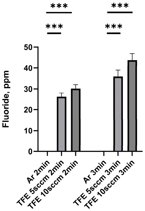
Figure 1 shows the concentrations of fluoride generated in PBS exposed to NTAP.
The fluoride concentrations in PBS treated with Ar NTAP for either 2 or 3 min were extremely low, at 0.007 ± 0.0008 ppm and 0.012 ± 0.002 ppm, respectively. The fluoride concentrations in PBS treated with FNTAP for 2 or 3 min were statistically significantly higher; the TFE 5 sccm 2 min, TFE 5 sccm 3 min, TFE 10 sccm 2 min and TFE 10 sccm 3 min groups had fluoride concentrations of 26.28 ± 1.78 ppm, 35.99 ± 2.99 ppm, 30.21 ± 1.81 ppm and 43.74 ± 3.06 ppm, respectively (p < 0.001).
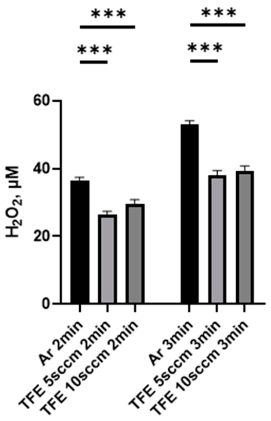
The H2O2 concentration in PBS treated with Ar NTAP was 36.59 ± 0.84 µM and 53.11 ± 0.96 µM for treatment times of 2 and 3 min, respectively (Figure 2). However, the H2O2 concentrations in PBS treated with FNTAP were statistically significantly lower than those in the corresponding Ar NTAP group (p < 0.001) for the same exposure time; the H2O2 concentrations for the TFE 5 sccm 2 min, TFE 5 sccm 3 min, TFE 10 sccm 2 min and TFE 10 sccm 3 min groups were 26.26 ± 1.10 µM, 37.99 ± 1.38 µM, 28.32 ± 1.77 µM and 39.28 ± 1.51 µM, respectively.
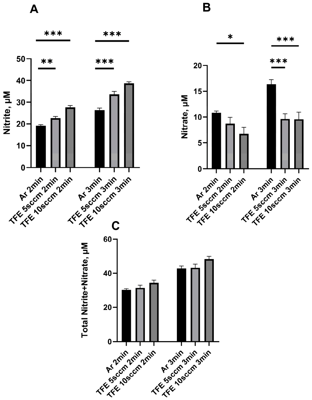
The NO2 concentration in PBS was 19.19 ± 0.48 µM and 26.35 ± 0.92 µM after PBS was exposed to Ar NTAP for 2 and 3 min, respectively. The NO2 concentrations in PBS were statistically significantly higher than those in the corresponding Ar NTAP group after PBS was exposed to FNTAP for either 2 or 3 min (p = 0.0049, p < 0.001). The nitrite concentrations for the TFE 5 sccm 2 min, TFE 5 sccm 3 min, TFE 10 sccm 2 min and TFE 10 sccm 3 min groups were 22.70 ± 0.76 µM, 33.61 ± 1.43 µM, 27.60 ± 0.91 µM and 38.75 ± 0.63 µM, respectively (Figure 3A). The NO3 concentrations in PBS were 10.84 ± 0.33 µM and 16.37 ± 0.85 µM for the Ar 2 min and Ar 3 min groups (Figure 3B), respectively. No statistically significant difference was observed between the Ar 2 min group and the TFE 5 sccm 2 min group, which had a concentration of 8.72 ± 1.22 µM. The TFE 10 sccm 2 min group had a statistically significantly lower NO3 concentration as compared to the Ar 2 min group: 6.73 ± 1.24 µM (p = 0.023). Both the TFE 5 sccm 3 min and TFE 10 sccm 3 min groups had statistically significantly lower NO3 concentrations (9.63 ± 0.97 µM and 9.58 ± 1.39 µM, respectively) than the Ar 3 min group (p < 0.001, Figure 3B). Figure 3C shows the total concentrations of NO3 and NO2 generated in PBS exposed to NTAP. An increasing trend in the total amount of NO3 and NO2 generated was observed as the plasma condition changed from Ar NTAP to TFE 5 sccm FNTAP and then to TFE 10 sccm FNTAP, for both the 2 min and 3 min treatments: 30.19 ± 0.70 µM, 42.72 ± 1.54 µM, 31.42 ± 1.53 µM, 43.24 ± 2.07 µM, 34.33 ± 1.63 µM and 48.32 ± 1.54 µM, respectively, for the Ar 2 min, Ar 3 min, TFE 5 sccm 2 min, TFE 5 sccm 3 min, TFE 10 sccm 2 min and TFE 10 sccm 3 min groups.

3.2. Metabolic Activity of Triple-Species Biofilms Treated by NTAP

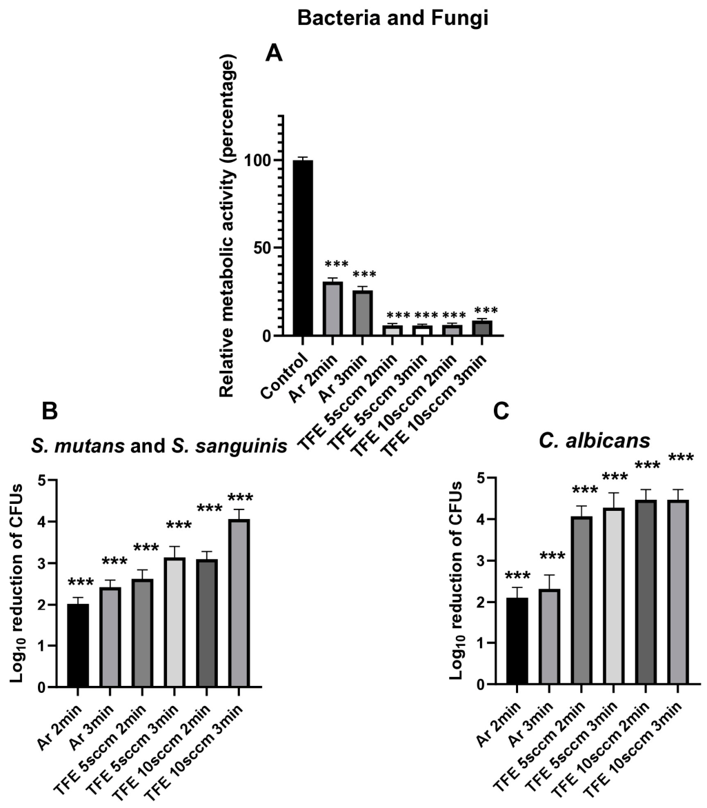
Figure 4A shows the total (bacterial and fungal) metabolic activity in in vitro triple-species biofilms after exposure to argon NTAP and FNTAPs. Ar plasma treatment resulted in a statistically significant reduction in total metabolic activity by 69.32 ± 2.05% (p < 0.001) and 74.32 ± 2.32% (p < 0.001) for the 2 and 3 min plasma exposures, respectively. FNTAP treatments reduced the total metabolic activity by 94.12 ± 1.03%, 94.22 ± 0.76%, 93.94 ± 1.05% and 91.43 ± 1.20% for the TFE 5 sccm 2 min, TFE 5 sccm 3 min, TFE 10 sccm 2 min and TFE 10 sccm 3 min, respectively (p < 0.001), compared to the untreated control group. Compared to Ar NTAP, the addition of TFE resulted in a statistically significantly greater reduction in total metabolic activity for both 2 and 3 min plasma exposures (p < 0.001). Of note, residual reactive oxygen and nitrogen species generated by nonthermal plasma may interfere with the resazurin-based PrestoBlue reaction. Therefore, CFU counts provide an independent measure that can be used to cross-validate the results of the metabolic activity assay.

3.3. Total Bacterial CFU Count of Triple-Species Biofilms Treated by NTAP

Figure 4B summarizes the results from total bacterial CFU assays for in vitro triple-species biofilms exposed to NTAP. Total bacterial log10 CFU was 6.83 ± 0.18 for the no-treatment group. Compared with the untreated control, a bacterial CFU log10 reduction of 2.02 ± 0.15 and 2.43 ± 0.17 was achieved following Ar NTAP treatment for 2 and 3 min, respectively (p < 0.001). FNTAP treatment achieved total bacterial CFU log10 reductions of 2.62 ± 0.22 and 3.14 ± 0.26, and 3.10 ± 0.18 and 4.06 ± 0.23 for the TFE 5 sccm 2 min, TFE 5 sccm 3 min, TFE 10 sccm 2 min and TFE 10 sccm 3 min groups, respectively (p < 0.001). Adding 10 sccm TFE significantly increased CFU log10 reductions compared to Ar NTAP treatment (p < 0.001) at the same treatment times.
Spearman correlation analysis was done for exploratory associations across experimental conditions and does not indicate causation. The Spearman r for the association between bacterial CFU log10 reduction and nitrite content in plasma-treated PBS was 0.94 (p = 0.0167). The Spearman r for the correlation between bacterial CFU log10 reduction and fluoride concentration in plasma-treated PBS was 1 (p = 0.0028). There was no significant correlation between bacterial CFU log10 reduction and the content of nitrate, total nitrates and nitrites, or H2O2 in plasma-treated PBS.

3.4. Fungal CFU Count of Triple-Species Biofilms Treated by NTAP

Figure 4C summarizes the results from C. albicans CFU assays for in vitro triple-species biofilms after plasma exposure. Total fungal log10 CFU was 5.77 ± 0.24 for the no-treatment group. A fungal CFU log10 reduction of 2.11 ± 0.25 and 2.32 ± 0.34 was achieved in the Ar 2 min and Ar 3 min groups, respectively (p < 0.001). With the addition of 5 sccm TFE, FNTAP treatments statistically significantly increased the fungal CFU log10 reduction to 4.07 ± 0.25 and 4.28 ± 0.36 after 2 and 3 min exposures, respectively (p < 0.001). With the addition of 10 sccm TFE, the fungal CFU counts were statistically significantly reduced, achieving a CFU log10 reduction of 4.47 ± 0.24 and 4.47 ± 0.24 after 2 and 3 min FNTAP treatments, respectively (p < 0.001). Seven out of nine CFU counts per well in each of the TFE 10 sccm 2 and 3 min FNTAP treatment groups were below the detection level (<20). Those data points were assigned the detection limit value (log10 20 = 1.3) for ease of calculations.
Spearman correlation analysis was done for exploratory associations across experimental conditions and does not indicate causation. The Spearman r for the correlation between fungal CFU log10 reduction and nitrite content in plasma-treated PBS was 0.84 (p = 0.0444). The Spearman r for the correlation between fungal CFU log10 reduction and fluoride concentration in plasma-treated PBS was 0.90 (p = 0.0278). There was no significant association between fungal CFU log10 reduction and the content of nitrate, total nitrates and nitrites, or H2O2 in plasma-treated PBS.

4. Discussion

C. albicans has been frequently detected in the oral cavities of children with severe early childhood caries [12] and is routinely seen in individuals who wear dentures [34]. C. albicans is believed to be an important contributor to the development of early childhood caries. Studies showed that C. albicans enhances bacterial colonization and biofilm formation when paired with bacteria such as S. gordonii, S. oralis, and S. sanguinis. C. albicans can also synergize the virulence of plaque biofilms in vivo in connection with S. mutans [18]. Enhanced physical interaction between C. albicans and S. mutans due to a high level of sucrose in the oral cavity increases the microbial burden and production of extracellular matrix. Therefore, we constructed in vitro triple-species oral biofilms from S. mutans, S. sanguinis, and C. albicans to further assess the antimicrobial and antibiofilm potency of FNTAP. Nonthermal plasma has been shown to be effective against C. albicans [45]. However, the effect of FNTAP on C. albicans-containing oral biofilms has not been studied extensively in the literature.
Both Ar NTAP and FNTAP demonstrated strong biofilm killing potency by both metabolic activity and CFU count assays as compared to the no-treatment group. This is consistent with our previous reports on both single-species and dual-species oral biofilms [31]. For TFE 10 sccm FNTAP, fungal CFU counts were below the detectable level (<20 per well) for both groups except for two data points each. Unsurprisingly, FNTAP possessed superior antibiofilm capability against the triple-species biofilms when compared with Ar NTAP (Figure 4). This observation is consistent with our previous report on the antibiofilm potency of FNTAP on in vitro dual-species oral biofilms of S. mutans and S. sanguinis [31]. When NTAPs were used to treat dual-species biofilms for 2 min, a bacterial CFU log10 reduction of 2.13, >5, and >5 was observed for Ar NTAP, 5 sccm FNTAP and 10 sccm FNTAP treatments, respectively. However, for triple-species oral biofilms generated from S. mutans, S. sanguinis, and C. albicans, a bacterial CFU log10 reduction of 2.02, 2.62, and 3.10 was observed for Ar NTAP, 5 sccm FNTAP and 10 sccm FNTAP treatments, respectively, which is lower than that for dual-species biofilms. Thus, it may be inferred that C. albicans increased the resilience of the resulting triple-species biofilm when compared with dual-species biofilms of S. mutans and S. sanguinis. However, it should be noted that dual and triple-species biofilms were constructed in independent sets of experiments, and the comparisons should be interpreted with caution.
In this study, we achieved a fungal CFU log10 reduction of 2.11 ± 0.25 for C. albicans when the triple-species biofilms were treated using Ar NTAP for 2 min. Figueira et al. reported a log10 CFU/mL reduction of 1.95 for C. albicans in a triple-species biofilms constructed from C. albicans, L. casei, and S.mutans after being treated using Ar NTAP for 2 min [26]. However, adding TFE to the plasma feeding gas Ar resulted in a significant increase in the fungal CFU log10 reduction for C. albicans in triple-species biofilms treated with the 5 sccm FNTAP or 10 sccm FNTAP for either 2 or 3 min (a fungal CFU log10 reduction of at least 4.07 across all FNTAP treatment groups). Our findings indicate that FNTAP has strong antifungal capability, suggesting that it has potential as a topical antimicrobial treatment approach for managing fungal-associated biofilm diseases. The marked reduction in C. albicans in triple-species biofilms highlights the promising nature of FNTAPs for targeting bacto–fungal biofilms associated with dental caries, particularly early childhood caries and severe caries phenotypes in which C. albicans may interact synergistically with cariogenic bacteria such as S. mutans.
It is well-known that ROS and RNS contribute to the antibacterial and antibiofilm potency of NTAP when it is used to treat bacteria and biofilms [46]. In order to understand the superior antibiofilm capability of FNTAP, the reactive species generated by both Ar NTAP and FNTAP were assessed by analyzing PBS exposed to plasma. Surprisingly, lower amounts of both H2O2 and NO3 were found in the PBS treated with FNTAPs compared with Ar NTAP, while a significantly higher amount of NO2 was detected in PBS treated with FNTAPs. Moreover, the total RNS of NO2 and NO3 slightly increased for FNTAP-treated PBS. Overall, no substantial increase in ROS and RNS was observed in PBS exposed to FNTAPs. On the other hand, almost no fluoride was detected in PBS treated with Ar NTAP, while a significantly high content of fluoride was found in PBS treated by each FNTAP.
The mechanisms of FNTAP’s antibiofilm activity are currently largely unknown and need further investigation. Rather, FNTAP likely acts through a synergistic plasma-chemical mechanism. NTAP generates multiple short- and long-lived reactive species, including ROS, RNS, free radicals and charged particles, which can penetrate and disrupt the biofilm matrix, damage microbial membranes, and impair essential metabolic pathways [47]. The addition of TFE appears to shift the plasma chemistry toward increased fluoride and nitrite generation. Fluoride at this concentration may not independently kill biofilm bacteria, but in the presence of plasma-induced membrane injury, oxidative/nitrosative stress, and biofilm matrix disruption, fluoride may more readily enter bacterial cells and inhibit acid production and stress-response systems. In parallel, nitrite and other RNS may contribute to antimicrobial activity through the formation of reactive nitrogen intermediates within the chemically altered biofilm microenvironment. Therefore, FNTAP’s antibiofilm effect is best explained not as a simple fluoride-dose effect, but as a synergistic interaction among fluoride delivery, RONS-mediated biofilm disruption, microbial membrane damage. However, this is speculative, and the mechanism for this observed superior antibiofilm capability for FNTAP warrants further thorough investigation.
NTAPs have been demonstrated to promote wound healing in animal models [48,49]. While we did not evaluate the in vivo safety of FNTAPs in this study, FNTAP could potentially have broader oral and translational applications, including the topical management of denture stomatitis, oral candidiasis, and other biofilm-associated mucosal infections. Beyond the oral cavity, FNTAP could potentially be developed as a topical antifungal technology for fungal infections on other body surfaces, such as skin, nail, or mucosal infections, where localized treatment and biofilm disruption are clinically important. By generating fluorinated species together with reactive oxygen and nitrogen species, FNTAP may provide enhanced antifungal and antibiofilm activity. Therefore, FNTAP could have a promising future direction as a multifunctional topical technology for the prevention and management of fungal-associated infections after rigorous in vivo efficacy and safety studies.
We demonstrated the robust ability of FNTAP to disrupt triple-species biofilms and significantly reduce bacterial and fungal CFU counts. However, the present study has several limitations. FNTAP was tested against triple-species biofilms under in vitro conditions. Oral biofilms are substantially more complex [34]; therefore, further studies are needed to assess the efficacy of FNTAP against bacto–fungal biofilms in vivo. We selected a 24 h biofilm model for this study because it is an appropriate model for assessing oral biofilms in vitro [50] and fluoride is demonstrated to be most effective in the early stages of cariogenic oral biofilm formation [44]. This model provides an established biofilm with sufficient bacterial growth and extracellular polymeric substance production to enable evaluation of antibiofilm efficacy. In addition, the 24 h incubation period provides a practical and reproducible model for comparative evaluation of antibiofilm efficacy and is compatible with microtiter plate-based assays. Nevertheless, further studies are warranted to evaluate the antibiofilm activity of FNTAPs against more mature oral biofilms. The model used in this study is an in vitro model that cannot account for salivary flow and host hygiene practices. Plus, tooth enamel cannot be approximated by polystyrene plates. Robust in vitro cytotoxicity studies and in vivo safety studies will be necessary to evaluate the translational potential of FNTAPs. This study measured fluoride ions in FNTAP-treated PBS with limited chemical characterization of fluorinated species. Further chemical characterization of reactive plasma species is warranted in future studies. Although significant correlations were observed between reductions in bacterial and fungal CFU counts and the production of RNS and fluorine-containing species by FNTAP, these were exploratory analyses. The precise mechanisms underlying the antimicrobial activity of FNTAP remain unclear and warrant further investigation through more rigorous mechanistic studies.

5. Conclusions

This study demonstrates that fluoride-generating nonthermal atmospheric plasma is especially effective against C. albicans in in vitro triple-species biofilms. It also has the potential for significant innovation in caries prevention through its generation of fluorinated species. Beyond traditional remineralization, FNTAP could significantly enhance antimicrobial efficacy against complex oral biofilms, with its particularly superior inactivation of C. albicans compared with argon NTAP. These findings highlight the potential of FNTAP as a potent, multifunctional tool for caries control.

Supplementary Materials

The following supporting information can be downloaded at: https://www.mdpi.com/article/10.3390/jfb17090477/s1, Table S1: Bacterial log10 CFU reduction as compared to no-treatment; Table S2: C. albicans log10 CFU reduction as compared to no-treatment; Table S3: Bacterial CFUs/well; Table S4: C. albicans CFUs/well; Table S5: Prestoblue metabolic activity analysis. Calculated as per kit instructions, no-treatment values adjusted to 1 or 100%; Table S6: Fluoride concentrations in PBS treated with Ar NTAP or FNTAP (ppm); Table S7: Concentrations of NO2- generated in PBS treated with Ar NTAP or FNTAP (µM); Table S8: Total concentrations of NO2- and NO3- generated in PBS treated with Ar NTAP or FNTAP (µM); Table S9: Concentrations of NO3- generated in PBS treated with Ar NTAP or FNTAP (µM); Table S10: H2O2 concentrations in PBS treated with Ar NTAP or FNTAP (µM).

Author Contributions

A.W.: contributed to design, data acquisition, analysis, and interpretation, drafted and critically revised the manuscript; L.W.: contributed to design, acquisition and analysis, drafted and critically revised the manuscript; Q.Y.: contributed to conception, design, interpretation, critically revised the manuscript; L.H.: contributed to conception, design, interpretation, critically revised the manuscript; H.S.: contributed to conception, design, interpretation, critically revised the manuscript. All authors have read and agreed to the published version of the manuscript.

Funding

This work was supported by NIH 1R21DE031868-01A1 to L.H., Q.Y. and H.S., Texas A&M University New Faculty Startup funds to L.H. and by Texas A&M Health 2026 Research Seedling Grant to L.H. The funders had no role in the study design, data collection and analysis, decision to publish or preparation of the manuscript.

Institutional Review Board Statement

Not applicable.

Informed Consent Statement

Not applicable.

Data Availability Statement

The original contributions presented in the study are included in the article/Supplementary Material, further inquiries can be directed to the corresponding authors.

Acknowledgments

Candida albicans: The following reagent was obtained through BEI Resources, NIAID, NIH: Candida albicans, Strain P37005, NR-29447.

Conflicts of Interest

H.S. and Q.Y. have financial interest in Nanova, Inc. The remaining authors declare no conflicts of interest.

Abbreviations

The following abbreviations are used in this manuscript:
BHIBrain Heart Infusion
CFUColony-forming units
C. albicansCandida albicans
DPBSDulbecco’s Phosphate-Buffered Saline
FNTAPFluoride-generating nonthermal atmospheric plasma
NTAPNonthermal atmospheric plasma
OD600Optical density at 600 nm
PBSPhosphate-Buffered Saline
RNSReactive nitrogen species
ROSReactive oxygen species
sccmStandard cubic centimeters per minute
SDASabouraud Dextrose Agar
S. sanguinisStreptococcus sanguinis
S. mutansStreptococcus mutans
TFE1,1,1,2-tetrafluoroethane

References

  1. Kassebaum, N.J.; Bernabé, E.; Dahiya, M.; Bhandari, B.; Murray, C.J.; Marcenes, W. Global burden of untreated caries: A systematic review and metaregression. J. Dent. Res. 2015, 94, 650–658. [Google Scholar] [CrossRef] [Scilit] [PubMed]
  2. Oral Health. 2025. Available online: https://www.who.int/news-room/fact-sheets/detail/oral-health (accessed on 21 April 2026).
  3. Mazurel, D.; Brandt, B.W.; Boomsma, M.; Crielaard, W.; Lagerweij, M.; Exterkate, R.A.M.; Deng, D.M. Streptococcus mutans and Caries: A Systematic Review and Meta-Analysis. J. Dent. Res. 2025, 104, 594–603. [Google Scholar] [CrossRef] [Scilit] [PubMed]
  4. Yue, Z.; Chenchen, Z.; Yalan, D.; Lei, L.; Tao, H. Oral microecological community- Streptococcus mutans dysbiosis and interaction provide therapeutic perspectives for dental caries. Arch. Oral Biol. 2025, 178, 106367. [Google Scholar] [CrossRef] [Scilit] [PubMed]
  5. AlEraky, D.M.; Madi, M.; El Tantawi, M.; AlHumaid, J.; Fita, S.; AbdulAzeez, S.; Borgio, J.F.; Al-Harbi, F.A.; Alagl, A.S. Predominance of non-Streptococcus mutans bacteria in dental biofilm and its relation to caries progression. Saudi J. Biol. Sci. 2021, 28, 7390–7395. [Google Scholar] [CrossRef] [Scilit] [PubMed]
  6. Wei, Y.; Dang, G.P.; Ren, Z.Y.; Wan, M.C.; Wang, C.Y.; Li, H.B.; Zhang, T.; Tay, F.R.; Niu, L.N. Recent advances in the pathogenesis and prevention strategies of dental calculus. npj Biofilms Microbiomes 2024, 10, 56. [Google Scholar] [CrossRef] [Scilit] [PubMed]
  7. Kanwar, I.L.; Sah, A.; Suresh, P. Biofilm-mediated Antibiotic-resistant Oral Bacterial Infections: Mechanism and Combat Strategies. Curr. Pharm. Des. 2017, 23, 2084–2095. [Google Scholar] [CrossRef] [Scilit] [PubMed]
  8. Bida, F.C.; Curca, F.R.; Lupusoru, R.V.; Virvescu, D.I.; Scurtu, M.; Rotundu, G.; Butnaru, O.M.; Tudorici, T.; Luchian, I.; Budala, D.G. The Systemic Link Between Oral Health and Cardiovascular Disease: Contemporary Evidence, Mechanisms, and Risk Factor Implications. Diseases 2025, 13, 354. [Google Scholar] [CrossRef] [Scilit] [PubMed]
  9. Kurtzman, G.; Horowitz, R.A.; Johnson, R.; Prestiano, J. Oral Biofilms and Their Connection to Cardiovascular Health. Med. Res. Arch. 2025, 13. [Google Scholar] [CrossRef] [Scilit]
  10. Kurtzman, G.M.; Horowitz, R.A.; Johnson, R.; Prestiano, R.A.; Klein, B.I. The systemic oral health connection: Biofilms. Medicine 2022, 101, e30517. [Google Scholar] [CrossRef] [Scilit] [PubMed]
  11. Witherden, E.A.; Shoaie, S.; Hall, R.A.; Moyes, D.L. The Human Mucosal Mycobiome and Fungal Community Interactions. J. Fungi 2017, 3, 56. [Google Scholar] [CrossRef] [Scilit] [PubMed]
  12. Xiao, J.; Huang, X.; Alkhers, N.; Alzamil, H.; Alzoubi, S.; Wu, T.T.; Castillo, D.A.; Campbell, F.; Davis, J.; Herzog, K.; et al. Candida albicans and Early Childhood Caries: A Systematic Review and Meta-Analysis. Caries Res. 2017, 52, 102–112. [Google Scholar] [CrossRef] [Scilit] [PubMed]
  13. O’Donnell, L.E.; Millhouse, E.; Sherry, L.; Kean, R.; Malcolm, J.; Nile, C.J.; Ramage, G. Polymicrobial Candida biofilms: Friends and foe in the oral cavity. FEMS Yeast Res. 2015, 15, fov077. [Google Scholar] [CrossRef] [Scilit] [PubMed]
  14. Allison, D.L.; Willems, H.M.E.; Jayatilake, J.A.M.S.; Bruno, V.M.; Peters, B.M.; Shirtliff, M.E. Candida–Bacteria Interactions: Their Impact on Human Disease, in Virulence Mechanisms of Bacterial Pathogens; ASM Press: Washington, DC, USA, 2016; pp. 103–136. [Google Scholar]
  15. Wan, S.X.; Tian, J.; Liu, Y.; Dhall, A.; Koo, H.; Hwang, G. Cross-Kingdom Cell-to-Cell Interactions in Cariogenic Biofilm Initiation. J. Dent. Res. 2021, 100, 74–81. [Google Scholar] [CrossRef] [Scilit] [PubMed]
  16. Delaney, C.; O’Donnell, L.E.; Kean, R.; Sherry, L.; Brown, J.L.; Calvert, G.; Nile, C.J.; Cross, L.; Bradshaw, D.J.; Brandt, B.W.; et al. Interkingdom interactions on the denture surface: Implications for oral hygiene. Biofilm 2019, 1, 100002. [Google Scholar] [CrossRef] [Scilit] [PubMed]
  17. Hwang, G. In it together: Candida-bacterial oral biofilms and therapeutic strategies. Environ. Microbiol. Rep. 2022, 14, 183–196. [Google Scholar] [CrossRef] [Scilit] [PubMed]
  18. Falsetta, M.L.; Klein, M.I.; Colonne, P.M.; Scott-Anne, K.; Gregoire, S.; Pai, C.H.; Gonzalez-Begne, M.; Watson, G.; Krysan, D.J.; Bowen, W.H.; et al. Symbiotic relationship between Streptococcus mutans and Candida albicans synergizes virulence of plaque biofilms in vivo. Infect. Immun. 2014, 82, 1968–1981. [Google Scholar] [CrossRef] [Scilit] [PubMed]
  19. Aliberti, A.; Gasparro, R.; Triassi, M.; Piscopo, M.; Ausiello, P.; Tribst, J.P.M. Fluoride Release from Pediatric Dental Restorative Materials: A Laboratory Investigation. Dent. J. 2025, 13, 224. [Google Scholar] [CrossRef] [Scilit] [PubMed]
  20. Aliberti, A.; Garcia-Godoy, F.; Borges, A.L.S.; Tribst, J.P.M.; Gasparro, R.; Mariniello, M.; Ausiello, P. Calcium, phosphate and fluoride ionic release from dental restorative materials for elderly population: An in vitro analysis. Front. Oral Health 2025, 6, 1609502. [Google Scholar] [CrossRef] [Scilit] [PubMed]
  21. Yeh, C.H.; Wang, Y.L.; Vo, T.T.T.; Lee, Y.C.; Lee, I.T. Fluoride in Dental Caries Prevention and Treatment: Mechanisms, Clinical Evidence, and Public Health Perspectives. Healthcare 2025, 13, 2246. [Google Scholar] [CrossRef] [Scilit] [PubMed]
  22. Philip, N.; Suneja, B.; Walsh, L.J. Ecological Approaches to Dental Caries Prevention: Paradigm Shift or Shibboleth? Caries Res. 2018, 52, 153–165. [Google Scholar] [CrossRef] [Scilit] [PubMed]
  23. Dang, M.-H.; Jung, J.-E.; Lee, D.-W.; Song, K.-Y.; Jeon, J.-G. Recovery of Acid Production in Streptococcus mutans Biofilms after Short-Term Fluoride Treatment. Caries Res. 2016, 50, 363–371. [Google Scholar] [CrossRef] [Scilit] [PubMed]
  24. Hong, Q.; Dong, X.; Jones, J.E.; Hong, L.; Yu, Q.; Sun, H.; Chen, M. A novel approach to expedite wound healing with plasma brush of cold flame. Rev. Sci. Instrum. 2023, 94, 084102. [Google Scholar] [CrossRef] [Scilit] [PubMed]
  25. Yang, B.; Chen, J.; Yu, Q.; Li, H.; Lin, M.; Mustapha, A.; Hong, L.; Wang, Y. Oral bacterial deactivation using a low-temperature atmospheric argon plasma brush. J. Dent. 2011, 39, 48–56. [Google Scholar] [CrossRef] [Scilit] [PubMed]
  26. Figueira, L.W.; Panariello, B.; Koga-Ito, C.Y.; Duarte, S. Exploring the efficacy of in-vitro low-temperature plasma treatment on single and multispecies dental cariogenic biofilms. Sci. Rep. 2024, 14, 20678. [Google Scholar] [CrossRef] [Scilit] [PubMed]
  27. Hong, Q.; Dong, X.; Chen, M.; Xu, Y.; Sun, H.; Hong, L.; Wang, Y.; Yu, Q. Disinfection of Streptococcus mutans biofilm by a non-thermal atmospheric plasma brush. Jpn. J. Appl. Phys. 2016, 55, 07LG02. [Google Scholar] [CrossRef] [Scilit]
  28. Hong, Q.; Dong, X.; Chen, M.; Sun, H.; Hong, L.; Wang, Y.; Li, H.; Yu, Q. An in vitro and in vivo study of plasma treatment effects on oral biofilms. J. Oral Microbiol. 2019, 11, 1603524. [Google Scholar] [CrossRef] [Scilit] [PubMed]
  29. Hong, Q.; Sun, H.; Chen, M.; Zhang, S.; Yu, Q. Plasma treatment effects on destruction and recovery of Porphyromonas gingivalis biofilms. PLoS ONE 2022, 17, e0274523. [Google Scholar] [CrossRef] [Scilit] [PubMed]
  30. Bapat, S.A.; Shashikiran, N.D.; Gugawad, S.; Gaonkar, N.; Taur, S.; Hadakar, S.; Chaudhari, P. Effect of non-thermal atmospheric pressure plasma and ErCr:YSGG LASER activation of three fluoride varnishes on surface re-mineralization of enamel: A SEM-EDX analysis. J. Indian Soc. Pedod. Prev. Dent. 2022, 40, 324–329. [Google Scholar] [CrossRef] [Scilit] [PubMed]
  31. Warang, A.; Deol, I.; Fakher, S.; Wu, L.; Hong, L.; Zhang, S.; Yu, Q.; Sun, H. Non-Thermal Atmospheric Plasma Enhances Biological Effects of Fluoride on Oral Biofilms. J. Funct. Biomater. 2025, 16, 132. [Google Scholar] [CrossRef] [Scilit] [PubMed]
  32. Koo, H.; Andes, D.R.; Krysan, D.J.; Hogan, D.A. Candida–streptococcal interactions in biofilm-associated oral diseases. PLoS Pathog. 2018, 14, e1007342. [Google Scholar] [CrossRef] [Scilit] [PubMed]
  33. Moreno-Florez, A.I.; Bedoya-Correa, C.M.; Garcia, C.; Pelaez-Vargas, A. Antimicrobial Activity of Ethanolic Propolis Extracts from Tame (Arauca) on Oral Biofilm Co-Cultures. Pathogens 2025, 14, 982. [Google Scholar] [CrossRef] [Scilit] [PubMed]
  34. Berger, D.; Rakhamimova, A.; Pollack, A.; Loewy, Z. Oral Biofilms: Development, Control, and Analysis. High-Throughput 2018, 7, 24. [Google Scholar] [CrossRef] [Scilit] [PubMed]
  35. Ritts, A.C.; Li, H.; Yu, Q.; Xu, C.; Yao, X.; Hong, L.; Wang, Y. Dentin surface treatment using a non-thermal argon plasma brush for interfacial bonding improvement in composite restoration. Eur. J. Oral Sci. 2010, 118, 510–516. [Google Scholar] [CrossRef] [Scilit] [PubMed]
  36. Lin, A.; Gorbanev, Y.; De Backer, J.; Van Loenhout, J.; Van Boxem, W.; Lemière, F.; Cos, P.; Dewilde, S.; Smits, E.; Bogaerts, A. Non-Thermal Plasma as a Unique Delivery System of Short-Lived Reactive Oxygen and Nitrogen Species for Immunogenic Cell Death in Melanoma Cells. Adv. Sci. 2019, 6, 1802062. [Google Scholar] [CrossRef] [Scilit] [PubMed]
  37. Chen, Z.; Simonyan, H.; Cheng, X.; Gjika, E.; Lin, L.; Canady, J.; Sherman, J.H.; Young, C.; Keidar, M. A Novel Micro Cold Atmospheric Plasma Device for Glioblastoma Both In Vitro and In Vivo. Cancers 2017, 9, 61. [Google Scholar] [CrossRef] [Scilit] [PubMed]
  38. Dong, X.; Wu, L.; Wang, Y.; Yu, Q.; Hong, L. Effects of Atmospheric Fluorine-Containing Nonthermal Plasmas on Demineralization and Remineralization of Tooth Enamel. IEEE Trans. Plasma Sci. 2022, 50, 2910–2916. [Google Scholar] [CrossRef] [Scilit]
  39. Diaz, F.E.; McGill, J.L. NRF2 agonists 4-octyl-itaconate and dimethyl fumarate reduce human and bovine RSV proliferation and RSV disease in a murine model. Immunohorizons 2025, 9, vlaf036. [Google Scholar] [CrossRef] [Scilit] [PubMed]
  40. Sul, J.O.; Choi, H.W.; Ra, S.W. Grape seed proanthocyanidin extract modulates cigarette smoke extract-induced epithelial cell apoptosis by inhibiting oxidative stress in chronic obstructive pulmonary disease. J. Funct. Foods 2024, 112, 105907. [Google Scholar] [CrossRef] [Scilit]
  41. Lee, E.-S.; Kang, S.-M.; Ko, H.-Y.; Kwon, H.-K.; Kim, B.-I. Association between the cariogenicity of a dental microcosm biofilm and its red fluorescence detected by Quantitative Light-induced Fluorescence-Digital (QLF-D). J. Dent. 2013, 41, 1264–1270. [Google Scholar] [CrossRef] [Scilit] [PubMed]
  42. Filoche, S.K.; Soma, K.J.; Sissons, C.H. Caries-related plaque microcosm biofilms developed in microplates. Oral Microbiol. Immunol. 2007, 22, 73–79. [Google Scholar] [CrossRef] [Scilit] [PubMed]
  43. Cai, J.N.; Jung, J.E.; Dang, M.H.; Kim, M.A.; Yi, H.K.; Jeon, J.G. Functional Relationship between Sucrose and a Cariogenic Biofilm Formation. PLoS ONE 2016, 11, e0157184. [Google Scholar] [CrossRef] [Scilit] [PubMed]
  44. Han, Y. Effects of brief sodium fluoride treatments on the growth of early and mature cariogenic biofilms. Sci. Rep. 2021, 11, 18290. [Google Scholar] [CrossRef] [Scilit] [PubMed]
  45. Song, Y.; Liu, D.; Ji, L.; Wang, W.; Niu, J.; Zhang, X. Plasma inactivation of candida albicans by an atmospheric cold plasma brush composed of hollow fibers. IEEE Trans. Plasma Sci. 2012, 40, 1098–1102. [Google Scholar] [CrossRef] [Scilit]
  46. Nicol, M.J.; Brubaker, T.R.; Honish, B.J., II; Simmons, A.N.; Kazemi, A.; Geissel, M.A.; Whalen, C.T.; Siedlecki, C.A.; Bilén, S.G.; Knecht, S.D.; et al. Antibacterial effects of low-temperature plasma generated by atmospheric-pressure plasma jet are mediated by reactive oxygen species. Sci. Rep. 2020, 10, 3066. [Google Scholar] [CrossRef] [Scilit] [PubMed]
  47. Chauvin, J.; Judée, F.; Yousfi, M.; Vicendo, P.; Merbahi, N. Analysis of reactive oxygen and nitrogen species generated in three liquid media by low temperature helium plasma jet. Sci. Rep. 2017, 7, 4562. [Google Scholar] [CrossRef] [Scilit] [PubMed]
  48. Wang, X.-F.; Fang, Q.-Q.; Jia, B.; Hu, Y.-Y.; Wang, Z.-C.; Yan, K.-P.; Yin, S.-Y.; Liu, Z.; Tan, W.-Q. Potential effect of non-thermal plasma for the inhibition of scar formation: A preliminary report. Sci. Rep. 2020, 10, 1064. [Google Scholar] [CrossRef] [Scilit] [PubMed]
  49. Kubinova, S.; Zaviskova, K.; Uherkova, L.; Zablotskii, V.; Churpita, O.; Lunov, O.; Dejneka, A. Non-thermal air plasma promotes the healing of acute skin wounds in rats. Sci. Rep. 2017, 7, 45183. [Google Scholar] [CrossRef] [Scilit] [PubMed]
  50. Li, P.; Zhang, Y.; Chen, D.; Lin, H. Investigation of a novel biofilm model close to the original oral microbiome. Appl. Microbiol. Biotechnol. 2024, 108, 330. [Google Scholar] [CrossRef] [Scilit] [PubMed]
Figure 1. Fluoride concentrations in PBS treated with Ar NTAP or FNTAP for 2 or 3 min. Sample size: 3 independent experiments with 3 individual replicates, n = 9 per group. *** for p < 0.001 as compared with that for Ar NTAP-treated for the same period. Error bars indicate standard error of the mean. Data are available in Supplementary Materials.
Jfb 17 00477 g001
Figure 2. H2O2 concentrations in PBS treated with Ar NTAP or FNTAP for 2 or 3 min. Sample size: 3 independent experiments with 3 individual replicates, n = 9 per group. *** for p < 0.001 as compared with that for Ar NTAP-treated for the same time. Error bars indicate standard error of the mean. Data are available in Supplementary Materials.
Jfb 17 00477 g002
Figure 3. Concentrations of NO2 (A), concentrations of NO3 (B) and total concentrations of NO2 and NO3 (C) generated in PBS treated with Ar NTAP or FNTAP for 2 min or 3 min. Sample size: 3 independent experiments with 3 individual replicates, n = 9 per group, except for Ar 2 min groups for Nitrate and Total Nitrite + Nitrate analysis where n = 8 (3 independent experiments with 3, 3, 2 individual replicates). * for p < 0.05, ** for p <0.01, *** for p < 0.001 compared with that for Ar NTAP-treated for the same period. Error bars indicate standard error of the mean. Data are available in Supplementary Materials.
Jfb 17 00477 g003
Figure 4. (A) Combined bacterial and fungal relative metabolic activity, with the metabolic activity set to be 100% for control group (i.e., untreated), *** = p <0.001 (all treatments compared to control). (B) Total bacteria CFU log10 reduction by FNTAP treatments at 2 min and 3 min treatment times, *** = p < 0.001 (all treatments compared to control). (C) Fungal CFU log10 reduction by FNTAP treatments at 2 min and 3 min treatment times,*** = p < 0.001 (all treatments compared to control). n = 9 per group (3 independent experiments with 3 individual replicates each). Error bars indicate standard error of the mean. Data are available in Supplementary Materials.
Jfb 17 00477 g004
Disclaimer/Publisher’s Note: The statements, opinions and data contained in all publications are solely those of the individual author(s) and contributor(s) and not of MDPI and/or the editor(s). MDPI and/or the editor(s) disclaim responsibility for any injury to people or property resulting from any ideas, methods, instructions or products referred to in the content.