Polyphosphate in Bone Tissue Engineering: From Molecular Mechanisms to Material Design




Abstract
1. Introduction
2. Molecular Structure and Basic Functions of PolyP
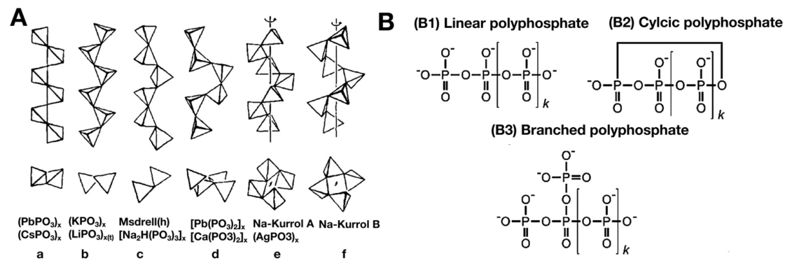
2.1. Chemical Structure and Physicochemical Properties
2.2. Biosynthesis and Metabolism in Organisms
3. Mechanisms of PolyP in Promoting Osteogenesis
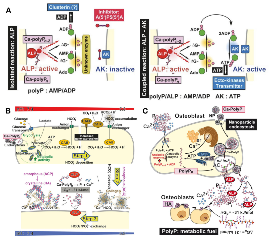
3.1. Participation in Bone Mineralization as a Phosphate Donor
3.2. Participation in Energy Metabolism as an Energy-Storing Molecule

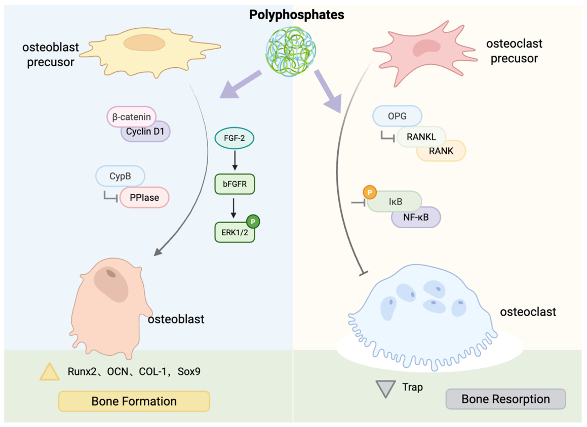
3.3. Regulation of Osteogenesis-Related Signaling Pathways
3.4. Other Mechanisms

| Domain | Mechanism | References | Detailed Mechanism and Effects |
|---|---|---|---|
| Bone Mineralization | Phosphate Donor | [33,34,35,36] | Serves as phosphate storage; hydrolyzed by alkaline phosphatase (ALP) to release inorganic phosphate (Pi), forming amorphous calcium phosphate and eventually hydroxyapatite (HA). |
| Calcium Ion Regulation | [33,34,35,36,37] | Regulates intracellular calcium ion concentration. Activates multiple osteogenic signaling pathways by inducing an increase in intracellular calcium ions. | |
| Energy Metabolism | Metabolic Fuel | [13,38,39,40] | High-energy phosphoanhydride bonds generate adenosine diphosphate (ADP) /adenosine triphosphate (ATP) upon hydrolysis, directly fueling extracellular bone mineralization. |
| Mitochondrial Regulation | [41,42,43,44] | Activates components of the mitochondrial permeability transition pore (mPTP); induces mitochondrial elongation/swelling; maintains growth/mineralization under hypoxia via hypoxia-inducible factor 1 alpha (HIF-1α) and carbonic anhydrase IX upregulation. | |
| Cell Signaling | Wnt/β-catenin Pathway | [45,46,47] | Activates the signaling pathway to upregulate β-catenin and cyclin D1, promoting osteogenic markers. |
| FGF/ERK Pathway | [48,49] | Stabilizes fibroblast growth factor (FGF)-2 for receptor binding, activating extracellular signal-regulated kinase (ERK)1/2 phosphorylation to upregulate osteogenic genes. | |
| Protein Folding | [51] | Binds to cyclophilin B (CypB), inhibiting peptidyl-prolyl cis-trans isomerase (PPIase) activity to regulate collagen folding. | |
| AMPK Pathway | [52] | Regulates intracellular free phosphate homeostasis, impacting phosphate transporter xenotropic and polytropic retrovirus receptor 1 (XPR1) and inositol hexakisphosphate kinase (IP6K) 1/2, thereby modulating AMP-activated protein kinase (AMPK) phosphorylation. | |
| Cell Homeostasis | Osteoclast Differentiation | [56,57,58] | Upregulates osteoprotegerin (OPG) expression to neutralize receptor activator of nuclear factor κB ligand (RANKL); inhibits inhibitor of nuclear factor kappa B alpha (IκBα) phosphorylation to block NF-κB signaling. |
| Bone Resorption | [59] | Long-chain PolyP (≥300 units) directly inhibits tartrate-resistant acid phosphatase (TRAP) activity, reducing the resorption pit area. | |
| Indirect Mechanisms | Angiogenesis | [61,62,63] | ATP produced by PolyP hydrolysis promotes human umbilical vein endothelial cell (HUVEC) tube formation; stimulates vascular endothelial growth factor (VEGF) and basic fibroblast growth factor (bFGF) secretion to couple angiogenesis. |
| Antibacterial Activity | [64,65,66] | Encapsulates and kills bacteria during wound healing while providing metabolic energy for tissue repair. |
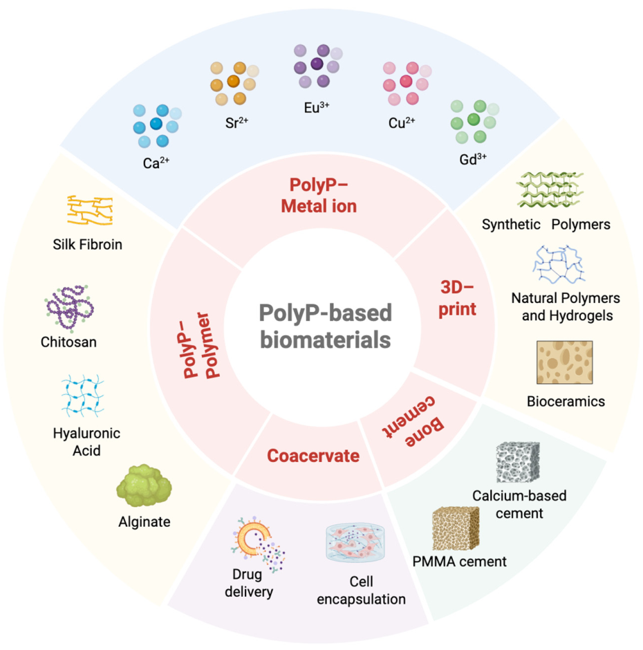
4. Design and Applications of PolyP in Biomaterials
4.1. Metal Ion–PolyP Complexes
4.2. PolyP–Polymer Composite Materials
4.3. PolyP-Incorporated Bone Cements
4.4. 3D-Printed and Electrospun Scaffolds
4.5. Coacervate Materials
| Material Type | Structure | Application | Binding Mode | |
|---|---|---|---|---|
| Metal ion composites | Ca | ~40 units | Amorphous calcium polyphosphate microparticles/nanoparticles (CPP-MP/NP): Slowly release Pi/Ca2+ and provide cellular energy [67]. | Ionic chelation |
| ~40 units | Calcium polyphosphate (CPP) and CPP/amorphous calcium carbonate (ACC) particles: Promote bone regeneration; synergistic effects with ACC [68,69]. | Ionic chelation | ||
| ~40 units | PolyP/ACC nanoparticles: Stabilize ACC for synergistic bone repair [70]. | Physical blending and encapsulation | ||
| Sr | ~40 units | Amorphous Sr-PolyP microparticles: Promote bone marrow mesenchymal stem cell (BMSC) mineralization and accelerate in vivo bone healing [71]. | Ionic substitution and doping | |
| NA | Strontium-doped calcium polyphosphate (SCPP) /BMSC composite: Treats femoral head necrosis [72]. | Ionic substitution and doping | ||
| Eu | NA | Europium-doped calcium polyphosphate (EuCPP) porous scaffold: Promotes BMSC migration and in vivo bone regeneration [73]. | Ionic substitution and doping | |
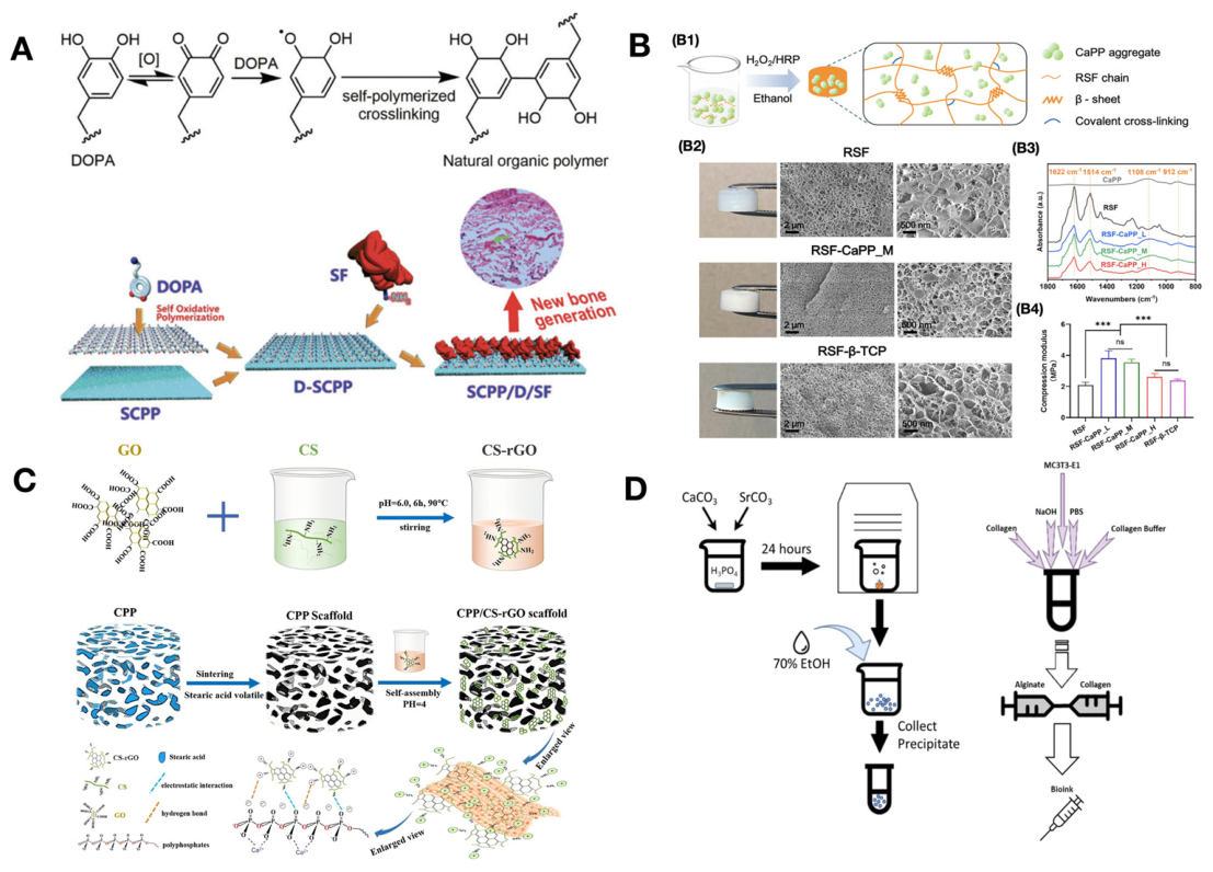
| Cu | NA | Calcium polyphosphate bone cement (CCPP)/dopamine (DOPA)/collagen scaffold: Upregulates osteogenic genes for new bone formation [74]. | Covalent and chemical bonding | |
| Gd | ~40 units | PolyP-Gd complex: Promotes hydroxyapatite (HA) formation and upregulates osteogenic genes [75]. | Ionic bonding and crosslinking | |
| Polymer composites | Silk fibroin | NA | SCPP/DOPA/silk fibroin (SF) scaffold: Promotes angiogenesis and accelerates in vivo bone formation [76]. | Electrostatic and hydrogen bonding; covalent and chemical bonding |
| Linear; 8/22/121 units | Regenerated silk fibroin (RSF)-CPP hydrogel: Promotes in vitro BMSC osteogenic differentiation [77]. | Electrostatic and hydrogen bonding | ||
| Chitosan | Linear | CPP/chitosan-reduced graphene oxide (CS-rGO) scaffold: Enhances BMSC biomineralization and osteogenic differentiation [78]. | Electrostatic and hydrogen bonding | |
| Linear | CS/CPP/pigeonite scaffold: Promotes BMSC differentiation and in vivo bone formation [79]. | Physical adsorption and adhesion | ||
| Hyaluronic acid | 13 units | HAX-PolyP hydrogel: Provides sustained osteogenic stimulation [80]. | Covalent and chemical bonding | |
| Alginate | NA | Alginate/CPP double-network hydrogel: Supports cell proliferation and osteogenic differentiation [81]. | Physical blending and encapsulation | |
| NA | SCPP-crosslinked alginate/collagen hydrogel: Promotes cell survival and early mineralization [82]. | Ionic bonding and crosslinking | ||
| Gelatin | NA | PolyP-modified gelatin: Improves compressive strength and osteogenic induction [83]. | Covalent and chemical bonding | |
| Bone cement | PMMA cement | NA | Polydopamine/SCPP/poly(methyl methacrylate) (PMMA) composite cement: Promotes osteoblast proliferation [87]. | Physical adsorption and adhesion |
| ~40 units | Sodium polyphosphate nanoparticles (NPP-NP) or calcium polyphosphate nanoparticles (CPP-NP) in PMMA: Modulates mechanics and improves self-healing properties [70]. | Physical blending and encapsulation | ||
| Calcium-based cement | Linear | CPPC: Induces in vitro mineralization and in vivo bone formation [88]. | Covalent and chemical bonding | |
| Linear; ~40 units | Tricalcium silicate/calcium polyphosphate/polyvinyl alcohol (C3S/CPP/PVA) composite cement: Promotes osteoblast proliferation and osteogenic differentiation [89]. | Physical adsorption and adhesion | ||
| 3D-printed and electrospun materials | Synthetic polymers | ~40 units | CPP-NP/polycaprolactone (PCL) three-dimensional (3D)-printed scaffold: Supports cell growth and upregulates chemokines [94]. | Physical blending and encapsulation |
| NA | PolyP-incorporated PCL/poly(L-lactic acid) (PLLA) nanofibers: Promotes induced pluripotent stem cell (iPSC) osteogenic differentiation [47]. | Physical blending and encapsulation | ||
| ~40 units | Amorphous CPP/collagen-coated PCL mat: Promotes BMSC attachment and angiogenesis [100]. | Physical blending and encapsulation | ||
| ~40 units | Sr-PolyP/ascorbyl palmitate/PCL fibers: Improves bioactivity and cell compatibility [101]. | Physical blending and encapsulation | ||
| Bioceramics | ~40 units | PolyP/beta-tricalcium phosphate (β-TCP) 3D-printed scaffold: Releases PolyP to promote BMSC proliferation and mineralization [96]. | Physical blending and encapsulation | |
| Linear | Powder-printed CPP: Supports in vitro cartilage formation for osteochondral repair [98]. | Physical blending and encapsulation | ||
| ~40 units | SCPP scaffold with tetracycline/matrix metalloproteinase (MMP)-2: Dual-drug release for promoting in vivo bone regeneration [102]. | Physical blending and encapsulation | ||
| Natural polymers and hydrogels | Linear | Alginate/poly(ethylene glycol) diacrylate/PolyP double-network hydrogel: Boosts cellular adenosine triphosphate (ATP), migration, and osteogenesis [97]. | Ionic chelating | |
| Linear | Printable CPP/alginoplast powder: Forms porous carbonate apatite for cell attachment [99]. | Physical blending and encapsulation | ||
| Coacervate-related | Cell encapsulation | Linear; ~40 units | PolyP coacervate matrix: Supports BMSC survival, proliferation, and differentiation [104]. | Ionic bonding and crosslinking |
| Drug delivery | ~40 units | Dexamethasone (Dex)-CPP/ascorbate–coacervate core–shell system: Enables multikinetic drug delivery [106]. | Ionic bonding and crosslinking | |
5. Clinical Translation Studies
5.1. Preclinical Animal Studies
5.2. Clinical Trials
6. Challenges and Outlook
Author Contributions
Funding
Institutional Review Board Statement
Informed Consent Statement
Data Availability Statement
Acknowledgments
Conflicts of Interest
Abbreviations
| AA | ascorbic acid |
| ACC | amorphous calcium carbonate |
| ADP | adenosine diphosphate |
| ALP | alkaline phosphatase |
| AMPK | AMP-activated protein kinase |
| ATP | adenosine triphosphate |
| BCP | biphasic calcium phosphate |
| β-TCP | beta-tricalcium phosphate |
| bFGF | basic fibroblast growth factor |
| bFGFR | basic fibroblast growth factor receptor |
| BMP-2 | bone morphogenetic protein 2 |
| BMSC | bone marrow mesenchymal stem cell |
| CCPP | copper-doped calcium polyphosphate |
| CPP | calcium polyphosphate |
| CPPC | calcium polyphosphate bone cement |
| CPP-MP | calcium polyphosphate microparticles |
| CPP-NP | calcium polyphosphate nanoparticles |
| COL-1 | collagen type I |
| CS | chitosan |
| C3S | tricalcium silicate |
| CypB | cyclophilin B |
| Dex | dexamethasone |
| DOPA | dopamine |
| ERK | extracellular signal-regulated kinase |
| EuCPP | europium-doped calcium polyphosphate |
| FGF | fibroblast growth factor |
| HA | hydroxyapatite |
| HyA | hyaluronic acid |
| hBMSC | human bone marrow mesenchymal stem cell |
| HIF-1α | hypoxia-inducible factor 1 alpha |
| HUVEC | human umbilical vein endothelial cell |
| IκBα | inhibitor of nuclear factor kappa B alpha |
| iPSC | induced pluripotent stem cell |
| IP6K1/2 | inositol hexakisphosphate kinase 1/2 |
| LLPS | liquid–liquid phase separation |
| MFAT | microfragmented fat |
| MMP-1 | matrix metalloproteinase 1 |
| mPTP | mitochondrial permeability transition pore |
| NF-κB | nuclear factor kappa B |
| NPP-NP | sodium polyphosphate nanoparticles |
| OCN | osteocalcin |
| OPG | osteoprotegerin |
| OPN | osteopontin |
| PCL | polycaprolactone |
| Pi | inorganic phosphate |
| PLLA | poly(L-lactic acid) |
| PMMA | poly(methyl methacrylate) |
| PolyP | polyphosphate |
| PPIase | peptidyl-prolyl cis-trans isomerase |
| PPK | polyphosphate kinase |
| PVA | polyvinyl alcohol |
| RANKL | receptor activator of nuclear factor κB ligand |
| rGO | reduced graphene oxide |
| RSF | regenerated silk fibroin |
| Runx2 | Runt-related transcription factor 2 |
| SBF | simulated body fluid |
| SCPP | strontium-doped calcium polyphosphate |
| SDF-1 | stromal cell-derived factor 1 |
| SF | silk fibroin |
| SOST | sclerostin |
| Sox9 | SRY-box transcription factor 9 |
| 3D | three-dimensional |
| TRAP | tartrate-resistant acid phosphatase |
| VEGF | vascular endothelial growth factor |
| XPR1 | xenotropic and polytropic retrovirus receptor 1 |
References
- Ni, X.; Feng, J.; Liang, M.; Zhou, F.; Xia, Y.; Dong, Z.; Xue, Q.; Li, Z.; Pu, F.; Xia, P. Enhancing Bone Repair with β-TCP-Based Composite Scaffolds: A Review of Design Strategies and Biological Mechanisms. Orthop. Res. Rev. 2025, 17, 313–340. [Google Scholar] [CrossRef] [Scilit] [PubMed]
- Zou, W.; Li, X.; Li, N.; Guo, T.; Cai, Y.; Yang, X.; Liang, J.; Sun, Y.; Fan, Y. A comparative study of autogenous, allograft and artificial bone substitutes on bone regeneration and immunotoxicity in rat femur defect model. Regen. Biomater. 2021, 8, rbaa040. [Google Scholar] [PubMed]
- Li, C.; Lv, H.; Du, Y.; Zhu, W.; Yang, W.; Wang, X.; Wang, J.; Chen, W. Biologically modified implantation as therapeutic bioabsorbable materials for bone defect repair. Regen. Ther. 2022, 19, 9–23. [Google Scholar] [CrossRef] [Scilit] [PubMed]
- Wei, S.; Ma, J.-X.; Xu, L.; Gu, X.-S.; Ma, X.-L. Biodegradable materials for bone defect repair. Mil. Med. Res. 2020, 7, 54. [Google Scholar] [CrossRef] [Scilit] [PubMed]
- Wu, Y.; Yang, L.; Chen, L.; Geng, M.; Xing, Z.; Chen, S.; Zeng, Y.; Zhou, J.; Sun, K.; Yang, X.; et al. Core-Shell Structured Porous Calcium Phosphate Bioceramic Spheres for Enhanced Bone Regeneration. ACS Appl. Mater. Interfaces 2022, 14, 47491–47506. [Google Scholar] [CrossRef] [Scilit] [PubMed]
- Miao, Q.; Jiang, N.; Yang, Q.; Hussein, I.M.; Luo, Z.; Wang, L.; Yang, S. Multi-stage controllable degradation of strontium-doped calcium sulfate hemihydrate-tricalcium phosphate microsphere composite as a substitute for osteoporotic bone defect repairing: Degradation behavior and bone response. Biomed. Mater. 2022, 17, 015014. [Google Scholar]
- Tang, J.; Hu, J.; Bai, X.; Wang, Y.; Cai, J.; Zhang, Z.; Geng, B.; Pan, D.; Shen, L. Near-Infrared Carbon Dots With Antibacterial and Osteogenic Activities for Sonodynamic Therapy of Infected Bone Defects. Small 2024, 20, e2404900. [Google Scholar] [CrossRef] [Scilit] [PubMed]
- Omelon, S.J.; Grynpas, M.D. Relationships between polyphosphate chemistry, biochemistry and apatite biomineralization. Chem. Rev. 2008, 108, 4694–4715. [Google Scholar] [CrossRef] [Scilit] [PubMed]
- Fleish, H.; Neuman, W.F. Mechanisms of calcification: Role of collagen, polyphosphates, and phosphatase. Am. J. Physiol. 1961, 200, 1296–1300. [Google Scholar] [CrossRef] [Scilit] [PubMed]
- Wang, X.; Schröder, H.C.; Müller, W.E.G. Amorphous polyphosphate, a smart bioinspired nano-/bio-material for bone and cartilage regeneration: Towards a new paradigm in tissue engineering. J. Mater. Chem. B 2018, 6, 2385–2412. [Google Scholar] [CrossRef] [Scilit] [PubMed]
- Wang, Y.; Li, M.; Li, P.; Teng, H.; Fan, D.; Du, W.; Guo, Z. Progress and Applications of Polyphosphate in Bone and Cartilage Regeneration. Biomed. Res. Int. 2019, 2019, 5141204. [Google Scholar] [CrossRef] [Scilit] [PubMed]
- Schröder, H.C.; Kurz, L.; Müller, W.E.; Lorenz, B. Polyphosphate in bone. Biochemistry 2000, 65, 296–303. [Google Scholar] [PubMed]
- Müller, W.E.G.; Schröder, H.C.; Wang, X. Inorganic Polyphosphates As Storage for and Generator of Metabolic Energy in the Extracellular Matrix. Chem. Rev. 2019, 119, 12337–12374. [Google Scholar] [CrossRef] [Scilit] [PubMed]
- Achbergerová, L.; Nahálka, J. Polyphosphate--an ancient energy source and active metabolic regulator. Microb. Cell Fact. 2011, 10, 63. [Google Scholar] [CrossRef] [Scilit] [PubMed]
- Chen, J.; Lei, D.; Liu, X.; Chen, Z.; Li, J.; Huang, L.; Liu, H.; Yang, X.; Wei, W.; Xie, S. Biosynthetic polyphosphate enhances osteogenesis of human periodontal ligament stem cells and promotes periodontal bone regeneration in a murine periodontal bone defect model. Front. Bioeng. Biotechnol. 2025, 13, 1672295. [Google Scholar] [CrossRef] [Scilit] [PubMed]
- Kulaev, I.S.; Vagabov, V.; Kulakovskaya, T. The Biochemistry of Inorganic Polyphosphates; John Wiley & Sons: Hoboken, NJ, USA, 2004. [Google Scholar]
- Albi, T.; Serrano, A. Inorganic polyphosphate in the microbial world. Emerging roles for a multifaceted biopolymer. World J. Microbiol. Biotechnol. 2016, 32, 27. [Google Scholar] [CrossRef] [Scilit] [PubMed]
- Christ, J.J.; Willbold, S.; Blank, L.M. Methods for the Analysis of Polyphosphate in the Life Sciences. Anal. Chem. 2020, 92, 4167–4176. [Google Scholar] [CrossRef] [Scilit] [PubMed]
- Kornberg, A.; Rao, N.N.; Ault-Riche, D. Inorganic polyphosphate: A molecule of many functions. Annu. Rev. Biochem. 1999, 68, 89–125. [Google Scholar] [CrossRef] [Scilit] [PubMed]
- Thilo, E. The structural chemistry of condensed inorganic phosphates. Angew. Chem. Int. Ed. Engl. 1965, 4, 1061–1071. [Google Scholar] [CrossRef] [Scilit]
- Rao, N.N.; Gómez-García, M.R.; Kornberg, A. Inorganic polyphosphate: Essential for growth and survival. Annu. Rev. Biochem. 2009, 78, 605–647. [Google Scholar] [CrossRef] [Scilit] [PubMed]
- Müller, W.E.G.; Wang, S.; Tolba, E.; Neufurth, M.; Ackermann, M.; Muñoz-Espí, R.; Lieberwirth, I.; Glasser, G.; Schröder, H.C.; Wang, X. Transformation of Amorphous Polyphosphate Nanoparticles into Coacervate Complexes: An Approach for the Encapsulation of Mesenchymal Stem Cells. Small 2018, 14, e1801170. [Google Scholar] [CrossRef] [Scilit] [PubMed]
- Mikami, Y.; Omagari, D.; Mizutani, Y.; Hayatsu, M.; Ushiki, T.; Tsuda, H. Dual effect of polyphosphate on mineralization of rat osteoblast ROS17/2.8 cells in a dose-dependent manner. J. Pharmacol. Sci. 2018, 138, 209–213. [Google Scholar] [CrossRef] [Scilit] [PubMed]
- Leyhausen, G.; Lorenz, B.; Zhu, H.; Geurtsen, W.; Bohnensack, R.; Müller, W.E.; Schröder, H.C. Inorganic polyphosphate in human osteoblast-like cells. J. Bone Miner. Res. 1998, 13, 803–812. [Google Scholar] [CrossRef] [Scilit] [PubMed]
- Kumble, K.D.; Kornberg, A. Inorganic polyphosphate in mammalian cells and tissues. J. Biol. Chem. 1995, 270, 5818–5822. [Google Scholar] [CrossRef] [Scilit] [PubMed]
- Morrissey, J.H.; Choi, S.H.; Smith, S.A. Polyphosphate: An ancient molecule that links platelets, coagulation, and inflammation. Blood 2012, 119, 5972–5979. [Google Scholar] [CrossRef] [Scilit] [PubMed]
- Hothorn, M.; Neumann, H.; Lenherr, E.D.; Wehner, M.; Rybin, V.; Hassa, P.O.; Uttenweiler, A.; Reinhardt, M.; Schmidt, A.; Seiler, J.; et al. Catalytic core of a membrane-associated eukaryotic polyphosphate polymerase. Science 2009, 324, 513–516. [Google Scholar] [CrossRef] [Scilit] [PubMed]
- Ruiz, F.A.; Lea, C.R.; Oldfield, E.; Docampo, R. Human platelet dense granules contain polyphosphate and are similar to acidocalcisomes of bacteria and unicellular eukaryotes. J. Biol. Chem. 2004, 279, 44250–44257. [Google Scholar] [CrossRef] [Scilit] [PubMed]
- Lorenz, B.; Schröder, H.C. Mammalian intestinal alkaline phosphatase acts as highly active exopolyphosphatase. Biochim. Biophys. Acta 2001, 1547, 254–261. [Google Scholar] [CrossRef] [Scilit] [PubMed]
- Tourkova, I.L.; Nelson, D.J.; Schlesinger, P.H.; Blair, H.C. Energy-Dependent Phosphate and Acid Transport for Bone Formation and Resorption. J. Cell Biochem. 2025, 126, e70039. [Google Scholar] [CrossRef] [Scilit] [PubMed]
- Prideaux, M.; Loveridge, N.; Pitsillides, A.A.; Farquharson, C. Extracellular matrix mineralization promotes E11/gp38 glycoprotein expression and drives osteocytic differentiation. PLoS ONE 2012, 7, e36786. [Google Scholar] [CrossRef] [Scilit] [PubMed]
- Bhadada, S.K.; Rao, S.D. Role of Phosphate in Biomineralization. Calcif. Tissue Int. 2021, 108, 32–40. [Google Scholar] [PubMed]
- Murshed, M. Mechanism of Bone Mineralization. Cold Spring Harb. Perspect. Med. 2018, 8, a031229. [Google Scholar] [CrossRef] [Scilit] [PubMed]
- Omelon, S.; Georgiou, J.; Henneman, Z.J.; Wise, L.M.; Sukhu, B.; Hunt, T.; Wynnyckyj, C.; Holmyard, D.; Bielecki, R.; Grynpas, M.D. Control of vertebrate skeletal mineralization by polyphosphates. PLoS ONE 2009, 4, e5634. [Google Scholar] [CrossRef] [Scilit] [PubMed]
- Vimalraj, S. Alkaline phosphatase: Structure, expression and its function in bone mineralization. Gene 2020, 754, 144855. [Google Scholar] [CrossRef] [Scilit] [PubMed]
- Armiento, A.R.; Hatt, L.P.; Sanchez Rosenberg, G.; Thompson, K.; Stoddart, M.J. Functional biomaterials for bone regeneration: A lesson in complex biology. Adv. Funct. Mater. 2020, 30, 1909874. [Google Scholar] [CrossRef] [Scilit]
- Wang, X.H.; Guo, Y.W.; Tolba, E.; Kokkinopoulou, M.; Wiens, M.; Schröder, H.C.; Müller, W.E.G. Two-Armed Activation of Bone Mineral Deposition by the Flavones Baicalin and Baicalein, Encapsulated in Polyphosphate Microparticles. Am. J. Chin. Med. 2017, 45, 533–555. [Google Scholar] [CrossRef] [Scilit] [PubMed]
- Müller, W.E.G.; Wang, S.; Neufurth, M.; Kokkinopoulou, M.; Feng, Q.; Schröder, H.C.; Wang, X. Polyphosphate as a donor of high-energy phosphate for the synthesis of ADP and ATP. J. Cell Sci. 2017, 130, 2747–2756. [Google Scholar] [CrossRef] [Scilit] [PubMed]
- Müller, W.E.G.; Neufurth, M.; Lieberwirth, I.; Wang, S.; Schröder, H.C.; Wang, X. Functional importance of coacervation to convert calcium polyphosphate nanoparticles into the physiologically active state. Mater. Today Bio 2022, 16, 404. [Google Scholar] [CrossRef] [Scilit] [PubMed]
- Müller, W.E.G.; Wang, S.; Wiens, M.; Neufurth, M.; Ackermann, M.; Relkovic, D.; Kokkinopoulou, M.; Feng, Q.; Schröder, H.C.; Wang, X. Uptake of polyphosphate microparticles in vitro (SaOS-2 and HUVEC cells) followed by an increase of the intracellular ATP pool size. PLoS ONE 2017, 12, e0188977. [Google Scholar] [CrossRef] [Scilit] [PubMed]
- Solesio, M.E.; Elustondo, P.A.; Zakharian, E.; Pavlov, E.V. Inorganic polyphosphate (polyP) as an activator and structural component of the mitochondrial permeability transition pore. Biochem. Soc. Trans. 2016, 44, 7–12. [Google Scholar] [CrossRef] [Scilit] [PubMed]
- Tsutsumi, K.; Sasase, T. Cellular calcification induced by inorganic polyphosphate involves ATP depletion and opening of the mitochondrial permeability transition pore (mPTP). FEBS Open Bio 2019, 9, 1617–1622. [Google Scholar] [CrossRef] [Scilit] [PubMed]
- Müller, W.E.; Tolba, E.; Feng, Q.; Schröder, H.C.; Markl, J.S.; Kokkinopoulou, M.; Wang, X. Amorphous Ca2+ polyphosphate nanoparticles regulate the ATP level in bone-like SaOS-2 cells. J. Cell Sci. 2015, 128, 2202–2207. [Google Scholar] [CrossRef] [Scilit] [PubMed]
- Müller, W.E.; Schröder, H.C.; Tolba, E.; Diehl-Seifert, B.; Wang, X. Mineralization of bone-related SaOS-2 cells under physiological hypoxic conditions. Febs J. 2016, 283, 74–87. [Google Scholar] [PubMed]
- Hassanian, S.M.; Ardeshirylajimi, A.; Dinarvand, P.; Rezaie, A.R. Inorganic polyphosphate promotes cyclin D1 synthesis through activation of mTOR/Wnt/β-catenin signaling in endothelial cells. J. Thromb. Haemost. 2016, 14, 2261–2273. [Google Scholar] [CrossRef] [Scilit] [PubMed]
- Ardeshirylajimi, A.; Golchin, A.; Khojasteh, A.; Bandehpour, M. Increased osteogenic differentiation potential of MSCs cultured on nanofibrous structure through activation of Wnt/β-catenin signalling by inorganic polyphosphate. Artif. Cells Nanomed. Biotechnol. 2018, 46, S943–S949. [Google Scholar] [CrossRef] [Scilit] [PubMed]
- Hosseini, F.S.; Soleimanifar, F.; Khojasteh, A.; Ardeshirylajimi, A. Promoting osteogenic differentiation of human-induced pluripotent stem cells by releasing Wnt/β-catenin signaling activator from the nanofibers. J. Cell Biochem. 2019, 120, 6339–6346. [Google Scholar] [PubMed]
- Kawazoe, Y.; Katoh, S.; Onodera, Y.; Kohgo, T.; Shindoh, M.; Shiba, T. Activation of the FGF signaling pathway and subsequent induction of mesenchymal stem cell differentiation by inorganic polyphosphate. Int. J. Biol. Sci. 2008, 4, 37–47. [Google Scholar] [CrossRef] [Scilit] [PubMed]
- Lui, E.L.; Ao, C.K.; Li, L.; Khong, M.L.; Tanner, J.A. Inorganic polyphosphate triggers upregulation of interleukin 11 in human osteoblast-like SaOS-2 cells. Biochem. Biophys. Res. Commun. 2016, 479, 766–771. [Google Scholar] [CrossRef] [Scilit] [PubMed]
- Müller, W.E.G.; Neufurth, M.; Wang, S.; Ackermann, M.; Muñoz-Espí, R.; Feng, Q.; Lu, Q.; Schröder, H.C.; Wang, X. Amorphous, Smart, and Bioinspired Polyphosphate Nano/Microparticles: A Biomaterial for Regeneration and Repair of Osteo-Articular Impairments In-Situ. Int. J. Mol. Sci. 2018, 19, 427. [Google Scholar] [CrossRef] [Scilit] [PubMed]
- Khong, M.L.; Li, L.; Solesio, M.E.; Pavlov, E.V.; Tanner, J.A. Inorganic polyphosphate controls cyclophilin B-mediated collagen folding in osteoblast-like cells. Febs J. 2020, 287, 4500–4524. [Google Scholar] [CrossRef] [Scilit] [PubMed]
- Da Costa, R.T.; Nichenko, A.; Perez, M.M.; Tokarska-Schlattner, M.; Kavehmoghaddam, S.; Hambardikar, V.; Scoma, E.R.; Seifert, E.L.; Schlattner, U.; Drake, J.C.; et al. Mammalian mitochondrial inorganic polyphosphate (polyP) and cell signaling: Crosstalk between polyP and the activity of AMPK. Mol. Metab. 2025, 91, 102077. [Google Scholar] [CrossRef] [Scilit] [PubMed]
- Kim, J.M.; Lin, C.; Stavre, Z.; Greenblatt, M.B.; Shim, J.H. Osteoblast-Osteoclast Communication and Bone Homeostasis. Cells 2020, 9, 2073. [Google Scholar] [CrossRef] [Scilit] [PubMed]
- Jones, D.H.; Kong, Y.Y.; Penninger, J.M. Role of RANKL and RANK in bone loss and arthritis. Ann. Rheum. Dis. 2002, 61, ii32-9. [Google Scholar] [CrossRef] [Scilit] [PubMed]
- Amin, N.; Boccardi, V.; Taghizadeh, M.; Jafarnejad, S. Probiotics and bone disorders: The role of RANKL/RANK/OPG pathway. Aging Clin. Exp. Res. 2020, 32, 363–371. [Google Scholar] [PubMed]
- Wang, X.; Schröder, H.C.; Feng, Q.; Draenert, F.; Müller, W.E. The deep-sea natural products, biogenic polyphosphate (Bio-PolyP) and biogenic silica (Bio-Silica), as biomimetic scaffolds for bone tissue engineering: Fabrication of a morphogenetically-active polymer. Mar. Drugs 2013, 11, 718–746. [Google Scholar] [CrossRef] [Scilit] [PubMed]
- Wang, X.; Schröder, H.C.; Wiens, M.; Ushijima, H.; Müller, W.E. Bio-silica and bio-polyphosphate: Applications in biomedicine (bone formation). Curr. Opin. Biotechnol. 2012, 23, 570–578. [Google Scholar] [CrossRef] [Scilit] [PubMed]
- Wang, X.; Schröder, H.C.; Diehl-Seifert, B.; Kropf, K.; Schlossmacher, U.; Wiens, M.; Müller, W.E. Dual effect of inorganic polymeric phosphate/polyphosphate on osteoblasts and osteoclasts in vitro. J. Tissue Eng. Regen. Med. 2013, 7, 767–776. [Google Scholar] [CrossRef] [Scilit] [PubMed]
- Harada, K.; Itoh, H.; Kawazoe, Y.; Miyazaki, S.; Doi, K.; Kubo, T.; Akagawa, Y.; Shiba, T. Polyphosphate-mediated inhibition of tartrate-resistant acid phosphatase and suppression of bone resorption of osteoclasts. PLoS ONE 2013, 8, e78612. [Google Scholar] [CrossRef] [Scilit] [PubMed]
- Kusumbe, A.P.; Ramasamy, S.K.; Adams, R.H. Coupling of angiogenesis and osteogenesis by a specific vessel subtype in bone. Nature 2014, 507, 323–328. [Google Scholar] [CrossRef] [Scilit] [PubMed]
- Müller, W.E.G.; Ackermann, M.; Wang, S.; Neufurth, M.; Muñoz-Espí, R.; Feng, Q.; Schröder, H.C.; Wang, X. Inorganic polyphosphate induces accelerated tube formation of HUVEC endothelial cells. Cell. Mol. Life Sci. 2018, 75, 21–32. [Google Scholar] [CrossRef] [Scilit] [PubMed]
- Gu, Z.; Xie, H.; Li, L.; Zhang, X.; Liu, F.; Yu, X. Application of strontium-doped calcium polyphosphate scaffold on angiogenesis for bone tissue engineering. J. Mater. Sci. Mater. Med. 2013, 24, 1251–1260. [Google Scholar] [CrossRef] [Scilit] [PubMed]
- Gu, Z.; Zhang, X.; Li, L.; Wang, Q.; Yu, X.; Feng, T. Acceleration of segmental bone regeneration in a rabbit model by strontium-doped calcium polyphosphate scaffold through stimulating VEGF and bFGF secretion from osteoblasts. Mater. Sci. Eng. C Mater. Biol. Appl. 2013, 33, 274–281. [Google Scholar] [CrossRef] [Scilit] [PubMed]
- Müller, W.E.G.; Neufurth, M.; Wang, S.; Schröder, H.C.; Wang, X. The Physiological Inorganic Polymers Biosilica and Polyphosphate as Key Drivers for Biomedical Materials in Regenerative Nanomedicine. Int. J. Nanomed. 2024, 19, 1303–1337. [Google Scholar] [CrossRef] [Scilit] [PubMed]
- Müller, W.E.; Schepler, H.; Neufurth, M.; Wang, S.; Ferrucci, V.; Zollo, M.; Tan, R.; Schröder, H.C.; Wang, X. The physiological polyphosphate as a healing biomaterial for chronic wounds: Crucial roles of its antibacterial and unique metabolic energy supplying properties. J. Mater. Sci. Technol. 2023, 135, 170–185. [Google Scholar] [CrossRef] [Scilit]
- Han, Z.; Xiong, J.; Jin, X.; Dai, Q.; Han, M.; Wu, H.; Yang, J.; Tang, H.; He, L. Advances in reparative materials for infectious bone defects and their applications in maxillofacial regions. J. Mater. Chem. B 2024, 12, 842–871. [Google Scholar] [CrossRef] [Scilit] [PubMed]
- Müller, W.E.; Tolba, E.; Schröder, H.C.; Wang, S.; Glaßer, G.; Muñoz-Espí, R.; Link, T.; Wang, X. A new polyphosphate calcium material with morphogenetic activity. Mater. Lett. 2015, 148, 163–166. [Google Scholar] [CrossRef] [Scilit]
- Wang, X.; Wang, S.; He, F.; Tolba, E.; Schröder, H.C.; Diehl-Seifert, B.; Müller, W.E. Polyphosphate as a bioactive and biodegradable implant material: Induction of bone regeneration in rats. Adv. Eng. Mater. 2016, 18, 1406–1417. [Google Scholar] [CrossRef] [Scilit]
- Wang, X.; Ackermann, M.; Wang, S.; Tolba, E.; Neufurth, M.; Feng, Q.; Schröder, H.C.; Müller, W.E. Amorphous polyphosphate/amorphous calcium carbonate implant material with enhanced bone healing efficacy in a critical-size defect in rats. Biomed. Mater. 2016, 11, 035005. [Google Scholar] [CrossRef] [Scilit] [PubMed]
- Tolba, E.; Wang, X.; Wang, S.; Neufurth, M.; Ackermann, M.; Schröder, H.C.; Müller, W.E.G. Amorphous Polyphosphate and Ca-Carbonate Nanoparticles Improve the Self-Healing Properties of both Technical and Medical Cements. Biotechnol. J. 2020, 15, e2000101. [Google Scholar] [CrossRef] [Scilit] [PubMed]
- Müller, W.E.G.; Tolba, E.; Ackermann, M.; Neufurth, M.; Wang, S.; Feng, Q.; Schröder, H.C.; Wang, X. Fabrication of amorphous strontium polyphosphate microparticles that induce mineralization of bone cells in vitro and in vivo. Acta Biomater. 2017, 50, 89–101. [Google Scholar] [CrossRef] [Scilit] [PubMed]
- Kang, P.; Xie, X.; Tan, Z.; Yang, J.; Shen, B.; Zhou, Z.; Pei, F. Repairing defect and preventing collapse of femoral head in a steroid-induced osteonecrotic of femoral head animal model using strontium-doped calcium polyphosphate combined BM-MNCs. J. Mater. Sci. Mater. Med. 2015, 26, 80. [Google Scholar] [CrossRef] [Scilit] [PubMed]
- Feng, S.; Peng, X.; Wu, Y.; Lei, N.; Cheng, C.; Deng, Y.; Yu, X. Europium-Doped 3D Dimensional Porous Calcium Phosphate Scaffolds as a Strategy for Facilitating the Comprehensive Regeneration of Bone Tissue: In Vitro and In Vivo. ACS Biomater. Sci. Eng. 2024, 10, 7086–7099. [Google Scholar] [CrossRef] [Scilit] [PubMed]
- Wang, X.; Wu, C.; Qi, H.; Tian, M.; Xie, H.; Wang, Y.; Gu, Z.; Peng, X.; Yu, X. Introducing copper and collagen (via poly(DOPA)) coating to activate inert ceramic scaffolds for excellent angiogenic and osteogenic capacity. RSC Adv. 2018, 8, 15575–15586. [Google Scholar] [CrossRef] [Scilit] [PubMed]
- Wang, X.; Huang, J.; Wang, K.; Neufurth, M.; Schröder, H.C.; Wang, S.; Müller, W.E.G. The morphogenetically active polymer, inorganic polyphosphate complexed with GdCl3, as an inducer of hydroxyapatite formation in vitro. Biochem. Pharmacol. 2016, 102, 97–106. [Google Scholar] [CrossRef] [Scilit] [PubMed]
- Wang, X.; Gu, Z.; Jiang, B.; Li, L.; Yu, X. Surface modification of strontium-doped porous bioactive ceramic scaffolds via poly(DOPA) coating and immobilizing silk fibroin for excellent angiogenic and osteogenic properties. Biomater. Sci. 2016, 4, 678–688. [Google Scholar] [CrossRef] [Scilit] [PubMed]
- Liu, Z.; Tilieke, M.; Zhou, Y.; Ming, M.; Zhang, H.; Chen, L.; Zheng, R.; Jie, Y.; Shu, X.; Guan, J.; et al. Calcium polyphosphate endows silk fibroin hydrogels with enhanced osteogenesis for bone function restoration. J. Mater. Chem. B 2025, 13, 10331–10342. [Google Scholar] [CrossRef] [Scilit] [PubMed]
- Ding, H.; Peng, X.; Yu, X.; Hu, M.; Wan, C.; Lei, N.; Luo, Y.; Yu, X. The construction of a self-assembled coating with chitosan-grafted reduced graphene oxide on porous calcium polyphosphate scaffolds for bone tissue engineering. Biomed. Mater. 2022, 17, 045016. [Google Scholar] [CrossRef] [Scilit] [PubMed]
- Dhivya, S.; Keshav Narayan, A.; Logith Kumar, R.; Viji Chandran, S.; Vairamani, M.; Selvamurugan, N. Proliferation and differentiation of mesenchymal stem cells on scaffolds containing chitosan, calcium polyphosphate and pigeonite for bone tissue engineering. Cell Prolif. 2018, 51, e12408. [Google Scholar] [CrossRef] [Scilit] [PubMed]
- Wu, A.T.; Aoki, T.; Sakoda, M.; Ohta, S.; Ichimura, S.; Ito, T.; Ushida, T.; Furukawa, K.S. Enhancing osteogenic differentiation of MC3T3-E1 cells by immobilizing inorganic polyphosphate onto hyaluronic acid hydrogel. Biomacromolecules 2015, 16, 166–173. [Google Scholar] [CrossRef] [Scilit] [PubMed]
- Guo, F.; Huang, K.; Niu, J.; Kuang, T.; Zheng, Y.; Gu, Z.; Zou, J. Enhanced osseointegration of double network hydrogels via calcium polyphosphate incorporation for bone regeneration. Int. J. Biol. Macromol. 2020, 151, 1126–1132. [Google Scholar] [CrossRef] [Scilit] [PubMed]
- Tharakan, S.; Khondkar, S.; Lee, S.; Ahn, S.; Mathew, C.; Gresita, A.; Hadjiargyrou, M.; Ilyas, A. 3D Printed Osteoblast-Alginate/Collagen Hydrogels Promote Survival, Proliferation and Mineralization at Low Doses of Strontium Calcium Polyphosphate. Pharmaceutics 2022, 15, 11. [Google Scholar] [CrossRef] [Scilit] [PubMed]
- Wang, L.; Huang, Y.; Ding, K.; Lai, Y.; Mao, R.; Luo, F.; Zhang, B.; Zhu, J.; Fan, Y.; Zhou, C.; et al. Polyphosphate enhanced biomimetic mineralization of 3D printing scaffolds for bone regeneration. Compos. Part B Eng. 2022, 239, 109989. [Google Scholar] [CrossRef] [Scilit]
- Jahani, A.; Nakhaei, M.; Moradi, A.; Ebrahimzadeh, M.H.; Mirbagheri, M.S.; Gharib, A.; Koohzad, F.; Jirofti, N. Exploring bioactive bone cement pastes: A promising solution for orthopedic surgeries. Biomater. Adv. 2025, 177, 214416. [Google Scholar] [CrossRef] [Scilit] [PubMed]
- Jiang, J.; Wang, J.; Fan, P.; Zhao, Z.; Deng, H.; Li, J.; Wang, Y.; Wang, Y. Biomaterial-based strategies for bone cement: Modulating the bone microenvironment and promoting regeneration. J. Nanobiotechnol. 2025, 23, 343. [Google Scholar] [CrossRef] [Scilit] [PubMed]
- Seesala, V.S.; Sheikh, L.; Basu, B.; Mukherjee, S. Mechanical and Bioactive Properties of PMMA Bone Cement: A Review. ACS Biomater. Sci. Eng. 2024, 10, 5939–5959. [Google Scholar] [CrossRef] [Scilit] [PubMed]
- Liu, X.; Cheng, C.; Peng, X.; Xiao, H.; Guo, C.; Wang, X.; Li, L.; Yu, X. A promising material for bone repair: PMMA bone cement modified by dopamine-coated strontium-doped calcium polyphosphate particles. R. Soc. Open Sci. 2019, 6, 191028. [Google Scholar] [CrossRef] [Scilit] [PubMed]
- Lee, B.H.; Hong, M.H.; Kim, M.C.; Kwon, J.S.; Ko, Y.M.; Choi, H.J.; Lee, Y.K. Bone cement with a modified polyphosphate network structure stimulates hard tissue regeneration. J. Biomater. Appl. 2016, 31, 344–356. [Google Scholar] [CrossRef] [Scilit] [PubMed]
- Zhang, R.; Hu, J.; Chen, H.; Ding, Z.; Ouyang, Y.; Zhang, Q.; Yan, Y. A novel degradable tricalcium silicate/calcium polyphosphate/polyvinyl alcohol organic-inorganic composite cement for bone filling. J. Biomater. Appl. 2021, 36, 772–788. [Google Scholar] [CrossRef] [Scilit] [PubMed]
- Yuan, X.; Zhu, W.; Yang, Z.; He, N.; Chen, F.; Han, X.; Zhou, K. Recent Advances in 3D Printing of Smart Scaffolds for Bone Tissue Engineering and Regeneration. Adv. Mater. 2024, 36, e2403641. [Google Scholar] [CrossRef] [Scilit] [PubMed]
- Yan, C.; Zhang, P.; Qin, Q.; Jiang, K.; Luo, Y.; Xiang, C.; He, J.; Chen, L.; Jiang, D.; Cui, W.; et al. 3D-printed bone regeneration scaffolds modulate bone metabolic homeostasis through vascularization for osteoporotic bone defects. Biomaterials 2024, 311, 122699. [Google Scholar] [CrossRef] [Scilit] [PubMed]
- Zhang, S.; Huang, L.; Bian, M.; Xiao, L.; Zhou, D.; Tao, Z.; Zhao, Z.; Zhang, J.; Jiang, L.B.; Li, Y. Multifunctional Bone Regeneration Membrane with Flexibility, Electrical Stimulation Activity and Osteoinductive Activity. Small 2024, 20, e2405311. [Google Scholar] [CrossRef] [Scilit] [PubMed]
- Liu, Y.; Wang, Y.; Meng, W.; Zhang, W.; Feng, S.; Wu, S. Synergistic enhancement of desferrioxamine and strontium-doped hydroxyapatite via coaxial electrostatic spinning on osteogenic differentiation. Biomater. Adv. 2025, 176, 214342. [Google Scholar] [CrossRef] [Scilit] [PubMed]
- Neufurth, M.; Wang, X.; Wang, S.; Steffen, R.; Ackermann, M.; Haep, N.D.; Schröder, H.C.; Müller, W.E.G. 3D printing of hybrid biomaterials for bone tissue engineering: Calcium-polyphosphate microparticles encapsulated by polycaprolactone. Acta Biomater. 2017, 64, 377–388. [Google Scholar] [CrossRef] [Scilit] [PubMed]
- Yazdanpanah, Z.; Johnston, J.D.; Cooper, D.M.L.; Chen, X. 3D Bioprinted Scaffolds for Bone Tissue Engineering: State-Of-The-Art and Emerging Technologies. Front. Bioeng. Biotechnol. 2022, 10, 824156. [Google Scholar] [CrossRef] [Scilit] [PubMed]
- Neufurth, M.; Molter, D.; La, X.; Wu, C.; Ushijima, H.; Schröder, H.C.; Wang, X.; Müller, W.E.G. Inorganic polyphosphate, a paradigm changer in 3D printing of β-tricalcium phosphate based materials for bone tissue surgery. Biomed. Mater. 2025, 20, 065002. [Google Scholar] [CrossRef] [Scilit] [PubMed]
- Zhang, M.; Qian, T.; Deng, Z.; Hang, F. 3D printed double-network alginate hydrogels containing polyphosphate for bioenergetics and bone regeneration. Int. J. Biol. Macromol. 2021, 188, 639–648. [Google Scholar] [CrossRef] [Scilit] [PubMed]
- Shanjani, Y.; De Croos, J.N.; Pilliar, R.M.; Kandel, R.A.; Toyserkani, E. Solid freeform fabrication and characterization of porous calcium polyphosphate structures for tissue engineering purposes. J. Biomed. Mater. Res. B Appl. Biomater. 2010, 93, 510–519. [Google Scholar] [CrossRef] [Scilit] [PubMed]
- Darvishnia, F.; Rabiee, S.M.; Sabour, D. Bioactivity evaluation of printable calcium polyphosphate/alginoplast cement for bone tissue engineering; In vitro study. Bioprinting 2022, 27, e00210. [Google Scholar] [CrossRef] [Scilit]
- Wang, X.; Ackermann, M.; Neufurth, M.; Wang, S.; Schröder, H.C.; Müller, W.E.G. Morphogenetically-Active Barrier Membrane for Guided Bone Regeneration, Based on Amorphous Polyphosphate. Mar. Drugs 2017, 15, 142. [Google Scholar] [CrossRef] [Scilit] [PubMed]
- Abdel-Hady, B.M.; Ekram, B.; Müller, W.E.; Gad, A.A.M.; Wang, X.; Schröder, H.C.; Tolba, E. Ascorbyl palmitate–PCL fiber mats loaded with strontium polyphosphate nanoparticles for guided bone regeneration. Polym. Bull. 2024, 81, 3355–3374. [Google Scholar] [CrossRef] [Scilit]
- Ding, Z.; Yuan, Q.; Huang, K.; Gu, Z.; Xuan, M.; Xu, Q.; Xu, M.; Ye, Q.; Li, L.; Xie, H. Double-Layer Microsphere Incorporated with Strontium Doped Calcium Polyphosphate Scaffold for Bone Regeneration. J. Biomed. Nanotechnol. 2019, 15, 1223–1231. [Google Scholar] [CrossRef] [Scilit] [PubMed]
- Silva, M.A.; Franco, D.F.; de Oliveira, L.F. New insight on the structural trends of polyphosphate coacervation processes. J. Phys. Chem. A 2008, 112, 5385–5389. [Google Scholar] [CrossRef] [Scilit] [PubMed]
- Schröder, H.C.; Neufurth, M.; Zhou, H.; Wang, S.; Wang, X.; Müller, W.E.G. Inorganic Polyphosphate: Coacervate Formation and Functional Significance in Nanomedical Applications. Int. J. Nanomed. 2022, 17, 5825–5850. [Google Scholar] [CrossRef] [Scilit] [PubMed]
- Wang, S.; Neufurth, M.; Schepler, H.; Muñoz-Espí, R.; Ushijima, H.; Schröder, H.C.; Wang, X.; Müller, W.E.G. Liquid-liquid phase transition as a basis for novel materials for skin repair and regeneration. J. Mater. Chem. B 2024, 12, 9622–9638. [Google Scholar] [CrossRef] [Scilit] [PubMed]
- Müller, W.E.G.; Tolba, E.; Wang, S.; Neufurth, M.; Lieberwirth, I.; Ackermann, M.; Schröder, H.C.; Wang, X. Nanoparticle-directed and ionically forced polyphosphate coacervation: A versatile and reversible core-shell system for drug delivery. Sci. Rep. 2020, 10, 17147. [Google Scholar] [CrossRef] [Scilit] [PubMed]
- Yuan, Y.; Yuan, Q.; Wu, C.; Ding, Z.; Wang, X.; Li, G.; Gu, Z.; Li, L.; Xie, H. Enhanced Osteoconductivity and Osseointegration in Calcium Polyphosphate Bioceramic Scaffold via Lithium Doping for Bone Regeneration. ACS Biomater. Sci. Eng. 2019, 5, 5872–5880. [Google Scholar] [CrossRef] [Scilit] [PubMed]
- Comeau, P.A.; Frei, H.; Yang, C.; Fernlund, G.; Rossi, F.M. In vivo evaluation of calcium polyphosphate for bone regeneration. J. Biomater. Appl. 2012, 27, 267–275. [Google Scholar] [CrossRef] [Scilit] [PubMed]
- Pilliar, R.M.; Kandel, R.A.; Grynpas, M.D.; Theodoropoulos, J.; Hu, Y.; Allo, B.; Changoor, A. Calcium polyphosphate particulates for bone void filler applications. J. Biomed. Mater. Res. B Appl. Biomater. 2017, 105, 874–884. [Google Scholar] [CrossRef] [Scilit] [PubMed]
- Zhu, W.; Zhou, H.; Wei, Z.; Weng, X.; Li, B.; Chen, S.; Engqvist, H.; Cui, S.; Xia, W. M(II)-polyphosphate-based wax-like material with osteogenesis and blood occlusion. Bioact. Mater. 2025, 52, 564–573. [Google Scholar] [CrossRef] [Scilit] [PubMed]
- Alkaabi, S.A.; Natsir Kalla, D.S.; Alsabri, G.A.; Fauzi, A.; Tajrin, A.; Müller, W.E.G.; Schröder, H.C.; Wang, X.G.; Forouzanfar, T.; Helder, M.N.; et al. Polyphosphate (PolyP) for alveolar cleft repair: Study protocol for a pilot randomized controlled trial. Trials 2021, 22, 393. [Google Scholar] [CrossRef] [Scilit] [PubMed]
- Alkaabi, S.A.; Kalla, D.S.N.; Alsabri, G.A.; Fauzi, A.; Jansen, N.; Tajrin, A.; Nurrahma, R.; Müller, W.; Schröder, H.C.; Xiaohong, W.; et al. Safety and feasibility study of using polyphosphate (PolyP) in alveolar cleft repair: A pilot study. Pilot Feasibility Stud. 2021, 7, 199. [Google Scholar] [CrossRef] [Scilit] [PubMed]


Disclaimer/Publisher’s Note: The statements, opinions and data contained in all publications are solely those of the individual author(s) and contributor(s) and not of MDPI and/or the editor(s). MDPI and/or the editor(s) disclaim responsibility for any injury to people or property resulting from any ideas, methods, instructions or products referred to in the content. |
© 2026 by the authors. Licensee MDPI, Basel, Switzerland. This article is an open access article distributed under the terms and conditions of the Creative Commons Attribution (CC BY) license.
Share and Cite
Nie, Z.; Lu, B.; Krut’ko, V.K.; Kulak, A.I.; Chen, F. Polyphosphate in Bone Tissue Engineering: From Molecular Mechanisms to Material Design. J. Funct. Biomater. 2026, 17, 422. https://doi.org/10.3390/jfb17080422
Nie Z, Lu B, Krut’ko VK, Kulak AI, Chen F. Polyphosphate in Bone Tissue Engineering: From Molecular Mechanisms to Material Design. Journal of Functional Biomaterials. 2026; 17(8):422. https://doi.org/10.3390/jfb17080422
Chicago/Turabian StyleNie, Zhangling, Bingqiang Lu, Valentina K. Krut’ko, Anatoly I. Kulak, and Feng Chen. 2026. "Polyphosphate in Bone Tissue Engineering: From Molecular Mechanisms to Material Design" Journal of Functional Biomaterials 17, no. 8: 422. https://doi.org/10.3390/jfb17080422
APA StyleNie, Z., Lu, B., Krut’ko, V. K., Kulak, A. I., & Chen, F. (2026). Polyphosphate in Bone Tissue Engineering: From Molecular Mechanisms to Material Design. Journal of Functional Biomaterials, 17(8), 422. https://doi.org/10.3390/jfb17080422