Nanotopography-Mediated Mechanotransduction Enhances hBMSCs Adhesion on TiO2 Nanotubes
 and
and
Abstract
1. Introduction
2. Materials and Methods
2.1. Material Preparation
2.2. Material Characterization
2.3. Protein Adsorption Assay
2.4. Cell Culture
2.5. Cell Adhesion Assay
2.6. Cell Proliferation Evaluation
2.7. Cell Morphology and Focal Adhesion Staining
2.8. Proteomics-Based Adhesion Mechanism Research
2.9. Statistical Analysis
2.10. Utilization of AI Tools
3. Results and Discussion
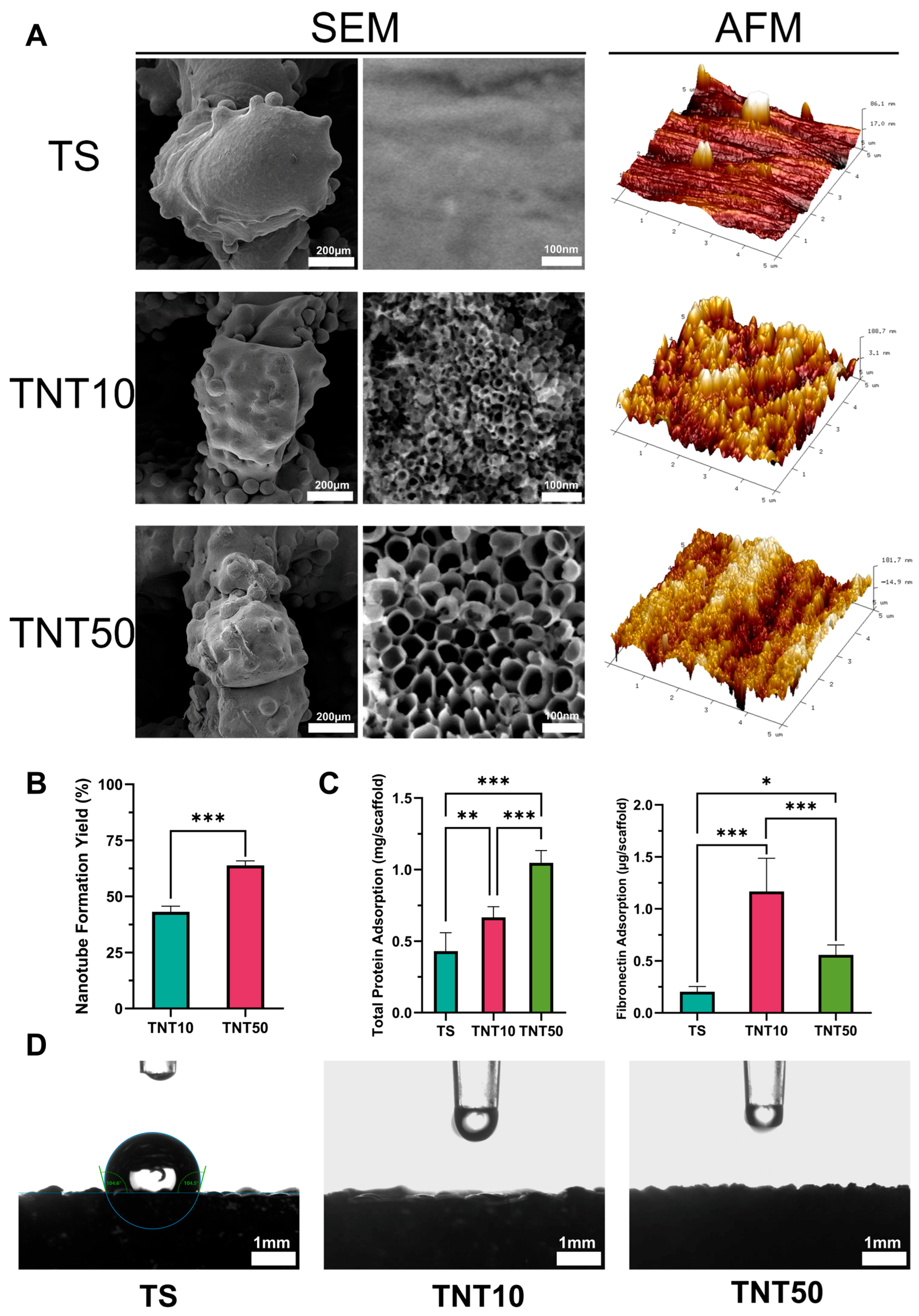
3.1. Material Characterization
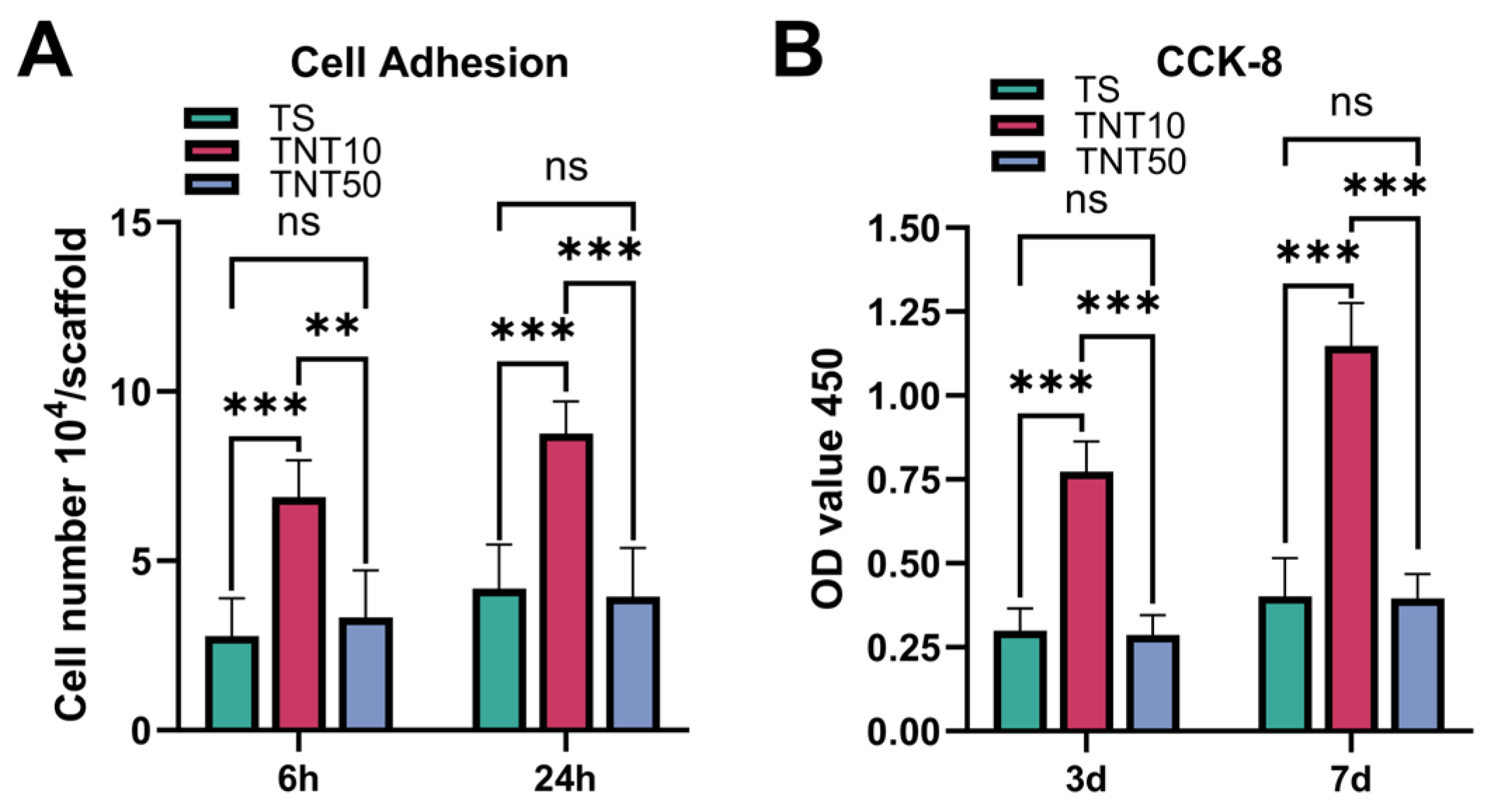
3.2. Cell Adhesion and Proliferation
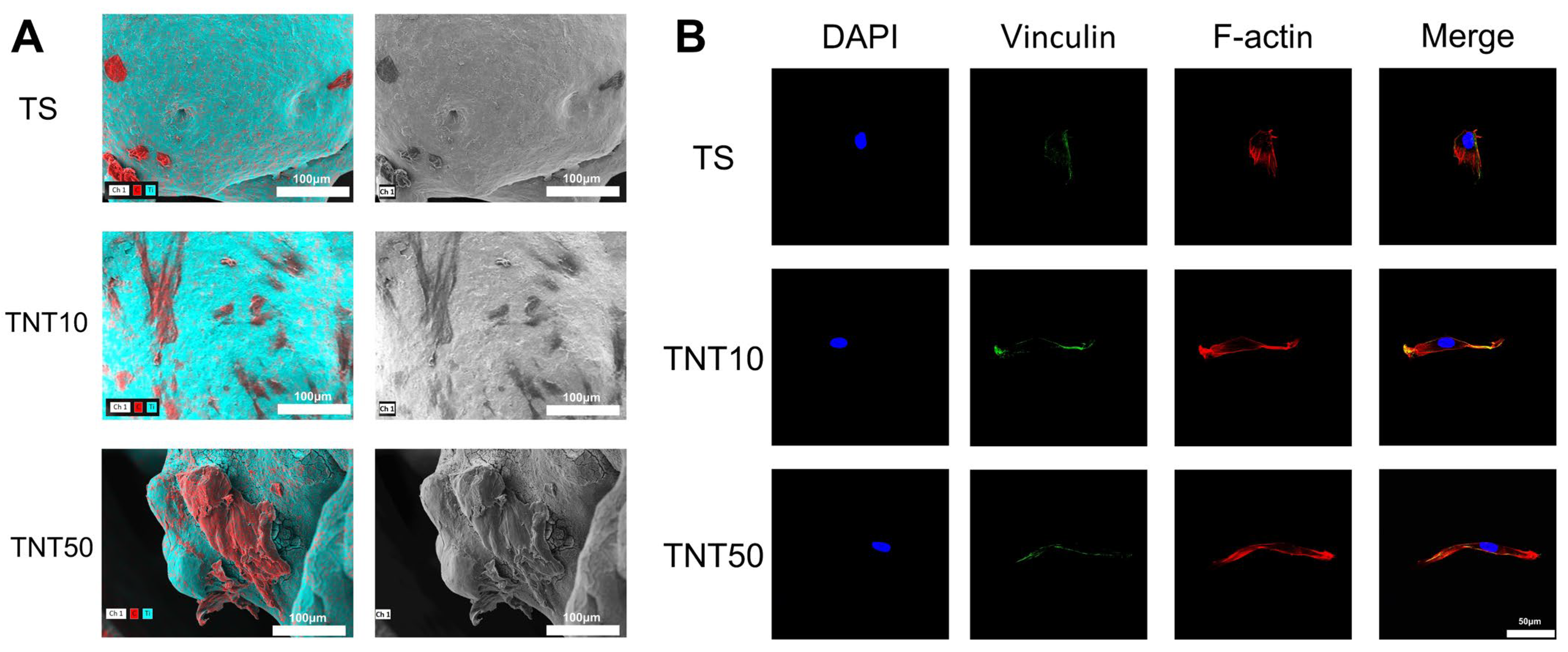
3.3. Cell Morphology and Focal Adhesion Staining
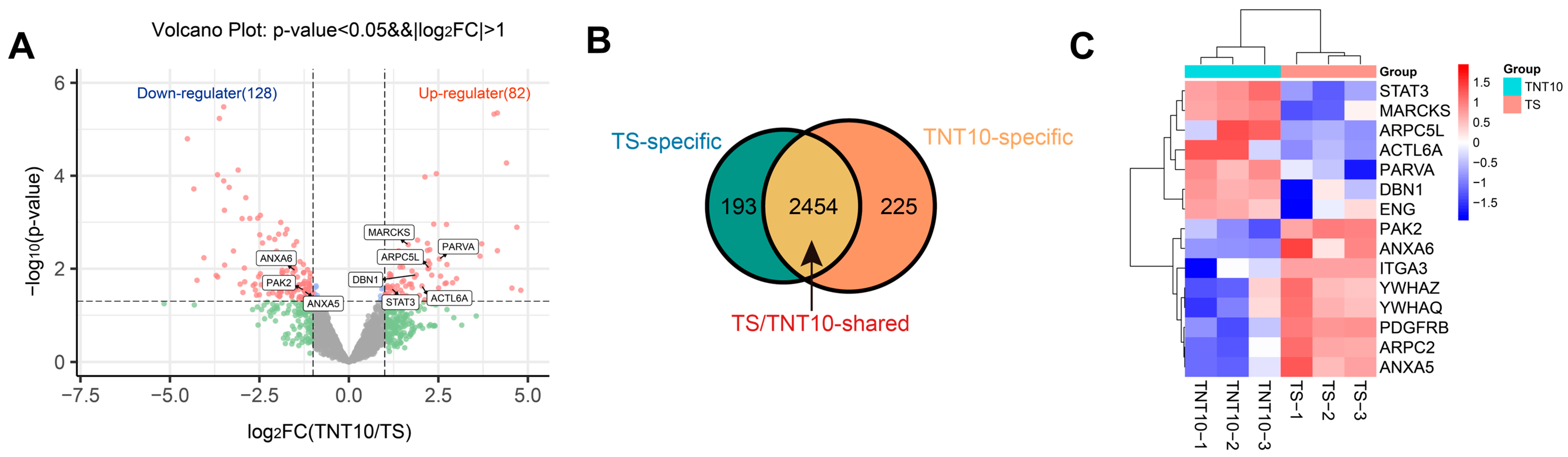
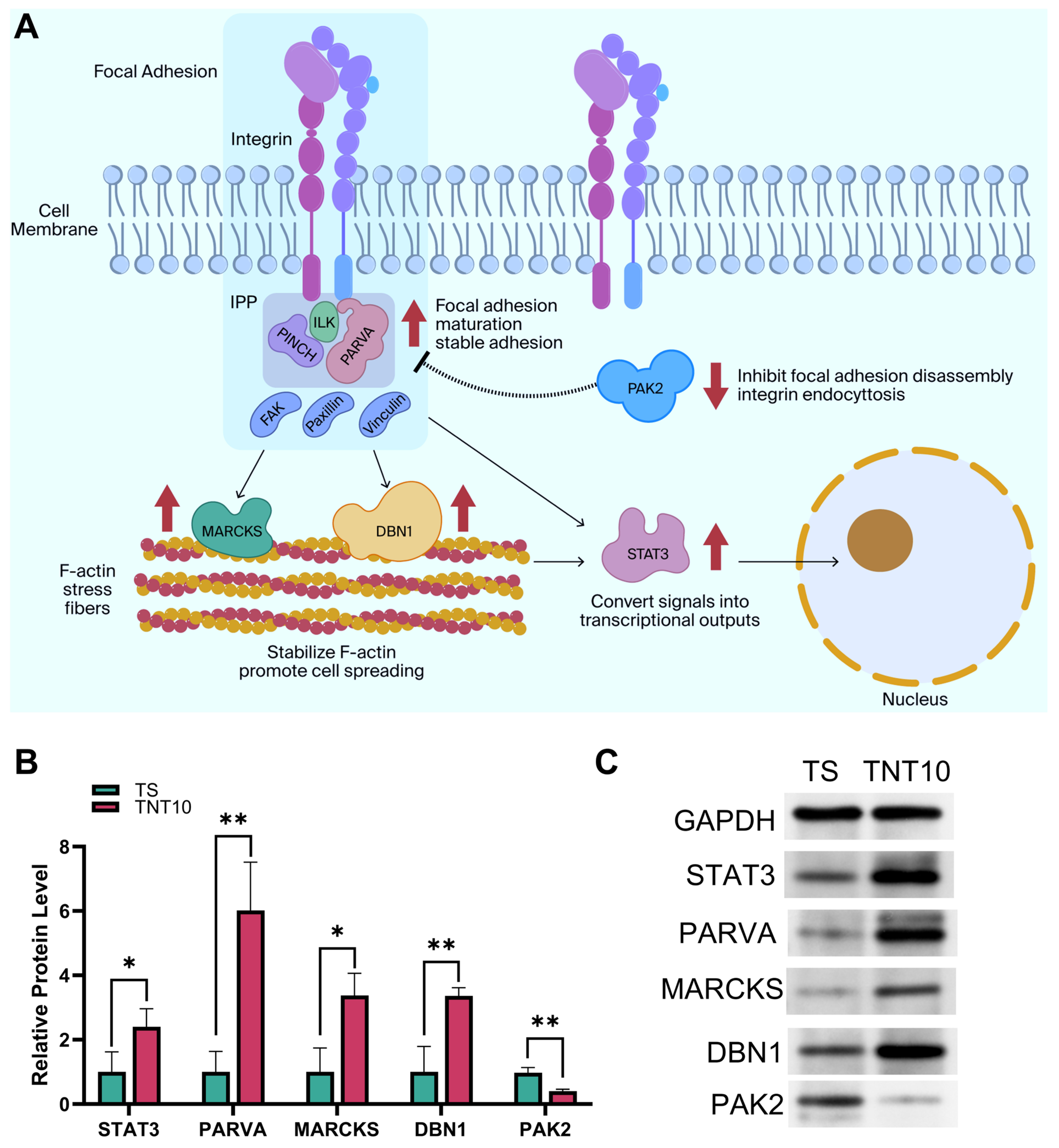
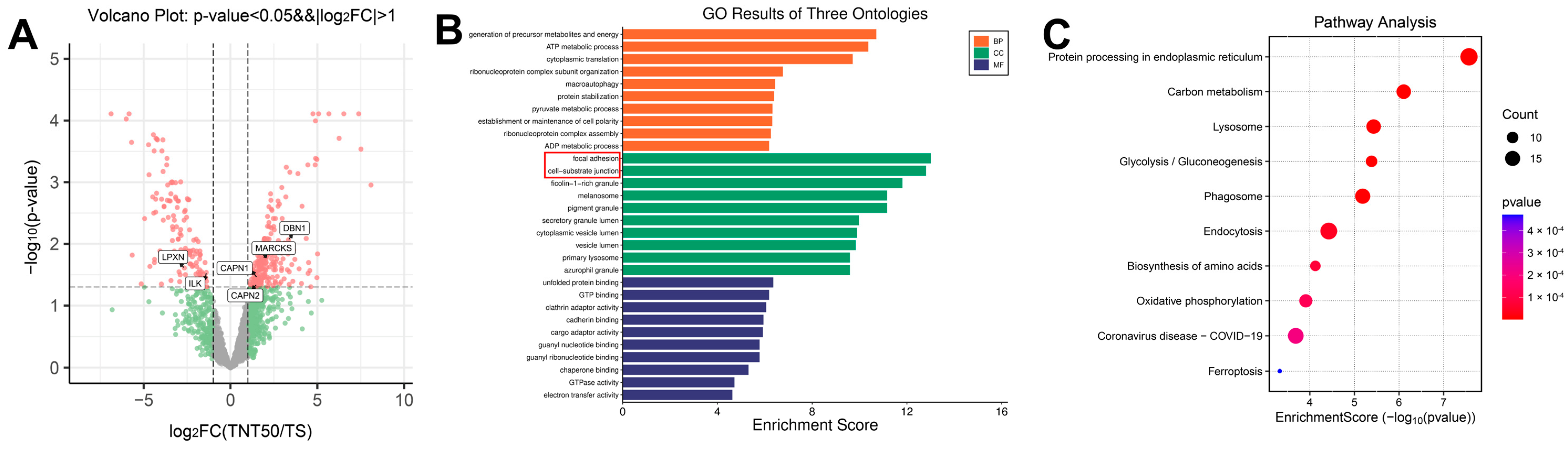
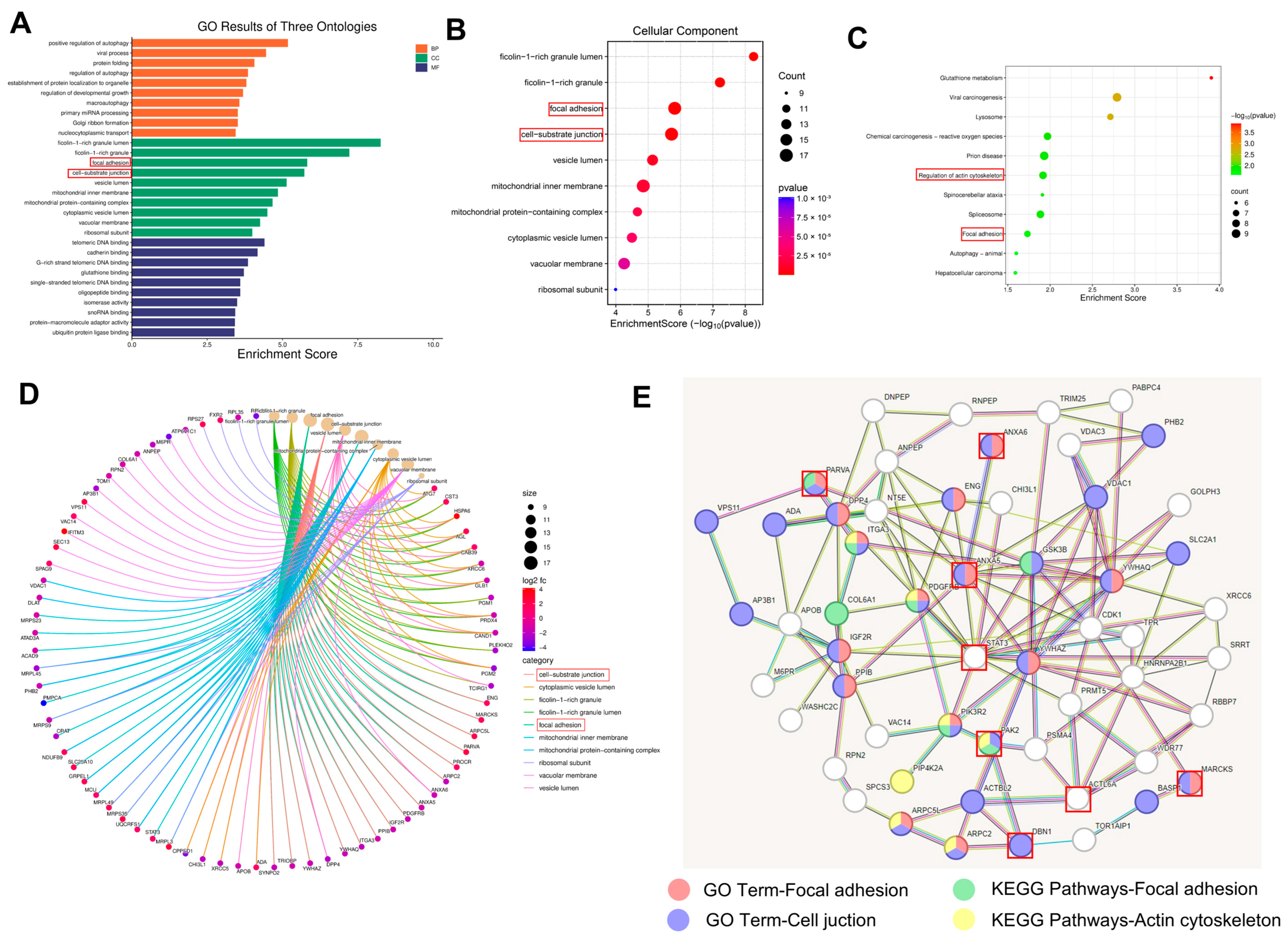
3.4. Proteomics—Mechanisms Promoting Adhesion
4. Conclusions
Supplementary Materials
Author Contributions
Funding
Institutional Review Board Statement
Informed Consent Statement
Data Availability Statement
Acknowledgments
Conflicts of Interest
Abbreviations
| AFM | atomic force microscope |
| ANOVA | analysis of variance |
| BCA | bicinchoninic acid |
| CCK-8 | cell counting kit-8 |
| CLSM | confocal laser scanning microscope |
| DBN1 | drebrin 1 |
| DEPs | differentially expressed proteins |
| EDS | energy-dispersive X-ray spectroscopy |
| FBS | fetal bovine serum |
| FN | fibronectin |
| GO | Gene Ontology |
| hBMSC | human bone marrow mesenchymal stem cell |
| ILK | integrin-linked kinase |
| IPP | ILK–PINCH–parvin |
| KEGG | Kyoto Encyclopedia of Genes and Genomes |
| LFQ | Label-Free Quantification |
| LPXN | leupaxin |
| MARCKS | Myristoylated alanine-rich C-kinase substrate |
| OD | optical density |
| PAK2 | p21-activated kinase 2 |
| PARVA | alpha-parvin |
| PBS | phosphate-buffered saline |
| PPI | protein–protein interaction |
| Ra | arithmetic mean roughness |
| Rq | root mean square roughness |
| SAD | surface area difference |
| SD | standard deviation |
| SEM | scanning electron microscope |
| STAT3 | signal transducers and activators of transcription 3 |
| TNT | titanium dioxide nanotube |
| TS | titanium alloy scaffold |
References
- Abd-Elaziem, W.; Darwish, M.A.; Hamada, A.; Daoush, W.M. Titanium-Based Alloys and Composites for Orthopedic Implants Applications: A Comprehensive Review. Mater. Des. 2024, 241, 112850. [Google Scholar] [CrossRef]
- Zhou, K.; Wang, M.; Zhang, S.; Huo, J.; Zheng, W.; Abueida, M.R.I.; Wang, Q.; Liu, H.; Yi, Z. Titanium alloys for orthopedic applications: A review on the osteointegration induced by physicomechanical stimuli. J. Mater. Res. Technol. 2024, 30, 8260–8276. [Google Scholar] [CrossRef]
- Long, E.G.; Buluk, M.; Gallagher, M.B.; Schneider, J.M.; Brown, J.L. Human mesenchymal stem cell morphology, migration, and differentiation on micro and nano-textured titanium. Bioact. Mater. 2019, 4, 249–255. [Google Scholar] [CrossRef] [PubMed]
- Huo, S.C.; Yue, B. Approaches to promoting bone marrow mesenchymal stem cell osteogenesis on orthopedic implant surface. World J. Stem Cells 2020, 12, 545–561. [Google Scholar] [CrossRef] [PubMed]
- Zhang, C.; Xie, B.; Zou, Y.; Zhu, D.; Lei, L.; Zhao, D.; Nie, H. Zero-dimensional, one-dimensional, two-dimensional and three-dimensional biomaterials for cell fate regulation. Adv. Drug Deliv. Rev. 2018, 132, 33–56. [Google Scholar] [CrossRef]
- Zhao, D.-P.; Kong, Y.-K.; Tang, J.-C.; Bai, F.; Ren, R.; Ma, F. Surface topography and cytocompatibility of metal injection molded Ti-22Nb alloy as biomaterial. Trans. Nonferrous Met. Soc. China 2018, 28, 1342–1350. [Google Scholar] [CrossRef]
- Tang, H.-C.; Chen, D.-P.; Xu, J.-C.; Xu, S.; Jin, W.; Yang, C.; Yang, K. Influence of heterogeneous microstructures on anodization behavior and biocompatibility of Ti-15Zr alloy. Trans. Nonferrous Met. Soc. China 2023, 33, 2076–2089. [Google Scholar] [CrossRef]
- Chen, W.; Shao, Y.; Li, X.; Zhao, G.; Fu, J. Nanotopographical surfaces for stem cell fate control: Engineering mechanobiology from the bottom. Nano Today 2014, 9, 759–784. [Google Scholar] [CrossRef]
- Newman, P.; Galenano-Niño, J.L.; Graney, P.; Razal, J.M.; Minett, A.I.; Ribas, J.; Ovalle-Robles, R.; Biro, M.; Zreiqat, H. Relationship between nanotopographical alignment and stem cell fate with live imaging and shape analysis. Sci. Rep. 2016, 6, 37909. [Google Scholar] [CrossRef]
- Zakir, O.; Ait-Karra, A.; Dikici, B.; Aityoub, A.; Abouelfida, A.; Outzourhit, A. A review on TiO2 nanotubes: Synthesis strategies, modifications, and applications. J. Solid State Electrochem. 2023, 27, 2289–2307. [Google Scholar] [CrossRef]
- Mydin, S.M.N.; Hazan, R.; Wan Eddis Effendy, W.N.; Romli, N.A.A.; Shaharuddin, S.; Khan, I.H.; Sreekantan, S. Medical implant surface technology: Geometrical formation of titania nanotube arrays by divergence anodization parameters. J. Bio- Tribo-Corros. 2023, 9, 69. [Google Scholar] [CrossRef]
- Tasdemir, N.K.; Kilicarslan, B.; Imren, G.; Karaosmanoglu, B.; Taskiran, E.Z.; Bayram, C. Hierarchical TiO2 nanotube arrays enhance mesenchymal stem cell adhesion and regenerative potential through surface nanotopography. J. R. Soc. Interface 2025, 22, 20240642. [Google Scholar] [CrossRef] [PubMed]
- Li, Y.; Wang, S.; Dong, Y.; Mu, P.; Yang, Y.; Liu, X.; Lin, C.; Huang, Q. Effect of size and crystalline phase of TiO2 nanotubes on cell behaviors: A high throughput study using gradient TiO2 nanotubes. Bioact. Mater. 2020, 5, 1062–1070. [Google Scholar] [CrossRef] [PubMed]
- Aguirre Ocampo, R.; Echeverry-Rendón, M.; Robledo, S.; Echeverría Echeverría, F. Effect of TiO2 nanotubes size, heat treatment, and UV irradiation on osteoblast behavior. Mater. Chem. Phys. 2022, 275, 125137. [Google Scholar] [CrossRef]
- Kanchanawong, P.; Calderwood, D.A. Organization, dynamics and mechanoregulation of integrin-mediated cell–ECM adhesions. Nat. Rev. Mol. Cell Biol. 2023, 24, 142–161. [Google Scholar] [CrossRef]
- Jo, J.; Abdi Nansa, S.; Kim, D.-H. Molecular regulators of cellular mechanoadaptation at cell–material interfaces. Front. Bioeng. Biotechnol. 2020, 8, 608569. [Google Scholar] [CrossRef]
- Zhao, D.-P.; Tang, J.-C.; Nie, H.-M.; Zhang, Y.; Chen, Y.-K.; Zhang, X.; Li, H.-X.; Yan, M. Macro-micron-nano-featured surface topography of Ti-6Al-4V alloy for biomedical applications. Rare Met. 2018, 37, 1055–1063. [Google Scholar] [CrossRef]
- Puga, M.L.; Venturini, J.; Caten, C.T.; Bergmann, C. Influencing parameters in the electrochemical anodization of TiO2 nanotubes: Systematic review and meta-analysis. Ceram. Int. 2022, 48, 19513–19526. [Google Scholar] [CrossRef]
- Kulkarni, M.; Mazare, A.; Park, J.; Gongadze, E.; Killian, M.S.; Kralj, S.; von der Mark, K.; Iglič, A.; Schmuki, P. Protein interactions with layers of TiO2 nanotube and nanopore arrays: Morphology and surface charge influence. Acta Biomater. 2016, 45, 357–366. [Google Scholar] [CrossRef]
- Wu, B.; Tang, Y.; Wang, K.; Zhou, X.; Xiang, L. Nanostructured titanium implant surface facilitating osseointegration from protein adsorption to osteogenesis: The example of TiO2 NTAs. Int. J. Nanomed. 2022, 17, 1865–1879. [Google Scholar] [CrossRef]
- Wu, S.; Zhang, D.; Bai, J.; Zheng, H.; Deng, J.; Gou, Z.; Gao, C. Adsorption of serum proteins on titania nanotubes and its role on regulating adhesion and migration of mesenchymal stem cells. J. Biomed. Mater. Res. Part A 2020, 108, 2305–2318. [Google Scholar] [CrossRef] [PubMed]
- Parvate, S.; Dixit, P.; Chattopadhyay, S. Superhydrophobic surfaces: Insights from theory and experiment. J. Phys. Chem. B 2020, 124, 1323–1360. [Google Scholar] [CrossRef] [PubMed]
- Park, J.; Bauer, S.; von der Mark, K.; Schmuki, P. Nanosize and vitality: TiO2 nanotube diameter directs cell fate. Nano Lett. 2007, 7, 1686–1691. [Google Scholar] [CrossRef] [PubMed]
- Yang, W.; Xi, X.; Ran, Q.; Liu, P.; Hu, Y.; Cai, K. Influence of the titania nanotubes dimensions on adsorption of collagen: An experimental and computational study. Mater. Sci. Eng. C 2014, 34, 410–416. [Google Scholar] [CrossRef]
- Tong, Z.; Liu, Y.; Xia, R.; Chang, Y.; Hu, Y.; Liu, P.; Zhai, Z.; Zhang, J.; Li, H. F-actin regulates osteoblastic differentiation of mesenchymal stem cells on TiO2 nanotubes through MKL1 and YAP/TAZ. Nanoscale Res. Lett. 2020, 15, 183. [Google Scholar] [CrossRef]
- Chang, Y.; Kong, K.; Tong, Z.; Qiao, H.; Jin, M.; Wu, X.; Ouyang, Z.; Zhang, J.; Zhai, Z.; Li, H. TiO2 nanotube topography enhances osteogenesis through filamentous actin and XB130-protein-mediated mechanotransduction. Acta Biomater. 2024, 177, 525–537. [Google Scholar] [CrossRef]
- Voltrova, B.; Hybasek, V.; Blahnova, V.; Sepitka, J.; Lukasova, V.; Vocetkova, K.; Sovkova, V.; Matejka, R.; Fojt, J.; Joska, L.; et al. Different diameters of titanium dioxide nanotubes modulate Saos-2 osteoblast-like cell adhesion and osteogenic differentiation and nanomechanical properties of the surface. RSC Adv. 2019, 9, 11341–11355. [Google Scholar] [CrossRef]
- Khaw, J.S.; Bowen, C.R.; Cartmell, S.H. Effect of TiO2 nanotube pore diameter on human mesenchymal stem cells and human osteoblasts. Nanomaterials 2020, 10, 2117. [Google Scholar] [CrossRef]
- Yim, E.K.; Darling, E.M.; Kulangara, K.; Guilak, F.; Leong, K.W. Nanotopography-induced changes in focal adhesions, cytoskeletal organization, and mechanical properties of human mesenchymal stem cells. Biomaterials 2010, 31, 1299–1306. [Google Scholar] [CrossRef]
- Ain, U.; Fatma, B.; Firdaus, H. The ILK–PINCH–parvin complex: A conserved primary adhesome regulating biological processes. Histochem. Cell Biol. 2025, 163, 78. [Google Scholar] [CrossRef]
- Vakaloglou, K.M.; Chrysanthis, G.; Rapsomaniki, M.A.; Lygerou, Z.; Zervas, C.G. IPP complex reinforces adhesion by relaying tension-dependent signals to inhibit integrin turnover. Cell Rep. 2016, 16, 596–610. [Google Scholar] [CrossRef] [PubMed]
- Chen, Z.; Zhang, W.; Selmi, C.; Ridgway, W.M.; Leung, P.S.C.; Zhang, F.; Gershwin, M.E. The myristoylated alanine-rich C-kinase substrate (MARCKS): A membrane-anchored mediator of cell function. Autoimmun. Rev. 2021, 20, 102942. [Google Scholar] [CrossRef] [PubMed]
- McGowan, S.E.; Gilfanov, N.; Chandurkar, M.K.; Stiber, J.A.; Han, S.J. Drebrin is required for myosin-facilitated actin cytoskeletal remodeling during pulmonary alveolar development. Am. J. Respir. Cell Mol. Biol. 2024, 70, 308–321. [Google Scholar] [CrossRef] [PubMed]
- Snyder, M.; Huang, X.Y.; Zhang, J.J. Signal transducers and activators of transcription 3 (STAT3) directly regulates cytokine-induced fascin expression and is required for breast cancer cell migration. J. Biol. Chem. 2011, 286, 38886–38893. [Google Scholar] [CrossRef]
- Wu, M.; Sarkar, C.; Guo, B. Regulation of cancer metastasis by PAK2. Int. J. Mol. Sci. 2024, 25, 13443. [Google Scholar] [CrossRef]
- Chan, P.M.; Lim, L.; Manser, E. PAK is regulated by PI3K, PIX, CDC42, and PP2Cα and mediates focal adhesion turnover in the hyperosmotic stress-induced p38 pathway. J. Biol. Chem. 2008, 283, 24949–24961. [Google Scholar] [CrossRef]
- Elad, N.; Volberg, T.; Patla, I.; Hirschfeld-Warneken, V.; Grashoff, C.; Fässler, R.; Spatz, J.P.; Geiger, B.; Medalia, O. The role of integrin-linked kinase in the molecular architecture of focal adhesions. J. Cell Sci. 2013, 126, 4099–4107. [Google Scholar] [CrossRef]
- Alpha, K.M.; Xu, W.; Turner, C.E. Paxillin family of focal adhesion adaptor proteins and regulation of cancer cell invasion. Int. Rev. Cell Mol. Biol. 2020, 355, 1–52. [Google Scholar]
- Chen, P.-W.; Kroog, G.S. Leupaxin is similar to paxillin in focal adhesion targeting and tyrosine phosphorylation but has distinct roles in cell adhesion and spreading. Cell Adhes. Migr. 2010, 4, 527–540. [Google Scholar] [CrossRef]
- Franco, S.J.; Rodgers, M.A.; Perrin, B.J.; Han, J.; Bennin, D.A.; Critchley, D.R.; Huttenlocher, A. Calpain-mediated proteolysis of talin regulates adhesion dynamics. Nat. Cell Biol. 2004, 6, 977–983. [Google Scholar] [CrossRef]
- Chan, K.T.; Bennin, D.A.; Huttenlocher, A. Regulation of adhesion dynamics by calpain-mediated proteolysis of focal adhesion kinase (FAK). J. Biol. Chem. 2010, 285, 11418–11426. [Google Scholar] [CrossRef]





| Parameter | TS | TNT10 | TNT50 | Significance |
|---|---|---|---|---|
| Surface roughness | ||||
| Ra (nm) | 34.1 ± 9.2 | 47.8 ± 5.0 | 61.2 ± 13.4 | * p < 0.05 |
| Rq (nm) | 44.2 ± 11.7 | 59.6 ± 5.7 | 76.5 ± 13.8 | * p < 0.05 |
| SAD (%) | 2.6 ± 1.6 | 22.2 ± 0.8 | 28.0 ± 6.1 | *** p < 0.001 |
| Static water contact angle (°) | 104.2 ± 1.0 | 0 | 0 | *** p < 0.001 |
| Nanotube diameter (nm) | / | 11.5 ± 5.5 | 65.9 ± 11.2 | *** p < 0.001 |
| Nanotube length (nm) | / | 432 ± 61.9 | 1023 ± 85.0 | *** p < 0.001 |
| Sample | Ti | O | Al | V | F | C |
|---|---|---|---|---|---|---|
| TS | 64.9 | 13.5 | 9.0 | 2.4 | 0.4 | 9.6 |
| TNT10 | 60.3 | 23.4 | 4.8 | 2.5 | 3.9 | 4.8 |
| TNT50 | 44.2 | 32.7 | 5.5 | 2.1 | 5.8 | 5.9 |
Disclaimer/Publisher’s Note: The statements, opinions and data contained in all publications are solely those of the individual author(s) and contributor(s) and not of MDPI and/or the editor(s). MDPI and/or the editor(s) disclaim responsibility for any injury to people or property resulting from any ideas, methods, instructions or products referred to in the content. |
© 2026 by the authors. Licensee MDPI, Basel, Switzerland. This article is an open access article distributed under the terms and conditions of the Creative Commons Attribution (CC BY) license.
Share and Cite
Xiong, C.; Feng, H.; Lu, L.; Jing, Z.; Wang, Y.; Yang, Y.; Meng, D.; Zhang, Y.; Li, W.; Cai, H. Nanotopography-Mediated Mechanotransduction Enhances hBMSCs Adhesion on TiO2 Nanotubes. J. Funct. Biomater. 2026, 17, 200. https://doi.org/10.3390/jfb17040200
Xiong C, Feng H, Lu L, Jing Z, Wang Y, Yang Y, Meng D, Zhang Y, Li W, Cai H. Nanotopography-Mediated Mechanotransduction Enhances hBMSCs Adhesion on TiO2 Nanotubes. Journal of Functional Biomaterials. 2026; 17(4):200. https://doi.org/10.3390/jfb17040200
Chicago/Turabian StyleXiong, Chenao, Hui Feng, Liyang Lu, Zehao Jing, Youhao Wang, Yiyuan Yang, Dexuan Meng, Yichen Zhang, Weishi Li, and Hong Cai. 2026. "Nanotopography-Mediated Mechanotransduction Enhances hBMSCs Adhesion on TiO2 Nanotubes" Journal of Functional Biomaterials 17, no. 4: 200. https://doi.org/10.3390/jfb17040200
APA StyleXiong, C., Feng, H., Lu, L., Jing, Z., Wang, Y., Yang, Y., Meng, D., Zhang, Y., Li, W., & Cai, H. (2026). Nanotopography-Mediated Mechanotransduction Enhances hBMSCs Adhesion on TiO2 Nanotubes. Journal of Functional Biomaterials, 17(4), 200. https://doi.org/10.3390/jfb17040200