Article

Stress Distribution on Endodontically Treated Anterior Teeth Restored via Different Ceramic Materials with Varying Post Lengths Versus Endocrown—A 3D Finite Element Analysis

1
Department of Clinical Dental Sciences, College of Dentistry, Princess Nourah bint Abdulrahman University, Riyadh 11671, Saudi Arabia
2
Department of Conservative Dental Sciences, College of Dentistry, Qassim University, Buraydah 52571, Saudi Arabia
3
Department of Prosthetic Dental Sciences, College of Dentistry, Qassim University, Buraydah 52571, Saudi Arabia
4
General Dentistry, College of Dentistry, Qassim University, Buraydah 52571, Saudi Arabia
5
Department of Fixed Prosthodontics, Faculty of Dental Surgery, MUST University, 6th of October City, Giza 12566, Egypt
*
Author to whom correspondence should be addressed.
J. Funct. Biomater. 2025, 16(6), 221; https://doi.org/10.3390/jfb16060221
Submission received: 5 April 2025 / Revised: 12 May 2025 / Accepted: 10 June 2025 / Published: 12 June 2025
(This article belongs to the Special Issue Advances in Restorative Dentistry Materials)

Abstract

Objective: This study aims to evaluate the stress distribution on endodontically treated anterior teeth restored using different restorative materials and different post lengths versus endocrowns employing finite element analysis (FEA). Methods: An extracted human central incisor tooth with a fully formed apex was scanned using high-resolution cone beam computed tomography (CBCT) to generate 3D finite element models. Six models of restorations of badly destructed central incisor were grouped according to the type of ceramic material and post length versus endocrown restorations. Group V-L: Vita Enamic, long post (10 mm intra-radicular), Group C-L: Celtra Duo, long post (10 mm intra-radicular), Group V-Sh: Vita Enamic, short post (3 mm intra-radicular), Group C-Sh: Celtra Duo, short post (3 mm intra-radicular), Group V-E: Vita Enamic endocrown (3 mm intra-radicular), and Group C-E: Celtra Duo endocrown (3 mm intra-radicular). A static load of 200 N was applied to the palatal surface at a 45 degree angle to the tooth’s long axis. The maximum equivalent von Mises stress and maximum principal stress were analyzed at four locations: the finish line, coronal third of the root (12 mm from the apex), middle third of the root (8 mm from the apex), and apical third of the root (4 mm from the apex). Results: Group C-L exhibited the highest maximum VM stress and PS at the finish line, in addition to the highest maximum VM stress and PS at the root apical third, while group C-Sh reported the least maximum VM stress at the root apical third among the groups. All Celtra Duo groups reported higher maximum VM stress than the corresponding groups of Vita Enamic at the finish line and root coronal thirds. However, at the root middle and apical thirds, both materials recorded similar stresses. Conclusions: Short posts and Vita Enamic endocrowns showed minimal stress, especially at the finish line, while long posts increased stress and fracture risk. The findings support conservative restorations without posts, although clinical validation is needed to confirm their long-term effectiveness and safety.

1. Introduction

Endodontically treated teeth are inherently more susceptible to structural damage and fractures compared to vital teeth. This increased risk of fracture is primarily due to loss of tooth structure during endodontic therapy and subsequent restorative procedures, which can compromise the tooth’s integrity and biomechanical properties. The presence of a post and core, along with various restorative prostheses, plays a crucial role in the prognosis and durability of these treated teeth [1,2,3,4,5].
Finite element analysis (FEA) is a computational tool that simulates the behavior of structures under various conditions. It employs the finite element method (FEM) to divide a structure into small, discrete elements and solve the equations of equilibrium for each element. This method allows for the detailed analysis of stress distribution, identifying potential weak spots and design flaws [6]. FEA has been successfully adopted in various engineering fields and has proven to be particularly beneficial in the field of dentistry for studying the biomechanical behaviors of dental structures and materials [7,8].
The application of FEA in dentistry has provided valuable insights into the stress distribution and fracture resistance of endodontically treated teeth restored with different materials and techniques. Studies have shown that the type of post, core, and crown material, as well as the length and design of the post, significantly affect the stress distribution within the restored tooth [9]. Proper selection of these components is essential to enhance the longevity and performance of the restoration.
Endodontically treated teeth often require the placement of a post to retain the core material and provide adequate support for the final restoration. The post and core system compensates for the loss of tooth structure and helps to distribute occlusal forces along the length of the root, reducing the risk of fracture [10,11,12]. Various materials are used for posts, including metal, fiber-reinforced composite, and ceramic, each with its own biomechanical properties and clinical implications [13,14,15,16].
FEA has become an indispensable tool in evaluating the biomechanical performance of various post, core, and crown materials used in the restoration of endodontically treated teeth. The selection of these materials significantly influences stress distribution within the tooth structure, thereby affecting the longevity and success of dental restorations [17].
Core materials are essential in reinforcing structurally compromised teeth, particularly following endodontic treatment. Their mechanical properties, notably the elastic modulus, significantly influence stress distribution within the tooth–restoration complex. FEA studies have demonstrated that composite resin cores, due to their elastic modulus being closer to that of dentin, facilitate a more uniform stress distribution. This compatibility reduces the risk of stress concentrations that could lead to structural failures. For instance, a study by Ouldyerou et al. (2023) [17] revealed that increasing the Young’s modulus of composite materials from 10 GPa to 26 GPa resulted in decreased stress levels in both enamel and dentin under occlusal loading, highlighting the importance of selecting core materials with mechanical properties akin to natural tooth structures [17].
By contrast, metallic cores, such as cast gold alloys, possess a significantly higher elastic modulus compared to dentin, leading to increased stress concentrations at the core–dentin interface. This disparity can result in unfavorable stress distributions, potentially compromising the integrity of the tooth [17].
Fiber posts have gained popularity due to their favorable biomechanical properties, such as a modulus of elasticity similar to dentin, which helps in reducing stress concentrations and improving fracture resistance [9,18,19]. However, the effectiveness of fiber posts depends on several factors, including the length of the post, the type of core material, and the design of the final restoration. The choice of crown material also plays a crucial role in the overall performance of the restored tooth [20,21,22,23].
The elastic modulus of post materials plays a crucial role in stress distribution. Materials with an elastic modulus closer to that of dentin, such as glass fiber posts, have been shown to produce more homogeneous stress distributions, potentially reducing the risk of root fractures [24]. Conversely, stiffer materials like cobalt-chromium can lead to higher stress concentrations, particularly in areas with complex root canal morphologies. This underscores the importance of material selection based on the specific anatomical considerations of the tooth being restored [25].
Vita Enamic and Celtra Duo are two widely used materials for dental crowns. Vita Enamic is a hybrid ceramic material that combines the properties of ceramic and composite, offering high strength, durability, and esthetics. Celtra Duo is a zirconia-reinforced lithium silicate ceramic known for its excellent mechanical properties and esthetic qualities [26]. Both materials have been extensively studied for their performance in dental restorations, but their impact on stress distribution in endodontically treated teeth requires further investigation [27].
Crown materials also significantly impact stress distribution. A study by Lin, J. et al. (2020) [23] compared zirconia and lithium disilicate-reinforced glass ceramic crowns, finding that zirconia crowns exhibited higher stress concentrations in the crown structure, while lithium disilicate crowns showed more favorable stress distributions in the underlying tooth structure. These findings highlight the need for a comprehensive approach in selecting crown materials, considering both the mechanical properties of the materials and their interaction with the tooth–restoration complex [23].
In addition to material properties, the design of the restoration, particularly the length of the post, has a significant impact on the stress distribution within the tooth. Longer posts are generally believed to provide better retention and stability for the core and crown, but they can also introduce higher stress concentrations at the apical end, potentially leading to root fractures [28,29,30,31]. Conversely, shorter posts may reduce the risk of apical stress but may not provide sufficient support for the restoration, especially in teeth with extensive structural loss [32].
The concept of endocrowns, which integrate the core and crown into a single restoration with an intra-radicular extension, has gained attention as an alternative approach for restoring endodontically treated teeth. Endocrowns are designed to distribute occlusal forces more evenly and minimize stress concentrations at critical points within the tooth structure [33,34,35]. Studies have shown that endocrowns can be particularly effective in restoring molars and premolars, but their application in anterior teeth requires further exploration [36,37,38].
This study aims to evaluate the stress distribution on endodontically treated anterior teeth using different restoration materials with variable post lengths through FEA. By comparing the performance of Vita Enamic and Celtra Duo crowns, with both long and short posts, as well as endocrowns with 3 mm intra-radicular extensions, this study seeks to identify the optimal restorative approach for enhancing the durability and prognosis of endodontically treated anterior teeth.

2. Materials and Methods

2.1. Model Construction

This in vitro study was granted ethical approval from the Committee of Research Ethics, Deanship of Scientific Research, Qassim University, under study number IRB 24-76-09. All methods were performed in accordance with the relevant guidelines and regulations. A fully developed human central incisor tooth with fully formed apex was scanned using high-resolution cone beam computed tomography (CBCT) (Planmeca ProMax 3D MID; Planmeca, Helsinki, Finland) operating at 90 kV and 12 mA with a voxel dimension of 75 mm, to obtain about 800 images. The images were processed using the Materialize Interactive Medical Image Control System (MIMICS 19.0; Materialise, Leuven, Belgium) to identify enamel and dentin tissues using voxel intensity and the mathematical region growing feature. A three-dimensional stereolithography (3D-STL) file format was then exported for both enamel and dentin structures, and the data were optimized using 3-Matic Medical 11.0 software (Materialise NV).
The file was imported into SolidWorks software 2018 (Dassault Systems, Vélizy-Villacoublay, France), where enamel and dentin were combined. The surrounding periodontal ligaments (PDL) were then established, extending from the cementoenamel junction (CEJ) to the apical portion of the root with a uniform thickness of 200 µm, and supported by alveolar bone 3 mm below the CEJ.

2.2. Restoration Modeling and Grouping (Table 1 and Table 2)

Six three-dimensional finite element models were created, with each representing a restorative scenario:
  • Group V-L: Vita Enamic crown—Long post; 10 mm intra-radicular length, and composite core.
  • Group C-L: Celtra Duo crown—Long post; 10 mm intra-radicular length, and composite core.
  • Group V-Sh: Vita Enamic crown—Short post; 3 mm intra-radicular length, and composite core.
  • Group C-Sh: Celtra Duo crown—Short post; 3 mm intra-radicular length, and composite core.
  • Group V-E: Vita Enamic—Endocrown with 3 mm intra-radicular extension.
  • Group C-E: Celtra Duo—Endocrown with 3 mm intra-radicular extension.

2.3. Model Preparation

The coronal portion of the created models was prepared to simulate a badly broken central incisor, leaving 2.0 mm ferrule coronal to the cementoenamel junction (CEJ) and a 0.5 mm chamfer finish line.
The root canal was prepared following the dimensions of the Protaper Next (PTN) rotary filing system (Dentsply; Maillefer, Ballaigu, Switzerland) with an estimated size X3: 30/0.07 taper and shaped to a centralized conical dimension. Afterward, the canal was obturated using gutta-percha points size X3 of the same PTN rotary filling system, together with Bioceramic sealer (Brasseler USA, Savannah, GA, USA), starting 0.5 mm from the apex and extending coronally.
The gutta-percha was then seared to the desired lengths according to the estimated groups. As for the groups using long posts [Group V-L and C-L], 10 mm of the gutta-percha was removed coronally. However, for the groups using short posts [Group V-Sh and C-Sh], only 3 mm of gutta-percha was removed coronally from the canal. For the endocrown restorations [Group V-E and C-E], 3 mm of the gutta-percha was removed coronally as well.
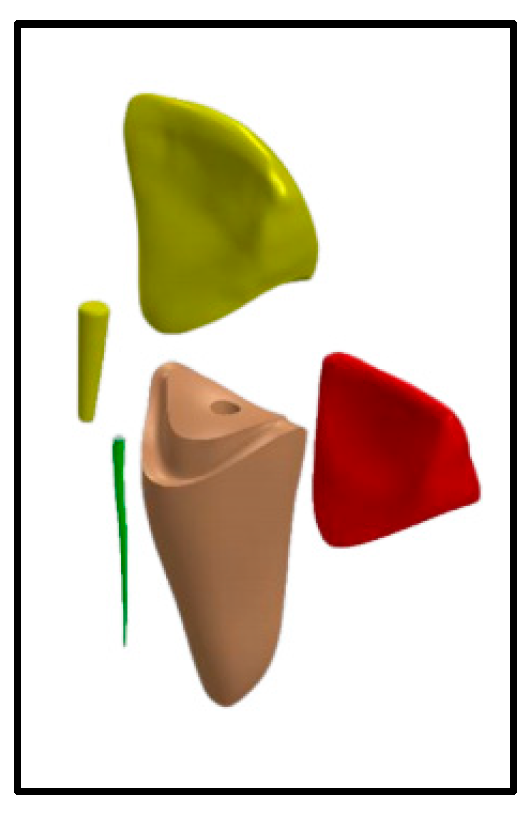
For the groups using long posts [Group V-L and C-L], a size 1/yellow drill was used for post space preparation, then size 1/yellow fiber post (3M™ RelyX™ Fiber Post 3D Glass Fiber Post, 3M Canada, London, ON, Canada) was cemented using self-adhesive resin cement (3M™ RelyX™ Unicem 2 Automix, 3M Canada, Canada) (Figure 1).
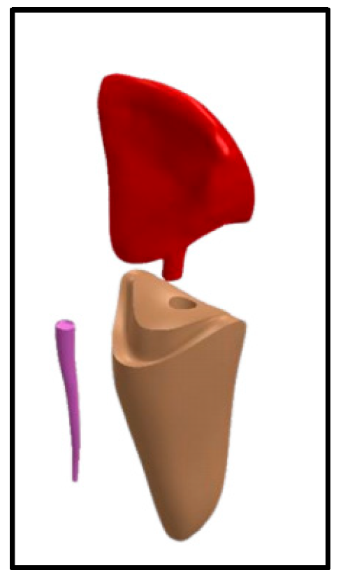
For the groups using short posts [Group V-Sh and C-Sh], a size 2/red drill was used for post space preparation, then size 2/red fiber post (3M™ RelyX™ Fiber Post 3D Glass Fiber Post, 3M Canada, London, ON, Canada) was cemented using self-adhesive resin cement (3M™ RelyX™ Unicem 2 Automix, 3M Canada, London, ON, Canada) (Figure 2).
For both the short and long post groups [Group V-L, C-L, V-Sh, and C-Sh], adhesive for core build up (3M™ Scotchbond™ Universal Adhesive, 3M Canada, Canada) and core build-up material (3M™ Filtek™ One Bulk Fill Restorative, 3M Canada, Canada) were used to build the composite core according to manufacturer’s instructions. Composite cores were prepared to receive ceramic crowns that were fabricated using Vita Enamic (Vita, Vita Zahnfabrik, Bad Säckingen, Germany) and Celtra Duo (Dentsply, Sirona, Dental Systems Gmbh FabrikstraBe, Bensheim, Germany), respectively (Figure 1 and Figure 2).
For the groups restored by endocrown [Group V-E and C-E], a size 2/red post drill was used for post space preparation, the endocrowns were CAD/CAM-fabricated using Vita Enamic (Vita, Vita Zahnfabrik, Bad Säckingen, Germany) and Celtra Duo (Dentsply, Sirona, Dental Systems Gmbh FabrikstraBe, Bensheim, Germany), respectively, and then finished and polished according to the manufacturer’s instruction (Figure 3).
The ceramic crowns and endocrowns were finally cemented on their respective models using self-adhesive resin cement (3M™ RelyX™ Unicem 2 Automix, 3M Canada, Canada).

Boundary Conditions and Load Application

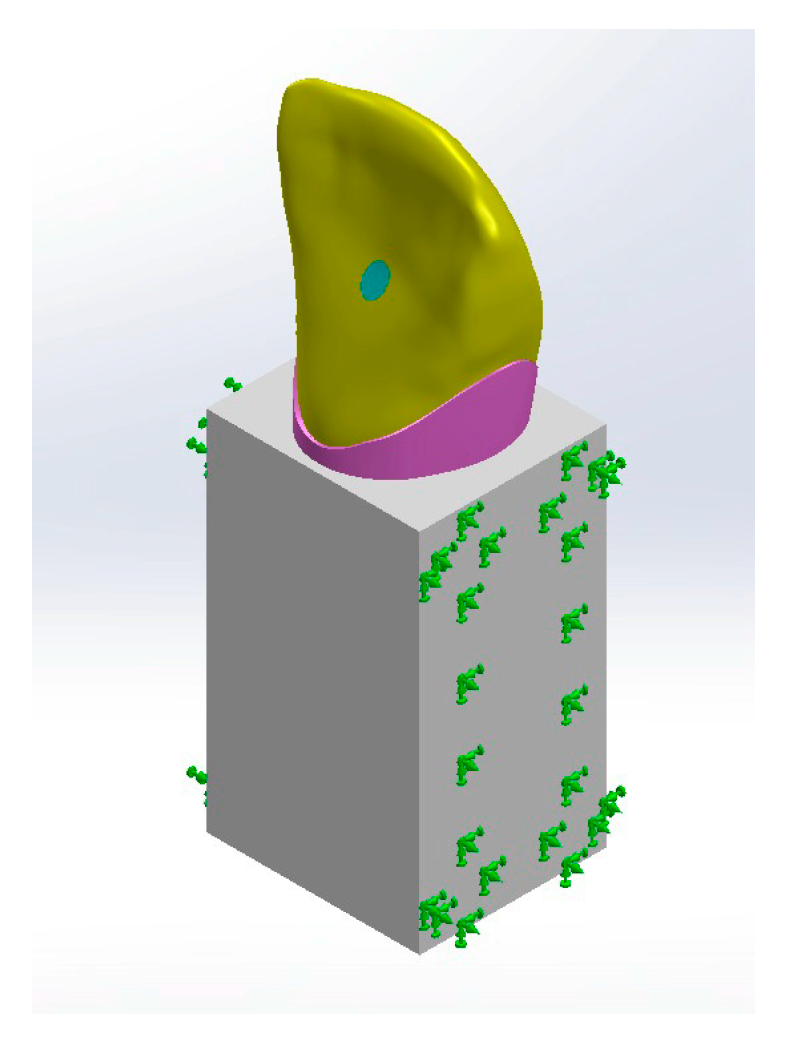
All bodies were simulated to be bonded to restrict the movement of a specific node or group of nodes within the finite element model. This constraint simulated a real-world situation where a physical object is fixed or attached to a stationary surface or structure, preventing it from moving freely in one or more directions. Boundary bonding means “no body is allowed to move relative to the body touching it” to any touching body. The cancellous block was fixed at both mesial and distal surfaces to isolate the region of interest (ROI) from outer effects (Figure 4).
The 3D models of the maxillary central incisor were subjected to a static load of 200 N magnitude directed on the lingual surface at a 45 degree angle to the tooth axis [39,40]. The evaluation of von Mises (VM) stress and principal stress (PS) focused on the pattern of stress distribution, and the maximum values were observed (Figure 4).
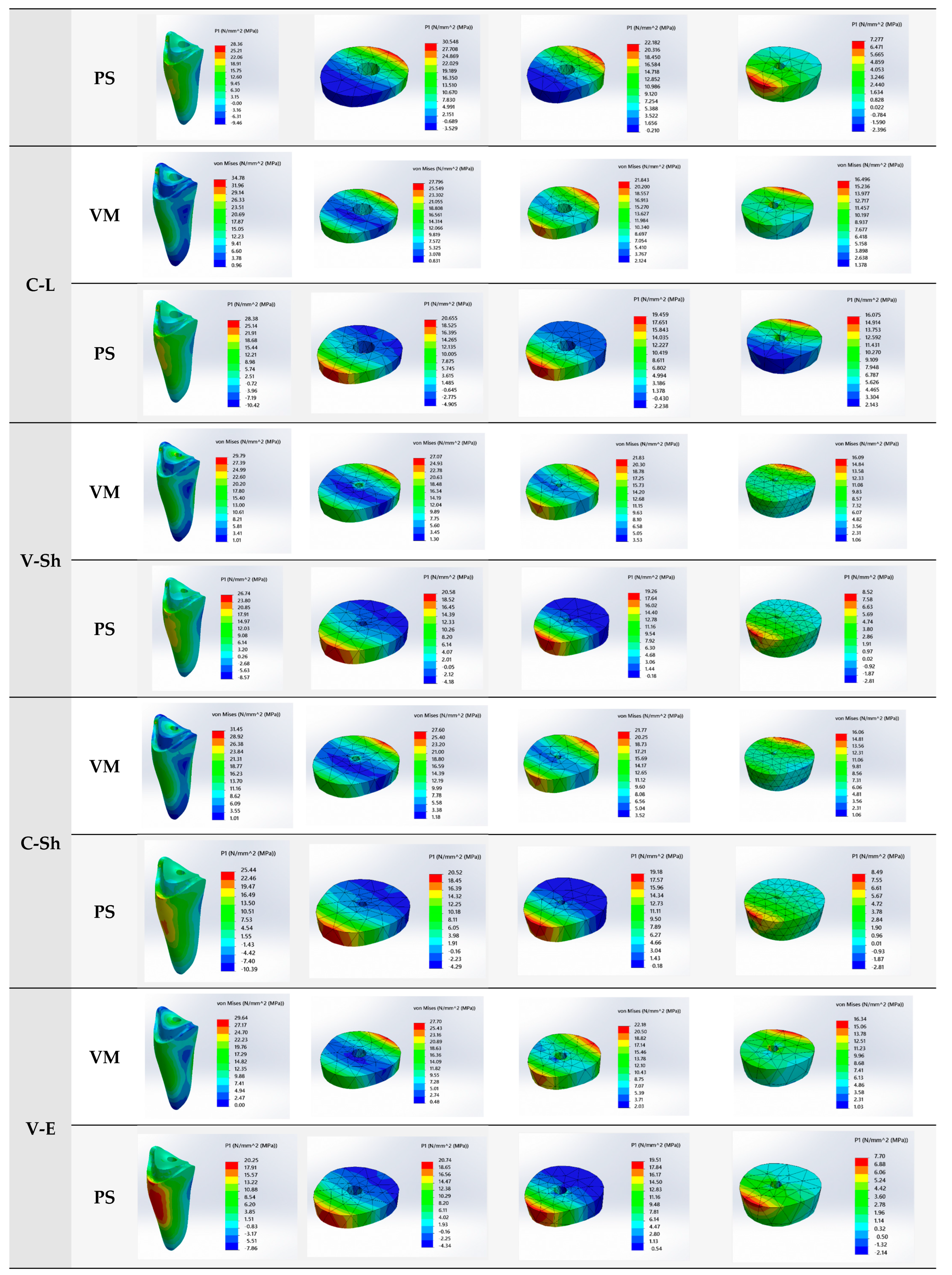
The magnitudes of the VM stress and PS were observed at four locations: the finish line, coronal third of the root (12 mm from the apex), middle third of the root (8 mm from the apex), and apical third of the root (4 mm from the apex) of the models [41,42]. Stress distribution was analyzed as colorimetric maps, with adjustable color scales corresponding to stress magnitude comparisons among the designs for each analyzed group.
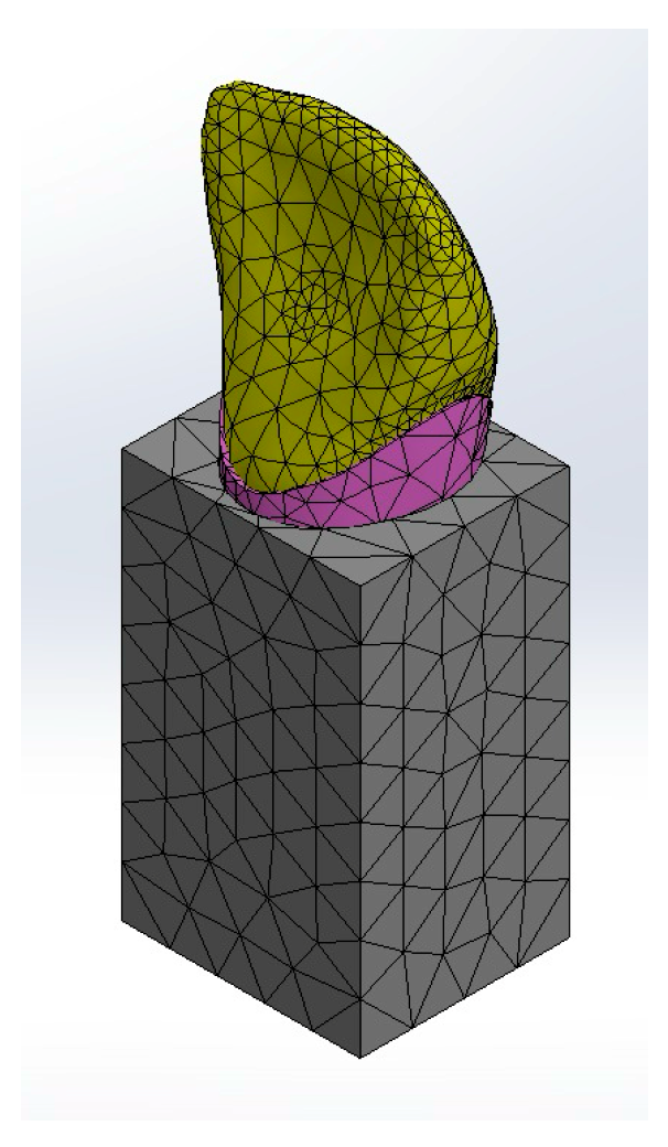
The 3 models were meshed with six degrees of freedom per node: (Figure 5).
  • For long post groups (V-L & C-L): 13,108 quadratic tetrahedral elements and 23,169 nodes.
  • For short post groups (V-Sh& C-Sh): 15,705 quadratic tetrahedral elements and 26,733 nodes.
  • For Endocrown groups (V-E & C-E): 14,037 quadratic tetrahedral elements and 24,084 nodes
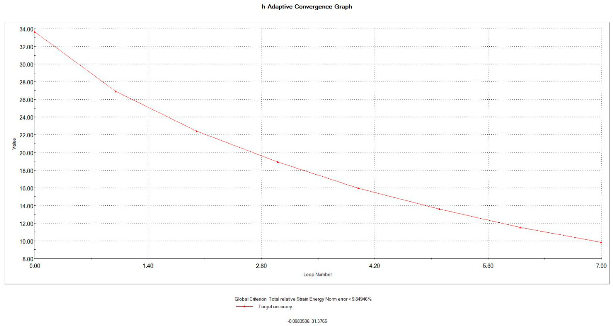
The behaviors of the meshed models during stress application as the real situation were validated by mesh convergence analysis to calculate the error percentage to reduce the error to the minimum acceptable error, which was considered to be 3% in our study (Figure 6).
The material properties used in the FEA models, including various elastic moduli and Poisson’s ratios for dentin, cementum, cancellous bone, periodontal ligament, gutta-percha, composite resin, adhesive resin cement, Vita Enamic, Celtra Duo, and fiber posts, are detailed in Table 3.

3. Results

Among all tested groups, Group C-L exhibited the highest values of maximum VM stress and PS at the finish line location (34.78 MPa and 28.38 MPa, respectively), in addition to the highest values of maximum VM stress and PS at the root apical third (16.496 MPa and 16.075 MPa, respectively), while group C-Sh reported the least VM stress at the root apical third among the groups (16.06 MPa) (Table 4, Figure 7).
Regarding finish line location, Group C-L exhibited the highest values of maximum VM stress and PS at the finish line (34.78 MPa and 28.38 MPa, respectively), while group V-E reported the least values of maximum VM stress and PS among the groups (29.64 MPa and 20.25 MPa, respectively) (Table 4, Figure 7).
Regarding coronal third location of the root (12 mm from the apex), Group C-E showed the highest value of maximum VM stress (28.05 MPa), group V-L showed the highest value of maximum PS (30.548 MPa), while group V-Sh displayed the least value of VM stress among the groups (27.07 MPa) (Table 4, Figure 7).
Regarding middle third location of the root (8 mm from the apex), both the V-E and C-E groups showed the highest value of maximum VM stress at the root middle third (22.18 MPa), group V-L showed the highest value of maximum PS (22.182 MPa), while group C-Sh displayed the least values of maximum VM stress and PS among the groups (21.77 MPa and 19.18 MPa, respectively) (Table 4, Figure 7).
Regarding apical third location of the root (4 mm from the apex), Group C-L exhibited the highest values of maximum VM stress and PS at the root apical third (16.496 MPa and 16.075 MPa, respectively), while group C-Sh reported the least value of maximum VM stress among the groups (16.06 MPa) (Table 4, Figure 7).
The three Celtra Duo groups reported higher VM stress than the corresponding groups of Vita Enamic at the finish line and root coronal thirds. However, at the root middle and apical thirds, both materials recorded similar value of VM stress (Table 4, Figure 7).

4. Discussion

The current study employed three-dimensional FEA to evaluate the stress distribution on endodontically treated anterior teeth restored using different restorative options, crown materials, and post lengths. This method allowed the quantification and characterization of the stress distribution after having applied external functional forces [50]. The findings from this study provide significant insights into the biomechanical performance of various restorative options, which are crucial for optimizing the prognosis and longevity of endodontically treated teeth.
While laboratory tests yield valuable insights into tooth fracture, they typically rely on destructive methods and cannot fully capture the stress–strain behavior of dental restorations [7,8,9,10,11,12,13,14,15,16,17,18,19,20,21,22,23,24,25,26,27,28,29,30,31,32,33,34,35,36,37,38,39,40,41,42,43,44,45,46,47,48,49,50,51]. Finite element analysis (FEA), on the other hand, is a powerful, non-destructive tool widely used in dental research to evaluate stress distribution and mechanical behavior within dental structures. By constructing a geometric model and subdividing it into smaller elements, FEA applies mathematical equations to simulate how the structure responds to various forces. This method is highly accurate and capable of modeling complex anatomical structures, such as teeth and their supporting tissues, in detail. Three-dimensional FEA (3D FEA) is especially prevalent, offering detailed simulations based on the mechanical properties of materials [52,53,54,55]. However, it is important to note that FEA cannot account for certain factors influencing restoration performance, including microleakage, polymerization shrinkage, and postoperative sensitivity [56].
Although dental tissues possess anisotropic properties, their elastic modulus shows only minimal variations in different directions under applied forces [57]. Similarly, the periodontal ligament exhibits negligible differences in stress distribution when modeled with either linear or nonlinear force responses. Therefore, like many other studies, the current research assumed that all materials are isotropic, linearly elastic, and homogeneous as a practical simplification [58,59]. All interfaces were also modeled as fully bonded, without any gaps or voids.
Endodontically treated teeth exhibit a high risk of biomechanical failure due to loss of tooth structure by dental trauma or caries. Therefore, their rehabilitation is considered as a clinical challenge [1]. They can be restored by post–core, crown, inlay only, and endocrown. Endocrown can be defined as an adhesive restoration that has a single unit of core and crown. The advantages of endocrown restorations are that it is a conservative technique, is less time consuming, and reduces treatment costs. It is indicated to restore endodontically treated teeth with anatomically variated roots, calcified, or curved root canals that would hinder the placement of regular post–core and with teeth that have extensive loss of coronal structure [60].
This study examines two CAD/CAM ceramic materials: Celtra Duo, a zirconia-reinforced lithium silicate (ZLS) containing 10% zirconium dioxide, which enhances strength while maintaining machinability for crowns, inlays, onlays, and veneers; and Vita Enamic, a polymer-infiltrated ceramic network (PICN) with 75% glass ceramic and 25% polymer, which combines ceramic strength with polymer resilience, enabling precise milling, efficient finishing, and suitability for minimally invasive restorations [60].
In the current study, the glass fiber post system was used as it offers several advantages over metallic posts, which often generate high stresses and lead to non-restorable root fractures. With an elastic modulus similar to dentin, glass fiber posts improve bending resistance and absorb forces more effectively, reducing fracture risk. Unlike high-modulus posts, which increase the likelihood of severe fractures, glass fiber posts distribute stress more evenly, minimizing damage and allowing for easier restoration if failure occurs [61].
The role of post length is particularly important in determining the tooth’s flexural behavior and fracture resistance. In posterior teeth, where masticatory forces are primarily compressive, the post aids in core retention [62]. However, in anterior teeth, which experience transverse loading, post length can influence fracture resistance. Recent studies suggest that shortening the post length does not negatively affect fracture resistance or compromise the apical seal. With modern adhesive techniques reducing the need for macro retentive elements, radicular posts may no longer be essential in posterior teeth, although limited data exist on their necessity in anterior teeth [61].
Endocrowns offer a convenient and cost-effective restorative option for endodontically treated teeth. Compared to conventional crowns with short or long posts, they require less clinical time and simplify the process. By eliminating the post and filling core, endocrowns also reduce bonding interfaces to potentially enhance structural integrity. Moreover, endocrowns are designed to distribute occlusal forces more evenly and minimize stress concentrations at critical points within the tooth structure [61].
The results of the current study revealed that Group C-L exhibited the highest maximum VM stress and PS values at the finish line (34.78 MPa and 28.38 MPa, respectively), as well as the highest maximum VM stress and PS values at the root apical third (16.496 MPa and 16.075 MPa, respectively), while group C-Sh reported the least stress at the root apical third among the groups (16.06 MPa). The results indicated that long post groups transferred more stress to the apical region, while short post groups had the least stress transfer, with the endocrown groups showing intermediate values. Similar findings [28] highlighted that although longer posts enhance retention, they also concentrate stress at the apical third, potentially raising the risk of root fractures. These findings emphasize the need to balance post length with tooth integrity and crown material selection.
Similar results were reported by Cruzado-Oliva FH [63], who observed that the stress distribution in endocrowns without ferrules was significantly lower than in traditional restorations with crowns and posts, aligning with other studies [64,65,66]. This is because fewer material interfaces allow better stress distribution, as seen in the monolithic structure of endocrowns [67,68], which also reduces the cement layer. By contrast, crown posts require three to four layers of composite resin for customization; otherwise, a thick cement layer forms, increasing fracture risk [69] Additionally, if an endocrown-treated tooth fractures, the break typically occurs at the cervical region [52], allowing for potential repair through crown lengthening surgery or orthodontic traction, unlike post-core crown restorations [67,68,69,70,71].
In addition, the stress distribution in long posts may be influenced by depth, as greater depth leads to higher stress, consistent with findings from other studies [67,68,72], possibly due to an increased contact surface area [73].
The ferrule effect is well documented in enhancing the fracture resistance of endodontically treated teeth. Studies have shown that increasing the ferrule height can lead to improved stress distribution and mechanical stability. For example, a study by Sherfudhin et al. [74] demonstrated that different ferrule designs significantly affect the fracture resistance and failure patterns of restored teeth. Similarly, finite element analysis by Eraslan et al. [75] indicated that a 2 mm ferrule height reduced the stress concentration within the root structure. These findings suggest that variations in ferrule height can have a substantial impact on the biomechanical performance of restorations.
The results demonstrated that endocrowns provided favorable stress distribution patterns. For instance, at the finish line location, Group V-E (Vita Enamic endocrown) demonstrated a maximum stress of 29.64 MPa, which was lower than the stress observed in the groups with traditional post and core systems. Similarly, Group C-E (Celtra Duo endocrown) showed a maximum stress of 31.26 MPa at the finish line. These findings suggest that endocrowns can effectively distribute stress and potentially enhance the biomechanical performance of restorations in endodontically treated teeth [32].
Bindl and Mormann [33] reported similar findings, indicating that endocrowns are particularly effective in restoring molars due to their ability to distribute occlusal forces more evenly. Biacchi and Basting [32] further supported the use of endocrowns, demonstrating their superior fracture resistance compared to traditional post and core systems. Our study extends these findings to anterior teeth, highlighting the potential of endocrowns to enhance the durability and prognosis of restorations.
However, one study found that teeth restored with endocrowns and fiberglass-reinforced posts exhibited similar fracture strength, as endocrowns function like short posts [37]. By contrast, other studies suggest that the depth of endocrown preparation does not significantly affect fracture resistance [68] or stress distribution [66]. Therefore, restoring extensively damaged anterior teeth with an endocrown or a short glass fiber post may offer advantages over a longer glass fiber post [61].
Going through the effect of crown material on stress distribution, all Celtra Duo groups reported higher stresses than the corresponding groups of Vita Enamic at the finish line and root coronal thirds. However, at the root middle and apical thirds, both materials recorded similar stresses, and it was clearly observed that the choice of crown material significantly impacts the stress distribution in endodontically treated teeth.
These findings are in line with those reported by Heintze et al. [26], who found that hybrid ceramic materials like Vita Enamic provide better stress distribution and lower fracture risk compared to zirconia-reinforced ceramics. Another study conducted by Conserva et al. [76] reported that composite materials with a Young’s modulus similar to that of dentin should be selected for the restoration of endodontically-treated teeth when using carbon fiber posts. However, Kelly and Benetti [27] highlighted that the choice of crown material should also consider aesthetic requirements and patient-specific factors, suggesting that Celtra Duo’s superior aesthetics could make it a preferable choice in certain clinical scenarios despite its slightly higher stress concentrations.
The findings from this study have several important clinical implications. First, the choice of post length should be carefully considered based on the specific clinical scenario. While longer posts generally provide better retention, they can also introduce higher stress concentrations at the apical third, which may increase the risk of root fractures. Therefore, the decision on post length should be balanced with other factors, such as the type of crown material and the overall condition of the tooth.
Second, the choice of crown material is critical for optimizing the biomechanical performance of restorations. Vita Enamic crowns demonstrated lower stress concentrations at the finish line compared to Celtra Duo crowns, suggesting that they may be a better option for reducing the risk of fractures and enhancing the durability of the restoration. However, Celtra Duo crowns also performed well, particularly in endocrown configurations, indicating that they can be a viable option depending on the specific clinical requirements [1,10,28,77].
The use of endocrowns with a 3 mm intra-radicular extension offers a promising alternative to traditional post and core systems. The favorable stress distribution patterns observed in this study suggest that endocrowns can effectively minimize stress concentrations and enhance the biomechanical performance of restorations. This can be particularly beneficial in cases where there is significant structural loss or when preserving as much tooth structure as possible is a priority [9,10,36,38].
In spite of the fact that the finite element method is widely used due to its rapid mechanical analysis and versatility, it cannot yet fully simulate the complex forces generated in the oral cavity. It only evaluates ideal stress conditions under static loads, whereas real chewing involves dynamic forces. Factors such as the presence of periodontal disease, variations in bone density, and patient-specific occlusal forces were not considered. With advancements in finite element analysis technology, future studies may achieve more complex biomechanical simulations or include supporting in vitro studies of mechanical testing, which in turn will validate the finite analysis outcomes.
The current study focused on a specific tooth type to maintain controlled conditions and reduce variability. However, we recognize that anatomical differences between anterior and posterior teeth, such as root morphology, canal configuration, and loading patterns, can significantly influence stress distribution and fracture resistance. For instance, posterior teeth typically endure higher occlusal forces and have different structural characteristics compared to anterior teeth. Therefore, while our findings provide valuable insights, caution should be exercised when extrapolating these results to other tooth types. Future studies should aim to include a variety of tooth types to enhance the applicability of the results across different clinical scenarios.
Future studies should aim to validate these findings through clinical trials and in vivo studies to confirm the biomechanical performance of different restorative options under real-world conditions. Additionally, further research should explore the impact of other factors, such as tooth morphology, occlusal loading patterns, and the presence of adjacent teeth, on stress distribution. This will provide a more comprehensive understanding of the factors that influence the success and longevity of restorations in endodontically treated teeth.

5. Conclusions

Post length has a non-significant impact on stress distribution in endodontically treated anterior teeth. However, short posts generate the least stress at all root levels, while long posts increase stress at the finish line and apical third, raising the risk of fractures.
Endocrowns, especially those made from Vita Enamic, exhibit favorable stress distribution at the finish line, making them a strong alternative to traditional post and core systems. Their mechanical performance suggests they could replace long post restorations while preserving more tooth structure.
Restorations without posts offer the advantage of preserving more tooth substance and simplifying clinical procedures. This supports the use of endocrowns or short posts for anterior teeth as a viable restorative approach.
Although the study demonstrates the mechanical superiority of endocrowns and short posts, in vivo validation is necessary before this technique can be widely recommended for clinical use.

Author Contributions

Conceptualization, M.S., N.A., A.A., R.A., A.A.A.A.-E. and M.M.A.; Data curation, M.S., A.A. and A.A.A.A.-E.; Funding acquisition, M.S.; Investigation, N.A. and R.A.; Methodology, M.S., N.A., A.A., R.A., A.A.A.A.-E. and M.M.A.; Project administration, N.A. and M.M.A.; Resources, M.S.; Software, A.A.; Supervision, A.A.A.A.-E.; Writing—original draft, M.S., N.A., A.A., R.A., A.A.A.A.-E. and M.M.A.; Writing—review & editing, M.S. and M.M.A. All authors have read and agreed to the published version of the manuscript.

Funding

This research was funded by Princess Nourah bint Abdulrahman University Researchers Supporting Project number (PNURSP2025R822), Princess Nourah bint Abdulrahman University, Riyadh, Saudi Arabia.

Institutional Review Board Statement

The study was conducted in accordance with the Declaration of Helsinki, and approved by the Committee of Research Ethics, Deanship of Scientific Research, Qassim University (protocol code IRB 24-76-09, date of approval: 6 February 2024).

Informed Consent Statement

Informed consent was obtained from all subjects involved in the study.

Data Availability Statement

The original contributions presented in the study are included in the article, further inquiries can be directed to the corresponding author.

Conflicts of Interest

The authors declare no conflicts of interest.

References

  1. Schwartz, R.S.; Robbins, J.W. Post Placement and Restoration of Endodontically Treated Teeth: A Literature Review. J. Endod. 2004, 30, 289–301. [Google Scholar] [CrossRef]
  2. Morgano, S.M.; Rodrigues, A.H.; Sabrosa, C.E. Restoration of Endodontically Treated Teeth. Dent. Clin. N. Am. 2004, 48, 397–416. [Google Scholar] [CrossRef]
  3. Slutzky-Goldberg, I.; Slutzky, H.; Gorfil, C.; Smidt, A. Restoration of Endodontically Treated Teeth: Review and Treatment Recommendations. Int. J. Dent. 2009, 2009, 150251. [Google Scholar] [CrossRef]
  4. Mannocci, F.; Cowie, J. Restoration of Endodontically Treated Teeth. Br. Dent. J. 2014, 216, 341–346. [Google Scholar] [CrossRef]
  5. Stenhagen, S.; Skeie, H.; Bårdsen, A.; Laegreid, T. Influence of the Coronal Restoration on the Outcome of Endodontically Treated Teeth. Acta Odontol. Scand. 2020, 78, 81–86. [Google Scholar] [CrossRef]
  6. Cook, R.D.; Malkus, D.S.; Plesha, M.E.; Witt, R.J. Concepts and Applications of Finite Element Analysis, 4th ed.; John Wiley & Sons: New York, NY, USA, 2002. [Google Scholar]
  7. Zarow, M.; Vadini, M.; Chojnacka-Brozek, A.; Szczeklik, K.; Milewski, G.; Biferi, V.; D’Arcangelo, C.; De Angelis, F. Effect of Fiber Posts on Stress Distribution of Endodontically Treated Upper Premolars: Finite Element Analysis. Nanomaterials 2020, 10, 1708. [Google Scholar] [CrossRef]
  8. Abdelhafeez, M.M. Applications of Finite Element Analysis in Endodontics: A Systematic Review and Meta-Analysis. J. Pharm. Bioallied Sci. 2024, 16, S1977–S1980. [Google Scholar] [CrossRef]
  9. Zhou, L.; Wang, Q. Comparison of Fracture Resistance Between Cast Posts and Fiber Posts: A Meta-Analysis of Literature. J. Endod. 2013, 39, 11–15. [Google Scholar] [CrossRef]
  10. Boudrias, P.; Sakkal, S.; Petrova, Y. Anatomical Post Design Meets Quartz Fiber Technology: Rationale and Case Report. Compend. Contin. Educ. Dent. 2001, 22, 337–340. [Google Scholar]
  11. Yee, K.; Bhagavatula, P.; Stover, S.; Eichmiller, F.; Hashimoto, L.; MacDonald, S.; Barkley, G., III. Survival Rates of Teeth with Primary Endodontic Treatment After Core/Post and Crown Placement. J. Endod. 2018, 44, 220–225. [Google Scholar] [CrossRef]
  12. Chen, S.; Hong, X.; Ye, Z.; Wu, M.; Chen, L.; Wu, L.; Zhao, S. The Effect of Root Canal Treatment and Post-Crown Restorations on Stress Distribution in Teeth with Periapical Periodontitis: A Finite Element Analysis. BMC Oral Health 2023, 23, 973. [Google Scholar] [CrossRef] [PubMed]
  13. Ferrari, M.; Vichi, A.; Garcia-Godoy, F. Clinical Evaluation of Fiber-Reinforced Epoxy Resin Posts and Cast Post and Cores. Am. J. Dent. 2000, 13, 15B–18B. [Google Scholar]
  14. Theodosopoulou, J.N.; Chochlidakis, K.M. A Systematic Review of Dowel (Post) and Core Materials and Systems. J. Prosthodont. 2009, 18, 464–472. [Google Scholar] [CrossRef]
  15. Nakamura, T.; Ohyama, T.; Waki, T.; Kinuta, S.; Wakabayashi, K.; Mutobe, Y.; Yatani, H. Stress Analysis of Endodontically Treated Anterior Teeth Restored with Different Types of Post Material. Dent. Mater. J. 2006, 25, 145–150. [Google Scholar] [CrossRef]
  16. Lamichhane, A.; Xu, C.; Zhang, F.Q. Dental Fiber-Post Resin Base Material: A Review. J. Adv. Prosthodont. 2014, 6, 60–65. [Google Scholar] [CrossRef]
  17. Ouldyerou, A.; Mehboob, H.; Mehboob, A.; Merdji, A.; Aminallah, L.; Mukdadi, O.M.; Barsoum, I.; Junaedi, H. Biomechanical Performance of Resin Composite on Dental Tissue Restoration: A Finite Element Analysis. PLoS ONE 2023, 18, e0295582. [Google Scholar] [CrossRef]
  18. Cagidiaco, M.C.; Goracci, C.; Garcia-Godoy, F.; Ferrari, M. Clinical Studies of Fiber Posts: A Literature Review. Int. J. Prosthodont. 2008, 21, 328–336. [Google Scholar]
  19. Goracci, C.; Ferrari, M. Current Perspectives on Post Systems: A Literature Review. Aust. Dent. J. 2011, 56 (Suppl. S1), 77–83. [Google Scholar] [CrossRef]
  20. Dietschi, D.; Duc, O.; Krejci, I.; Sadan, A. Biomechanical Considerations for the Restoration of Endodontically Treated Teeth: A Systematic Review of the Literature—Part 1. Composition and Micro- and Macrostructure Alterations. Quintessence Int. 2007, 38, 733–743. [Google Scholar] [PubMed]
  21. Stavropoulou, A.F.; Koidis, P.T. A Systematic Review of Single Crowns on Endodontically Treated Teeth. J. Dent. 2007, 35, 761–767. [Google Scholar] [CrossRef] [PubMed]
  22. Ramirez-Sebastia, A.; Bortolotto, T.; Roig, M.; Krejci, I. Composite vs Ceramic Computer-Aided Design/Computer-Assisted Manufacturing Crowns in Endodontically Treated Teeth: Analysis of Marginal Adaptation. Oper. Dent. 2013, 38, 663–673. [Google Scholar] [CrossRef]
  23. Lin, J.; Lin, Z.; Zheng, Z. Effect of Different Restorative Crown Design and Materials on Stress Distribution in Endodontically Treated Molars: A Finite Element Analysis Study. BMC Oral Health 2020, 20, 226. [Google Scholar] [CrossRef] [PubMed]
  24. Tekin, S.; Adiguzel, O.; Cangul, S.; Atas, O.; Erpacal, B. Evaluation of the Use of PEEK Material in Post-Core and Crown Restorations Using Finite Element Analysis. Am. J. Dent. 2020, 33, 251–257. [Google Scholar] [PubMed]
  25. Huang, M.; Wang, B.; Zhang, K.; Yan, X.; Chen, Z.; Zhang, X. Comparative Analysis of Stress Distribution in Residual Roots with Different Canal Morphologies: Evaluating CAD/CAM Glass Fiber and Other Post-Core Materials. BMC Oral Health 2024, 24, 337. [Google Scholar] [CrossRef]
  26. Heintze, S.D.; Cavalleri, A.; Forjanic, M.; Zellweger, G.; Rousson, V. Wear of Ceramic and Antagonist—A Systematic Evaluation of Influencing Factors In Vitro. Dent. Mater. 2008, 24, 433–449. [Google Scholar] [CrossRef]
  27. Kelly, J.R.; Benetti, P. Ceramic Materials in Dentistry: Historical Evolution and Current Practice. Aust. Dent. J. 2011, 56 (Suppl. S1), 84–96. [Google Scholar] [CrossRef]
  28. Sorensen, J.A.; Martinoff, J.T. Intracoronal Reinforcement and Coronal Coverage: A Study of Endodontically Treated Teeth. J. Prosthet. Dent. 1984, 51, 780–784. [Google Scholar] [CrossRef]
  29. McLaren, J.D.; McLaren, C.I.; Yaman, P.; Bin-Shuwaish, M.S.; Dennison, J.D.; McDonald, N.J. The Effect of Post Type and Length on the Fracture Resistance of Endodontically Treated Teeth. J. Prosthet. Dent. 2009, 101, 174–182. [Google Scholar] [CrossRef] [PubMed]
  30. Pereira, J.R.; Ribeiro Neto, E.M.; Pamato, S.; Valle, A.L.D.; Paula, V.G.D.; Vidotti, H.A. Fracture Resistance of Endodontically Treated Teeth Restored with Different Intraradicular Posts with Different Lengths. Braz. J. Oral Sci. 2013, 12, 1–4. [Google Scholar]
  31. Franco, É.B.; do Valle, A.L.; de Almeida, A.L.P.F.; Rubo, J.H.; Pereira, J.R. Fracture Resistance of Endodontically Treated Teeth Restored with Glass Fiber Posts of Different Lengths. J. Prosthet. Dent. 2014, 111, 30–34. [Google Scholar] [CrossRef]
  32. Naumann, M.; Koelpin, M.; Beuer, F.; Meyer-Lueckel, H. 10-Year Survival Evaluation for Glass-Fiber-Supported Post Endodontic Restoration: A Prospective Observational Clinical Study. J. Endod. 2012, 38, 432–435. [Google Scholar] [CrossRef]
  33. Bindl, A.; Mormann, W.H. Clinical Evaluation of Adhesively Placed Cerec Endocrowns After 2 Years—Preliminary Results. J. Adhes. Dent. 1999, 1, 255–265. [Google Scholar] [PubMed]
  34. Sevimli, G.; Cengiz, S.; Selçuk, O.R.U.Ç. Endocrowns. J. Istanbul Univ. Fac. Dent. 2015, 49, 57–63. [Google Scholar] [CrossRef] [PubMed]
  35. Govare, N.; Contrepois, M. Endocrowns: A Systematic Review. J. Prosthet. Dent. 2020, 123, 411–417. [Google Scholar] [CrossRef] [PubMed]
  36. Biacchi, G.R.; Basting, R.T. Comparison of Fracture Strength of Endocrowns and Glass Fiber Post-Retained Conventional Crowns. Oper. Dent. 2012, 37, 130–136. [Google Scholar] [CrossRef]
  37. Biacchi, G.R.; Mello, B.; Basting, R.T. The Endocrown: An Alternative Approach for Restoring Extensively Damaged Molars. J. Esthet. Restor. Dent. 2013, 25, 383–390. [Google Scholar] [CrossRef]
  38. Al-Dabbagh, R.A. Survival and Success of Endocrowns: A Systematic Review and Meta-Analysis. J. Prosthet. Dent. 2021, 125, 415.e1. [Google Scholar] [CrossRef]
  39. Wendling, M.M.; Mantovani, G.; Fernandes, B.V.; Carneiro, D.E.; Santos, R.V.; Sánchez-Ayala, A. Occlusal Loading Effect on Stress Distribution of Endodontically Treated Teeth: Finite Element Analysis Study. Eur. J. Prosthodont. Restor. Dent. 2024, 32, 102–108. [Google Scholar] [CrossRef]
  40. Monteiro, J.B.; Dal Piva, A.M.O.; Tribst, J.P.M.; Borges, A.L.S.; Tango, R.N. The Effect of Resection Angle on Stress Distribution after Root-End Surgery. Iran. Endod. J. 2018, 13, 188–194. [Google Scholar]
  41. Yamakami, S.A.; Gallas, J.A.; Petean, I.B.F.; Souza-Gabriel, A.E.; Sousa-Neto, M.; Macedo, A.P.; Palma-Dibb, R.G. Impact of Endodontic Kinematics on Stress Distribution During Root Canal Treatment: Analysis of Photoelastic Stress. J. Endod. 2022, 48, 255–262. [Google Scholar] [CrossRef]
  42. Kahyaie Aghdam, M.; Bahari, M.; Mohammadi, N.; Savadi Oskoee, S.; Ebrahimi Chaharom, M.E. Push-Out Bond Strength of Fiber Posts to Overflared Root Canals in Different Root Regions: Effect of Reinforcement Techniques. Front Dent. 2024, 10, 24. [Google Scholar]
  43. Yoon, H.G.; Oh, H.K.; Lee, D.Y.; Shin, J.H. 3-D finite element analysis of the effects of post location and loading location on stress distribution in root canals of the mandibular 1st molar. J. Appl. Oral. Sci. 2018, 26, e20160406. [Google Scholar] [CrossRef]
  44. Bucchi, C.; Del Fabbro, M.; Marce-Nogue, J. Orthodontic Loads in Teeth after Regenerative Endodontics: A Finite Element Analysis of the Biomechanical Performance of the Periodontal Ligament. Appl. Sci. 2022, 12, 7063. [Google Scholar] [CrossRef]
  45. Syed, A.U.Y.; Rokaya, D.; Shahrbaf, S.; Martin, N. Three-Dimensional Finite Element Analysis of Stress Distribution in a Tooth Restored with Full Coverage Machined Polymer Crown. Appl. Sci. 2021, 11, 1220. [Google Scholar] [CrossRef]
  46. Capra, A.; Biclesanu, C.; Buruiana, A.M. The role of periodontal ligament elasticity in periodontal changes—Numerical simulation. Ro. J. Stomatol. 2022, 68, 37–45. [Google Scholar] [CrossRef]
  47. Tammineedi, S.; Kakollu, S.; Thota, M.M.; Basam, R.C.; Basam, L.C.; Vemuri, S. Comparison of Stress Distribution in Teeth Restored with Fiber Post and Dentin Post by Applying Orthotropic Properties: A Three-Dimensional Finite Element Analysis. J. Conserv. Dent. 2020, 23, 589–592. [Google Scholar] [CrossRef]
  48. Soares, P.V.; Santos-Filho, P.C.F.; Queiroz, E.C.; Araújo, T.C.; Campos, R.E.; Araújo, C.A.; Soares, C.J. Fracture resistance and stress distribution in endodontically treated maxillary premolars restored with composite resin. J. Prosthodont. 2008, 17, 114–119. [Google Scholar] [CrossRef]
  49. Dentsply Sirona. Celtra® Duo: Zirconia-Reinforced Lithium Silicate (ZLS) Block FactFile; Dentsply Sirona Inc.: Charlotte, NC, USA, 2017. [Google Scholar]
  50. Rodrigues, Y.L.; Mathew, M.T.; Mercuri, L.G.; da Silva, J.S.P.; Henriques, B.; Souza, J.C.M. Biomechanical Simulation of Temporomandibular Joint Replacement (TMJR) Devices: A Scoping Review of the Finite Element Method. Int. J. Oral Maxillofac. Surg. 2018, 47, 1032–1042. [Google Scholar] [CrossRef]
  51. Alp, Ş.; Gulec Alagoz, L.; Ulusoy, N. Effect of Direct and Indirect Materials on Stress Distribution in Class II MOD Restorations: A 3D-Finite Element Analysis Study. Biomed Res. Int. 2020, 2020, 7435054. [Google Scholar] [CrossRef]
  52. Cantó-Navés, O.; Medina-Gálvez, R.; Marimon, X.; Ferrer, M.; Figueras-Álvarez, Ó.; Cabratosa-Termes, J. A 3D Finite Element Analysis Model of Single Implant-Supported Prosthesis Under Dynamic Impact Loading for Evaluation of Stress in the Crown, Abutment and Cortical Bone Using Different Rehabilitation Materials. Materials 2021, 14, 3519. [Google Scholar] [CrossRef]
  53. Lee, J.-H.; Jang, H.Y.; Lee, S.Y. Finite Element Analysis of Dental Implants with Zirconia Crown Restorations: Conventional Cement-Retained vs. Cementless Screw-Retained. Materials 2021, 14, 2666. [Google Scholar] [CrossRef] [PubMed]
  54. Cicciù, M. Bioengineering Methods of Analysis and Medical Devices: A Current Trends and State of the Art. Materials 2020, 13, 797. [Google Scholar] [CrossRef] [PubMed]
  55. Nabih, S.M.; Ibrahim, N.I.M.; Elmanakhly, A.R. Mechanical and Thermal Stress Analysis of Hybrid Ceramic and Lithium Disilicate-Based Ceramic CAD-CAM Inlays Using 3-D Finite Element Analysis. Braz. Dent. Sci. 2021, 24, 1–10. [Google Scholar] [CrossRef]
  56. Ausiello, P.; Dal Piva, A.M.; Borges, A.L.S.; Lanzotti, A.; Zamparini, F.; Epifania, E.; Tribst, J.P.M. Effect of Shrinking and No Shrinking Dentine and Enamel Replacing Materials in Posterior Restoration: A 3D-FEA Study. Appl. Sci. 2021, 11, 2215. [Google Scholar] [CrossRef]
  57. González-Lluch, C.; Pérez-González, A.; Sancho-Bru, J.L.; Rodríguez-Cervantes, P.J. Mechanical Performance of Endodontic Restorations with Prefabricated Posts: Sensitivity Analysis of Parameters with a 3D Finite Element Model. Comput. Methods Biomech. Biomed. Eng. 2014, 17, 1108–1118. [Google Scholar] [CrossRef] [PubMed]
  58. de Andrade, G.S.; Tribst, J.P.; Dal Piva, A.O.; Bottino, M.A.; Borges, A.L.; Valandro, L.F.; Özcan, M. A Study on Stress Distribution to Cement Layer and Root Dentin for Post and Cores Made of CAD/CAM Materials with Different Elasticity Modulus in the Absence of Ferrule. J. Clin. Exp. Dent. 2019, 11, e0–e8. [Google Scholar] [CrossRef]
  59. Dal Piva, A.M.; Tribst, J.P.; Souza, R.O.; Borges, A.L. Influence of Alveolar Bone Loss and Cement Layer Thickness on the Biomechanical Behavior of Endodontically Treated Maxillary Incisors: A 3-Dimensional Finite Element Analysis. J. Endod. 2017, 43, 791–795. [Google Scholar] [CrossRef]
  60. Soliman, M.; Alzahrani, G.; Alabdualataif, F.; Eldwakhly, E.; Alsamady, S.; Aldegheishem, A.; Abdelhafeez, M.M. Impact of Ceramic Material and Preparation Design on Marginal Fit of Endocrown Restorations. Materials 2022, 15, 5592. [Google Scholar] [CrossRef]
  61. Ramírez-Sebastià, A.; Bortolotto, T.; Cattani-Lorente, M.; Giner, L.; Roig, M.; Krejci, I. Adhesive Restoration of Anterior Endodontically Treated Teeth: Influence of Post Length on Fracture Strength. Clin. Oral Investig. 2014, 18, 545–554. [Google Scholar] [CrossRef]
  62. Garbin, C.A.; Spazzin, A.O.; Meira-Junior, A.D.; Loretto, S.C.; Lyra, A.M.; Braz, R. Biomechanical Behaviour of a Fractured Maxillary Incisor Restored with Direct Composite Resin Only or with Different Post Systems. Int. Endod. J. 2010, 43, 1098–1107. [Google Scholar] [CrossRef]
  63. Cruzado-Oliva, F.H.; Alarco-La Rosa, L.F.; Vega-Anticona, A.; Arbildo-Vega, H.I. Biomechanics of Anterior Endocrowns with Different Designs and Depths: Study of Finite Elements. J. Clin. Exp. Dent. 2023, 15, e1016–e1021. [Google Scholar] [CrossRef] [PubMed]
  64. de Carvalho, M.A.; Lazari-Carvalho, P.C.; Del Bel Cury, A.A.; Magne, P. Accelerated Fatigue Resistance of Endodontically Treated Incisors without Ferrule Restored with CAD/CAM Endocrowns. Int. J. Esthet. Dent. 2021, 16, 534–552. [Google Scholar]
  65. El-Damanhoury, H.M.; Haj-Ali, R.N.; Platt, J.A. Fracture Resistance and Microleakage of Endocrowns Utilizing Three CAD-CAM Blocks. Oper. Dent. 2015, 40, 201–210. [Google Scholar] [CrossRef] [PubMed]
  66. Bankoğlu Güngör, M.; Turhan Bal, B.; Yilmaz, H.; Aydin, C.; Karakoca Nemli, S. Fracture Strength of CAD/CAM Fabricated Lithium Disilicate and Resin Nano Ceramic Restorations Used for Endodontically Treated Teeth. Dent. Mater. J. 2017, 36, 135–141. [Google Scholar] [CrossRef]
  67. Li, X.; Kang, T.; Zhan, D.; Xie, J.; Guo, L. Biomechanical Behavior of Endocrowns vs Fiber Post-Core-Crown vs Cast Post-Core-Crown for the Restoration of Maxillary Central Incisors with 1 mm and 2 mm Ferrule Height: A 3D Static Linear Finite Element Analysis. Medicine 2020, 99, e22648. [Google Scholar] [CrossRef] [PubMed]
  68. Kanat-Ertürk, B.; Sarıdağ, S.; Köseler, E.; Helvacıoğlu-Yiğit, D.; Avcu, E.; Yıldıran-Avcu, Y. Fracture Strengths of Endocrown Restorations Fabricated with Different Preparation Depths and CAD/CAM Materials. Dent. Mater. J. 2018, 37, 256–265. [Google Scholar] [CrossRef]
  69. Fernandes, V.; Silva, A.S.; Carvalho, O.; Henriques, B.; Silva, F.S.; Özcan, M.; Souza, J.C. The Resin-Matrix Cement Layer Thickness Resultant from the Intracanal Fitting of Teeth Root Canal Posts: An Integrative Review. Clin. Oral Investig. 2021, 25, 5595–5612. [Google Scholar] [CrossRef]
  70. Dejak, B.; Młotkowski, A. Strength Comparison of Anterior Teeth Restored with Ceramic Endocrowns vs Custom-Made Post and Cores. J. Prosthodont. Res. 2018, 62, 171–176. [Google Scholar] [CrossRef]
  71. Zhen, M.; Wei, Y.P.; Hu, W.J.; Rong, Q.G.; Zhang, H. Finite Element Analysis of the Maxillary Central Incisor with Traditional and Modified Crown Lengthening Surgery and Post-Core Restoration in Management of Crown-Root Fracture. Zhonghua Kou Qiang Yi Xue Za Zhi 2016, 51, 362–367. [Google Scholar]
  72. Einhorn, M.; DuVall, N.; Wajdowicz, M.; Brewster, J.; Roberts, H. Preparation Ferrule Design Effect on Endocrown Failure Resistance. J. Prosthodont. 2019, 28, e237–e242. [Google Scholar] [CrossRef]
  73. Bozkurt, D.A.; Buyukerkmen, E.B.; Terlemez, A. Comparison of the Pull-Out Bond Strength of Endodontically Treated Anterior Teeth with Monolithic Zirconia Endocrown and Post-and-Core Crown Restorations. J. Oral Sci. 2023, 65, 1–5. [Google Scholar] [CrossRef] [PubMed]
  74. Sherfudhin, H.; Hobeich, J.; Carvalho, C.A.; Aboushelib, M.N.; Sadig, W.; Salameh, Z. Effect of Different Ferrule Designs on the Fracture Resistance and Failure Pattern of Endodontically Treated Teeth Restored with Fiber Posts and All-Ceramic Crowns. J. Appl. Oral Sci. 2011, 19, 28–33. [Google Scholar] [CrossRef] [PubMed]
  75. Eraslan, O.; Aykent, F.; Yücel, M.T.; Akman, S. The Finite Element Analysis of the Effect of Ferrule Height on Stress Distribution at Post-and-Core-Restored All-Ceramic Anterior Crowns. Clin. Oral Investig. 2009, 13, 223–227. [Google Scholar] [CrossRef] [PubMed]
  76. Conserva, E.; Consolo, U.; Gimenez Sancho, A.; Foschi, F.; Paolone, G.; Giovarrusscio, M.; Sauro, S. Stress Distribution in Carbon-Post Applied with Different Composite Core Materials: A Three-Dimensional Finite Element Analysis. J. Adhes. Sci. Technol. 2017, 31, 2435–2444. [Google Scholar] [CrossRef]
  77. Nahar, R.; Mishra, S.K.; Chowdhary, R. Evaluation of Stress Distribution in an Endodontically Treated Tooth Restored with Four Different Post Systems and Two Different Crowns—A Finite Element Analysis. J. Oral Biol. Craniofac. Res. 2020, 10, 719–726. [Google Scholar] [CrossRef]
Figure 1. Model for Group V-L and Group C-L; Vita Enamic/Celtra Duo crown, Long post; 10 mm intra-radicular length, and composite core. Jfb 16 00221 i001 crown, Jfb 16 00221 i002 composite core, Jfb 16 00221 i003 fiber post, Jfb 16 00221 i004 gutta-percha, Jfb 16 00221 i005 tooth structure.
Jfb 16 00221 g001
Figure 2. Model for Group V-Sh and Group C-Sh; Vita Enamic/Celtra Duo crown, Short post; 3 mm intra-radicular length, and composite core. Jfb 16 00221 i006 crown, Jfb 16 00221 i007 composite core, Jfb 16 00221 i008 fiber post, Jfb 16 00221 i009 gutta-percha, Jfb 16 00221 i010 tooth structure.
Jfb 16 00221 g002
Figure 3. Model for Group V-E and Group C-E: Vita Enamic/Celtra Duo Endocrown with 3 mm intra-radicular extension. Jfb 16 00221 i011 Endocrown, Jfb 16 00221 i012 gutta-percha, Jfb 16 00221 i013 tooth structure.
Jfb 16 00221 g003
Figure 4. Jfb 16 00221 i014 Boundary conditions and Jfb 16 00221 i015 load application.
Jfb 16 00221 g004
Figure 5. Mesh of the models.
Jfb 16 00221 g005
Figure 6. Mesh h-adaptive convergence graph.
Jfb 16 00221 g006
Figure 7. Von Mises (VM) stress distribution and principal stress (PS) at finish line and different root sections for each group.
Jfb 16 00221 g007aJfb 16 00221 g007bJfb 16 00221 g007c
Table 1. Model Grouping.
DesignLong Post
(10 mm Intra-Radicular)
Short Post
(3 mm Intra-Radicular)
Endocrown
(3 mm Intra-Radicular)
Material
Vita EnamicGroup V-LGroup V-ShGroup V-E
Celtra DuoGroup C-LGroup C-ShGroup C-E
Table 2. The two tested materials’ compositions (Vita Enamic and Celta Duo).
MaterialManufacturerCeramic TypeChemical Composition
Vita EnamicVITA-Zahnfabrik, Bad Säckingen, Germany Polymer-infiltrated ceramicPolymer-infiltrated feldspathic ceramic network material (UDMA, TEGDMA) with 86 wt% ceramic (SiO2, Al2O3, Na2O, K2O, B2O3, CaO, TiO2, coloring oxides)
Celtra DuoDentsply, Charlotte, NC, USAZirconia-reinforced lithium silicate ceramic SiO2, P2O5, Al2O3, Li2O, ZnO, 10% ZrO2
Table 3. Elastic moduli and Poisson’s ratios of the materials used in the model.
MaterialElastic Modulus (GPa)Poisson’s Ratio
Dentin [43,44,45]18.60.31
Cementum [44]8.20.3
Cancellous bone [43,44,45,46,47]1.370.3
Periodontal ligament [43,47]0.050.45
Gutta percha [43]0.140.45
Fiber post [43]330.33
Composite resin [47,48]16.60.24
Adhesive resin cement [43]18.60.28
Vita Enamic [45]300.23
Celtra Duo [49]700.23
Table 4. Maximum Von Mises (VM) stress and principal stress (PS) values at different sections for each group.
LocationFinish LineRoot Coronal Third
(12 mm from Apex)
Root Middle Third
(8 mm from Apex)
Root Apical Third
(4 mm from Apex)
Group
V-LVM31.1227.48521.99916.489
PS28.3630.54822.1827.277
C-LVM34.7827.79621.84316.496
PS28.3820.65519.45916.075
V-ShVM29.7927.0721.8316.09
PS26.7420.5819.268.52
C-ShVM31.4527.6021.7716.06
PS25.4420.5219.188.49
V-EVM29.6427.7022.1816.34
PS20.2520.7419.517.70
C-EVM31.2628.0522.1816.34
PS20.3120.7519.507.70
Disclaimer/Publisher’s Note: The statements, opinions and data contained in all publications are solely those of the individual author(s) and contributor(s) and not of MDPI and/or the editor(s). MDPI and/or the editor(s) disclaim responsibility for any injury to people or property resulting from any ideas, methods, instructions or products referred to in the content.