Article

Impact of a Novel Pretreatment on Bond Strength of Universal Adhesive to Conventional and CAD/CAM Resin Composites: In Vitro Study

by
Ali A. Elkaffas
1,2,
Abdullah Alshehri
1,
Feras Alhalabi
1,*,
Rania Bayoumi
1,3,
Abdullah Ali Alqahtani
1,
Abdulellah Almudahi
1,
Abdulaziz Fahd Alsubaie
4 and
Abdulaziz Fahd Alharbi
4
1
Conservative Dental Science Department, College of Dentistry, Prince Sattam bin Abdulaziz University, Al-Kharj 11942, Saudi Arabia
2
Department of Operative Dentistry, Faculty of Dentistry, Mansoura University, Mansoura 35516, Egypt
3
Biomaterials Department, Faculty of Dental Medicine for Girls, Al-Azhar University, Cairo 11754, Egypt
4
College of Dentistry, Prince Sattam bin Abdulaziz University, Al-Kharj 11942, Saudi Arabia
*
Author to whom correspondence should be addressed.
J. Funct. Biomater. 2025, 16(6), 197; https://doi.org/10.3390/jfb16060197
Submission received: 26 April 2025 / Revised: 21 May 2025 / Accepted: 22 May 2025 / Published: 27 May 2025
(This article belongs to the Special Issue Advances in Restorative Dentistry Materials)

Abstract

:
Novel dentin bonding pretreatment using copper sulfate (CuSO4) and dipotassium hydrogen phosphate (K2HPO4) may create a more hydrophobic environment for dentin bonding. Thus, this study aims to investigate the impact of a CuSO4 + K2HPO4 pretreatment on dentin μTBS when bonded with a universal adhesive to conventional and CAD/CAM resin composites. Eighty recently extracted human molars (n = 80) were chosen and placed in transparent acrylic blocks to expose the crowns entirely. Nano-filled resin composite and CAD/CAM resin blocks were selected. Based on the dentin pretreatment, type of resin composite, and adhesion strategy, the teeth were randomly allocated into eight equal groups (n = 10). The microtensile bond strength (μTBS) and fracture mode were determined. A three-way analysis of variance (ANOVA) was used to analyze the μTBS data, followed by Tukey’s post hoc test. The μTBS values were not significantly affected by either the resin composite type (p > 0.05) or the adhesive strategy (p > 0.05) according to the three-way ANOVA results. Conversely, significant differences were detected between no dentin pretreatment (24.20 ± 4.54 MPa) and CuSO4 + K2HPO4 pretreatment (33.66 ± 5.22 MPa) using an etch-and-rinse adhesive strategy for nano-filled composites (p < 0.001). Additionally, significant differences were detected between no dentin pretreatment (24.71 ± 4.33 MPa) and CuSO4 + K2HPO4 pretreatment (32.49 ± 4.92 MPa) using an etch-and-rinse adhesive strategy for CAD/CAM resin blocks (p < 0.001). Moreover, significant differences were detected between no dentin pretreatment (21.20 ± 3.40 MPa) and CuSO4 + K2HPO4 pretreatment (30.31 ± 3.87 MPa) using a self-etching adhesive strategy for nano-filled composites (p < 0.001). Also, significant differences were detected between no dentin pretreatment (23.89 ± 3.89 MPa) and CuSO4 + K2HPO4 pretreatment (31.22 ± 4.71 MPa) using a self-etching adhesive strategy for CAD/CAM resin blocks (p < 0.001). In conclusion, dentin μTBS was enhanced by a copper-based treatment when used with nano-filled and CAD/CAM resin blocks.

1. Introduction

The discipline of adhesive dentistry has significantly advanced over recent decades. This innovative approach advocates a more conservative cavity design reliant on the efficacy of contemporary simplified adhesives within the confines of minimally invasive dentistry. Three pretreatment stages have been amalgamated into a single bottle to simplify adhesive systems. These so-called “universal adhesives” include solvents, initiators, functional and base monomers, and optional fillers. The term “universal” signifies the adhesives’ compatibility with etch-and-rinse, self-etching, and selective enamel etching application modalities. In vitro and clinical studies indicate that selective enamel etching produces optimal results without affecting dentin when using universal adhesives [1,2]. An innovative universal adhesive, Scotchbond Universal Plus, has been developed; it is the first universal adhesive exhibiting radiopacity comparable to dentin [3].
Dentin bonding is characterized by insufficient resin monomer infiltration into the demineralized dentin matrix and the ensuing degradation of collagen and resin monomers, adversely affecting the quality of the hybrid layer [4]. Wet bonding utilizing ethanol [5], using an electric current to enhance resin infiltration [6], integrating crosslinking agents [7], eliminating proteoglycans [8], and other approaches have all been investigated as potential ways to increase the permeability of adhesive monomers to demineralized dentin [1]. Nevertheless, additional study is necessary because no technique has yet been able to fully address the underlying issues of dentin bonding.
Metals added to dentin bonding materials may make them more hydrophobic. Copper, a bivalent metal, reduces collagen breakdown by inhibiting matrix metalloproteinase (MMP). By acting as an indirect cross-linking agent, copper strengthens the collagen network [9]. Moreover, both gram-negative and gram-positive bacteria are effectively inhibited by copper’s antibacterial capabilities [4]. These benefits reflect copper’s significant application value and promising development potential in dentin bonding [9].
Dental prosthodontics, orthodontic equipment, dental implants, and restorative materials are all potential applications for nanoparticles containing copper in the dental field. The toxicity of nanoparticles containing copper has prompted researchers to look for less harmful versions of the material [10]. Actually, it is possible to manage the concentration and morphological properties of copper-containing nanoparticles so that they cause no harm to normal cells [11]. Additionally, it is rare for nanoparticles applied to dental materials to enter the body. Adding adhesives containing copper nanoparticles will not further cytotoxicate the pulp or oral soft tissues, according to multiple research studies. As an example, adhesives containing copper iodide nanoparticles coated with polyacrylic acid were created by Sabatini et al. [12]. Once the adhesive has been aged for one year, it will be able to effectively inhibit the growth of bacteria without compromising its bond strength or cytotoxicity.
Data on the bonding characteristics of CAD/CAM resin composites are lacking in the scientific literature because these materials are still in their infancy [13]. It is well known that the composition and structure of a material determine its bonding properties; this is especially true with conventional resin composites and CAD/CAM resin blocks [14]. Aladag et al. [15] assessed the influence of both thermal cycling and surface treatment techniques on the bonding efficacy of multistep adhesive resin cements for CAD/CAM resin blocks. They demonstrated that comparable bond strength values can be attained using either hydrofluoric acid or sandblasting with Al2O3. Moreover, Senol et al. [16] investigated the bond strength of conventional resin-based adhesive cement and self-adhesive resin cement for CAD-CAM resin blocks, revealing that the application of thermal cycling resulted in a reduction in the bond strength values of both types of resin cements for CAD-CAM resin blocks.
Currently, research on the application of copper for dentin bonding pretreatment is limited. However, one study conducted by Pan et al. [17] proved that dentin microtensile bond strength (μTBS) was enhanced by a copper-based preparation when used with universal adhesives. Thus, this study aims to investigate the impact of a CuSO4 + K2HPO4 pretreatment on dentin–resin interface μTBS when bonded with a universal adhesive to conventional and CAD/CAM resin composites. The null hypothesis is that there is no significant difference in dentin μTBS with and without CuSO4 + K2HPO4 pretreatment when bonded with a universal adhesive to conventional and CAD/CAM resin composites.

2. Materials and Methods

2.1. Sample Size Calculation

To test the null hypothesis that no differences exist among the different groups that were investigated, a power analysis was carried out. Based on the results of Pan et al.’s [17] investigation, the minimum required sample size (n) was found to be 60, which is equivalent to 10 samples per group, using an alpha (α) level of 0.05 (5%), a beta (β) level of 0.2 (20%), and an effect size (ϋ) of 0.517. The G*Power software, version 3.1.9.7 (Heinrich-Heine-Universität Düsseldorf, Düsseldorf, Germany) was used to calculate the sample size [13]. Statistical analysis utilized each tooth as the statistical unit; the mean μTBS derived from the 10 sticks of each tooth represented the bond strength of that tooth.

2.2. Study Design and Ethical Approval

The present in-vitro experimental study was approved by the institution’s Research Ethics Committee (approval No. SCBR-363/2024 and approval date 1 December 2024). It was conducted at the College of Dentistry, Prince Sattam bin Abdulaziz University. All procedures followed the standards set by CONSORT [18]. Because this was an in vitro experiment, no Clinical Trials Registry registration was necessary.

2.3. Randomization, Grouping, and Blinding

A total of eighty recently extracted human molars were placed in transparent acrylic blocks to expose the crowns entirely and given numbers. Based on the dentin pretreatment, adhesion strategy, and type of resin composite, teeth were randomly allocated into eight equal groups (n = 10) as shown in Table 1 and Figure 1. Using Excel 2010 (Microsoft, Redmond, WA, USA), the lab assistant created a six-block randomization sequence. To hide the randomization sequence, opaque sealed envelopes were used. The observer was totally blinded to the experimental procedures; however, the operator was informed about the randomization sequence right before they began. Reliability tests to assess the intra-examiner reproducibility between the two investigators were performed.

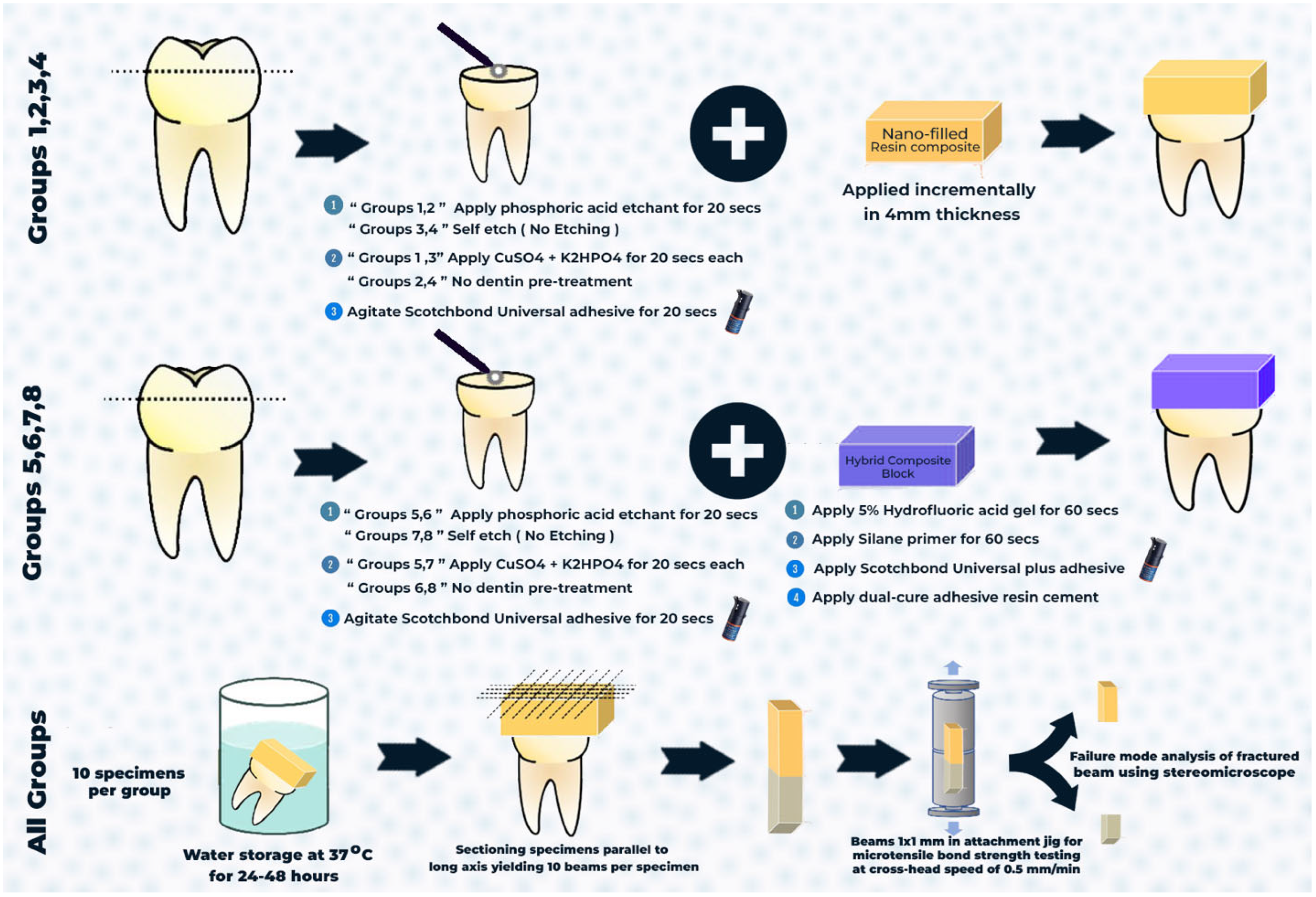
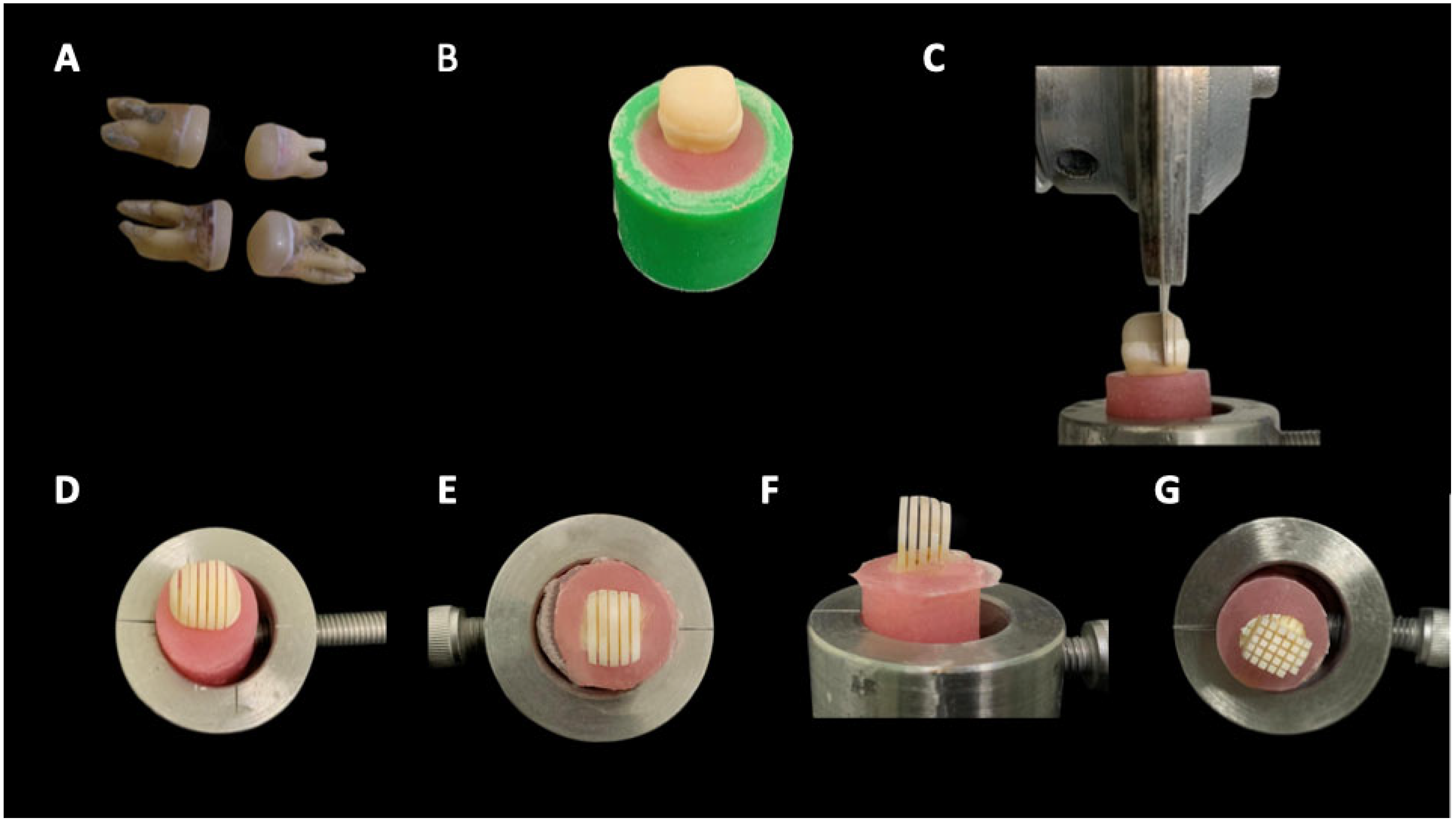
2.4. Specimen Preparation

Eighty human third molars were obtained from Department of Oral and Maxillofacial Surgery at the College of Dentistry, Prince Sattam bin Abdulaziz University. Teeth were taken from unhealthy patients with periodontal disease who had their teeth removed after their written informed consent had been obtained. The teeth were preserved in a 0.5% chloramine solution; these were utilized in a period of one month. To expose the flat dentin surface, a diamond saw (IsoMet 4000, Buehler, Lake Bluff, IL, USA) was utilized to horizontally cut the occlusal enamel at a level 1 mm below the dentin–enamel junction. An Olympus light microscope (Olympus SZ61, Munster, Germany), used at a magnification of 20×, was used to verify the exact removal of the occlusal enamel.

2.5. Restorative Procedures

2.5.1. Conventional Composite

The dentin surfaces of the tooth samples that were part of the etch-and-rinse groups were first polished and then acid-etched for 20 s using 37% phosphoric acid (Scotchbond Etchant, 3M Oral Care; St. Paul, MN, USA). The samples were then washed with deionized water for 20 s. No etching was done on the dentin surface for the self-etching groups. Then, either a new dentin-bonding pretreatment with 0.015 mol/L copper sulfate (CuSO4) and 0.01 mol/L dipotassium hydrogen phosphate (K2HPO4) was used (experimental group; Figure 2), or no treatment was carried out (control group). CuSO4 was mixed with distilled water at 37 °C to form a 0.015 mol/L blue CuSO4 solution (pH = 4). K2HPO4 was mixed with distilled water at 37 °C to form a 0.01 mol/L colorless liquid (pH = 7). In the control group, we used a tiny brush to gently remove excess water from the dentin surface while keeping it moist. The initial step was to apply 20 μL of CuSO4 solution to the dentin surface for 20 s in the experimental groups. Any surplus solution was then wiped away using a small, dry brush. The dentin surface was subsequently rubbed with a 20 μL K2HPO4 solution for 20 s. After rinsing with deionized water for 20 s, a microbrush was used to remove excess water and maintain moisture on the dentin surface.
Next, using either an etch-and-rinse or self-etching technique, the dentin surface was agitated for 20 s with the bonding adhesive Scotchbond Universal Plus (3M Oral Care, Seefeld, Germany). Using the incremental technique, a nano-filled resin composite was built. It was then light-cured for 20 s using a light-emitting diode curing unit (3M ESPE Elipar, Seefeld, Germany), which delivers 1200 mW/cm2 of light at 430–480 nm.

2.5.2. CAD/CAM Resin Blocks

To ensure accurate surface scanning without interference from laser beam reflections, tooth specimens were first covered with an opaque scanning spray. Then, a digital scanner (Freedom, DOF Inc., Seoul, Republic of Korea) was used for the scanning process. For design reasons, the acquired image was loaded into the CAD program (Exocad DentalCAD Galway 3.0; Align Technology, San José, CA, USA), and a cement spacer size of 50 μm was established. Subsequently, occlusal restorations of a consistent 4 mm thickness were manufactured (Figure 3).
Pressing the milling preview window’s button started the milling process. To start the wet milling operation, the 5-axis milling machine was turned on, and the Shufo hybrid ceramic disc (SHOFU Inc., Kyoto, Japan) was inserted into the spindle in a certain sequence. After the design was complete, the restoration STL file was sent into the milling machine (MC XL, Dentsply Sirona, Charlotte, NC, USA) in fine mode. After a series of cuts with 2.5 mm diamond burs, the disc was polished to eliminate any excess material at the connection point using a slow-speed diamond disk. Twenty specimens were milled in all (n = 20). To achieve the desired thickness, each restoration was polished using Kenda polishing tips (Kenda Unicus, Kenda AG, Vaduz, Liechtenstein), following the manufacturer’s recommendations. Multiple positions were used to measure the ultimate thickness with a caliper (lwanson’s caliper, Renfert, Hilzingen, Germany).
The interior surface of the specimen was etched for 60 s using a 5% hydrofluoric acid gel (VITA ADIVA CERAETCH, VITA Zahnfabrik, Bad Säckingen, Germany). After that, it was rinsed with water for 30 s, and then dried for 20 s using oil- and water-free air. It appeared as though the etched surface was frosty and dull. Phosphoric acid gel was used for cleaning, followed by rinsing and drying. A small amount of silane primer (RelyX® Ceramic Primer, 3M/ESPE, St. Paul, MN, USA) was applied to the etched surfaces of the specimens using microbrushing. After 60 s, it was evenly spread with air to achieve a thin coat.
The next step was to apply and blast a single coat of Scotchbond Universal Plus universal adhesive on the fitting surface of the resin block specimens. Lastly, to make sure it cured evenly, it was light-cured for 20 s. The dentin surface was then agitated for 20 s with bonding adhesive Scotchbond Universal Plus using either an etch-and-rinse or self-etching approach. Following the manufacturer’s directions, the tips were used to apply dual-cured adhesive resin cement (RelyX Ultimate, 3-M ESPE, St. Paul, MN, USA) to the surfaces of milled hybrid ceramic restorations and tooth specimens. To facilitate cement flow from all directions, all restorations were placed softly on the specimens. Any extra cement was carefully removed after lightly curing the filling edges for 4–5 s on each side. The restorations were light-cured for 20 s on each side after they were established on the stumps. A water bath was used to keep the samples at 37 °C for 24 h after they were generated (Figure 4). Materials utilized in the current study are shown in Table 2.

2.6. Microtensile Bond Strength Test (μTBS)

The specimens were sectioned longitudinally through the bonded interface using an IsoMet diamond saw (IsoMetTM 4000, Buehler Ltd., Lake Bluff, IL, USA; Figure 5). Ten beams were obtained from each tooth. Each beam was secured to a testing jig using cyanoacrylate glue and then stressed until failure using a universal testing machine (Instron 4000, Canton, MA, USA) at a cross-head speed of 0.5 mm/mm. Microtensile bond strength was calculated in MPa.

2.7. Statistical Analysis

The results were statistically evaluated utilizing the Statistical Package for the Social Sciences (SPSS 25.0, IBM/SPSS Inc., Chicago, IL, USA). The Kolmogorov–Smirnov test at α = 0.05 was utilized to verify the normal distribution of the results. A three-way ANOVA test was used to estimate how the mean of a quantitative variable changed according to the levels of three independent variables, followed by Tukey’s post hoc test for multiple comparisons. p-values > 0.05 are statistically non-significant, p-values ≤ 0.05 are significant, and p-values ≤ 0.001 are highly significant.

3. Results

3.1. Microtensile Bond Strength Values

Intra-rater and inter-rater reliability for all outcomes measured were >0.80 kappa statistics. The Shapiro–Wilk test indicated that the data from all groups followed a normal distribution (p > 0.05). The μTBS values were shown to be not significantly affected by either the resin composite type (p > 0.05) or the adhesive strategy (p > 0.05) according to the three-way ANOVA results. Conversely, the μTBS was shown to be significantly affected by dentin pretreatment (p < 0.001; Table 3). Comparison between the study groups based on mean μTBS is shown in Table 4. Hence, significant differences were detected between the no dentin pretreatment (24.20 ± 4.54 MPa) and CuSO4 + K2HPO4 pretreatment (33.66 ± 5.22 MPa) using the etch-and-rinse adhesive strategy for nano-filled composites (p < 0.001). Additionally, significant differences were detected between the no dentin pretreatment (24.71 ± 4.33 MPa) and CuSO4 + K2HPO4 pretreatment (32.49 ± 4.92 MPa) using the etch-and-rinse adhesive strategy for CAD/CAM resin blocks (p < 0.001). Moreover, significant differences were detected between the no dentin pretreatment (21.20 ± 3.40 MPa) and CuSO4 + K2HPO4 pretreatment (30.31 ± 3.87 MPa) using the self-etching adhesive strategy for nano-filled composites (p < 0.001). Also, significant differences were detected between the no dentin pretreatment (23.89 ± 3.89 MPa) and CuSO4 + K2HPO4 pretreatment (31.22 ± 4.71 MPa) using the self-etching adhesive strategy for CAD/CAM resin blocks (p < 0.001; Figure 6 and Figure 7). For nano-filled composites, there was no discernible difference between the etch-and-rinse and self-etching groups (p > 0.001). For the CAD/CAM resin blocks, no significant difference was detected between the etch-and-rinse and self-etching groups (p > 0.001; Figure 7). In addition, a comparison of the nano-filled and CAD/CAM resin blocks without the dentin pretreatment (p > 0.001) and with the CuSO4 + K2HPO4 pretreatment (p > 0.001) found no statistically significant difference (Figure 8).

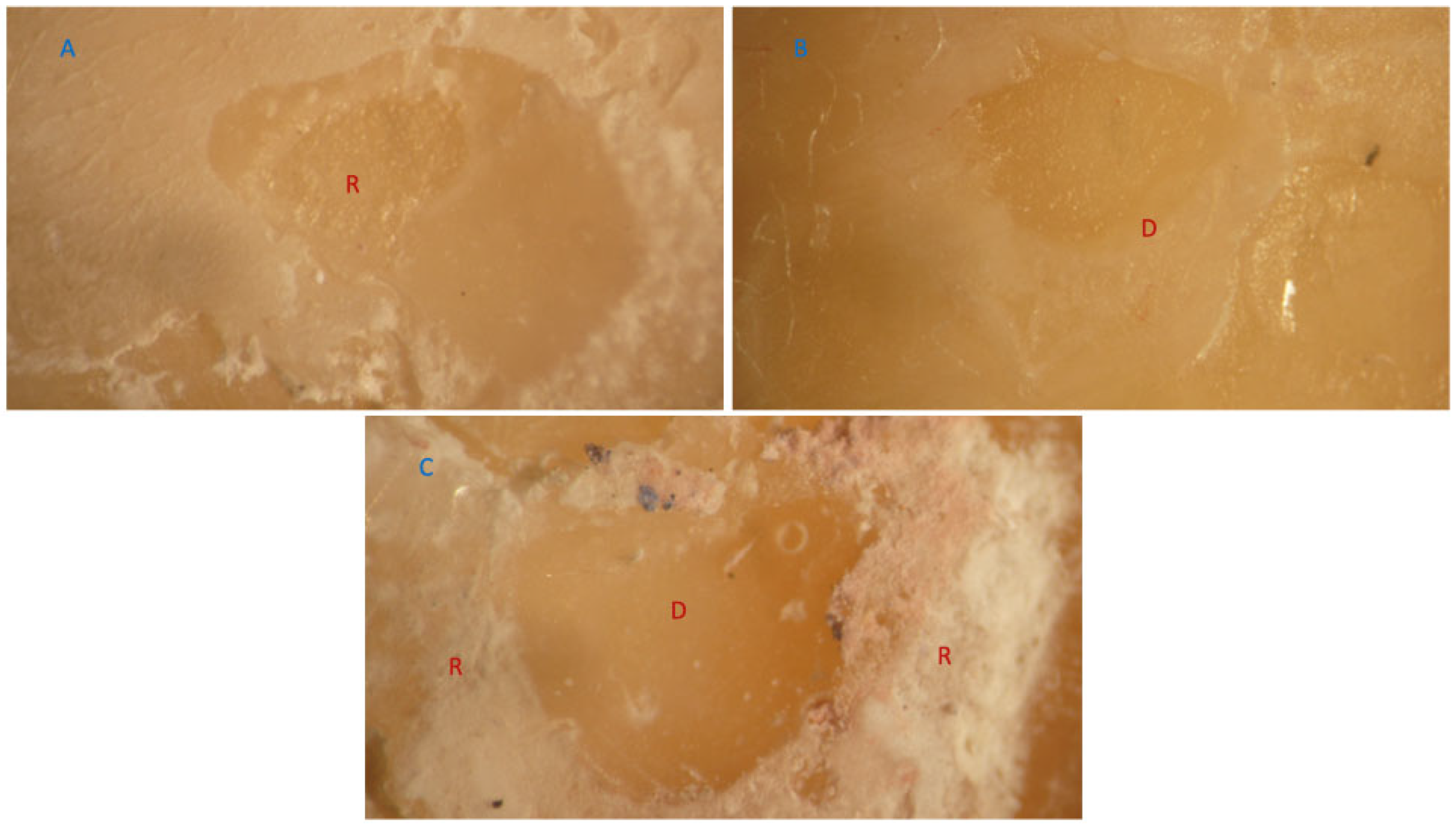
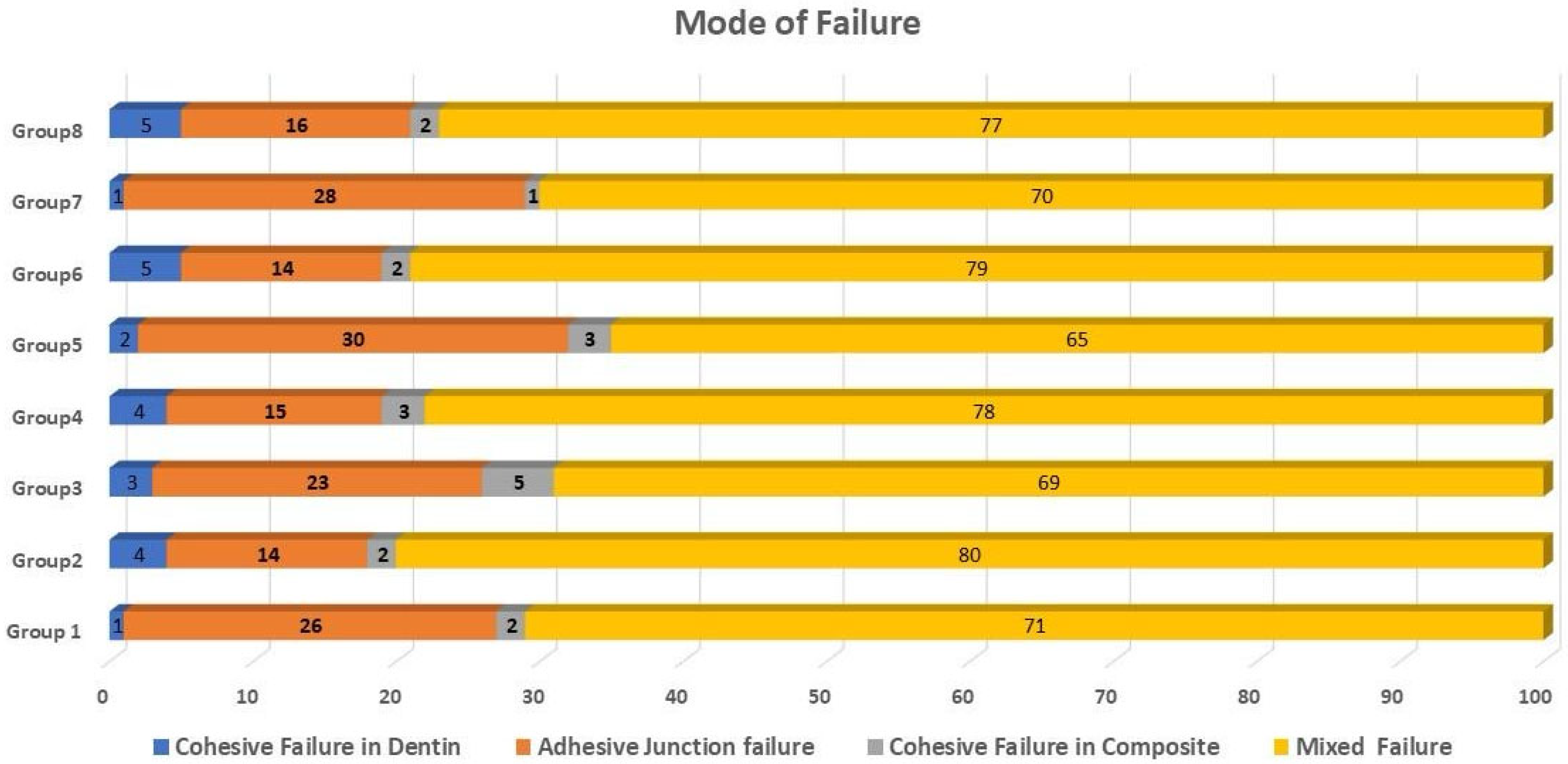
3.2. Failure Mode Analysis

The most prevalent type of fracture in all groups was a mixed failure, followed by an adhesive junction failure. The dentin and composite had minimal cohesive failures. Cohesive resin failures and adhesive junction failures occurred simultaneously throughout each mixed failure. Figure 9 and Figure 10 show representative stereomicroscope images of different failure modes. Figure 11 summarizes the distribution of the failure modes.

4. Discussion

Pretreatment of dentin with CuSO4 and K2HPO4 might render the entire hybrid layer more hydrophobic, which would improve dentin bonding. The results rejected the null hypothesis that there is no significant difference in dentin μTBS with and without CuSO4 + K2HPO4 pretreatment when bonded with a universal adhesive to conventional and CAD/CAM resin composites.
According to the literature, resin cements used in ceramic crowns should have a cement layer thickness of approximately 50–100 μm [19]. In addition, research has demonstrated that bonding characteristics are considerably diminished for a cement thickness ranging from 450 to 500 μm as a result of the residual tension caused by polymerization shrinkage [20]. For this reason, a cement spacer size of 50 μm was determined in this investigation.
In the present study, the μTBS significantly increased following CuSO4 and K2HPO4 pretreatment with nano-filled and CAD/CAM resin composites using both the etch-and-rinse and self-etching adhesive strategies. The bonding efficiency of different adhesives can be enhanced by the rapid formation of amorphous CaF2 nanoparticles in the demineralized dentin matrix following a simple sequential treatment of CaCl2 and NaF solutions. Endogenous, non-collagenous negatively charged groups are present in these nanoparticles [21].
Similarly, prior research demonstrated that nanoparticles produced by pretreatment of CaCl2 and K2HPO4 solutions enhanced dentin bonding characteristics. Like the aforementioned nanoparticles, Cu3(PO4)2 nanoparticles produced by Cu-P pretreatment may play a role in enhancing microtensile bond strength [21]. Theoretically, demineralized dentin might have its hydrophilicity mitigated by Cu-P pretreatment, and the resulting copper phosphate nanoparticles could provide structural support to the demineralized collagen fibrils. This structural support enhances the hybrid layer, and adhesive monomers are more effectively able to penetrate the interstices of the demineralized collagen fibrils network [8,22]. In the future, it will be necessary to thoroughly verify the exact process after Cu-P pretreatment using methods such as atomic force microscopy, solid surface potential, ion staining, water absorption property, and contact-angle tests.
Copper has characteristics that assist with dentin adhesion. Copper is essential for the activity of the collagen cross-linking enzyme lysyl oxidase [23]. The hybrid layer’s copper component strengthens the collagen network by acting as a cross-linking agent [22]. New evidence suggests that copper ions, which inhibit MMPs and can decrease MMP-2 synthesis, may regulate the activity of MMPs in oral tissues [24]. The impact of CuSO4 on collagen cross-linking and MMP inhibition will be subsequently validated. The findings of the present investigation align with those of Pan et al. [17], who demonstrated that copper-based pretreatment, when used with universal adhesives, enhanced the μTBS of dentin.
Cavity disinfectants based on CuSO4 have been widely employed in clinical practice due to their wettability and disinfecting effect [25]. This impact was probably caused by CuSO4’s ability to degrade the auto-associative propensity of water, which improves wettability in demineralized dentin, and dissociate strongly cross-linked collagen into a more distributed network of apparent fibrils [17]. Controversial findings persist despite claims that deproteinization causes dentin to become more porous and prone to abnormalities and anastomoses, which in turn increases bond strength [26].
The antibacterial properties of adhesives incorporating copper nanoparticles have been demonstrated in numerous investigations to be both long-lasting and effective. As an example, research conducted by Gutiérrez et al. [27] demonstrated that adhesive formulations can be enhanced with antibacterial capabilities when Cu nanoparticles are added at a concentration of 0.1 wt%. This enhancement was observed to occur without a compromise to the optical and mechanical properties of the adhesive solutions. Addition of 0.1 weight percent Cu nanoparticles to the adhesive can enhance the clinical efficacy of universal adhesive systems in non-carious cervical lesions without increasing cytotoxicity, according to a study by Matos et al. [28].
Dentin demineralization and smear layer removal are both aided by etching in the etch-and-rinse adhesive strategy, which increases the amount of monomer impregnation and exposes collagen fibrils; the result is a thick hybrid layer that is well integrated with the dentin surface [29,30]. This enhances the amount of monomer impregnation and reveals the collagen fibrils [31]. A downside is that it lowers the calcium phosphate concentration, which is practically nonexistent in hydroxyapatite crystals after etching [32]. Therefore, the primary diffusion-based bonding mechanism of the etch-and-rinse adhesive method for dentin is the hybridization or penetration of the resin into the exposed collagen fibril scaffold. Actual chemical bond formation is very unlikely due to the low affinity of the MDP monomers’ functional groups for the hydroxyapatite-depleted collagen [33,34].
Conversely, in the self-etching adhesive strategy, dentin priming and conditioning are accomplished by acidic resin monomers dissolving the smear layer, but the calcium phosphate in the hydroxyapatite surface remains intact [35,36]. The improved chemical link between the adhesive and the dentin substrate is a result of this closer chemical interaction with the functional groups of the MDP monomers [37]. Hence, the μTBS results of the Scotch Bond Universal Plus adhesive investigation showed that both etching adhesive strategies produced similar outcomes, a conclusion that is in line with previous research [38,39,40].
The self-etching and etch-and-rinse groups did not differ significantly from one another in this study. One component of Scotch Bond Universal Plus adhesive is a monomer of phosphate acid, which forms “nano-layering” on hydroxyapatite by chemically bonding to it. This results in the formation of hydrolytically stable calcium salts [41]. The similar adhesion–decalcification process also occurs when using Scotch Bond Universal Plus adhesive on apatite surfaces due to the presence of a polyalkenoic acid copolymer in this product [41]. Therefore, chemical adhesion and micromechanical retention through resin monomer diffusion were both enhanced by the bonding mechanisms of Scotch Bond Universal Plus adhesive.
In this investigation, we found no statistically significant difference between the self-etching and etch-and-rinse groups. Scotch Bond Universal Plus adhesive contains a phosphate acid monomer that chemically bonds to hydroxyapatite, forming hydrolytically stable calcium salts in the form of “nano-layering” on hydroxyapatite [41]. Additionally, Scotch Bond Universal Plus adhesive contains a polyalkenoic acid copolymer in its composition that interacts with apatite substrates following the same adhesion–decalcification reaction [42]. Consequently, the Scotch Bond Universal Plus adhesive facilitated micromechanical retention through the diffusion of resin monomers and chemical adhesion [43,44].
Munoz et al. [45] states that the bonding strength of MDP to the dentin substrate is negatively affected by the polyalkenoic acid copolymer because it hinders the chemical bond. In hydroxyapatite crystals, it competes with the MDP functional monomer for Ca-bonding sites and might even stop the monomers from approximation during polymerization. In addition, research has shown that 2-hydroxyethyl methacrylate can bind to the calcium in hydroxyapatite, which weakens the bond strength of MDP to dentin [46,47,48].
The bond strength findings were validated by the fracture beam analysis, which demonstrated an increased incidence of mixed failure in all groups and a reduced occurrence of cohesive failure in both the composite and dentin. Substrate fracture level is a good indicator of adhesive retentive strength, according to previous studies [17,49,50]. This study’s bond strength estimates were thus corroborated by the mode of failure. This agrees with the findings of Molla et al. [51], who also discovered that, at substantial bond strengths, most bond failure modes are admixed.
Furthermore, a comparison of nano-filled and CAD/CAM resin blocks, both without dentin pretreatment and with CuSO4 + K2HPO4 pretreatment, revealed no statistically significant difference. Hussain et al. [52] performed a study to compare conventional resin blocks with CAD/CAM resin blocks. They determined that the CAD/Cam resin blocks surpassed traditional composites in mechanical properties and monomer elution, although they exhibited concerning outcomes related to cytotoxicity. Mechanical research studies indicate that CAD/CAM resin blocks have superior characteristics compared to conventional composites and are equivalent to enamel and dentine. The filler content, morphology of filler particles, and filler composition primarily align with the producer’s specifications and resemble conventional composite materials [52].
Nevertheless, additional research into the bonding’s endurance and potential processes is required. Findings from this research will pave the way for novel approaches to enhancing the dentin–adhesive interface. Additionally, future research may adjust the aforementioned components to include aspects such as scanning electron microscopy, energy-dispersive X-ray, Fourier transform infrared spectroscopy, and atomic force microscopy. A more thorough investigation of their long-term degradation, particularly under in vivo conditions, is required. The main drawback of the present study is that in vitro investigations cannot replicate all the specific conditions to which a restoration is subjected in the oral cavity. To properly understand the differences between dentin pretreatment agents over time, it is crucial to evaluate the bonding performance of aged specimens using thermocycling or long-term water storage. This is given that our study only looked at immediate bond strength, but prior research has shown that there are negative effects associated with this thermocycling and long-term water storage.

5. Conclusions

Within the limitations of the study, dentin’s immediate μTBS was enhanced by a pretreatment using CuSO4 and K2HPO4 solutions when used with nano-filled and CAD/CAM resin blocks. Furthermore, the adhesion of Scotch Bond Universal Plus adhesive to dentin remains largely consistent regardless of whether the etch-and-rinse or self-etching adhesive technique is adopted.

6. Clinical Relevance

Dentin pretreatment with CuSO4 and K2HPO4 proved to be effective, which could enable general practitioners to use them in dental practice routinely.

Author Contributions

Conceptualization, A.A. (Abdulellah Almudahi); Methodology, A.A.E., F.A., A.F.A. (Abdulaziz Fahd Alsubaie) and A.F.A. (Abdulaziz Fahd Alharbi); Validation, A.A.E. and A.A.A.; Formal analysis, F.A.; Investigation, A.A.E., A.A. (Abdulellah Almudahi), A.F.A. (Abdulaziz Fahd Alsubaie) and A.F.A. (Abdulaziz Fahd Alharbi); Resources, A.A. (Abdullah Alshehri), R.B., A.A. (Abdulellah Almudahi), A.F.A. (Abdulaziz Fahd Alsubaie) and A.F.A. (Abdulaziz Fahd Alharbi); Writing—original draft, A.A.E., A.A. (Abdullah Alshehri) and F.A.; Writing—review & editing, A.A.E., R.B. and A.A.A.; Supervision, A.A. (Abdullah Alshehri); Project administration, A.A. (Abdullah Alshehri) and A.A.A.; Funding acquisition, R.B. and A.A. (Abdulellah Almudahi). All authors have read and agreed to the published version of the manuscript.

Funding

The study was supported via funding from Prince Sattam bin Abdulaziz University project number (PSAU/2025/3/32637).

Institutional Review Board Statement

The study was conducted according to the guidelines of the Declaration of Helsinki, and approved by the Ethics Committee of Prince Sattam bin Abdulaziz University (#SCBR-363/2024, approval date 1 December 2024).

Informed Consent Statement

Informed consent was obtained from the participants involved in the study before the teeth extraction.

Data Availability Statement

The original contributions presented in the study are included in the article, further inquiries can be directed to the corresponding author.

Conflicts of Interest

The authors declare no conflicts of interest.

References

  1. Nagarkar, S.; Theis-Mahon, N.; Perdigão, J. Universal dental adhesives: Current status, laboratory testing, and clinical performance. J. Biomed. Mater. Res. Part B Appl. Biomater. 2019, 107, 2121–2131. [Google Scholar] [CrossRef] [PubMed]
  2. Cuevas-Suárez, C.E.; da Rosa, W.L.O.; Lund, R.G.; da Silva, A.F.; Piva, E. Bonding Performance of Universal Adhesives: An Updated Systematic Review and Meta-Analysis. J. Adhes. Dent. 2019, 21, 7–26. [Google Scholar] [PubMed]
  3. Jäggi, M.; Karlin, S.; Zitzmann, N.U.; Rohr, N. Shear bond strength of universal adhesives to human enamel and dentin. J. Esthet. Restor. Dent. 2024, 36, 804–812. [Google Scholar] [CrossRef]
  4. Betancourt, D.E.; Baldion, P.A.; Castellanos, J.E. Resin-Dentin Bonding Interface: Mechanisms of Degradation and Strategies for Stabilization of the Hybrid Layer. Int. J. Biomater. 2019, 2019, 5268342. [Google Scholar] [CrossRef]
  5. Pashley, D.H.; Tay, F.R.; Carvalho, R.M.; Rueggeberg, F.A.; Agee, K.A.; Carrilho, M.; Donnelly, A.; García-Godoy, F. From dry bonding to water-wet bonding to ethanol-wet bonding. A review of the interactions between dentin matrix and solvated resins using a macromodel of the hybrid layer. Am. J. Dent. 2007, 20, 7. [Google Scholar]
  6. Bottene Guarda, M.; Tulio Di Nizo, P.; Flores Abuna, G.; Catelan, A.; Coelho Sinhoreti, M.A.; Pino Vitti, R. Effect of Electric Current-assisted Application of Adhesives on their Bond Strength and Quality. J. Adhes. Dent. 2020, 22, 393–398. [Google Scholar]
  7. Matinlinna, J.P.; Lung, C.Y.K.; Tsoi, J.K.H. Silane adhesion mechanism in dental applications and surface treatments: A review. Dent. Mater. 2018, 34, 13–28. [Google Scholar] [CrossRef] [PubMed]
  8. Farina, A.P.; Cecchin, D.; Vidal, C.M.; Leme-Kraus, A.A.; Bedran-Russo, A.K. Removal of water binding proteins from dentin increases the adhesion strength of low-hydrophilicity dental resins. Dent. Mater. 2020, 36, e302–e308. [Google Scholar] [CrossRef]
  9. Rucker, R.B.; Kosonen, T.; Clegg, M.S.; Mitchell, A.E.; Rucker, B.R.; Uriu-Hare, J.Y.; Keen, C.L. Copper, lysyl oxidase, and extracellular matrix protein cross-linking. Am. J. Clin. Nutr. 1998, 67 (Suppl. S5), 996s–1002s. [Google Scholar] [CrossRef]
  10. Kubo, A.-L.; Vasiliev, G.; Vija, H.; Krishtal, J.; Tõugu, V.; Visnapuu, M.; Kisand, V.; Kahru, A.; Bondarenko, O.M. Surface carboxylation or PEGylation decreases CuO nanoparticles’ cytotoxicity to human cells in vitro without compromising their antibacterial properties. Arch. Toxicol. 2020, 94, 1561–1573. [Google Scholar] [CrossRef]
  11. Tiwari, M.; Narayanan, K.; Thakar, M.B.; Jagani, H.V.; Venkata Rao, J. Biosynthesis and wound healing activity of copper nanoparticles. IET Nanobiotechnol. 2014, 8, 230–237. [Google Scholar] [CrossRef]
  12. Sabatini, C.; Mennito, A.S.; Wolf, B.J.; Pashley, D.H.; Renné, W.G. Incorporation of bactericidal poly-acrylic acid modified copper iodide particles into adhesive resins. J. Dent. 2015, 43, 546–555. [Google Scholar] [CrossRef]
  13. Grzebieluch, W.; Mikulewicz, M.; Kaczmarek, U. Resin composite materials for chairside CAD/CAM restorations: A comparison of selected mechanical properties. J. Healthc. Eng. 2021, 2021, 8828954. [Google Scholar] [CrossRef] [PubMed]
  14. Grzebieluch, W.; Kowalewski, P.; Grygier, D.; Rutkowska-Gorczyca, M.; Kozakiewicz, M.; Jurczyszyn, K. Printable and machinable dental restorative composites for CAD/CAM application—Comparison of mechanical properties, fractographic, texture and fractal dimension analysis. Materials 2021, 14, 4919. [Google Scholar] [CrossRef] [PubMed]
  15. Aladağ, S.; Ayaz, E.A. Bonding effectiveness of multi-step adhesive resin cements to CAD/CAM blocks: Impact of thermal cycling and surface treatment methods. BMC Oral Health 2024, 24, 1326. [Google Scholar] [CrossRef] [PubMed]
  16. Şenol, M.; Gürbüz, A.; Oyar, P. Bond strength of conventional resin-based adhesive cement and self-adhesive resin cement to CAD-CAM restorative materials. BMC Oral Health 2025, 25, 296. [Google Scholar] [CrossRef]
  17. Pan, Y.; Xu, J.; Cai, X.; Li, X.; Wang, X. Effect of a novel pretreatment on the microtensile bond strength of universal adhesives with dentin. J. Dent. Sci. 2023, 18, 1148–1155. [Google Scholar] [CrossRef]
  18. Schulz, K.F.; Altman, D.G.; Moher, D. CONSORT 2010 Statement: Updated guidelines for reporting parallel group randomised trials. J. Clin. Epidemiol. 2010, 63, 834–840. [Google Scholar] [CrossRef]
  19. Tribst, J.P.M.; Dos Santos, A.F.C.; da Cruz Santos, G.; da Silva Leite, L.S.; Lozada, J.C.; Silva-Concílio, L.R.; Baroudi, K.; Amaral, M. Effect of Cement Layer Thickness on the Immediate and Long-Term Bond Strength and Residual Stress between Lithium Disilicate Glass-Ceramic and Human Dentin. Materials 2021, 14, 5153. [Google Scholar] [CrossRef]
  20. Aker Sagen, M.; Dahl, J.E.; Matinlinna, J.P.; Tibballs, J.E.; Rønold, H.J. The influence of the resin-based cement layer on ceramic-dentin bond strength. Eur. J. Oral Sci. 2021, 129, e12791. [Google Scholar] [CrossRef]
  21. Xu, J.; Chen, Y.; Li, X.; Lei, Y.; Shu, C.; Luo, Q.; Chen, L.; Li, X. Reconstruction of a demineralized dentin matrix via rapid deposition of CaF2 nanoparticles in situ promotes dentin bonding. ACS Appl. Mater. Interfaces 2021, 13, 51775–51789. [Google Scholar] [CrossRef] [PubMed]
  22. Souza, A.; Gerlach, R.; Line, S. Inhibition of human gelatinases by metals released from dental amalgam. Biomaterials 2001, 22, 2025–2030. [Google Scholar] [CrossRef]
  23. Marelli, B.; Le Nihouannen, D.; Hacking, S.A.; Tran, S.; Li, J.; Murshed, M.; Doillon, C.J.; Ghezzi, C.E.; Zhang, Y.L.; Nazhat, S.N. Newly identified interfibrillar collagen crosslinking suppresses cell proliferation and remodelling. Biomaterials 2015, 54, 126–135. [Google Scholar] [CrossRef] [PubMed]
  24. Guo, H.; Lee, J.-D.; Uzui, H.; Toyoda, K.; Geshi, T.; Yue, H.; Ueda, T. Effects of copper and zinc on the production of homocysteine-induced extracellular matrix metalloproteinase-2 in cultured rat vascular smooth muscle cells. Acta Cardiol. 2005, 60, 353–359. [Google Scholar] [CrossRef]
  25. Sharma, V.; Nainan, M.T.; Shivanna, V. The effect of cavity disinfectants on the sealing ability of dentin bonding system: An in vitro study. J. Conserv. Dent. 2009, 12, 109–113. [Google Scholar] [CrossRef] [PubMed]
  26. Wakabayashi, Y.; Kondou, Y.; Suzuki, K.; Yatani, H.; Yamashita, A. Effect of dissolution of collagen on adhesion to dentin. Int. J. Prosthodont. 1994, 7, 302–306. [Google Scholar]
  27. Gutiérrez, M.F.; Malaquias, P.; Hass, V.; Matos, T.P.; Lourenço, L.; Reis, A.; Loguercio, A.D.; Farago, P.V. The role of copper nanoparticles in an etch-and-rinse adhesive on antimicrobial activity, mechanical properties and the durability of resin-dentine interfaces. J. Dent. 2017, 61, 12–20. [Google Scholar] [CrossRef]
  28. Matos, T.P.; Gutiérrez, M.F.; Hanzen, T.A.; Malaquias, P.; de Paula, A.M.; de Souza, J.J.; Hass, V.; Fernández, E.; Reis, A.; Loguercio, A.D. 18-month clinical evaluation of a copper-containing universal adhesive in non-carious cervical lesions: A double-blind, randomized controlled trial. J. Dent. 2019, 90, 103219. [Google Scholar] [CrossRef]
  29. De Munck, J.; Van Landuyt, K.; Peumans, M.; Poitevin, A.; Lambrechts, P.; Braem, M.; Van Meerbeek, B. A critical review of the durability of adhesion to tooth tissue: Methods and results. J. Dent. Res. 2005, 84, 118–132. [Google Scholar] [CrossRef]
  30. Breschi, L.; Maravic, T.; Mazzitelli, C.; Josic, U.; Mancuso, E.; Cadenaro, M.; Pfeifer, C.S.; Mazzoni, A. The evolution of adhesive dentistry: From etch-and-rinse to universal bonding systems. Dent. Mater. 2024, 41, 141–158. [Google Scholar] [CrossRef]
  31. Pashley, D.H.; Tay, F.R.; Breschi, L.; Tjäderhane, L.; Carvalho, R.M.; Carrilho, M.; Tezvergil-Mutluay, A. State of the art etch-and-rinse adhesives. Dent. Mater. 2011, 27, 1–16. [Google Scholar] [CrossRef] [PubMed]
  32. Rosa, W.L.; Piva, E.; Silva, A.F. Bond strength of universal adhesives: A systematic review and meta-analysis. J. Dent. 2015, 43, 765–776. [Google Scholar] [CrossRef]
  33. Elkaffas, A.A.; Hamama, H.H.; Mahmoud, S.H.; Fawzy, A.S. Effect of acid etching on dentin bond strength of ultra-mild self-etch adhesives. Int. J. Adhes. Adhes. 2020, 99, 102567. [Google Scholar] [CrossRef]
  34. Hardan, L.; Bourgi, R.; Kharouf, N.; Mancino, D.; Zrow, M.; Jakubowicz, N.; Haikel, Y.; Cuevas-Suárez, C.E. Bond Strength of Universal Adhesives to Dentin: A Systematic Review and Meta-Analysis. Polymers 2021, 13, 814. [Google Scholar] [CrossRef]
  35. Giannini, M.; Makishi, P.; Ayres, A.P.; Vermelho, P.M.; Fronza, B.M.; Nikaido, T.; Tagami, J. Self-etch adhesive systems: A literature review. Braz. Dent. J. 2015, 26, 3–10. [Google Scholar] [CrossRef]
  36. Costa, M.P.; Giacomini, M.C.; Zabeu, G.S.; Mosquim, V.; Dallavilla, G.G.; da Silva Santos, P.S.; Wang, L. Impact of functional monomers, bioactive particles, and HEMA, on the adhesive performance of self-etch adhesive systems applied to simulated altered dentin. J. Dent. 2024, 151, 105379. [Google Scholar] [CrossRef] [PubMed]
  37. Chersoni, S.; Suppa, P.; Grandini, S.; Goracci, C.; Monticelli, F.; Yiu, C.; Huang, C.; Prati, C.; Breschi, L.; Ferrari, M. In vivo and in vitro permeability of one-step self-etch adhesives. J. Dent. Res. 2004, 83, 459–464. [Google Scholar] [CrossRef] [PubMed]
  38. Wagner, A.; Wendler, M.; Petschelt, A.; Belli, R.; Lohbauer, U. Bonding performance of universal adhesives in different etching modes. J. Dent. 2014, 42, 800–807. [Google Scholar] [CrossRef]
  39. Chen, C.; Niu, L.-N.; Xie, H.; Zhang, Z.-Y.; Zhou, L.-Q.; Jiao, K.; Chen, J.-H.; Pashley, D.H.; Tay, F. Bonding of universal adhesives to dentine–old wine in new bottles? J. Dent. 2015, 43, 525–536. [Google Scholar] [CrossRef]
  40. Brkanović, S.; Sever, E.K.; Vukelja, J.; Ivica, A.; Miletić, I.; Krmek, S.J. Comparison of different universal adhesive systems on dentin bond strength. Materials 2023, 16, 1530. [Google Scholar] [CrossRef]
  41. Yoshida, Y.; Yoshihara, K.; Nagaoka, N.; Hayakawa, S.; Torii, Y.; Ogawa, T.; Osaka, A.; Meerbeek, B.V. Self-assembled nano-layering at the adhesive interface. J. Dent. Res. 2012, 91, 376–381. [Google Scholar] [CrossRef] [PubMed]
  42. Sezinando, A.; Linares Serrano, M.; Morales Perez, V.; Garcia Munoz, R.A.; Ceballos, L.; Perdigao, J. Chemical Adhesion of Polyalkenoate-based Adhesives to Hydroxyapatite. J. Adhes. Dent. 2016, 18, 257–265. [Google Scholar] [PubMed]
  43. Bourgi, R.; Kharouf, N.; Cuevas-Suárez, C.E.; Lukomska-Szymanska, M.; Haikel, Y.; Hardan, L. A literature review of adhesive systems in dentistry: Key components and their clinical applications. Appl. Sci. 2024, 14, 8111. [Google Scholar] [CrossRef]
  44. Alomran, W.K.; Nizami, M.Z.I.; Xu, H.H.; Sun, J. Evolution of Dental Resin Adhesives—A Comprehensive Review. J. Funct. Biomater. 2025, 16, 104. [Google Scholar] [CrossRef]
  45. Muñoz, M.; Luque-Martinez, I.; Malaquias, P.; Hass, V.; Reis, A.; Campanha, N.; Loguercio, A. In vitro longevity of bonding properties of universal adhesives to dentin. Oper. Dent. 2015, 40, 282–292. [Google Scholar] [CrossRef]
  46. Turp, V.; Sen, D.; Tuncelli, B.; Özcan, M. Adhesion of 10-MDP containing resin cements to dentin with and without the etch-and-rinse technique. J. Adv. Prosthodont. 2013, 5, 226. [Google Scholar] [CrossRef]
  47. Carrilho, E.; Cardoso, M.; Marques Ferreira, M.; Marto, C.M.; Paula, A.; Coelho, A.S. 10-MDP Based Dental Adhesives: Adhesive Interface Characterization and Adhesive Stability-A Systematic Review. Materials 2019, 12, 790. [Google Scholar] [CrossRef]
  48. Pimentel de Oliveira, R.; de Paula, B.L.; Ribeiro, M.E.; Alves, E.; Costi, H.T.; Silva, C. Evaluation of the Bond Strength of Self-Etching Adhesive Systems Containing HEMA and 10-MDP Monomers: Bond Strength of Adhesives Containing HEMA and 10-MDP. Int. J. Dent. 2022, 2022, 5756649. [Google Scholar] [CrossRef]
  49. Santander-Rengifo, F.; Carreras-Presas, C.M.; Aroste-Andía, R.; Hernández-Huamaní, E.; Gavilán-Chávez, P.; Cervantes-Ganoza, L.; Cayo-Rojas, C. Microtensile bond strength and failure mode of different universal adhesives on human dentin. Int. Dent. J. 2024, 74, 1239–1247. [Google Scholar] [CrossRef]
  50. Alam, A.; Yamauti, M.; Chowdhury, A.F.M.A.; Wang, X.; Álvarez-Lloret, P.; Zuñiga-Heredia, E.-E.; Cifuentes-Jiménez, C.; Dua, R.; Iijima, M.; Sano, H. Evaluating the advancements in a recently introduced universal adhesive compared to its predecessor. J. Dent. Sci. 2024, 19, 1609–1619. [Google Scholar] [CrossRef]
  51. Molla, K.; Park, H.J.; Haller, B. Bond strength of adhesive/composite combinations to dentin involving total- and self-etch adhesives. J. Adhes. Dent. 2002, 4, 171–180. [Google Scholar] [PubMed]
  52. Hussain, B.; Thieu, M.K.L.; Johnsen, G.F.; Reseland, J.E.; Haugen, H.J. Can CAD/CAM resin blocks be considered as substitute for conventional resins? Dent. Mater. 2017, 33, 1362–1370. [Google Scholar] [CrossRef] [PubMed]
Figure 1. Schematic flow diagram showing the study design.
Jfb 16 00197 g001
Figure 2. Images showing copper sulfate (CuSO4; blue) and dipotassium hydrogen phosphate (K2HPO4; white) powders.
Jfb 16 00197 g002
Figure 3. Images showing the design of specimens with 4 mm thickness using CAD software (Exocad DentalCAD Galway 3.0; Align Technology, San José, CA, USA).
Jfb 16 00197 g003
Figure 4. Schematic diagram shows step-by-step procedures, including surface treatment, composite application, storage, specimen sectioning, beam fixation in the attachment jig, and failure mode analysis. Group 1, no dentin pretreatment + etch-and-rinse + nano-filled composite; Group 2, CuSO4 + K2HPO4 pretreatment + etch-and-rinse + nano-filled composite; Group 3, no dentin pretreatment + self-etching + nano-filled composite; Group 4, CuSO4 + K2HPO4 pretreatment + self-etching + nano-filled composite; Group 5, no dentin pretreatment + etch-and-rinse + CAD/CAM resin blocks; Group 6, CuSO4 + K2HPO4 pretreatment + etch-and-rinse + CAD/CAM resin blocks; Group 7, no dentin pretreatment + self-etching + CAD/CAM resin blocks, and Group 8, CuSO4 + K2HPO4 pretreatment + self-etching + CAD/CAM resin blocks.
Jfb 16 00197 g004
Figure 5. Representative images showing specimen-cutting procedures for obtaining beams for μTBS testing: (A), specimens (10 specimens per group); (B), specimen fixed in acrylic mold; (C), lateral view showing IsoMet saw-sectioning in one direction for CAD/CAM resin; (D), occlusal view for sectioning in one direction for CAD/CAM resin; (E), occlusal view for sectioning in one direction for conventional resin; (F), lateral view for sectioning in one direction for conventional resin; and (G), occlusal view for sectioning in opposite direction for conventional resin to obtain beams 1 × 1 mm thickness.
Jfb 16 00197 g005
Figure 6. Graphically drawn mean values ± standard deviations showing the effect of resin composite and adhesive strategy on μTBS in MPa.
Jfb 16 00197 g006
Figure 7. Graphically drawn mean values ± standard deviations comparing the effect of adhesive strategy and dentin pretreatment on μTBS in MPa.
Jfb 16 00197 g007
Figure 8. Graphically drawn mean values ± standard deviations showing the effect of resin composite and dentin pretreatment on μTBS in MPa.
Jfb 16 00197 g008
Figure 9. Representative stereomicroscopic images depicting various failure modes: (A) mixed failure; (B) adhesive junction failure; (C) cohesive failure in composites.
Jfb 16 00197 g009
Figure 10. Stereomicroscope images of different failure modes: (A) cohesive failure in composite; (B) adhesive junction failure; (C) mixed failure; D, dentin; R, resin.
Jfb 16 00197 g010
Figure 11. Distribution of failure modes.
Jfb 16 00197 g011
Table 1. Dentin pretreatment and adhesive strategy performed in nano-filled and CAD/CAM resin composite groups.
GroupsDentin PretreatmentAdhesive StrategyResin Composite
Group 1No dentin pretreatmentEtch-and-rinseNano-filled composite
Group 2CuSO4 + K2HPO4Etch-and-rinseNano-filled composite
Group 3No dentin pretreatmentSelf-etchingNano-filled composite
Group 4CuSO4 + K2HPO4Self-etchingNano-filled composite
Group 5No dentin pretreatmentEtch-and-rinseCAD/CAM resin blocks
Group 6CuSO4 + K2HPO4Etch-and-rinseCAD/CAM resin blocks
Group 7No dentin pretreatmentSelf-etchingCAD/CAM resin blocks
Group 8CuSO4 + K2HPO4Self-etchingCAD/CAM resin blocks
Table 2. Composition, commercial name, and manufacturer of materials utilized in the current study.
MaterialCommercial Name and ManufacturerComposition
CAD/CAM resin blockSHOFU Inc., Kyoto, JapanTriethylene glycol dimethacrylate, urethane dimethacrylate, silica powder, zirconium silicate, fine particulate silica and colorant
Nano-filled resin compsiteFiltek Z350 XT, 3M Oral Care, St. Paul, MN, USAUrethane dimethacrylate, Bisphenol-A-glycidyl methacrylate, ethoxylated bisphenol-A dimethacrylate, in addition to trace amounts of triethylene glycol dimethacrylate and nanoagglomerated nanoparticles of zirconium/silica particles with sizes ranging from 0.6 to 1.4 mm, accounting for 78.5% of the total weight (59.5% of the volume)
Adhesive resin cementRelyX Ultimate, 3-M ESPE, St. Paul, MN, USASilane-treated glass powder, 2-methyl, 1,10-[1-(hydroxymethyl) 1,2-ethanediyl] ester, 2-propenoic acid, TEGDEMA, silane-treated silica, oxide glass chemicals, sodium persulfate, tert-butyl peroxy-3,5,5 trimethylhexanoate, reaction products with 2-hydroxy-1,3-propanediyl dimethacrylate and phosphorus oxide, copper (II) acetate monohydrate, silane-treated silica, substituted dimethacrylate, 1,12-dodecane dimethacrylate, -benzyl-5-phenyl-barbic-acid, sodium p-toluenesulfinate, 2-propenoic acid, calcium salt, calcium hydroxide, and titanium dioxide
Scotchbond Universal Plus AdhesiveSurefil® SDR™flow, Dentsply Caulk, Milford, DE, USA2-hydroxyethyl methacrylate, 2-propenoic acid, 10-methacryloyloxydecyl dihydrogen phosphate, 1,3-benzenediol 2-(2-hydroxyethoxy) ethyl 3-hydroxypropyl diethers, 2-methyl-, 3 triethoxysilyl) propyl ester, reaction products with silica and 3-aminopropyl triethoxysilane, ethanol, water, camphorquinone, a copolymer of acrylic and itaconic acid, and copper (II) acetate monohydrate
Table 3. Three-way ANOVA for μTBS in MPa.
SourceSum of SquaresdfMean SquaresFp-ValuePartial Eta Squared
(Effect Size)
Corrected Model1497.787 a7213.97012.0730.0000.540
Intercept61,217.113161,217.1133453.9860.0000.980
Dentin pretreatment1386.11311386.11378.2070.0000.521
Adhesive strategy82.012182.0124.6270.0350.060
Resin composite6.61316.6130.3730.5430.005
Dentin pretreatment * adhesive strategy0.11310.1130.0060.9370.000
Dentin pretreatment * resin composite5.51315.5130.3110.5790.004
Adhesive strategy * resin composite17.113117.1130.9660.3290.013
Dentin pretreatment * Adhesive strategy * resin composite0.31310.3130.0180.8950.000
Error1276.1007217.724
Total63,991.00080
Corrected Total2773.88779
a. R squared = 0.540; adjusted R squared = 0.495. *: Statistically significant (p ≤ 0.05).
Table 4. Microtensile bond strength (MPa) data (mean ± SD) in all groups.
Dentin Pretreatment
+ Adhesive Strategy
Resin Composite
Nano-Filled Resin Composite
Mean ± SD
CAD/CAM Resin Block
Mean ± SD
p-Value
No dentin pretreatment
Etch-and-rinse24.20 ± 4.54 Ac24.71 ± 4.33 Ac1.000
Self-etching21.20 ± 3.40 Ac23.89 ± 3.89 Ac1.000
CuSO4 + K2HPO4
Dentin pretreatment
Etch-and-rinse33.66 ± 5.22 Aa32.49 ± 4.92 Aa1.000
Self-etching30.31 ± 3.87 Ab31.22 ± 4.71 Aab1.000
Distinct upper-case letters in the same row signify a significant difference between groups; distinct lower-case letters in the same column denote a significant difference between groups (Tukey’s test, p < 0.05).
Disclaimer/Publisher’s Note: The statements, opinions and data contained in all publications are solely those of the individual author(s) and contributor(s) and not of MDPI and/or the editor(s). MDPI and/or the editor(s) disclaim responsibility for any injury to people or property resulting from any ideas, methods, instructions or products referred to in the content.

