


Regeneration of Critical Calvarial Bone Defects Using Bovine Xenograft, Magnesium-Enriched Bovine Xenograft and Autologous Dentin in Rats: Micro-CT, Gene Expression and Immunohistochemical Analysis

 ,
,  , ,
, ,  , ,
, ,  and
and 
Abstract
1. Introduction
2. Materials and Methods
2.1. Experimental Animals and Study Design
2.2. Materials
- Cerabone® (botiss GmbH, Zossen, Germany) with magnesium (BX + Mg group), produced at Biotrics Biomiplants AG (Berlin, Germany) in the form of Mg granules in which the mass fraction of the magnesium alloy (a solid solution of magnesium with yttrium, zinc, manganese and calcium) is 3%. Additional information is not available because the biomaterial itself is still the subject of research, and thus all data are confidential.
- Cerabone® (BX group) made from trabecular bovine bone from which the organic component of the bone tissue was removed by physical and chemical processes.
- Autologous dentin from rat teeth (AD group) ground with a dentin grinder (KometaBio Smart Dentin Grinder, Tenafly, NJ, USA) and then prepared according to a protocol previously described by our group of authors before being applied to the defect [21]. To collect dentin material for the AD group, tooth extractions were performed on donor inbred Wistar rats six days prior to the CCBD surgery [21,22,23].
2.3. Surgical Protocol and Bone Sample Harvesting
2.4. Micro-CT Analysis of 3D Parameters
2.5. Real-Time Polymerase Chain Reaction (RT-PCR) Analysis
2.6. Immunohistochemical Analysis
- BMP-2/4 (sc-137087 SCBT, Santa Cruz Biotechnology, Dallas, TX, USA, SAD), 1:200;
- VEGF (ab 231260 abcam, Abcam, Cambridge, UK), 1:200;
- SMAD-1/5/8 (#95115 CST, Cell Signaling Technology, Danvers, MA, USA, SAD), 1:100.
2.7. Statistical Analysis
3. Results
3.1. Mirco-CT Analysis
3.2. The Results of Real-Time Polymerase Chain Reaction (RT-PCR) Analysis
3.3. Results of Immunohistochemical Analysis
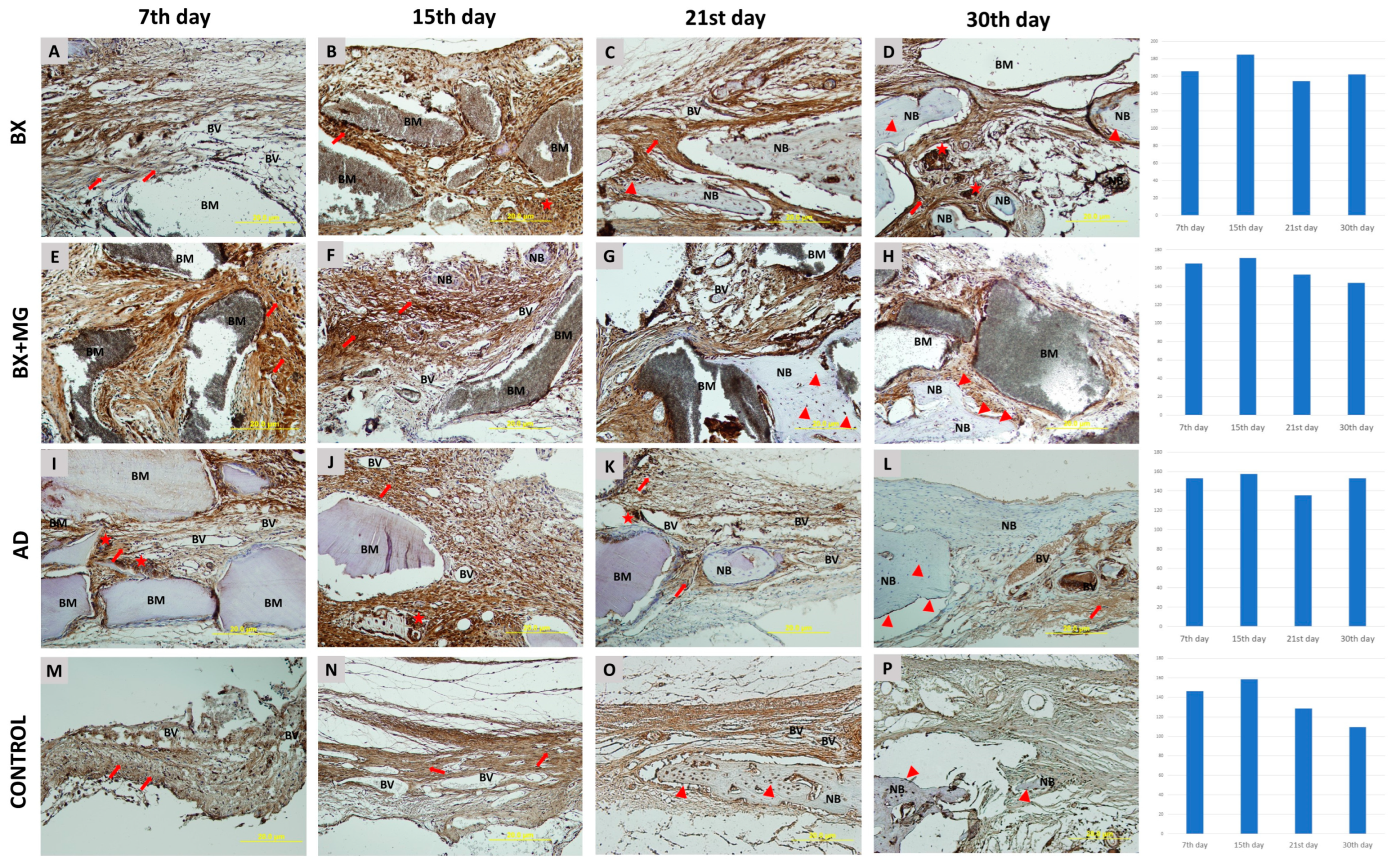
3.3.1. Immunohistochemical Analysis of BMP-2/4 Expression
3.3.2. Immunohistochemical Analysis of SMAD-1/5/8 Expression
3.3.3. Immunohistochemical Analysis of VEGF Expression
4. Discussion
Author Contributions
Funding
Institutional Review Board Statement
Informed Consent Statement
Data Availability Statement
Conflicts of Interest
References
- Rowe, P.; Koller, A.; Sharma, S. Physiology, Bone Remodeling. In StatPearls; StatPearls Publishing: Treasure Island, FL, USA, 2024. [Google Scholar]
- Albrektsson, T.; Johansson, C. Osteoinduction, Osteoconduction and Osseointegration. Eur. Spine J. 2001, 10 (Suppl. S2), S96–S101. [Google Scholar] [CrossRef] [Scilit]
- Kopf, J.; Petersen, A.; Duda, G.N.; Knaus, P. BMP2 and Mechanical Loading Cooperatively Regulate Immediate Early Signalling Events in the BMP Pathway. BMC Biol. 2012, 10, 37. [Google Scholar] [CrossRef] [Scilit]
- Rath, B.; Nam, J.; Deschner, J.; Schaumburger, J.; Tingart, M.; Grässel, S.; Grifka, J.; Agarwal, S. Biomechanical Forces Exert Anabolic Effects on Osteoblasts by Activation of SMAD 1/5/8 through Type 1 BMP Receptor. Biorheology 2011, 48, 37–48. [Google Scholar] [CrossRef] [Scilit]
- Zou, M.-L.; Chen, Z.-H.; Teng, Y.-Y.; Liu, S.-Y.; Jia, Y.; Zhang, K.-W.; Sun, Z.-L.; Wu, J.-J.; Yuan, Z.-D.; Feng, Y.; et al. The Smad Dependent TGF-β and BMP Signaling Pathway in Bone Remodeling and Therapies. Front. Mol. Biosci. 2021, 8. [Google Scholar] [CrossRef] [Scilit]
- Chen, Y.; Yin, Y.; Luo, M.; Wu, J.; Chen, A.; Deng, L.; Xie, L.; Han, X. Occlusal Force Maintains Alveolar Bone Homeostasis via Type H Angiogenesis. J. Dent. Res. 2023, 102, 1356–1365. [Google Scholar] [CrossRef] [Scilit]
- Rodan, G.A. Bone Homeostasis. Proc. Natl. Acad. Sci. USA 1998, 95, 13361–13362. [Google Scholar] [CrossRef] [Scilit]
- Hansson, S.; Halldin, A. Alveolar Ridge Resorption after Tooth Extraction: A Consequence of a Fundamental Principle of Bone Physiology. J. Dent. Biomech. 2012, 3, 1758736012456543. [Google Scholar] [CrossRef] [Scilit]
- Sodek, J.; Mckee, M.D. Molecular and Cellular Biology of Alveolar Bone. Periodontology 2000, 24, 99–126. [Google Scholar] [CrossRef] [Scilit]
- Pesic, Z.; Pejcic, A. Biomaterials in Dentistry—Implantology and Guided Bone Regeneration. In Biomaterials in Clinical Practice: Advances in Clinical Research and Medical Devices; Zivic, F., Affatato, S., Trajanovic, M., Schnabelrauch, M., Grujovic, N., Choy, K.L., Eds.; Springer International Publishing: Cham, Switzerland, 2018; pp. 697–739. ISBN 978-3-319-68025-5. [Google Scholar]
- Inchingolo, A.M.; Patano, A.; Di Pede, C.; Inchingolo, A.D.; Palmieri, G.; de Ruvo, E.; Campanelli, M.; Buongiorno, S.; Carpentiere, V.; Piras, F.; et al. Autologous Tooth Graft: Innovative Biomaterial for Bone Regeneration. Tooth Transformer® and the Role of Microbiota in Regenerative Dentistry. A Systematic Review. J. Funct. Biomater. 2023, 14, 132. [Google Scholar] [CrossRef] [Scilit]
- Minetti, E.; Palermo, A.; Ferrante, F.; Schmitz, J.H.; Lung Ho, H.K.; Dih Hann, S.N.; Giacometti, E.; Gambardella, U.; Contessi, M.; Celko, M.; et al. Autologous Tooth Graft after Endodontical Treated Used for Socket Preservation: A Multicenter Clinical Study. Appl. Sci. 2019, 9, 5396. [Google Scholar] [CrossRef] [Scilit]
- Mazor, Z.; Horowitz, R.A.; Prasad, H.; Kotsakis, G.A. Healing Dynamics Following Alveolar Ridge Preservation with Autologous Tooth Structure. Int. J. Periodontics Restor. Dent. 2019, 39, 697–702. [Google Scholar] [CrossRef] [Scilit]
- Pang, K.-M.; Um, I.-W.; Kim, Y.-K.; Woo, J.-M.; Kim, S.-M.; Lee, J.-H. Autogenous Demineralized Dentin Matrix from Extracted Tooth for the Augmentation of Alveolar Bone Defect: A Prospective Randomized Clinical Trial in Comparison with Anorganic Bovine Bone. Clin. Oral Implant. Res. 2017, 28, 809–815. [Google Scholar] [CrossRef] [Scilit]
- Mahesh, L.; Mascarenhas, G.; Bhasin, M.T.; Guirado, C.; Juneja, S. Histological Evaluation of Two Different Anorganic Bovine Bone Matrixes in Lateral Wall Sinus Elevation Procedure: A Retrospective Study. Natl. J. Maxillofac. Surg. 2020, 11, 258–262. [Google Scholar] [CrossRef] [Scilit]
- Kloss, F.R.; Kämmerer, P.W.; Kloss-Brandstätter, A. First Clinical Case Report of a Xenograft–Allograft Combination for Alveolar Ridge Augmentation Using a Bovine Bone Substitute Material with Hyaluronate (Cerabone® Plus) Combined with Allogeneic Bone Granules (Maxgraft®). J. Clin. Med. 2023, 12, 6214. [Google Scholar] [CrossRef] [Scilit]
- Park, J.-W.; Ko, H.-J.; Jang, J.-H.; Kang, H.; Suh, J.-Y. Increased New Bone Formation with a Surface Magnesium-Incorporated Deproteinized Porcine Bone Substitute in Rabbit Calvarial Defects. J. Biomed. Mater. Res. A 2012, 100, 834–840. [Google Scholar] [CrossRef] [Scilit]
- de Freitas Silva, L.; de Carvalho Reis, E.N.R.; Barbara, T.A.; Bonardi, J.P.; Garcia, I.R.; de Carvalho, P.S.P.; Ponzoni, D. Assessment of Bone Repair in Critical-Size Defect in the Calvarium of Rats after the Implantation of Tricalcium Phosphate Beta (β-TCP). Acta Histochem. 2017, 119, 624–631. [Google Scholar] [CrossRef] [Scilit]
- Kasuya, S.; Inui, S.; Kato-Kogoe, N.; Omori, M.; Yamamoto, K.; Inoue, K.; Ito, Y.; Nakajima, Y.; Hirata, A.; Ueno, T. Evaluation of Guided Bone Regeneration Using the Bone Substitute Bio-Oss® and a Collagen Membrane in a Rat Cranial Bone Defect Model. J. Hard Tissue Biol. 2018, 27, 79–84. [Google Scholar] [CrossRef] [Scilit]
- Jerbić Radetić, A.T.; Zoričić Cvek, S.; Tomas, M.; Erjavec, I.; Oguić, M.; Perić Kačarević, Ž.; Cvijanović Peloza, O. CSBD Healing in Rats after Application of Bovine Xenogeneic Biomaterial Enriched with Magnesium Alloy. Int. J. Mol. Sci. 2021, 22, 9089. [Google Scholar] [CrossRef] [Scilit]
- Cvijanović Peloza, O.; Jerbić Radetić, A.T.; Baričić, M.; Bukovac, L.; Zoričić Cvek, S. Dynamics of CSBD Healing after Implementation of Dentin and Xenogeneic Bone Biomaterial. Materials 2023, 16, 1600. [Google Scholar] [CrossRef] [Scilit]
- Bormann, K.-H.; Suarez-Cunqueiro, M.M.; Sinikovic, B.; Kampmann, A.; von See, C.; Tavassol, F.; Binger, T.; Winkler, M.; Gellrich, N.-C.; Rücker, M. Dentin as a Suitable Bone Substitute Comparable to SS-TCP--an Experimental Study in Mice. Microvasc. Res. 2012, 84, 116–122. [Google Scholar] [CrossRef] [Scilit]
- Rücker, M.; Laschke, M.W.; Junker, D.; Carvalho, C.; Schramm, A.; Mülhaupt, R.; Gellrich, N.-C.; Menger, M.D. Angiogenic and Inflammatory Response to Biodegradable Scaffolds in Dorsal Skinfold Chambers of Mice. Biomaterials 2006, 27, 5027–5038. [Google Scholar] [CrossRef] [Scilit]
- Rao, X.; Huang, X.; Zhou, Z.; Lin, X. An Improvement of the 2ˆ(–Delta Delta CT) Method for Quantitative Real-Time Polymerase Chain Reaction Data Analysis. Biostat. Bioinforma Biomath. 2013, 3, 71–85. [Google Scholar]
- Shive, M.S.; Anderson, J.M. Biodegradation and Biocompatibility of PLA and PLGA Microspheres. Adv. Drug Deliv. Rev. 1997, 28, 5–24. [Google Scholar] [CrossRef] [Scilit]
- Pinholt, E.M.; Bang, G.; Haanaes, H.R. Alveolar Ridge Augmentation by Osteoinduction in Rats. Scand. J. Dent. Res. 1990, 98, 434–441. [Google Scholar] [CrossRef] [Scilit]
- Ike, M.; Urist, M.R. Recycled Dentin Root Matrix for a Carrier of Recombinant Human Bone Morphogenetic Protein. J. Oral Implantol. 1998, 24, 124–132. [Google Scholar] [CrossRef] [Scilit]
- Witte, F. The History of Biodegradable Magnesium Implants: A Review. Acta Biomater. 2010, 6, 1680–1692. [Google Scholar] [CrossRef] [Scilit]
- Witte, F.; Kaese, V.; Haferkamp, H.; Switzer, E.; Meyer-Lindenberg, A.; Wirth, C.J.; Windhagen, H. In Vivo Corrosion of Four Magnesium Alloys and the Associated Bone Response. Biomaterials 2005, 26, 3557–3563. [Google Scholar] [CrossRef] [Scilit]
- Witte, F.; Fischer, J.; Nellesen, J.; Crostack, H.-A.; Kaese, V.; Pisch, A.; Beckmann, F.; Windhagen, H. In Vitro and in Vivo Corrosion Measurements of Magnesium Alloys. Biomaterials 2006, 27, 1013–1018. [Google Scholar] [CrossRef] [Scilit]
- Rider, P.; Kačarević, Ž.P.; Elad, A.; Rothamel, D.; Sauer, G.; Bornert, F.; Windisch, P.; Hangyási, D.; Molnar, B.; Hesse, B.; et al. Analysis of a Pure Magnesium Membrane Degradation Process and Its Functionality When Used in a Guided Bone Regeneration Model in Beagle Dogs. Materials 2022, 15, 3106. [Google Scholar] [CrossRef] [Scilit]
- Jung, O.; Hesse, B.; Stojanovic, S.; Seim, C.; Weitkamp, T.; Batinic, M.; Goerke, O.; Kačarević, Ž.P.; Rider, P.; Najman, S.; et al. Biocompatibility Analyses of HF-Passivated Magnesium Screws for Guided Bone Regeneration (GBR). Int. J. Mol. Sci. 2021, 22, 12567. [Google Scholar] [CrossRef] [Scilit]
- Witte, F.; Ulrich, H.; Rudert, M.; Willbold, E. Biodegradable Magnesium Scaffolds: Part 1: Appropriate Inflammatory Response. J. Biomed. Mater. Res. A 2007, 81, 748–756. [Google Scholar] [CrossRef] [Scilit] [PubMed]
- Hu, K.; Olsen, B.R. The Roles of Vascular Endothelial Growth Factor in Bone Repair and Regeneration. Bone 2016, 91, 30–38. [Google Scholar] [CrossRef] [Scilit] [PubMed]
- Hassumi, J.S.; Mulinari-Santos, G.; da S. Fabris, A.L.; Jacob, R.G.M.; Gonçalves, A.; Rossi, A.C.; Freire, A.R.; Faverani, L.P.; Okamoto, R. Alveolar Bone Healing in Rats: Micro-CT, Immunohistochemical and Molecular Analysis. J. Appl. Oral. Sci. 2018, 26, e20170326. [Google Scholar] [CrossRef] [Scilit] [PubMed]
- Kanbur, N.O.; Derman, O.; Sen, T.A.; Kinik, E. Osteocalcin. A Biochemical Marker of Bone Turnover during Puberty. Int. J. Adolesc. Med. Health 2002, 14, 235–244. [Google Scholar] [CrossRef] [Scilit] [PubMed]
- Deckers, M.M.; Karperien, M.; van der Bent, C.; Yamashita, T.; Papapoulos, S.E.; Löwik, C.W. Expression of Vascular Endothelial Growth Factors and Their Receptors during Osteoblast Differentiation. Endocrinology 2000, 141, 1667–1674. [Google Scholar] [CrossRef]
- da S. Pires, J.L.; de Carvalho, J.J.; Pereira, M.J.D.S.; da S. Brum, I.; Nascimento, A.L.R.; Dos Santos, P.G.P.; Frigo, L.; Fischer, R.G. Repair of Critical Size Bone Defects Using Synthetic Hydroxyapatite or Xenograft with or without the Bone Marrow Mononuclear Fraction: A Histomorphometric and Immunohistochemical Study in Rat Calvaria. Materials 2021, 14, 2854. [Google Scholar] [CrossRef] [Scilit]
- Verna, C.; Dalstra, M.; Wikesjö, U.M.E.; Trombelli, L. Carles Bosch Healing Patterns in Calvarial Bone Defects Following Guided Bone Regeneration in Rats. A Micro-CT Scan Analysis. J. Clin. Periodontol. 2002, 29, 865–870. [Google Scholar] [CrossRef] [Scilit]
- Gurtner, G.C.; Werner, S.; Barrandon, Y.; Longaker, M.T. Wound Repair and Regeneration. Nature 2008, 453, 314–321. [Google Scholar] [CrossRef] [Scilit]
- Devlin, H. Early Bone Healing Events Following Rat Molar Tooth Extraction. Cells Tissues Organs 2000, 167, 33–37. [Google Scholar] [CrossRef] [Scilit]
- Komori, T. Regulation of Bone Development and Extracellular Matrix Protein Genes by RUNX2. Cell Tissue Res. 2010, 339, 189–195. [Google Scholar] [CrossRef] [Scilit]
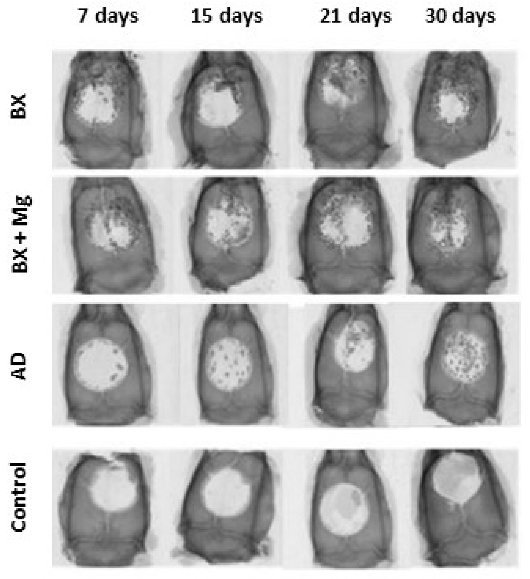
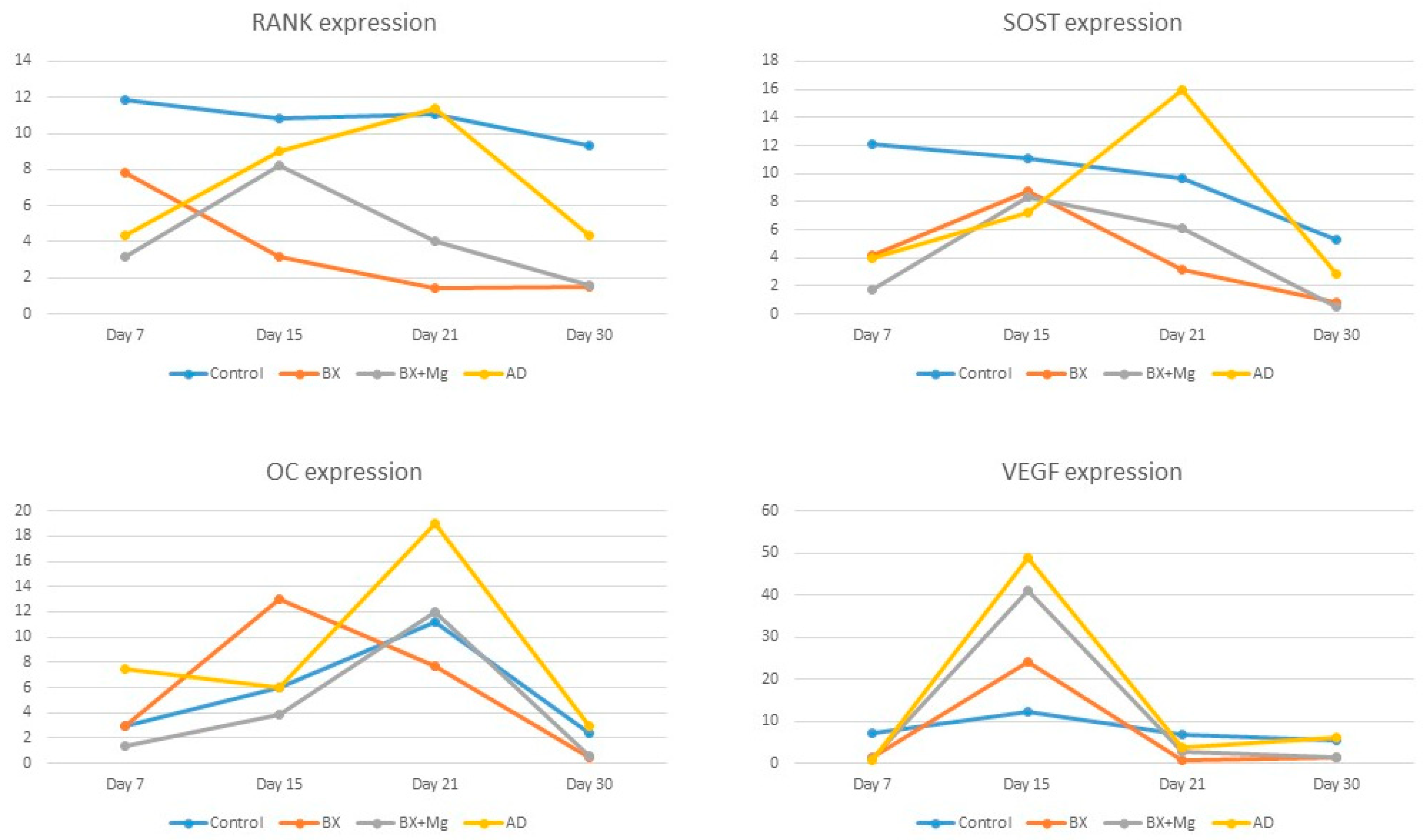
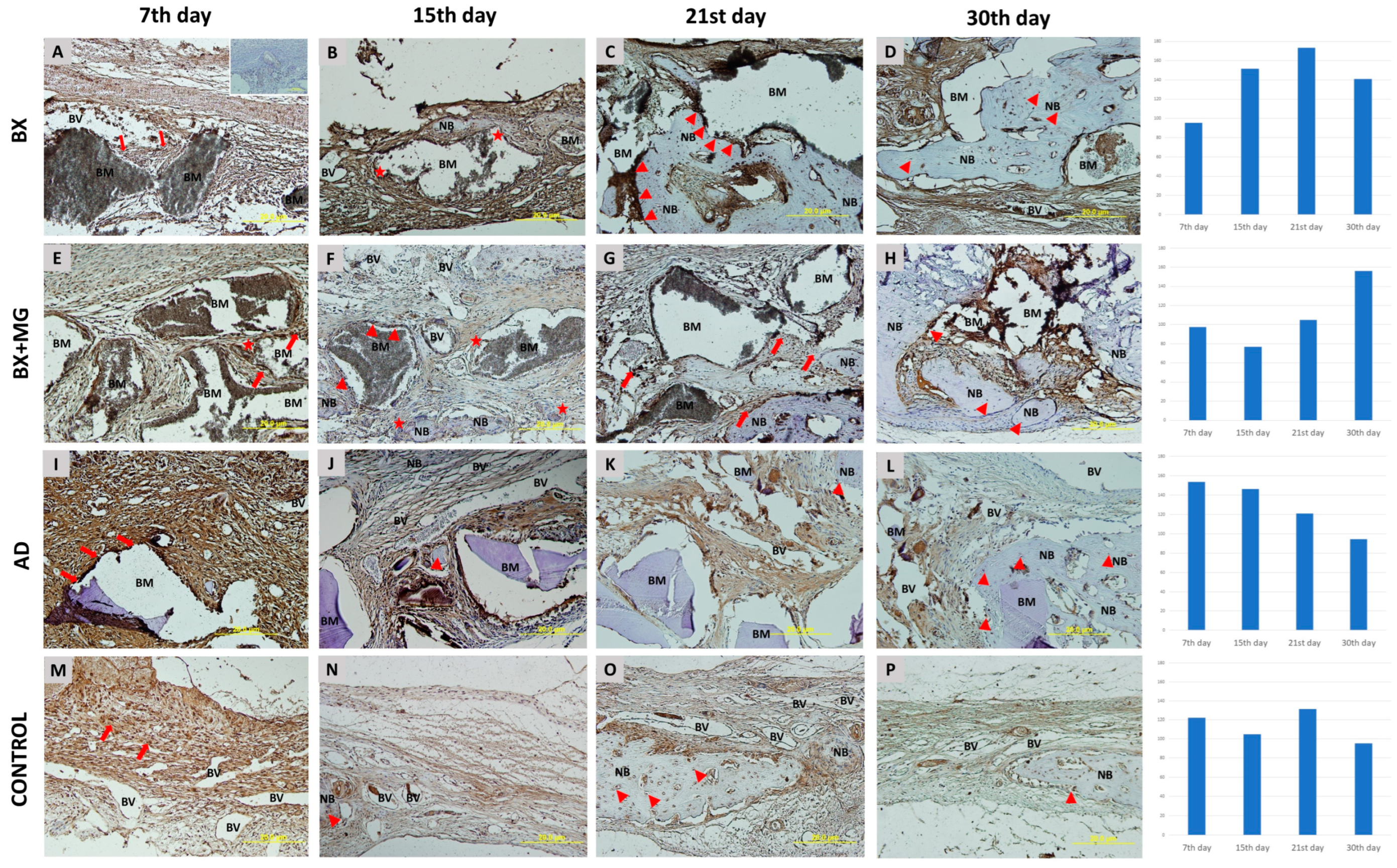
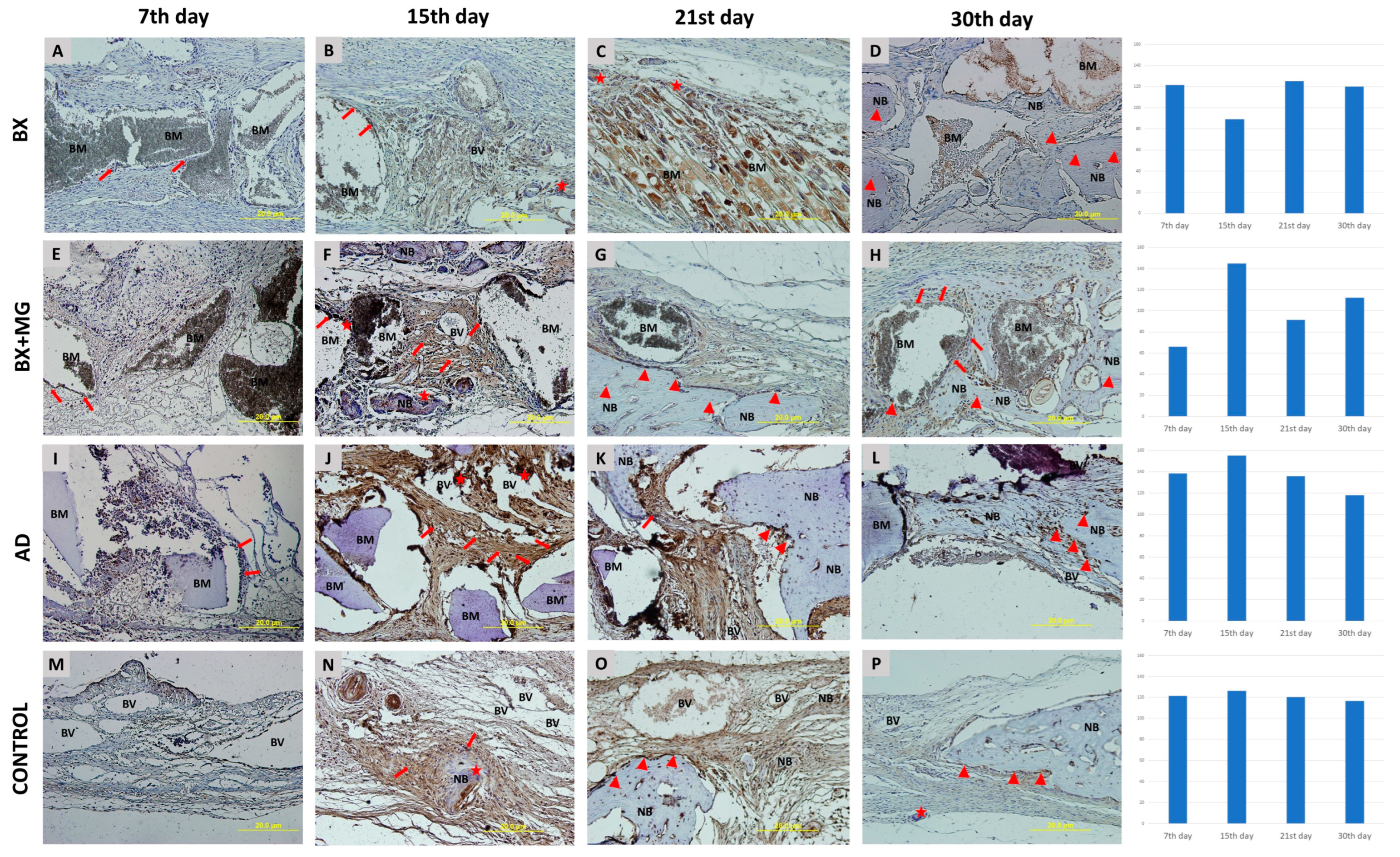

| Group Name | Number of Animals | Time Points | Total |
|---|---|---|---|
| BX 1 | 5 | 7th, 15th, 21st and 30th day | 20 |
| BX + Mg 2 | 5 | 7th, 15th, 21st and 30th day | 20 |
| AD 3 | 5 | 7th, 15th, 21st and 30th day | 20 |
| Control | 5 | 7th, 15th, 21st and 30th day | 20 |
| Total | 80 |
| Biomaterials | |||||
|---|---|---|---|---|---|
| BX 3 (N = 5) | BX + Mg 4 (N = 5) | AD 5 (N = 5) | Control 6 (N = 5) | ||
| Days | μCT Parametrs | Mean ± SD | Mean ± SD | Mean ± SD | Mean ± SD |
| 7 | BV/TV 1 (%) | 19,795 ± 1943 a,g | 19,813 ± 1643 a,e,g | 18,935 ± 1601 a,g | 6952 ± 1002 f |
| RB 2 (%) | 27,847 ± 1468 f | 24,626 ± 1520 f | 29,459 ± 1164 c,g | / | |
| 15 | BV/TV (%) | 24,238 ± 1943 a,f | 23,848 ± 1643 a,f | 24,967 ± 1601 a,g | 9906 ± 1002 |
| RB (%) | 30,989 ± 1468 b,c,g | 20,904 ± 1520 f | 22,886 ± 1164 g | / | |
| 21 | BV/TV (%) | 31,033 ± 1943 a | 31,885 ± 1643 a | 37,909 ± 1601 a,c,f | 11,921 ± 1002 |
| RB (%) | 22,912 ± 1468 b | 21,986 ± 1520 f,b | 12,916 ± 1164 | / | |
| 30 | BV/TV (%) | 33,015 ± 1943 a | 39,501 ± 1643 a,d | 48,994 ± 1601 a,c | 16,938 ± 1002 |
| RB (%) | 19,905 ± 1468 b,c | 12,726 ± 1520 | 11,996 ± 1164 | / | |
Disclaimer/Publisher’s Note: The statements, opinions and data contained in all publications are solely those of the individual author(s) and contributor(s) and not of MDPI and/or the editor(s). MDPI and/or the editor(s) disclaim responsibility for any injury to people or property resulting from any ideas, methods, instructions or products referred to in the content. |
© 2024 by the authors. Licensee MDPI, Basel, Switzerland. This article is an open access article distributed under the terms and conditions of the Creative Commons Attribution (CC BY) license (https://creativecommons.org/licenses/by/4.0/).
Share and Cite
Čandrlić, M.; Jerbić Radetić, A.T.; Omrčen, H.; Franović, B.; Batičić, L.; Gulić, T.; Čaljkušić-Mance, T.; Zoričić Cvek, S.; Malešić, L.; Perić Kačarević, Ž.; et al. Regeneration of Critical Calvarial Bone Defects Using Bovine Xenograft, Magnesium-Enriched Bovine Xenograft and Autologous Dentin in Rats: Micro-CT, Gene Expression and Immunohistochemical Analysis. J. Funct. Biomater. 2024, 15, 270. https://doi.org/10.3390/jfb15090270
Čandrlić M, Jerbić Radetić AT, Omrčen H, Franović B, Batičić L, Gulić T, Čaljkušić-Mance T, Zoričić Cvek S, Malešić L, Perić Kačarević Ž, et al. Regeneration of Critical Calvarial Bone Defects Using Bovine Xenograft, Magnesium-Enriched Bovine Xenograft and Autologous Dentin in Rats: Micro-CT, Gene Expression and Immunohistochemical Analysis. Journal of Functional Biomaterials. 2024; 15(9):270. https://doi.org/10.3390/jfb15090270
Chicago/Turabian StyleČandrlić, Marija, Ana Terezija Jerbić Radetić, Hrvoje Omrčen, Barbara Franović, Lara Batičić, Tamara Gulić, Tea Čaljkušić-Mance, Sanja Zoričić Cvek, Lucija Malešić, Željka Perić Kačarević, and et al. 2024. "Regeneration of Critical Calvarial Bone Defects Using Bovine Xenograft, Magnesium-Enriched Bovine Xenograft and Autologous Dentin in Rats: Micro-CT, Gene Expression and Immunohistochemical Analysis" Journal of Functional Biomaterials 15, no. 9: 270. https://doi.org/10.3390/jfb15090270
APA StyleČandrlić, M., Jerbić Radetić, A. T., Omrčen, H., Franović, B., Batičić, L., Gulić, T., Čaljkušić-Mance, T., Zoričić Cvek, S., Malešić, L., Perić Kačarević, Ž., & Cvijanović Peloza, O. (2024). Regeneration of Critical Calvarial Bone Defects Using Bovine Xenograft, Magnesium-Enriched Bovine Xenograft and Autologous Dentin in Rats: Micro-CT, Gene Expression and Immunohistochemical Analysis. Journal of Functional Biomaterials, 15(9), 270. https://doi.org/10.3390/jfb15090270

