Article

Exposure to Decreased pH and Caffeine Affects Hemocyte Parameters in the Mussel Mytilus galloprovincialis

1
Department of Biology, University of Padova, via Ugo Bassi 58/B, 35121 Padova, Italy
2
Department of Chemical Sciences, University of Padova, Via Marzolo 1, 35131 Padova, Italy
*
Author to whom correspondence should be addressed.
Current address: Department of Integrative Marine Ecology, Ischia Marine Centre, Stazione Zoologica Anton Dohrn, Punta San Pietro, 80077, Ischia (Naples), Italy.
J. Mar. Sci. Eng. 2020, 8(4), 238; https://doi.org/10.3390/jmse8040238
Submission received: 13 February 2020 / Revised: 27 March 2020 / Accepted: 28 March 2020 / Published: 1 April 2020
(This article belongs to the Special Issue Biomarkers of Stress Response in Aquatic Life)

Abstract

:
Combined effects of reduced pH, as predicted under climate change scenarios, and the most popular and widely used stimulant caffeine were assessed in hemocyte parameters of the mussel Mytilus galloprovincialis, being hemocytes involved in immune defense. Bivalves were exposed for one week to natural pH (8.1) and two reduced pH values (pH −0.4 units and pH −0.7 units). Exposure continued for additional two weeks, both in the absence and in the presence of environmentally relevant concentrations of caffeine (0.05 and 0.5 µg/L). Hemocyte parameters (total hemocyte count, hemocyte volume and diameter, neutral red uptake and hemocyte proliferation) were measured after 7 days of exposure to pH only, and after 14 (T1) and 21 (T2) days of exposure to the various pH*caffeine combinations. At all sampling times, pH significantly affected all the biological variables considered, whereas caffeine exhibited a significant influence at T2 only. Among the various hemocyte parameters, caffeine caused a significant increase in total hemocyte count at T2, and in hemocyte volume and diameter at both T1 and T2, when a significant interaction between pH and caffeine was also found. Overall, results demonstrated that hemocyte functionality was strongly influenced by the experimental conditions tested. Further studies are needed to assess combined effects of climate changes and emerging contaminants on bivalve immune system when challenged with environmental pathogens.

1. Introduction

Organic wastewater contaminants, such as pharmaceuticals, occurring in fluvial, estuarine and coastal environments have produced increasing concern on their potential ecotoxicological effects to aquatic species [1,2,3]. In particular, the presence of caffeine (3,7-dihydro-1,3,7-trimethyl-1h-purine-2,6-dione) in surface and ground waters has been detected worldwide [4,5,6,7,8]. Caffeine is considered as the most common stimulant consumed by humans [9,10]. It is used in medicine as a cardiac, cerebral, and respiratory stimulant, and as a diuretic [11]. Furthermore, it is a key ingredient of coffee, chocolate, tea and soft and energy drinks. Globally, annual coffee consumption increased from about 4.5 Tg in 1990 to about 10 Tg in 2013, with Europe having the highest general mean value per capita [12]. Since coffee consumption is a valuable proxy for caffeine consumption, an increasing amount of caffeine is expected to enter aquatic environments. Indeed, caffeine was found in U.S. streams at maximal levels of 6.0 µg/L (median 0.1 µg/L) [13]. Ferguson et al. [14] reported concentrations of caffeine ranging from 0.018 to 1.25 µg/L in near-shore habitats of southern Lake Michigan, while Siegener and Chen [15] found concentrations ranging from 0.14 to 1.6 µg/L in Boston Harbor seawater. As recently reviewed by Rodriguez-Gil et al. [16], the highest caffeine concentrations measured in estuarine water and seawater were 5.86 µg/L in United States [17] and 11.0 µg/L in Australia [18], respectively. However, when considering the whole worldwide data set, the 50th percentile concentration was 0.101 μg/L in estuary water and 0.019 μg/L in seawater. Despite that the high consumption and environmental occurrence of caffeine are well documented, the effects of caffeine to aquatic organisms need to be more fully investigated, especially in marine species living in estuarine and coastal environments, which are mostly affected by human activities.
In addition to emerging contaminants, marine organisms have to face global climate changes (GCCs). Climate change is a threat to marine biota because increased atmospheric CO2 due to human activity is causing ocean warming, acidification, hypercapnia and decreased carbonate saturation [19]. The capability of the oceans to take up CO2 may influence seawater carbonate chemistry, with a consequent decrease in pH values, concentration of carbonate ions and the related calcium carbonate (CaCO3) saturation state of seawater [20]. Several reports have implied that the regular and constant release of CO2 into the atmosphere has already caused a reduction in ocean pH values of approximately 0.1 pH units with respect to the preindustrial levels [21]. pH reductions of 0.06–0.32 and 0.7 units are predicted to occur by the year 2100 and by the year 2300, respectively [22,23]. It is known that changes in environmental parameters, such as temperature, salinity and pH can affect marine organisms by altering many biological pathways. Generally, when an organism is subject to stressful conditions it can cope with stress modifying its physiological, biochemical and behavioral responses. At the immunological level, responses comprise a complex network of specific and non-specific humoral and cell-mediated components. In bivalves, hemocytes are involved in many crucial physiological functions, including nutrient transport and digestion, tissue and shell formation, maintenance of homeostasis, and immune response [24,25,26].
Hemocyte-mediated immune parameters are suggested to be particularly sensitive to variations in environmental factors [26]. For example, in clams (Ruditapes philippinarum) kept for seven days at various temperature/salinity combinations (5, 15, 30 °C and 18, 38 and 38 salinity) the number of circulating hemocytes (total hemocyte count, THC) significantly decreased in animals exposed to 5 and 30 °C at both the lowest (18) and the highest (38) salinities tested [27]. Matozzo et al. [28] showed that the exposure to various combinations of temperature, salinity and reduced pH can significantly modify hemocyte functionality in Mytilus galloprovincialis and Chamelea gallina. In another study, high temperatures affected some important functional responses of hemocytes in the clam C. gallina [29]. In that study, clams were exposed for 7 days to 20, 25 and 30 °C before THC, phagocytosis, lysozyme activity (in both hemocyte lysate and cell-free hemolymph), activity and expression of the antioxidant enzyme superoxide dismutase (SOD) (in both hemocyte lysate and cell-free hemolymph) were measured. Results showed that the highest temperature increased significantly THC in C. gallina, while total SOD activity significantly decreased in hemocytes with increasing temperature.
However, GCCs will not only affect aquatic organisms, but they will also influence the behavior and fate of chemical toxicants, by modifying their environmental distribution and bioavailability [30]. As an instance, ocean acidification is predicted to affect the speciation of metals [31]. Nevertheless, variations in environmental parameters could alter marine organisms’ susceptibility to pollutants. In mussels (M. galloprovincialis) and clams (R. philippinarum) exposed for 14 days to seawater acidification in combination with diclofenac, a non-steroidal anti-inflammatory drug, the overall hemocyte response was significantly affected by both the stressors applied, even though a significant interaction between pH and diclofenac was mainly evident in mussels [32].
With the aim of elucidating better the possible impact of emerging contaminants and acidification to marine organisms, in this study the combined effects of lowered pH (as predicted by climate change scenarios) and caffeine (a widely used substance in many formulations) on some hemocyte parameters, such as THC, hemocyte diameter and volume, neutral red (NR) uptake and hemocyte proliferation were investigated for the first time in the Mediterranean mussel M. galloprovincialis. This species is widespread in marine coastal areas and is extensively used as model organism in ecotoxicological studies [33]. We choose to evaluate the combined effects of lowered pH and caffeine on mussel hemocytes, as such cells are involved in immune responses in bivalves [24,25,26] and hemocyte parameters (or immunomarkers) are widely used to evaluate the effects of environmental contaminants in molluscs [34].

2. Materials and Methods

2.1. Animals

Specimens of M. galloprovincialis (5.0 ± 0.5 cm shell length) were collected along the west coast of the Northern Adriatic Sea (near Cavallino Treporti, Italy, temperature 25 °C, salinity 35.2, pH 8.2), and transported immediately to the laboratory. Only bivalves with undamaged shells were chosen for the exposure, and epibionts (such as algae and barnacles) were gently removed from the mussels. In the laboratory, the organisms were maintained for 7 days in aerated seawater at salinity, temperature and pH values similar to those measured during the field collection.

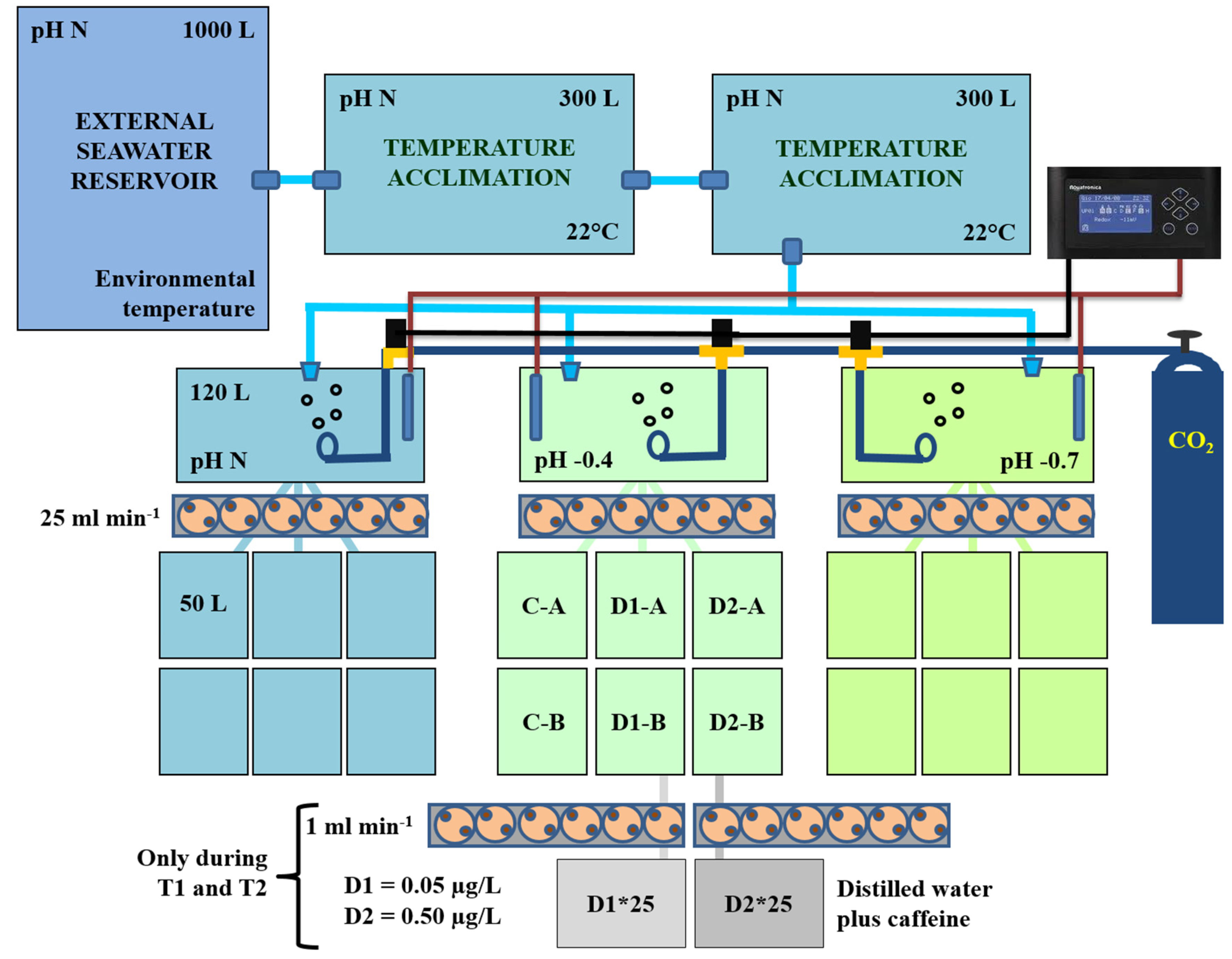
2.2. Experimental Set-Up for Mussel Exposure

Before starting exposure, bivalves were acclimated to the experimental conditions by means of a gradual decrease in seawater temperature (1 °C per day) to 22 °C and a gradual decrease in pH values of approximately −0.4 or −0.7 units (U). Mussels were fed daily with microalgae (Isochrysis galbana). The experiments were carried out during the resting phase of mussels, when animals were not ripe (June/July), to avoid stress possibly arising from gamete emissions during the experiments. The experimental flow-through system (Figure 1) used for mussel exposure was set-up at the Hydrobiological Station ‘Umberto D’Ancona’ in Chioggia. A main outside reservoir containing natural seawater (1000 L of capacity) was used to supply two tanks (about 300 L each) inside the laboratory. The seawater (salinity of 34.4 ± 0.3), equilibrated to the controlled laboratory temperature (22 °C), moved on to three tanks of about 120 L capacity, where the pH was adjusted to experimental values by bubbling CO2. To this end, an automatic control system (ACQ110 Aquarium Controller Evolution by Aquatronica) connected with pH electrodes (ACQ310N-PH by Aquatronica) was used. Lastly, the seawater was pumped (25 mL/min, using ACQ450 Dosing pumps by Aquatronica) into the experimental tanks (A, B and C, approximately 50 L each) containing the mussels.
Throughout the first week of exposure, animals were exposed to three pH values: natural pH (pH N; mean value of 8.07) and decreased pHs as projected for the year 2100 (−0.4 pH units; pH −0.4) and 2300 (−0.7 pH units; pH −0.7) [22,23]. Throughout the second and the third week, they were exposed to three concentrations of caffeine (0, 0.05 and 0.5 µg/L) for each pH value. For each concentration, two replicate tanks (with 70 bivalves each) were prepared (control without contaminant: C-A, C-B; caffeine 0.05 µg/L: D1-A, D1-B; caffeine 0.5 µg/L: D2-A, D2-B).
The experimental concentrations of caffeine were kept constant during the experiments by distributing continuously the stock solutions of the chemical (prepared in distilled water) using peristaltic pumps (1 mL/min, by means of a MCP Process Pump, mod. ISM915A, provided with a MS/CA pumphead, mod. ISM724A, ISMATEC) from two 15 L glass jars. Based on the two combined flows (25 mL/min for seawater and 1 mL/min for the caffeine solution, respectively), the concentrations of caffeine in the two glass jars were calculated to be 25 times higher than those required in the respective tanks with mussels.
Of the experimental combinations, the control without caffeine at pH N was considered as a reference. During the experiments, mussels were fed twice a day by supplying 250 mL of microalgae (3.5 − 106 cells/mL) (Isochrysis galbana) in each 120 L tank, thus maintaining an algal concentration of approximately 2000 cells/mL in the experimental tanks with bivalves. Parameters of seawater carbonate chemistry (i.e., pHT, total alkalinity, dissolved inorganic carbon, pCO2, calcite and aragonite saturation state) were monitored two to three times a week; methods in detail are reported in Munari et al. [35] and data are shown in Table 1. Mussel mortality was checked daily. At the end of the first (T0), the second (T1) and the third (T2) week of exposure, hemolymph was collected from mussels. For each experimental condition, 6 pools of hemolymph (3 pools per tank from 7 animals each) were used to assess hemocyte parameters.

2.3. Hemolymph Parameters

Hemolymph (about 300 µL per mussel) was collected from the adductor muscles with a 1 mL plastic syringe and stored in ice. To prevent clotting, an equal volume of 0.38% sodium citrate (Sigma) in 0.45 µm-filtered sea water (FSW), pH of 7.5, was added to hemolymph samples. One hundred µL of pooled hemolymph were used to measure the THC, whereas 500 and 400 µL of hemolymph were used to assess NR uptake and hemocyte proliferation, respectively.
To determine THC and hemocyte volume and diameter, a ScepterTM Handheld Automated Cell Counter (Millipore) was used for each measurement, 20 µl of pooled hemolymph were added to 2 mL of isotonic solution (COULTER ISOTON II Diluent). THC results were expressed as the number of hemocytes (−106)/mL hemolymph, whereas hemocyte diameter and volume were expressed in µm and picoliters (pL), respectively.
NR uptake assay was performed following the method reported in previous studies [36,37]. Pooled hemolymph was centrifuged at 780 g for 10 min. Hemocytes (at a final concentration of 106 cells/mL) were resuspended in an equal volume of 8 mg/L NR dye (Merck) solution in FSW. They were incubated at room temperature for 30 min and then centrifuged at 780 g for 10 min; subsequently, they were resuspended in distilled water, sonicated at 4 °C for 30 s with a Braun Labsonic U sonifier at 50% duty cycles, and centrifuged at 12,000 g for 15 min at 4 °C. After that, the supernatant, i.e., hemocyte lysate (HL), was collected for the NR uptake assay. Absorbance at 550 nm was measured with a Beckman 730 spectrophotometer. The results were expressed as optical density per ml hemolymph (OD/mL hemolymph).
Hemocyte proliferation was assessed using a colorimetric method of a commercial kit (Cell proliferation Kit II, Roche). The assay is based on the cleavage of the yellow tetrazolium salt XTT to produce an orange formazan dye in viable cells. In brief, the XTT labelling reagent and electron-coupling reagent were thawed at 37 °C and mixed immediately before use to obtain the XTT labelling mixture. Two hundred µL of the mixture were added to 400 µL of pooled hemolymph and then incubated for 4 h in a dark humidified chamber. The absorbance at 450 nm was then determined using a Beckman 730 spectrophotometer. The data were expressed as optical density (OD)/mL hemolymph.

2.4. Statistical Analysis
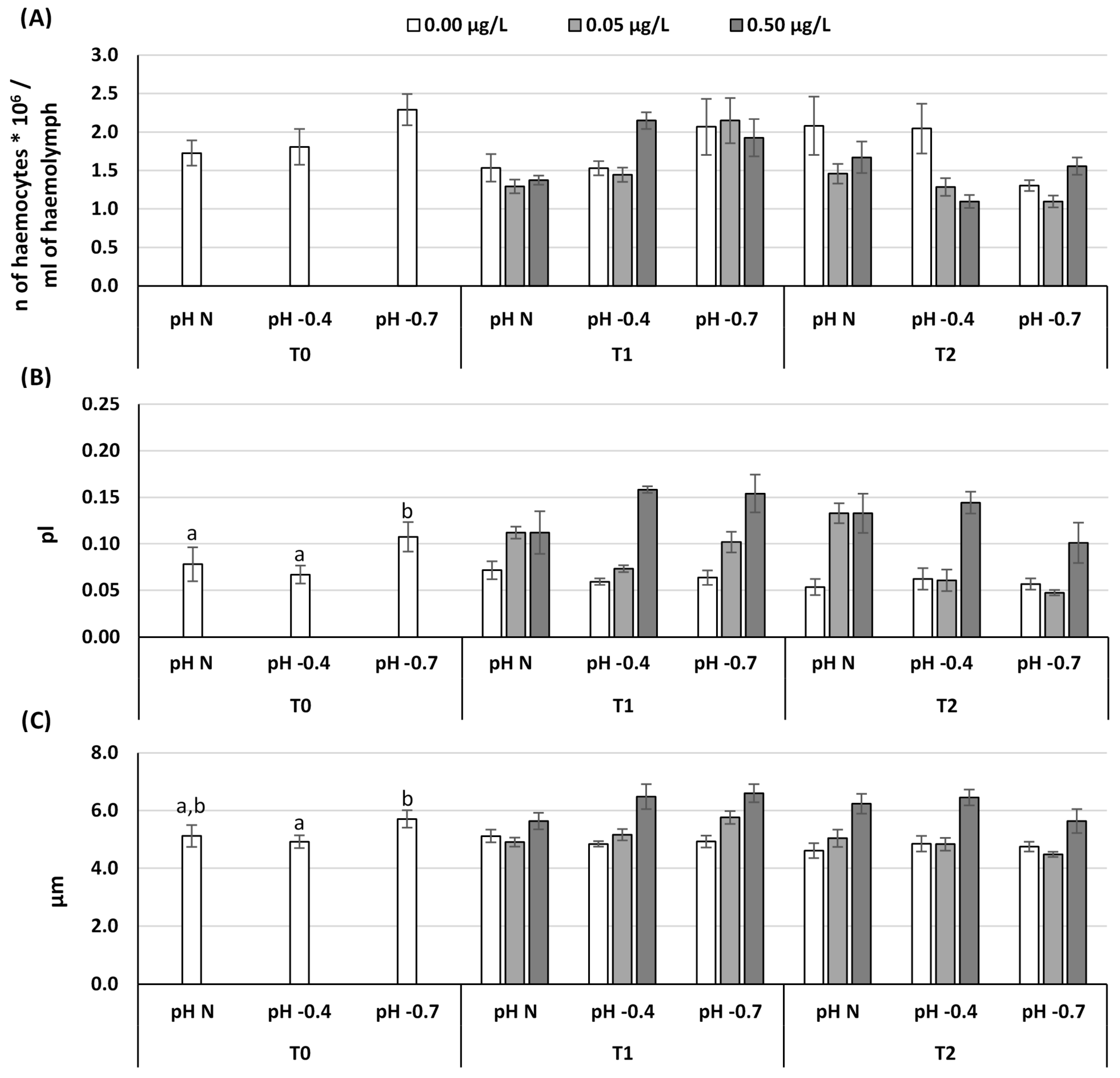
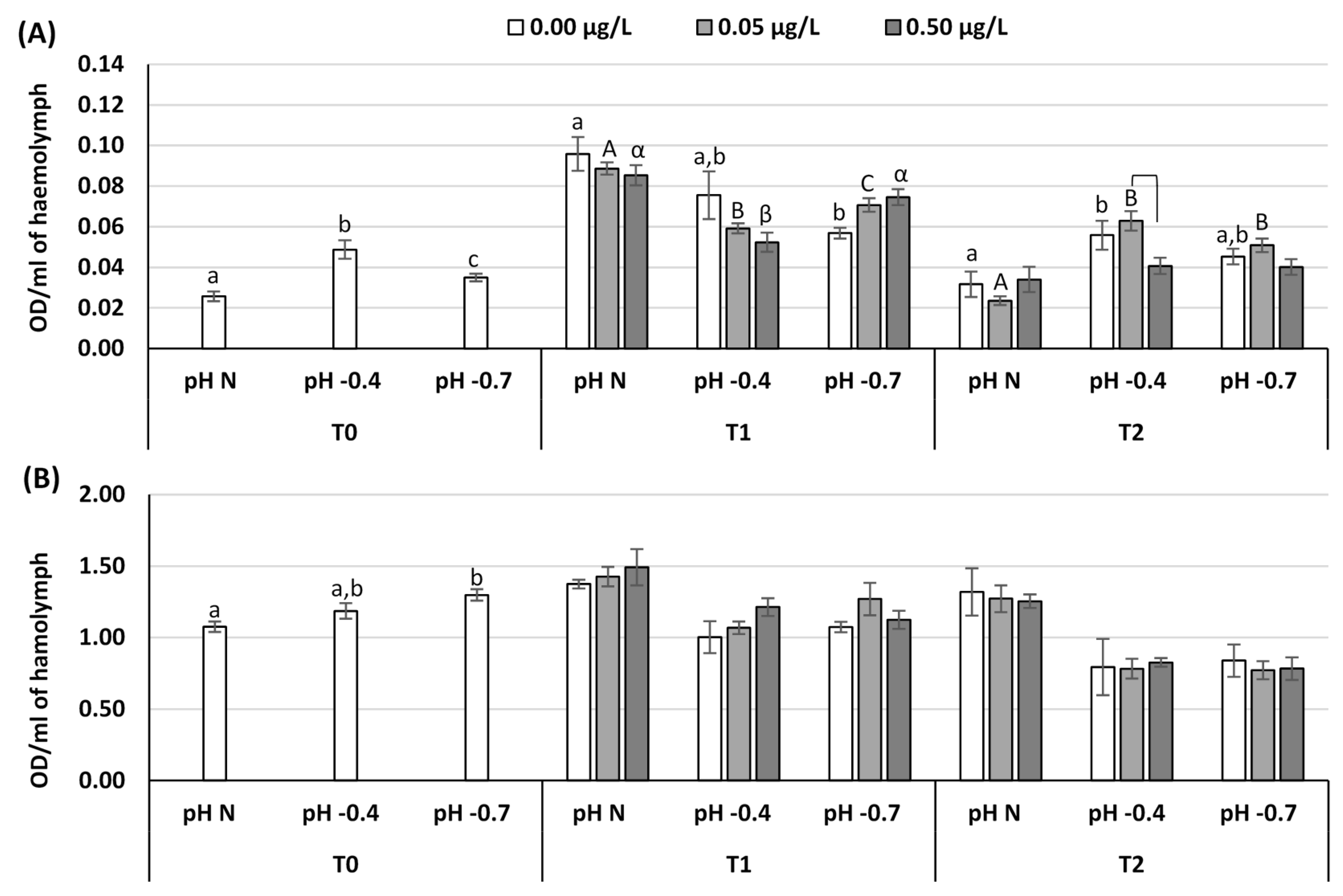
Data were checked for normal distribution (Shapiro–Wilk test) and homogeneity of variances (Bartlett’s test). As ANOVA assumptions were not fulfilled, a non-parametric approach was used for comparison of the results. The whole data set obtained at each sampling time was statistically analyzed using permutational multivariate analyses of variance (PERMANOVA, with 9999 permutations), in order to detect significant effects of pH, caffeine and interactions between pH and caffeine. Data from each biomarker were then analyzed with PERMANOVA to determine significant effects on the single hemocyte parameters. Furthermore, PERMANOVA was used to run relevant pair-wise comparisons among and within pH and caffeine levels. For each biomarker, the null hypotheses tested were: at T0, no significant difference exists among pH levels (N, −0.4 and −0.7); at T1 and T2, (i) for each pH tested, no significant difference exists among caffeine concentrations (0.00, 0.05 and 0.50 µg/L) and (ii) for each caffeine concentration tested, no significant difference exists among pH levels (N, −0.4 and −0.7). Statistical significance was set at p ≤ 0.05. Since low number of unique values from permutations sometimes impeded to make inference at the desired level of significance, the Monte-Carlo procedure was used to calculate p-values. The software package PRIMER 6 PERMANOVA Plus (PRIMER-E Ltd., Plymouth, UK) was used for the statistical analyses.

3. Results

Results of the PERMANOVA analysis are reported in Table 2. Significant effects of pH on the overall cellular responses of mussels were detected during the whole experiment, whereas a significant effect of caffeine was observed only at T2. The interaction between pH and caffeine did not cause significant alterations (Table 2). For each hemocyte-related biomarker measured, significant effects of the experimental factors, namely pH at T0, pH, caffeine and pH*caffeine at T1 and T2, were also observed (Table 2).
Significant differences between treatments are shown in Figure 2 and Figure 3, at T1 and T2, only when a significant effect of the pH*caffeine interaction was found. Furthermore, statistically significant differences between groups of animals exposed to different experimental pHs or different caffeine concentrations are reported in Table 3.
THC was significantly affected by reduced pH at both T1 and T2, with an opposite trend between the two sampling times (Table 2). While at T1 a significant increase in THC was observed at pH −0.7 compared to organisms kept at pH N at all caffeine concentrations (Figure 2A, Table 3), at T2 a significant reduction of THC values was observed in mussels kept at pH −0.7, compared to pH N. Caffeine had significant effects at T2 only, while a significant interaction between pH and caffeine was never observed (Table 2). In particular, a reduction in THC was observed in mussels exposed to 0.05 and 0.5 µg/L of caffeine compared to controls at both pH N and pH −0.4 (Figure 2A, Table 3).
In details, hemocyte volume was significantly influenced by reduced pH only at T0 (Table 2, Figure 2B), with significantly bigger hemocytes in mussels reared at pH −0.7 compared to those at pH N and pH −0.4. Volume was significantly increased by caffeine at both T1 and T2 especially at the highest concentration tested (Table 3). Hemocytes diameter was significantly influenced by pH at both T0 and T1.While at T0 there was no clear pattern of variation, at T1 a significant increase in hemocyte size was observed in mussels reared at pH −0.7 compared to those at pH N and pH −0.4 (Table 2 and Table 3, Figure 2C). In addition, diameter was significantly influenced by caffeine at both T1 and T2 at all pH levels, especially in mussels kept at 0.5 µg/L that showed to have significantly bigger hemocytes when compared to control mussels and those exposed to 0.05 µg/L.
NR uptake was significantly affected by pH at all sampling times (Table 2 and Table 3, Figure 3A). However, a clear trend was observed at T1 only, with an increased uptake capability of the hemocytes of mussels maintained at pH −0.4 and −0.7 compared to the pH N. A significant combined effect of pH and caffeine was also observed at both T1 and T2. After the first week of exposure to caffeine, at both pH N and pH −0.4 in presence of caffeine the NR uptake capability was reduced, while at pH −0.7 an opposite trend was revealed, even if the differences among caffeine concentrations within the different pH levels were not significant. At T2, a significant difference between 0.05 and 0.5 µg/L condition was found at pH −0.4, with reduced values of NR uptake at the highest dose.
Cell proliferation was affected only by pH during the whole experiment (Table 2, Figure 3B). A significant increase with decreasing pH was observed at T0, while cell proliferation recorded in mussels kept at pH −0.4 and −0.7 was significantly lower than pH N at both T1 and T2 (Table 3).

4. Discussion

Alterations in hemocyte morpho-functional parameters have been reported for bivalves after exposure, both in vitro and in vivo, to organic and inorganic contaminants, such as heavy metals [38,39,40], organotins [37] and polycyclic aromatic hydrocarbons (PAHs) [41,42]. More recently, increasing attention has been addressed to the evaluation of potential immunotoxic effects of emerging contaminants, such as pharmaceuticals and personal care products (PPCPs) [43,44,45,46,47] and nanoparticles [48,49,50].
In this study, the effects of caffeine, commonly used as a stimulant in a variety of products, were investigated under different seawater pH values for the first time in various hemocyte parameters of M. galloprovincialis. Literature about caffeine toxicity in non-target aquatic organisms is limited. As for bivalves, the sub-lethal cellular toxicity of environmentally relevant concentrations of caffeine was investigated in the mussel Mytilus californianus [51]. In that study, Hsp70 concentrations in the gill and mantle tissues of mussels exposed to 0.05, 0.2, and 0.5 µg/L of caffeine for 10, 20, and 30 days were compared to basal levels of the control mussels. Exposure to caffeine at a dose of 0.05 µg/L induced a moderate up-regulation of Hsp70 in the gill lamellae of M. californianus after 20 days of exposure, while in the highest caffeine concentration (0.5 µg/L), no increase in Hsp70 expression was observed throughout the experiment. In M. galloprovincialis, exposure to 0.005, 0.05 and 0.5 µg/L of caffeine for 7 days triggered a significant decrease of lysosomal membrane stability in both hemocytes and digestive glands (at 0.05 and 0.5 µg/L) and a significant increase of lysosomal content of neutral lipids (at 0.5 µg/L). A significant increase in the activity of the biotrasformation enzyme glutathione-S-transferase was also observed in digestive glands (at 0.005 and 0.5 µg/L) [52].
After 14 days of exposure to caffeine in the range of 0.1–50 µg/L, significant detrimental effects were observed in the clam Ruditapes philippinarum, with a dose-dependent reduction in hemocyte lysosomal membrane stability [51]. Values of biomarkers of oxidative stress and biotrasformation phase I and II were significantly higher in digestive glands, mostly at the highest caffeine concentrations tested. Acetylcholinesterase (AChE) activity, as a biomarker of neurotoxicity, increased significantly at 0.1 µg/L, but was significantly inhibited at all the other concentrations [53]. Although AChE activity was not assessed, very similar responses were obtained in the freshwater clam Corbicula fluminea exposed for 21 days to the same caffeine concentrations tested in R. philippinarum [54]. Impairment in lysosomal membrane stability of hemocytes after exposure to caffeine at doses as low as 0.05–0.1 µg/L suggest that immune cells can be a target for this substance in invertebrates [52,53,54,55], showing immune modulatory effects like those observed in rodents and humans [56].
Not only the exposure to contaminants can modify hemocyte parameters in bivalves. It is well documented that changes in environmental parameters can influence hemocyte functionality as shown in various studies concerning the effects of temperature [29,57,58] and salinity [59,60], as well as their combination [27]. Similarly, effects of pH alone [61,62,63] under various temperature and salinity values [28] were demonstrated in several bivalve species.
When evaluating potential effects of environmental stressors, variations in hemocyte functionality represent crucial endpoints as they are expression of altered physiological performance and, most of all, they may result in a reduced immunosurveillance, which increases susceptibility to diseases and lowers survival capability. THC is one of the most used bivalve immunomarkers to evaluate negative effects of stressors (including pollutants) to animals [64]. Generally, increased THC may be due to either proliferation or movement of cells from tissues into hemolymph, whereas decreased THC are caused by cell lysis or increased cell movement from hemolymph to tissues [65,66]. In this study, THC was significantly affected by reduced pH, confirming previous results obtained in both M. galloprovincialis and R. philippinarum [32]. After the first week of exposure, an increase in THC was observed at reduced pH. It is important to note that circulating hemocytes from bivalves, as demonstrated in mussels and clams, can divide in the hemolymph [67,68] and that contaminants, including pharmaceuticals, can stimulate the mitotic activity of bivalve hemocytes [45,69]. At T1, higher THC levels at low pH may be a consequence of the increased hemocyte proliferation recorded at T0 as a response to decreased pH, with a significant difference at pH −0.7 compared to pH N. However, the same increasing trend in hemocyte proliferation was not maintained at T1, but a significant reduction was revealed under reduced pH conditions, showing similar values in the absence and in the presence of caffeine. At the end of the experiment, a reverse pattern of THC variation respect to T1 was observed, higher values being occurred in the absence of caffeine at pH N and −0.4, and lower ones in all conditions tested at pH −0.7. At pH −0.7, prevailing effect of reduced pH masked the detrimental effect of caffeine on THC.
Unlike this study, Bibby et al. [61] found that a 32-day exposure to different pH levels (7.8, 7.7, 7.5 and 6.7) did not cause significant effects on total and differential cell counts in Mytilus edulis. Interestingly, both hemocyte volume and diameter showed to be affected by the experimental conditions tested, in particular following exposure to caffeine. Indeed, both parameters were significantly increased by caffeine at T1 and T2, with the highest values at the higher caffeine concentration (0.5 µg/L) at all pH tested. Overall, hemocyte size measurement indicated a dose-dependent effect of caffeine and additive effect of reduced pH and caffeine. Our results are consistent with those reported for hemocyte size in mussels exposed to reduced pH and diclofenac [32], even though the effects of caffeine were more evident than that of diclofenac.
The cationic probe NR is commonly used to evaluate the effects of stressors on lysosomal membrane stability in bivalve hemocytes [38,53,70,71,72]. The uptake of NR by hemocytes occurs either by pinocytosis or by passive diffusion across cell membranes [73]. Consequently, alterations in dye uptake reflect damage to cell membranes (including lysosomal membranes) and/or weakening of hemocyte pinocytotic capability. In this study, we demonstrated that the maintenance of bivalves at reduced pH significantly increased NRU by hemocytes after one week of exposure, suggesting an increase in pinocytotic activity, probably related to the increased THC levels and the greater hemocyte volume and diameter observed at T0 in animals kept at pH −0.7. In the study of Matozzo et al. [28], the exposure of C. gallina to reduced pH, high temperature (28 °C) and low salinity (28) induced hemocytes to become more active, increasing NRU. Additionally, Bibby et al. [61] found increased levels of phagocytosis in M. edulis exposed to reduced pH values. In a recent study [52], a 7-day exposure of M. galloprovincialis to caffeine caused a significant decrease in lysosomal membrane stability evaluated by neutral red retention time (NRRT) at the concentration of 0.5 µg/L. Similarly, a significant NRRT decrease was found in R. philippinarum exposed for 14 days to 0.1 µg/L [54]. Using the same experimental design adopted in the present study to assess the combined effects of pH and diclofenac, only exposure to reduced pH was shown to significantly affect NRU in both M. galloprovincialis and R. philippinarum at all sampling time, even though different patterns of variation were observed in the two species [32]. Additionally, in our study a significant effect of pH persisted during the whole experiment. As for caffeine, although a slight dose-dependent reduction in NRU by hemocytes was observed in animals kept at pH N and −0.4 at T1, the trend was opposite at pH −0.7. At T2, a significant NRU increase was observed at the lower caffeine concentration under reduced pH, however NRU values were quite similar at −0.7 pH for all caffeine concentrations tested. These findings highlighted the significant interaction between the two stressors at both T1 and T2.
Overall, our results suggest that environmental concentrations of caffeine and seawater acidification may impact the physiological condition and functionality of the hemocytes in mussels, mostly by altering their size and pinocytotic activity. Further investigations, however, are needed to address the toxicity of this widespread contaminant on sentinel species. In particular, since effects on cell membrane functionality were highlighted also in other studies under acidified conditions [61], further studies should investigate the combined effects of global climate changes and emerging contaminants on marine bivalves when their immune system is challenged with environmental pathogens.

Author Contributions

Conceptualization, M.M., V.M. and M.G.M.; methodology, M.M., V.M. and M.G.M.; data analysis, M.M., V.M., G.B., V.R., P.P., D.B. and M.G.M.; investigation, M.M., G.B. and V.R.; chemical analysis, P.P. and D.B.; writing—original draft preparation, M.M.; writing—review and editing, M.M., V.M. and M.G.M. All authors have read and agreed to the published version of the manuscript.

Funding

This research received no external funding.

Conflicts of Interest

The authors declare no conflict of interest.

References

  1. Gaw, S.; Thomas, K.V.; Hutchinson, T.H. Sources, impacts and trends of pharmaceuticals in the marine and coastal environment. Phil. Trans. R. Soc. B 2014, 369, 20130572. [Google Scholar] [CrossRef] [PubMed] [Green Version]
  2. Mezzelani, M.; Gorbi, S.; Regoli, F. Pharmaceuticals in the aquatic environments: Evidence of emerged threat and future challenges for marine organisms. Mar. Environ. Res. 2018, 140, 41–60. [Google Scholar] [CrossRef] [PubMed]
  3. Fekadu, S.; Alemayehu, E.; Dewil, R.; Van der Bruggen, B. Pharmaceuticals in freshwater aquatic environments: A comparison of the African and European challenge. Sci. Total Environ. 2019, 654, 324–337. [Google Scholar] [CrossRef] [PubMed]
  4. Comeau, F.; Surette, C.; Brun, G.L.; Losier, R. The occurrence of acidic drugs and caffeine in sewage effluents and receiving waters from three coastal watersheds in Atlantic Canada. Sci. Total Environ. 2008, 396, 132–146. [Google Scholar] [CrossRef] [PubMed]
  5. Loos, R.; Gawlik, B.M.; Locoro, G.; Rimaviciute, E.; Contini, S.; Bidoglio, G. EU-wide survey of polar organic persistent pollutants in European river waters. Environ. Pollut. 2009, 157, 561–568. [Google Scholar] [CrossRef] [PubMed]
  6. Rodriguez del Rey, Z.; Granek, E.F.; Sylvester, S. Occurrence and concentration of caffeine in Oregon coastal waters. Mar. Pollut. Bull. 2012, 64, 1417–1424. [Google Scholar] [CrossRef]
  7. Alygizakis, N.A.; Gago-Ferrero, P.; Borova, V.L.; Pavlidou, A.; Hatzianestis, I.; Thomaidis, N.S. Occurrence and spatial distribution of 158 pharmaceuticals, drugs of abuse and related metabolites in offshore seawater. Sci. Total Environ. 2016, 541, 1097–1105. [Google Scholar] [CrossRef] [Green Version]
  8. Brumovský, M.; Bečanová, J.; Kohoutek, J.; Borghini, M.; Nizzetto, L. Contaminants of emerging concern in the open sea waters of the Western Mediterranean. Environ. Pollut. 2017, 229, 976–983. [Google Scholar] [CrossRef]
  9. Nawrot, P.; Jordan, S.; Eastwood, J.; Rotstein, J.; Hugenholtz, A.; Feeley, M. Effects of caffeine on human health. Food Addit. Contam. 2003, 20, 1–30. [Google Scholar] [CrossRef]
  10. Fulgoni, V.L.; Keast, D.R.; Lieberman, H.R. Trends in intake and sources of caffeine in the diets of US adults: 2001–20101–4. Am. J. Clin. Nutr. 2015, 101, 1081–1087. [Google Scholar] [CrossRef]
  11. Buerge, I.J.; Poiger, T.; Müller, M.D.; Buser, H.R. Caffeine, an anthropogenic marker for wastewater contamination of surface waters. Environ. Sci. Technol. 2003, 37, 691–700. [Google Scholar] [CrossRef] [PubMed]
  12. Quadra, G.R.; Paranaíba, J.R.; Vilas-Boas, J.; Roland, F.; Amado, A.M.; Barros, N.; Dias, R.J.P.; Cardoso, S.J. A global trend of caffeine consumption over time and related environmental impacts. Environ. Pollut. 2020, 256, 113343. [Google Scholar] [CrossRef] [PubMed]
  13. Kolpin, D.W.; Furlong, E.T.; Meyer, M.T.; Thurman, E.M.; Zaugg, S.D.; Barber, L.B.; Buxton, H.T. Pharmaceuticals, hormones, and other organic wastewater contaminants in US streams, 1999-2000: A national reconnaissance. Environ. Sci. Technol. 2002, 36, 1202–1211. [Google Scholar] [CrossRef] [PubMed] [Green Version]
  14. Ferguson, P.J.; Bernot, M.J.; Doll, J.C.; Lauer, T.E. Detection of pharmaceuticals and personal care products (PPCPs) in near-shore habitats of southern Lake Michigan. Sci. Total Environ. 2013, 458, 187–196. [Google Scholar] [CrossRef]
  15. Siegener, R.; Chen, R.F. Caffeine in Boston Harbor seawater. Mar. Pollut. Bull. 2002, 44, 383–387. [Google Scholar] [CrossRef]
  16. Rodríguez-Gil, J.; Cáceres, N.; Dafouz, R.; Valcárcel, Y. Caffeine and paraxanthine in aquatic systems: Global exposure distributions and probabilistic risk assessment. Sci. Total Environ. 2018, 612, 1058–1071. [Google Scholar] [CrossRef]
  17. Benotti, M.J.; Brownawell, B.J. Distributions of pharmaceuticals in an urban estuary during both dry- and wet-weather conditions. Environ. Sci. Technol. 2007, 41, 5795–5802. [Google Scholar] [CrossRef]
  18. French, V.A.; King, S.C.; Kumar, A.; Northcott, G.; McGuinness, K.; Parry, D. Characterisation of microcontaminants in Darwin Harbour, a tropical estuary of northern Australia undergoing rapid development. Sci. Total Environ. 2015, 536, 639–647. [Google Scholar] [CrossRef]
  19. IPCC. Summary for policymakers. In Climate Change 2007: The Physical Science Basis. Contribution of Working Group I to the Fourth Assessment Report of the Intergovernmental Panel on Climate Change; Solomon, S., Qin, D., Manning, M., Chen, Z., Marquis, M., Averyt, K.B., Tignor, M., Miller, H.L., Eds.; Cambridge University Press: Cambridge, UK; New York, NY, USA, 2007; p. 996. [Google Scholar]
  20. Orr, J.C.; Fabry, V.I.; Aumont, O.; Bopp, L.; Doney, S.C.; Feely, R.A.; Gnanadesikan, A.; Gruber, N.; Ishida, A.; Joos, F.; et al. Anthropogenic ocean acidification over the twenty-first century and its impact on calcifying organisms. Nature 2005, 437, 681–686. [Google Scholar] [CrossRef]
  21. Williamson, P.; Turley, C. Ocean acidification in a geoengineering context. Phil. Trans. R. Soc. A 2012, 370, 4317–4342. [Google Scholar] [CrossRef] [Green Version]
  22. Hartin, C.A.; Bond-Lamberti, B.; Patel, P.; Mundra, A. Ocean acidification over the next three centuries using a simple global climate carbon-cycle model: Projections and sensitivities. Biogeosciences 2016, 13, 4329–4342. [Google Scholar] [CrossRef] [Green Version]
  23. IPCC. Summary for Policymakers. In IPCC Special Report on the Ocean and Cryosphere in a Changing Climate; Pörtner, H.O., Roberts, D.C., Masson-Delmotte, V., Zhai, P., Tignor, M., Poloczanska, E., Mintenbeck, K., Alegría, A., Nicolai, M., Okem, A., et al., Eds.; Available online: https://report.ipcc.ch/srocc/pdf/SROCC_FinalDraft_FullReport.pdf (accessed on 10 February 2020).
  24. Cheng, T.C. Hemocytes: Forms and functions. In The Eastern Oyster Crassostrea Virginica; Kennedy, V.S., Newell, R.I.E., Eble, A.F., Eds.; Maryland Sea Grant Book: College Park, MD, USA, 1996; pp. 299–333. [Google Scholar]
  25. Cheng, T.C. Cellular defense mechanisms in oysters. In Recent Advances in Marine Biotechnology: Immunobiology and Pathology; Fingerman, N., Nagabhushanam, R., Eds.; Science Publishers: Enfield, NH, USA, 2000; pp. 43–83. [Google Scholar]
  26. Chu, F.L.E. Defense mechanisms of marine bivalves. In Recent Advances in Marine Biotechnology: Immunobiology and Pathology; Fingerman, N., Nagabhushanam, R., Eds.; Science Publishers: Enfield, NH, USA, 2000; pp. 1–42. [Google Scholar]
  27. Munari, M.; Matozzo, V.; Marin, M.G. Combined effects of temperature and salinity on functional responses of hemocytes and survival in air of the clam Ruditapes philippinarum. Fish Shellfish Immun. 2011, 30, 1024–1030. [Google Scholar] [CrossRef] [PubMed]
  28. Matozzo, V.; Chinellato, A.; Munari, M.; Finos, L.; Bressan, M.; Marin, M.G. First evidence of immunomodulation in bivalves under seawater acidification and increased temperature. PLoS ONE 2012, 7, e33820. [Google Scholar] [CrossRef] [PubMed] [Green Version]
  29. Monari, M.; Matozzo, V.; Foschi, J.; Cattani, O.; Serrazanetti, G.P.; Marin, M.G. Effects of high temperatures on functional responses of hemocytes in the clam Chamelea gallina. Fish Shellfish Immun. 2007, 22, 98–114. [Google Scholar] [CrossRef] [PubMed]
  30. Noyes, P.D.; Mcelwee, M.K.; Miller, H.D.; Clark, B.W.; Van Tiem, L.A.; Walcott, K.C.; Erwin, K.N.; Levin, E.D. The toxicology of climate change: Environmental contaminants in a warming world. Environ. Int. 2009, 35, 971–986. [Google Scholar] [CrossRef] [PubMed]
  31. Millero, F.J.; Woosley, R.; Ditrolio, B.; Waters, J. Effect of ocean acidification on the speciation of metals in seawater. Oceanography 2009, 22, 72–85. [Google Scholar] [CrossRef]
  32. Munari, M.; Matozzo, V.; Chemello, G.; Riedl, V.; Pastore, P.; Badocco, D.; Marin, M.G. Seawater acidification and emerging contaminants: A dangerous marriage for hemocytes of marine bivalves. Environ. Res. 2019, 175, 11–21. [Google Scholar] [CrossRef]
  33. Gagné, F.; Burgeot, T. Bivalves in Ecotoxicology. In Encyclopedia of Aquatic Ecotoxicology; Férard, J.F., Blaise, C., Eds.; Springer: Dordrecht, The Netherlands, 2013. [Google Scholar]
  34. Matozzo, V.; Gagné, F. Immunotoxicology approaches in ecotoxicology: Lessons from mollusks. In Lessons in Immunity: From Single-Cell Organisms to Mammals, 1st ed.; Ballarin, L., Cammarata, M., Eds.; Academic Press-Elsevier: London, UK, 2016; pp. 29–51. [Google Scholar]
  35. Munari, M.; Matozzo, V.; Gagné, F.; Chemello, G.; Riedl, V.; Finos, L.; Pastore, P.; Badocco, D.; Marin, M.G. Does exposure to reduced pH and diclofenac induce oxidative stress in marine bivalves? A comparative study with the mussel Mytilus galloprovincialis and the clam Ruditapes philippinarum. Environ. Pollut. 2018, 240, 925–937. [Google Scholar]
  36. Cajaraville, M.P.; Olabarrieta, I.; Marigomez, I. In vitro activities in mussel hemocytes as biomarkers of environmental quality: A case study in the Abra Estuary (Biscay Bay). Ecotox. Environ. Safe. 1996, 35, 253–260. [Google Scholar] [CrossRef]
  37. Matozzo, V.; Ballarin, L.; Marin, M.G. In vitro effects of tributyltin on functional responses of hemocytes in the clam Tapes philippinarum. Appl. Organomet. Chem. 2002, 16, 169–174. [Google Scholar] [CrossRef]
  38. Matozzo, V.; Ballarin, L.; Pampanin, D.M.; Marin, M.G. Effects of copper and cadmium exposure on functional responses of hemocytes in the clam, Tapes philippinarum. Arch. Environ. Con. Tox. 2001, 41, 163–170. [Google Scholar] [CrossRef] [PubMed]
  39. Sauve, S.; Brousseau, P.; Pellerin, J.; Morin, Y.; Senecal, L.; Goudreau, P.; Fournier, M. Phagocytic activity of marine and freshwater bivalves: In vitro exposure of hemocytes to metals (Ag, Cd, Hg and Zn). Aquat. Toxicol. 2002, 58, 189–200. [Google Scholar] [CrossRef]
  40. Duchemin, M.B.; Auffret, M.; Wessel, N.; Fortier, M.; Morin, Y.; Pellerin, J.; Fournier, M. Multiple experimental approaches of immunotoxic effects of mercury chloride in the blue mussel, Mytilus edulis, through in vivo, in tubo and in vitro exposures. Environ. Pollut. 2008, 153, 416–423. [Google Scholar] [CrossRef] [PubMed]
  41. Guerra-Rivas, G.; Gomez-Gutierrez, C.M.; Marquez-Rocha, F.J. Effect of polycyclic aromatic hydrocarbons on the pallial fluid buffering capacity of the marine mussel, Mytilus galloprovincialis. Comp. Biochem. Phys. C 2002, 132, 171–179. [Google Scholar] [CrossRef]
  42. Giannapas, M.; Karnis, L.; Dailianis, S. Generation of free radicals in hemocytes of mussels after exposure to low molecular weight PAH components: Immune activation, oxidative and genotoxic effects. Comp. Biochem. Phys. C 2012, 155, 182–189. [Google Scholar]
  43. Parolini, M.; Binelli, A.; Cogni, D.; Riva, C.; Provini, A. An in vitro biomarker approach for the evaluation of the ecotoxicity of non-steroidal anti-inflammatory drugs (NSAIDs). Toxicol. In Vitro 2009, 23, 935–942. [Google Scholar] [CrossRef] [PubMed]
  44. Matozzo, V.; Bertin, V.; Battistara, M.; Guidolin, A.; Masiero, L.; Marisa, I.; Orsetti, A. Does the antibiotic amoxicillin affect hemocyte parameters in non-target aquatic invertebrates? The clam Ruditapes philippinarum and the mussel Mytilus galloprovincialis as model organisms. Mar. Environ. Res. 2016, 119, 51–58. [Google Scholar] [CrossRef]
  45. Matozzo, V.; Rova, S.; Marin, M.G. The nonsteroidal anti-inflammatory drug, ibuprofen, affects the immune parameters in the clam Ruditapes philippinarum. Mar. Environ. Res. 2012, 79, 116–121. [Google Scholar] [CrossRef]
  46. Matozzo, V.; Costa Devoti, A.; Marin, M.G. Immunotoxic effects of triclosan in the clam Ruditapes philippinarum. Ecotoxicology 2012, 21, 66–74. [Google Scholar] [CrossRef]
  47. Luna-Acosta, A.; Renault, T.; Thomas-Guyon, H.; Faury, N.; Saulnier, D.; Budzinski, H.; Le Menach, K.; Pardon, P.; Fruitier-Arnaudin, I.; Bustamante, P. Detection of early effects of a single herbicide (Diuron) and a mix of herbicides and pharmaceuticals (Diuron, Isoproturon, Ibuprofen) on immunological parameters of pacific oyster (Crassostrea gigas) spat. Chemosphere 2012, 87, 1335–1340. [Google Scholar] [CrossRef] [Green Version]
  48. Canesi, L.; Ciacci, C.; Betti, M.; Fabbri, R.; Canonico, B.; Fantinati, A.; Marcornini, A.; Pojana, G. Immunotoxicity of carbon black nanoparticles to blue mussel hemocytes. Environ. Int. 2008, 34, 1114–1119. [Google Scholar] [CrossRef] [PubMed]
  49. Marisa, I.; Matozzo, V.; Munari, M.; Binelli, A.; Parolini, M.; Martucci, A.; Franceschinis, E.; Brianese, N.; Marin, M.G. In vivo exposure of the marine clam Ruditapes philippinarum to zinc oxide nanoparticles: Responses in gills, digestive gland and haemolymph. Environ. Sci. Pollut. Res. 2016, 23, 15275–15293. [Google Scholar] [CrossRef] [PubMed]
  50. Shi, W.; Han, Y.; Guo, C.; Zhao, X.; Liu, S.; Su, W.; Zha, S.; Wang, Y.; Liu, G. Immunotoxicity of nanoparticle nTiO2 to a commercial marine bivalve species, Tegillarca granosa. Fish Shellfish Immun. 2017, 66, 300–306. [Google Scholar] [CrossRef] [PubMed]
  51. Rodriguez del Rey, Z.; Granek, E.F.; Buckley, B.A. Expression of Hsp70 in Mytilus californianus following exposure to caffeine. Ecotoxicology 2011, 20, 855–861. [Google Scholar] [CrossRef] [PubMed]
  52. Capolupo, M.; Valbonesi, P.; Kiwan, A.; Buratti, S.; Franzellitti, S.; Fabbri, E. Use of an integrated biomarker-based strategy to evaluate physiological stress responses induced by environmental concentrations of caffeine in the Mediterranean mussel Mytilus galloprovincialis. Sci. Total Environ. 2016, 563, 538–548. [Google Scholar] [CrossRef] [PubMed]
  53. Aguirre-Martínez, G.V.; DelValls, T.A.; Martín-Díaz, M.L. General stress, detoxification pathways, neurotoxicity and genotoxicity evaluated in Ruditapes philippinarum exposed to human pharmaceuticals. Ecotoxicol. Environ. Safe. 2016, 124, 18–31. [Google Scholar] [CrossRef]
  54. Aguirre-Martínez, G.V.; DelValls, A.T.; Martín-Díaz, M.L. Yes, caffeine, ibuprofen, carbamazepine, novobiocin and tamoxifen have an effect on Corbicula fluminea (Muller, 1774). Ecotoxicol. Environ. Safe. 2015, 120, 142–154. [Google Scholar] [CrossRef]
  55. Aguirre-Martínez, G.V.; Buratti, S.; Fabbri, E.; DelValls, A.T.; Martín-Díaz, M.L. Using lysosomal membrane stability of hemocytes in Ruditapes philippinarum as a biomarker of cellular stress to assess contamination by caffeine, ibuprofen, carbamazepine and novobiocin. J. Environ. Sci. 2013, 25, 1408–1418. [Google Scholar] [CrossRef]
  56. Al Reef, T.; Ghanem, E. Caffeine: Well-known as psychotropic substance, but little as immunomodulatory. Immunobiology 2018, 223, 818–825. [Google Scholar] [CrossRef]
  57. Chen, M.; Yang, H.; Delaporte, M.; Zhao, S. Immune condition of Chlamys farreri in response to acute temperature challenge. Aquaculture 2007, 271, 479–487. [Google Scholar] [CrossRef]
  58. Perrigault, M.; Dahl, S.F.; Espinosa, E.P.; Gambino, L.; Allam, B. Effects of temperature on hard clam (Mercenaria mercenaria) immunity and QPX (Quahog Parasite Unknown) disease development: II. Defense parameters. J. Invertebr. Pathol. 2011, 106, 322–332. [Google Scholar] [CrossRef] [PubMed]
  59. Matozzo, V.; Monari, M.; Foschi, J.; Serrazanetti, G.P.; Cattani, O.; Marin, M.G. Effects of salinity on the clam Chamelea gallina. Part I: Alterations in immune responses. Mar. Biol. 2007, 151, 1051–1058. [Google Scholar] [CrossRef]
  60. Bamber, S.D. Does sustained tolerance of reduced salinity seawater alter phagocytosis efficiency in hemocytes of the blue mussel Mytilus edulis (L.)? J. Exp. Mar. Biol. Ecol. 2018, 500, 132–139. [Google Scholar] [CrossRef]
  61. Bibby, R.; Widdicombe, S.; Parry, H.; Spicer, J.; Pipe, R. Effects of ocean acidification on the immune response of the blue mussel Mytilus edulis. Aquat. Biol. 2008, 2, 67–74. [Google Scholar] [CrossRef] [Green Version]
  62. Wang, Q.; Cao, R.; Ning, X.; You, L.; Mu, C.; Wang, C.; Wei, L.; Cong, M.; Wu, H.; Zhao, J. Effects of ocean acidification on immune responses of the Pacific oyster Crassostrea gigas. Fish Shellfish Immun. 2016, 49, 24–33. [Google Scholar] [CrossRef]
  63. Su, W.; Rong, J.; Zha, S.; Yan, M.; Fang, J.; Liu, G. Ocean acidification affects the cytoskeleton, lysozymes, and nitric oxide of hemocytes: A possible explanation for the hampered phagocytosis in blood clams, Tegillarca granosa. Front. Physiol. 2018, 9, 619. [Google Scholar] [CrossRef] [Green Version]
  64. Oliver, L.M.; Fisher, W.S. Appraisal of prospective bivalve immunomarkers. Biomarkers 1999, 4, 510–530. [Google Scholar]
  65. Pipe, R.K.; Coles, J.A. Environmental contaminants influencing immune function in marine bivalve molluscs. Fish Shellfish Immun. 1995, 5, 581–595. [Google Scholar] [CrossRef]
  66. Parry, H.E.; Pipe, R.K. Interactive effects of temperature and copper on immunocompetence and disease susceptibility in mussels (Mytilus edulis). Aquat. Toxicol. 2004, 69, 311–325. [Google Scholar] [CrossRef]
  67. Matozzo, V.; Marin, M.G.; Cima, F.; Ballarin, L. First evidence of cell division in circulating hemocytes from the Manila clam Tapes philippinarum. Cell Biol. Int. 2008, 32, 865–868. [Google Scholar] [CrossRef]
  68. Renwrantz, L.; Siegmund, E.; Woldmann, M. Variations in hemocyte counts in the mussel, Mytilus edulis: Similar reaction patterns occur in disappearance and return of molluscan hemocytes and vertebrate leukocytes. Comp. Biochem. Phys. A 2013, 164, 629–637. [Google Scholar] [CrossRef] [PubMed]
  69. Mayrand, E.; St-Jean, S.D.; Courtenay, S.C. Hemocyte responses of blue mussels (Mytilus edulis L.) transferred from a contaminated site to a reference site: Can the immune system recuperate? Aquac. Res. 2005, 36, 962–971. [Google Scholar] [CrossRef]
  70. Hauton, C.; Hawkins, L.E.; Hutchinson, S. The use of the neutral red retention assay to examine the effects of temperature and salinity on hemocytes of the European flat oyster Ostrea edulis (L). Comp. Biochem. Phys. B 1998, 119, 619–623. [Google Scholar] [CrossRef]
  71. Canesi, L.; Lorusso, L.C.; Ciacci, C.; Betti, M.; Regoli, F.; Pojana, G.; Gallo, G. Effects of blood lipid lowering pharmaceuticals (bezafibrate and gemfibrozil) on immune and digestive gland functions of the bivalve mollusc, Mytilus galloprovincialis. Chemosphere 2007, 69, 994–1002. [Google Scholar] [CrossRef]
  72. Binelli, A.; Cogni, D.; Parolini, M.; Riva, C.; Provini, A. Cytotoxic and genotoxic effects of in vitro exposure to triclosan and trimethoprim on zebra mussel (Dreissena polymorpha) hemocytes. Comp. Biochem. Phys. C 2009, 150, 50–56. [Google Scholar] [CrossRef]
  73. Coles, J.A.; Farley, S.R.; Pipe, R.K. Alteration of the immune response of the common marine mussel Mytilus edulis resulting from exposure to cadmium. Dis. Aquat. Org. 1995, 22, 59–65. [Google Scholar] [CrossRef]
Figure 1. The experimental flow-through system.
Jmse 08 00238 g001
Figure 2. THC (number of hemocytes*106/mL hemolymph) (A), hemocyte volume (pL) (B) and hemocyte diameter (µm) (C) in M. galloprovincialis. Values are the means ± SE (n = 6). Columns are white for 0.00 µg/L caffeine concentration, light grey for 0.05 µg/L caffeine concentration and dark grey for 0.50 µg/L caffeine concentration. At T0, differences among pH N, pH −0.4 and pH −0.7 are presented with lower-case letters (a–c).
Jmse 08 00238 g002
Figure 3. Hemocyte NR uptake (A) and hemocyte proliferation (B) in M. galloprovincialis. Values are the means ± SE (n = 6). Columns are white for 0.00 µg/L caffeine concentration, light grey for 0.05 µg/L caffeine concentration and dark grey for 0.50 µg/L caffeine concentration. Significant differences (p < 0.05) among treatments are shown at T1 and T2 only when a significant effect of the pH*diclofenac interaction was found. Differences among pH N, pH −0.4 and pH −0.7 at the same caffeine concentration are presented with lower-case letters (a–c) at the caffeine concentration of 0.00 µg/L, with capital letters (A–C) at the caffeine concentration 0.05 µg/L and with Greek letters (α–β) at the caffeine concentration of 0.50 µg/L. At each pH value, asterisks represent significant biomarker variations between different caffeine concentrations.
Jmse 08 00238 g003
Table 1. Seawater carbonate chemistry parameters (mean values ± SE) throughout the experiment. TA = total alkalinity; DIC = total dissolved inorganic carbon; pCO2 = CO2 partial pressure; Ωcal = calcite saturation state; Ωarg = aragonite saturation state.
Sampling TimeConditions
(pH and Diclofenac)
pHTTADICpCO2calarg
T0N pH8.07 ± 0.012884.42 ± 32.082665.50 ± 30.58631.47 ± 14.395.72 ± 0.093.76 ± 0.06
pH −0.47.76 ± 0.012800.33 ± 21.062681.52 ± 17.341080.65 ± 31.213.62 ± 0.102.38 ± 0.06
pH −0.77.34 ± 0.032894.53 ± 30.642923.77 ± 25.363248.09 ± 214.451.56 ± 0.111.02 ± 0.07
T1–T2N pH0.00 µg/L8.14 ± 0.032848.91 ± 10.382539.97 ± 12.27399.19 ± 39.007.71 ± 0.345.07 ± 0.22
0.05 µg/L8.14 ± 0.042840.80 ± 6.342529.65 ± 13.70391.60 ± 37.967.76 ± 0.385.10 ± 0.25
0.50 µg/L8.14 ± 0.042841.81 ± 8.032531.97 ± 15.17395.75 ± 31.857.73 ± 0.315.09 ± 0.20
pH −0.40.00 µg/L7.71 ± 0.042837.30 ± 10.422734.99 ± 19.681262.87 ± 122.783.34 ± 0.252.20 ± 0.17
0.05 µg/L7.73 ± 0.032825.60 ± 10.792713.87 ± 13.891167.10 ± 87.113.49 ± 0.182.30 ± 0.12
0.50 µg/L7.74 ± 0.042820.23 ± 9.652708.33 ± 19.471143.96 ± 152.523.49 ± 0.192.29 ± 0.13
pH −0.70.00 µg/L7.39 ± 0.012879.65 ± 18.052886.92 ± 19.912734.47 ± 156.341.72 ± 0.121.13 ± 0.08
0.05 µg/L7.42 ± 0.022883.54 ± 19.372882.98 ± 26.642566.18 ± 162.271.81 ± 0.121.19 ± 0.08
0.50 µg/L7.43 ± 0.022878.18 ± 16.182872.24 ± 24.442504.41 ± 181.901.87 ± 0.121.23 ± 0.08
Table 2. PERMANOVA results. Pseudo-F values (indicated as F) and Monte Carlo p-values for all the parameters and for each single parameter measured in hemocytes of Mytilus galloprovincialis throughout the experiment performed at three pH levels (pH N, pH −0.4 and pH −0.7), in the absence (T0: 7-days exposure) or in the presence of 0.05 and 0.50 µg/L caffeine (T1: from day 7 to 14; T2: from day 14 to 21) are listed. Significant results are in bold. Abbreviations: THC = total hemocyte count; NRU = neutral red uptake.
Sampling TimeFactorsAll Variables
in
THCHaemocytes Volume (pl)Haemocytes Diameter (µm)NRUCell
Proliferation
T0pHF(2,89) = 3.181
p(MC) = 0.047
F(2,89) = 2.281
p(MC) = 0.106
F(2,89) = 5.301
p(MC) = 0.006
F(2,89) = 4.562
p(MC) = 0.013
F(2,89) = 13.403
p(MC) < 0.001
F(2,89) = 6.320
p(MC) < 0.001
T1pHF(2,53) = 8.968
p(MC) < 0.001
F(2,53) = 8.285
p(MC) < 0.005
F(2,53) = 1.825
p(MC) = 0.175
F(2,53) = 3.363
p(MC) = 0.046
F(2,53) = 19.306
p(MC) < 0.001
F(2,53) = 14.833
p(MC) < 0.001
caffeineF(2,53) = 0.416
p(MC) = 0.6624
F(2,53) = 0.275
p(MC) = 0.758
F(2,53) = 19.994
p(MC) < 0.001
F(2,53) = 20.295
p(MC) < 0.005
F(2,53) = 0.643
p(MC) = 0.522
F(2,53) = 2.125
p(MC) = 0.132
pH*caffeineF(4,53) = 1.268
p(MC) = 0.291
F(4,53) = 1.108
p(MC) = 0.360
F(4,53) = 1.209
p(MC) = 0.283
F(4,53) = 2.051
p(MC) = 0.102
F(4,53) = 3.497
p(MC) = 0.014
F(4,53) = 0.914
p(MC) = 0.459
T2pHF(2,53) = 3.712
p(MC) = 0.032
F(2,53) = 3.621
p(MC) = 0.033
F(2,53) = 1.997
p(MC) = 0.150
F(2,53) = 2.019
p(MC) = 0.142
F(2,53) = 17.974
p(MC) < 0.001
F(2,53) = 20.194
p(MC) < 0.001
caffeineF(2,53) = 5.684
p(MC) = 0.007
F(2,53) = 5.489
p(MC) = 0.008
F(2,53) = 25.008
p(MC) < 0.001
F(2,53) = 41.010
p(MC) < 0.001
F(2,53) = 1.970
p(MC) = 0.158
F(2,53) = 0.144
p(MC) = 0.869
pH*caffeineF(4,53) = 1.079
p(MC) = 0.375
F(4,53) = 1.138
p(MC) = 0.355
F(4,53) = 0.772
p(MC) = 0.553
F(4,53) = 0.802
p(MC) = 0.529
F(4,53) = 2.960
p(MC) = 0.028
F(4,53) = 0.0548
p(MC) = 0.992
Table 3. Statistical comparisons between experimental groups. Significant results are in bold.
T1 pH N−0.7pH −0.4pH N−0.7pH −0.7pH −0.4−0.7pH −0.7
THC0.0130.0030.035
Haemocytes Diameter (µm)0.2000.0090.225
NRU<0.001<0.0010.052
Cell proliferation<0.001<0.0010.348
0.00 µg/L−0.70.05 µg/L0.00 µg/L−0.70.50 µg/L0.05 µg/L−0.70.50 µg/L
Haemocytes Volume (pl)0.042<0.001<0.001
Haemocytes Diameter (µm)0.052<0.001<0.001
T2 pH N−0.7pH −0.4pH N−0.7pH −0.7pH −0.4−0.7pH −0.7
THC0.5660.0150.021
NRU<0.001<0.0010.052
Cell Proliferation<0.001<0.0010.938
0.00 µg/L−0.70.05 µg/L0.00 µg/L−0.70.50 µg/L0.05 µg/L−0.70.50 µg/L
THC0.0050.2620.005
Haemocytes Volume (pl)0.796<0.001<0.001
Haemocytes Diameter (µm)0.790<0.001<0.001

