Article

Comparative Untargeted LC-HRMS-Based Metabolomic Profiling of Gracilaria edulis (Gracilariales, Rhodophyta) from Three Different Beaches Using Sequential Solvent Extraction


1
Faculty of Fisheries and Marine Sciences, Universitas Jenderal Soedirman, Purwokerto 53122, Indonesia
2
Magister of Agricultural Biotechnology Study Program, Graduate School, Universitas Jenderal Soedirman, Purwokerto 53122, Indonesia
3
Faculty of Agricultural Sciences, Nutritional Sciences and Environmental Management, Justus Liebig University, 35390 Giessen, Germany
4
Natural Product Department, Fraunhofer-Institute for Molecular Biology and Applied Ecology (IME), Ohlebergsweg 12, 35392 Giessen, Germany
5
Department of Aquaculture, Faculty of Fisheries and Marine Science, Universitas Diponegoro, Semarang 50275, Indonesia
6
Doctoral Program of Aquatic Resources Management, Faculty of Fisheries and Marine Science, Universitas Diponegoro, Semarang 50275, Indonesia
7
Department of Seafood Science and Technology, The Institute of Marine Industry, Gyeongsang National University, 38 Cheondaegukchi-gil, Tongyeong-si 53064, Gyeongsangnam-do, Republic of Korea
*
Authors to whom correspondence should be addressed.
J. Mar. Sci. Eng. 2026, 14(7), 672; https://doi.org/10.3390/jmse14070672
Submission received: 17 February 2026 / Revised: 30 March 2026 / Accepted: 30 March 2026 / Published: 3 April 2026

Abstract

Gracilaria edulis, a red seaweed that is widely known as an agar-producing seaweed, has a unique biosynthetic pathway for producing bioactive compounds. However, most of the bioactive compounds of this species have not explored yet. The environmental conditions as well as the extraction method might influence the bioactive compound production. Hence, this study aimed to explore untargeted liquid chromatography–high resolution mass spectrometry (LCHRMS)-based metabolomics profiling of G. edulis (Gracilariales, Rhodophyta) from three different beaches along the southern part of the Special Region of Yogyakarta (Java Island, Indonesia), which has different environmental characteristics. We also observed the effect of extraction solvent on metabolomic profiling of G. edulis extract using three different solvents with the different polarity. Metabolomic profiling was performed using an LC-HRMS instrument and analyzed using the Global Natural Product Social Molecular Networking (GNPS) database. The results revealed differences in the metabolomic profiles of G. edulis’s extracts based on coastal location and solvent. G. edulis from Kukup Beach exhibited the highest metabolomic diversity (277 nodes), followed by G. edulis from Sepanjang Beach (268) and G. edulis from Krakal Beach (204). Among the solvents, n-hexane was the most effective, extracting 311 nodes, followed by methanol (293) and ethyl acetate (197). Nine tentatively dereplicated compounds were found, i.e., pumilacidine C, pumilacidine E, lichenysin, cholesterol, AC1L1X1Z, sarmentoside B, 7-dehydrocholesterol, pheophytin A and porphyra-334. Some dereplicated compounds were found in a specific area. For example, lichenysin and pumilacidin compounds were produced by G. edulis from Sepanjang Beach, while cholesterol and AC1L1X1Z were found in from G. edulis extract, which were collected from Sepanjang and Kukup Beaches, and 7-dehydrocholesterol and sarmentoside B compounds from all beach locations, while pheophytin A and porphyra-334 from Krakal Beach. Further research is needed to obtain pure compounds that have the potential to be antibacterial, as well as antioxidant, anti-inflammatory, and photoprotective compounds.

1. Introduction

Red seaweed of the genus Gracilaria sp. has high economic value and is known as the main raw material for agar production. Based on data from the Food and Agriculture Organization of the United Nations (FAO), more than 80% of the world’s production comes from Gracilaria spp. Indonesia is the second largest producer of Gracilaria after China, accounting for 28% of the world’s total production [1,2,3]. The genus of Gracilaria belongs to the phylum Rhodophyta, class Florideophyceae, order Gracilariales, and family Gracilariaceae [4,5]. This genus is highly diverse, with 206 species names, 4 subspecies, and 14 varieties listed in the seaweed database (https://www.algaebase.org/ (accessed on 19 January 2026)) [5]. Native to tropical and subtropical regions, Gracilaria species exhibit abundant diversity across the Indo-Pacific. Gracilaria edulis and Gracilaria salicornia are common in the Indo-Pacific; Gracilaria corticata occurs in the Western Indo-Pacific; Gracilaria changii occurs in the central Indo-Pacific; and Gracilaria gracilis occurs in several global regions [3]. Previous studies have reported that G. edulis, G. salicornia, Gracilaria textorii, Gracilaria firma, and Gracilaria arcuata were found on the southern coast of Java Island [6,7,8].
Besides producing polysaccharides such as agar and carrageenan, which are widely used in the food and cosmetic industries [9,10], Gracilaria species are also rich in bioactive compounds. Bioactive compounds of Gracilaria, such as alkaloids, flavonoids, steroids, saponins, tannins, glycosides, and amino acids, have been reported to be useful in the fields of biotechnology, pharmaceuticals, and health as antioxidants, immunomodulators, anti-obesity, anti-inflammatory, antiviral, antiangiogenic, and antibacterial [11]. G. edulis is one of the Gracilaria species that possess unique biosynthetic pathways that enable them to produce a variety of bioactive compounds essential for survival and play a role in adaptation.
Recent advances in metabolomic analysis of seaweed have provided detailed insights into various metabolic systems and their bioactive compounds [12]. Metabolomics analysis, based on liquid chromatography–high resolution mass spectrometry (LC-HRMS), involves comprehensive, qualitative, and quantitative analyses of metabolites, which are the final products of cellular regulatory processes. Untargeted metabolomics approaches can provide a broad overview of the various compounds in each sample, as well as information on the physiological status of seaweed in response to genetic and environmental changes [13,14]. The production of bioactive compounds in seaweed is influenced by biotic and abiotic factors, including species, physiological status, growth conditions, and environmental factors such as climate, location, salinity, temperature, associated organisms, and the presence or absence of pollutants. The red seaweed samples of G. edulis in this study were collected from three beaches with different environmental characteristics, that is, Kukup Beach, Sepanjang Beach, and Krakal Beach in the South of the Special Region of Yogyakarta (Java Island, Indonesia). Furthermore, the isolation results of bioactive compounds are significantly influenced by the solvent used [15].
Hence, this study aimed to apply LC-HRMS metabolomic profiling to identify untargeted bioactive compounds in G. edulis across three different coastal habitats. Concurrently, the study evaluates how varying solvent polarities and environmental conditions influence the diversity and abundance of the detected metabolites.

2. Materials and Methods

2.1. Collection and Sample Preparation

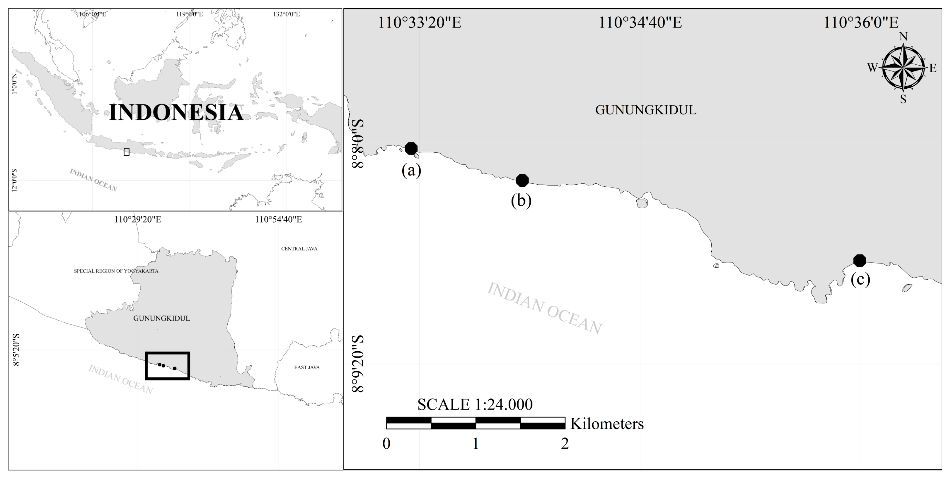
Sampling was carried out at Kukup Beach (8°8′3.31″ S 110°33′15.85″ E), Sepanjang Beach (8°8′18.89″ S 110°33′52.50″ E), and Krakal Beach (8°8′44.99″ S 110°36′0.17″ E) (Figure 1) in the tidal area at the lowest tide. The three beaches are pocket beaches with different morphological and hydrological characteristics. Kukup Beach has a flat, undulating topography with a sloping slope, and there is a barrier in the form of a coral island to the east. The seaweed G. edulis on this beach is commonly found in symbiosis with Sargassum spp. and Ophiuroidea spp. On Sepanjang Beach, the topographic characteristics in the form of a coastline extend from west to east and do not have coral islands as barriers; they tend to receive a large wave energy. G. edulis on this beach grows a lot next to seagrass on rocky sand substrates. Krakal Beach also has almost the same topographic characteristics, but this beach has undulating slopes and coral islands as barriers. The seaweed G. edulis on this beach coexists with Ulva spp., and several species of sea worms and mollusca.
Samples were collected using a hand sampling method, then cleaned to remove dirt, sand, and other adhering organisms. The samples were stored in a cooler to maintain freshness during transportation. The samples were washed with running water, identified, and dried at room temperature.

2.2. Extraction of Secondary Metabolites

The dry samples were cut into small pieces, ground into fine powder, and extracted using n-hexane, ethyl acetate, and methanol (Brataco, Jakarta, Indonesia) [16]. The isolation of the secondary metabolite compounds of the samples was carried out by a sequential solvent extraction method based on the degree of polarity, starting from non-polar solvents (n-hexane), semi-polar (ethyl acetate), and polar (methanol). Extraction was performed by adding 60 mL of solvent to 2 g of seaweed powder. The ratio between the weight of the sample and the volume of solvent added is 1:30 [17]. The sample solution was then extracted for 2 × 24 h at a speed of 140 rpm. The sequential solvent extraction process was carried out with the same steps but with different solvents sequentially based on the degree of polarity from the smallest to the largest. The sample solution was filtered using Whatman No. 41 filter paper (Cytiva, Marlborough, MA, USA) and evaporated. The crude extract was stored at −20 °C for the isolation of secondary metabolites.

2.3. LC-HRMS Measurements

Metabolite characterization was carried out using an LC-HRMS approach to identify bioactive compounds in the extract [18]. This analysis used a microTOF-QII mass spectrometer (Bruker, Billerica, MA, USA) equipped with an electrospray ionization (ESI) source and connected to a Dionex Ultimate 3000 high-performance liquid chromatography system (Thermo Scientific, Darmstadt, Germany). Separation was done on a reversed-phase EC10/2 Nucleoshell C18 column (2.7 µm) (Macherey-Nagel, Duren, Germany), with the column temperature kept at 25 °C [19]. Before analysis, the extract was redissolved in methanol to a final concentration of 10 mg mL−1. The sample was then homogenized and centrifuged at 10,000 rpm for 10 min under cold conditions (<4°C). The supernatant was transferred to a 96-well microplate (200 µL) and evaporated. This process of adding supernatant and evaporating was repeated until a dry residue remained. LC-HRMS analysis was performed in positive ionization mode using two mobile phases: phase A was water with 0.1% formic acid, and phase B was methanol with the same amount of formic acid. The gradient started at 95% phase A (0–0.30 min), then gradually decreased to 4.75% by the 18th minute. After that, phase A was quickly reduced to 0% at 18.10 min and held until 22.50 min. The initial condition of 95% phase A was restored at 22.60 min and maintained until the end of the run at 25 min. The flow rate was set to 600 µL/min [20].

2.4. Molecular Networking

The LC-HRMS measurement result of MS/MS data transformed from MassHunter data (.d files) into an .mzXML file using MSConvert (ProteoWizard 3.0). The files were processed and analyzed using the Global Natural Product Social Molecular Networking website (GNPS, https://gnps.ucsd.edu/ (accessed on 17 September 2025)) [21], visualized using Cytoscape 3.9.1 [22], analysed using DataAnalysis 4.0 (Bruker, Billerica, MA, USA), and RStudio 2025.09.0. The variables used in data processing were based on differences in extraction solvents and coastal locations. Then, Extracted Ion Chromatogram (EIC) is defined as a chromatogram obtained by extracting the m/z value to visualize the intensity and retention time, were analysed using DataAnalysis 4.0 (Bruker, Billerica, MA, USA). The variables used in data processing were based on differences in extraction solvents and coastal locations. Dereplicated compounds were considered putative, as identification was primarily based on accurate mass and MS/MS spectral similarity.

2.5. Heatmap and Hierarchical Clustering Analysis (HCA)

The intensities obtained using DataAnalysis 4.0 were visualized to determine the grouping of putative metabolite patterns. The process began by standardizing the intensity of each extract containing a putative metabolite using the Z-score transformation. Then, clustering was performed using the Ward D2 hierarchical method and Euclidean distance. All analyses were performed using RStudio 2025.09.0 and R 4.5.1.

2.6. Statistical Analysis

Differences in the distribution of G. edulis nodes from beaches (Kukup, Sepanjang, and Krakal) or solvents (n-hexane, ethyl acetate, and methanol) were analyzed using the chi-square goodness-of-fit test [23]. All statistical analyses were performed using R 4.5.1 and RStudio 2025.09.0 software. The results of the analysis were in the form of chi-square statistical values (X-square), degrees of freedom (df), and p-values. If significant differences (p < 0.05) were observed, standard residual analysis was performed for each beach or solvent category.

3. Results

3.1. Metabolomic Profile of G. edulis

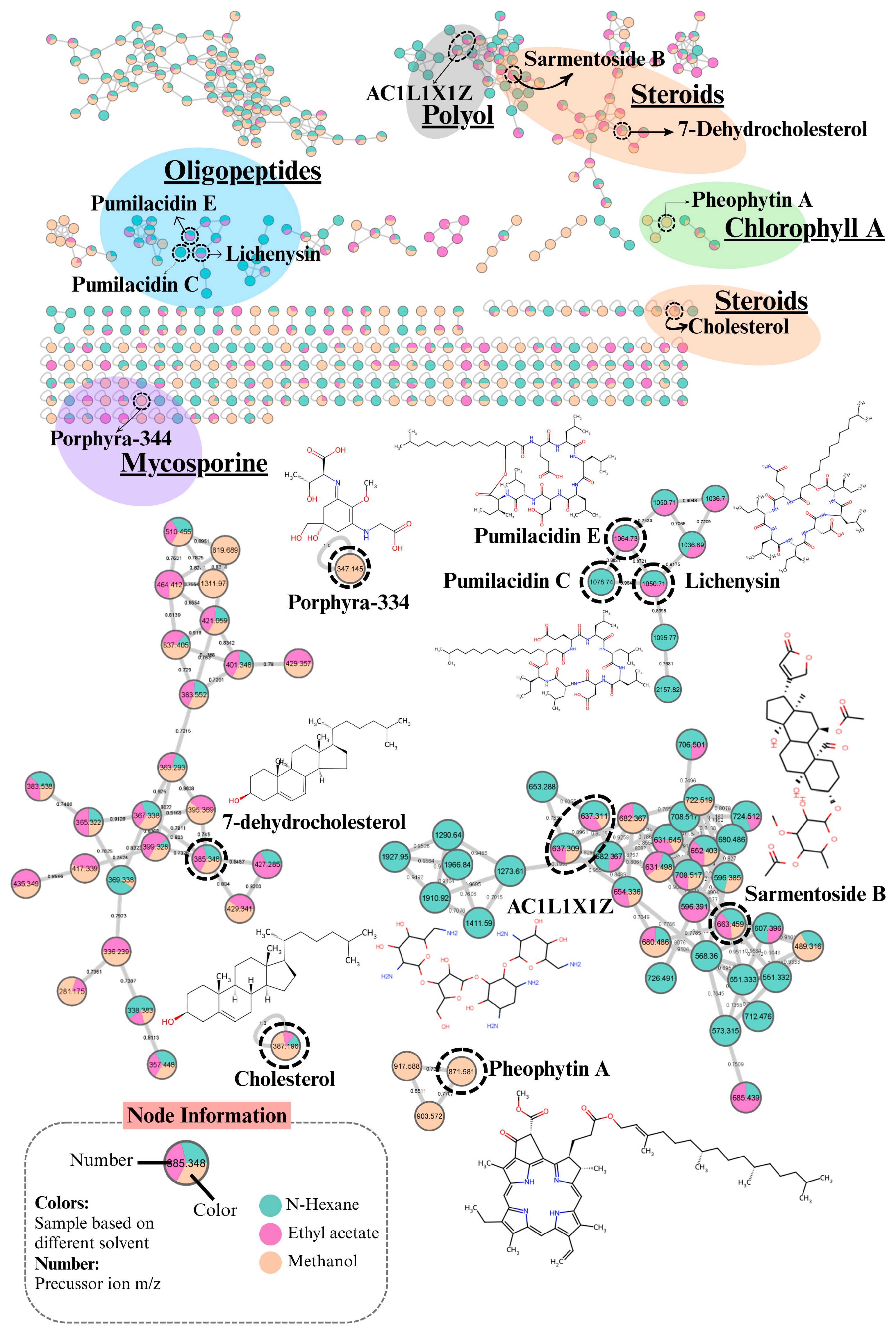
The results of the metabolomics analysis of G. edulis from Kukup, Sepanjang, and Krakal beaches are shown in Figure 2. In total, 425 metabolites were detected. Among these, nine compounds were tentatively identified based on the GNPS spectral library and are listed in Table 1. An edge was drawn between two or more metabolites from clusters based on biochemical correlation, if their pairwise cosine score was ≥0.7, indicating a high degree of biochemical correlation or relatedness. Network analysis revealed a fragmented structure, consisting of 38 distinct clusters and 169 single nodes with cosine scores ranging from 0.7 to 1.0. Each cluster group is represented by a different color. There are 9 of 425 compounds identified by the GNPS database. Mismatches between metabolite samples and library data may occur because the metabolite samples are new compounds or the data have not yet been deposited in the GNPS database.
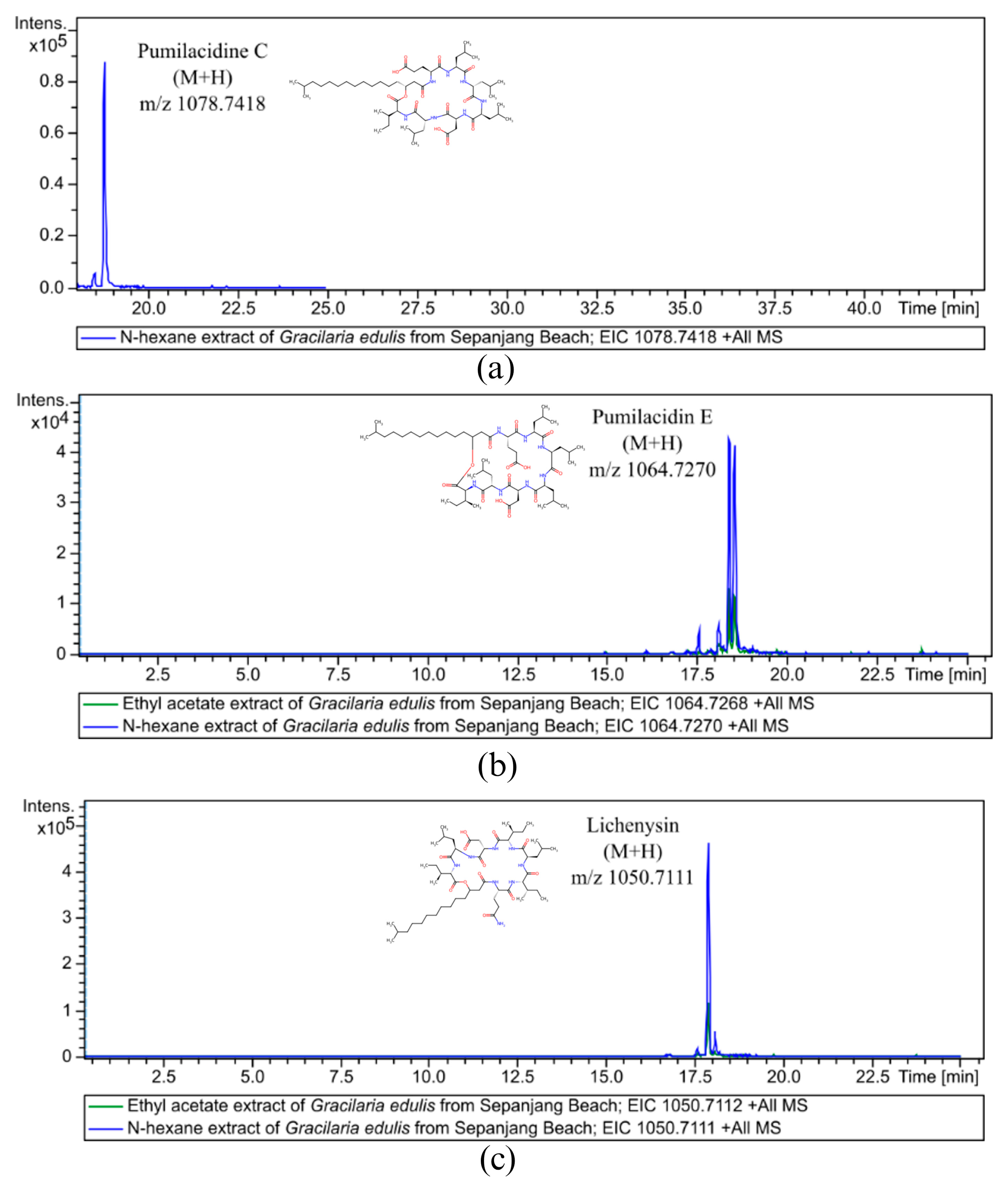
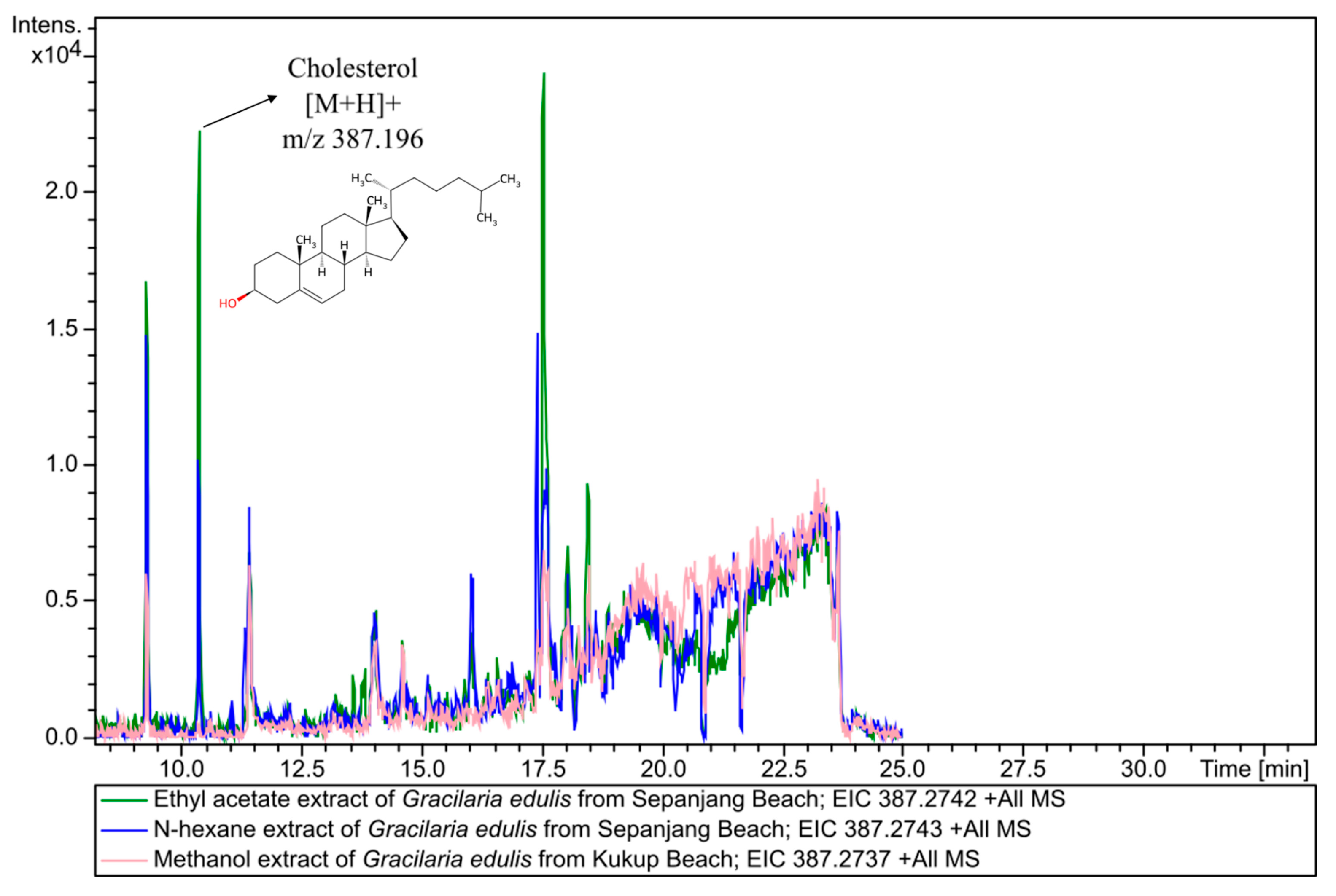
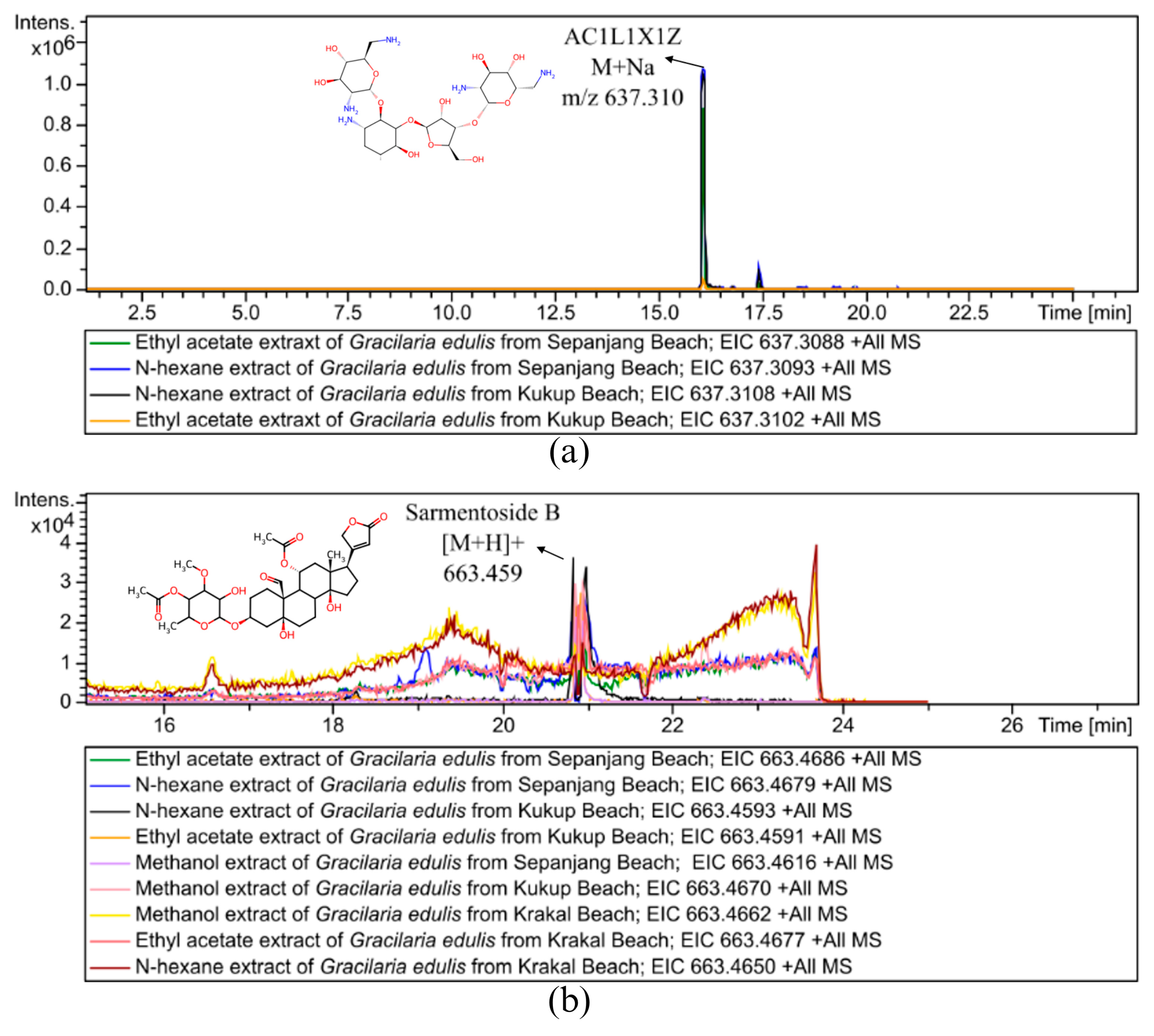
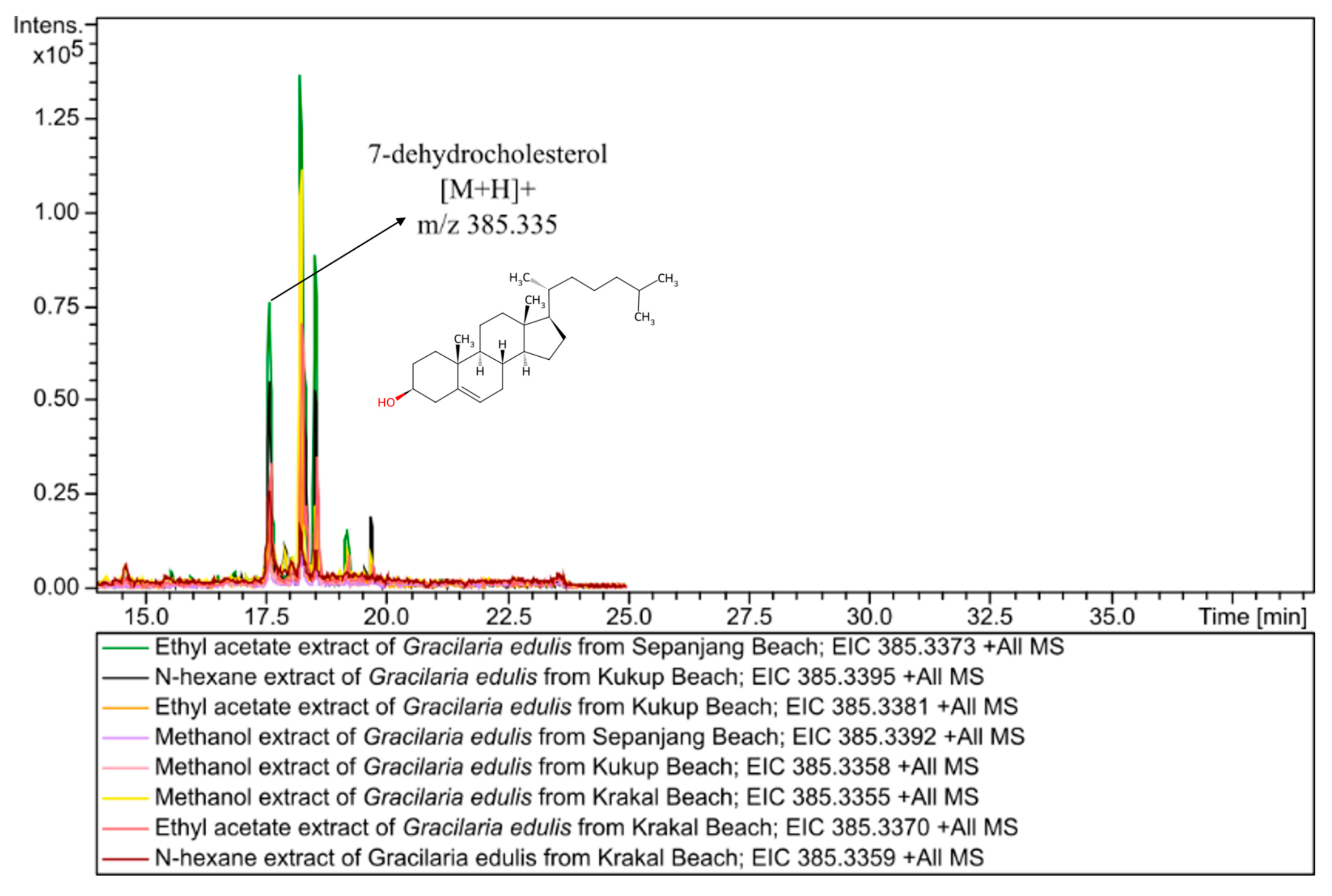
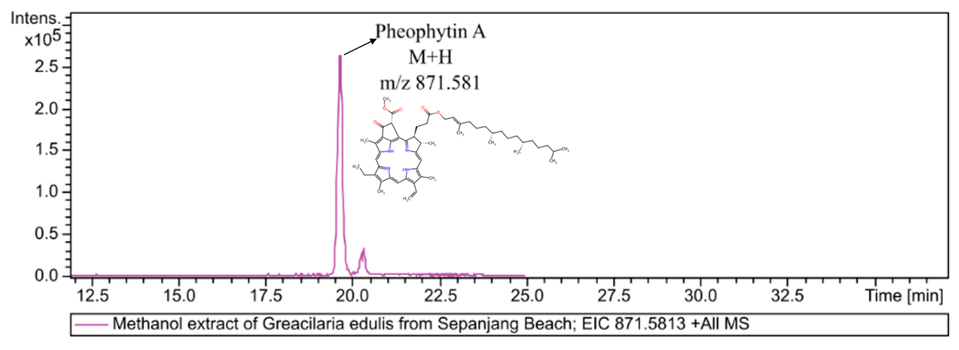
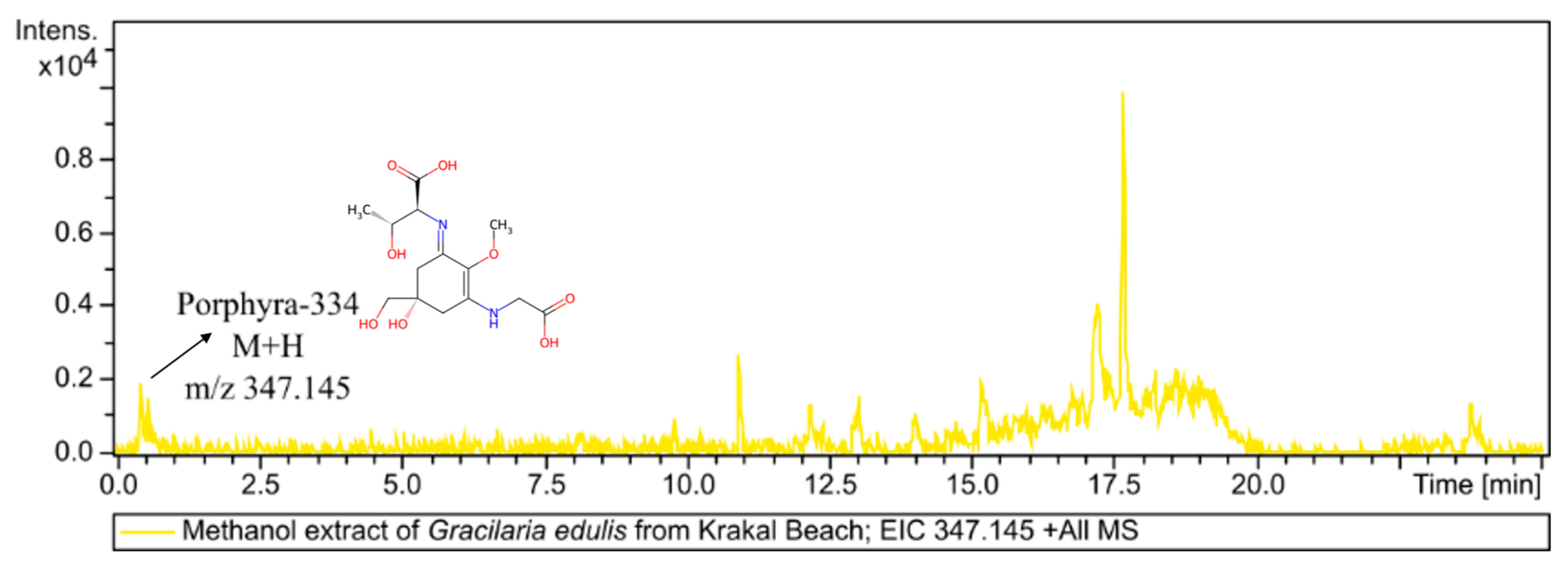
Pumilacidin C (m/z 1078.74 M + H), pumilacidin E (m/z 1064.73 M + H), and lichenysin (m/z 1050.71 M + H) were specifically produced by G. edulis from Sepanjang Beach. Pumilacidin C was found in G. edulis, which was extracted using n-hexane, whereas pumilacidin E and lichenysin were found in G. edulis, which was extracted using n-hexane and ethyl acetate. The chromatogram in Figure 3 shows that pumilacidin C, pumilacidin E, and lichenysin occurred at retention times of 18.82 min, 18.45 min, and 17.85 min. Cholesterol (m/z 387.196 [M + H]+) was produced by G. edulis from Kukup and Sepanjang Beaches, specifically extracted using methanol, n-hexane, and ethyl acetate. The chromatogram in Figure 4 showed that cholesterol was produced at a retention time of 10.43 min. The n-hexane extract of G. edulis from Sepanjang Beach yielded the highest compound intensity, whereas the other two extracts showed comparable levels. AC1L1X1Z (m/z 637.31 M + Na) and sarmentoside B (m/z 663.459 [M + H]+) are compounds that belong to the same molecular group and form a network. AC1L1X1Z was found at a retention time of 16.28 min in G. edulis extract, which was collected from Kukup and Sepanjang Beaches and extracted using metanol, n-hexane, and ethyl acetate (Figure 5). Sarmentoside B was found in all extracts at a retention time of 20.91 min, whereas 7-dehydrocholesterol (m/z 385.335 [M + H]+) appeared at a retention time of 17.87 min and was found in almost all G. edulis extracts, except for the one extracted with n-hexane from Sepanjang beach (Figure 6). The highest intensity was shown by the ethyl acetate extract of G. edulis from the Sepanjang Beach. Pheophytin A (m/z 871.582 [M + H]+) was detected at a retention time of 19.92 min in G. edulis from Sepanjang Beach which was extracted by methanol (Figure 7). Porphyra-334 (m/z 347.145 M + H) was detected at a retention time of 0.56 min in G. edulis from Krakal Beach which was extracted by methanol (Figure 8).

3.2. Putative Bioactive Compound from Three Different Beaches and Various Solvents

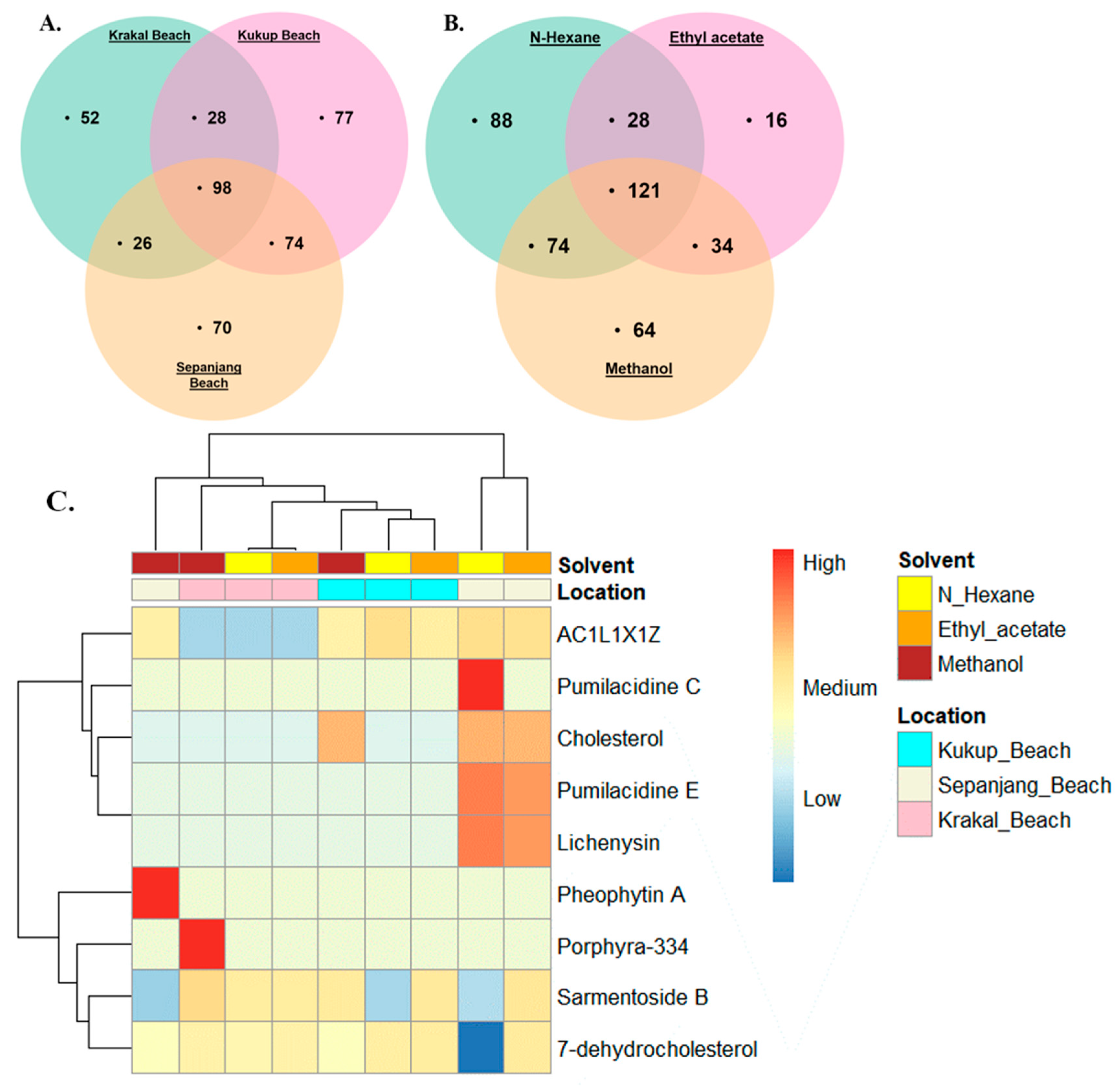
The number of metabolomic profiling nodes based on two different parameters, i.e., geographical location (Kukup, Sepanjang, and Krakal Beaches) and various solvents (n-hexane, ethyl acetate, and methanol is shown in Figure 9. Based on geographical location, Kukup Beach showed the richest metabolomic diversity, with 277 total nodes, followed by Sepanjang Beach with 268 total nodes and Krakal Beach with 204 total nodes, respectively. Based on the solvents, n-hexane extracted the highest number of metabolites with 311 nodes, followed by methanol with 293 nodes and ethyl acetate with 197 nodes, respectively (Figure 9A).
There was a difference in the intensity of the compounds in G. edulis extract based on the solvent and beach location (Figure 9C). Five dereplicated compounds, consisting of pumilacidin C, pumilacidin E, lichenysin, cholesterol, and AC1L1X1Z, showed the effectiveness of extraction using a solvent with non-polar properties, such as n-hexane. Sarmentoside B, pheophytin A, and porphyra-334 were effectively extracted using polar solvents such as methanol, whereas 7-dehydrocholesterol was effectively extracted using a semi-polar solvent such as ethyl acetate. Based on the sampling location, lichenysin and pumilacidin compounds were found in G. edulis extract from Sepanjang Beach, while cholesterol and AC1L1X1Z were found in G. edulis extract from Sepanjang and Kukup Beaches. Pheophytin A and porphyra-334 were specifically found in G. edulis from Krakal Beach. Meanwhile, the 7-dehydrocholesterol and sarmentoside B compounds were found in G. edulis extract from all beach locations. The highest intensity was produced by pumilacidin C, which was specifically found in G. edulis extract from Sepanjang Beach, which was extracted with n-hexane.

4. Discussion

Seaweed contains a variety of potential compounds in the form of primary and secondary metabolites, of which more than 1600 compounds are isolated from various types of red seaweed [43,44] and have been reported to have potential as antioxidants, antivirals, anti-obesity, antitumor, and antibacterials [15,45]. In the genus Gracilaria, it has been reported that there are various bioactive compounds, including mycosporine-like amino acids, lipids, agarans, phenolic acids, steroids, diterpenes, sulfonic acids, bromophenols, oxylipins, heterosides, and pigments [3]. Previous studies have reported that G. edulis extracted with ethyl acetate solvent has identified three putative compounds through Gas Chromatography–Mass Spectrometry (GC-MS), namely eicosane, methyl palmitate, and palmitic acid, and three putative compounds through Liquid Chromatography–Mass Spectrometry (LC-MS), namely manuealide A, manuealide C, and furanyl compounds, based on the Comprehensive Marine Natural Products Database (CMNPD) [8]. In contrast, G. edulis extracted with various solvents (chloroform < ethyl acetate < acetone < ethanol < methanol < water) successfully identified a large number of putative compounds, including 42 via GC-MS and 51 via LC-MS, based on the National Institute of Standards and Technology (NIST) database [46]. In this study, 425 putative metabolites were detected, and network analysis revealed a fragmented structure consisting of 38 distinct clusters and 169 single nodes. Among these, nine putative compounds were tentatively identified based on the GNPS spectral library: pumilacidin C (m/z 278 1078.74 [M + H]), pumilacidin E (m/z 1064.73 [M + H]), lichenysin (m/z 1050.71 [M + H]), choles- 279 terol (m/z 387.196 [M + H]+), AC1L1X1Z (m/z 637.31 [M + Na]), sarmentoside B (m/z 663.459 280 [M + H]+), 7-dehydrocholesterol (m/z 385.335 [M + H]+), pheophytin A (m/z 871.582 [M + H]+), and porphyra-334 (m/z 347.145 [M + H]) (Table 1). The low number of putative compounds identified in this study is likely due to the limited MS/MS spectra available in the GNPS database, resulting in many MS/MS spectra being detected but not annotated in this study. Differences in analytical platforms, such as LC-HRMS, LC-MS, and GC-MS, can also lead to variations in the number of putative compounds detected in the samples. Furthermore, sample origin, environmental conditions, and extraction methods can significantly affect metabolite profiles, resulting in different putative compounds compared to those reported in previous studies.
The pumilacidin C, pumilacidin E, and lichenysin were specifically produced by G. edulis from Sepanjang Beach. These three compounds belong to the surfactin family, which is a cyclic lipopeptide consisting of a heptapeptide backbone and one β-hydroxyl fatty acid with variable chain lengths ranging from 12 to 16 carbons [27,47]. Surfactin compounds have the ability to form ion channels and pores in cell membranes that can change permeability, resulting in an imbalance in osmotic pressure inside and outside the cell membrane; hence, vital ions in the cell are released, resulting in the death of microorganisms [48]. The antibacterial activity of pumilacidin has been reported to have the ability to kill Staphylococcus aureus bacteria. This compound is reported to have some advantages in terms of stability to heat, pH, and treatments with chemicals and enzymes [24]. Pumilacidin isolated from Bacillus sp. has been reported to suppress the motility of the pathogenic bacteria Vibrio alginolyticus and reduce biofilm formation, making it a potential drug candidate targeting bacterial motility or biofilm formation with low antibiotic resistance potential [25,26]. Lichenysin is reported to have antibacterial and antiadherence activity, which can form biofilms by the bacteria Pseudomonas aeruginosa and S. aureus [27,28]. Furthermore, lichenysin is also reported to have bactericidal properties against pathogenic and decomposing bacteria such as Bacillus cereus, Listeria monocytogenes, Erwinia carotovora, and Streptococcus spp. [29]. Until now, there have been no reports of the discovery of pumilacidin and lichenysin compounds in red seaweed, especially in G. edulis species. Both compounds are commonly found in marine bacteria, Bacillus sp., where specifically pumilacidin is produced by Bacillus pumilus bacteria, while lichenysin is specifically produced by Bacillus licheniformis bacteria [27]. In addition to being produced by B. licheniformis, lichenysin compounds can also be produced in aerobic and anaerobic conditions by Bacillus mojavensis bacteria under aerobic and anaerobic conditions [29]. Seaweed, as a holobiont organism host complex surface biofilms and microbiomes, containing diverse bacteria that interact with the algae across ecological and biochemical gradients. These interactions can be mutualistic, commensal, or competitive depending on species and environment. Within this system, chemical collaboration rather than direct biosynthesis by the algae is the dominant mechanism [49]. Seaweeds might provide a chemically rich microenvironment, while bacteria such as Bacillus spp. respond by producing secondary metabolites with ecological functions [50].
Cholesterol is a lipid molecule of the steroid group with a steroid skeleton consisting of four fused rings, with a 1,2-cycllopentanoperahydrofenanthane ring system [51]. Steroids are multi-cyclic compounds that have pharmacological activities such as antimicrobial, antioxidant, anticancer, and anti-inflammatory. Although cholesterol is primarily synthesized by humans and animals, its production has also been reported in numerous red seaweed species, including Kappaphycus alvarezii [33], Laurencia sp. [52] Laurencia complanata, Grateloupia sp., G. corticata, Halymenia sp., Spyridia sp., Metamastophora sp., Calloseris sp., and Neurymenia fraxinifolia [31]. This algal cholesterol demonstrates significant antibacterial properties. It reveals antibacterial activity against Gram-negative bacteria such as E. coli and P. vulgaris [30]. Furthermore, cholesterol from G. corticata extract exhibits antibacterial activity against S. aureus, while in Laurencia complanata extracts, it is effective against the pathogens B. cereus, S. aureus, Streptococcus pneumoniae, and Candida albicans [31]. Beyond its antibacterial potential, cholesterol from K. alvarezii also exhibits neurotrophic activity, as demonstrated by its high neurite-outgrowth-promoting activity (NOPA) in fetal rat hippocampal neuron cultures in vitro [32,33].
The AC1L1X1Z and sarmentoside B are in the same molecular group, hence they form a network. The compound AC1L1X1Z is annotated as neomycin and has been reported to be produced by Undaria pinnatifida [53]. In addition, this compound is also reported to have antibacterial activity against E. coli [34], and S. aureus [35]. Sarmentoside B is a glycoside of the Strophanthus sarmentosus plant [36]. Until now, reports of the discovery of sarmentoside B compounds in marine organisms have not been widely reported, including in red seaweed species. Sarmentoside B is produced by marine bacteria that are symbiotic with the sea cucumbers Stichopus vastus and Holothuria leucospilota and has antibacterial potential against the pathogenic bacteria E. coli, Pseudomonas aeruginosa, B. subtilis, and S. aureus [37].
The 7-dehydrocholesterol is a precursor of vitamin D3. In general, 7-dehydrocholesterol is found in animals and mammals, especially in parts of the skin exposed to ultraviolet B (UVB) rays, where it can cause the cleavage of the C bond in 7-dehydrocholesterol to form vitamin D3 [54]. In addition to humans and animals, 7-dehydrocholesterol has been found in several types of plants, such as tomato leaves (Lycopersicon esculentum), potatoes (Solanum tuberosum), and zucchini (Curcubita pepo) [55]. In seaweed, 7-dehydrocholesterol has been found in Sargassum muticum species using HPLC instruments, with a content of 90 µg/100 g [56]. Other studies have reported the presence of 7-dehydrocholesterol in small concentrations in kombu (Lessonia corrugata), with a content of 0.01 µg/100 g [38].
Pheophytin A is a pigment derived from chlorophyll a that has undergone thermal or weak acid treatment, where pheophytin A does not have magnesium ions (Mg2+) like chlorophyll a [57,58]. Pheophytin A has been found in many types of green plants, including seaweeds. The biological activity of pheophytin A has also been reported in seaweeds, including Ulva rigida [59] and Codium adhaerens [39] as an antioxidant, and Sargassum fulvellum and Enteromorpha prolifera as anti-inflammatory agents [40].
Porphyra-334 is one of 70 types of mycosporin-like amino acids (MAAs). MAAs themselves are compounds synthesized through the shikimate and/or pentose phosphate pathways by various marine organisms, including marine microorganisms and seaweed, to protect themselves from light exposure, such as ultraviolet (UV) rays [42,60]. The photoprotective potential of porphyra-334 was reported by Nunez (2025) and found in several species of red seaweed, including Gracilaria sp., Laurencia obtusa, Acanthopora spicifera, Ceramium sp., and Dasya sp., using an ultrahigh-performance liquid chromatography coupled with diode array detection and tandem mass spectrometry (UHPLC-DAD-MS/MS) instrument, where the highest intensity was produced by A. spicifera [61]. In addition, porphyra-334 is also found in the red seaweeds Porphyra linearis [62], Porphyra dentata, Porphyra yezoensis [63], Porphyra umbilicalis dan Grateloupia turuturu [64], which are reported to have antioxidant potential.
The productivity of bioactive compounds in seaweed is influenced by various biotic factors, such as the symbiotic relationship of seaweed with other surrounding organisms, and abiotic factors, such as environmental conditions, including temperature, salinity, light intensity, and geographical conditions [15,45,65]. In this study, the metabolomic profiles of G. edulis extracts varied based on beach location and solvent (Figure 9). Based on geographical location, Kukup Beach showed the richest metabolomic diversity, with 277 nodes, followed by Sepanjang Beach with 268 nodes and Krakal Beach with 204 nodes. A chi-square goodness-of-fit test revealed a significant difference in the number of detected nodes between beaches (X-squared = 12.692, df = 2, p < 0.002). Residual analysis also showed that Kukup Beach contributed the most nodes (residual = 2.12), while Krakal Beach produced significantly fewer nodes than Sepanjang Beach (residual = −3.54).
Based on the GNPS analysis, nine putative compounds were dereplicated: lichenysin and pumilacidin compounds were produced by G. edulis from Sepanjang Beach, cholesterol and AC1L1X1Z were produced by G. edulis from Sepanjang and Kukup Beaches, 7-dehydrocholesterol and sarmentoside B compounds were produced by G. edulis from all beach locations, while specific pheophytin A and porphyra-334 were produced by G. edulis from Krakal Beach. In this study, the highest intensity of the putative compound was found in G. edulis originating from the Sepanjang Beach. Sepanjang Beach has high tourist activity, which contributes to an increase in marine debris abundance [66]. Seaweed that grows in extreme environmental conditions tends to produce more secondary metabolites as a self-defense mechanism. Seaweed living in aquatic environments with high nutrient levels focuses on growth and development, reducing the amount of phenolic compounds produced. Conversely, in aquatic environments with contaminants and metals, seaweed releases phenolic compounds in the form of phlorotannins as a natural detoxification mechanism to protect itself from contaminants [67].
The composition and bioactivity of essential oils are influenced by the biomass drying method, extraction technique, extraction temperature, extraction duration, and solvent ratio [65,68]. Based on the differences in solvents, n-hexane extracted the highest number of metabolites, with 311 nodes, followed by methanol, with 293 nodes, and ethyl acetate, with 197 nodes. The chi-square goodness-of-fit test showed significant differences in the distribution of detected nodes between the solvents used in the extraction of G. edulis bioactive compounds (X-squared = 28.135, df = 2, p-value < 0.001). Standardized residue analysis showed that G. edulis extracted with n-hexane gave the highest node distribution with a residue value of 3.30, while G. edulis extracted with ethyl acetate gave a lower node distribution compared to the node distribution obtained from G. edulis extracted with methanol, where the residue value of G. edulis extracted with ethyl acetate was −5.25.
In this study, nine compounds were dereplicated, producing different intensities. As many as five compounds, consisting of pumilacidin C, pumilacidin E, lichenysin, cholesterol, and AC1L1X1Z, showed effective extraction using a solvent with non-polar properties, such as n-hexane. Sarmentoside B, pheophytin A, and porphyra-334 were effectively extracted using polar solvents such as methanol, whereas 7-dehydrocholesterol was effectively extracted using the semi-polar solvent, ethyl acetate. Each solvent selectively extracts bioactive compounds based on the principle of “like dissolves like,” targeting compounds with similar polarity levels [68]. Non-polar solvents, including n-hexane, have been reported to extract various bioactive compounds that act as antibacterial agents, such as pigments, terpenoids, alkaloids, hydrocarbons, and fatty acids [69]. Differences in bioactivity were reported in Gracilaria tenuistipitata extracted using methanol, ethanol, and water, where G. tenuistipitata extracted with methanol had a higher phenolic content of flavonoid compounds than extracts extracted using ethanol and water [70].

5. Conclusions

In this study, we observed differences in the metabolomic profiles of G. edulis extracts based on variations in coastal location and solvent composition. G. edulis extracts, which were collected from Kukup Beach, showed the richest metabolomic diversity, with 277 nodes. Lichenysin and pumilacidin compounds were produced by G. edulis from Sepanjang Beach, while cholesterol and AC1L1X1Z were produced by G. edulis from Sepanjang and Kukup Beaches. The 7-dehydrocholesterol and sarmentoside B compounds were produced by G. edulis from all beach locations, while specific pheophytin A and porphyra-334 were produced by G. edulis from Krakal Beach. Among the solvents tested, n-hexane was the most effective for the extraction of the target compounds. Studies have reported that nonpolar solvents such as n-hexane can extract various bioactive compounds with antibacterial properties, such as pigments, terpenoids, alkaloids, hydrocarbons, and fatty acids [69]. Further research is needed to obtain pure compounds that have the potential to be antibacterial, antioxidant, anti-inflammatory, and photoprotective compounds. Purification can be carried out using chromatography such as column chromatography, thin-layer chromatography, and preparative HPLC, followed by characterization using LC-HRMS and NMR.

Author Contributions

Conceptualization, M.D.N.M. and J.-S.C.; Methodology, M.D.N.M., R. (Riyanti), T.F.S. and J.-S.C.; Validation, R. (Riviani), D.S., T.F.S. and M.A.P.; formal analysis, F.N.A., R. (Riviani), D.S., T.F.S., and M.A.P.; investigation, F.N.A.; data curation, F.N.A. and D.H.; writing—original draft preparation, F.N.A., D.H., M.D.N.M. and J.-S.C.; writing—review and editing, M.D.N.M., R. (Riyanti), H.S., D.H., T.F.S., M.A.P. and J.-S.C.; supervision, M.D.N.M., R. (Riyanti), H.S., T.F.S. and J.-S.C. All authors have read and agreed to the published version of the manuscript.

Funding

This research was supported by the International Collaboration Research Grant, Jenderal Soedirman University (Grant No. 26.278/UN23.35.5/PT.01/II/2024). We thank Jenderal Soedirman University, Indonesia; Justus Liebig University, Germany; and Gyeongsang National University, South Korea; for the international research collaboration.

Data Availability Statement

The original contributions presented in the study are included in the article; further inquiries can be directed to the corresponding authors.

Conflicts of Interest

The authors declare no conflicts of interest.

References

  1. FAO. Cultured Aquatic Species Information Programme Gracilaria spp.; FAO: Rome, Italy, 2014; Volume 14, pp. 171–184. [Google Scholar]
  2. Arivoli, P.; Antony, C.; Ruby, P.; Nadu, T.; Fisheries, J.J. Global Seaweed Production: Data and Trends. Vigyan Varta Int. E-Mag. Sci. Enthus. 2024, 5, 61–64. [Google Scholar]
  3. Torres, P.; Santos, J.P.; Chow, F.; dos Santos, D.Y.A.C. A Comprehensive Review of Traditional Uses, Bioactivity Potential, and Chemical Diversity of the Genus Gracilaria (Gracilariales, Rhodophyta). Algal Res. 2019, 37, 288–306. [Google Scholar] [CrossRef]
  4. Greville, R.K. Algae Britannicae or Descriptions of the Marine and Other Inarticulated Plants of the British Islands, Belonging to the Order Algae; With Plates Illustrative of the Genera; MacLachlan & Stewart: Edinburgh, UK, 1830. [Google Scholar]
  5. Guiry, M.D.; Guiry, G.M. AlgaeBase; World-Wide Electronic Publication; National University of Ireland: Galway, Ireland, 2024. [Google Scholar]
  6. Meinita, M.D.N.; Akromah, N.; Andriyani, N.; Setijanto; Harwanto, D.; Liu, T. Molecular Identification of Gracilaria Species (Gracilariales, Rhodophyta) Obtained from The South Coast of Java Island, Indonesia. Biodiversitas 2021, 22, 3046–3056. [Google Scholar] [CrossRef]
  7. Susanti, F.; Adharini, R.I.; Rahmi, K.A.; Sari, D.W.K.; Kandasamy, G. Identification of Gracilaria spp. In Gunungkidul Regency, Yogyakarta Indonesia Based on DNA Barcoding Target Cytochrome Oxidase Subunit 1. Ilmu Kelaut. Indones. J. Mar. Sci. 2022, 27, 189–198. [Google Scholar]
  8. Kurnia, D.S.; Kasanah, N. Bioprospecting of Indonesian Red Seaweeds Gracilaria edulis and Pterocladiella sp. 4: Anti-Vibrio Activity and Chemical Composition. Appl. Phycol. 2025, 6, 126–144. [Google Scholar] [CrossRef]
  9. Arulkumar, A.; Rosemary, T.; Paramasivam, S.; Rajendran, R.B. Phytochemical Composition, In Vitro Antioxidant, Antibacterial Potential and GC-MS Analysis of Red Seaweeds (Gracilaria corticata and Gracilaria edulis) from Palk Bay, India. Biocatal. Agric. Biotechnol. 2018, 15, 63–71. [Google Scholar] [CrossRef]
  10. Hewa, I.U.P.; Epage, N.S.; Gunathilake, V.K.; Isuru, U. Comparative Morphoanatomy and Phycochemical Screening of Three Selected Gracilaria (Rhodophyta) Species from Sri Lanka. Appl. Phycol. 2024, 5, 129–142. [Google Scholar] [CrossRef]
  11. Jiménez-González, C.; Agrasar, A.M.T.; Mallo, F.; Rúa, M.L.; Fuciños, C. Red Seaweed Proteins: Valuable Marine-Origin Compounds with Encouraging Applications. Algal Res. 2023, 75, 103262. [Google Scholar] [CrossRef]
  12. Carpena, M.; Pereira, C.S.G.P.; Silva, A.; Barciela, P.; Jorge, A.O.S.; Perez-Vazquez, A.; Pereira, A.G.; Barreira, J.C.M.; Oliveira, M.B.P.P.; Prieto, M.A. Metabolite Profiling of Macroalgae: Biosynthesis and Beneficial Biological Properties of Active Compounds. Mar. Drugs 2024, 22, 478. [Google Scholar] [CrossRef]
  13. Goh, P.T.; Poong, S.W.; Zheng, X.; Liu, T.; Qi, Z.; Beardall, J.; Lim, P.E. Metabolomic Profiling of A Red Alga, Gracilaria changii, Under Current Ambient and Elevated PCO2 Levels Using An Untargeted Gas Chromatography-Mass Spectrometry (GC–MS) Approach. J. Appl. Phycol. 2025, 37, 1573–1591. [Google Scholar] [CrossRef]
  14. Meinita, M.D.N.; Riyanti; Sanjayasari, D.; Riviani; Harwanto, D.; Jiso, A.; Schäberle, T.F.; Mettal, U.; Moon, I.-S.; Choi, J.-S. Bioactive Compound Profiling of Agarophyte Seaweed (Gelidiella acerosa, Gracilaria arcuata, and Gracilaria verrucosa) Based on LC-HRMS Metabolomic and Molecular Networking Approach. Foods 2025, 14, 4042. [Google Scholar] [CrossRef]
  15. Pérez, M.J.; Falqué, E.; Domínguez, H. Antimicrobial Action of Compounds from Marine Seaweed. Mar. Drugs 2016, 14, 52. [Google Scholar] [CrossRef]
  16. El Shafay, S.M.; Ali, S.S.; El-Sheekh, M.M. Antimicrobial Activity of Some Seaweeds Species from Red Sea, Against Multidrug Resistant Bacteria. Egypt. J. Aquat. Res. 2016, 42, 65–74. [Google Scholar] [CrossRef]
  17. Topuz, O.K.; Gokoglu, N.; Yerlikaya, P.; Ucak, I.; Gumus, B. Optimization of Antioxidant Activity and Phenolic Compound Extraction Conditions from Red Seaweed (Laurencia obtuse). J. Aquat. Food Prod. Technol. 2016, 25, 414–422. [Google Scholar] [CrossRef]
  18. Sapkota, B.K.; Khadayat, K.; Aryal, B.; Bashyal, J.; Jaisi, S.; Parajuli, N. LC-HRMS-Based Profiling: Antibacterial and Lipase Inhibitory Activities of Some Medicinal Plants for the Remedy of Obesity. Sci. Pharm. 2022, 90, 55. [Google Scholar] [CrossRef]
  19. Riyanti; Balansa, W.; Liu, Y.; Sharma, A.; Mihajlovic, S.; Hartwig, C.; Leis, B.; Rieuwpassa, F.J.; Ijong, F.G.; Wägele, H.; et al. Selection of Sponge-Associated Bacteria with High Potential for the Production of Antibacterial Compounds. Sci. Rep. 2020, 10, 19614. [Google Scholar] [CrossRef]
  20. Riyanti; Marner, M.; Hartwig, C.; Patras, M.A.; Wodi, S.I.M.; Rieuwpassa, F.J.; Ijong, F.G.; Balansa, W.; Schaberle, T. Sustainable Low-Volume Analysis of Environmental Samples by Semi-Automated Prioritization of Extracts for Natural Product Research (SeaPEPR). Mar. Drugs 2020, 18, 649. [Google Scholar] [CrossRef]
  21. Wang, M.; Carver, J.J.; Phelan, V.V.; Al, E. Sharing and Community Curation of Mass Spectrometry Data with Global Natural Products Social Molecular Networking. Nat. Biotechnol. 2016, 34, 828–837. [Google Scholar] [CrossRef]
  22. Shannon, P.; Markiel, A.; Ozier, O.; Baliga, N.S.; Wang, J.T.; Ramage, D.; Amin, N.; Schwikowski, B.; Ideke, T. Cytoscape: A Software Environment for Integrated Models. Genome Res. 2003, 13, 2498–2504. [Google Scholar] [CrossRef]
  23. Hosmer, D.W.; Hosmer, T.; Cessie, S.L.E.; Lemeshow, S. A Comparison of Goodness-of-Fit Test for the Logistic Regression Model. Stat. Med. 1997, 16, 965–980. [Google Scholar] [CrossRef]
  24. Saggese, A.; Culurciello, R.; Casillo, A.; Corsaro, M.M.; Ricca, E.; Baccigalupi, L. A Marine Isolate of Bacillus pumilus Secretes a Pumilacidin Active Against Staphylococcus aureus. Mar. Drugs 2018, 16, 180. [Google Scholar] [CrossRef]
  25. Xiu, P.; Liu, R.; Zhang, D.; Sun, C. Pumilacidin-Like Lipopeptides Derived from Marine Bacterium Bacillus sp. Strain 176 Suppress the Motility of Vibrio alginolyticus. Appl. Environ. Microbiol. 2017, 83, e00450-17. [Google Scholar] [CrossRef]
  26. Dasgupta, A.; De, D.; Chaudhuri, S. Anti-biofilm and disinfectant-like activity of pumilacidin, a lipopeptide biosurfactant produced by Bacillus pumilus NITDID1. Biocatal. Agric. Biotechnol. 2024, 56, 103024. [Google Scholar] [CrossRef]
  27. Zammuto, V.; Rizzo, M.G.; De Pasquale, C.; Ferlazzo, G.; Caccamo, M.T.; Magazù, S.; Guglielmino, S.P.P.; Gugliandolo, C. Lichenysin-like Polypeptide Production by Bacillus licheniformis B3-15 and Its Antiadhesive and Antibiofilm Properties. Microorganisms 2023, 11, 1842. [Google Scholar] [CrossRef]
  28. Castaldi, A.; Truong, B.N.; Vu, Q.T.; Le, T.H.M.; Marie, A.; Le Pennec, G.; Rouvier, F.; Brunel, J.M.; Longeon, A.; Pham, V.C.; et al. Computational Methods Reveal a Series of Cyclic and Linear Lichenysins and Surfactins from the Vietnamese Marine Sediment-Derived Streptomyces Strain G222. Molecules 2024, 29, 1458. [Google Scholar] [CrossRef]
  29. Shleeva, M.O.; Kondratieva, D.A.; Kaprelyants, A.S. Bacillus licheniformis: A Producer of Antimicrobial Substances, Including Antimycobacterials, Which Are Feasible for Medical Applications. Pharmaceutics 2023, 15, 1893. [Google Scholar] [CrossRef]
  30. Sribalan, R.; Padmini, V.; Lavanya, A.; Ponnuvel, K. Evaluation of Antimicrobial Activity of Glycinate and Carbonate Derivatives of Cholesterol: Synthesis and Characterization. Saudi Pharm. J. 2016, 24, 658–668. [Google Scholar] [CrossRef]
  31. Rahelivao, M.P.; Gruner, M.; Andriamanantoanina, H.; Andriamihaja, B.; Bauer, I.; Knölker, H.J. Red Algae (Rhodophyta) from the Coast of Madagascar: Preliminary Bioactivity Studies and Isolation of Natural Products. Mar. Drugs 2015, 13, 4197–4216. [Google Scholar] [CrossRef]
  32. Tirtawijaya, G.; Mohibbullah, M.; Meinita, M.D.N.; Moon, I.S.; Hong, Y.K. The Ethanol Extract of the Rhodophyte Kappaphycus Alvarezii Promotes Neurite Outgrowth in Hippocampal Neurons. J. Appl. Phycol. 2016, 28, 2515–2522. [Google Scholar] [CrossRef]
  33. Tirtawijaya, G.; Haque, M.A.; Choi, J.S.; Moon, I.S.; Meinita, M.D.N.; Choi, J.S.; Hong, Y.-K. Spinogenesis and Synaptogenesis Effects of the Red Seaweed Kappaphycus Alvarezii and Its Isolated Cholesterol on Hippocampal Neuron Cultures. Prev. Nutr. Food Sci. 2019, 24, 418–425. [Google Scholar] [CrossRef]
  34. Kumar, L.; Brice, J.; Toberer, L.; Klein-Seetharaman, J.; Knauss, D.; Sarkar, S.K. Antimicrobial Biopolymer Formation from Sodium Alginate and Algae Extract Using Aminoglycosides. PLoS ONE 2019, 14, e0214411. [Google Scholar] [CrossRef]
  35. Blanchard, C.; Brooks, L.; Beckley, A.; Colquhoun, J.; Dewhurst, S.; Dunman, P.M. Neomycin Sulfate Improves the Antimicrobial Activity of Mupirocin-Based Antibacterial Ointments. Antimicrob. Agents Chemother. 2016, 60, 862–872. [Google Scholar] [CrossRef]
  36. Callow, R.K.; Taylor, D.A.H. The Cardio-Active Glycosides of Strophanthus sarmentosus P.DC. ”Sarmentoside B“ and Its Relation to an Original Sarmentobioside. J. Chem. Soc. 1952, 2299–2304. [Google Scholar] [CrossRef]
  37. Wibowo, J.T.; Kellermann, M.Y.; Versluis, D.; Putra, M.Y.; Murniasih, T.; Mohr, K.I.; Wink, J.; Engelmann, M.; Praditya, D.F.; Steinmann, E.; et al. Biotechnological Potential of Bacteria Isolated from the Sea Cucumber Holothuria leucospilota and Stichopus vastus from Lampung, Indonesia. Mar. Drugs 2019, 17, 635. [Google Scholar] [CrossRef]
  38. Hughes, L.J.; Black, L.J.; Sherriff, J.L.; Dunlop, E.; Strobel, N.; Lucas, R.M.; Bornman, J.F. Vitamin D Content of Australian Native Food Plants and Australian-Grown Edible Seaweed. Nutrients 2018, 10, 876. [Google Scholar] [CrossRef]
  39. Radman, S.; Cikoš, A.M.; Flanjak, I.; Babić, S.; Čižmek, L.; Šubarić, D.; Čož-Rakovac, R.; Jokić, S.; Jerković, I. Less Polar Compounds and Targeted Antioxidant Potential (In Vitro and in Vivo) of Codium adhaerens c. Agardh 1822. Pharmaceuticals 2021, 14, 944. [Google Scholar] [CrossRef]
  40. Pangestuti, R.; Kim, S.K. Biological Activities and Health Benefit Effects of Natural Pigments Derived from Marine Algae. J. Funct. Foods 2011, 3, 255–266. [Google Scholar] [CrossRef]
  41. Yang, W.; Sakamoto, T. Functional Characterization of the Mys Genes for Porphyra-334 Biosynthesis from the Terrestrial Cyanobacterium Nostoc Commune by Heterologous Expression. J. Gen. Appl. Microbiol. 2025, 71, 19–27. [Google Scholar] [CrossRef]
  42. Hatakeyama, M.; Nakamura, S. Button-on-a-String Mechanism in Water, the Ultrafast UV-to-Heat Conversion by Mycosporine-like Amino Acid Porphyra-334 of Natural Sunscreen Compound. ACS Phys. Chem. Au 2025, 5, 274–282. [Google Scholar] [CrossRef]
  43. Leal, M.C.; Munro, M.H.G.; Blunt, J.W.; Puga, J.; Jesus, B.; Calado, R.; Rosa, R.; Madeira, C. Biogeography and Biodiscovery Hotspots of Macroalgal Marine Natural Products. Nat. Prod. Rep. 2013, 30, 1380–1390. [Google Scholar] [CrossRef]
  44. Carpena, M.; Garcia-Perez, P.; Garcia-Oliveira, P.; Chamorro, F.; Otero, P.; Lourenço-Lopes, C.; Cao, H.; Simal-Gandara, J.; Prieto, M.A. Biological Properties and Potential of Compounds Extracted from Red Seaweeds. Phytochem. Rev. 2023. online ahead of print. [Google Scholar] [CrossRef]
  45. Das, D.; Arulkumar, A.; Paramasivam, S.; Lopez-Santamarina, A.; del Carmen Mondragon, A.; Miranda Lopez, J.M. Phytochemical Constituents, Antimicrobial Properties and Bioactivity of Marine Red Seaweed (Kappaphycus alvarezii) and Seagrass (Cymodocea serrulata). Foods 2023, 12, 2811. [Google Scholar] [CrossRef]
  46. Asghar, A.; Tan, Y.; Shahid, M.; Yow, Y.; Lahiri, C. Metabolite Profiling of Malaysian Gracilaria edulis Reveals Eplerenone as Novel Antibacterial Compound for Drug Repurposing Against MDR Bacteria Collection of Materials. Front. Microbiol. 2021, 12, 653562. [Google Scholar] [CrossRef]
  47. Torres-Mendoza, D.; Coronado, L.M.; Pineda, L.M.; Guzmán, H.M.; Dorrestein, P.C.; Spadafora, C.; Gutiérrez, M. Pumilacidins from the Octocoral-Associated Bacillus sp. DT001 Display Anti-Proliferative Effects in Plasmodium falciparum. Molecules 2018, 23, 2179. [Google Scholar] [CrossRef]
  48. Markelova, N.; Chumak, A. Antimicrobial Activity of Bacillus Cyclic Lipopeptides and Their Role in the Host Adaptive Response to Changes in Environmental Conditions. Int. J. Mol. Sci. 2025, 26, 336. [Google Scholar] [CrossRef]
  49. Ren, C.; Liu, Z.; Wang, X. Minireview The Seaweed Holobiont: From Microecology to Biotechnological Applications. Microb. Biotechnol. 2022, 15, 738–754. [Google Scholar] [CrossRef]
  50. Egan, S.; Harder, T.; Burke, C.; Steinberg, P.; Kjelleberg, S.; Thomas, T. The Seaweed Holobiont: Understanding Seaweed–Bacteria Interactions. Fed. Eur. Microbiol. Soc. 2013, 37, 462–476. [Google Scholar] [CrossRef]
  51. Albuquerque, H.M.T.; Santos, C.M.M.; Silva, A.M.S. Cholesterol-Based Compounds: Recent Advances in Synthesis and Applications. Molecules 2019, 24, 116. [Google Scholar] [CrossRef]
  52. Pokharkar, O.; Anumolu, H.; Zyryanov, G.V.; Tsurkan, M.V. Natural Products from Red Algal Genus Laurencia as Potential Inhibitors of RdRp and nsp15 Enzymes of SARS-CoV-2: An In Silico Perspective. Microbiol. Res. 2023, 14, 1020–1048. [Google Scholar] [CrossRef]
  53. Villalobos, J.P. Biogeographical and Phylogenetic Boundaries of the Specialised Metabolism of Pseudonocardia from Marine Origin; University of Strathclyde: Glasgow, UK, 2022. [Google Scholar]
  54. Prabhu, A.V.; Luu, W.; Li, D.; Sharpe, L.J.; Brown, A.J. DHCR7: A Vital Enzyme Switch Between Cholesterol and Vitamin D Production. Prog. Lipid Res. 2016, 64, 138–151. [Google Scholar] [CrossRef]
  55. Aburjai, T.; Al-Khalil, S.; Abuirjeie, M. Vitamin D3 and Its Metabolites in Tomato, Potato, Egg Plant and Zucchini Leaves. Phytochemistry 1998, 49, 2497–2499. [Google Scholar] [CrossRef]
  56. De Roeck-Holtzhauer, Y.; Quere, I.; Claire, C. Vitamin Analysis of Five Planktonic Microalgae and One Macroalga. J. Appl. Phycol. 1991, 3, 259–264. [Google Scholar] [CrossRef]
  57. Liu, X.; Gu, Y.; Xu, W.; Zhang, R.; Koyama, T. Analysis for Inhibition Mechanisms of Pheophytin A and Pyropheophytin A Against Alpha-Glucosidase by Multi-Spectroscopy, Molecular Docking, and Molecular Dynamics Simulation. LWT 2025, 224, 117828. [Google Scholar] [CrossRef]
  58. Semizo, H.; Horii, I.; Ueda, R.; Takahashi, Y.; Matsuo, Y.; Kawakami, H. Role of Pheophytin in Power Generation of Photosynthetic Fuel Cells. Chem. Phys. Impact 2025, 11, 100948. [Google Scholar] [CrossRef]
  59. Malea, P.; Patronia, M.M.; Dermentzis, M.; Kevrekidou, A.; Kevrekidis, D.P. ROS Production-Related Phenomena and Biochemical Reactions of the Seaweed Ulva rigida in Response to the Effect of Environmentally Relevant Bisphenol A Concentrations. Aquat. Toxicol. 2026, 290, 107638. [Google Scholar] [CrossRef]
  60. Song, S.Y.; Cho, J.Y.; Park, D.H.; Song, S.H.; Lee, S.H.; Park, J.W.; Lim, H.K.; Cho, S.S. Biological Effect of Mycosporine-Gly-Ser (Shinorine) Against Bis-Retinoid N-Retinyl-N-Retinylidene Ethanolamine- and Blue-Light-Induced Retinal Pigment Epithelium Cell Damage. Nutrients 2025, 17, 1363. [Google Scholar] [CrossRef]
  61. Angulo-Nuñez, E.; Rincón-Díaz, N.; Puyana, M.; Costa, G.; Urrea-Victoria, V.; Castellanos, L.; Ramos, F.A. Photoprotective Potential of Mycosporine-Like Amino Acids in Red Algae (Rhodophyta) from San Andrés and Providencia Islands, Colombian Caribbean. Rev. Bras. Farmacogn. 2025, 35, 754–762. [Google Scholar] [CrossRef]
  62. Pereira, D.T.; Korbee, N.; Vega, J.; Figueroa, F.L. The Role of Nitrate Supply in Bioactive Compound Synthesis and Antioxidant Activity in the Cultivation of Porphyra linearis (Rhodophyta, Bangiales) for Future Cosmeceutical and Bioremediation Applications. Mar. Drugs 2024, 22, 222. [Google Scholar] [CrossRef]
  63. Kim, B.S.; Im, J.H.; Yoon, Y.S.; Kim, H.; Cho, J.Y.; Ham, J.R.; Heo, Y.J.; Lee, H.I. Analysis of Nutritional Composition and Flavor Patterns by Variety (Porphyra dentata and Porphyra yezoensis) in Dried Laver from Jeonnam, Korea. Foods 2025, 14, 335. [Google Scholar] [CrossRef]
  64. Ferreira, J.; Pacheco, M.; Silva, A.M.; Gaivão, I. Unlocking the Potential of Red Seaweeds: A Special Focus on Grateloupia turuturu Yamada and Porphyra umbilicalis Kütz. Mar. Drugs 2025, 23, 347. [Google Scholar] [CrossRef]
  65. Cadar, E.; Popescu, A.; Dragan, A.M.L.; Pesterau, A.M.; Pascale, C.; Anuta, V.; Prasacu, I.; Velescu, B.S.; Tomescu, C.L.; Bogdan-Andreescu, C.F.; et al. Bioactive Compounds of Marine Algae and Their Potential Health and Nutraceutical Applications: A Review. Mar. Drugs 2025, 23, 152. [Google Scholar] [CrossRef] [PubMed]
  66. Hibatullah, M.F.; Mutaqin, B.W. Marine Debris Characteristics in Various Coastal Typologies in the Gunungkidul Coastal Area of Yogyakarta—Indonesia. Discov. Geosci. 2024, 2, 28. [Google Scholar] [CrossRef]
  67. Lomartire, S.; Cotas, J.; Pacheco, D.; Marques, J.C.; Pereira, L.; Gonçalves, A.M.M. Environmental Impact on Seaweed Phenolic Production and Activity: An Important Step for Compound Exploitation. Mar. Drugs 2021, 19, 245. [Google Scholar] [CrossRef] [PubMed]
  68. Gori, A.; Boucherle, B.; Rey, A.; Rome, M.; Fuzzati, N.; Peuchmaur, M. Development of an Innovative Maceration Technique to Optimize Extraction and Phase Partition of Natural Products. Fitoterapia 2021, 148, 104798. [Google Scholar] [CrossRef]
  69. Sukrri, N.N.A.N.M.; Farizan, A.F.; Farizan, A.F.; Ramzi, M.M.; Rawi, N.N.; Rahman, N.I.A.; Bakar, K.; Siong, J.Y.F.; Azemi, A.K.; Ismail, N. Antifouling Activity of Malaysian Green Seaweed Ulva lactuca and Its Isolated Non-Polar Compound. Heliyon 2024, 10, e38366. [Google Scholar] [CrossRef]
  70. Sobuj, M.K.A.; Islam, M.A.; Islam, M.S.; Islam, M.M.; Mahmud, Y.; Rafiquzzaman, S.M. Effect of Solvents on Bioactive Compounds and Antioxidant Activity of Padina tetrastromatica and Gracilaria tenuistipitata Seaweeds Collected from Bangladesh. Sci. Rep. 2021, 11, 19082. [Google Scholar] [CrossRef] [PubMed]
Figure 1. Sampling locations at three beaches along the Southern Coast of Gunungkidul, Indonesia: (a). Kukup Beach (8°8′3.31″ S 110°33′15.85″ E), (b). Sepanjang Beach (8°8′18.89″ S 110°33′52.50″ E), (c). Krakal Beach (8°8′44.99″ S 110°36′0.17″ E).
Jmse 14 00672 g001
Figure 2. Metabolomic profile of Gracilaria edulis extract from the Southern Coast of Gunungkidul, Indonesia. Each node represents a metabolite with a total of 425 nodes. The pie chart at the node shows the relative abundance of detected molecular ions. Edges act as connections between nodes with cosine scores ranging from 0.7 to 1.0. There are 38 clusters of nodes and 169 single nodes.
Jmse 14 00672 g002
Figure 3. LC-HRMS profile of the (a) pumilacidin C, (b) pumilacidin E, and (c) lichenysin. The x-axis shows the retention time, the y-axis shows the intensity, while the color on the chromatogram shows the source of the extract. EIC is Extracted Ion Chromatogram.
Jmse 14 00672 g003
Figure 4. LC-HRMS profile of the cholesterol compound. The x-axis shows the retention time, the y-axis shows the intensity, while the color on the chromatogram shows the source of the extract. EIC is Extracted Ion Chromatogram.
Jmse 14 00672 g004
Figure 5. LC-HRMS profile of (a) AC1L1X1Z and (b) sarmentoside B. The x-axis shows the retention time, the y-axis shows the intensity, while the color on the chromatogram shows the source of the extract. EIC is Extracted Ion Chromatogram.
Jmse 14 00672 g005
Figure 6. LC-HRMS profile of 7-dehydrocholesterol. The x-axis shows the retention time, the y-axis shows the intensity, while the color on the chromatogram shows the source of the extract. EIC is Extracted Ion Chromatogram.
Jmse 14 00672 g006
Figure 7. LC-HRMS profile of Pheophytin A. The x-axis shows the retention time, the y-axis shows the intensity, while the color on the chromatogram shows the source of the extract. EIC is Extracted Ion Chromatogram.
Jmse 14 00672 g007
Figure 8. LC-HRMS profile of Porphyra-334. The x-axis shows the retention time, the y-axis shows the intensity, while the color on the chromatogram shows the source of the extract. EIC is Extracted Ion Chromatogram.
Jmse 14 00672 g008
Figure 9. Number of metabolomic profiling nodes of G. edulis extract based on: (A) Different locations. (B) Different solvents. (C) Heatmap of putative bioactive compound intensity of G. edulis samples based on differences in coastal location and various solvents. The number of nodes in venh indicates the diversity of metabolites, while the heatmap color indicates the intensity: Red indicates high intensity and blue indicates low intensity.
Jmse 14 00672 g009
Table 1. Putative identified metabolites in the positive ion mode of G. edulis extract based on differences in location and solvent.
Compound
Name
Chem. FormulaMol. WeightParent Mass (m/z)AdductRT Mean (Min)Solvent/SourcePotential
Pumilacidine CC56H99N7O131078.4441078.74M + H18.82N-hexane/Sepanjang BeachAntibacterial [24,25,26]
Pumilacidine EC55H97N7O131063.7141064.73M + H18.45Ethyl acetate and n-hexane/Sepanjang BeachAntibacterial [24,25,26]
LichenysinC54H96N8O121049.4061050.71M + H17.85Ethyl acetate and n-hexane/Sepanjang BeachAntibacterial [27,28,29]
CholesterolC27H46O386.354387.196[M + H]+10.43Ethyl acetate and n-hexane/Sepanjang Beach;
Methanol/Kukup Beach
Antibacterial [30,31], Anti-neuroinflammation [32,33]
AC1L1X1ZC23H46N6O13614.3122637.310M + Na16.28Ethyl acetate and methanol/Sepanjang Beach;
Ethyl acetate and n-hexane/Kukup Beach
Antibacterial [34,35,36]
Sarmentoside BC34H48O13664.7663.459[M + H]+20.91N-hexane, ethyl acetate, and methanol/Sepanjang Beach;
N-hexane, ethyl acetate, and methanol/Kukup Beach;
N-hexane, ethyl acetate, and methanol/Krakal Beach
Antibacterial [37]
7-dehydrocholesterolC27H44O384.64385.348[M + H]+17.87N-hexane, ethyl acetate, and methanol/Sepanjang Beach;
N-hexane, ethyl acetate, and methanol/Kukup Beach;
N-hexane, ethyl acetate, and methanol/Krakal Beach
Precussor of Vitamin D3 [38]
Pheophytin AC55H74N4O5870.2871.581M + H19.92Methanol/Sepanjang BeachAntioxidant [39] and Anti-inflammatory [40].
Porphyra-334C14H22N2O8346.33347.145M + H0.56Methanol/Krakal BeachUV-protective compound [41,42]
Disclaimer/Publisher’s Note: The statements, opinions and data contained in all publications are solely those of the individual author(s) and contributor(s) and not of MDPI and/or the editor(s). MDPI and/or the editor(s) disclaim responsibility for any injury to people or property resulting from any ideas, methods, instructions or products referred to in the content.