1. Introduction
The unarmored dinoflagellate
Akashiwo sanguinea (Hirasaka) G. Hansen & Moestrup is a cosmopolitan species commonly observed in estuarine and coastal waters worldwide [
1,
2]. This species is notorious for forming extensive, persistent, and often monospecific harmful algal blooms (HABs) [
3]. These blooms pose a significant threat to marine ecosystems and coastal economies. For instance, large-scale blooms have been directly linked to mass mortalities of fish and invertebrates, primarily due to the generation of anoxic conditions upon bloom decay [
4,
5]. Furthermore,
A. sanguinea blooms can produce massive proteinaceous surfactants that coat the feathers of seabirds, neutralizing their natural water repellency and insulation and leading to death from hypothermia [
6]. Given these severe ecological and economic impacts, understanding the mechanisms that regulate the life cycle and bloom dynamics of
A. sanguinea is of paramount importance.
The life history of
A. sanguinea, like many dinoflagellates, involves a biphasic cycle that alternates between a motile, planktonic vegetative stage and a dormant, benthic resting cyst stage [
7,
8]. The formation of these resting cysts, typically through sexual reproduction, is a critical ecological strategy that confers numerous advantages. Cysts can remain viable in sediments for extended periods, with
A. sanguinea cysts reported to survive for up to 9 months [
7]. This dormancy allows the species to endure unfavorable environmental conditions, such as extreme temperatures, nutrient depletion, or darkness [
9,
10]. Subsequently, these benthic cyst beds act as a seed bank for re-populating the water column when conditions become favorable again, thereby initiating recurrent blooms and contributing to their geographical dispersal and long-term persistence [
8,
11].
The transition from vegetative growth to sexual reproduction and encystment is governed by a complex interplay of environmental cues. Factors such as temperature, light, and grazing pressure are known to play a role [
9,
10]. However, nutrient availability is widely recognized as one of the most critical triggers [
12]. For many dinoflagellate species, including
A. sanguinea, limitation of macronutrients, particularly nitrogen, is a potent inducer of encystment [
13,
14]. Under nitrogen-limited conditions,
A. sanguinea undergoes significant physiological and biochemical changes, diverting resources away from cell division and towards the production of energy-rich storage compounds. Studies have shown that newly formed cysts contain significantly lower levels of chlorophyll and protein but exhibit a massive accumulation of carbohydrates and lipids, which serve as energy reserves to sustain the cell during dormancy and fuel subsequent germination [
14,
15].
While the role of macronutrients (N, P) in regulating dinoflagellate life cycles has been extensively studied, the influence of mineral particles and their associated trace elements remains a significant knowledge gap. Natural waters, especially in coastal and estuarine systems, contain a complex matrix of suspended minerals that can influence phytoplankton physiology. These mineral dusts can act as a source of essential trace metals (e.g., Fe, Mn, Zn), which are required as cofactors for numerous metabolic enzymes involved in photosynthesis and nutrient assimilation [
16]. Conversely, they can also act as stressors or alter the bioavailability of other nutrients in the water column. The precise impact of such mineral-rich microenvironments on the life-cycle progression of HAB species like
A. sanguinea is not well characterized.
Taebaek coal ore is a refused coal ore (Shell type) from the coal mining region of Taebaek, Republic of Korea. This material is primarily composed of silica (SiO
2, 61.1%) and alumina (Al
2O
3, 17.7%), with significant amounts of carbon (9.31%), iron oxide (Fe
2O
3, 5.17%), and various trace elements including potassium, calcium, magnesium, and manganese [
17]. Due to its complex mineralogical composition, Taebaek coal powder has been investigated for various recycling applications, including as a raw material for glass and ceramics. Given this composition, we hypothesized that an aqueous extract from Taebaek coal powder could provide a controlled source of dissolved mineral-derived trace elements in sterile culture and serve as a simplified model system to test how mineral/trace-element enrichment influences the physiological state and life-cycle progression of
A. sanguinea.
To test this hypothesis in a controlled setting, we prepared a sterile coal-derived leachate (coal powder-conditioned seawater) and compared cultures grown in coal powder–amended versus standard f/2 media. The primary research question of this study was: Does supplementation with a coal powder extract enhance vegetative growth and, more importantly, the production of resting cysts in A. sanguinea? To address this, we provided a comparative analysis of vegetative growth, life-stage transitions, and, most importantly, the efficiency of resting cyst production. By employing high-throughput flow imaging microscopy (FlowCam) alongside traditional fluorometric methods, we characterized the impact of the mineral amendment on both population growth and life-stage transitions. The results provide novel insights into the role of natural mineral supplements in regulating dinoflagellate life cycles and offer a practical method for enhancing cyst yields for research, while also laying a foundation for future studies examining how mineral-induced encystment may influence HAB dynamics in a context-dependent manner.
2. Materials and Methods
2.1. Algal Culture and Maintenance
A clonal, axenic culture of
Akashiwo sanguinea AS01 was obtained from the Korean Collection for Type Cultures (KCTC). Stock cultures were maintained in silicate-free f/2 medium, prepared using natural seawater that was filtered through a 0.22 µm membrane and subsequently autoclaved at 121 °C for 30 min. The salinity of the medium was adjusted to 35 psu using sterile distilled water or concentrated seawater brine. The cultures were incubated at 20 ± 1 °C under a 12:12 h light–dark cycle, with illumination provided by cool white fluorescent lamps at a photon flux of approximately 100 µmol photons m
−2 s
−1 [
18]. To maintain the cultures in the exponential growth phase, they were transferred to fresh f/2 medium every two weeks.
2.2. Taebaek Coal Powder and Preparation of Amended Medium
The refused coal ore used in this study was sourced from a mine in Taebaek, Republic of Korea. The material was finely ground to a powder with a particle size of less than 100 µm. A conditioned seawater medium was prepared by adding 10 g of the Taebaek coal powder to 1 L of 0.22 µm-filtered natural seawater. The mixture was autoclaved at 121 °C for 15 min, allowed to cool, and then filtered again through a 0.22 µm membrane to remove remaining particulates. This process yielded a clear, mineral-enriched seawater. This conditioned seawater was then used as the base for preparing the f/2 medium, following the standard f/2 nutrient enrichment protocol. This final medium is referred to as the “coal powder-amended medium.” The control medium consisted of standard f/2 prepared with the same batch of filtered and autoclaved seawater but without the coal powder treatment. It should be noted that this process creates a leachate, and the precise chemical composition of the dissolved components was not characterized in this study. Because the extract was produced by autoclaving and re-filtration, the resulting chemical speciation and bioavailability of dissolved constituents may differ from natural coastal mineral inputs (e.g., dust deposition or sediment resuspension), which often include particle-associated phases and occur without sterilization. Accordingly, this treatment should be considered a simplified, sterile leachate model rather than a direct environmental analog; importantly, the control medium underwent the same filtration and autoclaving steps to ensure internal comparability.
2.3. Growth and Encystment of A. sanguinea
Time-course experiments using batch cultures were conducted to determine the effects of the Taebaek coal powder amendment on the growth and cyst formation of A. sanguinea. All treatments were conducted in triplicate in 2 L Erlenmeyer culture flasks containing 1 L of medium. Algal cells from an exponential growth phase culture were inoculated into the flasks to achieve an initial cell density of approximately 300 cells mL−1. The flasks were incubated under the same temperature and light conditions as the stock cultures (20 ± 1 °C, 12:12 h L:D cycle, 100 µmol photons m−2·−1).
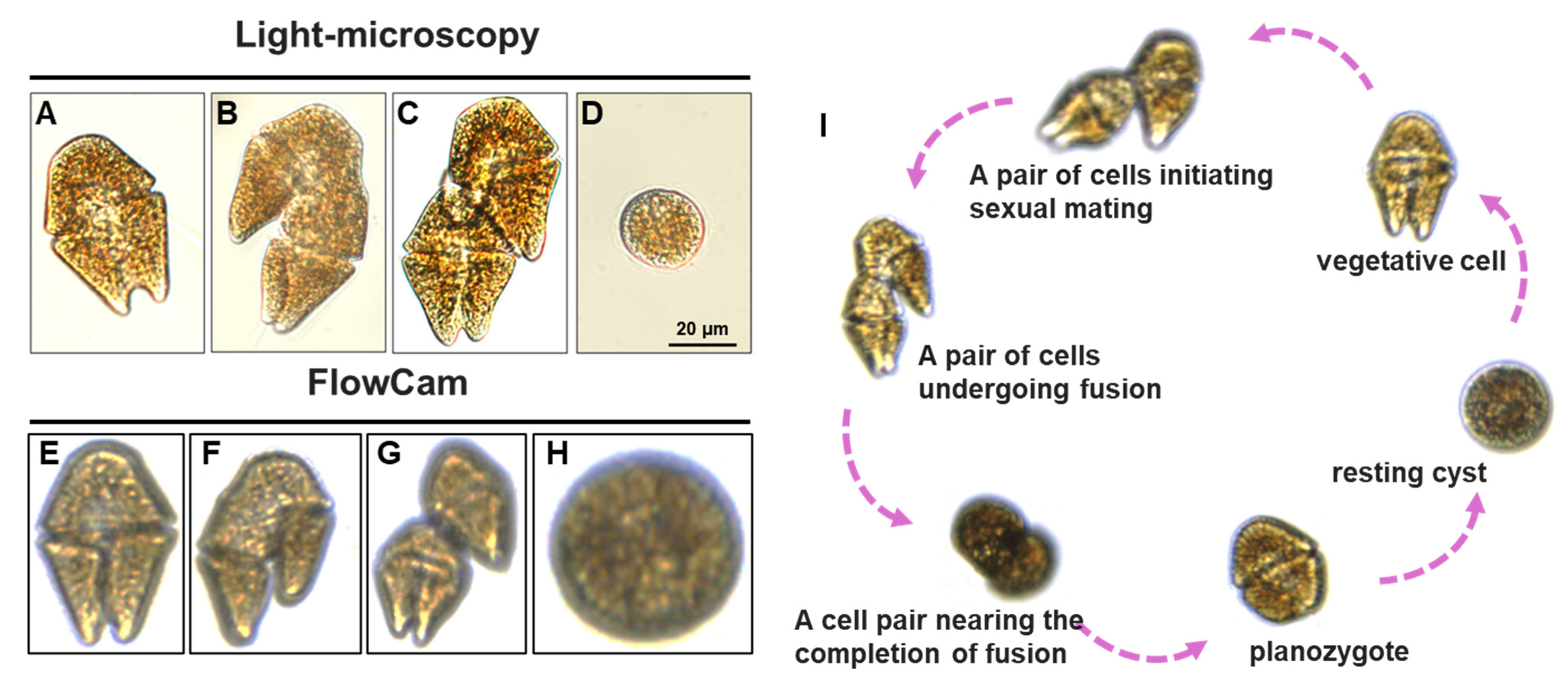
Morphological observations were performed on an inverted microscope at 200× to 400× magnification. Aliquots (2 mL) were settled in Utermöhl chambers for 30 min prior to observation. Vegetative cell dimensions were measured using an ocular micrometer. Resting cysts were identified by their spherical shape and thick wall. Images were recorded with a digital camera.
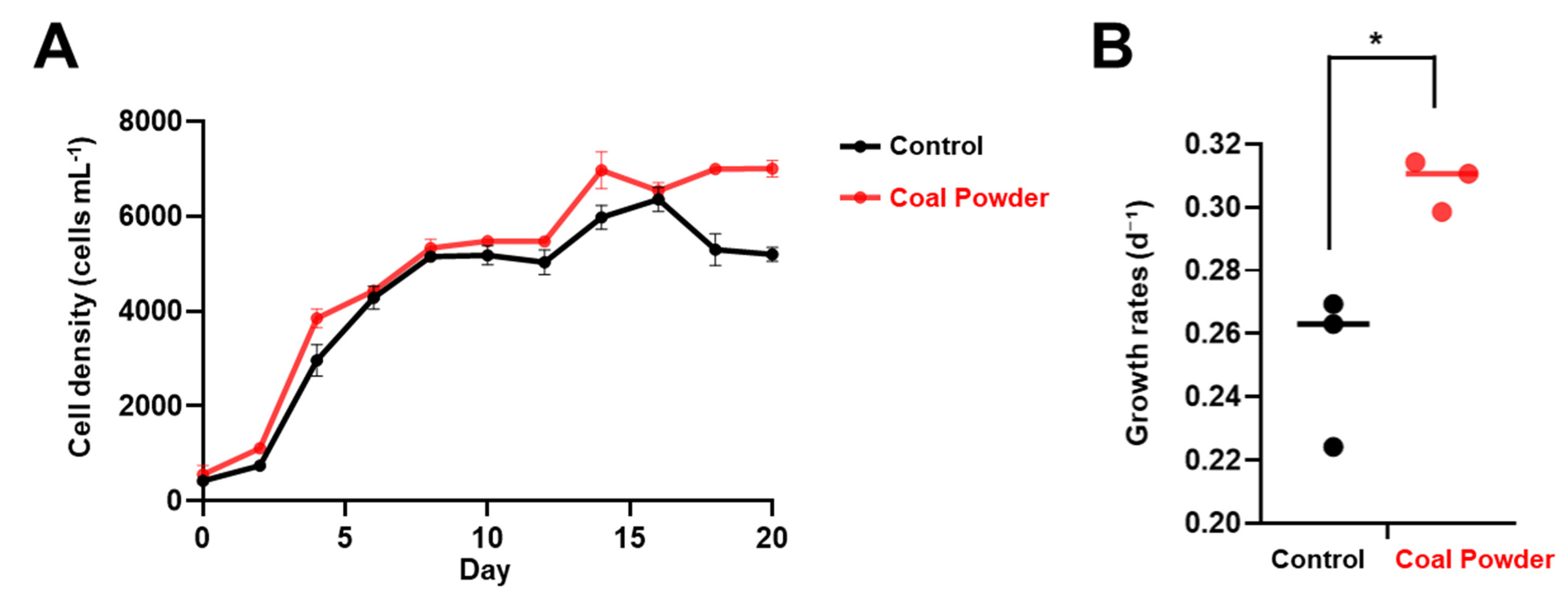
The specific growth rate (µ) was calculated for the exponential growth phase using the formula: µ = (ln(N2) − ln(N1))/(t2 − t1), where N1 and N2 are the cell densities at times t1 and t2.
Then, the Chl
a concentrations were measured with a fluorometer (Trilogy, Turner Design, San Jose, CA, USA) by a fluorometric technique [
19]. To convert fluorescence readings into cell densities, a calibration curve was established prior to the experiment by correlating fluorescence values with direct microscopic counts from a serial dilution of an
A. sanguinea culture (R
2 > 0.99).
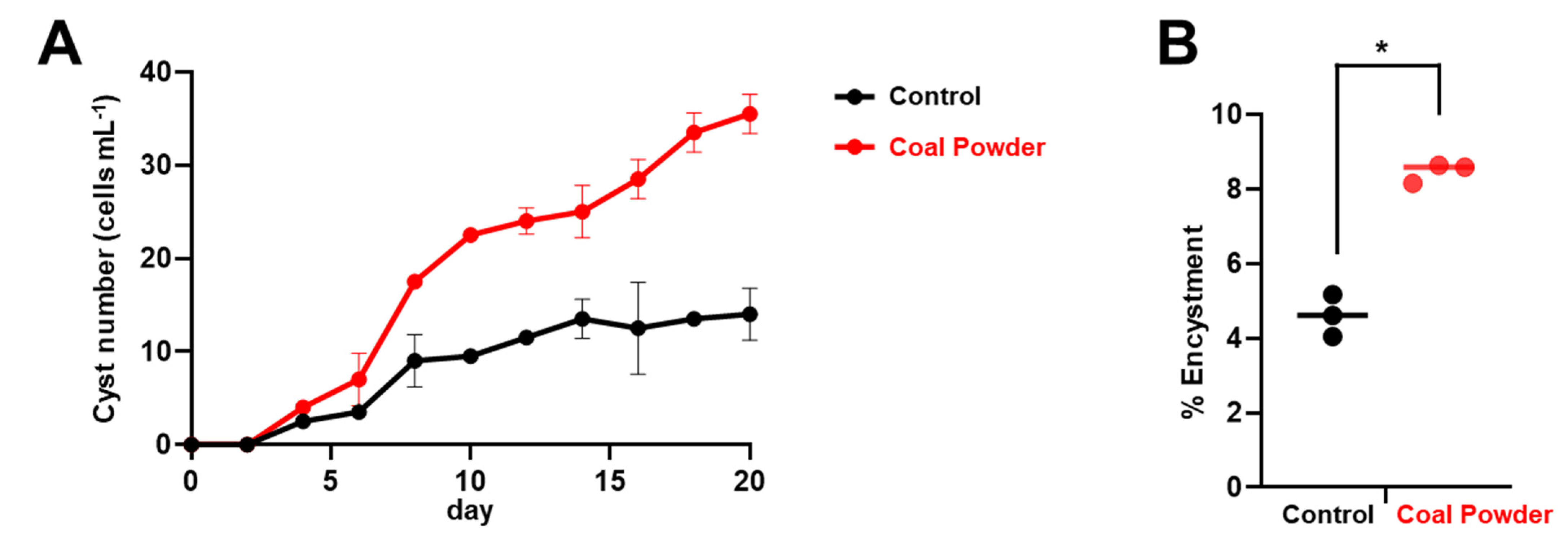
Resting cyst counts were also recorded while monitoring algal growth according to the morphological characteristics described in Chen et al. (2015) [
13]. Resting cysts were identified by their characteristic spherical shape and thick, smooth cell wall. The encystment ratio (S, %) during the experiment was calculated using the following equation:
where C is the cyst density (cysts mL
−1) and M is the vegetative cell density (cells mL
−1).
2.4. Life-Stage Characterization by Imaging Flow Cytometry
Morphological characteristics, size distribution, and life-stage composition of the cultures were monitored every two days throughout the 20-day experimental period using a Benchtop FlowCam Cyano imaging flow cytometer (Yokogawa Fluid Imaging Technologies, Scarborough, ME, USA). This instrument combines flow cytometry with high-resolution digital imaging, enabling qualitative observation and morphological analysis of individual cells [
20]. The FlowCam system was equipped with a 532 nm excitation laser and a color digital camera capable of capturing both brightfield and fluorescence images. For this study, a 10× objective lens was used in combination with a 100 μm flow cell to optimize image quality for
A. sanguinea cells within the typical size range of this species. The system was operated in laser-trigger mode to ensure consistent detection of fluorescent particles while minimizing background interference. At each sampling time point, 1 mL aliquots were withdrawn from each culture bottle using sterile pipettes to prevent contamination. Each sample was processed immediately after collection to minimize potential artifacts from cell settling or behavioral changes. The FlowCam analysis parameters were standardized as follows: flow rate of 0.5 mL min
−1, trigger threshold set to detect particles with equivalent spherical diameter (ESD) ≥ 10 μm, and image capture rate optimized to ensure high-quality images of all detected particles [
21].
2.5. Image Analysis and Morphometric Measurements
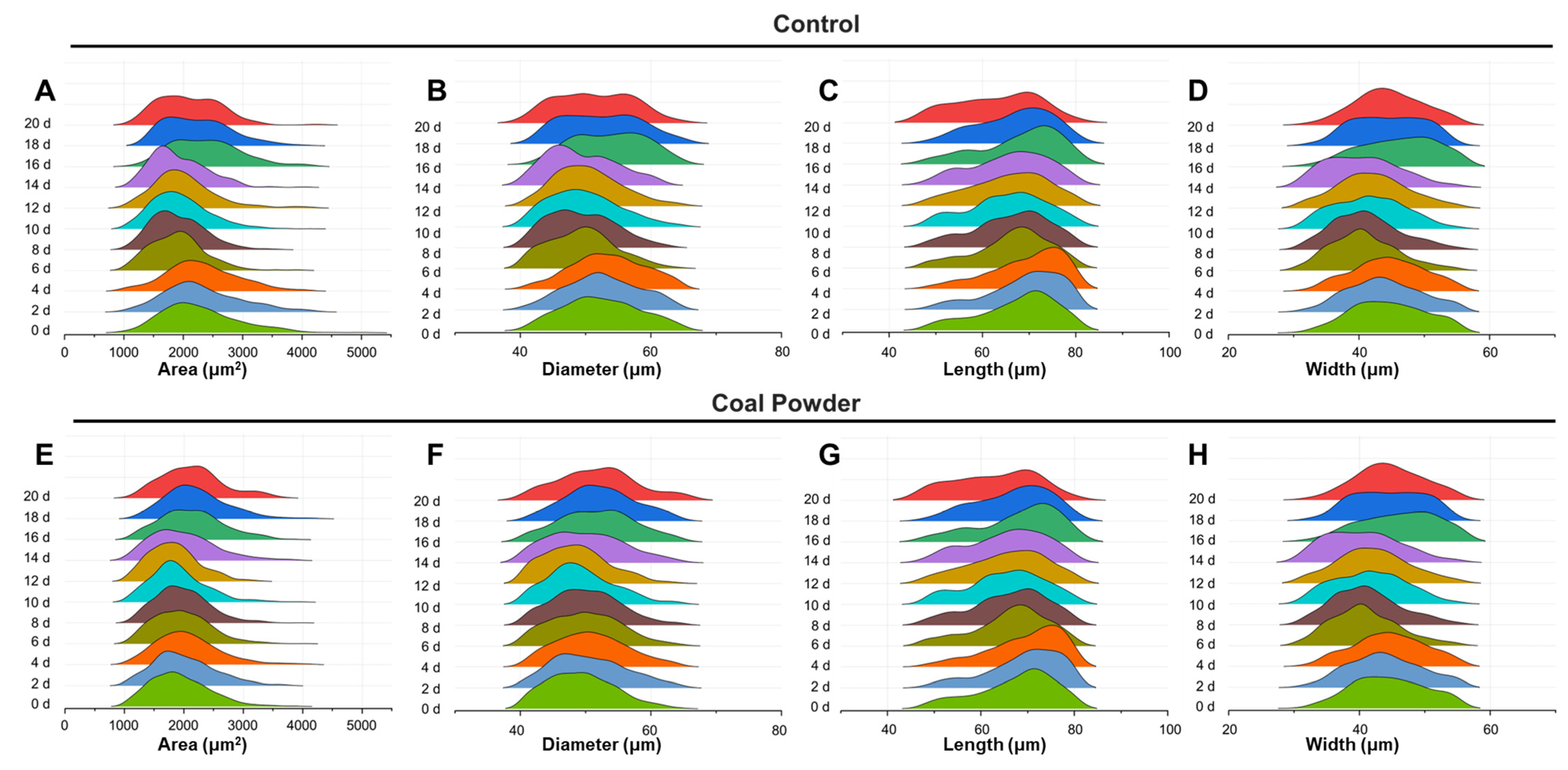
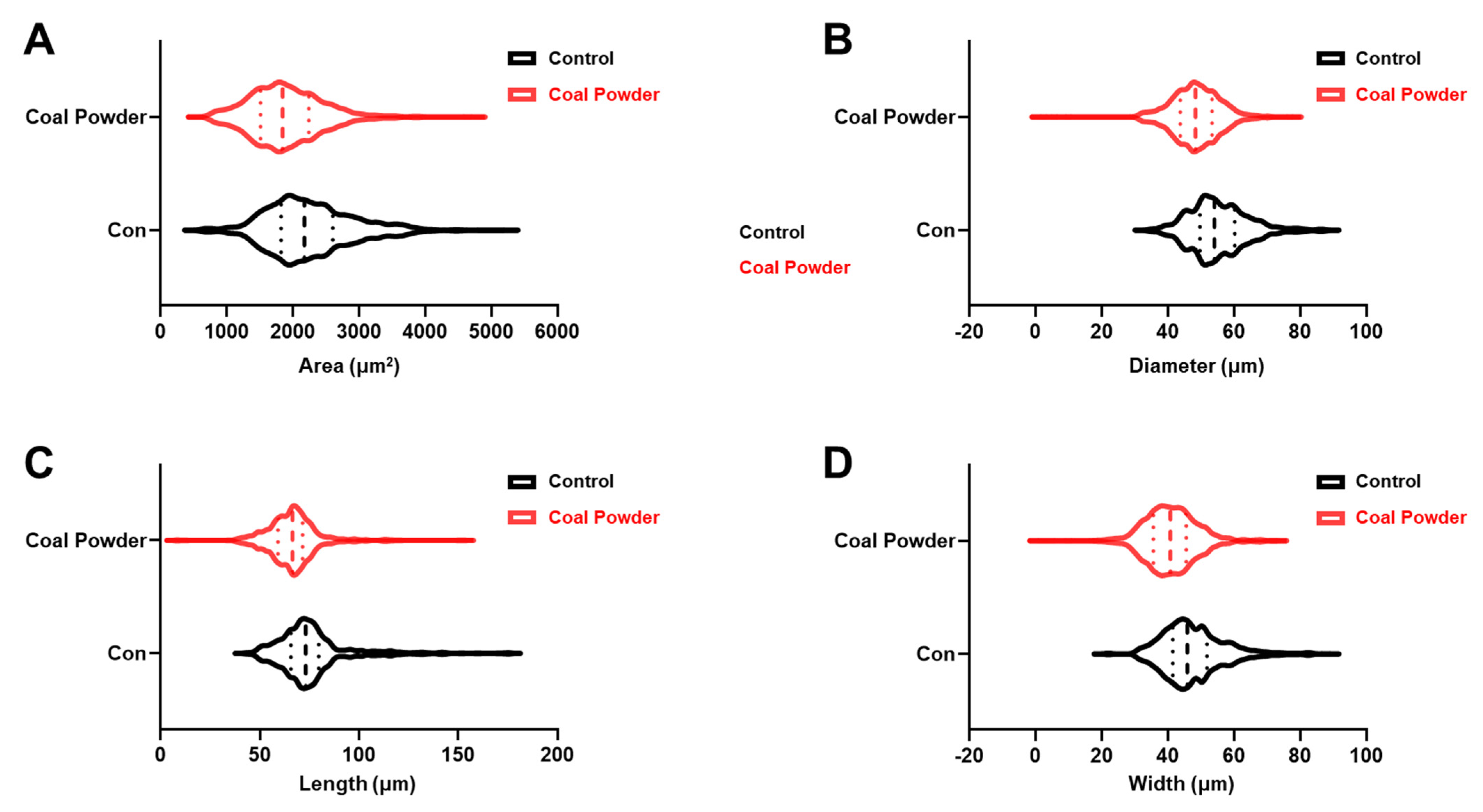
Captured images were processed using Visual Spreadsheet software (Version 6, Fluid Imaging Technologies) to extract morphological parameters for each detected cell. The analysis protocol included automated particle detection followed by manual verification to ensure accurate identification of A. sanguinea cells and exclusion of debris or other particles. For each cell, the following morphometric parameters were measured: diameter, length, width and area. Cell size distributions were analyzed using 5 μm size bins to track temporal changes in population structure. Morphological classification was based on established criteria, including cell shape, size, and fluorescence characteristics.
2.6. Statistical Analysis
All data are presented as mean ± standard deviation (SD) from at least three independent experiments and analyzed by the GraphPad Prism 9.0 software (GraphPad Software Inc., La Jolla, CA, USA). Statistical comparisons between the control and coal powder-amended treatments were performed using a two-tailed Student’s t-test. If the p-value was less than 0.05 (* p < 0.05), the differences were considered statistically significant.
4. Discussion
This study demonstrates that a mineral-rich supplement derived from Taebaek coal powder acts as a potent stimulant for both vegetative growth and sexual encystment in the harmful algal bloom dinoflagellate, Akashiwo sanguinea. Cultures amended with the coal powder extract exhibited significantly higher growth rates, achieved greater peak population densities, and, most critically, produced a substantially higher yield of resting cysts compared to the standard f/2 medium control. These results underscore the pivotal role that mineral-derived micronutrients can play in modulating the life-cycle progression and population dynamics of this key HAB species.
The central finding of this research is the pronounced enhancement of encystment. Notably, the higher cyst abundance and encystment ratio observed here should be interpreted as enhanced net cyst accumulation, rather than definitive evidence of increased sexual induction efficiency. Although we morphologically observed sexual stages (e.g., paired cells/planozygote-like forms) by microscopy/FlowCam, we did not quantify key sexual reproduction dynamics such as gamete pairing efficiency or planozygote formation rates, nor did we assess cyst survival. Therefore, while the coal extract clearly increased the final number of morphologically defined resting cysts, the specific step(s) of the sexual cycle that are affected remain to be resolved. The transition from a motile, vegetative state to a dormant, benthic cyst is a cornerstone of the ecological strategy of many dinoflagellates, yet the precise environmental cues that govern this process are multifaceted. While macronutrient limitation, especially of nitrogen, is a well-documented trigger for encystment in
A. sanguinea [
13], our findings reveal that the mineralogical context is also a critical regulatory factor. However, because the culture-medium chemistry was not characterized (i.e., dissolved macronutrients and dissolved trace/heavy-metal concentrations were not quantified), the present study cannot provide preliminary validation for micronutrient modulation or metal-associated stress as causal drivers; therefore, these mechanistic explanations are advanced as working hypotheses rather than evidence-based conclusions.
One plausible mechanism is the alleviation of trace metal limitation. Standard f/2 medium, while robust, may not perfectly replicate the complex suite of micronutrients available in natural coastal waters. Trace metals such as iron (Fe), manganese (Mn), zinc (Zn), and selenium (Se) are indispensable for phytoplankton, serving as essential cofactors in a vast array of metabolic pathways, including photosynthesis, respiration, and nitrate assimilation [
16]. The silicate-rich Taebaek coal powder likely leached a cocktail of these bioavailable trace elements into the medium. The observed increase in vegetative growth rates and overall biomass in the amended cultures strongly supports this hypothesis, as it points to a direct stimulation of cellular metabolism, likely by satisfying a latent micronutrient demand. For instance, Fe is a notorious limiting nutrient in many marine systems, and its role in the photosynthetic apparatus is paramount. Similarly, Mn is a core component of the oxygen-evolving complex in Photosystem II, and Se is known to be required by some dinoflagellates for optimal growth.
Alternatively, the increased cyst accumulation is consistent with a stress-associated shift in life-history allocation. While trace metals are required at low levels, elevated concentrations of certain metals can impose physiological constraints, including oxidative stress and cellular damage [
16], and have been discussed as potential triggers of encystment in dinoflagellates under specific chemical regimes [
16]. Under this framework, coal-derived dissolved constituents could, in principle, contribute to conditions under which enhanced growth and resting-stage formation co-occur. However, in the absence of measurements of dissolved trace-metal concentrations/speciation, macronutrients, and cellular stress indicators, support for a stress-mediated pathway remains indirect, and the mechanism underlying the observed response cannot be resolved in the present study.
A third possibility involves the indirect alteration of macronutrient dynamics. The fine mineral particles of the coal powder could have acted as surfaces for the adsorption of dissolved inorganic nitrogen from the medium. This process would accelerate the onset of nitrogen limitation within the culture, which is a known, potent trigger for encystment in
A. sanguinea [
13]. This pathway constitutes a plausible, non-exclusive hypothesis whereby initial growth stimulation may accelerate macronutrient drawdown and, potentially in conjunction with adsorption, bias cultures toward nitrogen limitation-associated sexual induction and resting cyst formation; importantly, any application-oriented extrapolation would need to explicitly account for heavy-metal contamination and nutrient-regime perturbation (including eutrophication), which were not evaluated in this study.
It is noteworthy that these significant physiological shifts occurred without a major alteration in cell size. The FlowCam imaging confirmed that the cell area distribution remained stable, indicating that the coal powder amendment influenced the rate of cell cycle progression and the direction of the life-history pathway, rather than general cell enlargement. This suggests a targeted regulatory effect on the cellular machinery controlling division and sexual induction, a phenomenon that merits further molecular investigation.
From a methodological standpoint, this work validates the power of combining traditional fluorometric quantification with high-throughput flow imaging. While fluorometry provided robust quantitative data on population growth, the FlowCam offered valuable qualitative insights into population structure and life-stage transitions with a speed and detail that would be unattainable with manual microscopy alone, offering a comprehensive view of the dynamic changes in the cultures.
In conclusion, this study demonstrates that supplementing standard f/2 culture medium with an extract from Taebaek coal powder significantly promotes both the vegetative growth and, more importantly, the sexual encystment of the dinoflagellate Akashiwo sanguinea. The mineral amendment resulted in higher cell densities and a substantially greater yield of resting cysts compared to the control. The underlying chemical drivers remain unresolved because dissolved macronutrients and dissolved trace/heavy-metal concentrations in the culture medium were not quantified; accordingly, micronutrient enrichment, metal-associated stress, and altered macronutrient availability should be interpreted as alternative hypotheses rather than validated mechanisms. Taken together, our axenic laboratory experiments show that supplementation with the Taebaek coal extract increased vegetative cell density and, in parallel, markedly elevated resting cyst yields in A. sanguinea. These results indicate that coal-derived mineral cues can modulate both growth and the life-cycle transition toward encystment under controlled conditions. However, several limitations of the present study should be acknowledged to guide future research. Primarily, the use of a single concentration of the coal powder extract prevents a clear distinction between the dual hypotheses of micronutrient stimulation and concentration-dependent physiological stress. Future investigations should prioritize a dose–response experimental design to decouple these potential mechanisms. Additionally, this study was conducted with a single axenic clonal strain, which limits the generalizability of our findings to genetically diverse natural populations. Finally, as cyst viability and germination were not assessed, our results reflect net cyst accumulation rather than the production of viable resting cysts. Incorporating dose–response relationships, strain diversity, and viability assays will be critical for advancing the understanding of how mineral inputs regulate dinoflagellate life cycles.