Article

Assessment of the Ecotoxicity of Marine Sediments from the Eastern Kamchatka Using Bioassays

by
Valentina Vladimirovna Slobodskova
1,
Victor Pavlovich Chelomin
1,
Sergey Petrovich Kukla
1,*,
Andrey Alexandrovich Mazur
1,*,
Nadezhda Vladimirovna Dovzhenko
1,
Aleksandra Anatolyevna Istomina
1 and
Elena Vladimirovna Zhuravel
2
1
V.I. Il’ichev Pacific Oceanological Institute, Far Eastern Branch of the Russian Academy of Sciences, Baltiyskaya, 43, 690041 Vladivostok, Russia
2
Institute of the World Ocean, Far Eastern Federal University, Ayaks, 10, Russky Island, 690922 Vladivostok, Russia
*
Authors to whom correspondence should be addressed.
J. Mar. Sci. Eng. 2025, 13(10), 1891; https://doi.org/10.3390/jmse13101891
Submission received: 4 September 2025 / Revised: 22 September 2025 / Accepted: 28 September 2025 / Published: 2 October 2025
(This article belongs to the Special Issue Ecological Risk Assessments in Marine Pollutants)

Abstract

Because of the active influx of chemical compounds into the marine environment, a significant portion is transformed and accumulates in bottom sediments (BS), posing a threat to benthic organisms. The eastern coast of the Kamchatka Peninsula, with its characteristic volcanic, seismic, and gas–chemical features, is of particular interest to our research. This study is the first to assess the cyto- and genotoxicity of BS in coastal waters off the eastern coast of the Kamchatka Peninsula using biotesting on representatives of the benthic community (the mussel Mytilus trossulus and the sand dollar Scaphehinus mirabilis). Of the aqueous extracts exposure of BS from all stations, M. trossulus showed destabilization of lysosomal membranes in gills and digestive gland cells. It was shown that aqueous extracts from BS of Kamchatka Bay (station 1) and Avachinskaya Bay (station 3) had no negative effect on DNA molecules in the gills and digestive gland cells of mussels, and the values obtained corresponded to the control. Extracts from BS of Kronotsky Bay (station 2) and Avacha Bay (station 4) damaged the integrity of the genome in the cells of the gills M. trossulus and sperm of S. mirabilis. The level of DNA damage in sperm increased by 3 and 3.5 times, respectively, compared with that in control gametes. The results of the biotests on two biological models show that sediments from Kronotsky Bay and Avacha Bay exhibit cytotoxicity and induce DNA damage in both somatic cells and gametes.

1. Introduction

Over time, a variety of chemical compounds of natural and anthropogenic origin end up in the marine environment in various ways, where they are actively involved in various physicochemical and biological processes. As a result, a significant portion of these chemical compounds is transformed, deposited, and accumulated in bottom sediments (BS). In this regard, marine sediments are a kind of final reservoir and storage of numerous anthropogenic pollutants, including persistent and highly toxic polycyclic aromatic hydrocarbons (PAHs), halogen-containing pesticides, heavy metals, etc. [1,2,3,4]. Complex processes of deposition of organic and inorganic pollutants contribute to the purification of water masses, where the resulting contaminated sediments pose a threat to benthic organisms. Toxic compounds present in polluted sediments can be slowly released and re-enter the water column under the influence of complex biological, hydrochemical, and hydrodynamic processes [2,5], posing a serious threat not only to benthic [2,6,7,8], but also to planktonic organisms [9,10]. Therefore, unlike coastal waters, BS studies provide a more stable and realistic approach to studying the history of pollution and related adverse effects on aquatic ecosystems. According to experts in this field, the identification of toxic substances by chemical analysis is insufficient to assess the true toxic potential of sediments due to possible additive, synergistic, or antagonistic interactions of various types of chemical compounds [11,12]. In addition, chemical analysis does not allow the assessment of the bioavailability of inorganic and organic pollutants present in BS, which introduces uncertainty in the prediction of potential toxicity and the likelihood of their transport through trophic networks [13,14].
In contrast, a complex analysis based on the reaction of biological systems may reflect the integral toxic effects of all pollutants present in the samples. In these cases, the most reliable method of assessing the condition and predicting the quality of sediments is the method of biotesting. When in the laboratory using live test objects, it is possible to unlock the full potential of contamination, contributing to bioavailability, for example, by resuspending or extracting sediments [15,16,17]. The assessment of sediment pollution using various biotesting techniques is widely used in environmental monitoring to characterize the environmental risks posed by polluted water sediments and to take scientifically sound protective measures [10,18,19].
Considering that many chemical pollutants cause genotoxicity and mutagenicity or are potentially carcinogenic, the characterization of the genotoxic potential of aquatic sediments has become widespread in biotesting methods using bacteria, invertebrates, and fish and is included in various biomonitoring programs [7,9,12,14,20]. The potential of sediments genotoxicity can be assessed using the DNA comet assay by analyzing the DNA damage of animal cells exposed to BS extracts [21,22,23,24]. Such laboratory analyses are especially useful when the main goal is to assess the ecotoxic potential of BS in areas with unstable marine environments affected by a complex of anthropogenic and abnormal natural phenomena.
In this regard, the waters of eastern Kamchatka are of particular interest. The remoteness of Kamchatka from the mainland has contributed to the preservation of its unique nature and wild ecosystem. However, this isolation also creates difficulties for the full study and development of the region. The coastal waters of Kamchatka have significant and diverse natural resource potential, the preservation of which is one of the main tasks of the socio-economic development of the Kamchatka. This poorly studied region is subject to active volcanic and seismotectonic processes with numerous gas–fluid emissions, which can have a negative impact on the marine environment on both local and regional scales. The ecological disaster of September 2020, which led to the mass death of bottom marine organisms, confirmed the instability of the region’s marine waters. The causes of this are still not entirely clear; however, it is assumed that it was caused by the bloom of toxic microalgae. According to experts, the mass death of animals has caused significant damage to the benthic communities of the eastern coast of Kamchatka [25,26,27]. Therefore, a comprehensive study of the prerequisites for the development of such ecological disasters will enable the prediction of the sustainability of the coastal ecosystems of the eastern coast of Kamchatka. As the first stage of systematic studies of this unexplored region, we assessed the genotoxicity of BS in the coastal waters of the eastern coast of Kamchatka using two biological models: the benthic filter-feeding mollusk Mytilus trossulus and the sperm of the sand dollar Scaphehinus mirabilis.

2. Materials and Methods

2.1. Sampling

Surface sediments were collected with a bottom sediment catcher type Van Veen in the water areas of the east coast of Kamchatka in October 2023: in Kamchatka Bay (station 1), Kronotsky Bay (station 2), Avachinsky Bay (station 3) (in the area of the ecological disaster in September 2020), and in Avacha Bay (station 4) (Figure 1). The sampling coordinates are provided in Supplementary File Table S1. Three samples were collected from each station in three replicates (n = 9).
Under laboratory conditions, water extracts were obtained from all BS samples, which were further used for biotesting. To prepare the aqueous extract, the BS was transferred into a wide-mouth flask for 750–1000 cm3, and filtered and UV-treated seawater was added in the ratio of 1:4. The flask with a closed stopper was shaken for 2 h, followed by settling for a day. The extract was filtered through a funnel 15 cm in diameter and a large pleated filter placed in it. All the filtrates were transparent [28].

2.2. Biotest Conditions

To assess the quality of BS of Kamchatka coastal water areas, the biotesting method was applied [19]. Mussels (Mytilus trossulus) were kept for 5 days in samples of water extracts obtained from all submitted samples. The control groups were maintained in filtered seawater. Sperm sampled by the “dry method” from sand dollars (Scaphechinus mirabilis) were exposed to short-term (1 h) exposure to the obtained aqueous extracts.
In experiments involving mussels, two biochemical indicators were used as bioindicators: DNA damage and membrane stability. In experiments involving sand dollar sperm, DNA damage was determined.
Lysosome membrane stability analysis is based on the retention of the vital neutral red (NR) dye [8]. The dye is a lipophilic chemical substance that passively diffuses through the cell membrane. The efficiency of neutral red retention over time depends on the state of the lysosome membrane.
The amount of DNA molecule damage was determined using the alkaline version of the comet assay, which allows us to assess the genome state of an individual cell [29].
The visualization and registration of DNA-comets were performed using a fluorescence microscope (Zeiss, Oberkochen, Germany, Axio Imager A1) equipped with an AxioCam MRc digital camera. The computer program Casp 1.2.2 (CASPlab, Wrocław, Poland) was used to process the digital images, which allows the calculation of various parameters of comets indicating the degree of cellular DNA damage. For each comet, the proportion of DNA in the comet tail (% DNA in the comet tail) was determined.

2.3. Statistical Analysis

Statistical processing of the results was performed using Statistica 7 (StatSoft, Tulsa, OK, USA). Each experiment was repeated three times. The Mann–Whitney U test for non-parametric variables was used to assess the reliability of parameter changes. Significance was established at p < 0.05.

3. Results

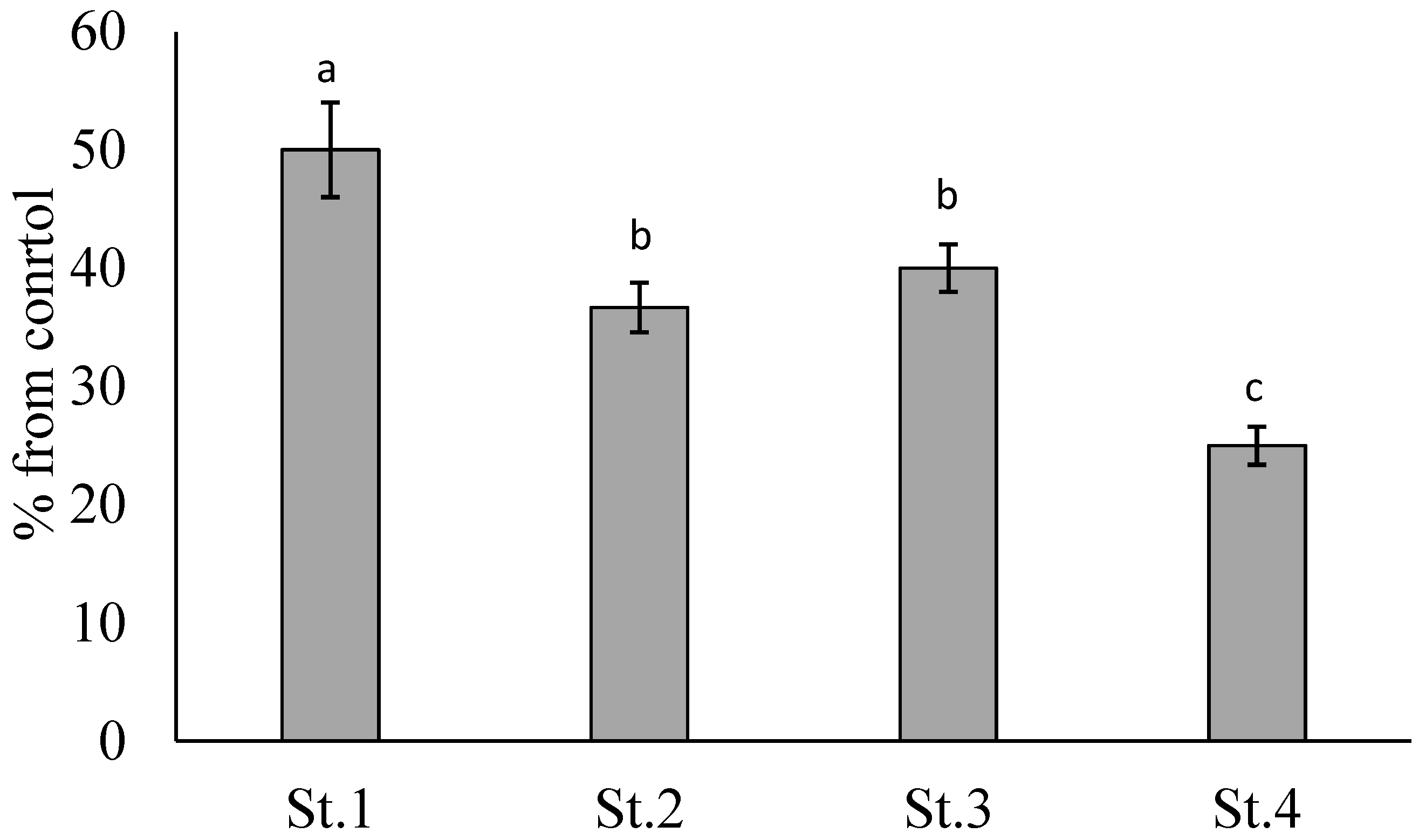
The results of determining the stability of the membranes of lysosomes of mussel hemocytes, presented in Figure 2, showed that all the studied samples contained xenobiotics that significantly reduced the retention time of the dye in these subcellular structures.
Thus, the bottom sediment extracts from stations 4 (Avacha Bay) and 2 (Kronotsky Bay) exhibited higher toxic effects on lysosome membrane stability. At these stations, this indicator decreased to 25 ± 1.6% and 36.7 ± 2.1%, respectively, relative to the control values. Extracts from bottom sediments at stations 3 and 1 had a lesser effect, reducing the parameter under study to 40 ± 2% (Avachinsky Bay) and 50 ± 4% (Kamchatka Bay), respectively. Notably, the effect was most pronounced when exposed to BS extracts from Avacha Bay. In experimental mussels exposed to BS extracts from this bay, the retention time of the dye in lysosomes decreased by almost 3 times compared with control mollusks.
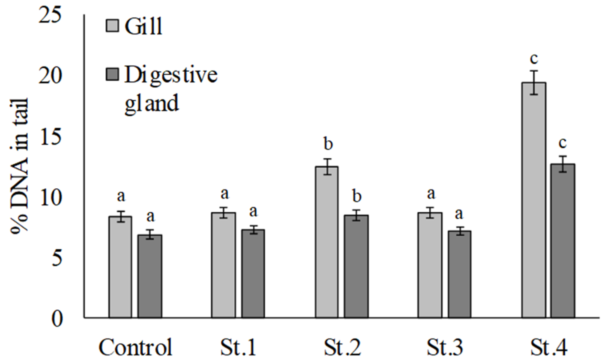
The comet assay allowed us to assess the level of DNA damage in the cells of Mytilus trossulus tissues (Figure 3) and in sand dollar sperm (Figure 4) after exposure to aqueous BS extracts obtained from the waters of the eastern coast of Kamchatka. In experimental mollusks exposed to extracts from the BS of Kamchatka Bay (st. 1) and Avachinsky Bay (st. 3), a small level (7–8%) of nuclear DNA fragmentation is observed in the cells of the gills and digestive gland, corresponding to control mollusks, which is formed in the cells during the functioning of life-support systems due to the accumulation of alkali-labile regions and/or single- and double-strand breaks. Exposure of mussels to a medium containing extracts obtained from the BS of Kronotsky Bay and Avacha Bay resulted in a significant increase in the level of DNA damage, mainly in gill cells (12.44 ± 1.22% and 19.39 ± 1.71%, respectively), indicating the initiation of damage to the integrity of the genome (Figure 3).
A similar trend was observed during biotesting of extracts from BS of the studied water areas using the sand dollar spermatozoa as a biological model. From the results of biotesting, it follows that the BS of all the studied water areas contain chemical components that can increase the fragmentation of the DNA molecule to varying degrees. It is noteworthy that, as in the experiments with the mussel, the greatest degree of DNA damage was observed in gametes after short-term exposure to extracts of BS from Avacha Bay (9.72 ± 0.44%) and Kronotsky Bay (10.24 ± 0.57%). In this case, the level of DNA damage in sperm compared to control gametes increased by 3 and 3.5 times, respectively (Figure 4).
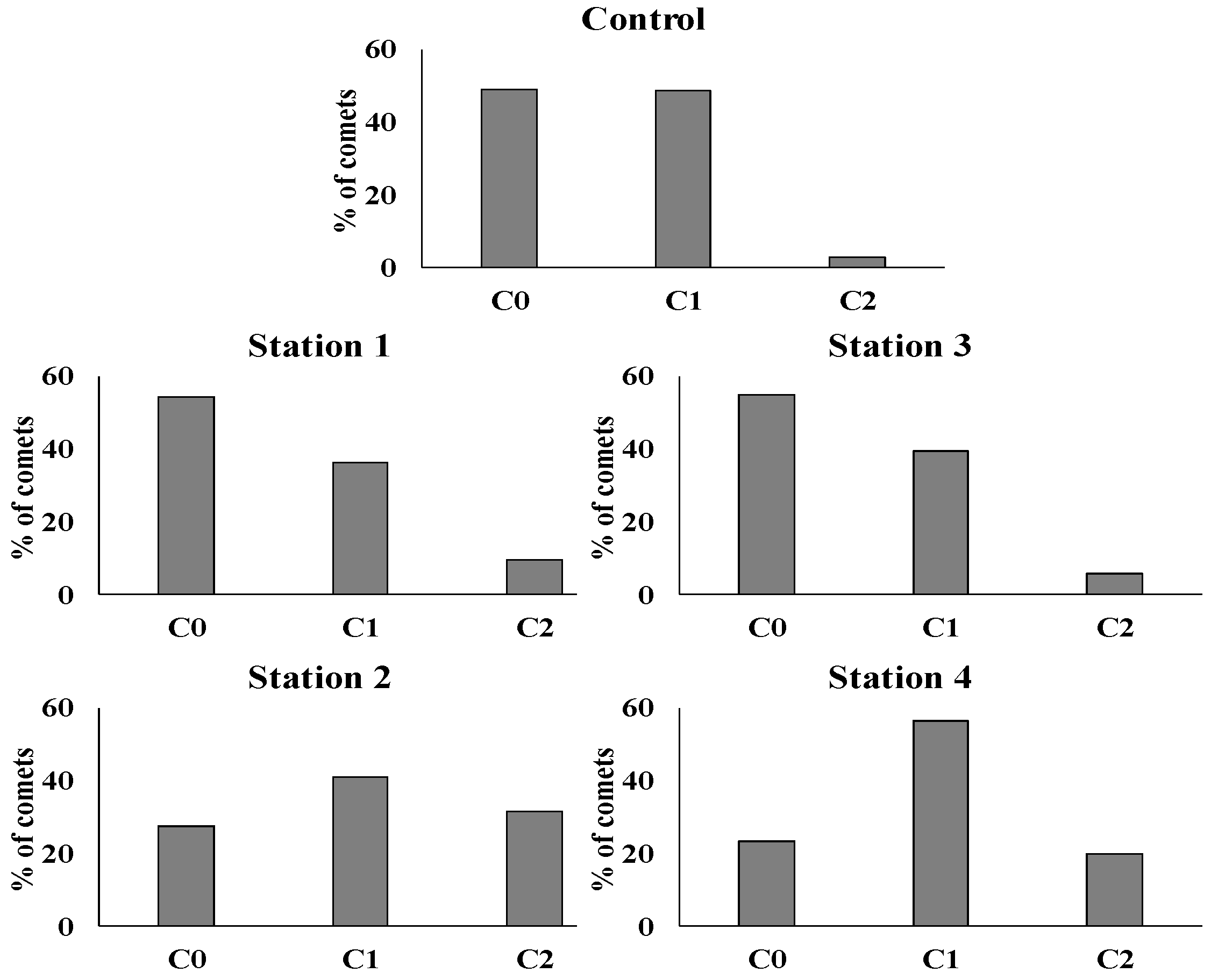
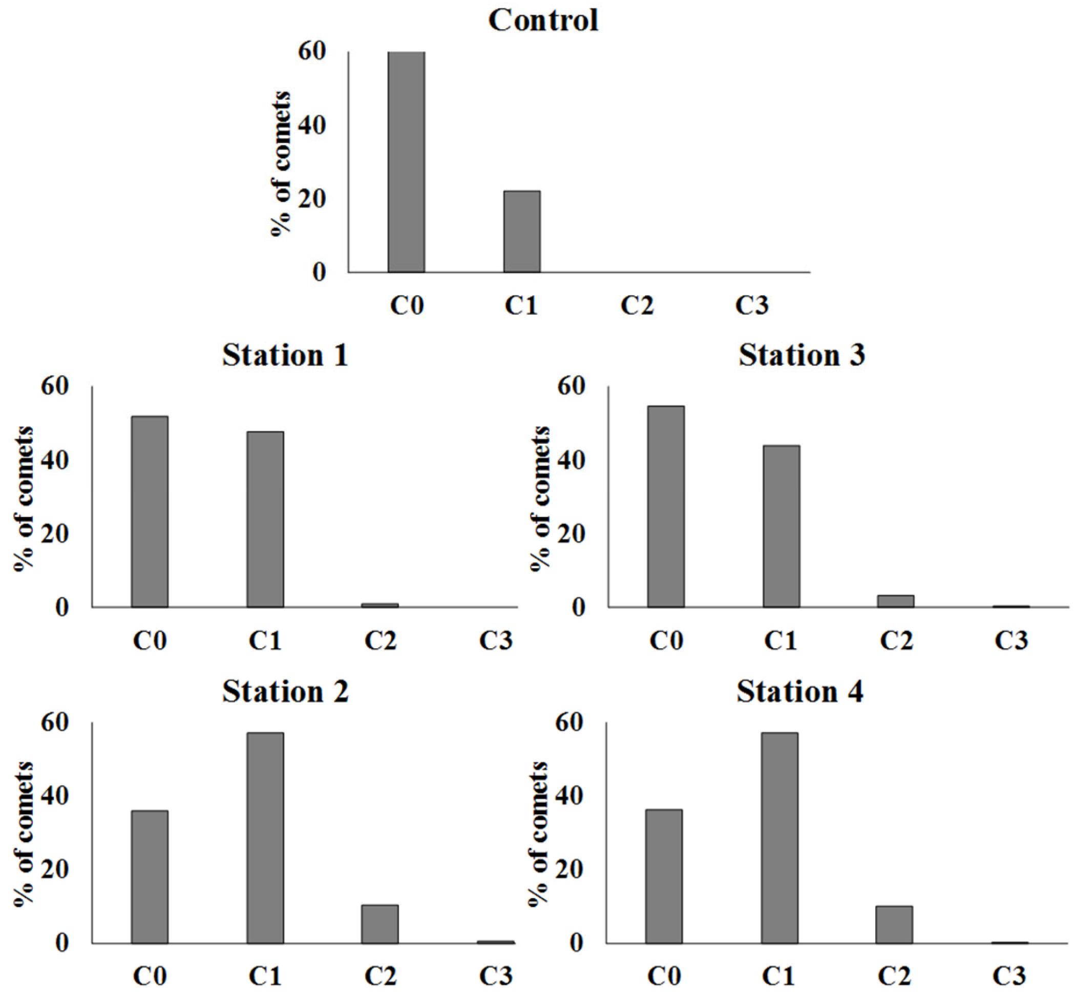
Analysis of the distribution of comets by the degree of DNA destruction, grouped into classes, as suggested by Cavaş and colleagues [30], showed that in all mollusks, in the cells of both tissues, comets of the C0 (0–5% DNA in the comet tail) and C1 (5–20%) classes, characteristic of intact and viable cells, predominated. The total number of comets of these classes (C0 + C1) was approximately 98%, while the share of comets with a DNA damage level of more than 20% (C2 class) accounted for no more than 2%. In addition, changes in the ratio of the proportion of comets of classes C0 and C1 were observed in the experimental mussels, but the content of intact cells did not change, and there was no increase in the number of comets of class C2 (Figure 5 and Figure 6).
Based on the obtained data on the distribution of comet DNA by class, it is evident that in the sperm samples of all the studied groups, as well as in mussels, comets belonging to classes C0 and C1 predominated. At stations, 2 and 3, an increase in comets of class C2 is observed (Figure 7).

4. Discussion

In our study, we used typical representatives of the Far Eastern marine fauna, which, due to their high sensitivity to pollution, are widely used in various ecotoxicological programs [28,31,32,33]. The mussel Mytilus trossulus is a typical eurybiont representative of filter-feeding mollusks. With this method of nutrition, not only dissolved organic and mineral nutrients enter the body through the gills, but also relatively small (up to 10 µm) organo-mineral particles. The sand dollar Scaphehinus mirabilis is also an epibenthous species, and during the spawning period it releases gametes directly into seawater, where they are exposed to various chemicals. Gametes and larval stages of various aquatic organisms, especially sea urchins, are actively used as unique models for assessing water pollution and BS, which is an important direction in modern ecotoxicology [34,35,36,37,38,39]. Spermatozoa, unlike somatic cells, are more susceptible to genotoxicants, because they are practically devoid of the ability to repair DNA and have weak antioxidant activity and biotransformation systems [35,40,41,42]. Moreover, biotesting on sea urchins showed that their sensitivity to BS quality is higher than that of many other models [43].
We conducted biotesting experiments in the laboratory using aqueous extracts from sediment samples. During this treatment, a complex of hazardous chemicals that tend to be adsorbed on solid particles and precipitate with them are released into seawater and become bioavailable to benthic and planktonic organisms. This approach is widely used to assess the potential toxic hazard of sediments [10,20,44,45,46,47]. At present, highly sensitive physiological and biochemical methods are actively introduced into ecotoxicological practice, two of which—the stability of lysosome membranes and DNA damage in individual cells—were used in this study. The present results confirm the high sensitivity of this subcellular biomarker, which has been repeatedly emphasized in various ecotoxicological studies [8,18,48,49,50,51,52]. This is especially evident in the example of mollusks, in which pollutants of various nature, present in BS at low concentrations and short-term exposure, caused destabilization of lysosomal membranes [8,53,54,55,56].
Without diminishing the importance and information content of the cytochemical marker, special attention should be paid to damage to the DNA molecule because this marker is not only diagnostic but also prognostic in nature. The comet assay is widely used to assess the toxic effects of various pollutants on marine organisms [8,12,18,21,23] and is the most informative method for cell-by-cell recording of nuclear DNA integrity, allowing the assessment of changes in the cell nucleus at the early stages of DNA damage [57,58]. According to experts, this molecular approach is tens of times more sensitive than any biomarker used to assess the degree of toxicity at the organism level [59]. The increase in the level of damage to nuclear DNA indicates that the repair system cannot cope with the rate of destructive changes in the genome induced by chemicals present in the sediments. In addition, there are concerns that DNA damage may trigger cell death mechanisms (e.g., in mussel somatic cells) or affect reproduction by initiating abnormalities in embryonic and larval development (e.g., after fertilization of eggs by sperm with DNA damage). It is worth noting that gill cells showed higher levels of DNA damage than digestive gland cells, which is apparently related to the mussels’ feeding and breathing patterns. When filtering, a large amount of polluted water passes through the gills and, accordingly, the gill cells come into direct contact with toxic compounds. At the same time, mussels can absorb a small amount of microparticles from BS directly through the mouth.
In support of our results, we can cite a number of studies in which, based on the determination of genotoxic properties (using comet assay), BS with unfavorable properties in this regard were identified in a number of marine areas, including estuaries [5,6,7,12,14,21,60]. In this regard, genotoxic potential analyses have proven to be particularly productive and informative when toxic substances are present in sediments at low concentrations. Thus, in mollusks (Manila clam, Tapes semidecussatus), damage to the genome was detected after exposure to BS collected not only in the polluted area of the Douglas River estuary but also in the relatively clean area of the Ballymacoda River estuary [6]. In addition, Pinto et al. [12] showed that in the Sado estuary (Portugal), which is classified as moderately polluted overall, sediments from an industrial area were more genotoxic than those from an agricultural area. The high sensitivity of the comet assay is demonstrated by the results of a study by Jeong et al. [61], which revealed the genotoxic properties of BS in the water area 5 years after an oil spill.
Even such a short list of examples, along with our results, provide grounds to believe that the use of cyto- and genotoxicity markers in assessing the ecotoxicological hazard of sediments is relevant and informative. The results on the potential risks of BS from Avacha Bay (station 4) are a good example of the use of markers to characterize BS in water areas subject to anthropogenic pressure. High toxicity in areas of anthropogenic activity is most often associated with the impact of potentially toxic compounds [62]. However, organisms in such water areas will be exposed to the cumulative toxic effects of a complex of various pollutants, ranging from radionuclides, petroleum products, and heavy metals to microplastics and pathogenic bacteria [63,64]. Thus, semi-closed Avacha Bay with weak water exchange is characterized by a higher level of pollution in the open bays of the eastern coast of Kamchatka. This is due to the fact that two rivers (Avacha and Paratunka) flow into the water area, and on the banks there are two cities (Petropavlovsk-Kamchatsky and Vilyuchinsk), industrial enterprises, and a port serving numerous fishing and cargo vessels. The higher cyto- and genotoxic responses of the two biological models used in our study to the exposure of BS of Avacha Bay are obviously due to the influx of pollutants from river waters, domestic and industrial wastewater, as well as a result of oil leaks from ships and intensive economic activity on the coast.
However, in the case of sediments of the bays of eastern Kamchatka (Kamchatsky, Kronotsky, and Avachinsky Bays) we have a different picture. Owing to the low population density and remoteness from large industrial and technical enterprises, the eastern coast of Kamchatka does not experience noticeable anthropogenic pressure and preserves a unique natural environment. However, in this poorly studied region, natural phenomena are constantly recorded that can negatively affect the stability of marine ecosystems. The marine shelf and adjacent waters are exposed to potential threats from abnormal natural factors such as seismic activity, volcanic activity and numerous gas–fluid emissions. The leaching of volcanic ash that enters the marine environment via river flows and atmospheric transport leads to the enrichment of waters with biogenic elements, which in turn can serve as a stimulus for large-scale microalgal blooms, organic matter formation, and eutrophication. Thus, Semkin et al. studied the rivers of this region and concluded that the main source of biogenic substances in them, and as a result of eutrophication in coastal areas, is volcanic, not anthropogenic activity [65]. The large-scale bloom of toxic microalgae that caused mass mortality in September 2020 is associated with such eutrophication [25]. However, in order to establish the causes of large-scale microalgae blooms, additional regular monitoring studies are required to record the precursor signals of such phenomena. Furthermore, a number of model organisms have shown that sulfur compounds can be potentially toxic to aquatic organisms [66,67]. Given the accumulation capacity of BS, there is reason to believe that these periodically occurring negative events can affect the quality of sediments, both locally and regionally. Extreme natural factors play an important role in understanding the possible causes of cytotoxicity of BS in these water areas and, especially, genotoxicity of BS in Kronotsky Bay (station 2).
The chemical composition of the sediments and the concentrations of these compounds that pass into solution and become bioavailable during the resuspension process were not investigated. We proceeded from the fact that for a reliable assessment of ecotoxicological hazard, the primary importance is not so much the presence of hazardous chemicals in BS but their biological effects. Risk assessments based on chemical analysis of sediments can be misleading and do not reflect true toxicity. However, chemical analyses can complement the results of bioassays and play a leading role in identifying sources of hazardous chemical compounds and mechanisms of toxic effects. Bioassays, especially those using highly sensitive and informative methods such as LMS and comet assay, can be considered as the most adequate tool for predicting risks to aquatic biota.

5. Conclusions

Based on the biotesting of the two biological models, there is every reason to assert that the sediments of Kronotsky Bay and Avacha Bay most likely contain relatively accessible chemical components that exhibit cytotoxicity and induce genome destabilization in both somatic cells and gametes.
In general, our results, although preliminary, nevertheless revealed an additional problem of potential toxic risk, which can come from both marine sediments located in the zone of human influence, and sediments formed under the influence of natural factors. To understand the causes of disasters such as the one that occurred in Kamchatka in 2020, further regular comprehensive monitoring programs are needed, including hydrochemical, hydrological, and biological diagnostic methods. Such programs will make it possible to predict the sustainability of the unique marine ecosystem of the eastern coast of Kamchatka and contribute to the preservation of its high biodiversity.

Supplementary Materials

The following supporting information can be downloaded at: https://www.mdpi.com/article/10.3390/jmse13101891/s1, Table S1. Coordinates of sediment sampling.

Author Contributions

Conceptualization, V.P.C. and E.V.Z.; Methodology, N.V.D. and A.A.I.; Software, S.P.K.; Formal analysis, V.V.S., S.P.K., A.A.M. and N.V.D.; Investigation, V.V.S., S.P.K., A.A.M., N.V.D. and A.A.I.; Resources, V.P.C.; Data curation, V.V.S., S.P.K., A.A.M. and A.A.I.; Writing—original draft, V.V.S. and V.P.C.; Writing—review & editing, V.V.S., S.P.K., A.A.M., N.V.D. and E.V.Z.; Visualization, S.P.K.; Project administration, V.P.C.; Funding acquisition, V.P.C. All authors have read and agreed to the published version of the manuscript.

Funding

This research was supported by the state assignment for research work of V.I. Il’ichev Pacific Oceanological Institute, FEB RAS (No. 124022100077-0 and 124072200009-5).

Institutional Review Board Statement

All procedures in the present work, as well as the sand dollars and mussels’ disposal methods, were approved by the Commission on Bioethics at the V.I. Il’ichev Pacific Oceanological Institute, Far Eastern Branch of Russian Academy of Science (protocol №29 and date of approval 26 June 2024), Vladivostok, Russia.

Informed Consent Statement

Not applicable.

Data Availability Statement

The original contributions presented in this study are included in the article. Further inquiries can be directed to the corresponding authors.

Conflicts of Interest

The authors declare no conflicts of interest.

References

  1. Chapman, P.M. Determining when contamination is pollution—Weight of evidence determinations for sediments and effluents. Environ. Int. 2007, 33, 492–501. [Google Scholar] [CrossRef] [PubMed]
  2. Benedetti, M.; Gorbi, S.; Fattorini, D.; D’Errico, G.; Piva, F.; Pacitti, D.; Regoli, F. Environmental hazards from natural hydrocarbons seepage: Integrated classification of risk from sediment chemistry, bioavailability and biomarkers responses in sentinel species. Environ. Pollut. 2014, 185, 116–126. [Google Scholar] [CrossRef] [PubMed]
  3. Mennillo, E.; Adeogun, A.O.; Arukwe, A. Quality screening of the Lagos lagoon sediment by assessing the cytotoxicity and toxicological responses of rat hepatoma H4IIE and fish PLHC-1 cell-lines using different extraction approaches. Environ. Res. 2020, 182, 108986. [Google Scholar] [CrossRef] [PubMed]
  4. Hidayati, N.V.; Syakti, A.D.; Nuning, V.; Asia, L.; Khabouchi, I.; Torre, F.; Widowati, I.; Sabdono, A.; Doumenq, P.; Syakti, A.D. Ecological risk assessment of persistent organic pollutants (POPs) in surface sediments from aquaculture system. Chemosphere 2021, 263, 128372. [Google Scholar] [CrossRef]
  5. Kammann, U.; Riggers, J.C.; Theobald, N.; Steinhart, H. Genotoxic potential of marine sediments from the North Sea. Mutat. Res. 2000, 467, 161–168. [Google Scholar] [CrossRef]
  6. Coughlan, B.M.; Hartl, M.G.J.; O’Reilly, S.J.; Sheehan, D.; Morthersill, C.; van Pelt, F.N.A.M.; O’Halloran, J.; O’Brien, N.M. Detecting genotoxicity using the Comet assay following chronic exposure of Manila clam Tapes semidecussatus to polluted estuarine sediments. Mar. Pollut. Bull. 2002, 44, 1359–1365. [Google Scholar] [CrossRef]
  7. Costa, P.M.; Pinto, M.; Vicente, A.M.; Gonçalves, C.; Rodrigo, A.P.; Louro, H.; Costa, M.H.; Caeiro, S.; Silva, M.J. An integra-tive assessment to determine the genotoxic hazard of estuarine sediments: Combining cell and whole-organism responses. Front. Genet. 2014, 5, 437. [Google Scholar] [CrossRef]
  8. Guidi, P.; Bernardeschi, M.; Scarcelli, V.; Cantafora, E.; Benedetti, M.; Falleni, A.; Frenzilli, G. Lysosomal, genetic and chromosomal damage in haemocytes of the freshwater bivalve (Unio pictorum) exposed to polluted sediments from the River Cecina (Italy). Chem. Ecol. 2017, 33, 516–527. [Google Scholar] [CrossRef]
  9. Chen, G.; White, P.A. The Mutagenic Hazards of Aquatic Sediments: A Review. Mutat. Res. 2004, 567, 151–225. [Google Scholar] [CrossRef]
  10. Charry, M.P.; Keesing, V.; Costello, M.; Tremblay, L.A. Assessment of the ecotoxicity of urban estuarine sediment using benthic and pelagic copepod bioassays. PeerJ 2018, 6, e4936. [Google Scholar] [CrossRef]
  11. Costa, P.M.; Neuparth, T.S.; Caeiro, S.; Lobo, J.; Martins, M.; Ferreira, A.M.; Caetano, M.; Vale, C.; Ángel DelValls, T.; Costa, M.H. Assessment of the genotoxic potential of contaminated estuarine sediments in fish peripheral blood: Laboratory versus in situ studies. Environ. Res. 2011, 111, 25–36. [Google Scholar] [CrossRef] [PubMed]
  12. Pinto, M.; Costa, P.M.; Louro, H.; Costa, M.H.; Lavinha, J.; Caeiro, S.; Silva, M.J. Determining oxidative and non-oxidative genotoxic effects driven by estuarine sediment contaminants on a human hepatoma cell line. Sci. Total Environ. 2014, 478, 25–35. [Google Scholar] [CrossRef] [PubMed]
  13. Chapman, P.M.; Anderson, B.; Carr, S.; Engle, V.; Green, R.; Hameedi, J.; Harmon, M.; Haverland, P.; Hyland, J.; Ingersoll, C.; et al. General guidelines for using the sediment quality triad. Mar. Pollut. Bull. 1997, 34, 368–372. [Google Scholar] [CrossRef]
  14. Rigaud, S.; Di Giorgio, C.; Radakovitch, O.; Garnier, J.; De Méo, M. Genotoxicity of sediment extracts of the Berre lagoon (France). Chemosphere 2012, 88, 937–944. [Google Scholar] [CrossRef] [PubMed]
  15. Kammann, U.; Biselli, S.; Hühnerfuss, H.; Reineke, N.; Theobald, N.; Vobach, M.; Wosniok, W. Genotoxic and teratogenic potential of marine sediment extracts investigated with comet assay and zebrafish test. Environ. Pollut. 2004, 132, 279–287. [Google Scholar] [CrossRef]
  16. do Nascimento Monte, C.; de Castro Rodrigues, A.P.; de Freitas, A.R.; Braz, B.F.; Freire, A.S.; Cordeiro, R.C.; Santelli, R.E.; Machado, W.T.V. Ecological risks associated to trace metals of contaminated sediments from a densely urbanized tropical eutrophic estuary. Environ. Monit. Assess. 2021, 193, 767. [Google Scholar] [CrossRef]
  17. Giorgioa, C.; Malleretb, L.; Gueydon-Morinb, C.; Rigaudc, S.; Méoa, M. Comparison of two extraction procedures for the assessment of sediment genotoxicity: Implication of polar organic compounds. Mutat. Res. 2011, 725, 1–12. [Google Scholar] [CrossRef]
  18. Hylland, K.; Burgeot, T.; Martínez-Gomez, C.; Lang, T.; Robinson, C.D.; Svavarsson, J.; Thain, J.E.; Vethaak, A.D.; Gubbins, M.J. How can we quantify impacts of contaminants in marine ecosystems? Mar. Environ. Res. 2017, 124, 2–10. [Google Scholar] [CrossRef]
  19. Baran, A.; Klimkowicz-Pawlas, A.; Ukalska-Jaruga, A. Distribution of polycyclic aromatic hydrocarbons (PAHs) in the bottom sediments of a dam reservoir, their interaction with organic matter and risk to benthic fauna. J. Soils Sediments 2021, 21, 2418–2431. [Google Scholar] [CrossRef]
  20. Mazur, M.A.; Zhuravel, E.V. Assessment of the Toxicity of Bottom Sediments from Coastal Areas of Peter the Great Gulf (Sea of Japan). Contemp. Probl. Ecol. 2022, 15, 699–708. [Google Scholar] [CrossRef]
  21. Šrut, M.; Traven, L.; Štambuk, A.; Kralj, S.; Žaja, R.; Mićović, V.; Klobučar, G. Genotoxicity of marine sediments in the fish hepatoma cell line PLHC-1 as assessed by the Comet assay. Toxicol. Vitr. 2011, 25, 308–314. [Google Scholar] [CrossRef] [PubMed]
  22. Kilemade, M.F.; Hartl, M.G.; Sheehan, D.; Mothersill, C.; Van Pelt, F.N.; O’Halloran, J.; O’Brien, N.M. Genotoxicity of field-collected inter-tidal sediments from Cork Harbor, Ireland, to juvenile turbot (Scophthalmus maximus L.) as measured by the Comet assay. Environ. Mol. Mutagen. 2004, 44, 56–64. [Google Scholar] [CrossRef] [PubMed]
  23. Martins, M.; Costa, P.M. The comet assay in Environmental Risk Assessment of marine pollutants: Applications, assets and handicaps of surveying genotoxicity in non-model organisms. Mutagenesis 2015, 30, 89–106. [Google Scholar] [CrossRef]
  24. Li, Q.; Chen, L.; Liu, L. Embryotoxicity and genotoxicity evaluation of sediments from Yangtze River estuary using zebrafish (Danio rerio) embryos. Environ. Sci. Pollut. Res. 2016, 23, 4908–4918. [Google Scholar] [CrossRef] [PubMed]
  25. Orlova, T.Y.; Aleksanin, A.I.; Lepskaya, E.V.; Efimova, K.V.; Selina, M.S.; Morozova, T.V.; Stonik, I.V.; Kachur, V.A.; Karpenko, A.A.; Vinnikov, K.A.; et al. A massive bloom of Karenia species (Dinophyceae) off the Kamchatka coast, Russia, in the fall of 2020. Harmful Algae 2022, 120, 102337. [Google Scholar] [CrossRef]
  26. Alexanin, A.; Kachur, V.; Khramtsova, A.; Orlova, T. Methodology and Results of Satellite Monitoring of Karenia Microalgae Blooms, That Caused the Ecological Disaster off Kamchatka Peninsula. Remote Sens. 2023, 15, 1197. [Google Scholar] [CrossRef]
  27. Morozov, T.B.; Sanamyan, N.P.; Sanamyan, K.E. Consequences of the Impacts of a Harmful Algal Bloomon Benthic Invertebrates in the Eastern Kamchatka Shelf. Rus. J. Mar. Biol. 2024, 50, 319–334. [Google Scholar] [CrossRef]
  28. Zhuravel, E.V.; Mazur, M.A.; Abdrakhmanova, O.T.; Tunina, M.A. Application of a Test Battery for Assessing the Toxicity of Marine Sediments in Vostok Bay (Peter the Great Bay, Sea of Japan). Russ. J. Mar. Biol. 2024, 50, 203–211. [Google Scholar] [CrossRef]
  29. Slobodskova, V.V.; Dovzhenko, N.V.; Kukla, S.P.; Chelomin, V.P.; Mazur, A.A. Express Diagnosis and Prediction of Remote Mass Mortality of Scallop Mizuhopecten yessoensis in Mariculture Farms Using Biomarkers. J. Mar. Sci. Eng. 2024, 12, 1151. [Google Scholar] [CrossRef]
  30. Cavaş, T.; Könen, S. In vivo genotoxicity testing of the amnesic shellfish poison (domoic acid) in piscine erythrocytes using the micronucleus test and the comet assay. Aquat. Toxicol. 2008, 90, 154–159. [Google Scholar] [CrossRef]
  31. Kukla, S.P.; Slobodskova, V.V.; Zhuravel, E.V.; Mazur, A.A.; Chelomin, V.P. Exposure of adult sand dollars (Scaphechinus mirabilis) (Agassiz, 1864) to copper oxide nanoparticles induces gamete DNA damage. Environ. Sci. Pollut. Res. 2022, 29, 39451–39460. [Google Scholar] [CrossRef]
  32. Chelomin, V.P.; Slobodskova, V.V.; Kukla, S.P.; Zhuravel, E.V.; Chernyaev, A.P. Genotoxic Effects of Exposure to Water-Soluble Fraction of Diesel Fuel in Sand Dollar Scaphechinus mirabilis Gametes. Toxics 2023, 11, 29. [Google Scholar] [CrossRef]
  33. Mazur, M.; Zhuravel, E.; Kovekovdova, L. Pollution and Potential Ecological Risk Evaluation of Heavy Metals and Arsenic in Surface Marine Sediments of the Coastal Vostok Bay (Peter the Great Bay, Sea of Japan, Russia). Pollution 2025, 11, 510–524. [Google Scholar] [CrossRef]
  34. Faggio, C.; Pagano, M.; Alampi, R.; Vazzana, I.; Felice, M.R. Cytotoxicity, haemolymphatic parameters, and oxidative stress following exposure to sub-lethal concentrations of quaternium-15 in Mytilus galloprovincialis. Aquat. Toxicol. 2016, 180, 258–265. [Google Scholar] [CrossRef] [PubMed]
  35. Lacaze, E.; Devaux, A.; Jubeaux, G.; Mons, R.; Gardette, M.; Bony, S.; Garric, J.; Geffard, O. DNA damage in Gammarus fossarum sperm as a biomarker of genotoxic pressure: Intrinsic variability and reference level. Sci. Total Environ. 2011, 409, 3230–3236. [Google Scholar] [CrossRef] [PubMed]
  36. Châtel, A.; Bruneau, M.; Lièvre, C.; Goupil, A.; Mouneyrac, C. Spermatozoa: A relevant biological target for genotoxicity assessment of contaminants in the estuarine bivalve Scrobicularia plana. Mar. Pollut. Bull. 2017, 116, 488–490. [Google Scholar] [CrossRef]
  37. Thomas, M.C.; Flores, F.; Kaserzon, S.; Fisher, R.; Negri, A.P. Toxicity of ten herbicides to the tropical marine microalgae Rhodomonas salina. Sci. Rep. 2020, 10, 7612. [Google Scholar] [CrossRef]
  38. Shaliutina, O.; Materiienko, A.; Shaliutina-Kolešová, A.; Gazo, I. Using fish spermatozoa in in vitro toxicity tests: A potential toxicology tool. Aquaculture 2021, 539, 736647. [Google Scholar] [CrossRef]
  39. Lin, Z.; Zhou, W.; Ke, Z.; Wu, Z. Ecotoxicity of perfluorooctanoic acid and perfluorooctane sulfonate on aquatic plant Vallisneria natans. Environ. Sci. Pollut. Res. Int. 2024, 31, 26646–26664. [Google Scholar] [CrossRef]
  40. Aitken, R.J.; Baker, M.A. Oxidative stress, sperm survival and fertility control. Mol. Cell. Endocrinol. 2006, 250, 66–69. [Google Scholar] [CrossRef]
  41. Lewis, C.; Galloway, T. Genotoxic damage in polychaetes: A study of species and cell-type sensitivities. Mutat. Res. 2008, 654, 69–75. [Google Scholar] [CrossRef] [PubMed]
  42. Lacaze, E.; Geffard, O.; Bony, S.; Devaux, A. Genotoxicity assessment in the amphipod Gammarus fossarum by use of the alkaline Comet assay. Mutat. Res. 2010, 700, 32–38. [Google Scholar] [CrossRef] [PubMed]
  43. Ferraro, A.; Siciliano, A.; Spampinato, M.; Morello, R.; Trancone, G.; Race, M.; Guida, M.; Fabbricino, M.; Spasiano, D.; Fratino, U. A multi-disciplinary approach based on chemical characterization of foreshore sediments, ecotoxicity assessment and statistical analyses for environmental monitoring of marine-coastal areas. Mar. Environ. Res. 2024, 202, 106780. [Google Scholar] [CrossRef] [PubMed]
  44. Brinkmann, M.; Eichbaum, K.; Reininghaus, M.; Koglin, S.; Kammann, U.; Baumann, L.; Segner, H.; Zennegg, M.; Buchinger, S.; Reifferscheid, G.; et al. Towards science-based sediment quality standards-Effects of field-collected sediments in rainbow trout (Oncorhynchus mykiss). Aquat. Toxicol. 2015, 166, 50–62. [Google Scholar] [CrossRef]
  45. Carotenuto, Y.; Vitiello, V.; Gallo, A.; Libralato, G.; Trifuoggi, M.; Toscanesi, M.; Lofrano, G.; Esposito, F.; Buttino, I. Assessment of the relative sensitivity of the copepods Acartia tonsa and Acartia clausi exposed to sediment-derived elutriates from the Bagnoli-Coroglio industrial area. Mar. Environ. Res. 2020, 155, 104878. [Google Scholar] [CrossRef]
  46. Cheney, C.; Pothier, M.; Thomas, P.J.; Sarma, S.N.; Poulain, A.J.; Blais, J.M. Paleoecotoxicology: Developing methods to assess the toxicity of lake sediment records influenced by legacy gold mining. Aquat. Toxicol. 2022, 250, 106248. [Google Scholar] [CrossRef]
  47. Li, J.; He, X.; Wei, J.; Tang, Q.; Bao, Y.; Gao, X.; Mao, Z.; Tang, X.; Huang, P.; Wu, S. Sedimentation substantially buries more carbon than nitrogen or phosphorus in the Three Gorges Reservoir, China. Environ. Res. 2025, 285, 122490. [Google Scholar] [CrossRef]
  48. Moore, M.N.; Allen, J.I.; McVeigh, A.; Shaw, J. Lysosomal and autophagic reactions as predictive indicators of environmental impact in aquatic animals. Autophagy 2006, 2, 217–220. [Google Scholar] [CrossRef]
  49. Brown, R.J.; Galloway, T.S.; Lowe, D.; Browne, M.A.; Dissanayake, A.; Jones, M.B.; Depledge, M.H. Differential sensitivity of three marine invertebrates to copper assessed using multiple biomarkers. Aquat. Toxicol. 2004, 66, 267–278. [Google Scholar] [CrossRef]
  50. Mamaca, E.; Bechmann, R.K.; Torgrimsen, S.; Aas, E.; Bjørnstad, A.; Baussant, T.; Floch, S.L. The neutral red lysosomal retention assay and Comet assay on haemolymph cells from mussels (Mytilus edulis) and fish (Symphodus melops) exposed to styrene. Aquat. Toxicol. 2005, 75, 191–201. [Google Scholar] [CrossRef]
  51. Guidi, P.; Frenzilli, G.; Benedetti, M.; Bernardeschi, M.; Falleni, A.; Fattorini, D.; Regoli, F.; Scarcelli, V.; Nigro, M. Antioxidant, genotoxic and lysosomal biomarkers in the freshwater bivalve (Unio pictorum) transplanted in a metal polluted river basin. Aquat. Toxicol. 2010, 100, 75–83. [Google Scholar] [CrossRef]
  52. Istomina, A.; Mazur, A.; Chelomin, V.; Kukla, S.; Slobodskova, V.; Zvyagintsev, A.; Fedorets, Y.; Yelovskaya, O.; Kolosova, L. The Integrated Response of Biomarkers in the Assessment of the Quality of the Marine Environment Based on the Example of the Bivalve Mollusk Mytilus trossulus (Gould, 1850). Russ. J. Mar. Biol. 2021, 47, 185–192. [Google Scholar] [CrossRef]
  53. Regoli, F.; Frenzilli, G.; Bocchetti, R.; Annarumma, F.; Scarcelli, V.; Fattorini, D.; Nigro, M. Time-course variations of oxyradical metabolism, DNA integrity and lysosomal stability in mussels, Mytilus galloprovincialis, during a field translocation experiment. Aquat. Toxicol. 2004, 68, 167–178. [Google Scholar] [CrossRef]
  54. Werner, I.; Teh, S.; Datta, S.; Lu, X.; Young, T. Biomarker responses in Macoma nasuta (Bivalvia) exposed to sediments from northern San Francisco Bay. Mar. Environ. Res. 2004, 58, 299–304. [Google Scholar] [CrossRef] [PubMed]
  55. Martínez-Gómez, C.; Robinson, C.D.; Burgeot, T.; Gubbins, M.; Halldorsson, H.P.; Albentosa, M.; Bignell, J.P.; Hylland, K.; Vethaak, A.D. Biomarkers of general stress in mussels as common indicators for marine biomonitoring programmes in Europe. Mar. Environ. Res. 2017, 124, 70–80. [Google Scholar] [CrossRef]
  56. Zhao, C.; Li, X.; Luo, S.; Chang, Y. Assessments of lysosomal membrane responses to stresses with neutral red retention assay and its potential application in the improvement of bivalve aquaculture. Afr. J. Biotechnol. 2011, 10, 13968–13973. [Google Scholar] [CrossRef]
  57. Jiang, W.J.; Hu, C.; Lai, F.; Pang, W.; Yi, X.; Xu, Q.; Wang, H.; Zhou, J.; Zhu, H.; Zhong, C.; et al. Assessing base-resolution DNA mechanics on the genome scale. Nucleic Acids Res. 2023, 51, 9552–9566. [Google Scholar] [CrossRef] [PubMed]
  58. Marques, M.B.; Luvizotto-Santos, R.; Hauser-Davis, R.A. Genetic damage in elasmobranchs: A review. Environ. Toxicol. Pharmacol. 2025, 113, 104607. [Google Scholar] [CrossRef]
  59. Smith, M.A.; Fernandez-Triana, J.; Roughley, R.; Hebert, P.D. DNA barcode accumulation curves for understudied taxa and areas. Mol. Ecol. Resour. 2009, 9, 208–216. [Google Scholar] [CrossRef]
  60. Williams, T.; Davies, I.; Wu, H.; Diab, A.; Webster, L.; Viant, M.; Chipman, J.; Leaver, M.; George, S.; Moffat, C.; et al. Molecular responses of European flounder (Platichthys flesus) chronically exposed to contaminated estuarine sediments. Chemosphere 2014, 108, 152–158. [Google Scholar] [CrossRef]
  61. Jeong, H.J.; Lee, H.J.; Hong, S.; Khim, J.S.; Shim, W.J.; Kim, G.B. DNA damage caused by organic extracts of contaminated sediment, crude, and weathered oil and their fractions recovered up to 5 years after the 2007 Hebei Spirit oil spill off Korea. Mar. Pollut. Bull. 2015, 95, 452–457. [Google Scholar] [CrossRef]
  62. Xiongyi, M.; Xueqin, W.; Yupei, H.; Xiqian, Z.; Xiaohua, Z. The sources of bioavailable toxic metals in sediments regulated their aggregated form, environmental responses and health risk—A case study in Liujiang River Basin, China. Water Res. 2025, 278, 123369. [Google Scholar] [CrossRef]
  63. Sazykina, M.; Barabashin, T.; Konstantinova, E.; Al-Rammahi, A.A.K.; Pavlenko, L.; Khmelevtsova, L.; Karchava, S.; Klimova, M.; Mkhitaryan, I.; Sazykin, I. Non-corresponding contaminants in marine surface sediments as a factor of ARGs spread in the Sea of Azov. Mar. Pollut. Bull. 2022, 184, 114196. [Google Scholar] [CrossRef]
  64. Fuentes Molina, N.; L’opez P’erez, T.M.; Puerta Cerpa, Y.D. Molecular mechanisms of microplastic toxicity in coastal sediments of La Guajira Colombia and emerging ecological risks. Case Stud. Chem. Environ. 2025, 11, 101108. [Google Scholar] [CrossRef]
  65. Semkin, P.; Pavlova, G.; Lobanov, V.; Barabanshchikov, Y.; Kukla, S.; Sagalaev, S.; Shvetsova, M.; Shkirnikova, E.; Tishchenko, P.; Tibenko, E.; et al. Nutrient Flux under the Influence of Melt Water Runoff from Volcanic Territories and Ecosystem Response of Vilyuchinskaya and Avachinskaya Bays in Southeastern Kamchatka. J. Mar. Sci. Eng. 2023, 11, 1299. [Google Scholar] [CrossRef]
  66. Cieszynska-Semenowicz, M.; Rogowska, J.; Ratajczyk, W.; Ratajczyk, J.; Wolska, L. Toxicity studies of elemental sulfur in marine sediments. Int. J. Sediment Res. 2018, 33, 191–197. [Google Scholar] [CrossRef]
  67. Karjalainen, J.; Hu, X.; Mäkinen, M.; Karjalainen, A.; Järvistö, J.; Järvenpää, K.; Sepponen, M.; Leppänen, M.T. Sulfate sensitivity of aquatic organism in soft freshwaters explored by toxicity tests and species sensitivity distribution. Ecotoxicol. Environ. Saf. 2023, 258, 114984. [Google Scholar] [CrossRef]
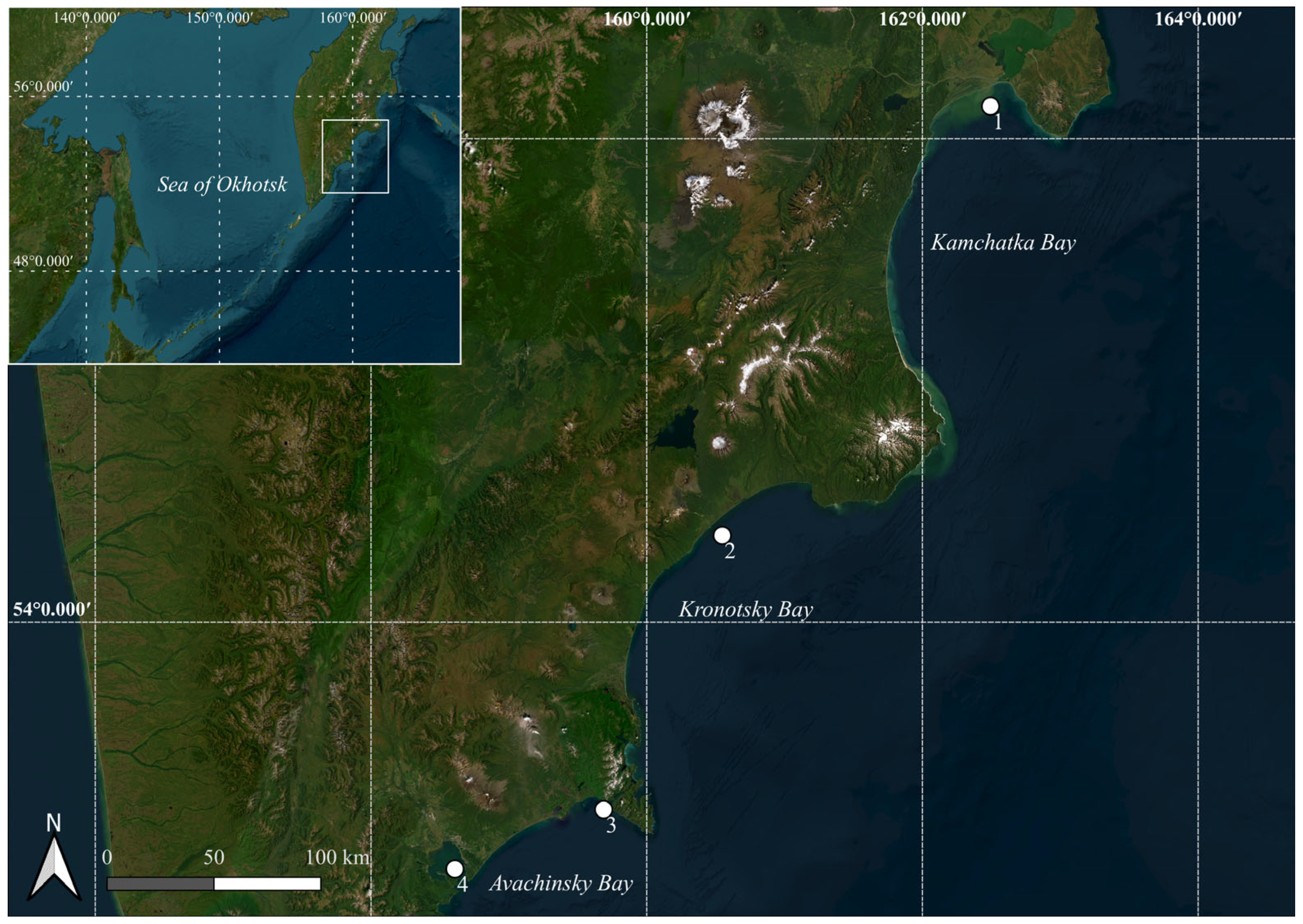
Figure 1. Map of bottom sediment sampling.
Jmse 13 01891 g001
Figure 2. Changes in M. trossulus hemocyte lysosome membrane stability exposed to extracts bottom sediment (mean ± standard deviation, n = 9). Lowercase letters indicate statistically significant differences between groups (p < 0.05).
Jmse 13 01891 g002
Figure 3. The degree of DNA fragmentation in tissue cells of the mussel M. trossulus after exposure to sediment extracts (mean ± standard deviation, n = 9). Lowercase letters indicate statistically significant differences between groups.
Jmse 13 01891 g003
Figure 4. The degree of DNA fragmentation of sperm of the sand dollar S. mirabilis after exposure to sediment extracts (mean ± standard deviation, n = 9). Lowercase letters indicate statistically significant differences between groups (p < 0.05).
Jmse 13 01891 g004
Figure 5. Distribution of comets formed by gill cells M. trossulus by degree of damage.
Jmse 13 01891 g005
Figure 6. Distribution of comets formed by digestive gland cells M. trossulus by degree of damage.
Jmse 13 01891 g006
Figure 7. Distribution of comets formed by spermatozoa of Scaphechinus mirabilis according to the degree of damage.
Jmse 13 01891 g007
Disclaimer/Publisher’s Note: The statements, opinions and data contained in all publications are solely those of the individual author(s) and contributor(s) and not of MDPI and/or the editor(s). MDPI and/or the editor(s) disclaim responsibility for any injury to people or property resulting from any ideas, methods, instructions or products referred to in the content.