
Article

Ralstonia solanacearum Species Complex Resists Bacteriophage and/or Antibiotic by Reducing Virulence and Expressing Resistance Genes
1
College of Resources and Environment, Yunnan Agricultural University, Kunming 650201, China
2
Yunnan Institute of Microbiology, School of Life Sciences, Yunnan University, Kunming 650500, China
*
Authors to whom correspondence should be addressed.
These authors contributed equally to this work.
Agriculture 2026, 16(5), 595; https://doi.org/10.3390/agriculture16050595
Submission received: 29 January 2026 / Revised: 28 February 2026 / Accepted: 3 March 2026 / Published: 5 March 2026
(This article belongs to the Special Issue Biological Control of Plant Diseases by Beneficial Microbes)

Abstract

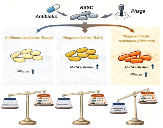
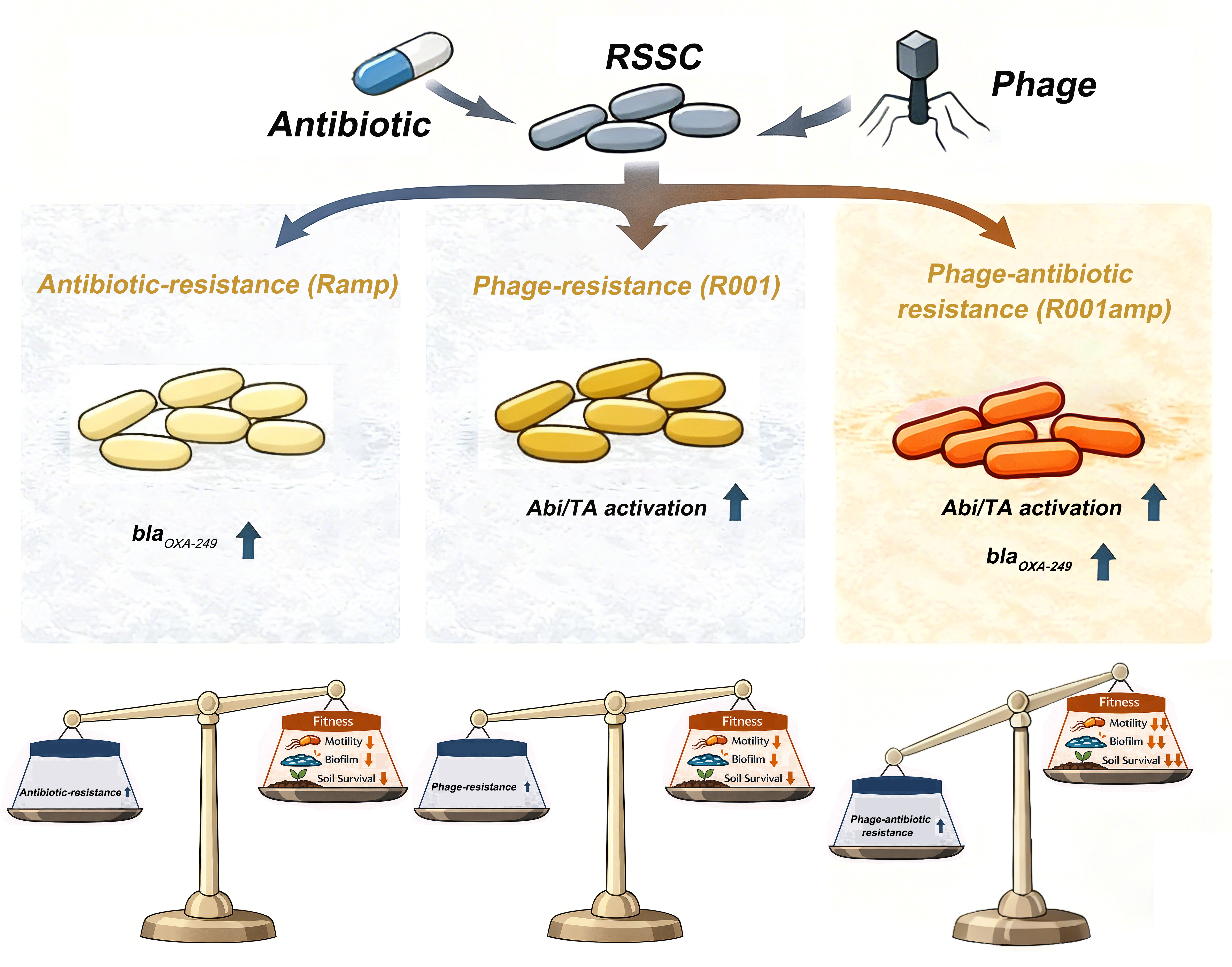
The Ralstonia solanacearum species complex (RSSC) is a globally significant plant pathogenic bacterium. Given the lack of effective chemical controls, phage therapy has emerged as a promising biocontrol alternative. While combining phages with antibiotics can counteract phage resistance, RSSC may still evolve concurrent resistance to both agents. However, the fitness consequences and underlying mechanisms of such resistance remain unclear. In this study, a novel RSSC phage was isolated to experimentally investigate the trade-offs between resistance and virulence in evolved strains. Compared to the wild-type, phage-resistant, antibiotic-resistant, and dual-resistant mutants showed no significant differences in growth rate, exopolysaccharide and lipopolysaccharide production. However, their motility, soil survival, and biofilm formation were significantly impaired, with the most severe decline observed in the dual-resistant mutants. Furthermore, phage-resistant strains exhibited enhanced antibiotic resistance, while antibiotic-resistant strains displayed cross-resistance. The antibiotic resistance gene blaOXA-249 was upregulated only in antibiotic-resistant strains. In phage-resistant bacteria, the abortive infection system was activated. A reduction in bacterial cell numbers post-infection indicated that phage resistance limits phage propagation via a “suicidal” mechanism. These findings reveal that resistance evolution in RSSC carries substantial fitness costs and highlight phage steering as a novel strategy for designing phage agents.

Graphical Abstract

1. Introduction

The Ralstonia solanacearum species complex (RSSC) causes bacterial wilt (BW) in more than 390 plant species in more than 75 families and is considered one of the most destructive plant pathogenic bacteria worldwide [1]. In Europe, members of the RSSC are regulated as quarantine organisms in Commission Implementing Regulation (EU) 2019/2072 [2]. The RSSC is classified into three distinct species and differentiated into four phylotypes: R. solanacearum (phylotype II), R. pseudosolanacearum (phylotypes I and III), and R. syzygii (phylotype IV) [1]. Strains of the RSSC typically invade susceptible hosts through wounds and subsequently colonize and reproduce within the xylem tissues. To promote disease progression, RSSC employs a range of virulence factors, including exopolysaccharide (EPS), lipopolysaccharide (LPS), motility, type III secretion system (T3SS) effectors, and the secretion of various enzymes—such as cell wall-degrading enzymes (e.g., pectinase and cellulase) and enzymes involved in detoxifying reactive oxygen species [3].
BW is difficult to control due to pathogen aggressiveness, persistence, wide range of hosts, and wide geographic distribution in tropical, subtropical, and temperate regions, and the agricultural damage increases annually. No agrochemicals are available to manage bacterial wilt effectively [4]. The crisis of BW has led some to propose phages as a therapy. Viruses that infect bacteria, phages are environmentally ubiquitous, host-specific, and effective at infecting pathogenic bacteria. Phages have been demonstrated to be effective at controlling RSSC population densities in lab and greenhouse experiments, resulting in reduced BW disease incidence [5,6,7]. However, the emergence of phage resistance in target bacteria is a pressing concern that undermines the reliability of phage therapy as a sustainable solution for managing BW.
One of the most promising alternative strategies to combat bacterial resistance is the application of lytic phages combined with antibiotics. Unfortunately, pathogenic bacteria may lead to the outbreak of phage-antibiotic co-resistant strains under the combined action of phages and antibiotics. It is established that bacteria may experience fitness costs when evolving resistance to antibiotics or phages. The mechanisms of this trade-off—whether synergistic or antagonistic—are highly species-specific [8]. However, how phage resistance, antibiotic resistance, or dual-resistance mutations influence key bacterial fitness traits—including growth rate, motility, biofilm formation, virulence factors, and antibiotic resistance—remain poorly characterized in the RSSC.
Bacteria have developed different mechanisms to defend against phages, such as preventing phages from being adsorbed on the surface of host bacteria, degradation of invading nucleic acids, inhibition of DNA or RNA synthesis, and induction of growth arrest or cell suicide for population-level protection [9]. Yet, among the various mechanisms used by bacteria to defend against phages, modifications to bacterial surface receptors resulting from genomic mutations are often considered the simplest route for acquiring phage resistance [10]. The fitness cost of receptor mutations varies depending on the strain and its environment, but in most cases, large competitive costs are generally associated with surface modification [11]. Another common outcome during defense is abortive infection (Abi), whereby cells die upon phage invasion, suppressing replication and the epidemic.
Unlike the “selfish” strategy of surface modification—which benefits the individual host without clearing phage—abortive infection (Abi) is an “altruistic” process that eliminates the infected cell to protect the population. Other intracellular defense systems, however, can both ensure the survival of the infected individual and suppress the phage epidemic, thereby offering collective protection [9].
Similarly, bacterial resistance to antibiotics is also accompanied by various changes in virulence or phage sensitivity [12]. Bacteria have evolved a wide range of mechanisms to resist antibiotics, including enzymatic degradation, target modification, reduced permeability, and active efflux [13]. In agricultural soils, plant-associated bacteria are exposed to multiple selective pressures, including naturally abundant phages and antibiotics introduced through human agricultural activities. Antibiotic residues can enter agricultural soils through anthropogenic antibiotic use, including manure application in intensive livestock production, aquaculture effluents, and contaminated irrigation water, thereby imposing selection pressure on soil bacteria [14,15,16]. Together with naturally abundant phages, these inputs create concurrent selective pressures on RSSC. Several phage defense systems have been identified in RSSC [17]; however, which defense strategies are most suitable for RSSC under simultaneous phage and antibiotic pressures remains unclear.
This study aimed to elucidate the strategies employed by the RSSC to resist phages and/or antibiotics and to assess the associated fitness and virulence costs in resistant strains. To this end, we isolated phage-resistant, antibiotic-resistant, and dual-resistant mutants and systematically evaluated their growth, motility, biofilm formation, EPS and LPS production, and antibiotic sensitivity. The underlying resistance mechanisms were further investigated. Understanding these mechanisms and trade-offs will facilitate the design of more effective biocontrol strategies against RSSC.

2. Materials and Methods

2.1. Bacterial Strain and Culture Conditions

The wild-type RSSC strain, LcA12172, was originally isolated from the rhizosphere soil of BW of tobacco in Lincang, Yunnan Province, China. The genome sequence of strain LcA12172 is available in the National Microbiology Data Center (NMDC) under accession number NMDC60215644. Strains were incubated at 32 °C in SM broth (glucose 5 g/L, peptone 10 g/L, acid-hydrolyzed casein 1 g/L, and yeast extract 1 g/L) or on SM agar plates supplemented with 1% 2,3,5-triphenyltetrazolium chloride (TTC).

2.2. Phage Isolation, Purification, and Transmission Electron Microscopy (TEM)

Phage YIM V22001R used in this study was isolated from the rhizosphere soil of diseased tobacco plants collected in Nanchang, Jiangxi Province, China. The phage isolation process followed a previous protocol [18]. Briefly, 10 g of soil sample was mixed with 25 mL of SM buffer (100 mmol/L NaCl, 8 mmol/L MgSO4, 0.05 mol/L Tris-HCl, 0.01% w/v gelatin, pH 7.4–7.6), followed by the addition of 10 μL of a logarithmic-phase LcA12172 culture. The mixture was incubated at 32 °C with shaking at 150 r/min for 24 h. Subsequently, the suspension was spun at 16,100× g for 10 min at 4 °C, and the fluid was filtered via a 0.22 μm membrane filter (Millipore, Bedford, MA, USA) and subjected to serial dilutions. Per dilution, 500 μL of the concentrated phage solution was blended with the same volume of host bacterial culture and incubated at 32 °C for 15 min to allow adsorption. The mixture was then combined with 0.7% agar and immediately poured onto solid agar plates to prepare double-layer agar plates. After solidification of the top agar, the plates were incubated at 32 °C. After 24 h, large and well-defined single plaques were selected and transferred into 100 μL of SM broth. Purification via the double-layer agar method was repeated for five cycles to generate purified phage stocks.
For phage morphological characterization, samples of the phage suspension were negatively stained with 2% (w/v) sodium phosphotungstate for 1 min, air-dried, and inspected using a transmission electron microscope (JEM-2100; JEOL, Tokyo, Japan) [19].

2.3. Bacterial and Phage Genomic DNA Extraction and Sequencing

Genomic DNA from bacterial samples was isolated utilizing the DNeasy PowerSoil Kit (QIAGEN, Hilden, Germany) and sent to E-GENE Tech Co., Ltd. (Shenzhen, China) for whole-genome sequencing. Libraries were prepared using the SQK-LSK109 ligation sequencing kit (Oxford Nanopore Technologies, Oxford, UK) and sequenced on the Oxford Nanopore Technologies (ONT) platform. Genome assembly was executed using Canu (v1.5) [20]. Open reading frame (ORF) prediction was conducted with Prodigal (v2.6.3) [21]. Antibiotic resistance genes and resistance-related information were analyzed using the Resistance Gene Identifier (RGI) tool in the Comprehensive Antibiotic Resistance Database (CARD) (https://card.mcmaster.ca/analyze, accessed on 1 June 2021) [22]. Phage defense system–related genes were predicted using the online tool DefenseFinder (https://defensefinder.mdmlab.fr/, accessed on 5 June 2022) [23].
Purified phage suspensions were subjected to DNase I (ultimate concentration 50 U/mL) and RNase A (ultimate concentration 250 μg/mL) and incubated at 37 °C for 1 h, followed by heat inactivation at 80 °C for 15 min. Thereafter, EDTA (0.5 M), 10% SDS, and proteinase K (final concentration 250 μg/mL) were introduced sequentially, and the solution was incubated at 55 °C for 3 h. Phage genomic DNA was isolated and purified via phenol–chloroform extraction and ethanol precipitation [18]. Genomic DNA libraries were generated from fragmented short DNA and sequenced in paired-end mode on the Illumina NovaSeq platform by Guangdong Magigen Biotechnology Co., Ltd. (Guangzhou, China). Raw sequencing reads were filtered using SOAPnuke (v2.1.6) [24] and BWA (v0.7.17) [25], and the clean data were assembled using MEGAHIT (v1.2.9) [26]. Viral sequences were identified and retrieved using CheckV (v2.9.0) [27]. Open reading frames (ORFs) were predicted using Prokka (v1.14.6) [28].
The complete genome sequence of phage YIMV22001R, together with 18 closely related phage genome sequences retrieved from the GenBank and International Committee on Taxonomy of Viruses (ICTV) databases, was used for comparative genomic analyses. Whole-genome phylogenetic analysis was performed using the Virus Classification and Tree Building Online Resource (VICTOR, https://ggdc.dsmz.de/victor.php, accessed on 8 January 2026) [29], which infers phylogenetic relationships based on the Genome-BLAST Distance Phylogeny (GBDP) method. EzBioCloud (https://www.ezbiocloud.net/tools/ani, accessed on 24 October 2025) [30] was used to perform pairwise average nucleotide identity (ANI) comparisons among the 18 phage genome sequences, and ANI diagrams were plotted using R (v4.5.1). Unless otherwise specified, default parameters were used for all software tools.

2.4. Isolation of Phage- and/or Antibiotic-Resistant Mutants

Phage-resistant mutants were isolated as described by Li et al. [31], with minor modifications. Briefly, 100 mL aliquots of overnight cultures of the wild-type strain LcA12172 (108 CFU/mL) were incubated with 10 mL of phage YIM V22001R (109 PFU/mL) at 32 °C for 48 h and then plated on SM–TTC agar plates and incubated overnight at 32 °C. Single colonies were selected and sequentially streaked at least three times on SM–TTC agar plates. All phage-resistant mutants were tested for resistance using the double-layer agar method. Strains that did not produce plaques were considered phage-resistant.
Ampicillin (final concentration 50 μg/mL) was added to logarithmic-phase cultures of strains, followed by incubation at 32 °C for 24 h. This concentration was selected to impose a strong selection pressure for resistant mutants and is within the range commonly used for antibiotic selection in Ralstonia studies [32]. The cultures were streaked on SM–TTC agar plates containing the same concentration of ampicillin. Single colonies were then selected and streaked on fresh SM–TTC agar plates supplemented with ampicillin. This procedure was repeated five times to obtain antibiotic-resistant mutants and phage–antibiotic dual-resistant mutants.

2.5. Motility and Biofilm Formation Assays

Swimming, swarming, and twitching motility assays were performed on SM-TTC agar plates containing 0.3%, 0.5%, and 1.5% agar, respectively, as previously described by Yahyaoui et al. [33], with minor modifications. Briefly, overnight bacterial cultures were adjusted to an optical density at 600 nm (OD600) of 0.8, and 1 μL of each culture was spotted onto the center of the corresponding agar plate. The plates were incubated at 32 °C for 48 h, and motility was evaluated by measuring bacterial diameter of growth zone.
Biofilm formation was assessed using the crystal violet staining method as described by Su et al. [34] with minor modifications. Briefly, bacterial strains were inoculated into 96-well microtiter plates containing 200 μL of SM broth per well and incubated at 32 °C for 36 h. Unattached cells were discarded, and the wells were washed three times with 200 μL of sterile ddH2O. Subsequently, 200 μL of 1% (w/v) crystal violet ammonium oxalate solution was added to each well and incubated at room temperature for 15 min. Excess stain was removed by washing with ddH2O. The bound crystal violet was then solubilized by adding 200 μL of 75% ethanol and incubating at room temperature for 15 min. Biofilm formation was quantified by measuring the absorbance at 570 nm using a microplate reader.

2.6. Antibiotic Susceptibility Assay

Antibiotic susceptibility of different bacterial strains was evaluated using the disk diffusion method as described by Humphries et al. [35]. The antibiotics tested included imipenem (10 μg/disk), lincomycin (2 μg/disk), vancomycin (30 μg/disk), sulfamethoxazole (300 μg/disk), nalidixic acid (30 μg/disk), tetracycline (30 μg/disk), erythromycin (15 μg/disk), gentamicin (10 μg/disk), polymyxin B (300 IU/disk), chloramphenicol (30 μg/disk), ceftriaxone (30 μg/disk), norfloxacin (10 μg/disk), tobramycin (10 μg/disk), and amikacin (30 μg/disk). Logarithmic-phase bacterial cultures were mixed with SM soft agar (0.7%) and poured onto SM agar plates. Antibiotic-embedded disks were subsequently applied to the agar, and the Petri dishes were cultured at 32 °C for 24 h. Antibiotic susceptibility was assessed by measuring the diameter of the inhibition zones.
In addition, the sensitivity of strains to ampicillin or cephalexin was determined using the broth dilution method [36]. Briefly, 200 μL of SM broth containing different concentrations of ampicillin or cephalexin (0, 0.25, 0.5, 1, 2, 3, 4, 5, 10, 15, 20, 25, 30, and 35 μg/mL) was added to each well, followed by the addition of 4 μL of bacterial suspension (OD600 = 0.8). The plates were incubated at 32 °C for 48 h, and bacteria were measured by OD600 by Microplate Reader (Molecular Devices, San Jose, CA, USA).

2.7. Bacterial Growth Curve

To evaluate the growth rates of different strains, 1.5 mL of bacterial culture (OD600 = 0.8) was inoculated into 150 mL of SM liquid medium in a conical flask. The cultures were incubated at 32 °C at 150 r/min for 24 h. A control group containing only SM medium without bacterial inoculation was included. The OD600 was measured every 3 h, and growth curves were generated based on the OD600 values as previously described [37].

2.8. Determination of EPS, LPS, Pectinase, and Cellulase Activities

Extraction and quantification of EPS were performed according to the method described by Zhang et al. [38] with minor modifications. Specifically, 1 mL of bacterial culture in the logarithmic stage (OD600 = 0.8) was subjected to centrifugation at 11,200× g for 10 min at 4 °C, and the clarified supernatant was passed through a 0.22 μm filter. Proteins were eliminated via the Sevag procedure (chloroform/n-butanol ratio 4:1). Subsequently, four volumes of 95% ethanol were added, and EPS was precipitated at 4 °C overnight. The precipitate was dissolved in ultrapure water, and EPS content was determined using the phenol–sulfuric acid method with glucose as the standard. Absorbance was measured at 490 nm.
LPS was extracted from 1 mL of bacterial culture (OD600 = 0.8) for each strain using a bacterial lipopolysaccharide extraction kit (Bestbio, Beijing, China). Specifically, 200 μL of LPS solution was combined with 600 μL of anthrone reagent and incubated within a 95 °C water bath for 10 min. Following cooling, absorbance was measured at 625 nm. A standard curve was generated using anhydrous glucose, and results were expressed as anthrone-reactive carbohydrate equivalents in the LPS extracts [39].
Extraction and activity assays of pectinase and cellulase were performed following the method of Ling et al. [40] with minor modifications. Enzyme activities were determined via the 3,5-dinitrosalicylic acid (DNS) colorimetric technique, with galacturonic acid and glucose standard curves used to calculate pectinase and cellulase activities, respectively.
Pectinase activity was determined using the DNS method as described previously, with minor modifications [41,42]. Activity was calculated based on the D-galacturonic acid standard curve and expressed as U/mL. For pectinase activity, 1 mL of bacterial culture (OD600 = 0.8) was spun down at 16,100× g for 10 min at 4 °C, and the supernatant was isolated as the raw enzyme preparation. A total of 100 μL of crude enzyme solution was mixed with 500 μL of 0.4% pectin solution and 400 μL of glycine–NaOH buffer (pH 9.4) and incubated at 50 °C for 30 min. Subsequently, 200 μL of the reaction mixture was mixed with 200 μL of distilled water and 500 μL of DNS reagent, boiled for 5 min, cooled, and diluted to a final volume of 2.5 mL. Absorbance was measured at 540 nm using a microplate reader. Pectinase activity (U/mL) was calculated using the formula: Pectinase activity (U/mL) = [(A − A0) × N × 5 × 1000]/(k × t). where A is the absorbance of the sample, A0 is the absorbance of the blank, N is the dilution factor, k is the slope of the standard curve, and t is the reaction time (30 min).
Cellulase activity (FPase) was determined using a filter paper–DNS assay as described previously, with minor modifications [43,44]. For cellulase activity, 100 μL of bacterial culture (OD600 = 1.4) was mixed with 900 μL of sodium citrate buffer and reacted at 32 °C for 30 min. Subsequently, the mixture was centrifuged, and the supernatant was retained as the crude enzyme preparation. Filter paper strips were placed in centrifuge tubes and moistened with 100 μL of buffer and equilibrated at 50 °C for 10 min, followed by the addition of 50 μL of crude enzyme solution and 150 μL of sterile water. Following 60 min of incubation at 50 °C, 200 μL of DNS reagent was applied, and the solution was heated for 5 min, cooled, and diluted to a final volume of 2.5 mL. After dilution, 200 μL of the mixture was transferred to a 96-well microplate, and absorbance was measured at 540 nm using a microplate reader. Cellulase activity (U/mL) was calculated using the formula: cellulase activity (U/mL) = [(A1 − A2) × K + b] × D/t. where A1 is the absorbance of the reaction mixture, A2 is the absorbance of the blank, K and b are the slope and intercept of the standard curve, respectively, D is the dilution factor, and t is the reaction time (60 min). One unit (U) of cellulase activity was defined as the amount of enzyme that releases 1 μg of reducing sugar (glucose equivalents) per minute under the assay conditions.

2.9. Bacterial Survival Rate

To evaluate bacterial survival in soil, 10 mL of logarithmic-phase bacterial culture (OD600 = 0.8) was vigorously blended with 30 g of sterilized soil (autoclaved at 121 °C for 30 min). Samples of 1 g of soil were harvested at 0 h, 48 h, and 96 h post-inoculation, and viable cell concentrations were quantified by employing serial dilution combined with plate counting techniques [45]. The bacterial survival rate was calculated as follows: Survival rate (%) = [(bacterial count at a given time point)/(bacterial count at 0 h)] × 100%.
In addition, to determine whether bacteria activate abortive infection–mediated defense following phage infection, phage lysate was mixed with logarithmic-phase LcA12172r001amp at a multiplicity of infection (MOI) of 1 and incubated at 32 °C for 90 min. Bacterial cultures without phage addition were used as controls. At 30 min intervals, samples of the infection mixture were serially diluted and plated on SM–TTC agar plates. After incubation at 32 °C for 24 h, viable bacterial cells were enumerated [46].

2.10. Reverse Transcription-Quantitative Real-Time PCR (RT-qPCR) Analysis

Total bacterial RNA was extracted using a Total RNA Extraction Kit (Biosharp, Hefei, China) according to the manufacturer’s instructions. RNA concentration and purity were assessed spectrophotometrically; only samples with A260/A280 between 1.8 and 2.1 and A260/A230 > 2.0 were used for cDNA synthesis. Complementary DNA (cDNA) synthesis was performed using the TransScript One-Step gDNA Removal and cDNA Synthesis SuperMix (TransGen Biotech, Beijing, China). The expression levels of antibiotic resistance genes (ARGs) and defense system–related genes were analyzed by quantitative PCR (qPCR). The 16S rRNA gene was used as the internal control gene. Gene-specific primers were synthesized according to the genomic sequences and are detailed in Table S1.
Every qPCR assay (20 μL) comprised 10 μL of 2× TransStart TOP/Tip Green qPCR SuperMix, 0.4 μL of 50× Passive Reference Dye, 0.4 μL of every primer (10 μM), 1 μL of cDNA sample, and 7.8 μL of RNase-free water. The qPCR amplification was performed using a LightCycler 480 II system (Roche Diagnostics, Rotkreuz, Switzerland) with an initial denaturation at 94 °C for 30 s, followed by 45 cycles of 94 °C for 5 s, 60 °C for 20 s, and 72 °C for 30 s. Relative gene expression levels were calculated using the 2^(−ΔΔCt) method [47].

2.11. Phage Adsorption Assays

Phage adsorption assays were conducted according to the protocol by Diao et al. [48], with slight adjustments. Specifically, phages were mixed with wild-type and resistant mutants at an MOI of 1 and incubated at 32 °C for 15 min to allow adsorption. The mixtures were then centrifuged at 16,100× g for 2 min at 4 °C to pellet the bacterial cells with adsorbed phages. The titer of unadsorbed phages in the supernatant was determined using the double-layer agar method, with the initial phage inoculum serving as the control. The phage adsorption rate was calculated using the following formula: Adsorption rate (%) = [1 − (unadsorbed number of phage)/(initial number of phage)] × 100%.

2.12. Statistical Analysis

All experiments were performed with three replicates. Data are expressed as the mean ± standard deviation (SD). Comparisons among multiple groups were conducted using one-way analysis of variance (ANOVA), followed by Tukey’s honestly significant difference (HSD) post hoc test for multiple comparisons. Comparisons between two groups were performed using Student’s t-test. A p value < 0.05 was considered statistically significant. All statistical analyses were carried out using Origin 2024 software (OriginLab Corporation, North Andover, MA, USA).

3. Results

3.1. Isolation and Characterization of Phages

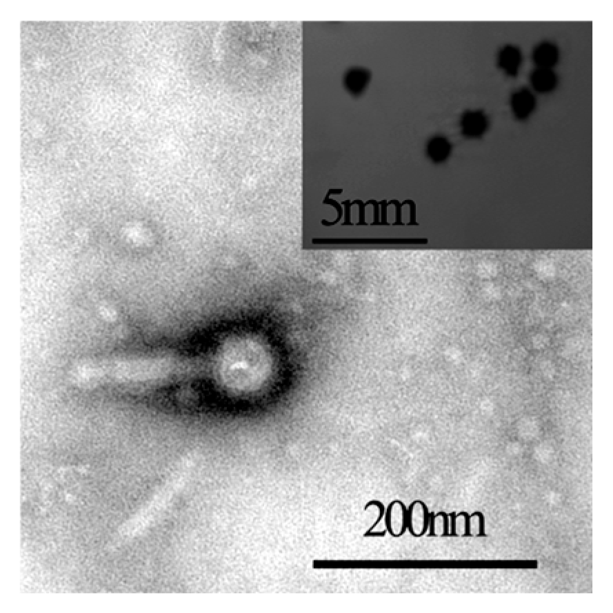
Using RSSC strain LcA12172 as the host strain, a lytic phage was isolated from the rhizosphere soil of diseased tobacco plants and designated YIM V22001R. This phage formed clear, transparent plaques with regular edges on SM double-layer agar plates, with an average diameter of approximately 0.83 ± 0.12 mm after 24 h of incubation (Figure 1). TEM observation demonstrated that YIM V22001R possesses head-tail architecture, featuring a head width of 51.79 ± 2.1 nm and a tail length of 138.99 ± 4.9 nm (Figure 1).
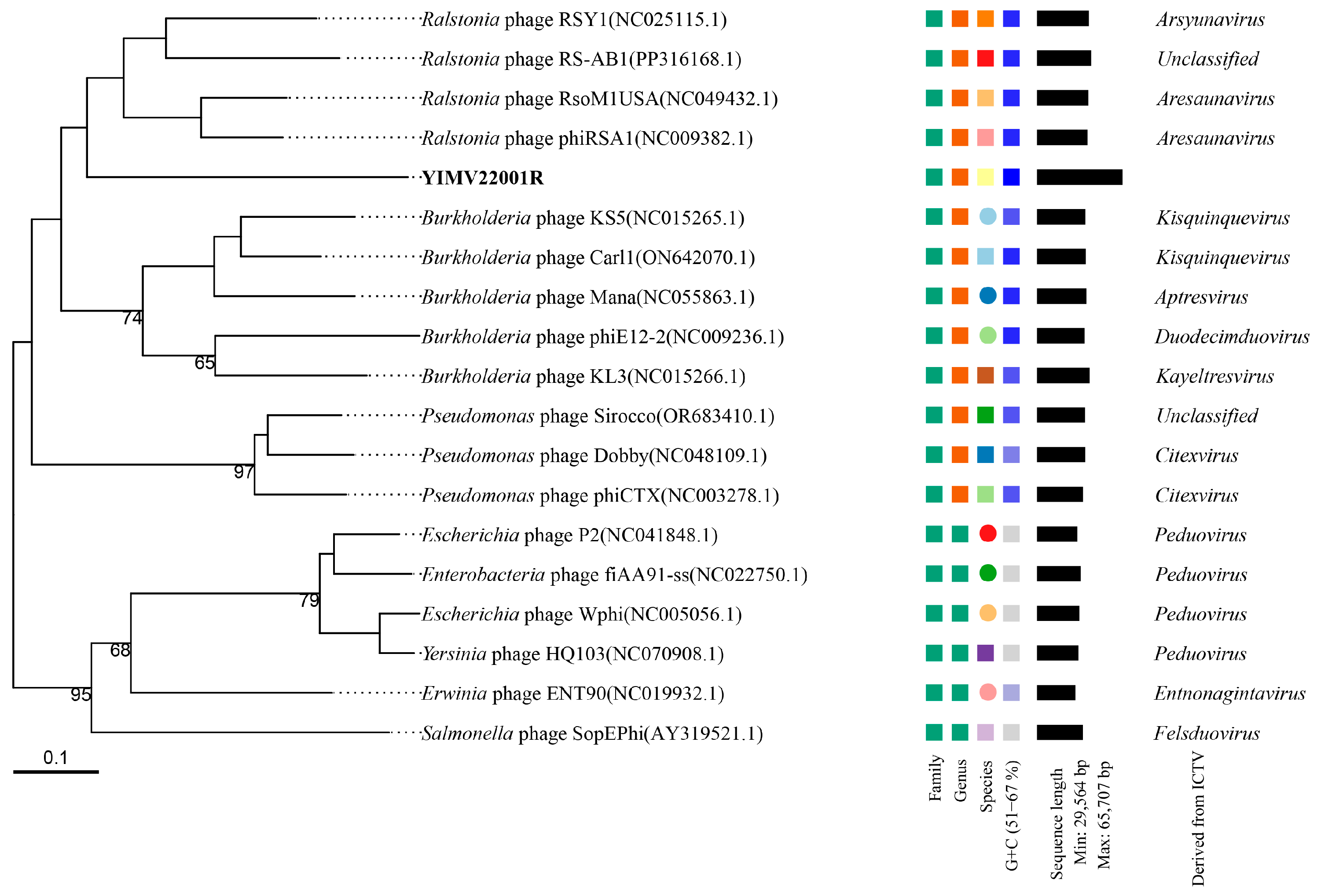
The complete genome of phage YIMV22001R is double-stranded DNA, with a total size of 65,707 bp and 66.91% GC content. A total of 70 ORFs were predicted. No tRNA genes, integrase genes, or antibiotic resistance genes were identified. Whole-genome phylogenetic analysis showed that phage YIMV22001R clustered with Ralstonia phage RSY1, Ralstonia phage RS-AB1, Ralstonia phage RsoM1USA, and Ralstonia phage phiRSA1, indicating a relatively close evolutionary relationship among these phages (Figure 2).
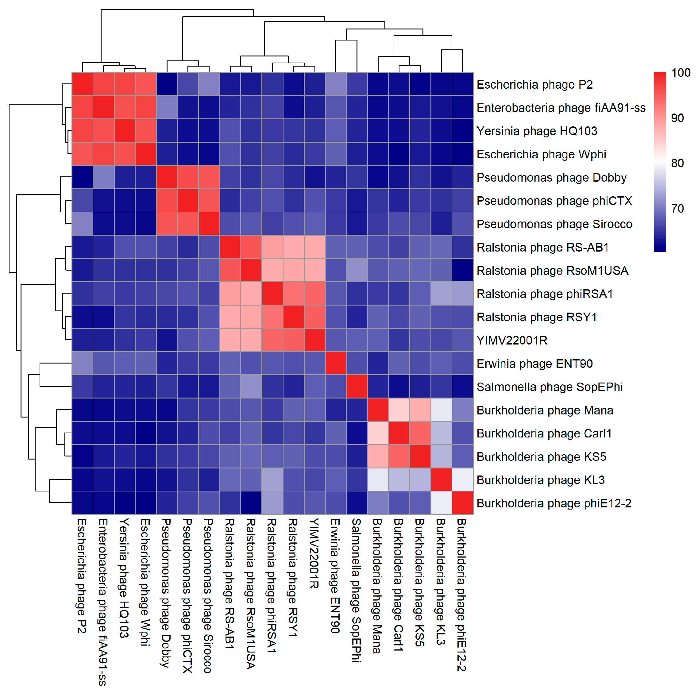
ANI analysis revealed relatively high ANI values between YIMV22001R and Ralstonia phage RSY1 (94.8%), Ralstonia phage phiRSA1 (94.27%), Ralstonia phage RsoM1USA (87.94%), and Ralstonia phage RS-AB1 (87.91%) (Figure 3). Among these, Ralstonia phage RSY1 belongs to the genus Arsyunavirus (Peduoviridae), while Ralstonia phages phiRSA1 and RsoM1USA belong to the genus Aresaunavirus (Peduoviridae); Ralstonia phage RS-AB1 has not yet been classified.
Notably, our genome annotation reveals that several predicted ORFs in YIMV22001R cannot be matched to any known genes in NCBI or other public databases, suggesting that these ORFs are unique to YIMV22001R. The absence of similarity to known genes, along with significant sequence divergence from related phages, may contribute to its distinct phylogenetic placement.
Taken together, although phage YIMV22001R shows relatively high genetic similarity to members of the genus Aresaunavirus and Arsyunavirus, it forms an independent phylogenetic branch. Therefore, YIMV22001R is currently assigned to an unclassified group within Peduoviridae.

3.2. Isolation of Resistant Mutants

A phage-resistant mutant derived from wild type LcA12172 was obtained by co-incubation with phage YIMV22001R. The resistant isolate, designated LcA12172r001 (R001 for short), was repeatedly restreaked and screened using spot assays to confirm the acquisition of a heritable phage-resistant phenotype (Figure S1).
Wild type LcA12172 was cultured in SM–TTC medium supplemented with ampicillin (50 μg/mL) for 24 h, and an ampicillin-resistant mutant, designated LcA12172ramp (Ramp for short), was selected and confirmed by repeated restreaking on SM–TTC medium containing ampicillin (Figure S1). When LcA12172r001 was inoculated into SM–TTC medium supplemented with ampicillin, a mutant co-resistant to both phage and ampicillin was obtained and designated LcA12172r001amp (R001amp for short) (Figure S1).

3.3. Fitness Trade-Offs of Resistant Mutants

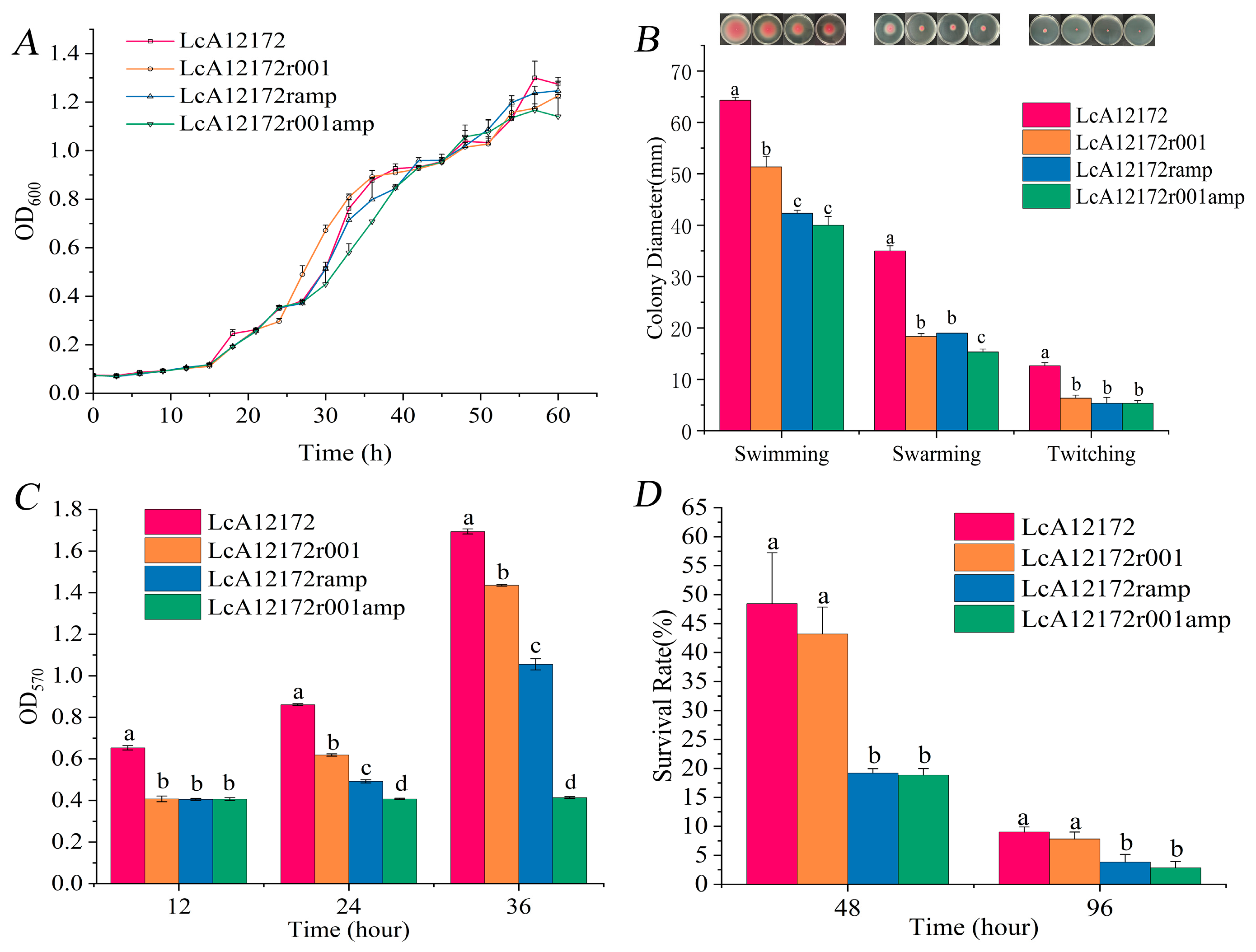
The growth of the resistant mutants did not differ significantly from that of the wild-type strain (p > 0.05) (Figure 4A), indicating that bacterial growth was not markedly affected by the acquisition of phage and/or antibiotic resistance.
Three forms of motility were tested on SM agar with varying concentrations: swimming at 0.3%, swarming at 0.5%, and twitching at 1.5%. As shown in Figure 4B and Figure S2, all resistant mutants exhibited reduced swimming, swarming, and twitching motility compared with the wild-type strain, and R001amp displayed the weakest motility among the mutants, indicating that simultaneous resistance to both phages and antibiotics further impaired bacterial motility.
After 12 h of incubation, all three mutants exhibited significantly reduced biofilm formation compared with the wild-type strain (p < 0.05). Notably, biofilm formation by the R001amp strain did not increase markedly over time (Figure 4C). These results indicate that the biofilm-forming ability of resistant mutants is reduced.
No significant differences were observed between the wild-type strain and the three resistant mutants in terms of EPS content (87.70–88.65 μg/mL), LPS content (34.38–36.25 μg/mL), pectinase activity (7.34–7.46 U/mL), or cellulase activity (1.22–1.23 U/mL) (p > 0.05) (Table S2), indicating that the acquisition of resistance had no significant effect on these virulence-associated extracellular polymers or hydrolytic enzyme activities.
Comparison of soil survival rates showed that the wild-type strain and the R001 strain exhibited significantly higher survival than the Ramp strain and the R001amp strain (Figure 4D). These results indicate that the acquisition of antibiotic resistance significantly compromises bacterial survival in the soil environment.
The antibiotic susceptibility assay showed that the wild-type strain and the R001 strain exhibited identical antibiotic resistance profiles. The Ramp and the R001amp strains shared the same resistance profile and, compared with the former two strains, additionally acquired resistance to chloramphenicol and ceftriaxone (Table 1). Meanwhile, the R001 strain exhibited increased tolerance to ampicillin (Figure S3A). In addition, the Ramp strain showed cross-resistance to cephalexin, which is also a β-lactam antibiotic (Figure S3B).

3.4. ARG Expression in Resistant Mutants

Comparison against the CARD revealed that the wild-type strain LcA12172 harbors three antibiotic resistance genes belonging to two types: adeF (present as two copies with different predicted functions) and blaOXA-249. The adeF gene is associated with resistance to tetracyclines and fluoroquinolones [49], whereas blaOXA-249 is related to resistance to β-lactam antibiotics [50].
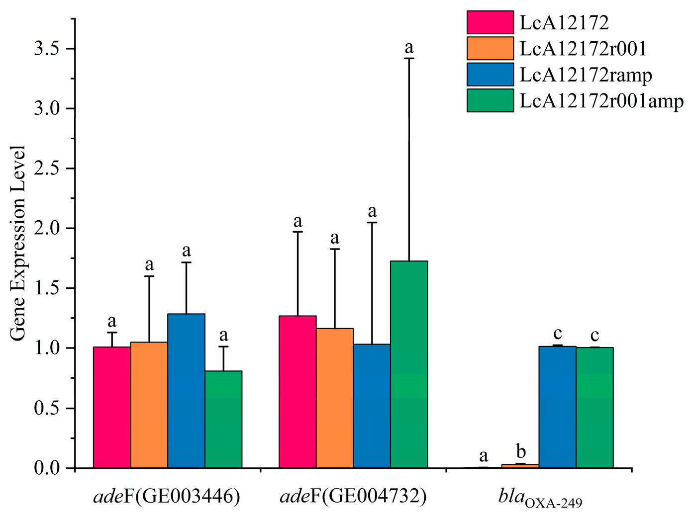
RT-qPCR analysis showed that blaOXA-249 was barely expressed in the wild-type strain but was highly expressed in the Ramp and R001amp strains. The expression level of blaOXA-249 in the phage-resistant strain was also significantly higher than that in the wild type, but significantly lower than that in the antibiotic-resistant strain (Figure 5). This expression pattern is consistent with the increased tolerance of the R001 strain to ampicillin. In contrast, no significant differences in the expression levels of the two adeF genes were observed between the wild-type strain and the resistant mutants (Figure 5). Together, these results indicate that resistance to ampicillin in the mutant strains is associated with the elevated expression of the blaOXA-249 gene.

3.5. Abortive Infection in Phage-Resistant Mutants

To investigate the mechanisms underlying phage resistance, we first compared the phage adsorption rates between the wild-type strain and the resistant mutants. The results showed that there were no significant differences in phage adsorption rates among the strains (Figure S4), indicating that the emergence of resistance was not due to failure of phage adsorption caused by mutations in host receptors.
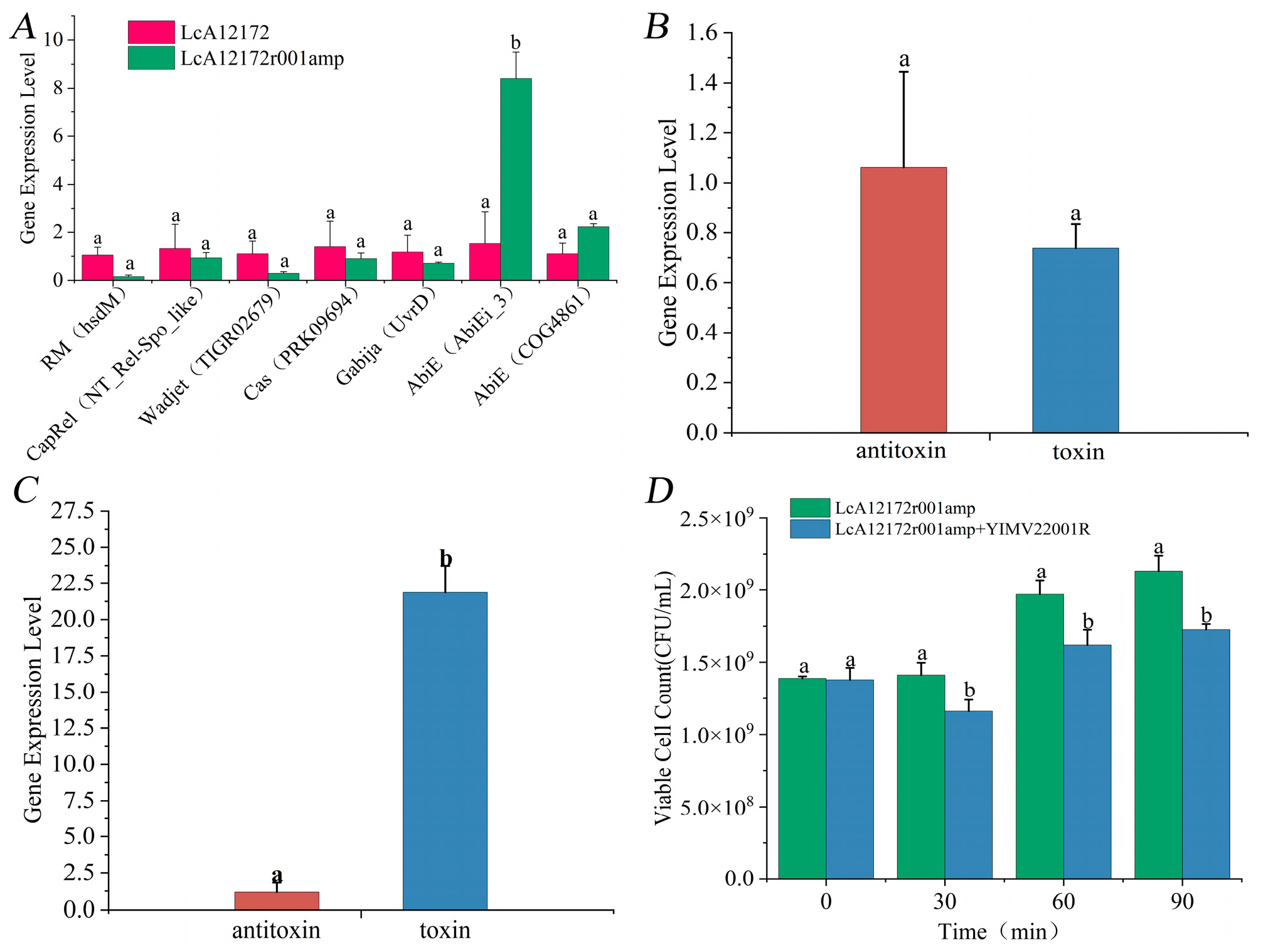
Prediction using DefenseFinder revealed that the genome of the wild-type strain harbors seven phage defense systems belonging to six types, namely restriction–modification (RM), CapRel, Wadjet, CRISPR-Cas (clustered regularly interspaced short palindromic repeats and CRISPR-associated proteins), Gabija, and two Abi systems, comprising a total of 21 genes. RT-qPCR analysis of key genes associated with these defense systems showed that the antitoxin gene (gene004117) from one Abi system (type IV toxin–antitoxin (TA) system) was significantly upregulated in the R001amp strain (Figure 6A), suggesting that phage resistance may be associated with an abortive infection system.
Additional investigation showed that without phage invasion, there existed no substantial variation in the transcriptional activity of the toxin and antitoxin genes within the R001amp strain (Figure 6B). Nevertheless, following phage infection, the toxin gene expression reached notably greater heights compared to the antitoxin gene (Figure 6C), demonstrating that this TA system was activated under phage stress.
To verify whether “suicidal” behavior occurs in the R001amp strain upon phage infection, we quantified the number of viable cells after co-culturing the phage with the R001amp strain. The results showed that although this strain was unable to form plaques, the number of viable cells decreased significantly after phage addition (Figure 6D). This result further confirms that the observed resistance is attributable to bacterial “suicide” triggered by activation of the Abi system, thereby restricting phage propagation.

4. Discussion

RSSC infects a wide range of plant hosts and causes bacterial wilt in many different crops. Phages, as a precise and efficient biocontrol technology, have received increasing attention. Currently, only about 100 phages infecting RSSC have been published [17]. The main reason that RSSC phages are less studied is due to difficulties with the extraction of phages from soil particles [51]. The lack of a sufficient number of phages remains one of the major challenges of phage therapy. In this study, an RSSC phage, YIM V22001R, was isolated from the rhizosphere soil of BW tobacco. Genome-based phylogenetic analysis using VICTOR, together with ANI, demonstrated that YIMV22001R is phylogenetically distinct from previously described Ralstonia phages and represents a novel species within the family Peduoviridae (Figure 2 and Figure 3). YIM V22001R does not contain integrase or virulence factors, making it a potential biocontrol phage.
One of the more attractive and feasible uses of phages is to combine them with antibiotics. However, pathogenic bacteria are still able to generate mutants that resist both phages and antibiotics. In this study, we found that although the growth rate of the resistant strains did not show significant changes (Figure 4A), their motility (Figure 4B), biofilm formation ability (Figure 4C), and soil survival rate (Figure 4D) were significantly reduced. Due to the limited availability of resources such as energy within the cells, it is common for bacteria to face trade-offs when under different stresses. Our findings agree with previous work that demonstrated how phage resistance in P. aeruginosa is often accompanied by fitness costs such as motility impairment, decrease in biofilm formation ability and resensitization towards some antibiotics [8,52]. In contrast, some strains exhibit reduced growth rates upon acquiring resistance [53]. These results indicate that trade-offs between resistance and virulence are strain-specific and strongly dependent on environmental context.
Motility is a key factor in the ecological adaptability and virulence of pathogens [54]. It mediates the movement required for host infection and plant colonization. Cells lacking motility either cannot infect the host or exhibit reduced virulence [55]. Our results showed that swimming, swarming, and twitching of the resistant strains were all reduced, with the dual-resistant strains exhibiting the poorest motility (Figure 4B). This result is consistent with studies on the phage-resistant mutants, which also showed a significant decrease in motility [56].
Plant pathogens form biofilms in the xylem of host plants, as well as on the surfaces of roots and leaves [57], leading to the blockage of xylem vessels, resistance to plant antimicrobial compounds, and colonization of specific niches. Therefore, biofilm formation is also an important factor in virulence [58]. In this study, we found that the biofilm formation ability of the resistant strains was significantly lower than that of the wild-type strains. Similarly, Castledine et al. [53] found that biofilm formation was reduced in P. aeruginosa phage-resistant strains. Karimi and colleagues suggested that the strength of biofilm formation in antibiotic-resistant Klebsiella pneumoniae strains was higher than in sensitive strains, with a remarkable correlation observed between antibiotic resistance and biofilm formation [59]. In this study, the biofilm formation ability of the antibiotic-resistant strains was weakened, indicating that their resistance is not dependent on biofilm formation but may mainly stem from other adaptive strategies. The crystal violet assay is a commonly used method for quantifying biofilm formation. Because crystal violet absorbance depends on wavelength and the resolubilizing agent, comparisons across studies using different wavelengths/solvents may require conversion based on extinction-coefficient ratio adjustment.
LPS and EPS are receptors for some phages and are also closely related to the virulence of RSSC [60]. In this study, we found no significant differences in EPS and LPS production between the wild-type and resistant strains (Table S2), and phage adsorption rates were unchanged across strains (Figure S3). Notably, because the anthrone assay detects total carbohydrates rather than LPS specifically, our LPS measurements reflect anthrone-reactive carbohydrates in the LPS extracts; more specific assays will be required for definitive LPS quantification. Together, these results suggest that phage resistance is unlikely to be driven by major receptor modifications. This suggests that the phage-resistant strains did not resist phages through receptor modifications. Previous research has shown that bacteria often resist phage infections through receptor mutations [61]. However, more than 100 phage resistance mechanisms, including CRISPR, have now been discovered [62], reflecting the diverse strategies bacteria use to resist phages. How bacteria select the most suitable strategy from such a wide array of resistance mechanisms remains an important scientific question.
The soil survival rate of the wild-type and r001 was significantly higher than that of Ramp and R001amp (Figure 4D), indicating that the acquisition of antibiotic resistance significantly reduced the soil survival ability of the strains. It has been shown that the continuous expression of antibiotic resistance genes consumes substantial energy and metabolic resources, potentially weakening the bacteria’s competitive ability in complex environments [13]. Ferenci [63] pointed out that under multiple selective pressures, bacteria prioritize the allocation of resources to critical survival traits (such as resistance mechanisms) while sacrificing other adaptive functions (such as environmental tolerance). These findings are consistent with the conclusions of Leon and Bastias [64], who noted that resistance evolution is often accompanied by a reduction in virulence or environmental adaptability. Meanwhile, the reduced motility (Figure 4B) and biofilm formation (Figure 4C) of the resistant strains also contributed to the lower soil survival rates of the bacteria.
In this study, the antibiotic sensitivity profiles of the wild-type strain and R001 were consistent, but the Ramp and R001amp strains exhibited cross-resistance to ceftriaxone (a β-lactam) and chloramphenicol (a phenicol) (Table 1). Multiple selective pressures may drive bacteria to evolve mutations with cross-adaptive potential, such as phage resistance being accompanied by a reduction in antibiotic resistance, an increase in antibiotic resistance, or the maintenance of the original resistance levels.
Garcia-Cruz et al. [8] observed that phage-resistant P. aeruginosa strains regained sensitivity to amikacin and showed reduced resistance to ciprofloxacin and other antibiotics. In contrast, Burmeister et al. [12] found in Escherichia coli that not only did the TolC mutant (phage-resistant strain) show reduced resistance to tetracycline, but some LPS mutants (phage-resistant strains) also exhibited enhanced resistance to tetracycline. Additionally, some evolved TolC mutants retained tetracycline resistance. This phenomenon is consistent with the cross-resistance observed in P. aeruginosa under single antibiotic pressure [65] and aligns with the findings of Ju et al. [66], who observed cross-resistance among aminoglycosides, β-lactams, and tetracyclines. However, cross-resistance is not a universal rule. Gonzales et al. [67] found that methicillin-resistant Staphylococcus aureus exhibited “collateral sensitivity” to certain β-lactams, meaning that resistance to one β-lactam antibiotic led to increased sensitivity to another β-lactam. Additionally, Master et al. [68] reported that P. aeruginosa developed strong resistance to ciprofloxacin after acquiring resistance to imipenem. These studies suggest that cross-resistance and collateral sensitivity in pathogens depend not only on the type of antibiotic but also on the bacterial species, resistance mechanisms, and environmental factors.
We found that LcA12172 harbors two resistance genes, blaOXA-249 and adeF. The gene blaOXA-249 is associated with resistance to β-lactams and aminoglycosides, such as piperacillin, piperacillin-tazobactam, ticarcillin-clavulanic acid, cefotaxime, and amikacin [50], while adeF is associated with resistance to tetracyclines and fluoroquinolones [49]. Compared to the wild-type strain, the blaOXA-249 gene was significantly upregulated in Ramp and R001amp, while the adeF gene showed no significant differences (Figure 5), indicating that the ampicillin resistance in Ramp is related to blaOXA-249, adeF may not be a primary determinant of the ampicillin-resistance phenotype under our conditions, or its contribution is not reflected at the transcriptional level.
Similarly, R001 and R001amp resist phage infections not by mutating or altering their surface receptors (Figure S3), but by activating the Abi system (Figure 6A). As shown in Figure S1, phages co-incubated with resistant bacteria fail to produce plaques, confirming the phage cannot produce progeny, and the decrease in bacterial numbers further supports suicide of cells (Figure 6D).
Bacteria encode multiple lines of defense against phages that infect them. We hypothesize that bacteria will prioritize the resistance strategy with the lowest cost in a specific environment. In general, bacteria are known to resist phage infections by losing or modifying bacterial surface receptors, a strategy often linked to reduced fitness in the absence of phage [69]. However, in this study, LcA12172 activated Abi for defense rather than surface modification (SM). Previous studies have also found that genes belonging to the CBASS Abi system were upregulated in phage-resistant mutants [70].
The choice of defense mechanism is influenced by factors such as nutrient availability and phage density. Previous research has shown that a functional CRISPR-Cas immune pathway has no detectable fitness cost when phages are absent, while a large competitive cost is generally associated with SM. Additionally, under high phage exposure, bacteria tend to select constitutive defenses (such as SM and Abi), while low exposure favors inducible defenses (such as CRISPR-Cas) [11,71]. Although LcA12172 contains a CRISPR-Cas system, it did not use it but instead adopted Abi, which, like SM, is a constitutive defense.
As previously described, Abi is considered an altruistic defense strategy that protects the population at a cost to the individual—in contrast to surface resistance, which advantages the individual but does not eliminate phages and is therefore “selfish” [9]. Therefore, our results are consistent with an Abi-mediated defense contributing to resistance in LcA12172; however, without genetic validation, we cannot exclude contributions from other defense systems. Using an Abi strategy may be advantageous for LcA12172 under our experimental conditions after balancing resource acquisition and the risk of phage infection. We strongly concur with Shudo’s perspective that the optimal defense strategy minimizes the sum of pathogen-induced damage and the costs associated with defense mechanisms [72]. The trade-off between virulence and resistance in pathogenic bacteria is species-specific and influenced by diverse ecological and genetic factors [73].
A limitation of this study is that these mutants were obtained after 48 h of phage co-incubation or antibiotic exposure; they likely represent spontaneously arising resistances rather than long-term experimental evolution. Meanwhile, phenotypic assays were conducted using only one representative mutant clone per resistance type. Future work using multiple independently derived mutants will help confirm the generality of the observed trade-offs. In addition, resistance evolution and fitness costs were characterized using a single phage isolate; therefore, the extent to which these findings generalize across diverse RSSC phages remains to be validated in future work.

5. Conclusions

In the present study, we isolated a novel phage YIMV22001R that infects RSSC. Phage-resistant, ampicillin-resistant, and phage–ampicillin dual-resistant RSSC strains were subsequently obtained. We observed a decrease in virulence among the resistant mutants, along with cross-resistance to chloramphenicol, ceftriaxone, and cephalexin in ampicillin-resistant strains. Notably, the dual-resistant mutants exhibited the lowest virulence. Mechanistically, our results are consistent with an Abi-like abortive infection response contributing to resistance against phage YIMV22001R in LcA12172. Collectively, these results indicate that phage YIMV22001R is a promising candidate for further evaluation as a biocontrol agent against BW, including validation in greenhouse and field settings. Furthermore, our findings suggest that conventional methods of assessing phage therapy efficacy based solely on pathogen density may underestimate the potential of phage therapy, because resistance-associated fitness costs (e.g., reduced motility, biofilm formation, and soil survival) can attenuate virulence and environmental persistence without necessarily causing large immediate reductions in bulk bacterial density. As the fitness trade-offs associated with phage and/or antibiotic resistance are further elucidated, phage steering may offer a novel strategy to enhance the effectiveness of phage-based biocontrol applications.

Supplementary Materials

The following supporting information can be downloaded at: https://www.mdpi.com/article/10.3390/agriculture16050595/s1, Figure S1: Screening of resistant mutants. Infection of LcA12172 (A) and LcA12172r001 (B) by phage YIMV22001R. Resistance of LcA12172 (C) and LcA12172ramp (D) to ampicillin. Resistance of LcA12172r001amp to phage YIMV22001R (E) and ampicillin (F).; Figure S2: Sensitivity of LcA12172 and LcA12172r001 to ampicillin (A), and sensitivity of LcA12172 and LcA12172ramp to cephalexin (B); Figure S3: Adsorption rate of phage YIMV22001R to different strains. Different letters indicate significant differences between strains (P < 0.05); Figure S4: Representative plates of swimming, swarming and twitching motility assays. Top to bottom: swimming, swarming, and twitching motility. Left to right: LcA12172, LcA12172r001, LcA12172ramp, and LcA12172r001amp. Quantitative measurements are summarized in Figure 4B; Table S1: The primers used in this study; Table S2: EPS, LPS, pectinase and cellulase activities of the wild-type strain and resistant mutants.

Author Contributions

Z.Z., Y.C., S.L., G.T., Y.D. and Q.H.: writing—original draft, formal analysis, visualization. Z.Z. and Y.C.: software. W.X. and S.Z.: supervision; writing—review and editing. methodology, conceptualization, funding acquisition. All authors have read and agreed to the published version of the manuscript.

Funding

This work was supported by Yunnan Province Basic Research Program (grant No. 202301BD070001-110).

Data Availability Statement

The original data presented in the study are openly available in the National Microbiology Data Center (NMDC) at https://nmdc.cn/ under the accession numbers NMDC60215644 and NMDC60215780.

Acknowledgments

This research was financially supported by Yunnan Province Basic Research Program (grant No. 202301BD070001-110). In addition, special thanks to Shifang Zheng for helpful sampling.

Conflicts of Interest

The authors declare no conflicts of interest.

References

  1. Ray, J.D.; Vala, B.; Mintoff, S.; Pathania, N.; Bellgard, S.E. Ralstonia solanacearum Species Complex in Australia. Plant Dis. 2024, 108, 3496–3507. [Google Scholar] [CrossRef]
  2. Commission Implementing Regulation (EU). 2019/2072 of 28 November 2019; Commission Implementing Regulation (EU): Brussels, Belgium, 2019. [Google Scholar]
  3. de Pedro-Jove, R.; Puigvert, M.; Sebastia, P.; Macho, A.P.; Monteiro, J.S.; Coll, N.S.; Setubal, J.C.; Valls, M. Dynamic expression of Ralstonia solanacearum virulence factors and metabolism-controlling genes during plant infection. BMC Genom. 2021, 22, 170. [Google Scholar] [CrossRef]
  4. Geng, R.; Cheng, L.; Cao, C.; Liu, Z.; Liu, D.; Xiao, Z.; Wu, X.; Huang, Z.; Feng, Q.; Luo, C.; et al. Comprehensive Analysis Reveals the Genetic and Pathogenic Diversity of Ralstonia solanacearum Species Complex and Benefits Its Taxonomic Classification. Front. Microbiol. 2022, 13, 854792. [Google Scholar] [CrossRef]
  5. Bae, J.Y.; Wu, J.; Lee, H.J.; Jo, E.J.; Murugaiyan, S.; Chung, E.; Lee, S.W. Biocontrol potential of a lytic bacteriophage PE204 against bacterial wilt of tomato. J. Microbiol. Biotechnol. 2012, 22, 1613–1620. [Google Scholar] [CrossRef]
  6. Fujiwara, A.; Fujisawa, M.; Hamasaki, R.; Kawasaki, T.; Fujie, M.; Yamada, T. Biocontrol of Ralstonia solanacearum by treatment with lytic bacteriophages. Appl. Environ. Microbiol. 2011, 77, 4155–4162. [Google Scholar] [CrossRef]
  7. Wang, X.; Wei, Z.; Yang, K.; Wang, J.; Jousset, A.; Xu, Y.; Shen, Q.; Friman, V.P. Phage combination therapies for bacterial wilt disease in tomato. Nat. Biotechnol. 2019, 37, 1513–1520. [Google Scholar] [CrossRef] [PubMed]
  8. Garcia-Cruz, J.C.; Rebollar-Juarez, X.; Limones-Martinez, A.; Santos-Lopez, C.S.; Toya, S.; Maeda, T.; Ceapa, C.D.; Blasco, L.; Tomas, M.; Diaz-Velasquez, C.E.; et al. Resistance against two lytic phage variants attenuates virulence and antibiotic resistance in Pseudomonas aeruginosa. Front. Cell. Infect. Microbiol. 2023, 13, 1280265. [Google Scholar] [CrossRef]
  9. Mayo-Munoz, D.; Pinilla-Redondo, R.; Birkholz, N.; Fineran, P.C. A host of armor: Prokaryotic immune strategies against mobile genetic elements. Cell Rep. 2023, 42, 112672. [Google Scholar] [CrossRef] [PubMed]
  10. Zhang, S.; Chu, M.; Sun, X. The arms race in bacteria-phage interaction: Deciphering bacteria defense and phage anti-defense mechanisms through metagenomics. Front. Microbiol. 2025, 16, 1687307. [Google Scholar] [CrossRef] [PubMed]
  11. Brockhurst, M.A.; Buckling, A.; Rainey, P.B. The effect of a bacteriophage on diversification of the opportunistic bacterial pathogen, Pseudomonas aeruginosa. Proc. R. Soc. B Biol. Sci. 2005, 272, 1385–1391. [Google Scholar] [CrossRef]
  12. Burmeister, A.R.; Fortier, A.; Roush, C.; Lessing, A.J.; Bender, R.G.; Barahman, R.; Grant, R.; Chan, B.K.; Turner, P.E. Pleiotropy complicates a trade-off between phage resistance and antibiotic resistance. Proc. Natl. Acad. Sci. USA 2020, 117, 11207–11216. [Google Scholar] [CrossRef]
  13. Andersson, D.I.; Hughes, D. Antibiotic resistance and its cost: Is it possible to reverse resistance? Nat. Rev. Microbiol. 2010, 8, 260–271. [Google Scholar] [CrossRef]
  14. Silva, C.P.; Louros, V.; Silva, V.; Otero, M.; Lima, D.L.D. Antibiotics in Aquaculture Wastewater: Is It Feasible to Use a Photodegradation-Based Treatment for Their Removal? Toxics 2021, 9, 194. [Google Scholar] [CrossRef]
  15. Jalloul, G.; Keniar, I.; Tehrani, A.; Boyadjian, C. Antibiotics Contaminated Irrigation Water: An Overview on Its Impact on Edible Crops and Visible Light Active Titania as Potential Photocatalysts for Irrigation Water Treatment. Front. Environ. Sci. 2021, 9, 767963. [Google Scholar] [CrossRef]
  16. Fang, L.; Chen, C.; Li, S.; Ye, P.; Shi, Y.; Sharma, G.; Sarkar, B.; Shaheen, S.M.; Lee, S.S.; Xiao, R.; et al. A comprehensive and global evaluation of residual antibiotics in agricultural soils: Accumulation, potential ecological risks, and attenuation strategies. Ecotoxicol. Environ. Saf. 2023, 262, 115175. [Google Scholar] [CrossRef] [PubMed]
  17. Castillo, J.A.; Secaira-Morocho, H.; Maldonado, S.; Sarmiento, K.N. Diversity and Evolutionary Dynamics of Antiphage Defense Systems in Ralstonia solanacearum Species Complex. Front. Microbiol. 2020, 11, 961. [Google Scholar] [CrossRef] [PubMed]
  18. Deng, B.; Che, R.; Zhu, P.; Wang, Y.; Li, Z.; Zhang, S.; Xiao, W. Genome Sequences of the First Phages Infecting Limnohabitans Reveal Their Global Distribution and Metabolic Potential. Microorganisms 2025, 13, 1324. [Google Scholar] [CrossRef] [PubMed]
  19. Hu, Q.; Zhang, Y.; Lv, N.; Kang, Y.; Nasir, S.; Wang, R.; Qu, J.; Jiang, J.; Li, X.; Wang, X. Lytic bacteriophages targeting multidrug-resistant Pseudomonas aeruginosa in Moschus berezovskii: Isolation, characterization, and therapeutic efficacy against bacteremia. Virol. J. 2025, 22, 285. [Google Scholar] [CrossRef]
  20. Koren, S.; Walenz, B.P.; Berlin, K.; Miller, J.R.; Bergman, N.H.; Phillippy, A.M. Canu: Scalable and accurate long-read assembly via adaptive k-mer weighting and repeat separation. Genome Res. 2017, 27, 722–736. [Google Scholar] [CrossRef]
  21. Hyatt, D.; Chen, G.L.; Locascio, P.F.; Land, M.L.; Larimer, F.W.; Hauser, L.J. Prodigal: Prokaryotic gene recognition and translation initiation site identification. BMC Bioinform. 2010, 11, 119. [Google Scholar] [CrossRef]
  22. Alcock, B.P.; Raphenya, A.R.; Lau, T.T.Y.; Tsang, K.K.; Bouchard, M.; Edalatmand, A.; Huynh, W.; Nguyen, A.V.; Cheng, A.A.; Liu, S.; et al. CARD 2020: Antibiotic resistome surveillance with the comprehensive antibiotic resistance database. Nucleic Acids Res. 2020, 48, D517–D525. [Google Scholar] [CrossRef]
  23. Tesson, F.; Herve, A.; Mordret, E.; Touchon, M.; d’Humieres, C.; Cury, J.; Bernheim, A. Systematic and quantitative view of the antiviral arsenal of prokaryotes. Nat. Commun. 2022, 13, 2561. [Google Scholar] [CrossRef]
  24. Chen, Y.; Chen, Y.; Shi, C.; Huang, Z.; Zhang, Y.; Li, S.; Li, Y.; Ye, J.; Yu, C.; Li, Z.; et al. SOAPnuke: A MapReduce acceleration-supported software for integrated quality control and preprocessing of high-throughput sequencing data. Gigascience 2018, 7, gix120. [Google Scholar] [CrossRef]
  25. Li, H.; Durbin, R. Fast and accurate short read alignment with Burrows-Wheeler transform. Bioinformatics 2009, 25, 1754–1760. [Google Scholar] [CrossRef]
  26. Li, D.; Liu, C.M.; Luo, R.; Sadakane, K.; Lam, T.W. MEGAHIT: An ultra-fast single-node solution for large and complex metagenomics assembly via succinct de Bruijn graph. Bioinformatics 2015, 31, 1674–1676. [Google Scholar] [CrossRef]
  27. Nayfach, S.; Camargo, A.P.; Schulz, F.; Eloe-Fadrosh, E.; Roux, S.; Kyrpides, N.C. CheckV assesses the quality and completeness of metagenome-assembled viral genomes. Nat. Biotechnol. 2021, 39, 578–585. [Google Scholar] [CrossRef]
  28. Seemann, T. Prokka: Rapid prokaryotic genome annotation. Bioinformatics 2014, 30, 2068–2069. [Google Scholar] [CrossRef] [PubMed]
  29. Meier-Kolthoff, J.P.; Goker, M. VICTOR: Genome-based phylogeny and classification of prokaryotic viruses. Bioinformatics 2017, 33, 3396–3404. [Google Scholar] [CrossRef]
  30. Yoon, S.H.; Ha, S.M.; Lim, J.; Kwon, S.; Chun, J. A large-scale evaluation of algorithms to calculate average nucleotide identity. Antonie van Leeuwenhoek 2017, 110, 1281–1286. [Google Scholar] [CrossRef] [PubMed]
  31. Li, C.; Shi, T.; Sun, Y.; Zhang, Y. A Novel Method to Create Efficient Phage Cocktails via Use of Phage-Resistant Bacteria. Appl. Environ. Microbiol. 2022, 88, e0232321. [Google Scholar] [CrossRef] [PubMed]
  32. Wang, K.H.; Zheng, D.H.; Yuan, G.Q.; Lin, W.; Li, Q.Q. A yceI Gene Involves in the Adaptation of Ralstonia solanacearum to Methyl Gallate and Other Stresses. Microorganisms 2021, 9, 1982. [Google Scholar] [CrossRef] [PubMed]
  33. Yahyaoui, A.; Oueslati, M.; Motyka-Pomagruk, A.; Kaczynska, N.; Sledz, W.; Tarhouni, B.; Hajlaoui, M.R.; Lojkowska, E.; Sadfi-Zouaoui, N. Characterisation of Pectinolytic Bacillus pumilus and Paenibacillus amyloliticus Strains, New Pathogens of Potato in Tunisia. Agriculture 2023, 13, 1275. [Google Scholar] [CrossRef]
  34. Su, Y.; Tang, K.; Liu, J.; Wang, Y.; Zheng, Y.; Zhang, X.H. Quorum Sensing System of Ruegeria mobilis Rm01 Controls Lipase and Biofilm Formation. Front. Microbiol. 2018, 9, 3304. [Google Scholar] [CrossRef] [PubMed]
  35. Humphries, R.; Bobenchik, A.M.; Hindler, J.A.; Schuetz, A.N. Overview of Changes to the Clinical and Laboratory Standards Institute Performance Standards for Antimicrobial Susceptibility Testing, M100, 31st Edition. J. Clin. Microbiol. 2021, 59, e0021321. [Google Scholar] [CrossRef] [PubMed]
  36. Le, K.D.; Kim, J.; Yu, N.H.; Kim, B.; Lee, C.W.; Kim, J.C. Biological Control of Tomato Bacterial Wilt, Kimchi Cabbage Soft Rot, and Red Pepper Bacterial Leaf Spot Using Paenibacillus elgii JCK-5075. Front. Plant Sci. 2020, 11, 775. [Google Scholar] [CrossRef] [PubMed]
  37. Clough, S.E.; Elphinstone, J.G.; Friman, V.P. Plant pathogenic bacterium Ralstonia solanacearum can rapidly evolve tolerance to antimicrobials produced by Pseudomonas biocontrol bacteria. J. Evol. Biol. 2024, 37, 225–237. [Google Scholar] [CrossRef]
  38. Zhang, M.; Hong, M.; Wang, Z.; Jiao, X.; Wu, C. Temperature stress improved exopolysaccharide yield from Tetragenococcus halophilus: Structural differences and underlying mechanisms revealed by transcriptomic analysis. Bioresour. Technol. 2023, 390, 129863. [Google Scholar] [CrossRef]
  39. Cao, H.; Ji, W.; Liu, Q.; Li, C.; Huan, Y.; Lei, L.; Fu, Y.; Gao, X.; Liu, Y.; Liu, S.; et al. Morus alba L. (Sangzhi) alkaloids (SZ-A) exert anti-inflammatory effects via regulation of MAPK signaling in macrophages. J. Ethnopharmacol. 2021, 280, 114483. [Google Scholar] [CrossRef] [PubMed]
  40. Ling, L.Z.; Chen, L.L.; Ma, J.Y.; Li, C.Y.; Zhang, D.R.; Hu, X.D.; Zhang, S.D. Characterization of Major Cell-Wall-Degrading Enzymes Secreted by Diaporthe spp. Isolate Z1-1N Causing Postharvest Fruit Rot in Kiwifruit in China. Biology 2024, 13, 1006. [Google Scholar] [CrossRef] [PubMed]
  41. Zhang, G.; Wang, F.; Deng, S.; Chen, G.; Liu, H.; Zhang, H. A pH-stable alkaline pectate lyase produced by the newly identified strain Bacillus altitudinis CAS-WZS-08. J. King Saud Univ.-Sci. 2023, 35, 102649. [Google Scholar] [CrossRef]
  42. Zhang, G.; Li, S.; Xu, Y.; Wang, J.; Wang, F.; Xin, Y.; Shen, Z.; Zhang, H.; Ma, M.; Liu, H. Production of alkaline pectinase: A case study investigating the use of tobacco stalk with the newly isolated strain Bacillus tequilensis CAS-MEI-2-33. BMC Biotechnol. 2019, 19, 45. [Google Scholar] [CrossRef]
  43. Yadav, P.S.; Prasad, B.V.S.; Chandra, M.S.; Srinivasulu, M.; Maddela, N.R.; Prasad, R. Recovery of filter paperase from mouldy rice husk in solid state fermentation by Aspergillus protuberus. Curr. Res. Microb. Sci. 2024, 7, 100254. [Google Scholar] [CrossRef]
  44. Chen, X.; Moran Torres, J.P.; Tedjai, S.V.K.; Lugones, L.G.; Wosten, H.A.B. Functional analysis of FlbA-regulated transcription factor genes in Aspergillus niger using a multiplexed CRISPRoff system. Int. J. Biol. Macromol. 2024, 277, 134326. [Google Scholar] [CrossRef]
  45. Somorin, Y.; O’Byrne, C. Determination of Survival of Wildtype and Mutant Escherichia coli in Soil. Bio Protoc. 2017, 7, e2414. [Google Scholar] [CrossRef] [PubMed]
  46. Lopatina, A.; Tal, N.; Sorek, R. Abortive Infection: Bacterial Suicide as an Antiviral Immune Strategy. Annu. Rev. Virol. 2020, 7, 371–384. [Google Scholar] [CrossRef] [PubMed]
  47. Calcuttawala, F.; Shaw, R.; Sarbajna, A.; Dutta, M.; Sinha, S.; Das Gupta, S.K. Apoptosis like symptoms associated with abortive infection of Mycobacterium smegmatis by mycobacteriophage D29. PLoS ONE 2022, 17, e0259480. [Google Scholar] [CrossRef]
  48. Diao, K.; Li, G.; Sun, X.; Yi, H.; Zhang, S.; Xiao, W. Genomic Characterization of a Halovirus Representing a Novel Siphoviral Cluster. Viruses 2023, 15, 1392. [Google Scholar] [CrossRef] [PubMed]
  49. Li, D.; Gao, J.; Dai, H.; Wang, Z.; Zhao, Y.; Cui, Y. Higher spreading risk of antibacterial biocide and heavy metal resistance genes than antibiotic resistance genes in aerobic granular sludge. Environ. Res. 2022, 212, 113356. [Google Scholar] [CrossRef]
  50. Izdebski, R.; Fiett, J.; Hryniewicz, W.; Gniadkowski, M. Molecular analysis of Acinetobacter baumannii isolates from invasive infections in 2009 in Poland. J. Clin. Microbiol. 2012, 50, 3813–3815. [Google Scholar] [CrossRef]
  51. Jansson, J.K. Soil viruses: Understudied agents of soil ecology. Environ. Microbiol. 2023, 25, 143–146. [Google Scholar] [CrossRef]
  52. Chan, B.K.; Sistrom, M.; Wertz, J.E.; Kortright, K.E.; Narayan, D.; Turner, P.E. Phage selection restores antibiotic sensitivity in MDR Pseudomonas aeruginosa. Sci. Rep. 2016, 6, 26717. [Google Scholar] [CrossRef] [PubMed]
  53. Castledine, M.; Padfield, D.; Sierocinski, P.; Soria Pascual, J.; Hughes, A.; Makinen, L.; Friman, V.P.; Pirnay, J.P.; Merabishvili, M.; de Vos, D.; et al. Parallel evolution of Pseudomonas aeruginosa phage resistance and virulence loss in response to phage treatment in vivo and in vitro. eLife 2022, 11, e73679. [Google Scholar] [CrossRef] [PubMed]
  54. Corral, J.; Sebastia, P.; Coll, N.S.; Barbe, J.; Aranda, J.; Valls, M. Twitching and Swimming Motility Play a Role in Ralstonia solanacearum Pathogenicity. mSphere 2020, 5, 10-1128. [Google Scholar] [CrossRef]
  55. Bhatt, S.; Raj, S.M.P.; Faridi, N.; Pathak, D.; Agarwal, A.; Mishra, S.P. Development of antibody to virulence factor flagellin and its evaluation in screening Ralstonia pseudosolanacearum. Braz. J. Microbiol. 2024, 55, 809–821. [Google Scholar] [CrossRef]
  56. Jorgensen, J.; Sundell, K.; Castillo, D.; Dramshoj, L.S.; Jorgensen, N.B.; Madsen, S.B.; Landor, L.; Wiklund, T.; Donati, V.L.; Madsen, L.; et al. Reversible mutations in gliding motility and virulence genes: A flexible and efficient phage defence mechanism in Flavobacterium psychrophilum. Environ. Microbiol. 2022, 24, 4915–4930. [Google Scholar] [CrossRef] [PubMed]
  57. Mansfield, J.; Genin, S.; Magori, S.; Citovsky, V.; Sriariyanum, M.; Ronald, P.; Dow, M.; Verdier, V.; Beer, S.V.; Machado, M.A.; et al. Top 10 plant pathogenic bacteria in molecular plant pathology. Mol. Plant Pathol. 2012, 13, 614–629. [Google Scholar] [CrossRef]
  58. Yao, J.; Allen, C. The plant pathogen Ralstonia solanacearum needs aerotaxis for normal biofilm formation and interactions with its tomato host. J. Bacteriol. 2007, 189, 6415–6424. [Google Scholar] [CrossRef]
  59. Karimi, K.; Zarei, O.; Sedighi, P.; Taheri, M.; Doosti-Irani, A.; Shokoohizadeh, L. Investigation of Antibiotic Resistance and Biofilm Formation in Clinical Isolates of Klebsiella pneumoniae. Int. J. Microbiol. 2021, 2021, 5573388. [Google Scholar] [CrossRef]
  60. Yang, L.; Guan, D.; Valls, M.; Ding, W. Sustainable natural bioresources in crop protection: Antimicrobial hydroxycoumarins induce membrane depolarization-associated changes in the transcriptome of Ralstonia solanacearum. Pest Manag. Sci. 2021, 77, 5170–5185. [Google Scholar] [CrossRef]
  61. Labrie, S.J.; Samson, J.E.; Moineau, S. Bacteriophage resistance mechanisms. Nat. Rev. Microbiol. 2010, 8, 317–327. [Google Scholar] [CrossRef]
  62. Murtazalieva, K.; Mu, A.; Petrovskaya, A.; Finn, R.D. The growing repertoire of phage anti-defence systems. Trends Microbiol. 2024, 32, 1212–1228. [Google Scholar] [CrossRef]
  63. Ferenci, T. Trade-off Mechanisms Shaping the Diversity of Bacteria. Trends Microbiol. 2016, 24, 209–223. [Google Scholar] [CrossRef]
  64. Leon, M.; Bastias, R. Virulence reduction in bacteriophage resistant bacteria. Front. Microbiol. 2015, 6, 343. [Google Scholar] [CrossRef] [PubMed]
  65. Hernando-Amado, S.; Laborda, P.; Valverde, J.R.; Martinez, J.L. Mutational background influences P. aeruginosa ciprofloxacin resistance evolution but preserves collateral sensitivity robustness. Proc. Natl. Acad. Sci. USA 2022, 119, e2109370119. [Google Scholar] [CrossRef]
  66. Ju, X.; Zhu, M.; Han, J.; Lu, Z.; Zhao, H.; Bie, X. Combined Effects and Cross-Interactions of Different Antibiotics and Polypeptides in Salmonella bredeney. Microb. Drug Resist. 2018, 24, 1450–1459. [Google Scholar] [CrossRef]
  67. Gonzales, P.R.; Pesesky, M.W.; Bouley, R.; Ballard, A.; Biddy, B.A.; Suckow, M.A.; Wolter, W.R.; Schroeder, V.A.; Burnham, C.A.; Mobashery, S.; et al. Synergistic, collaterally sensitive beta-lactam combinations suppress resistance in MRSA. Nat. Chem. Biol. 2015, 11, 855–861. [Google Scholar] [CrossRef] [PubMed]
  68. Master, R.N.; Clark, R.B.; Karlowsky, J.A.; Ramirez, J.; Bordon, J.M. Analysis of resistance, cross-resistance and antimicrobial combinations for Pseudomonas aeruginosa isolates from 1997 to 2009. Int. J. Antimicrob. Agents 2011, 38, 291–295. [Google Scholar] [CrossRef]
  69. Warring, S.L.; Malone, L.M.; Jayaraman, J.; Easingwood, R.A.; Rigano, L.A.; Frampton, R.A.; Visnovsky, S.B.; Addison, S.M.; Hernandez, L.; Pitman, A.R.; et al. A lipopolysaccharide-dependent phage infects a pseudomonad phytopathogen and can evolve to evade phage resistance. Environ. Microbiol. 2022, 24, 4834–4852. [Google Scholar] [CrossRef] [PubMed]
  70. Wang, J.; Wang, X.; Yang, K.; Lu, C.; Fields, B.; Xu, Y.; Shen, Q.; Wei, Z.; Friman, V.P. Phage selection drives resistance-virulence trade-offs in Ralstonia solanacearum plant-pathogenic bacterium irrespective of the growth temperature. Evol. Lett. 2024, 8, 253–266. [Google Scholar] [CrossRef]
  71. Westra, E.R.; van Houte, S.; Oyesiku-Blakemore, S.; Makin, B.; Broniewski, J.M.; Best, A.; Bondy-Denomy, J.; Davidson, A.; Boots, M.; Buckling, A. Parasite Exposure Drives Selective Evolution of Constitutive versus Inducible Defense. Curr. Biol. 2015, 25, 1043–1049. [Google Scholar] [CrossRef]
  72. Shudo, E.; Iwasa, Y. Inducible defense against pathogens and parasites: Optimal choice among multiple options. J. Theor. Biol. 2001, 209, 233–247. [Google Scholar] [CrossRef] [PubMed]
  73. Kogay, R.; Wolf, Y.I.; Koonin, E.V. Defence systems and horizontal gene transfer in bacteria. Environ. Microbiol. 2024, 26, e16630. [Google Scholar] [CrossRef] [PubMed]
Figure 1. TEM and plaque morphology of phage YIMV22001R. The inset shows the plaque morphology of YIMV22001R. Scale bars are labeled in the respective panels.
Agriculture 16 00595 g001
Figure 2. Phylogenetic analysis based on the genome sequence of YIMV22001R performed with VICTOR.
Agriculture 16 00595 g002
Figure 3. ANI heatmap of phage genomes. Phage names are shown on the horizontal and vertical axes, while the color of each intersecting square represents the corresponding ANI value between pairs of phages.
Agriculture 16 00595 g003
Figure 4. Fitness trade-offs of resistant mutants. (A): Growth curves of different strains. (B): Swimming, swarming, and twitching motility of different strains. (C): Biofilm formation ability of different strains. (D): Survival of different strains in soil. Different letters indicate significant differences between groups at the same time point (p < 0.05).
Agriculture 16 00595 g004
Figure 5. Expression of antibiotic resistance gene in different strains. Different letters indicate significant differences (p < 0.05).
Agriculture 16 00595 g005
Figure 6. Phage-resistant strains defend against phages through abortive infection. (A): Expression of phage defense system genes in different strains. Expression of the antitoxin and toxin genes in strain LcA12172r001amp in the absence (B) or presence (C) of phage YIMV22001R. (D): Viable cell counts of LcA12172r001amp infected with phage YIMV22001R. Different letters indicate significant differences between groups at the same time point (p < 0.05).
Agriculture 16 00595 g006
Table 1. Antibiotic susceptibility of different strains.
AntibioticsLcA12172LcA12172r001LcA12172rampLcA12172r001amp
Imipenem++++
Lincomycin
Vancomycin
Sulfamethoxazole++++
Nalidixic acid++++
Tetracycline++++
Erythromycin++++
Gentamicin++++
Polymyxin B
Chloramphenicol++
Ceftriaxone++
Cephalexin++
Ampicillin++
Norfloxacin++++
Tobramycin++++
Amikacin++++
Note: “+” indicates sensitivity, and “−” indicates resistance.
Disclaimer/Publisher’s Note: The statements, opinions and data contained in all publications are solely those of the individual author(s) and contributor(s) and not of MDPI and/or the editor(s). MDPI and/or the editor(s) disclaim responsibility for any injury to people or property resulting from any ideas, methods, instructions or products referred to in the content.