Article

Transcriptomic Analysis of the Uterine Mucosa Reveals a Gene Regulatory Network Associated with Reduced Eggshell Strength in Late-Laying Hens


Frontiers Science Center for Molecular Design Breeding (MOE), State Key Laboratory of Animal Biotech Breeding, College of Animal Science and Technology, China Agricultural University, Beijing 100193, China
*
Author to whom correspondence should be addressed.
These authors contributed equally to this work.
Agriculture 2026, 16(17), 1809; https://doi.org/10.3390/agriculture16171809
Submission received: 23 July 2026 / Revised: 19 August 2026 / Accepted: 22 August 2026 / Published: 24 August 2026
(This article belongs to the Section Farm Animal Production)

Abstract

The decline in eggshell strength (ESS) during the late laying period is an important issue that needs to be addressed in the laying hen industry. Given that eggshell formation depends heavily on uterine mucosa secretions, understanding the underlying transcriptomic changes is essential. To investigate this, eggshell phenotypes were compared between the peak laying period (30 weeks of age, 30 W) and the late laying period (65 weeks of age, 65 W) of White Leghorn laying hens. Furthermore, RNA sequencing of the uterine mucosa was performed, and the sequencing data were quality-controlled, aligned to the chicken reference genome (GRCg7b), quantified using StringTie, and analysed for differential expression using DESeq2. WGCNA was subsequently performed to identify candidate genes associated with eggshell quality. The results showed that ESS decreased from 31.31 ± 3.34 N in the 30 W group to 24.65 ± 2.11 N in the 65 W group (p < 0.001). Transcriptomic analysis identified 903 differentially expressed genes (DEGs) in the uterine mucosa, and these DEGs were significantly enriched in pathways related to eggshell mineralisation. By integrating WGCNA, protein–protein interaction (PPI) network analysis, and trait correlation analysis, six candidate genes were identified: GNAS, BMPR2, EDN2, ANXA1, PLCB2, and CX3CL1. In conclusion, this study identifies new candidate genes and provides a theoretical basis for elucidating the molecular mechanisms underlying the decline in eggshell quality in late-laying hens.

1. Introduction

Eggs are an important source of dietary protein for humans and contain abundant vitamins, minerals, and other nutrients. They are inexpensive, readily accessible, and widely consumed worldwide [1,2]. Egg production is a core trait that reflects the reproductive performance of laying flocks and directly affects the economic efficiency of the poultry industry [3,4]. In particular, eggshell quality directly affects the proportion of marketable eggs and losses during grading, transportation, and storage. The incidence of broken eggs is approximately 2%–12%, resulting in economic losses [5]. Therefore, improving eggshell quality remains a major scientific issue in laying-hen breeding and production.
The eggshell is formed in the uterus of the oviduct [6]. It is a highly mineralised structure consisting of the inner and outer shell membranes, mammillary layer, palisade layer, and cuticle [7]. Calcium carbonate (CaCO3) is the main inorganic component of the eggshell, accounting for 94% to 97% of shell weight [8], and enabling the egg to withstand external mechanical forces [9]. Ca2+ and HCO3 are the main substrates for eggshell mineralisation [10,11]. This process occurs in the acellular uterine fluid and depends on the transport of Ca2+ and HCO3 and the secretion of organic matrix components by the uterine mucosa.
Eggshell formation is affected by age [12], genetics [13], nutrition [14], and environmental factors [15], leading to variation in eggshell strength (ESS). Recent genetic studies have demonstrated that eggshell quality traits have a complex and age-dependent genetic basis. GWAS and genetic parameter analyses have identified genomic regions associated with ESS and shown that the heritability of ESS decreases with age, while ESS is also genetically correlated with egg weight and the ratio of eggshell weight to egg weight [12,16,17]. Among these factors, age has a particularly pronounced effect [18]. A decrease in ESS has been observed as laying hens age [19,20,21]. In aged laying hens with low ESS, multifocal oedema and loose connective tissue have been observed in the uterine stroma [22], and fibrosis has been detected in the central region of the mucosa [23]. In addition, disruption of Ca2+ homeostasis in the uterus may activate apoptotic pathways, resulting in excessive apoptosis [22].
Previous reports have explored eggshell regulatory mechanisms from the perspectives of ion transport, tissue morphology, and omics screening. These reports show that genes involved in uterine ion transport and the regulation of biomineralisation are closely associated with eggshell quality. These genes include calcium-transport-related genes, such as ATP2B1, TRPV6, and CACNA1C [24]; genes involved in ion transport during eggshell formation, such as ATP2A2, SCNN1G, CA2, and CALM1 [25]; and genes encoding eggshell matrix proteins, such as OCX-32 and OCX-36 [25]. As laying hens age, the expression of some genes in the uterus changes, indicating that ESS is regulated by multiple genes.
However, these studies differ in chicken breed, laying period, sampling time, and analytical strategy, and the key functional genes and coordinated regulatory networks underlying changes in ESS remain unclear. Therefore, this study compared uterine transcriptomic profiles between White Leghorn (WL) laying hens during the peak and late laying periods to identify new molecular targets and provide a theoretical basis for understanding the molecular mechanisms underlying the decline in ESS.

2. Materials and Methods

2.1. Animals and Ethics Statement

The experiment was conducted in 2025 at the Poultry Genetic Resources and Breeding Experimental Base of China Agricultural University using WL hens. All birds were maintained under the same management conditions and had free access to feed and water. Sixteen fresh eggs were collected from each age group during the peak laying period at 30 weeks of age (30 W) and the late laying period at 65 weeks of age (65 W), and eggshell traits were measured within 8 h. A total of 12 healthy hens with stable laying performance were selected for tissue collection, including six independent hens at 30 W and six different hens at 65 W.
The oviposition time of each hen was individually monitored and recorded. As eggshell mineralisation takes approximately 18–20 h [26], uterine mucosal tissue was collected 16 h after the recorded oviposition time, corresponding to the linear phase of eggshell mineral deposition. Before tissue collection, all hens were euthanised by cervical dislocation performed by trained personnel, a method consistent with the AVMA Guidelines for the Euthanasia of Animals and the institutional animal welfare requirements. The uterine mucosa was immediately snap-frozen in liquid nitrogen and stored at −80 °C for subsequent RNA extraction. All experimental procedures followed the guidelines of the Animal Welfare Committee of the State Key Laboratory of Agricultural Biotechnology, China Agricultural University (approval number: AW80203202-1-1).

2.2. Measurement of Eggshell Traits

The eggshell phenotypic traits measured included ESS, eggshell thickness (EST), shape index (SI), and egg weight (EW). An electronic analytical balance with an accuracy of 0.001 g was used (Ohaus Instruments Co., Ltd., Shanghai, China). Egg length and width were measured using a Vernier caliper with an accuracy of 0.01 mm (Wuxi Xishun Measuring Tools Co., Ltd., Wuxi, China), and SI was calculated as the egg length-to-width ratio. Eggshell thickness was measured at the blunt end, equatorial region, and sharp end using an EST tester with an accuracy of 0.01 mm (MA3; Nanjing Mingao Instruments and Equipment Co., Ltd., Nanjing, China). The inner shell membrane was removed before measurement, and the mean of the three measurements was used as the EST. For ESS determination at the blunt end, an ESS tester with an accuracy of 0.01 N was used (ASC-500-J; Nanjing Mingao Instruments and Equipment Co., Ltd., Nanjing, China).

2.3. Extraction of Total RNA from Uterine Mucosal Tissue

An appropriate amount of uterine mucosal tissue was homogenised using a grinding pestle, and total RNA was extracted using the TRIzol method (TIANGEN Biotech (Beijing) Co., Ltd., Beijing, China). The concentration of RNA was measured using a NanoDrop 2000 spectrophotometer (Thermo Fisher Scientific, Waltham, MA, USA), and RNA integrity was assessed using an Agilent 2100 Bioanalyzer (Agilent Technologies, Santa Clara, CA, USA).

2.4. Library Construction and Sequencing

After RNA quality assessment, complementary DNA (cDNA) libraries were constructed, with six biological replicates in each group. Messenger RNA (mRNA) was enriched using oligo (dT) magnetic beads and randomly fragmented, followed by first-strand and second-strand cDNA synthesis. After end repair, A-tailing, adaptor ligation, fragment selection, and purification, the second strand was digested using uracil-DNA glycosylase (UDG; New England Biolabs, Ipswich, MA, USA), and libraries with an insert size of 300 ± 50 bp were obtained by PCR amplification. Libraries were initially quantified using a Qubit 3.0 fluorometer (Thermo Fisher Scientific, Waltham, MA, USA), and only libraries with concentrations above 1 ng/µL were retained. Insert size was assessed using the Qsep400 high-throughput analysis system (BiOptic Inc., New Taipei City, Taiwan). Libraries that passed quality control were subjected to RNA sequencing (RNA-seq) on the Illumina NovaSeq 6000 platform (San Diego, CA, USA) using a paired-end 150 bp sequencing strategy.

2.5. Sequencing Data Processing and Gene Screening

To ensure sequencing quality, FastQC (version 0.12.1) was used for quality control. Adapters, poly-N sequences, and low-quality reads were removed to obtain clean reads, and Q20, Q30, and GC content and sequence duplication levels were calculated using FastQC. Clean reads were aligned to the chicken reference genome (GRCg7b) using HISAT2 (version 2.2.1), and mapped reads were assembled and quantified using StringTie (version 2.2.1) to generate transcriptomic data for each sample. Differential expression analysis was performed using DESeq2 (version 1.38.0). Genes with |log2 fold change| ≥ 1 and q value < 0.05 were defined as differentially expressed genes (DEGs) between the two groups.

2.6. GO and KEGG Enrichment Analysis

Gene Ontology (GO) functional enrichment analysis of DEGs was performed using the KOBAS (version 3.0) database (bioinfo.org/kobas, accessed on 22 April 2026). GO terms were classified into three categories: biological process (BP), cellular component (CC), and molecular function (MF). The Kyoto Encyclopedia of Genes and Genomes (KEGG) database integrates genomic, chemical, and systems-level functional information. In the KEGG pathway database, metabolic and biological pathways are grouped into six categories: cellular processes, environmental information processing, genetic information processing, human diseases, metabolism, and organismal systems. Enrichment analyses of DEGs based on GO and KEGG were performed using the KOBAS database, and the results were visualised using the CNSknowall platform (https://cnsknowall.com, accessed on 22 April 2026).

2.7. WGCNA Analysis

Weighted gene co-expression network analysis (WGCNA) was performed using the R package “WGCNA (version 1.73)” to investigate relationships between gene expression and phenotypic traits. Genes with similar expression patterns were clustered into modules with coordinated expression characteristics, and module–trait correlations were then analysed. ESS was used as the trait of interest and as an indicator of eggshell mineralisation capacity across different laying ages. Modules significantly correlated with eggshell mineralisation were identified for further analysis (p < 0.05). To reduce noise from weakly correlated modules, genes from the three modules showing the strongest correlations were prioritised for further analysis to identify key modules and hub genes potentially contributing to variation in the ESS phenotype.

2.8. Protein–Protein Interaction (PPI) Network Construction

The STRING database (cn.string-db.org, accessed on 24 May 2026) was used to predict and visualise the PPI network of genes. Gallus gallus was selected as the target species, and the minimum confidence score was set to 0.400. Hub genes in the interaction network were identified using betweenness centrality (BC) analysis in Cytoscape (version 3.10.4).

2.9. qRT-PCR Analysis

For qRT-PCR validation, RNA was extracted from uterine mucosal tissue samples and reverse-transcribed into cDNA. Gene-specific primers were designed using the National Center for Biotechnology Information (NCBI) website (www.ncbi.nlm.nih.gov, accessed on 5 June 2026) and synthesised by Sangon Biotech (Shanghai) Co., Ltd., Shanghai, China. The primer sequences used for qRT-PCR are listed Table 1. Quantitative real-time PCR (qRT-PCR) was performed on uterine mucosal samples from the 30 W and 65 W groups, with β-actin used as the internal reference gene. The 20 µL reaction mixture contained 1 µL of cDNA template, 10 µL of 2× qPCR PreMix, 0.6 µL each of forward and reverse primers, and 7.8 µL of ddH2O. The PCR programme was as follows: initial denaturation at 95 °C for 3 min; denaturation at 95 °C for 5 s; annealing at 60 °C for 10 s; and extension at 72 °C for 15 s, for 40 cycles. Relative gene expression was calculated using the 2−ΔΔCT method, and three technical replicates were included for each sample.

2.10. Statistical Analysis

Experimental data were compiled using Excel 2019 and are presented as the mean ± standard deviation (SD). IBM SPSS Statistics 27.0 was used to perform independent-sample t-tests for phenotypic traits and Pearson correlation analysis between ESS and gene expression. A p value < 0.05 was considered statistically significant (ns = not significant; * p < 0.05; ** p < 0.01; *** p < 0.001). Figures were generated using GraphPad Prism 10.1.2.

3. Results

3.1. Comparison of Eggshell Traits

The eggshell traits of WL hens at the two laying periods are shown in Table 2. Compared with the 30 W group, ESS decreased from 31.31 ± 3.34 N to 24.65 ± 2.11 N in the 65 W group (p < 0.001), whereas EST and SI did not differ significantly between the two groups (p > 0.05). In addition, EW was significantly higher in the 65 W group than in the 30 W group (p < 0.001). Collectively, eggs from 65 W hens were characterised by increased EW and reduced ESS, whereas EST and SI remained relatively stable.

3.2. Quality of Transcriptome Sequencing Data

After quality control of the uterine mucosal RNA-seq data, a total of 66.25 Gb of clean reads was obtained. The Q30 value was ≥97.63%, and the GC content ranged from 47.5% to 48.5% (Table S1). Clean reads were aligned to the chicken reference genome GRCg7b, and the read mapping rate ranged from 92.16% to 92.84%, indicating that the RNA-seq data were reliable.

3.3. Differential Expression Analysis

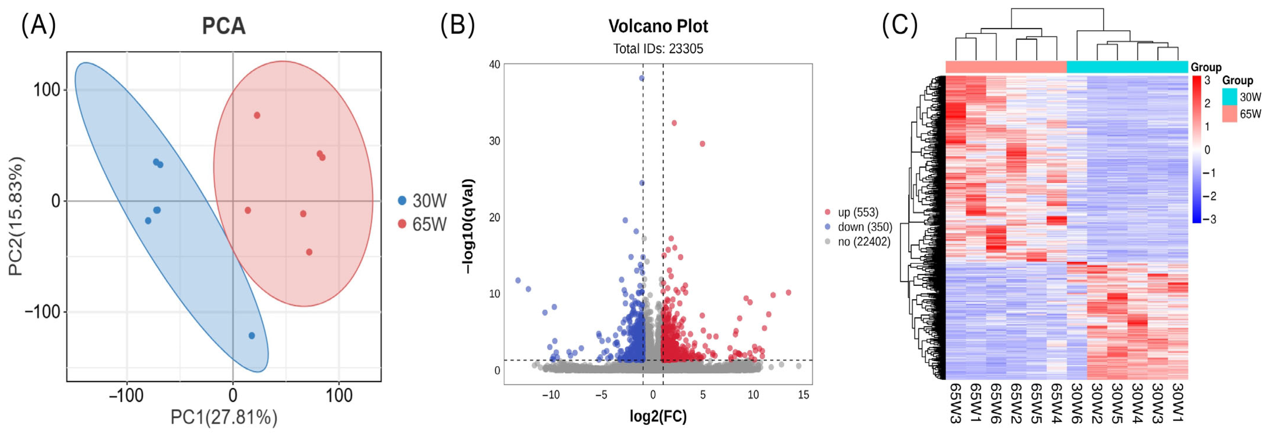
Principal component analysis showed clear separation between the two groups and close clustering of biological replicates within each group (Figure 1A). A total of 903 DEGs were identified. Using the 30 W group as the control, 553 genes were upregulated and 350 genes were downregulated in the 65 W group (Figure 1B). The DEGs also showed clear clustering patterns (Figure 1C).

3.4. Functional Enrichment of DEGs

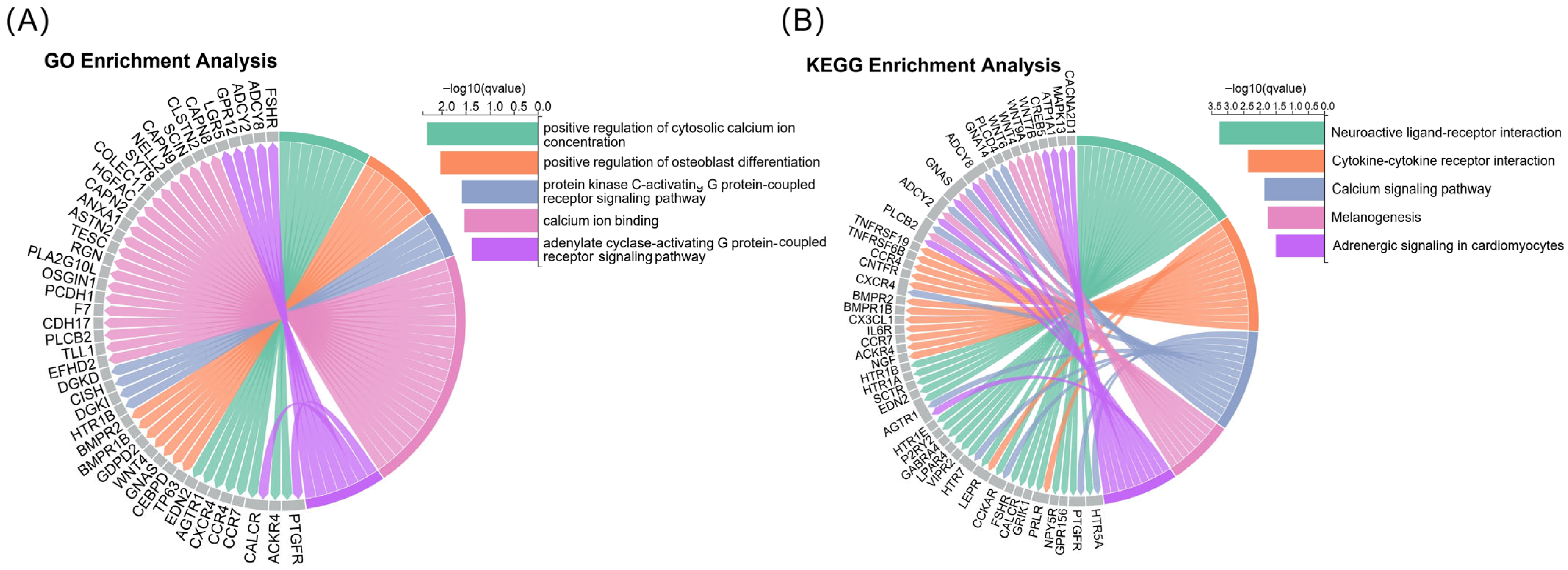
To investigate the functional and pathway enrichment of the identified genes, GO and KEGG enrichment analyses were performed for the DEGs. The enrichment analysis based on GO identified 1798 GO terms, of which 75 were significantly enriched (q < 0.05; Table S2). To specifically explore the molecular mechanisms of eggshell formation, five significantly enriched GO terms biologically relevant to eggshell mineralisation and ion transport were selected for further analysis: positive regulation of cytosolic calcium ion concentration, positive regulation of osteoblast differentiation, protein kinase C-activating G protein-coupled receptor signalling pathway, calcium ion binding, and adenylate cyclase-activating G protein-coupled receptor signalling pathway (Figure 2A). The pathway enrichment analysis based on KEGG identified 111 pathways, of which eight were significantly enriched (q < 0.05; Table S3). Following the same biologically-driven criteria, five significantly enriched KEGG pathways associated with eggshell mineralisation and uterine function were highlighted: neuroactive ligand–receptor interaction, cytokine–cytokine receptor interaction, calcium signalling pathway, melanogenesis, and adrenergic signalling in cardiomyocytes (Figure 2B). Genes in the selected GO terms and KEGG pathways were combined into a gene set designated Gene-G/K, comprising 75 DEGs. These enrichment patterns indicate that the transcriptional differences between the 30 W and 65 W groups were closely associated with processes involved in uterine ion homeostasis, Ca2+ signalling, and mineralisation-related regulation, providing a molecular link between altered uterine gene expression and the reduced ESS observed during the late laying period.

3.5. WGCNA Results

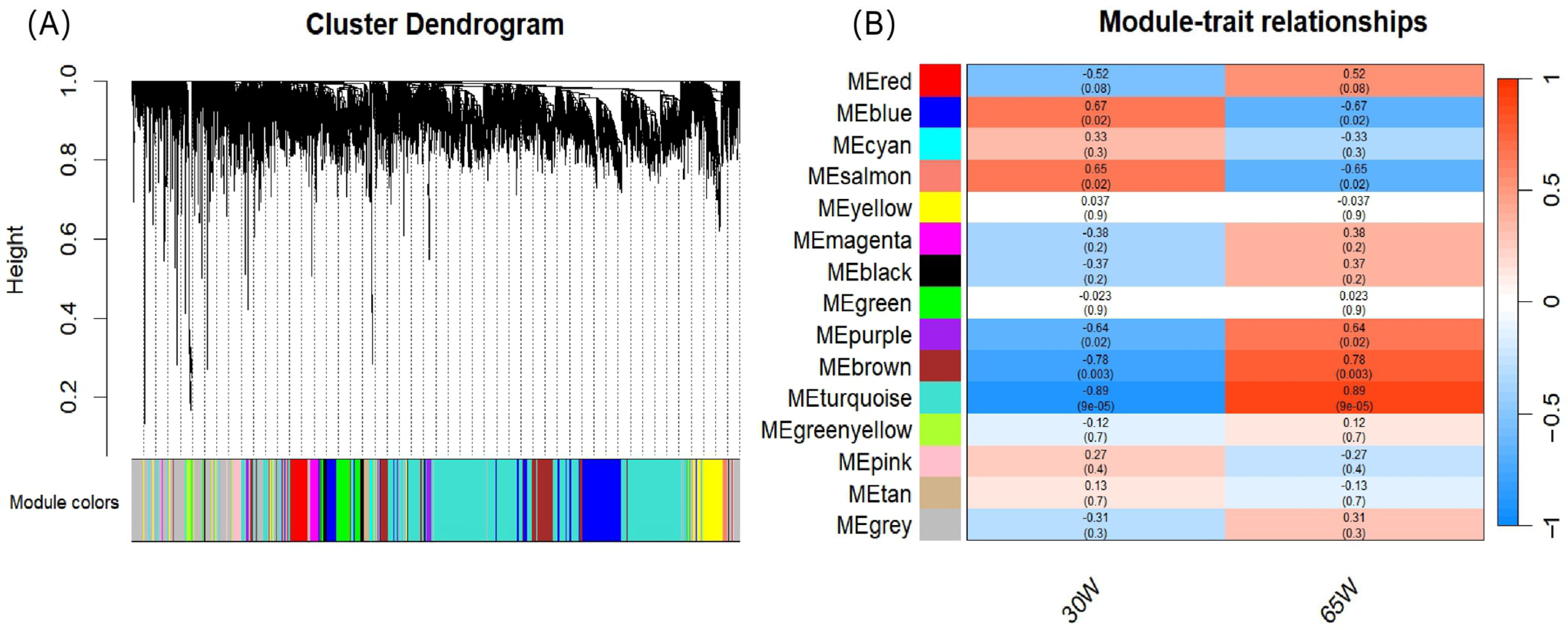
To further identify candidate genes associated with ESS, WGCNA was performed using genes identified in the uterine mucosa of the two groups. These genes were divided into 15 co-expression modules (Figure 3A). Module–trait correlation analysis showed that the turquoise and brown modules were significantly correlated with ESS (p < 0.01), with correlation coefficients of 0.89 and 0.78 and containing 2695 and 550 genes, respectively (Figure 3B and Table S4). The blue, salmon, and purple modules were significantly correlated with ESS (p < 0.05), with correlation coefficients of 0.67, 0.65, and 0.64 and gene numbers of 920, 51, and 106, respectively (Figure 3B and Table S4). Genes from the three modules with the highest correlation coefficients were combined into a gene set designated Gene-WGCNA, comprising 4039 genes.

3.6. Candidate Gene Screening

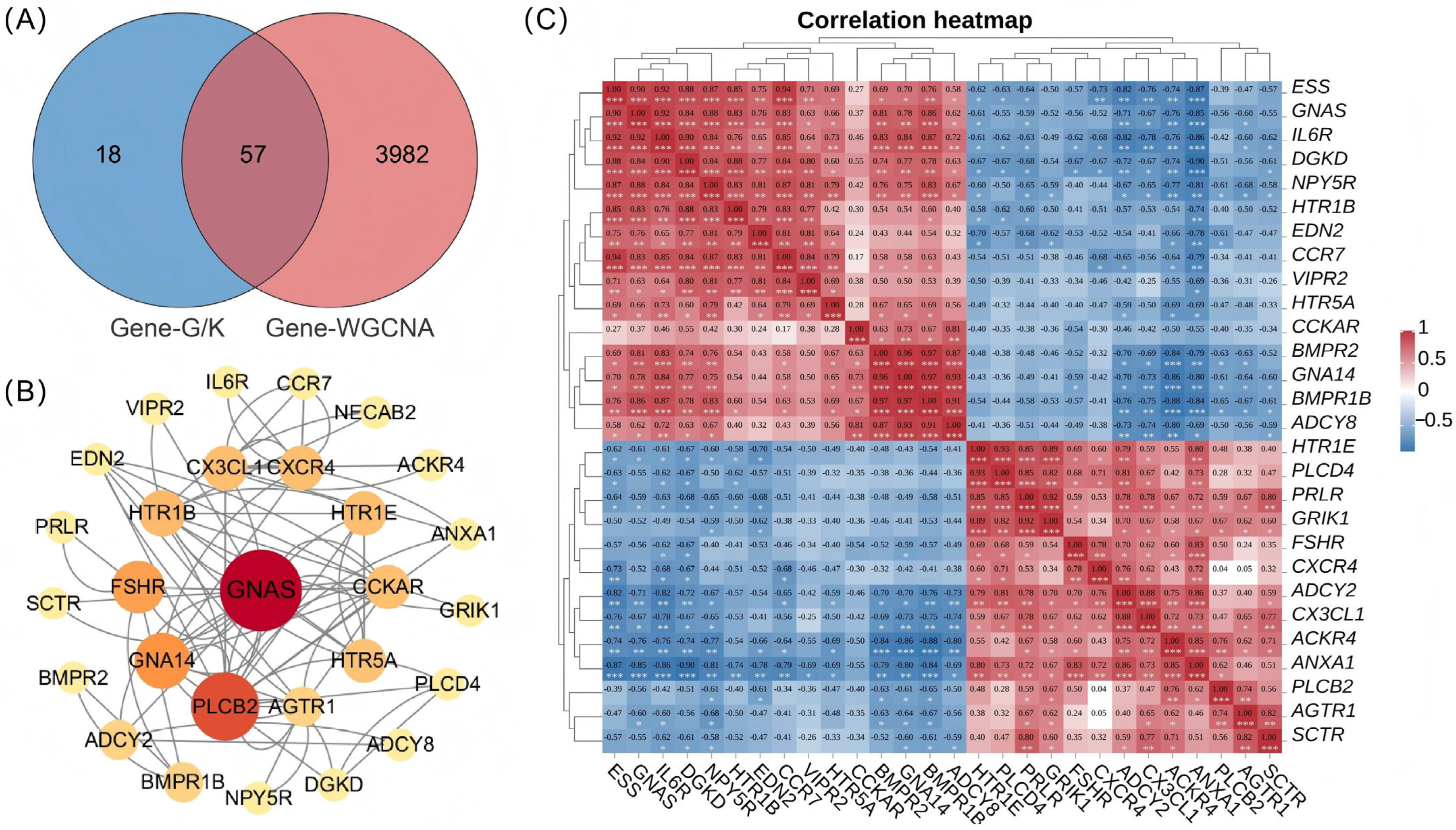
The intersection of the Gene-G/K and Gene-WGCNA gene sets yielded 57 DEGs that were highly associated with ESS and included in the pathways of interest (Figure 4A and Table S4). PPI analysis was performed for these 57 intersecting genes (Table S5), and GNAS had the highest BC value (Figure 4B), indicating that GNAS was a hub gene in the interaction network.
Pearson correlation analysis was performed between ESS and the interacting DEGs in the PPI network (Figure 4C). The results showed that GNAS (r = 0.90, p < 0.001), EDN2 (r = 0.75, p < 0.01), and BMPR2 (r = 0.69, p < 0.05) were significantly positively correlated with ESS. In contrast, ANXA1 (r = −0.87, p < 0.001) and CX3CL1 (r = −0.76, p < 0.01) were significantly negatively correlated with ESS. Notably, although PLCB2 showed only a negative correlation trend with ESS (r = −0.39, p > 0.05), its BC value ranked second only to that of GNAS in the PPI network, suggesting that it may have considerable topological importance within the network (Figure 4B,C). Collectively, six candidate genes were ultimately selected: GNAS, BMPR2, EDN2, ANXA1, PLCB2, and CX3CL1. The expression patterns and functional annotations of these genes further connected the age-related transcriptional changes in the uterine mucosa with biological processes relevant to eggshell mineralisation, including ion signalling, mineralisation regulation, and maintenance of the local uterine microenvironment.

3.7. qRT-PCR Validation of Candidate Genes

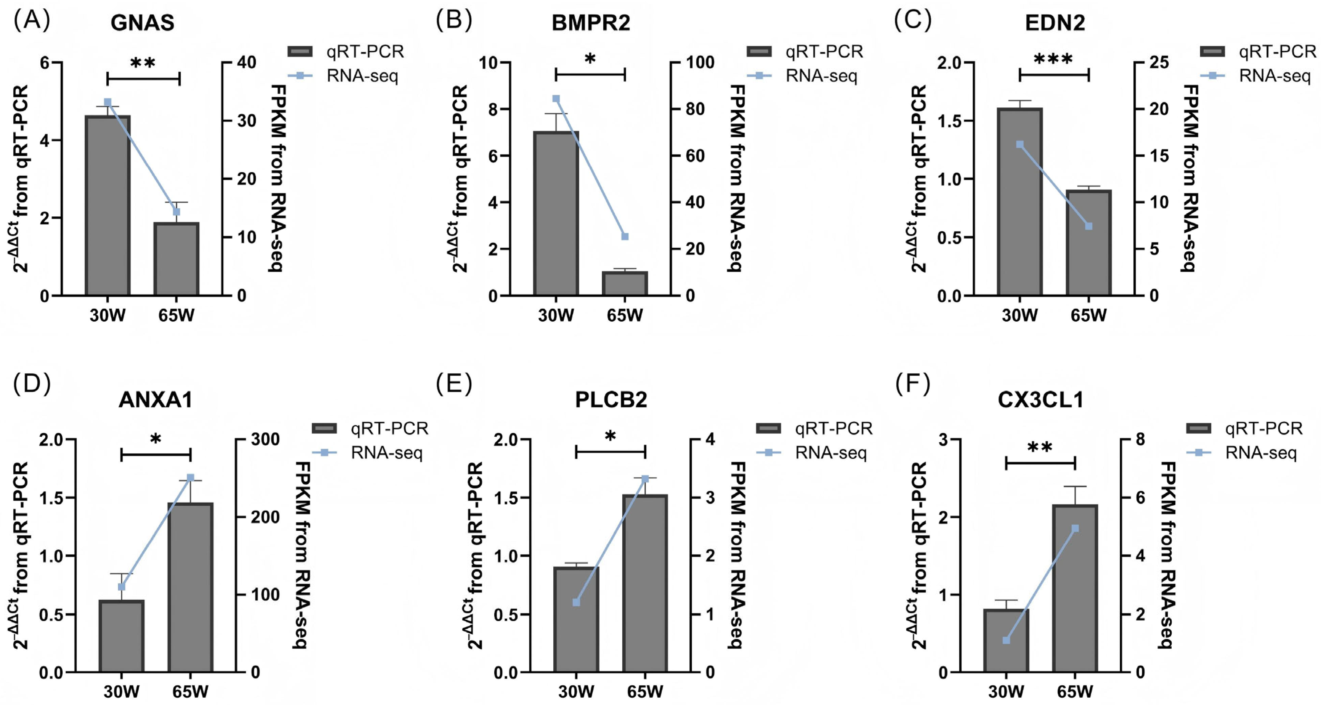
Candidate genes were validated by qRT-PCR in uterine mucosal tissue samples from each group. The qRT-PCR results were consistent with the expression trends obtained from RNA-seq, supporting the reliability of the RNA-seq results (Figure 5).

4. Discussion

The deterioration in eggshell quality during the late laying period increases egg breakage and causes economic losses. In the present study, phenotypic analysis showed that, compared with 30 W hens, 65 W hens had significantly higher EW but significantly lower ESS, whereas EST and SI did not differ significantly. These results suggest that the deterioration in eggshell quality during the late laying period is not necessarily accompanied by reduced shell thickness but may instead be associated with impaired shell mechanical properties and mineralisation. In addition to EST, shell weight may also influence eggshell quality, as greater shell deposition may contribute to improved shell mechanical properties; however, shell weight was not measured in the present study. It has been reported that eggshell breaking strength decreases from 5.8 kg at 33 weeks of age to 4.4 kg at 67 weeks of age, representing an approximately 25% reduction [26]. In a study of eggshells from laying hens aged 35 to 85 weeks, EW increased from 46.01 g to 52.75 g, and eggshell surface area increased from 60.06 cm2 to 65.78 cm2; in contrast, eggshell stiffness decreased from 67.45 N/mm to 45.96 N/mm. Eggshell organic matter content, phosphorus content, and mammillary density also decreased, suggesting that aging is accompanied by reduced eggshell mechanical performance and deterioration in mineralisation-related structures and chemical composition [9]. This may be closely associated with impaired uterine calcium transport [22]. A previous study classified 75-week-old laying hens into a high-eggshell-strength group (>42 N) and a low-eggshell-strength group (<32 N) and found marked differences in proteins and metabolites associated with uterine calcium transport between the two groups. Specifically, the low-strength group exhibited reduced calcium levels during the initial phase of eggshell mineralisation and redox imbalance during the growth phase [27]. In addition, increasing hen age alters the protein composition of the eggshell matrix, uterine fluid, and uterine extracellular vesicles. Extracellular vesicles in the uterine fluid can encapsulate and stabilise amorphous calcium carbonate (ACC) and deliver it to mineralisation sites, thereby preventing non-specific precipitation [28]. Therefore, age-related changes in the composition of the uterine fluid and its extracellular vesicles may impair the stable transport and deposition of mineralisation precursors. Meanwhile, increased inflammation of the uterine mucosa and impaired local health may affect ion transport, matrix deposition, and the stability of the mineralisation microenvironment, thereby contributing to deterioration in eggshell ultrastructure and mechanical properties [23,29].
Based on the reduced ESS observed in 65 W hens, we further investigated age-related transcriptional changes in the uterine mucosa. The DEGs were significantly enriched in pathways related to Ca2+ signalling, ion transport, GPCR-mediated signalling, and regulation of the local uterine microenvironment. Because eggshell mineralisation depends on the continuous supply and tightly coordinated transport of Ca2+ and HCO3 into the uterine fluid, these transcriptional alterations suggest that changes in uterine gene expression during the late laying period may affect the ionic and regulatory environment required for normal eggshell mineralisation. Further integration of functional enrichment, WGCNA, and PPI network analysis prioritised six candidate genes, namely GNAS, BMPR2, EDN2, ANXA1, PLCB2, and CX3CL1. These genes were mainly associated with second-messenger signalling, mineralisation-related regulation and local inflammatory homeostasis, providing a potential molecular link between uterine transcriptional changes and the decline in eggshell mineralisation during the late laying period.
GNAS was significantly downregulated in the uterine mucosa of 65 W hens and was positively correlated with ESS, suggesting that it may be an important node linking receptor signalling, ion transport, and mineralisation regulation. GNAS encodes Gsα, the stimulatory α-subunit of the heterotrimeric G protein, which mediates GPCR signalling [30,31]. Upon GPCR activation, Gsα stimulates adenylyl cyclase (AC), thereby increasing intracellular cAMP concentrations [32,33]. As a second messenger, cAMP can activate two major downstream effectors: protein kinase A (PKA) and exchange protein directly activated by cAMP (EPAC) [34,35]. Studies using uterine tissues mounted in Ussing chambers have shown that forskolin, an AC activator, increases cAMP levels and enhances transepithelial anion transport across the uterine epithelium, thereby increasing HCO3 secretion. Moreover, HCO3 secretion is positively correlated with Ca2+ secretion [36,37]. In the uterine epithelium, carbonic anhydrase (CA) catalyses the hydration of CO2 to generate HCO3, which is subsequently secreted into the uterine fluid via anion exchangers such as SLC26A9 [38]. In epithelial tissues such as the intestine and airway, cAMP induces HCO3 secretion through the CFTR–SLC26 axis, suggesting that this regulatory mechanism is conserved across epithelial tissues [39,40]. Therefore, downregulation of GNAS in 65 W hens may inhibit the GNAS–AC–cAMP–PKA/EPAC signalling pathway and reduce HCO3 secretion through the CFTR–SLC26 axis. However, direct functional evidence that GNAS regulates HCO3 secretion in the uterine epithelium is still lacking. In the present study, pathway enrichment analysis showed that GNAS was involved in the calcium signalling pathway. Recent studies have shown that activation of Gs-coupled GPCRs can increase cytosolic Ca2+ levels through several mechanisms, including PKA-mediated regulation of Ca2+ channels, cAMP–EPAC–PLCε signalling, enhanced sensitivity of inositol 1,4,5-trisphosphate (IP3) receptors, and the Gs–Gβγ–PLCβ–Ca2+ pathway [41]. These findings suggest that GNAS may participate in intracellular Ca2+ mobilisation in uterine cells. Therefore, the reduced expression of GNAS in 65 W hens may impair the coordinated supply of Ca2+ and HCO3 required for eggshell mineralisation, thereby affecting the stability of the ionic environment in uterine fluid and the supply of mineralisation precursors.
Unlike GNAS-mediated Gsα–AC–cAMP signalling, PLCB2 is mainly involved in intracellular Ca2+ mobilisation through membrane phospholipid hydrolysis. In the present study, PLCB2 was significantly upregulated in the uterine mucosa of 65 W hens. PLCB2 encodes phospholipase Cβ2 (PLCβ2), a member of the PLCβ family. As a canonical downstream effector of GPCRs, PLCβ2 catalyses the hydrolysis of the membrane phospholipid phosphatidylinositol 4,5-bisphosphate (PIP2) to generate two second messengers, IP3 and diacylglycerol (DAG) [42]. IP3 binds to its receptors and induces Ca2+ release from the endoplasmic reticulum, whereas DAG further activates downstream effectors such as protein kinase C (PKC). Together, these second messengers regulate intracellular Ca2+ mobilisation and membrane-associated responses [42]. Because anion secretion and transepithelial transport in epithelial tissues are regulated by the spatiotemporal coordination of cAMP and Ca2+ signalling [43], upregulation of PLCB2 may transiently enhance Ca2+ responsiveness but may not necessarily help maintain mineralisation homeostasis. Instead, elevated PLCB2 expression may reflect dysregulated Ca2+-dependent signalling in the aged uterus, thereby disturbing the coordinated ion transport and mineralisation precursor supply required for eggshell formation. Together, the downregulation of GNAS and upregulation of PLCB2 suggest that uterine Ca2+ signalling during the late laying period may be dysregulated rather than simply weakened, potentially contributing to impaired eggshell mineralisation.
BMPR2 and GNAS were both enriched in the GO-term positive regulation of osteoblast differentiation. BMPR2 was positively correlated with ESS (r = 0.69). BMPR2 encodes bone morphogenetic protein receptor type II, a member of the bone morphogenetic protein (BMP) receptor family, and functions as a transmembrane receptor with serine/threonine kinase activity. After BMP ligands bind to their receptors, SMAD1/5/8 phosphorylation is induced. Phosphorylated SMAD1/5/8 then forms a complex with SMAD4 and translocates into the nucleus, where it regulates the expression of osteogenic transcription factors such as RUNX2, thereby promoting osteoprogenitor cell differentiation, matrix formation, and mineral deposition [44]. Notably, BMPR2 has been identified as a bone-metabolism-related candidate gene that responds to changes in photoperiod. Its expression pattern is similar to fluctuations in serum 1,25-dihydroxyvitamin D3 [1,25(OH)2D3], and it exhibits distinct dynamic expression patterns under different photoperiodic conditions, suggesting that BMPR2 may participate in the rhythmic regulation of eggshell formation [15]. Although eggshell mineralisation occurs in the uterus rather than in bone tissue, both processes are forms of ordered biomineralisation. Therefore, downregulation of BMPR2 may reflect a weakened uterine response to mineralisation-regulatory signals.
The local uterine microenvironment in aged laying hens may also affect eggshell mineralisation. EDN2 encodes endothelin-2, a peptide molecule with potent vasoactive and contractile regulatory functions. The EDN2–EDNR signalling axis has been shown to be functionally active in chickens [45,46]. Before ovulation, EDN2 acts as a transient and local regulatory signal; its expression increases briefly in granulosa cells and then acts through receptors on smooth muscle-like cells in the outer follicular layer, inducing local contraction and promoting follicular rupture and oocyte release [46]. Activation of endothelin receptors can also stimulate intracellular Ca2+ signalling, the MAPK/ERK pathway, and the cAMP/PKA pathway [45]. Notably, EDN2 was annotated to the GO term “positive regulation of cytosolic calcium ion concentration” [22]. In proteomic studies of uterine fluid and extracellular vesicles, EDN2 was identified as a differentially abundant protein associated with intracellular calcium homeostasis in the low-eggshell-strength group [29]. Therefore, EDN2 may participate in eggshell formation by regulating local Ca2+ homeostasis and maintaining the uterine microenvironment.
The expression patterns of ANXA1 and CX3CL1 suggest inflammatory microenvironmental remodelling in the uterus during the late laying period. ANXA1 is a Ca2+-dependent phospholipid-binding protein that is mainly involved in membrane trafficking, regulation of inflammation, and tissue repair [47,48]. In chicken uterine omics studies, members of the annexin family have been identified as a candidate gene family associated with ESS, suggesting that this family may participate in maintaining local homeostasis in the uterus [49]. In contrast, CX3CL1 is a chemokine with both adhesive and chemotactic functions; it can mediate immune cell recruitment and participate in inflammatory responses, promote M1 macrophage polarisation, and enhance responses related to tissue remodelling [50,51]. The upregulation of both genes at 65 W suggests that the uterus may not only have reduced mineralisation capacity but also exhibit inflammatory activation and microenvironmental imbalance.
The altered expression patterns of the six candidate genes may collectively represent a putative regulatory network associated with impaired eggshell mineralisation during the late laying period. Specifically, reduced GNAS expression and increased PLCB2 expression suggest dysregulation of second-messenger and Ca2+-dependent signalling, whereas reduced BMPR2 expression may indicate attenuated mineralisation-related signalling. Altered EDN2 expression may reflect changes in local Ca2+ homeostasis, while the upregulation of ANXA1 and CX3CL1 suggests inflammatory and microenvironmental remodelling. Together, these transcriptional changes may disturb the coordinated supply of mineralisation precursors, ion homeostasis, and the local uterine microenvironment, thereby contributing to impaired eggshell mineralisation and reduced ESS during the late laying period. Specifically, GNAS and PLCB2 jointly reflect an imbalance in second-messenger signalling and ion secretion; BMPR2 and EDN2 point to weakened mineralisation signalling and reduced capacity to maintain local uterine function; and ANXA1 and CX3CL1 indicate changes in the inflammatory microenvironment in the uterus of aged laying hens. Together, these changes may weaken the regulation of mineralisation precursor supply and local homeostasis in the uterus, thereby contributing to reduced ESS.
This study has several limitations. Candidate gene selection was mainly based on phenotypic differences, transcriptomic analysis, and correlation results, and direct functional validation is still lacking. Future studies could integrate analyses of uterine fluid ion composition, eggshell ultrastructure, cell-level overexpression, and knockdown experiments to clarify the specific roles of these candidate genes in eggshell formation. In conclusion, this study compared eggshell phenotypes between 30 W and 65 W WL laying hens and identified six candidate genes through transcriptomic analysis of the uterine mucosa. This provides novel candidate targets for elucidating the molecular mechanisms underlying the decline in ESS and may facilitate improvements in eggshell quality and the sustainable development of the laying hen industry.

5. Conclusions

This study revealed important molecular features associated with the decline in ESS in WL laying hens during the late laying period. RNA-seq and bioinformatics analyses of the uterine mucosa identified key pathways closely related to eggshell mineralisation, as well as six candidate genes: GNAS, BMPR2, EDN2, ANXA1, PLCB2, and CX3CL1. Collectively, these genes may form a regulatory network involved in maintaining uterine ionic homeostasis, regulating mineralisation, and remodelling the local microenvironment. These findings provide a theoretical basis for elucidating the molecular mechanisms underlying the deterioration in eggshell quality in aged laying hens.

Supplementary Materials

The following supporting information can be downloaded at: https://www.mdpi.com/article/10.3390/agriculture16171809/s1, Table S1: Sequence alignment between the sequenced samples and the reference genome; Table S2: GO functional enrichment analysis results; Table S3: KEGG pathway enrichment analysis results; Table S4: Gene sets of WGCNA modules; Table S5: PPI pairs of overlapping genes retrieved from the STRING database.

Author Contributions

Conceptualization, H.F.; methodology, H.F.; visualization, H.F.; writing—original draft preparation, H.F. and L.W.; validation, X.H. and S.W.; resources, B.Z. and H.Z.; writing—review and editing, B.Z. and H.Z.; supervision, B.Z. and H.Z.; funding acquisition, H.Z. All authors have read and agreed to the published version of the manuscript.

Funding

This research was funded by Key Research and Development Program of Shandong (2025LZGC048), the China Agricultural Research System (CARS-39-K05), and the Chinese Universities Scientific Fund (2026TC124).

Institutional Review Board Statement

The study was conducted in accordance with the guidelines of the Animal Welfare Committee of the State Key Laboratory of Agricultural Biotechnology, China Agricultural University, and approved by the Animal Welfare Committee (approval number: AW80203202-1-1 and date of approval: [8 February 2023]).

Data Availability Statement

The original data presented in the study are openly available in NCBI (PRJNA1497917).

Acknowledgments

During the preparation of this manuscript, the authors used ChatGPT (GPT-5.5) for grammar-checking purposes. The authors have reviewed and edited the output and take full responsibility for the content of this publication.

Conflicts of Interest

The authors declare no conflicts of interest.

Abbreviations

The following abbreviations are used in this manuscript:
WLWhite Leghorn
30 W30 weeks of age
65 W65 weeks of age
ESSEggshell strength
ESTEggshell thickness
SIShape index
EWEgg weight
RNA-seqRNA sequencing
DEGsDifferentially expressed genes
GOGene Ontology
KEGGKyoto Encyclopedia of Genes and Genomes
WGCNAWeighted gene co-expression network analysis
PPIProtein–protein interaction

References

  1. Li, W.; Cao, Z.; Xu, F.; Zhang, X.; Sun, Y.; Xie, Z.; Ning, C.; Zhang, Q.; Wang, D.; Tang, H. Whole transcriptome sequencing reveals key genes and cerna regulatory networks associated with pimpled eggs in hens. Poult. Sci. 2024, 103, 12. [Google Scholar] [CrossRef] [Scilit] [PubMed]
  2. Ahmed, M.W.; Khaliduzzaman, A.; Emmert, J.L.; Kamruzzaman, M. An overview of recent advancements in hyperspectral imaging in the egg and hatchery industry. Comput. Electron. Agric. 2025, 230, 16. [Google Scholar] [CrossRef] [Scilit]
  3. Luo, J.; Sun, T.; Jiang, S.; Yang, Z.; Xiao, C.; Deng, J.; Zhou, B.; Yang, X. Comprehensive analysis of non-coding rnas in the ovaries of high and low egg production hens. Anim. Reprod. Sci. 2025, 276, 11. [Google Scholar] [CrossRef] [Scilit] [PubMed]
  4. Wu, L.; Zhang, T.; Luo, Z.; Xiao, H.; Wang, D.; Wu, C.; Fang, X.; Li, J.; Zhou, J.; Miao, J.; et al. Impact of gut microbial diversity on egg production performance in chickens. Microbiol. Spectr. 2025, 13, 17. [Google Scholar] [CrossRef] [Scilit] [PubMed]
  5. Hamilton, R.M.G.; Bryden, W.L. Relationship between egg shell breakage and laying hen housing systems—An overview. World’s Poult. Sci. J. 2021, 77, 249–266. [Google Scholar] [CrossRef] [Scilit]
  6. Brionne, A.; Nys, Y.; Hennequet-Antier, C.; Gautron, J. Hen uterine gene expression profiling during eggshell formation reveals putative proteins involved in the supply of minerals or in the shell mineralization process. BMC Genom. 2014, 15, 17. [Google Scholar] [CrossRef] [Scilit] [PubMed]
  7. Park, J.; Sohn, S. The influence of hen aging on eggshell ultrastructure and shell mineral components. Korean J. Food Sci. Anim. Resour. 2018, 38, 1080–1091. [Google Scholar] [CrossRef] [Scilit] [PubMed]
  8. Qiu, J.L.; Zhou, Q.; Zhu, J.M.; Lu, X.T.; Liu, B.; Yu, D.Y.; Lin, G.; Ao, T.; Xu, J.M. Organic trace minerals improve eggshell quality by improving the eggshell ultrastructure of laying hens during the late laying period. Poult. Sci. 2020, 99, 1483–1490. [Google Scholar] [CrossRef] [Scilit] [PubMed]
  9. Fu, Y.; Zhao, D.; Gao, L.; Zhang, H.; Feng, J.; Min, Y.; Qi, G.; Wang, J. Tmt-based quantitative proteomic analysis reveals age-related changes in eggshell matrix proteins and their correlation with eggshell quality in xinyang blue-shelled laying hens. Poult. Sci. 2025, 104, 11. [Google Scholar] [CrossRef] [Scilit] [PubMed]
  10. Khan, S.; Wu, S.; Roberts, J. Rna-sequencing analysis of shell gland shows differences in gene expression profile at two time-points of eggshell formation in laying chickens. BMC Genom. 2019, 20, 20. [Google Scholar] [CrossRef] [Scilit] [PubMed]
  11. Feng, J.; Lu, M.; Ma, L.; Zhang, H.; Wu, S.; Qiu, K.; Min, Y.; Qi, G.; Wang, J. Uterine inflammation status modulates eggshell mineralization via calcium transport and matrix protein synthesis in laying hens. Anim. Nutr. 2023, 13, 411–425. [Google Scholar] [CrossRef] [Scilit] [PubMed]
  12. Chen, X.; Li, X.; Zhong, C.; Jiang, X.; Wu, G.; Li, G.; Yan, Y.; Yang, N.; Sun, C. Genetic patterns and genome-wide association analysis of eggshell quality traits of egg-type chicken across an extended laying period. Poult. Sci. 2024, 103, 9. [Google Scholar] [CrossRef] [Scilit] [PubMed]
  13. Li, Z.; Wu, H.; Fu, J.; Mushtaq, M.; Khan, M.; Liu, Y.; Azeem, Z.; Shi, H.; He, Y.; Zhang, R.; et al. Eggshell quality traits and transcriptome gene screening between yunnong and jingfen chicken breeds. Biology 2024, 13, 1048. [Google Scholar] [CrossRef] [Scilit] [PubMed]
  14. Lu, Y.; Xu, H.; Hu, Z.; Li, D.; Rustempasic, A.; Zhou, Y.; Deng, Q.; Pu, J.; Zhao, X.; Zhang, Y.; et al. Probiotics improve eggshell quality via regulating microbial composition in the uterine and cecum. Poult. Sci. 2025, 104, 13. [Google Scholar] [CrossRef] [Scilit] [PubMed]
  15. Liu, X.; Shi, L.; Su, B.; Liu, A.; Wang, D.; Chen, Y.; Hao, E.; Bai, H.; Sun, Y.; Li, Y.; et al. Long-24-h ahemeral light cycle improved eggshell quality of hens in late laying period. Poult. Sci. 2025, 104, 8. [Google Scholar] [CrossRef] [Scilit] [PubMed]
  16. Li, S.; Zhao, X.; Zheng, X.; Chen, H.; Zhou, R.; Shi, L.; Liu, H.; Xu, L.; Ning, Z.; Wang, D. Study on changes in egg quality traits and genetic parameters of White Leghorn hens from 35 to 100 weeks of age. Poult. Sci. 2025, 104, 105502. [Google Scholar] [CrossRef] [Scilit] [PubMed]
  17. Xu, X.; Yang, Y.; Zhang, X.; Wang, D.; Li, Q.; Chang, Z.; Ni, A.; Huang, Z.; Yuan, J.; Sun, Y.; et al. Research note: Genetic parameters estimation of egg quality traits in Beijing-You chickens. Poult. Sci. 2025, 104, 105294. [Google Scholar] [CrossRef] [Scilit] [PubMed]
  18. Han, G.P.; Kim, J.H.; Kim, J.M.; Kil, D.Y. Transcriptomic analysis of the liver in aged laying hens with different eggshell strength. Poult. Sci. 2023, 102, 15. [Google Scholar] [CrossRef] [Scilit] [PubMed]
  19. Zhao, D.; Gao, L.; Gong, F.; Feng, J.; Zhang, H.; Wu, S.; Wang, J.; Min, Y. Tmt-based quantitative proteomic analysis reveals eggshell matrix protein changes correlated with eggshell quality in jing tint 6 laying hens of different ages. Poult. Sci. 2024, 103, 12. [Google Scholar] [CrossRef] [Scilit] [PubMed]
  20. Fu, Y.; Wang, J.; Schroyen, M.; Chen, G.; Zhang, H.; Wu, S.; Li, B.; Qi, G. Effects of rearing systems on the eggshell quality, bone parameters and expression of genes related to bone remodeling in aged laying hens. Front. Physiol. 2022, 13, 14. [Google Scholar] [CrossRef] [Scilit] [PubMed]
  21. Alfonso-Carrillo, C.; Benavides-Reyes, C.; de Los Mozos, J.; Dominguez-Gasca, N.; Sanchez-Rodriguez, E.; Garcia-Ruiz, A.I.; Rodriguez-Navarro, A.B. Relationship between bone quality, egg production and eggshell quality in laying hens at the end of an extended production cycle (105 weeks). Animals 2021, 11, 623. [Google Scholar] [CrossRef] [Scilit] [PubMed]
  22. Fu, Y.; Zhou, J.; Schroyen, M.; Zhang, H.; Wu, S.; Qi, G.; Wang, J. Decreased eggshell strength caused by impairment of uterine calcium transport coincide with higher bone minerals and quality in aged laying hens. J. Anim. Sci. Biotechnol. 2024, 15, 20. [Google Scholar] [CrossRef] [Scilit] [PubMed]
  23. Nii, T.; Sugiura, T.; Suzuki, N.; Isobe, N.; Yoshimura, Y. Effects of aging on the microbiota and inflammatory status of the intestinal and oviductal mucosa in laying hens. J. Poult. Sci. 2025, 62, 12. [Google Scholar] [CrossRef] [Scilit] [PubMed]
  24. Feng, J.; Zhang, H.; Wu, S.; Qi, G.; Wang, J. Uterine transcriptome analysis reveals mrna expression changes associated with the ultrastructure differences of eggshell in young and aged laying hens. BMC Genom. 2020, 21, 15. [Google Scholar] [CrossRef] [Scilit] [PubMed]
  25. Park, J.A.; Cho, E.J.; Park, J.Y.; Sohn, S.H. Histological change of uterus endometrium and expression of the eggshell-related genes according to hen age. Korean J. Poult. Sci. 2017, 44, 19–28. [Google Scholar] [CrossRef] [Scilit]
  26. Benavides-Reyes, C.; Folegatti, E.; Dominguez-Gasca, N.; Litta, G.; Sanchez-Rodriguez, E.; Rodriguez-Navarro, A.B.; Umar Faruk, M. Research note: Changes in eggshell quality and microstructure related to hen age during a production cycle. Poult. Sci. 2021, 100, 101287. [Google Scholar] [CrossRef] [Scilit] [PubMed]
  27. Yang, Y.; Dai, D.; Zhang, H.; Wu, S.; Qi, G.; Wang, J. The characterization of uterine calcium transport and metabolism during eggshell calcification of hens laying high or low breaking strength eggshell. Poult. Sci. 2025, 104, 105111. [Google Scholar] [CrossRef] [Scilit] [PubMed]
  28. Stapane, L.; Le Roy, N.; Ezagal, J.; Rodriguez-Navarro, A.B.; Labas, V.; Combes-Soia, L.; Hincke, M.T.; Gautron, J. Avian eggshell formation reveals a new paradigm for vertebrate mineralization via vesicular amorphous calcium carbonate. J. Biol. Chem. 2020, 295, 15853–15869. [Google Scholar] [CrossRef] [Scilit] [PubMed]
  29. Gao, L.; Fu, Y.; Guo, Y.; Zhang, H.; Qi, G.; Wang, J. Proteomic changes in extracellular vesicle and uterine fluid proteins correlated with eggshell quality in aged laying hens. Poult. Sci. 2025, 104, 105939. [Google Scholar] [CrossRef] [Scilit] [PubMed]
  30. de Oliveira, E.M.; Keogh, J.M.; Talbot, F.; Henning, E.; Ahmed, R.; Perdikari, A.; Bounds, R.; Wasiluk, N.; Ayinampudi, V.; Barroso, I.; et al. Obesity-associated gnas mutations and the melanocortin pathway. N. Engl. J. Med. 2021, 385, 1581–1592. [Google Scholar] [CrossRef] [Scilit] [PubMed]
  31. Cui, Q.; Aksu, C.; Ay, B.; Remillard, C.E.; Plagge, A.; Gardezi, M.; Dunlap, M.; Gerstenfeld, L.C.; He, Q.; Bastepe, M. Maternal gnas contributes to the extra-large g protein α-subunit (xlαs) expression in a cell type-specific manner. Front. Genet. 2021, 12, 10. [Google Scholar] [CrossRef] [Scilit] [PubMed]
  32. Ripoll, L.; von Zastrow, M.; Blythe, E.E. Intersection of gpcr trafficking and camp signaling at endomembranes. J. Cell Biol. 2025, 224, 16. [Google Scholar] [CrossRef] [Scilit] [PubMed]
  33. Zhang, M.; Chen, T.; Lu, X.; Lan, X.; Chen, Z.; Lu, S. G protein-coupled receptors (gpcrs): Advances in structures, mechanisms, and drug discovery. Signal Transduct. Target. Ther. 2024, 9, 43. [Google Scholar] [CrossRef] [Scilit] [PubMed]
  34. Zhang, H.; Liu, Y.; Liu, J.; Chen, J.; Wang, J.; Hua, H.; Jiang, Y. Camp-pka/epac signaling and cancer: The interplay in tumor microenvironment. J. Hematol. Oncol. 2024, 17, 31. [Google Scholar] [CrossRef] [Scilit] [PubMed]
  35. Ahmed, M.B.; Alghamdi, A.A.A.; Islam, S.U.; Lee, J.; Lee, Y. Camp signaling in cancer: A pka-creb and epac-centric approach. Cells 2022, 11, 2020. [Google Scholar] [CrossRef] [Scilit] [PubMed]
  36. Brockmeier, K.; Schultheiss, G. Regulation of anion transport across the uterine epithelium of Gallus domesticus. Poult. Sci. 2011, 90, 618–623. [Google Scholar] [CrossRef] [Scilit] [PubMed]
  37. Vetter, A.E.; O’Grady, S.A. Sodium and anion transport across the avian uterine (shell gland) epithelium. J. Exp. Biol. 2005, 208, 479–486. [Google Scholar] [CrossRef] [Scilit] [PubMed]
  38. Jonchere, V.; Brionne, A.; Gautron, J.; Nys, Y. Identification of uterine ion transporters for mineralisation precursors of the avian eggshell. BMC Physiol. 2012, 12, 10. [Google Scholar] [CrossRef] [Scilit] [PubMed]
  39. Becker, H.M.; Seidler, U.E. Bicarbonate secretion and acid/base sensing by the intestine. Pflug. Arch. 2024, 476, 593–610. [Google Scholar] [CrossRef] [Scilit] [PubMed]
  40. Mall, M.A.; Burgel, P.; Castellani, C.; Davies, J.C.; Salathe, M.; Taylor-Cousar, J.L. Cystic fibrosis. Nat. Rev. Dis. Prim. 2024, 10, 26. [Google Scholar]
  41. Brands, J.; Bravo, S.; Jürgenliemke, L.; Grätz, L.; Schihada, H.; Frechen, F.; Alenfelder, J.; Pfeil, C.; Ohse, P.G.; Hiratsuka, S.; et al. A molecular mechanism to diversify ca(2+) signaling downstream of gs protein-coupled receptors. Nat. Commun. 2024, 15, 7684. [Google Scholar] [CrossRef] [Scilit] [PubMed]
  42. Ubeysinghe, S.; Wijayaratna, D.; Kankanamge, D.; Karunarathne, A. Molecular regulation of plcβ signaling. Methods Enzymol. 2023, 682, 17–52. [Google Scholar] [CrossRef] [Scilit] [PubMed]
  43. Varga, A.; Kiss, A.; Crul, T.; Madácsy, T.; Pallagi, P.; Maléth, J. Beyond the mutations: Spatiotemporal regulation of cftr by camp and calcium signaling in epithelial physiology and cystic fibrosis. Cell. Mol. Biol. Lett. 2025, 31, 10. [Google Scholar] [CrossRef] [Scilit] [PubMed]
  44. Chen, S.; Jiang, T.; Liu, Q.; Liu, Z.; Su, Y.; Su, H. Hsa_circ_0001485 promoted osteogenic differentiation by targeting bmpr2 to activate the tgfβ-bmp pathway. Stem Cell Res. Ther. 2022, 13, 453. [Google Scholar] [CrossRef] [Scilit] [PubMed]
  45. Liu, H.; Luo, Q.; Zhang, J.; Mo, C.; Wang, Y.; Li, J. Endothelins (edn1, edn2, edn3) and their receptors (ednra, ednrb, ednrb2) in chickens: Functional analysis and tissue distribution. Gen. Comp. Endocrinol. 2019, 283, 113231. [Google Scholar] [CrossRef] [Scilit] [PubMed]
  46. Ko, C.J.; Cho, Y.M.; Ham, E.; Cacioppo, J.A.; Park, C.J. Endothelin 2: A key player in ovulation and fertility. Reproduction 2022, 163, R71–R80. [Google Scholar] [CrossRef] [Scilit] [PubMed]
  47. Dai, Q.; Zheng, J.; Fu, M.; Qin, S.; Fu, X. Research progress of annexin a1 and its derived peptides in the diagnosis and treatment of circulatory diseases. Immun. Inflamm. Dis. 2025, 13, e70249. [Google Scholar] [CrossRef] [Scilit] [PubMed]
  48. Gerke, V.; Gavins, F.N.E.; Geisow, M.; Grewal, T.; Jaiswal, J.K.; Nylandsted, J.; Rescher, U. Annexins-a family of proteins with distinctive tastes for cell signaling and membrane dynamics. Nat. Commun. 2024, 15, 1574. [Google Scholar] [CrossRef] [Scilit] [PubMed]
  49. Zhang, X.; Chen, J.; Sun, Y.; Li, Q.; Ma, P.; Du, H.; Yang, H.; Li, X.; Xu, X.; Ma, H.; et al. Multi-omics reveals key cell types and gene families regulating eggshell strength in chicken uteri. Zool. Res. 2025, 46, 1396–1410. [Google Scholar] [PubMed]
  50. Hu, X.; Huang, X.; Yin, T.; Chen, J.; Zhao, W.; Yu, M.; Liu, L.; Du, M. Cx3cl1 (fractalkine): An important cytokine in physiological and pathological pregnancies. J. Reprod. Immunol. 2024, 166, 104392. [Google Scholar] [CrossRef] [Scilit] [PubMed]
  51. Deng, Z.; Zhong, W. Cx3cl1 promotes m1 macrophage polarization and osteoclast differentiation via nsun5-mediated m5c modification. Sci. Rep. 2025, 15, 25246. [Google Scholar] [CrossRef] [Scilit] [PubMed]
Figure 1. Transcriptome analysis of the uterine mucosa. Note: (A) PCA of uterine mucosal transcriptomes from the 30 W and 65 W groups. The coloured shaded areas indicate the clustering of samples within the corresponding groups. (B) Volcano plot of DEGs. The vertical and horizontal dashed lines indicate the thresholds for differential expression (|log2 fold change| = 1 and q value = 0.05), respectively. (C) Hierarchical clustering heatmap of DEGs. 30 W, 30 weeks of age; 65 W, 65 weeks of age; PCA, principal component analysis; DEGs, differentially expressed genes.
Agriculture 16 01809 g001
Figure 2. GO and KEGG enrichment analysis of DEGs. Note: (A) Significantly enriched GO terms associated with eggshell mineralisation. (B) Significantly enriched KEGG pathways associated with eggshell mineralisation.
Agriculture 16 01809 g002
Figure 3. WGCNA of uterine mucosal genes at 30 W and 65 W. Note: (A) Cluster dendrogram. (B) Relationships between modules and ESS. Correlation coefficients and p values are shown in each cell. WGCNA, weighted gene co-expression network analysis; 30 W, 30 weeks of age; 65 W, 65 weeks of age.
Agriculture 16 01809 g003
Figure 4. Candidate gene screening. Note: (A) Intersection between the Gene-G/K and Gene-WGCNA gene sets. (B) PPI network of intersecting genes. (C) Correlation analysis between genes and ESS. Asterisks indicate statistical significance of the Pearson correlations: * p < 0.05, ** p < 0.01, and *** p < 0.001.
Agriculture 16 01809 g004
Figure 5. Candidate gene expression validated by qRT-PCR and RNA-seq. Note: (A) Relative expression of GNAS. (B) Relative expression of BMPR2. (C) Relative expression of EDN2. (D) Relative expression of ANXA1. (E) Relative expression of PLCB2. (F) Relative expression of CX3CL1. Bars indicate qRT-PCR results, and the line plot indicates RNA-seq data. * p < 0.05; ** p < 0.01; *** p < 0.001.
Agriculture 16 01809 g005
Table 1. qRT-PCR primers.
GeneSequence (5′ → 3′)Product Length/bp
GNASF: CATGCACCTTCGCCAATACG226
R: GGCCATCTCAAACTGTCCGA
BMPR2F: CTCCTCACAGGACGGCAAAT237
R: CAGTTCACTCCTGCGTCCTT
EDN2F: ACCCCCAAATCGAAACCCTC122
R: AGGCTGCCCCATACCATCTT
ANXA1F: TTCAGTACAGTCACGCCCAA185
R: AGTCTTCTTCCAGGCTCTTTCC
PLCB2F: CCACGGCCTGAGATTGATGA218
R: TCTGGAGACAACTGCCCTCT
CX3CL1F: GCACCAAGAAAGCCATCATATTTA213
R: AAGGTGGTACTTGGAGACCG
β-actinF: GATATTGCTGCGCTCGTTGT127
R: CAACCATCACACCCTGATGTC
Table 2. Comparison of eggshell traits of WL hens at 30 W and 65 W.
Indices30 W65 Wp Value
ESS (N)31.31 ± 3.3424.65 ± 2.11<0.001
EST (mm)0.32 ± 0.010.31 ± 0.020.413
SI1.29 ± 0.031.28 ± 0.040.546
EW (g)53.58 ± 2.6157.35 ± 3.04<0.001
Note: 30 W, 30 weeks of age; 65 W, 65 weeks of age; ESS, eggshell strength; EST, eggshell thickness; SI, shape index; EW, egg weight. Data are presented as mean ± SD.
Disclaimer/Publisher’s Note: The statements, opinions and data contained in all publications are solely those of the individual author(s) and contributor(s) and not of MDPI and/or the editor(s). MDPI and/or the editor(s) disclaim responsibility for any injury to people or property resulting from any ideas, methods, instructions or products referred to in the content.