
Article

Synergistic Interaction Between Southern Tomato Virus and Tomato Spotted Wilt Virus During Tomato Infection

1
Forest Resources Exploitation and Utilization Engineering Research Center for Grand Health of Yunnan Provincial Universities, College of Biological Science and Food Engineering, Southwest Forestry University, Kunming 650224, China
2
Yunnan Provincial Key Lab of Agricultural Biotechnology, Institute of Biotechnology and Germplasm Resources, Yunnan Academy of Agricultural Sciences, Kunming 650223, China
*
Author to whom correspondence should be addressed.
These authors contributed equally to this work.
Agriculture 2026, 16(15), 1625; https://doi.org/10.3390/agriculture16151625
Submission received: 25 March 2026 / Revised: 28 May 2026 / Accepted: 28 May 2026 / Published: 29 July 2026
(This article belongs to the Section Crop Protection, Diseases, Pests and Weeds)

Abstract

Synergistic infections by different viruses exacerbate symptoms, broaden host ranges, and cause severe economic losses, becoming a critical factor in viral disease epidemics. Mixed-infection by Amalgavirus lycopersici (commonly known as southern tomato virus, STV) and Orthotospovirus tomatomaculae (commonly known as tomato spotted wilt virus, TSWV) results in leaf necrosis, fruit hardening, and severe yield loss in tomatoes. However, the mechanisms by which their synergistic infections aggravate tomato diseases remain unclear. From 2020 to 2023, the incidence of STV and TSWV was monitored by RT-PCR in a total of 572 tomato samples. The annual incidence of STV was 29.0%, 31.5%, 31.1%, and 45.2% in 2020, 2021, 2022, and 2023, respectively, while that of TSWV was 23.3%, 18.9%, 22.5% and 16.1%, respectively. Shoot tip detoxification technology was applied to cultivate virus-free seedlings of Ninghuang-1, and 80% of virus-free plants survived in large-scale cultivation experiments. Mixed-infection markedly increased viral RNA and TSWV N protein expression levels, accompanied by severe damage to chloroplasts, mitochondria, and other cellular structures. Putative STV virions were observed. This study provides a theoretical foundation and has practical implications for green prevention and control of viral diseases in the field.

1. Introduction

The vegetable industry, as a vital component of modern agriculture, plays a strategic role in ensuring food security and increasing farmers’ incomes. It constitutes an essential part of plateau-specific modern agriculture in Yunnan (in southwestern China) and represents a key sector for developing green food brands. This industry has significantly enhanced profitability in both domestic and export markets for both farmers and enterprises. Tomato (Solanum lycopersicum L.) is one of the major vegetable crops; however, mixed-infections of multiple viruses have caused serious losses. Notable viruses include Tobamovirus fructirugosum (tomato brown rugose fruit virus, ToBRFV), Begomovirus solanumdelhiense (tomato leaf curl New Delhi virus, ToLCNDV), Begomovirus coheni (tomato yellow leaf curl virus, TYLCV), Crinivirus tomatichlorosis (tomato chlorosis virus, ToCV), Amalgavirus lycopersici (southern tomato virus, STV), Orthotospovirus tomatomaculae (tomato spotted wilt virus, TSWV), Cucumovirus CMV (cucumber mosaic virus, CMV), Tobamovirus tomatotessellati (tomato mosaic virus, ToMV), and Potyvirus yituberosi (potato virus Y, PVY) [1,2,3].
Seed transmission is one of the primary routes of virus dissemination in vegetables. Seed-transmitted viruses, including Tobamovirus species such as Tobamovirus tabaci (tobacco mosaic virus, TMV), Tobamovirus viridimaculae (cucumber green mottle mosaic virus, CGMMV), and ToBRFV, as well as Potyvirus species such as PVY, Potyvirus capsivenamaculae (chilli veinal mottle virus, ChiVMV), and Potyvirus cucurbitaflavitesselati (zucchini yellow mosaic virus, ZYMV), predominantly affect solanaceous and cucurbit crops [4,5,6]. Soybean vein necrosis virus (soybean vein necrosis virus, SVNV) was the first reported orthotospovirus transmitted by seeds, and TSWV was also confirmed to be seed-transmissible in 2022 [7,8]. Seed-transmitted viral diseases not only reduce germination rates and crop yields, causing economic losses, but also facilitate environmental spread via infected seeds, further complicating disease control [9].
Moreover, crops are often mixed-infected by multiple viruses during growth. Such mixed-infections, particularly those with synergistic pathogenicity, result in substantial economic losses and pose greater challenges for disease control [10,11]. Viral synergism can significantly enhance pathogenicity and transmission through multiple mechanisms, including suppression of host resistance, genomic complementation, and resource competition; it can also alter host physiology, disrupt cellular structures, or modulate vector behavior [12]. Notable examples include mixed-infection of PVY and Potexvirus ecspotati (potato virus X, PVX) in potato, ToCV and TYLCV in tomato, and mixed infection of CMV with PVY and/or Potyvirus nicotianainsculpentis (tobacco etch virus, TEV) in tomato, all of which have caused severe agricultural losses [13,14,15]. Additionally, ToCV mixed-infection with TSWV can render the resistance gene Sw-5 ineffective [16].
STV is a member of the species Amalgavirus lycopersici (genus Amalgavirus, family Amalgaviridae) and has a monopartite double-stranded RNA (dsRNA) genome [17]. In infected tomato plants, STV is widely distributed in various tissues, including leaves, stems, seeds, shoot apices, and root tips, and localizes to the cortex, vascular tissues, pith, seed coat, endosperm, cotyledons (inner and outer), hypocotyls, and radicles [18]. STV is primarily transmitted through seeds at high rates (up to 70%), while no evidence of mechanical transmission has been reported [19,20]. Although STV alone causes no obvious symptoms, mixed-infection with other viruses, such as CMV or Potexvirus pepini (PepMV), leads to more severe symptoms and greater yield losses [21].
TSWV, belonging to the family Tospoviridae, genus Orthotospovirus, is primarily transmitted by an insect vector in a persistent, propagative manner, but seed transmission also occurs [8,22]. TSWV is an enveloped virus with a virion diameter of 80–120 nm [23]. Its genome consists of large (L), medium (M), and small (S) RNAs, with the S RNA encoding the nucleocapsid (N) protein, which is used for phylogenetic analysis and identification of TSWV strains [24,25]. Annually, TSWV causes severe yield and economic losses in agriculture [26]. Infected plants exhibit leaf wilting and concentric ring-shaped spots, which can progress to systemic necrosis leading to whole-plant death [27].
Although STV and TSWV mixed-infection has been documented [28], the molecular mechanisms underlying this interaction, as well as the associated cytopathological and phenotypic changes, remain poorly understood. In this study, we aimed to characterize the pathological outcomes of STV and TSWV mixed-infection and investigate the underlying molecular and cellular changes using a combination of field surveys, controlled inoculation experiments, thermotherapy-meristem culture, RT-qPCR, transmission electron microscopy, and phenotypic assays.

2. Materials and Methods

2.1. Plant Materials and Virus Sources

STV-infected tomato seeds were provided by Chunmuyuan Agricultural Technology Co., Ltd. (Kunming, China), and the Nicotiana benthamiana seeds were provided by the authors’ laboratory. A total of 572 symptomatic leaves and fruits samples were collected over four years (from January 2020 to August 2023) from open-field areas planted with two cultivars (‘Ninghuang-1’ and ’Huangjia-1’) in Dali Bai Autonomous Prefecture, Yunnan Province, China. Samples were collected using a random sampling method across the fields. These field-grown plants were cultivated under open-field conditions following standard local agronomic practices (drip irrigation, regular fertilization, and pest management). Sample collection followed local regulations and verbal consent of the field owners was obtained. TSWV-KM, a pepper isolate of TSWV, was collected from diseased plants exhibiting necrosis and ring spot symptoms in Kunming, China, and was stored at −80 °C for subsequent use. The virus was mechanically inoculated onto N. benthamiana leaves for virus propagation. All plant materials used were commercial cultivars or laboratory- maintained germplasms, and no endangered or protected species were involved.

2.2. Tissue Culture Techniques

Seeds of the tomato cultivar ‘Ninghuang-1’ were surface-sterilized and germinated on MS medium supplemented with specific concentrations of 6-BA, NAA, and GA3 for 1–2 weeks. Shoot meristem tips (0.3–0.5 mm) were then excised and cultured on multiplication medium following the protocol of Yan et al. [29] under controlled culture conditions (temperature 25 ± 2 °C, 16 h light/8 h dark photoperiod, light intensity 2000 lx) to obtain virus-free plants. Successful virus elimination was confirmed by RT-PCR. For acclimatization, virus-free plantlets were transferred to a substrate consisting of humus soil, vermiculite, and perlite mixed at a ratio of 3:2:1.

2.3. Inoculation of Plants

For TSWV inoculation, symptomatic N. benthamiana leaves infected with TSWV were collected and homogenized in 0.05 M phosphate buffer (pH 7.0) at a ratio of 1:10 (w/v). The crude leaf extracts were rub-inoculated onto the leaves of 6-week-old tomatoes. Two groups received TSWV inoculation: (i) the TSWV group, consisting of virus-free plants inoculated with TSWV; and (ii) the STV+TSWV group, consisting of plants that were pre-infected with STV via seed transmission and subsequently inoculated with TSWV. As control, leaf extract from virus-free N. benthamiana was also inoculated onto tomato test plants (n = 3). Inoculated plants were maintained in a growth chamber under a 16 h light (28 °C)/8 h dark (25 °C) photoperiod. For controlled inoculation experiments, plants were grown in an insect-free greenhouse.

2.4. RT-PCR and RT-qPCR

Total RNA was extracted from tomato samples using RNA-Easy™ Isolation Reagent (Vazyme, Nanjing, China). Reverse transcription was performed with the HiScript® III 1st Strand cDNA Synthesis Kit (Vazyme, Nanjing, China). Conventional RT-PCR was used for field samples collected from 2020 to 2023, while greenhouse-inoculated plants (single-infected, mixed-infected, and virus-free controls) were analyzed by both conventional RT-PCR and RT-qPCR. For conventional PCR, each 25 μL reaction contained 1 × Q5 Reaction Buffer, 200 μM dNTPs, 0.5 μM each forward and reverse primer, 1 μL of cDNA template, and 0.5 units of Q5® High-Fidelity DNA Polymerase (NEB, Ipswich, MA, USA). PCR was carried out on a Bio-Rad C1000 Touch Thermal Cycler (Bio-Rad, Hercules, CA, USA) under the following conditions: initial denaturation at 98 °C for 30 s; 35 cycles of denaturation at 98 °C for 10 s, annealing at 55 °C for 20 s, and extension at 72 °C for 30 s; followed by a final extension at 72 °C for 2 min. All primers used for conventional PCR (including those for TSWV, STV, TMV, ToCV, and ToBRFV) are provided in Supplementary Table S1. For quantitative real-time PCR (RT-qPCR), each 20 μL reaction contained 10 μL of Hieff® qPCR SYBR Green Master Mix (YEASEN, Shanghai, China), 0.2 μM each forward and reverse primer, 2 μL of 10-fold diluted cDNA, and RNase-free water was added to the final volume. RT-qPCR was performed on an Applied Biosystems StepOnePlus™ instrument (Applied Biosystems, Foster City, CA, USA) using the following protocol: initial denaturation at 95 °C for 5 min; 40 cycles of 95 °C for 10 s and 60 °C for 30 s; followed by a melt curve analysis (from 60 °C to 95 °C, with 0.5 °C increments every 5 s) to confirm amplification specificity. Primers for RT-qPCR (TSWV N gene and STV CP gene) are also provided in Supplementary Table S1. A standard curve was generated from plasmid DNA for absolute quantification, and all samples were tested in triplicate.

2.5. Western Blot Detection of Target Proteins in Tomato

TSWV N protein levels were analyzed by Western blotting using 10% (w/v) SDS-PAGE. Total protein was extracted from tomato leaves using RNA-Easy™ Isolation Reagent (Vazyme, Nanjing, China) following the manufacturer’s protocol. Protein concentrations were determined using the Coomassie brilliant blue assay and adjusted to equal levels across samples. N protein was detected using a primary antibody (anti-TSWV N protein, rabbit polyclonal, 1:4000), followed by incubation with AP-coupled goat anti-rabbit IgG (1:8000; Sigma, Santa Clara, CA, USA). Protein bands were visualized and quantified with the ChemiDoc™ MP Imaging System (Bio-Rad, Hercules, CA, USA).

2.6. Ultrathin Sectioning and Electron Microscopy

Tomato leaf tissues (1 mm × 1 mm × 2 mm) from plants with conspicuous symptoms were fixed, dehydrated, embedded, sectioned, and stained as described by Zhang et al. [30]. Briefly, tissues were fixed in 2.5% glutaraldehyde in 0.2 M phosphate-buffered saline (PBS, pH 7.2) for 24 h, rinsed in 0.1 M PBS, and post-fixed in 1% osmium tetroxide for 2 h. The fixed tissues were dehydrated through a graded ethanol series followed by propylene oxide for 1 h, then embedded in Spurr’s resin. Ultrathin sections (60–70 nm) were cut, stained with 2% uranyl acetate for 15 min and 0.2% lead citrate for 15 min, and observed under a Tecnai Spirit G2 transmission electron microscope at 80 kV (FEI, Hillsboro, OR, USA). Images were captured with a Gatan 830 CCD camera.

2.7. Statistical Analysis

Data were analyzed using IBM SPSS Statistics version 22, and graphs were generated with GraphPad Prism 6. One-way analysis of variance (ANOVA) followed by Tukey’s test was used to determine statistical significance. Three biological replicates were performed for each condition.

3. Results

3.1. Detection and Incidence of STV and TSWV over Four Years

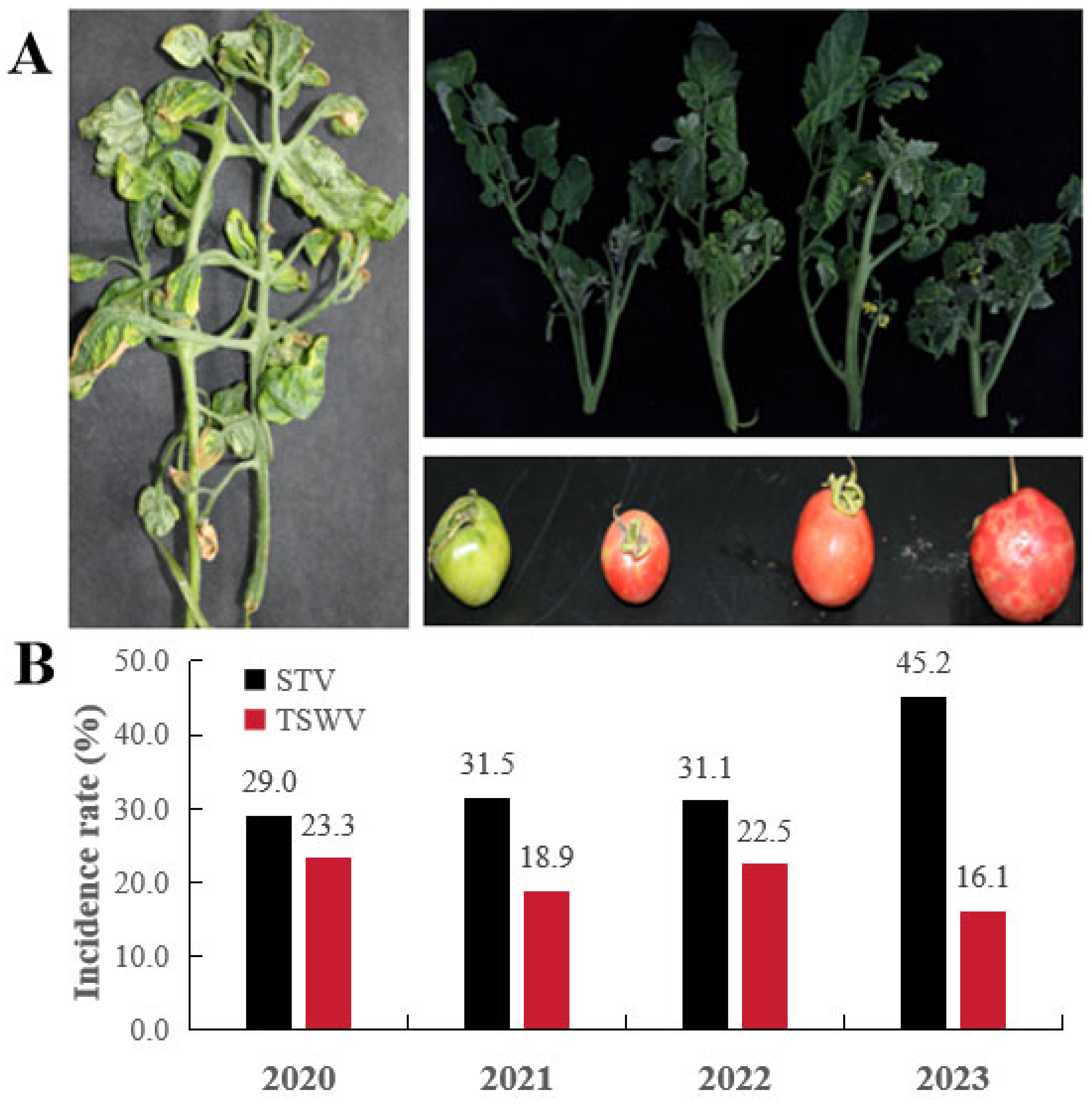
Severe viral disease symptoms were consistently observed in commercial tomato fields in Dali, Yunnan Province, China, from 2020 to 2023. The cultivars Ninghuang-1 and Huangjia-1, grown at large scale, exhibited typical symptoms including leaf curling, wilting, necrosis, and ring-shaped fruit spots of varying sizes (Figure 1A).
RT-PCR analysis of 572 symptomatic leaf samples detected only TSWV and STV among the five viruses tested (TSWV, TMV, STV, ToBRFV, ToCV). The annual incidence rates of TSWV were 23.3%, 18.9%, 22.5%, and 16.1% for 2020–2023, respectively, while STV incidence was 29.0%, 31.5%, 31.1%, and 45.2% over the same period (Figure 1B). All positive samples produced PCR amplicons of the expected sizes. Sequencing of randomly selected amplicons confirmed their identities as TSWV-YN1 (GenBank JF960235) and STV_GD-01 (PQ492145.1), with 98–100% nucleotide identity to the corresponding reference sequences.

3.2. Production of Virus-Free Seedlings

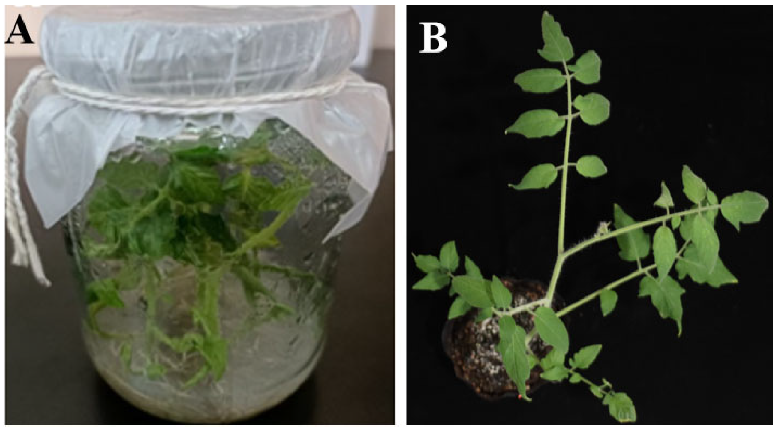
STV-free plants (Figure 2A) of ‘Ninghuang-1’ were obtained by direct shoot tip culture. RT-PCR analysis confirmed that all regenerated plants tested negative for STV, demonstrating successful virus elimination (Figure 3L and Figure 4L). After acclimatization, 80% of the plants survived (Figure 2B).

3.3. Phenotypic Characteristics and Virus Detection at Various Developmental Stages

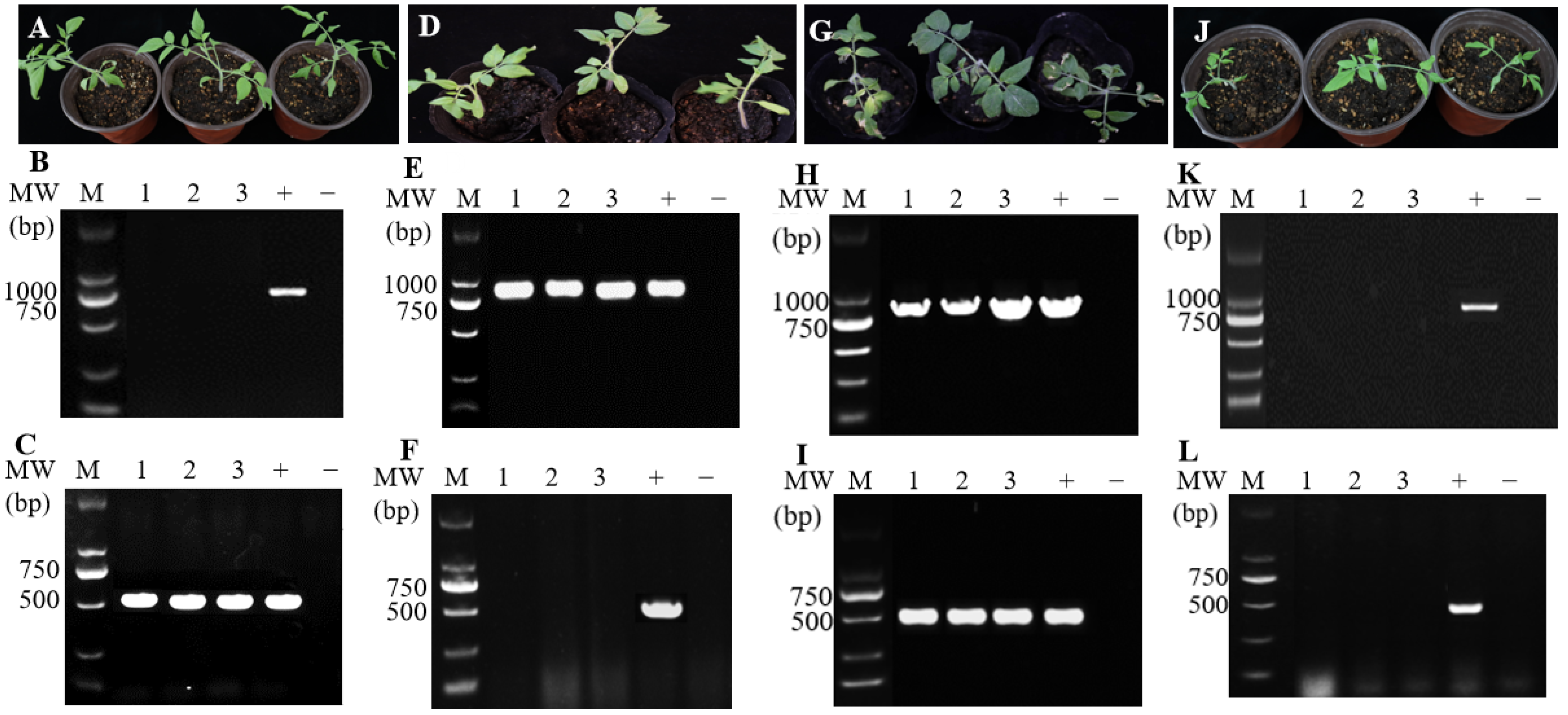
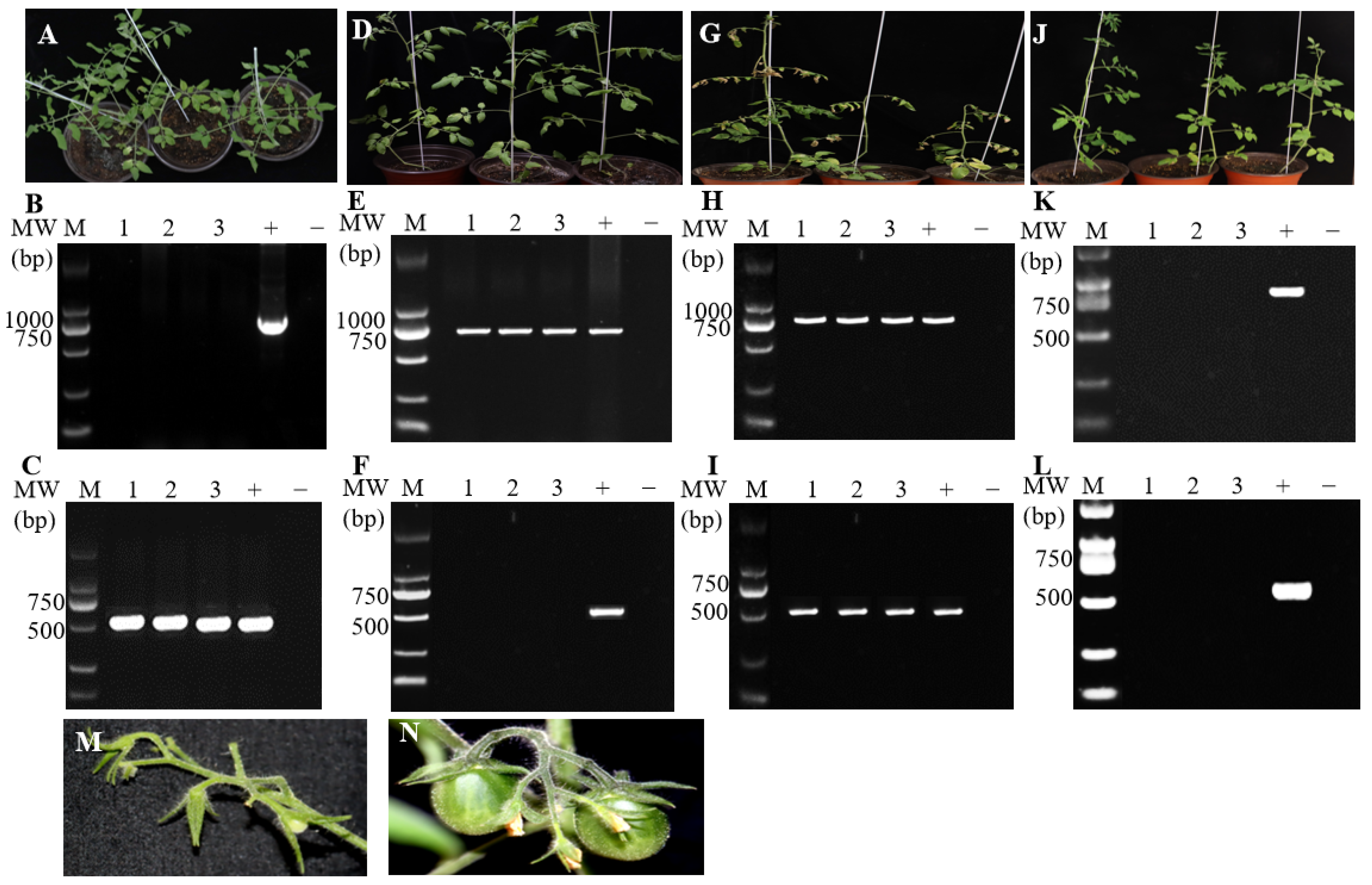
At the seedling stage, RT-PCR detection showed that seedlings inoculated with STV alone tested positive only for STV (Figure 3B,C), those inoculated with TSWV alone tested positive only for TSWV (Figure 3E,F), co-inoculated seedlings tested positive for both viruses (Figure 3H,I), while virus-free plants (healthy controls) tested negative for both (Figure 3K,L). Virus-free plants exhibited vigorous growth with healthy green leaves (Figure 3J). Seedlings infected with STV alone displayed no obvious viral symptoms (Figure 3A). Plants inoculated with TSWV alone developed mild leaf chlorosis and stunting (Figure 3D). Mixed-infection of STV and TSWV resulted in leaf yellowing and distinct necrotic lesions (Figure 3G). At the mature stage, viral infections were further verified by RT-PCR (Figure 4B,C,E,F,H,I), with results consistent with those at the seedling stage. Virus-free plants maintained vigorous growth with lush green foliage (Figure 4J). Plants infected with STV alone showed mild growth retardation and slight leaf deformation (Figure 4A). TSWV single infection caused more severe stunting and systemic chlorosis (Figure 4D). Mixed-infected plants exhibited wilting, extensive tissue necrosis, and plant decline (Figure 4G). At the reproductive stage, virus-free plants produced abundant, well-developed fruits (Figure 4N). STV-infected plants yielded fewer and smaller fruits (Figure 4M).

3.4. Histopathological Differences

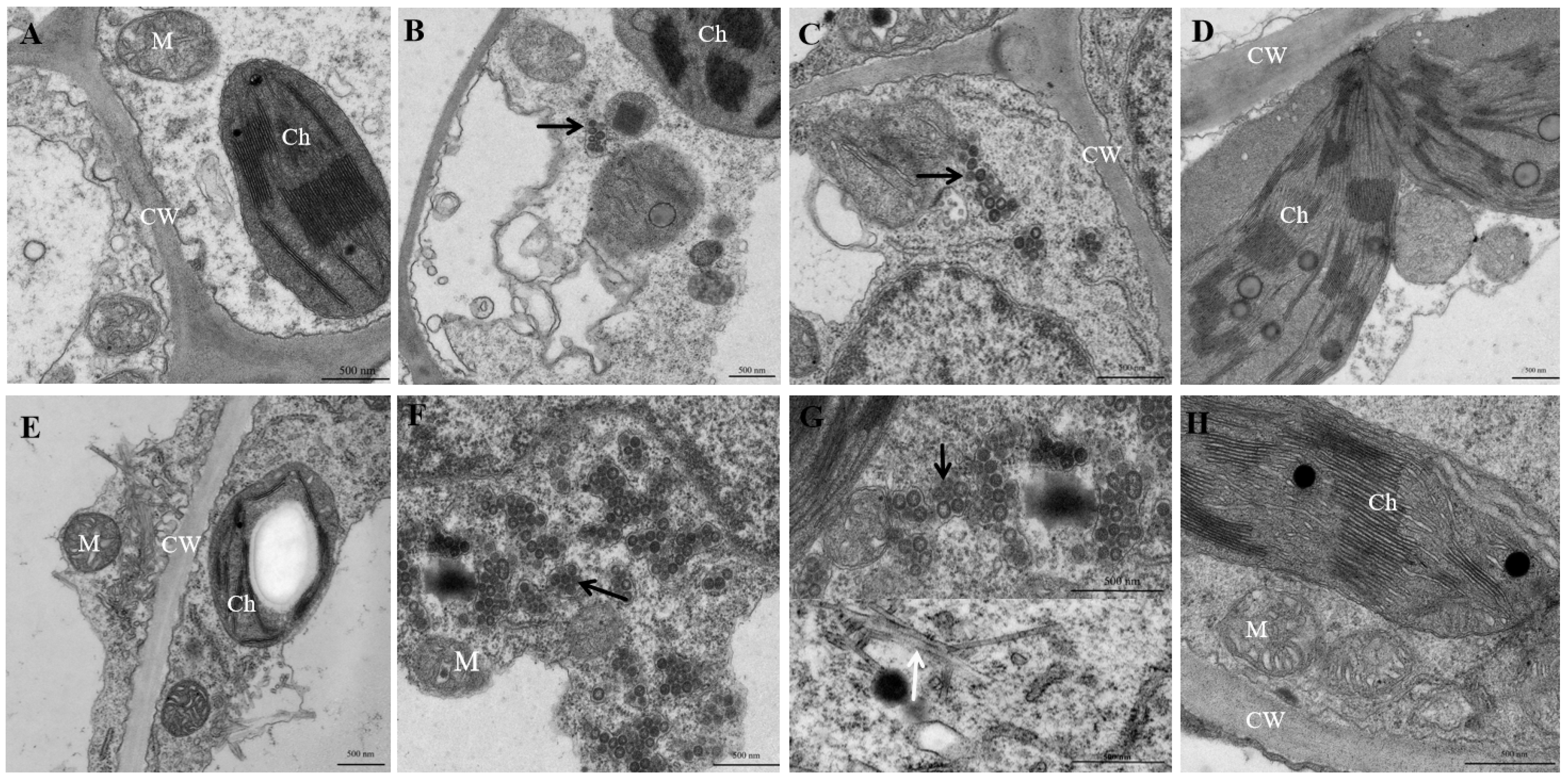
At the seedling stage, STV infection did not cause apparent structural damage, and chloroplasts and mitochondria remained intact (Figure 5A). TSWV particles (80–120 nm) were observed in TSWV-infected and STV+TSWV mixed-infected leaves, with higher particle abundance in mixed-infected plants (Figure 5B,C). These particles were enclosed in double-membrane vesicles and associated with disrupted chloroplast thylakoid structures. At the mature seedling stage, putative STV virions were observed in STV-infected leaves (Figure 5E). In mixed-infected plants, TSWV particles aggregated near these tubular structures, but not in the same field of view (Figure 5G). TSWV-infected leaves showed dense particle aggregation, and virus-infected tissues exhibited severe chloroplast and mitochondrial damage (Figure 5F). No virus particles were observed in virus-free plants at either stage, and cellular structures remained intact (Figure 5D,H).

3.5. Differences in Viral Gene and Protein Expression

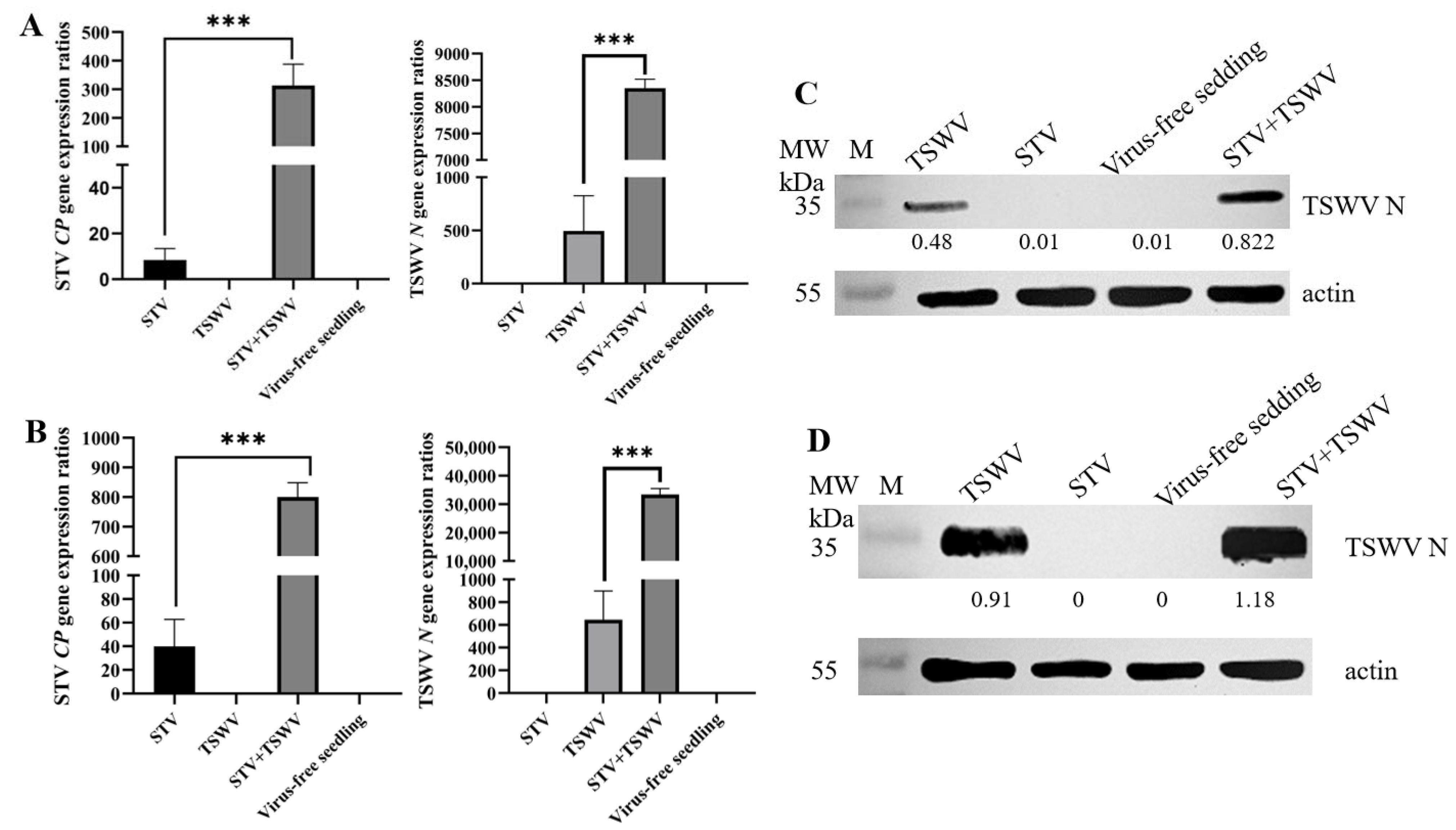
At both seedling and mature stages, STV CP expression in STV and TSWV mixed-infected plants was increased by up to 200-fold compared to STV-alone infection, and TSWV N expression was also elevated relative to TSWV-alone infection (Figure 6A,B). Similarly, densitometric quantification of Western blots showed that TSWV N protein accumulation in mixed-infected leaves was higher than that in TSWV-alone infection after normalization to β-actin (Figure 6C,D). No viral gene expression or viral protein was detected in virus-free plants at either stage.

4. Discussion

The overall upward trend in STV incidence over the four-year study period may be partly explained by the continuous cultivation of ‘Ninghuang-1’ and ‘Huangjia-1’, two widely grown cultivars that both showed high STV incidence in the field. Whether these cultivars are highly susceptible to STV infection remains to be determined. In our controlled inoculation experiments, conducted exclusively on ‘Ninghuang-1’, plants infected with STV alone developed only mild growth retardation and slight leaf deformation (Figure 4A), indicating that the virus alone does not cause severe symptoms. However, this does not preclude possible higher susceptibility at the level of viral accumulation or seed transmission efficiency. Additionally, because STV symptoms are often mild or inconspicuous, the virus has remained underrecognized, facilitating its continued spread through the cultivation of infected seed stocks. In contrast, the gradual decline of TSWV incidence over the same period likely reflects the successful implementation of integrated pest management (IPM) strategies in the Dali region since 2020, including insect-proof nets, insecticide applications against thrips vectors, and the removal of symptomatic plants. Nevertheless, mixed-infection by STV and TSWV remains a major concern due to more severe symptom expression and greater yield reduction compared with single infections. Given the increasing prevalence of STV, an environmental change or a surge in thrips vector populations could trigger a large-scale TSWV outbreak, which would cause substantial economic losses.
When plants are mixed-infected by multiple viruses or by different strains of the same virus, the interactions can be antagonistic or synergistic. Antagonistic interactions typically occur among closely related viruses or strains, whereas synergistic interactions are more common between viruses from different genera. In synergistic mixed-infections, viral interactions often increase the accumulation of one or both viruses and intensify disease symptoms compared to single infections. This can expand the host range, enhance transmission efficiency, and facilitate the emergence of new diseases [16,31,32]. For example, in mixed-infections of ToCV and Crinivirus contagichlorosis (tomato infectious chlorosis virus, TICV), TICV accumulation increases significantly [33]. Similarly, PVY and PVX mixed-infection in Nicotiana tabacum produces more severe symptoms and increased PVX accumulation [34]. In the present study, STV and TSWV mixed-infection significantly upregulated STV CP and TSWV N gene expression, increased TSWV N protein accumulation, and caused severe disease symptoms (Figure 3, Figure 4 and Figure 6). Histopathological observations (Figure 5) further revealed the cellular basis of this synergistic damage.
Plant tissue culture has been widely applied to produce virus-free plants in various crops, including orchids, potato, cassava, geraniums, and garlic [35,36,37,38,39]. Here, seed- and shoot tip-based tissue culture techniques successfully generated virus-free tomato seedlings. After multiplication (Figure 2A) and acclimatization under greenhouse conditions (Figure 2B), the survival rate to maturity was 80%. This rate is comparable to the 61–82% plantlet development rate reported for potato meristem-tip culture and slightly lower than the 85% acclimatization survival reported for virus-free orchids [31,33]. Thus, this method can effectively eliminate STV from seed stocks, reducing the primary source of infection in the field.
Based on these findings, we propose the following management strategies to mitigate STV and STV-TSWV mixed-infections: (i) production and use of STV-free seedlings via shoot tip culture, as validated in this study; (ii) elimination of primary infection sources, including STV-infected seed stocks and reservoir weed hosts; (iii) integrated thrips control, including insect-proof nets, insecticide rotation, and removal of symptomatic plants to prevent severe STV-TSWV mixed-infections; and (iv) routine molecular surveillance for early detection of STV, given its inconspicuous symptoms. The use of virus-free planting material can reduce initial inoculum in the field, lower the need for insecticide applications against virus vectors, and delay disease outbreaks. Therefore, this work provides both a technical foundation for large-scale production of virus-free tomato seedlings and a scientific basis for implementing environmentally sound integrated disease management practices in tomato production.
This study has two main limitations. First, all inoculation experiments were performed exclusively on the cultivar ‘Ninghuang-1’, whereas the field survey included both ‘Ninghuang-1’ and ‘Huangjia-1’. Consequently, the synergistic effects observed under controlled conditions may not fully represent the virus interactions in ‘Huangjia-1’ or other cultivars. Future studies should extend inoculation experiments to additional tomato cultivars, including ‘Huangjia-1’, to assess the generalizability of the observed synergy. Second, all inoculation experiments used mechanical inoculation rather than natural thrips-mediated transmission. While mechanical inoculation is a standard approach, it does not fully replicate natural infection processes that involve vector behavior and transmission efficiency. Thus, the observed synergistic effects may differ under field conditions where thrips are the primary vectors. Future studies should validate our findings using thrips-mediated inoculation.

5. Conclusions

Mixed-infection of STV and TSWV caused severe synergistic symptoms in tomato. This synergy was associated with markedly increased viral RNA accumulation, elevated TSWV N protein levels, and extensive ultrastructural damage to chloroplasts and mitochondria. We also observed putative STV virions, providing direct morphological evidence for this virus. A four-year field survey showed that STV incidence rose to 45.2%, while TSWV incidence dropped, probably due to integrated pest management. Shoot tip culture produced virus-free ‘Ninghuang-1’ seedlings with 80% survival after acclimatization, offering a way to remove primary infection sources.
One limitation is that our field survey did not include a systematic comparison of STV susceptibility among different tomato cultivars; future work should evaluate whether ‘Ninghuang-1’ is intrinsically more susceptible to STV infection or seed transmission than other cultivars. Another limitation is that our experiments used mechanical inoculation under controlled greenhouse conditions. Future work should test virus-free seedlings under natural thrips transmission in the field, clarify the molecular basis of the observed synergy, and evaluate the cost-effectiveness of large-scale production for commercial tomato growers.

Supplementary Materials

The following supporting information can be downloaded at: https://www.mdpi.com/article/10.3390/agriculture16151625/s1, Table S1: Primers used for virus detection by RT-PCR.

Author Contributions

R.Q.: Writing—review & editing, Methodology. J.L.: Review & editing, Resources, Conceptualization. S.C.: Validation, Investigation, Data curation. L.Z. (Lizhen Zhang): Validation, Methodology. X.B.: Investigation, Software. Y.Z.: Methodology, Data curation. L.J.: Methodology, Data curation. L.Z. (Lihua Zhao): Writing—original draft, Writing—review & editing, Funding acquisition. All authors have read and agreed to the published version of the manuscript.

Funding

This research was supported by the Natural Science Foundation of Yunnan Province (Grant Nos. 202401AS070031), Yunnan Young & Elite Talents Project (YNWR-QNRC-2022 to LHZ), the National Natural Science Foundation of China (Grant No. 32360639), and the Southwest Forestry University Talent Program (Grant No. 110225028).

Data Availability Statement

The raw data supporting the conclusions of this article will be made available by the authors on request.

Conflicts of Interest

The authors declare no conflicts of interest.

References

  1. Jewehan, A.; Kiemo, F.W.; Salem, N.; Tóth, Z.; Salamon, P.; Szabó, Z. Isolation and molecular characterization of a tomato brown rugose fruit virus mutant breaking the tobamovirus resistance found in wild Solanum species. Arch. Virol. 2022, 167, 1559–1563. [Google Scholar] [CrossRef]
  2. Tu, L.; Wu, S.; Gan, S.; Zhao, W.; Li, S.; Cheng, Z.; Zhou, Y.; Zhu, Y.; Ji, Y. A simplified RT-PCR assay for the simultaneous detection of tomato chlorosis virus and tomato yellow leaf curl virus in tomato. J. Virol. Methods 2022, 299, 114282. [Google Scholar] [CrossRef] [PubMed]
  3. Rivarez, M.P.S.; Pecman, A.; Bačnik, K.; Maksimović, O.; Vučurović, A.; Seljak, G.; Mehle, N.; Gutiérrez-Aguirre, I.; Ravnikar, M.; Kutnjak, D. In-depth study of tomato and weed viromes reveals undiscovered plant virus diversity in an agroecosystem. Microbiome 2023, 11, 60. [Google Scholar] [CrossRef] [PubMed]
  4. Tsvigun, V.O.; Levishko, A.S.; Gumeniuk, I.I.; Mazur, S.O. Monitoring of viral infections in vegetable crops in agroecosystems of Ukraine. Microbiol. J. 2024, 86, 87–101. [Google Scholar] [CrossRef]
  5. Dombrovsky, A.; Tran-Nguyen, L.T.T.; Jones, R.A.C. Cucumber green mottle mosaic virus: Rapidly increasing global distribution, etiology, epidemiology, and management. Annu. Rev. Phytopathol. 2017, 55, 231–256. [Google Scholar] [CrossRef] [PubMed]
  6. Salem, N.M.; Sulaiman, A.; Samarah, N.; Turina, M.; Vallino, M. Localization and Mechanical Transmission of Tomato Brown Rugose Fruit Virus in Tomato Seeds. Plant Dis. 2024, 106, 275–281. [Google Scholar]
  7. Groves, C.; German, T.; Dasgupta, R.; Mueller, D.; Smith, D.L. Seed transmission of soybean vein necrosis virus: The first tospovirus implicated in seed transmission. PLoS ONE 2016, 11, e0147342. [Google Scholar] [CrossRef] [PubMed]
  8. Wang, H.; Wu, X.; Huang, X.; Wei, S.; Lu, Z.; Ye, J. Seed transmission of tomato spotted wilt orthotospovirus in peppers. Viruses 2022, 14, 1873. [Google Scholar] [CrossRef] [PubMed]
  9. Escalante, C.; Sanz-Saez, A.; Jacobson, A.; Otulak-Koziel, K.; Kozieł, E.; Balkcom, K.S.; Zhao, C.; Conner, K. Plant virus transmission during seed development and implications to plant defense system. Front. Plant Sci. 2024, 15, 1385456. [Google Scholar] [CrossRef] [PubMed]
  10. Syller, J.; Grupa, A. Antagonistic within-host interactions between plant viruses: Molecular basis and impact on viral and host fitness. Mol. Plant Pathol. 2016, 17, 769–782. [Google Scholar] [PubMed]
  11. Singhal, P.; Nabi, S.U.; Yadav, M.K.; Dubey, A. Mixed infection of plant viruses: Diagnostics, interactions and impact on host. J. Plant Dis. Prot. 2022, 127, 353–368. [Google Scholar]
  12. Mascia, T.; Gallitelli, D. Synergies and antagonisms in virus interactions. Plant Sci. 2016, 252, 176–192. [Google Scholar] [CrossRef] [PubMed]
  13. Rochow, W.F.; Ross, A.F. Virus multiplication in plants doubly infected by potato viruses X and Y. Virology 1955, 1, 10–27. [Google Scholar] [CrossRef] [PubMed]
  14. Li, J.; Wang, J.C.; Ding, T.B.; Chu, D. Synergistic Effects of a Tomato chlorosis virus and Tomato yellow leaf curl virus Mixed Infection on Host Tomato Plants and the Whitefly Vector. Front. Plant Sci. 2021, 12, 672400. [Google Scholar] [CrossRef] [PubMed]
  15. Sikora, E.J.; Gudauskas, R.T.; Murphy, J.F.; Porch, D.W.; Andrianifahanana, M.; Zehnder, G.W.; Bauske, E.M.; Kemble, J.M.; Lester, D.F. A Multivirus Epidemic of Tomatoes in Alabama. Plant Dis. 1998, 82, 117–120. [Google Scholar] [CrossRef] [PubMed]
  16. García-Cano, E.; Resende, R.O.; Fernández-Muñoz, R.; Moriones, E. Synergistic interaction between tomato chlorosis virus and tomato spotted wilt virus results in breakdown of resistance in tomato. Phytopathology 2006, 96, 1263–1269. [Google Scholar] [CrossRef] [PubMed]
  17. Adams, M.J.; Lefkowitz, E.J.; King, A.M.Q.; Carstens, E.B.; Kuhn, J.H. Ratification vote on taxonomic proposals to the International Committee on Taxonomy of Viruses. Arch. Virol. 2014, 159, 2831–2841. [Google Scholar] [CrossRef] [PubMed]
  18. Hao, X.J.; Zheng, Y.Y.; Cui, B.M.; Xiang, B.C. Localization of southern tomato virus (STV) in tomato tissues. J. Plant Dis. Prot. 2023, 130, 1143–1147. [Google Scholar] [CrossRef]
  19. Elvira-González, L.; Puchades, A.V.; Carpino, C.; Alfaro-Fernández, A.; Font-San-Ambrosio, M.I.; Rubio, L.; Galipienso, L. Fast detection of southern tomato virus by one-step transcription loop-mediated isothermal amplification (RT-LAMP). J. Virol. Methods 2017, 241, 11–14. [Google Scholar] [CrossRef] [PubMed]
  20. Sabanadzovic, S.; Valverde, R.A.; Brown, J.K.; Martin, R.R.; Tzanetakis, I.E. Southern tomato virus: The link between the families Totiviridae and Partitiviridae. Virus Res. 2009, 140, 130–137. [Google Scholar] [CrossRef] [PubMed]
  21. González, L.E.; Peiró, R.; Rubio, L.; Galipienso, L. Persistent southern tomato virus (STV) interacts with cucumber mosaic and/or pepino mosaic virus in mixed infections modifying plant symptoms, viral titer and small RNA accumulation. Microorganisms 2021, 9, 689. [Google Scholar] [CrossRef]
  22. Zhu, M.; van Grinsven, I.L.; Kormelink, R.; Tao, X. Paving the way to tospovirus infection: Multilined interplays with plant innate immunity. Annu. Rev. Phytopathol. 2019, 57, 41–62. [Google Scholar] [CrossRef] [PubMed]
  23. Adkins, S. Tomato spotted wilt virus—Positive steps towards negative success. Mol. Plant Pathol. 2000, 1, 151–157. [Google Scholar] [CrossRef] [PubMed]
  24. de Avila, A.C.; de Haan, P.; Kormelink, R.; Resende, R.O.; Goldbach, R.W.; Peters, D. Classification of tospoviruses based on phylogeny of nucleoprotein gene sequences. J. Gen. Virol. 1993, 74, 153–159. [Google Scholar] [CrossRef] [PubMed]
  25. Lee, J.S.; Cho, W.K.; Kim, M.K.; Kwak, H.R.; Choi, H.S.; Kim, K.H. Complete genome sequences of three tomato spotted wilt virus isolates from tomato and pepper plants in Korea and their phylogenetic relationship to other TSWV isolates. Arch. Virol. 2011, 156, 725–728. [Google Scholar] [CrossRef] [PubMed]
  26. Sevik, M.; Arli-Sokmen, M. Estimation of the effect of tomato spotted wilt virus (TSWV) infection on some yield components of tomato. Phytoparasitica 2012, 40, 87–93. [Google Scholar]
  27. Choi, J.D.; Kim, J.S.; Kim, J.Y.; Kim, J.H.; Lee, S.H.; Choi, G.S.; Kim, H.R.; Chung, B.N. Occurrence and symptoms of tomato spotted wilt virus on vegetables in Korea (I). Radiat. Prot. Dosim. 2005, 11, 213–216. [Google Scholar] [CrossRef][Green Version]
  28. Verbeek, M.; Dullemans, A.M.; Espino, A.; Botella, M.; Font, M.I. First report of southern tomato virus in tomato in the Canary Islands, Spain. J. Plant Pathol. 2015, 97, 395. [Google Scholar]
  29. Yan, K.; Du, X.; Mao, B. Production of virus-free chrysanthemum (Chrysanthemum morifolium Ramat) by tissue culture techniques. Methods Mol. Biol. 2022, 2400, 171–186. [Google Scholar] [PubMed]
  30. Zhang, Z.; Zheng, K.; Dong, J.; Fang, Q.; Hong, J.; Wang, X. Clustering and cellular distribution characteristics of virus particles of tomato spotted wilt virus and tomato zonate spot virus in different plant hosts. Virol. J. 2016, 13, 11. [Google Scholar] [CrossRef] [PubMed]
  31. Zhang, X.S.; Holt, J.; Colvin, J. Synergism between plant viruses: A mathematical analysis of the epidemiological implications. Plant Pathol. 2001, 50, 732–746. [Google Scholar] [CrossRef]
  32. Elena, S.F.; Bernet, G.P.; Carrasco, J.L. The games plant viruses play. Curr. Opin. Virol. 2014, 8, 62–67. [Google Scholar] [CrossRef] [PubMed]
  33. Wintermantel, W.M.; Cortez, A.A.; Anchieta, A.G.; Gulati-Sakhuja, A.; Hladky, L.L. Co-infection by two criniviruses alters accumulation of each virus in a host-specific manner and influences efficiency of virus transmission. Phytopathology 2008, 98, 1340–1345. [Google Scholar] [CrossRef] [PubMed]
  34. Vance, V.B. Replication of potato virus X RNA is altered in coinfections with potato virus Y. Virology 1991, 182, 486–494. [Google Scholar] [CrossRef] [PubMed]
  35. Faccioli, G.; Rubies-Autonell, C. PVX and PVY distribution in potato meristem tips and their eradication by the use of thermotherapy and meristem-tip culture. J. Phytopathol. 1983, 103, 66–76. [Google Scholar]
  36. Gimenez, M.D.; Yañez-Santos, A.M.; Paz, R.C.; Quiroga, M.P.; Marfil, C.F.; Conci, V.C.; García-Lampasona, S.C. Assessment of genetic and epigenetic changes in virus-free garlic (Allium sativum L.) plants obtained by meristem culture followed by in vitro propagation. Plant Cell Rep. 2016, 35, 129–141. [Google Scholar] [PubMed]
  37. Pradhan, S.; Regmi, T.; Ranjit, M.; Pant, B. Production of virus-free orchid Cymbidium aloifolium (L.) Sw. by various tissue culture techniques. Heliyon 2016, 2, e00176. [Google Scholar] [CrossRef] [PubMed][Green Version]
  38. Maruthi, M.N.; Whitfield, E.C.; Otti, G.; Tumwegamire, S.; Kanju, E.; Legg, J.P.; Mkamilo, G.; Kawuki, R.; Benesi, I.; Zacarias, A.; et al. A method for generating virus-free cassava plants to combat viral disease epidemics in Africa. Physiol. Mol. Plant Pathol. 2019, 105, 77–87. [Google Scholar] [CrossRef] [PubMed]
  39. Alavijeh, M.K.; Bayat, H.; Kianpour, D.; Kalantari, S.; Zarei, A. Optimization of in vitro propagation and virus eradication using meristem culture and thermotherapy in two geranium species Pelargonium× hortorum (‘Zonal’) and Pelargonium× domesticum (‘Regal’). BMC Plant Biol. 2025, 25, 9. [Google Scholar] [CrossRef] [PubMed]
Figure 1. Symptoms of viral diseases and virus detection rates from January 2020 to August 2023. (A) Typical disease symptoms observed on tomato leaves and fruit. (B) Incidence rates of virus detection in tomato samples.
Agriculture 16 01625 g001
Figure 2. Production of virus-free tomato plants. (A) Multiplication of STV-free shoots following virus testing. (B) In vitro plantlets acclimatized in humus soil mixed with vermiculite and perlite in a 3:2:1 ratio.
Agriculture 16 01625 g002
Figure 3. Phenotypic characteristics and virus detection in tomato seedlings. (AC) Tomato seedlings infected with STV alone. (A) Seedlings infected with STV alone, showing no obvious viral symptoms. (B) RT-PCR detection of TSWV (negative). (C) RT-PCR detection of STV (positive). (DF) Tomato seedlings inoculated with TSWV after virus elimination. (D) Plants inoculated with TSWV alone, displaying mild leaf chlorosis and stunting. (E) RT-PCR detection of TSWV (positive). (F) RT-PCR detection of STV (negative). (GI) Tomato seedlings infected with STV followed by TSWV inoculation. (G) Plants mixed-infected with STV and TSWV, exhibiting severe symptoms including leaf yellowing and distinct necrotic lesions. (H) RT-PCR detection of TSWV (positive). (I) RT-PCR detection of STV (positive). (JL) Virus-free tomato seedlings. (J) Virus-free plants exhibiting vigorous growth and healthy green leaves. (K) RT-PCR detection of TSWV (negative). (L) RT-PCR detection of STV (negative). In the RT-PCR gel images (B,C,E,F,H,I,K,L), lanes M indicate DNA molecular weight marker; lanes 1, 2, 3 represent three independent biological replicates; ‘+’ denotes positive control (viral RNA-derived cDNA); ‘−’ denotes negative control (nuclease-free water). Three biological replicates were performed for each condition.
Agriculture 16 01625 g003
Figure 4. Phenotypic characteristics and virus detection in mature tomato plants. (AC) Mature tomato plants infected with STV alone. (A) STV-infected plants showing mild growth retardation and slight leaf deformation. (B) RT-PCR detection of TSWV (negative). (C) RT-PCR detection of STV (positive). (DF) Mature tomato plants inoculated with TSWV after virus elimination. (D) TSWV-inoculated plants displaying more severe stunting and systemic chlorosis. (E) RT-PCR detection of TSWV (positive). (F) RT-PCR detection of STV (negative). (GI) Mature tomato plants infected with STV followed by TSWV inoculation. (G) Mixed-infected plants exhibiting wilting, extensive tissue necrosis, and plant decline. (H) RT-PCR detection of TSWV (positive). (I) RT-PCR detection of STV (positive). (JL) Virus-free mature tomato plants. (J) Virus-free plants maintaining vigorous growth with lush green foliage. (K) RT-PCR detection of TSWV (negative). (L) RT-PCR detection of STV (negative). (M) Fruits from STV-infected tomato plants showing fewer and smaller fruits. (N) Fruits from virus-free tomato plants showing abundant, well-developed fruits. In the RT-PCR gel images (B,C,E,F,H,I,K,L), lanes M indicate DNA molecular weight marker; lanes 1, 2, 3 represent three independent biological replicates; ‘+’ denotes positive control (viral RNA-derived cDNA); ‘−’ denotes negative control (nuclease-free water). Three biological replicates were performed for each condition.
Agriculture 16 01625 g004
Figure 5. Histopathological differences at different developmental stages. (AD) Leaflet samples at the seedling stage: (A) Tomato infected with STV alone; (B) Tomato inoculated with TSWV alone; (C) Tomato mixed-infected with STV and TSWV; (D) Virus-free tomato. (EH) Leaflet samples at the mature stage: (E) Tomato infected with STV alone; (F) Tomato inoculated with TSWV alone; (G) Tomato mixed-infected with STV and TSWV; (H) Virus-free tomato. Note: M, mitochondria; CW, cell wall; Ch, chloroplast. Black arrows indicate TSWV particles; white arrows indicate putative STV virions. Three biological replicates were performed for each condition.
Agriculture 16 01625 g005
Figure 6. Expression analysis of STV CP and TSWV N genes and protein in tomato leaves. (A,B) RT-qPCR analysis of STV CP and TSWV N gene expression levels in tomato leaves at the seedling (A) and mature (B) stages. Error bars represent the standard deviation (SD) (n = 3). ***: P < 0.001 (one-way ANOVA with Tukey’s post hoc test). (C,D) Western blot analysis of TSWV N protein accumulation in tomato leaves at the seedling (C) and mature (D) stages. TSWV: plants inoculated with TSWV after virus elimination; STV: plants infected with STV only; virus-free: plants after virus elimination with no virus inoculation; STV+TSWV: plants infected with STV followed by TSWV inoculation. β-Actin (approximately 43 kDa) was used as the loading control. Three biological replicates were performed for each condition.
Agriculture 16 01625 g006
Disclaimer/Publisher’s Note: The statements, opinions and data contained in all publications are solely those of the individual author(s) and contributor(s) and not of MDPI and/or the editor(s). MDPI and/or the editor(s) disclaim responsibility for any injury to people or property resulting from any ideas, methods, instructions or products referred to in the content.