Article

Molecular Identification and Phylogenetic Analysis of Fusarium spp. Associated with Triticum aestivum L. Based on DNA Barcoding


1
Department of Fundamental Sciences in Animal Husbandry, Faculty of Agriculture, Trakia University, Students Campus, 6000 Stara Zagora, Bulgaria
2
Department of Biological Sciences, Faculty of Agriculture, Trakia University, Students Campus, 6000 Stara Zagora, Bulgaria
*
Author to whom correspondence should be addressed.
Agriculture 2026, 16(11), 1232; https://doi.org/10.3390/agriculture16111232
Submission received: 6 March 2026 / Revised: 15 May 2026 / Accepted: 18 May 2026 / Published: 2 June 2026
(This article belongs to the Section Crop Protection, Diseases, Pests and Weeds)

Abstract

Fusarium spp. are active producers of mycotoxins that enter the food chain and pose risks to human health. Identifying pathogenic agents is a key step in developing disease management strategies. For the first time in Bulgaria, we identified eight Fusarium species in wheat, harvest 2024–2025, through the application of DNA barcoding. For a genetic marker and construction of phylogenetic tree, the protein-coding gene β-tub was chosen. Among 26 identified isolates, F. sporotrichioides (42.3%) dominated, followed by F. proliferatum (23.1%), F. avenaceum (7.7%), F. armeniacum (7.7%), and F. poae (7.7%). F. tricinctum (3.8%), F. oxysporum (3.8%), and F. equiaseti (3.9%) were weakly expressed. Phylogenetic analysis classified the isolates into five species complexes: FSAMSC, FFSC, FTSC, FIESC, and FOSC and highlighted the genetic distances between them. Molecular genetic analysis showed that 84.6% of the wheat samples contained only one species of Fusarium, and in 15.4% the co-presence of two species was established. The largest share was in samples with a low infestation of 2–4%, which represented 35% (n = 32) of all positives. No statistically significant difference was found between varieties and contamination level, but a statistically significant positive correlation was demonstrated by the preceding crop (rapeseed, sunflower, and maize).

1. Introduction

Fusarium spp. is a cosmopolitan genus of ascomycete fungi, numbering more than 300 species of important economic and ecological importance [1]. Although species perform a variety of ecological roles, e.g., saprophytes, endophytes, and symbionts, they are among the best-known pathogens of wheat and cause serious diseases with huge financial losses in the agricultural sector [2]. These adaptive micromycetes are able to colonize roots, stems, leaves, ears and grain. Soil, plant residues and seeds serve as the main sources of inoculum, and chlamydospores can survive in the soil for years, providing initial outbreaks for subsequent infections in which two or more species can infect the same crop at the same time. On the other hand, the presence of species is also strongly influenced by agriculture practices in the different growing regions, which requires further studies [3]. Species F. graminearum, F. culmorum, F. avenaceum, F. poae, F. sporotrichioides, F. equiseti, F. verticillioides, and F. proliferatum are the predominant species that infect wheat and cause fusariosis by classes (FHB) and Fusarium crown rot (FCR) in Europe and Bulgaria including [4,5,6]. In Canada and Italy, the dominant pathogen of durum wheat is F. avenaceum, with F. graminearum present in a secondary role [7,8]. Worldwide, the distribution of Fusarium spp. in wheat shows significant regional variations related to climatic conditions, crop rotation and specific agrotechnical practices. For example, the combination “maize–wheat” increases the risk of pathogen transmission between crops, especially F. verticillioides and F. graminearum, which creates favorable conditions for the development of FHB in wheat [9]. Some Fusarium species are active producers of mycotoxins: trichothecenes, fumonisins, zearalenone, eniatin, etc. According to Perochon A. and Doohan FM. (2024), these secondary metabolites facilitate the development of cereal diseases during pathogenesis [10]. On the other hand, Fusarium mycotoxins accumulate in harvested produce and can have carcinogenic, neurotoxic, mutagenic, teratogenic, estrogenic, hepatotoxic and immunosuppressive effects on animal and human health [11].
Despite the availability of new cereal breeding lines with known resistance to FHB, the selection of resistant varieties is difficult due to the high proportion of asymptomatic infections and the difficulties encountered in selecting resistant genotypes [12,13,14]. The interaction between the parasite and the host triggers the expression of multiple genes and the secretion of secondary metabolites—mycotoxins and enzymes—facilitating penetration, enhancing virulence and facilitating the development of cereal diseases during pathogenesis [15,16]. Bashyal et al. (2017) noted that the genome of F. fujikuroi contains 1194 secretory proteins, 38% of which are likely related to its virulence [17]. Fusarium species are active producers of mycotoxins including trichothecenes, fumonisins, zearalenone, eniatin, etc., which accumulate in the crop and can exhibit carcinogenic, neurotoxic, mutagenic, teratogenic, estrogenic, hepatotoxic and immunosuppressive effects on animal and human health [10,11].
Classical identification methods based on morphological characteristics (colony appearance, color and growth rate of mycelium, observation of macroconidia, microconidia, chlamydospores and spore-producing structures [18]) are seriously hampered by the limited informativeness of the variability of the characters and have difficulty distinguishing species with related morphological characteristics [19].
With the advancement of molecular techniques, DNA barcoding accelerated technology has been developed, where species can be identified through variations in short gene sequences (so-called barcodes) located in a standardized genetic locus (specific gene or region) of the sample studied, which are highly variable at the species level but conservative at the genus level [20].
Although the non-coding region ITS was recognized as a major barcode marker for fungi by the International Consortium of Mycologists in Amsterdam, 2011 [21] due to its critical biological role in rRNA and ribosome maturation [22], it is not always a reliable tool for Fusarium identification [23,24]. According to Stielow et al. (2015) [25], unlike other fungal genera, the use of the ITS genetic locus for identifying representatives of the genus Fusarium is very difficult and can be reliably applied only in about 75% of all species. In the search for suitable genetic loci for the genus, protein-coding genes play an increasingly important role in molecular identification due to their high informativeness. As such genes, the gene encoding β-tubulin (β-tub), tef-1α, the gene sequence that encodes nuclear DNA-dependent RNA polymerase II (RPB2), etc., are widely used [26].
In filamentous fungi, the number of genes in the β-tubulin locus varies between genera and species. The β-tub gene encodes the β-tubulin subunit and includes several exons separated by introns, the location and length of which can vary between species. The combination of conserved and variable regions determines the potential of the β-tub gene and makes it a suitable and reliable marker for species identification and phylogenetic analyses. Within the genus Fusarium, the β-tub gene shows significant species differentiation, allowing its use to precisely distinguish closely related taxa. Many ascomycetes possess only one β-tub gene, but in F. graminearum, β-tub1 and β-tub2 stand out with different functions during vegetative growth and sexual reproduction, but with common roles in hyphal growth [27].
The primers for the β-tub gene were designed by Annis and Panaccione (1998) [28] and were originally designed to cover flank intron 3 of the Neotyphodium spp. [29]. Subsequently, Tooley et al. (2001) [30] designated the BT5 and BT3 primers for characterization of pathogenic Claviceps spp. on sorghum (Sorghum bicolor), which are reliably applied for species identification within the genus Fusarium [31,32].
The aim of this study is to identify Fusarium spp. species distributed in the Bulgarian common wheat population by applying the DNA barcoding method based on the β-tub region, to clarify the phylogenetic relationships between the species, as well as to establish the influence of the wheat variety on the level of contamination with Fusarium spp.

2. Material and Methods

2.1. Sample Collection

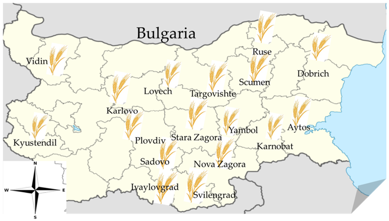
A total of 101 symptomless bread wheat (Triticum aestivum L.) samples were collected during the period June 2024–August 2025 from different main commercial wheat-growing regions in Bulgaria (Figure 1) from storage facilities and according to ISO 24333:2009 [33]. In 29 of the analyzed samples, the preceding crop was rapeseed, in 43 the preceding crop was sunflower, and in 29 samples it was maize. After collection, all samples were promptly brought back to the laboratory. The collection includes 59 samples of imported varieties, 28 samples of Bulgarian selected varieties and 14 samples without information on the varietal affiliation. Briefly, 1 kg spot samples of wheat with no visible disease symptoms were taken with a bulk profile sampler from 10 points throughout the bulk lot (lots ranged from 5 to 100 tons). Multiple samples from appropriate sites were aseptically poured into a sterile container and manually mixed to obtain a homogeneous composite sample. Following their complete mixing, a bulk of samples weighing 10 kg was formed and manually reduced by quartering to samples of 0.5 kg, which were then labeled. Sampling at storage facilities was chosen to focus on Fusarium spp. that persists post-harvest and presents a direct risk to food and feed safety. This strategy reflected the study’s aim of assessing contamination under realistic storage conditions. While field sampling may reveal broader pre-harvest fungal diversity, post-harvest sampling provides relevant insights into the fungal populations that are most likely to enter the food chain.

2.2. Fungal Isolation of Fusarium spp.

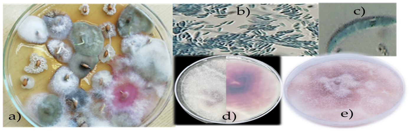
One hundred whole seeds from each sample were randomly selected and superficially sterilized with 70% ethanol for 6 min, followed by three rinses with distilled water to eliminate surface microflora. The isolates were dried on a sterile filter paper, and plated onto potato dextrose agar (PDA) (HiMedia©, Maharashtra, India) in 4 Petri dishes (d = 15 cm) (25 seeds in each) for 7 days at a constant temperature of 25 °C and a photoperiod of 16 h/8 h (day/night) [33]. As seen in Figure 2, all suspected Fusarium spp. colonies were subcultured using the single spore technique [34,35]. A spore suspension was prepared in a 10 mL sterile water sample so as to contain 1 to 10 spores. The prepared water agar (WA) medium plates were inoculated with 0.1 mL of suspension and incubated for 18–20 h at 25 °C. For further molecular identification, the resulting single spore cultures were transferred onto synthetic nutrient-poor agar (SNA) and incubated at 25 °C for 7 days to initiate spore formation [36]. Each isolate’s spores were examined under a microscope to identify distinctive characteristics unique to Fusarium spp. The identification of Fusarium spp. was done using keys by Leslie and Summerall, (2006), Gerlach and Nirenberg (1982), and Burgess et al. (1992) [19,36,37]. For the precision of the study, 26 representative isolates were selected from the 101 wheat samples, based on the year of sample collection, preliminary morphological identification, and colony morphology, to represent each species in percentage terms with similar traits, relative to all isolated strains, for molecular identification and subsequent Sanger sequencing.

2.3. Fusarium Species Identification

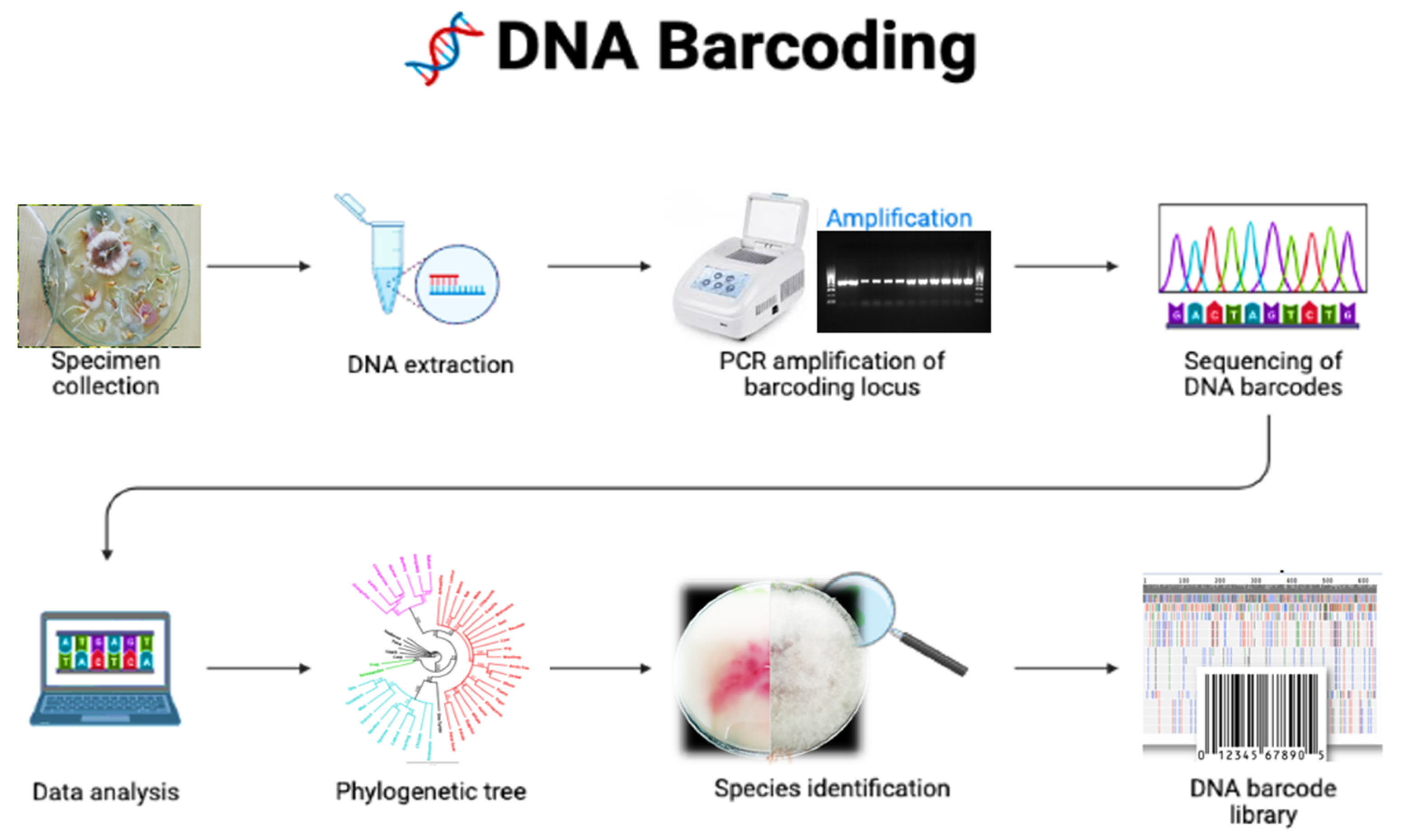
DNA barcoding stages for Fusarium species identification in this study are presented in Figure 3.

2.4. DNA Extraction, PCR Amplification and Sequencing

A 5-day-old mycelium of Fusarium spp. strains from SNA plates was used for genomic DNA isolation. Initially, to lysate the fungi’s polysaccharide cell wall, the mycelium was frozen for 24 h at −20 °C and then pulverized with quartz sand. DNA extraction from mycelium of monosporic Fusarium cultures was performed by using Animal and Fungi DNA Preparation Kit (Jena Bioscience GmbH, Jena, Germany) according to the manufacturer’s instructions. The concentration of the extracted genomic DNA of each sample was measured using a NanoView Plus spectrophotometer (GE HealthCare Technologies, Inc., Chicago, CA, USA) at a 260–280 nm wavelength. The DNA concentration of all samples was adjusted to 10 ng/µL, in a working volume of 70 µL, and the genomic DNA-extracted samples were stored at −18 °C until further analysis. The optimal annealing temperature was determined based on gradient PCR in the temperature range 51.1–61.2 °C. The following primers were used for amplification: region β-tub: F:5′-CGTCTAGAGGTACCCATACCGGCA-3′ and R:5′-GCTCTAGACTGCTTTCTGGCAGACC-3′ [30]. PCRs were performed in 25 μL volumes containing 2 μL of the extracted DNA, 12.5 μL 2x Master Mix (VWR International BV, Leuven, Belgium), 1 μL of each primer (FOR/REV), and 8.5 μL of nuclease-free water. β-tub gene was amplified using the following cycling parameters: 95 °C for 5 min, 30 cycles of 95 °C for 30 s, primers annealing at 51.1 °C for 0,45 min, 72 °C for 1 min, and a final extension at 72.0 °C for 9 min followed by a 4 °C soak in a thermal cycler (QB-96 Thermal Cycler, Quanta Biotech Ltd., Surrey, UK). Amplicons were size fractioned alongside a 100 bp molecular weight marker (Gene-Ruler™ Ladder Plus, Thermo Fisher Scientific Inc., Waltham, MA, USA) in 1% agarose gels in 1× TAE buffer, stained with 10,000× GelRed™ (Cat. No. 41003, Biotium Inc., Flermont, FL, USA) and visualized on a MiniBis photodocumentation system using a transilluminator (ECX-15M Bio Imaging Systems, Bio-Imaging Systems, Inc., Jackson, MS, USA). Sequencing reactions were purified using GeneMATRIX Short DNA Clean-Up Purification Kit (Cat. No. E3515, EURx Ltd., Gdansk, Poland) and sequenced in both directions by a PlateSeq kit (Eurofins Genomics Ebersberg, Gdansk, Germany).

2.4.1. Statistical Analyses

The one-way analysis of variance (ANOVA) was used to investigate the influence of the factors “varietal affiliation” and origin of the varieties (from other countries/Bulgarian) of wheat observed in our country. To establish relationships between the predecessor (sunflower, rapeseed, and maize) and Fusarium spp., the Shapiro–Wilk test was used to test for normal distribution. The significant deviation from normal distribution necessitated non-parametric statistical methods: Spearman’s rank correlation coefficient (ρ) to examine relationships and the Kruskal–Wallis H-test to compare the influence of individual predecessor crops.

2.4.2. Sequence Analysis

All obtained sequence data were manually edited and aligned using the MUSCLE algorithm [38] in the MEGA software v. 11.0.13 [39] (Tamura, 2021). To identify Fusarium spp. among the generated sequence data, the Basic Local Alignment Search Tool (BLAST) was used: https://blast.ncbi.nlm.nih.gov/Blast.cgi?CMD=Web&PAGE_TYPE=BlastHome, accessed on 4 March 2026.

2.4.3. Phylogenetic Analysis

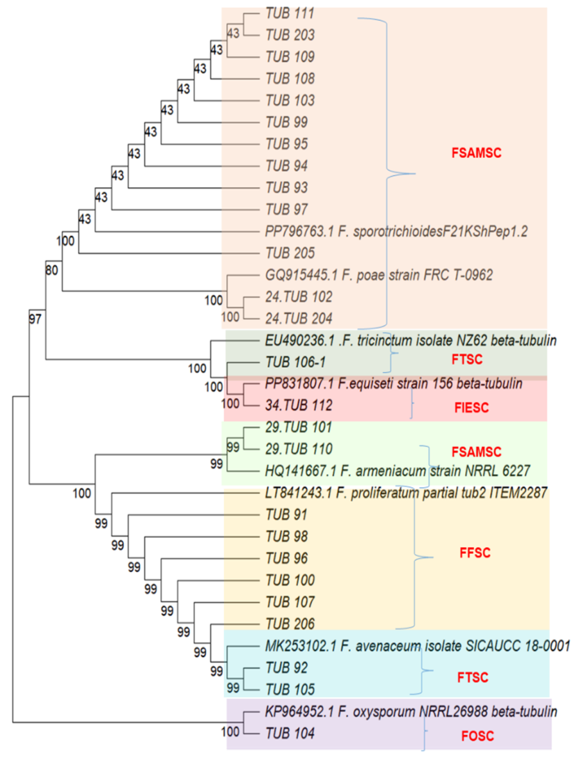
The phylogeny was inferred using the Maximum Likelihood method and Tamura (1992) model of nucleotide substitutions and the tree with the highest log likelihood (−1534.17) is shown [40]. The percentage of replicate trees in which the associated taxa clustered together (1000 replicates) is shown next to the branches. A phylogenetic tree was inferred through the maximum likelihood approach, applying the Tamura (1992) three-parameter substitution model combined with a discrete gamma distribution (TN93 + G) [41]. Support for branches was evaluated using 1000 bootstrap replicates, with bootstrap values of 50 or higher shown at the respective nodes. Positions with gaps or missing data were removed entirely (complete deletion method). Each sequence is labeled with the organism’s name followed by its GenBank accession number. The following color scheme was used: F. equiseti/FIESC—red; F. avenaceum/FTSC—blue; F. tricinctum/FTSC—dark green; F. armeniacum/FTSC—light green; F. sporotrichioides and F. poae/FSAMSC—orange; F. proliferatum/FFFC—yellow; F. oxysporum/FOSC—purple. The Neighbor-Joining (NJ) tree was generated using a matrix of pairwise distances computed using the p-distance. The evolutionary rate differences among sites were modeled using a discrete Gamma distribution across 5 categories (+G, parameter = 0.3006), with 0.00% of sites deemed evolutionarily invariant (+I). The analytical procedure encompassed 59 nucleotide sequences. The complete deletion option was applied to eliminate positions containing gaps and missing data resulting in a final data set comprising 433 positions. Evolutionary analyses were conducted in MEGA11 utilizing up to 7 parallel computing threads [39]. The sequences were submitted in Gene Bank of NCBI (SUB16139725/11.12 in 22 April 2026).

3. Results

Based on the mycological analysis of the samples studied, it was found that Fusarium spp. was identified in the epiphytic microflora of 82.2% (n = 83) of the samples studied, with the degree of contamination in individual samples varying between 0–46%. The average value for 2024 was 7.2% ± 8.30, and for 2025, 5.5% ± 2.80. The largest share, 35% (n = 32), was occupied by samples with relatively low levels of contamination between 2 and 4%, followed by samples with 5–6% content of Fusarium spp. (17%) (n = 16). In samples of wheat variety Factor from the Dobrich region, the highest levels were found (46%) in 2024, and for variety Nikolay, harvest 2024, in the Sadovo region (38%).
The influence of the factors “varietal affiliation” of wheat (Bulgarian/foreign) grown in Bulgaria on contamination with Fusarium spp. is presented in Table 1.
Data from the one-factor analysis of variance (ANOVA) showed a lack of statistically significant influence (p > 0.05) (Table 2). An additional analysis, establishing the dependence of the contamination with Fusarium spp. according to the origin of the varieties (from other countries/Bulgarian) of wheat observed in our country, also did not prove a statistically significant result between the origin of the selected varieties and the amount of contamination (p > 0.05) (Table 3).
To test the Ho hypothesis (no association between the preceding crop and Fusarium contamination), a Hypothesis Test Summary was performed and the p-Value < 0.001 was rejected. To establish the relationships between the ancestor (sunflower, rapeseed, and maize) and Fusarium spp., the Shapiro–Wilk test used demonstrated a significant deviation from normal distribution (p < 0.05) for all studied groups (Table 4). Therefore, a non-parametric statistical method was applied: the Kruskal–Wallis H-test to compare the influence of individual ancestors.
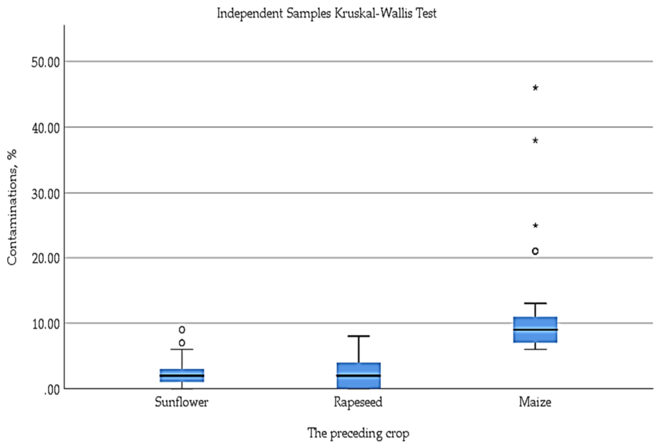
The Kruskal–Wallis test results reveal significant differences in infection levels among the three preceding crops (Figure 4). The highest mean rank for infection was recorded for maize (85.36), while rapeseed (38.19) and sunflower (36.47) showed substantially lower and similar values (Table 5).
The results of the Kruskal–Wallis test revealed statistically significant differences in the degree of insemination between the three types of crops studied as predecessors (χ2 = 56.79, p < 0.001). A subsequent post-hoc analysis using Dunn’s pairwise comparisons with Bonferroni correction found that sunflower was significantly different from rapeseed (p < 0.001) and from maize (p < 0.001). No statistically significant difference was reported between rapeseed and maize (p = 1.000) (Table 6).
The data analysis showed that corn as a preceding crop has a statistically higher impact compared to rapeseed and sunflower. However, rapeseed and sunflower act in a similar way and the differences between them are negligibly small.
To establish the relationships between the ancestor (sunflower, rapeseed, and maize) and Fusarium spp., the Shapiro–Wilk test used demonstrated a significant deviation from normal distribution (p < 0.05) for all studied groups. Therefore, non-parametric statistical methods were applied: Spearman’s rank correlation coefficient (ρ) to examine the relationships and Kruskal–Wallis H-test to compare the influence of individual ancestors (Table 4).
Results of molecular identification of Fusarium spp.
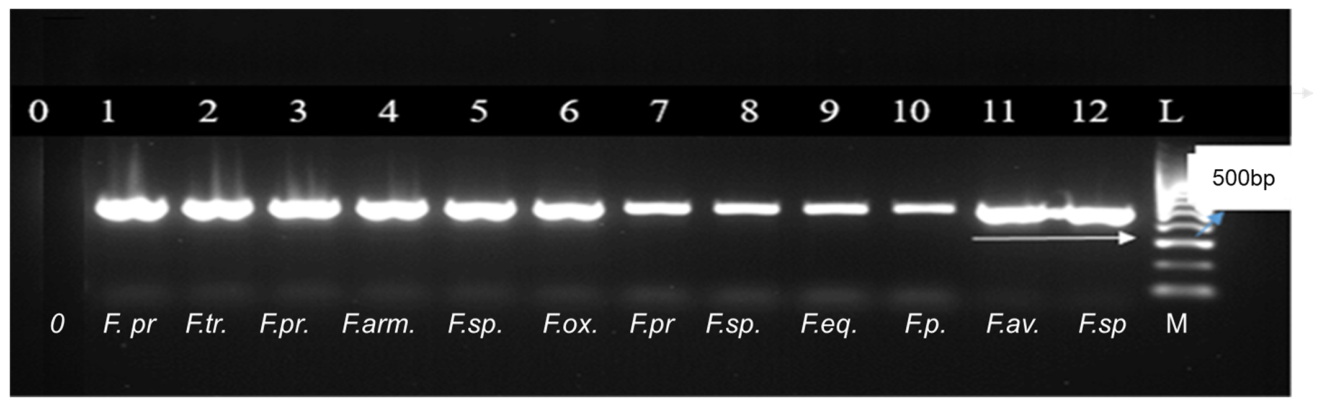
After purification of the amplification products (500 bp), 26 representative isolates were selected for sequencing by the Sanger method (Figure 5).
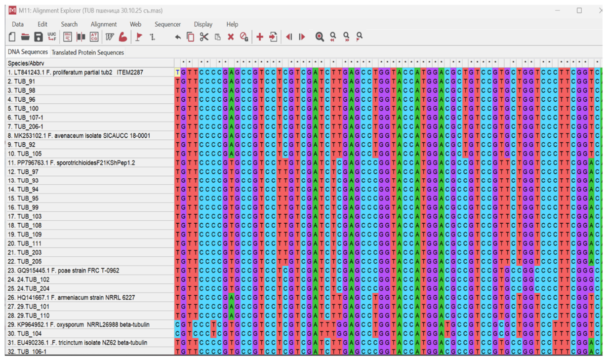
Analysis of the sequences using the β-tub DNA barcode revealed eight Fusarium species: F. sporotrichioides, F. proliferatum, F. poae, F. armeniacum, F. equiseti, F. tricinctum, F. avenaceum and F. oxisporum (Figure 4, Figure 5 and Figure 6).
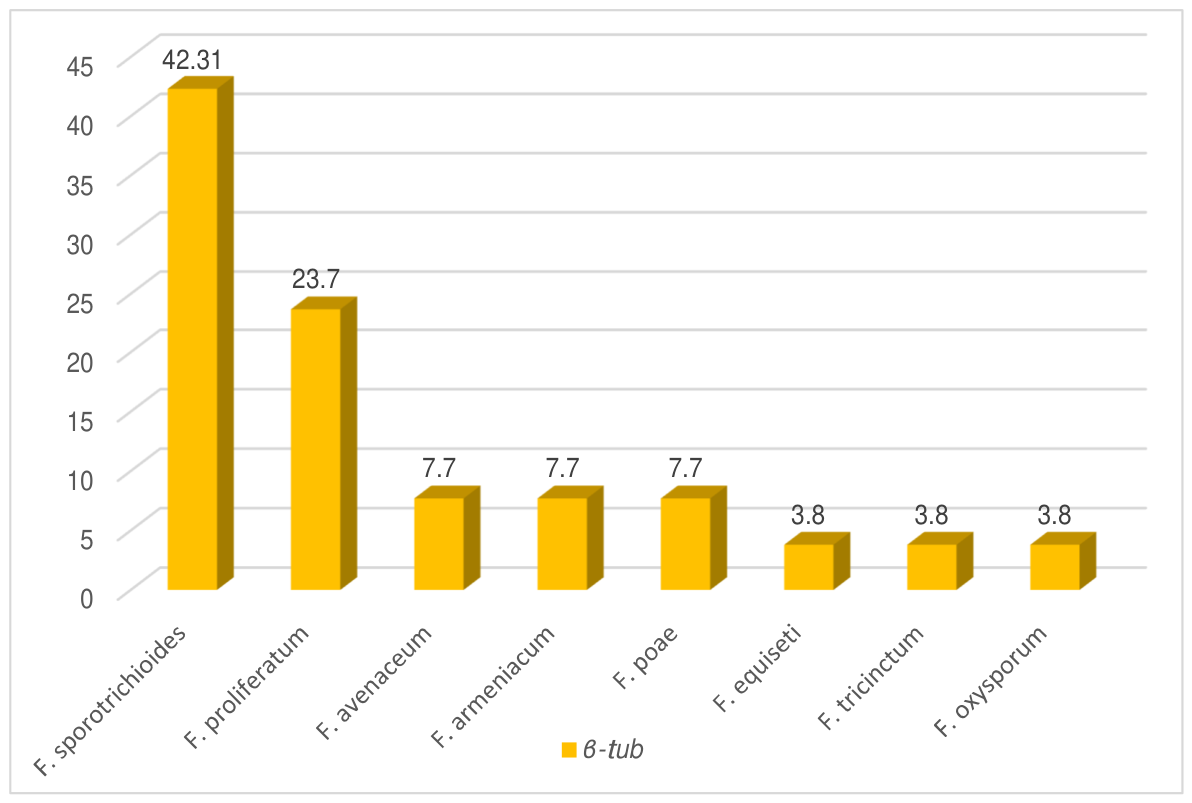
The percentage distribution of species from the analyzed varieties, determined by the β-tub barcode, are shown in Figure 7 and Table 7.
Molecular genetic analysis showed that 84.62% (n = 22) of the wheat samples contained only one Fusarium species, and in 15.38% (n = 4) the co-presence of two species was established. In the samples from the Ivaylovgrad region, Avenue variety, and the Sadovo region, Gizda variety, F. avenaceum/F. sporotrichioides and F. sporotrichioides/F. proliferatum were isolated and identified. In the samples from the Ivaylovgrad region (No. 2 and 3), Avenue variety, and the Sadovo region (No. 5, 6), Gizda variety, F. avenaceum/F. sporotrichioides and F. sporotrichioides/F. proliferatum were isolated and identified (Table 7).
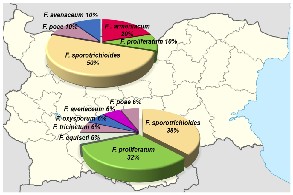
The results of the analysis show that in Southern Bulgaria the species diversity is wider (seven species), compared to Northern Bulgaria (five species). F. equiseti, F. oxysporum and F. tricinctum are found only in Southern Bulgaria, while in Northern Bulgaria the species F. armeniacum is found (Figure 8).
Phylogenetic analysis of the identified Fusarium spp.
Phylogenetic analyses based on β-tub gene sequences further separated the 26 isolates into five Fusarium species complexes, FSAMSC, FFSC, FIESC, FTSC, and FOSC, according to the modern phylogenetic concept of the genus. The isolates F. sporotrichioides and F. poae—members of the FSAMSC complex—are grouped in a separate independent cluster based on genetic similarity cluster. The six strains of F. proliferatum, showing a close relationship, were clearly distinguished in a separate group as members of the FFSC. The isolates designated as F. oxysporum are separated into a separate cluster. The two isolates of the species F. armeniacum (FSAMSC) were separated into a separate subcluster, while F. equiseti, F. tricinctum, and F. avenaceum were scattered throughout the tree (Figure 9).

4. Discussion

In the analysis of 101 samples of common wheat, harvest 2024–2025, an average value for contamination with microscopic fungi of the genus Fusarium was found in 2024 to be 7.2% ± 8.30, and for 2025—5.5% ± 2.80. For comparison, most data from other countries report higher values for contamination with this mold fungi. For example, Alkadri et al. (2013) [42] reported an average of 2.4% presence of the genus in the surface microbiome of wheat grown in Syria, harvest 2009–2010, with Fusarium spp. being found in 62.5% of the 48 analyzed samples. In wheat samples from Italy, Saudi Arabia, and Ukraine, the genus was present on average with 16–26%, 32.3%, and 71%, respectively [43,44,45]. Our results show a relatively low average infection rate with Fusarium spp. in wheat, compared to data from other countries. The proportion of isolated Fusarium spp. strains from the investigated varieties varies depending on the year and the growing location. Such fluctuations have been associated with the impact of climatic conditions, changes in agrotechnical practices, crop rotation, soil management and the application of biological control [3,46,47].
The types of crops in the crop rotation and their influence on the infection of wheat with Fusarium have been documented by several authors [48,49]. The reason has been proven to be the transfer through the rotation of Fusarium-contaminated residues from the earlier crop, acting as a reservoir for subsequent infection. In our study, we analyzed the influence of rapeseed, corn and sunflower, grown as previous crops, on the incidence of Fusarium. The highest infection rank recorded in corn (85.36), according to the Kruskal–Wallis test, compared to sunflower (36.47) and rapeseed (38.19), indicates the cultivation of corn as a predecessor in the plot is associated with the highest phytosanitary risk of development of Fusarium spp. and possible contamination of the crop with the mycotoxins produced by them. The results confirmed the data of Dill-Macky et al. (2000) [50], and Robertson, A. (2010) [51] that the use of maize before this significantly increases the infection risk due to the inoculum accumulated in the residues. Many authors recommend interrupting the pathogenesis by sowing with rapeseed or legumes [52]. The results on the presence of pathogens in Bulgarian wheat samples after growing rapeseed are significantly lower than in corn.
The samples after sunflower, which is an “unsuitable” host for pathogens, also have lower and stable levels of infection. According to Lori et al., 2009 [49], sunflower is not a suitable substrate for Fusarium, and Schoneberg et al., 2016 [48], even confirmed that wheat grown after rapeseed and sunflower has lower levels of the produced mycotoxin DON.
As a result of our descriptive analysis of the influence of wheat variety on contamination with Fusarium spp., no statistically significant difference was found (p > 0.05) (Table 2). A similar lack of a significant effect of genotype was reported by El Chami et al. (2023) and Spanic et al. (2021) [53,54]. In Bulgarian conditions, studies by Desheva and Chavdarov (2016) [55] also reported that although some varieties exhibit higher resistance, varieties completely resistant to Fusarium infections have not been established. Despite the application of different crop rotations and the use of new varieties in the fight against pathogens, in-depth studies of the population composition of these potential mycotoxin producers, including accurate species identification, are needed. All this highlights the need for in-depth studies of the population composition of these potential mycotoxin producers, with the first step being accurate identification of the species, to develop strategies to limit their presence on wheat crops. In our study, we analyzed the influence of rapeseed, corn and sunflower, grown as previous crops, on the incidence of Fusarium. The results confirmed the data that the use of maize before this significantly increases the infection risk due to the inoculum accumulated in the residues. Many authors recommend interrupting the pathogenesis by sowing with rapeseed or legumes [52]. The results of the presence of pathogens in Bulgarian wheat samples after growing rapeseed are significantly lower than compared to the maize.
In the present study, the results of the molecular genetic analysis conducted based on the β-tub barcode for the purpose of species identification of Fusarium spp. in common wheat show that the species composition of this genus in our country is diverse. A total of four key species were identified—F. sporotrichioides, F. proliferatum, F. poae and F. avenaceum—which constitute about 80% of the population in the country. Of all the identified species, F. sporotrichioides stands out as dominant (42.31%). Although the information about its ecological plasticity is still limited and contradictory, some data indicate that the species develops in a relatively wide temperature range, with the optimum being between 25 and 30 °C [56]. Since F. sporotrichioides is a known producer of more toxic type A trichothecenes (T-2 and HT-2) [57], its spread in Bulgaria may pose a serious risk to the health of domestic animals and humans.
According to other data, the species develops at low temperatures, with minimums around 0, to 35 °C as a maximum for growth in some sources, while the optimum remains 22.5–30 °C (https://www.fao.org/4/y1390e/y1390e04.htm?utm, accessed 4 March 2026). This is probably one of the reasons for its leading distribution among Fusarium spp. isolates in Bulgaria. In the present study, the frequency of detection of F. sporotrichioides was higher in Northern Bulgaria (50%) compared to Southern Bulgaria (38%), which agrees with the observation that the species is highly tolerant to temperature differences (Figure 7). A similar trend was also reported by Gagkaeva et al. (2019) [58], who detected F. sporotrichioides in 84.1% of grain samples in the cold regions of the Volga, Urals and Western Siberia, where the species dominates over the known pathogen F. graminearum (19.2%). However, in a study from Ukraine, the species F. sporotrichioides (18.5%) was the 2nd most common after F. graminearum (39.3%), followed by F. avenaceum (13.9%), F. poae (10.6%) and F. tricinctum (8.6%) [45]. Overall, F. graminearum was reported as a “traditional” species on cereal crops in Bulgaria before the 1970s [59,60], but was displaced by F. verticillioides (24.6%), F. poae (15.4%), and F. sporotrichioides (11%) in the period 2001–2005 [61] More recent data by Gencheva and Beev (2020) [62] for the 2018 harvest, from the Stara Zagora region, indicated that the leading species is F. tricinctum (41.7%), followed by F. poae (16.7%), F. graminearum (8.3%), F. proliferatum (8.3%), and F. equiseti (8.3%). In our study, F. graminearum was not identified. The species F. proliferatum (23.7%, n = 6) is the second most identified species in Bulgaria. The pathogen synthesizes fumonisins, which are particularly dangerous for grain crops and animal health. Its co-presence with F. sporotrichioides in sample (No. 6), Sadovo region, 2024, increases the risk of co-accumulation of various mycotoxins [63,64]. The F. poae identified in this study (7.7%) is widely documented in Europe: in Ukraine and in Italy with a frequency of 10.6% and 17.8%, respectively [7,65]. In our country, it has been documented by Beev et al. (2011) [66] with 16.7% and Gencheva and Beev, (2020) [62] with 16.7%. The species is considered to be sensitive to climatic conditions, with warmer and drier periods favoring its development [63]. Among the most common representatives of Fusarium spp. in temperate climate zones is F. avenaceum [7]. In our study it was present with 7.7% (n = 2). It is the leading pathogen within the F. tricinctum complex (FTSC) and demonstrates high genetic variability [7]. The species is known for the production of “emerging mycotoxins”–moniliformin, bovericin and eniatins [57]. Atypical species for wheat include F. equiseti, 3.8% (n = 1), whose presence in Bulgaria is probably due to changing climatic conditions globally and in the region. Until recently, this species was not associated with the wheat mycoflora, but data on its presence in our country are now available [62,67]. Of interest is the newly identified species F. armeniacum, which in our study was isolated for the first time in Bulgaria with 7.7% (n = 2). Although it is rarer and occurs mainly in North America and temperate climate zones [68,69]. this species deserves attention due to its proven ability to produce trichothecene T-2 toxin [70]. On the other hand, the species identified by us molecularly, such as F. sporotrichioides, F. poae, F. avenaceum, F. tricinctum, and F. equiseti, often coexist with the most virulent Fusarium pathogens—F. graminearum and F. culmorum [71,72,73]. Even species previously considered to be weakly aggressive, such as F. poae and F. sporotrichioides, turn out to be significant for the development of infection in cereals and are key producers of trichothecenes in wheat [74,75]. According to Stępień’s (2011) [76] studies on the optimal temperature range of F. proliferatum, there is a large genetic and phenotypic variability between isolates, which requires differences in temperature optima and water activity of the substrates. The species F. armeniacum, identified only in Northern Bulgaria, seems to develop better at lower temperatures (~25 °C) and higher humidity during the growing season [77].
Although the ITS region is a major barcode marker in fungi, its drawback is insufficient variability to distinguish different species within Fusarium species complexes. On the other hand, the combination of conserved and variable regions in the β-tub gene establishes it as a reliable locus for species identification in the genus. The use of β-tub (619 bp) and its complete sequencing in F. solani isolates from India proved that this species contains five exons and four introns, which retain their positions, but are shorter compared to other species of the genus and can serve a role in species distinction [78]. This structure confirms the evolutionary conservatism of the location of the intron regions and simultaneously reveals taxonomically significant differences in their length. According to the author, in many cases β-tub shows better resolution between closely related species than the ITS region and confirms its importance as an effective marker in the molecular identification and barcoding of Fusarium spp. According to O’Donnell et al. (2015) [79], however, the application of β-tubulin (the first protein-coding gene) for molecular identification and phylogeny in the genus has “limited utility in some species complexes due to the presence of different duplicated genes within the same species” (paralogs), which for future studies requires the use of several genetic markers as barcodes to improve the accuracy and reliability of species identification. Due to the high degree of similarity between species at each single locus, the current approach for identification of Fusarium spp. is recommended to be multilocus—combining ITS, tef-1α, RPB2, β-tub and CaM sequences, to enhance the discriminatory potential of the analysis [80,81,82].
In order to clarify the evolutionary relationships between the studied isolates and their affiliation to the species complexes of the genus Fusarium, phylogenetic trees were constructed. The phylogenetic analysis established that the Bulgarian wheat isolates belong mainly to five large evolutionarily related groups—species complexes with a characteristic mycotoxic profile. The representatives of the F. sambucinum species complex (FSAMSC) including F. sporotrichioides, F. armeniacum, F. poae—species associated with the production of trichothecenes—are the most common with 57.7% (n = 15). It is followed by the F. fujikuroj species complex (FFSC) with 23.1% (n = 6), known for the synthesis of fumonisins and other secondary metabolites. Members of the F. tricinctum species complex FTSC (F. tricinctum, F. avenaceum) are distinguished by a more moderate presence—11.5%, (n = 3)—but are proven producers of enyatins, moniliformin and bovericin [73]. The two complexes FIESC and FOSC are the least widespread (F. equiseti, 3.8%, n = 1; F. oxysporum, 3.8%, n = 1). The predominance of representatives from FSAMSC and FTSC suggests a significant risk of contamination of the wheat crop with mycotoxins, while the presence of FIESC and FOSC completes the picture of the pathogenic pressure on the crop. Therefore, molecular analysis not only reveals the species diversity of Fusarium spp. on a regional scale, but also demonstrates which complexes are of greatest importance for crop health and potential safety of production.
The phylogeny distinguished members of FSAMSC, FOSC, and FFSC in separate clusters with genetically similar species. However, the phylogenetic analysis based on the β-tub gene encountered some limitations. These are the cases with isolates F. equiseti, F. tricinctum and F. avenaceum, which are scattered throughout the tree. The barcode failed to reliably unite F. armeniacum with the other members of FSAMSC, as well as F. tricinctum and F. avenaceum in the FTSC complex, which put its discriminatory power for all species in doubt and thus reinforced the recommendations of O’Donnell et al. (2022) [82] for multilocus genotyping.

5. Conclusions

Fusarium species are some of the most widespread pathogens worldwide with significant economic and environmental impacts on agriculture and human health. In recent years, rapid advances in molecular technologies, genomics and diagnostic approaches have transformed our understanding of the genus, its host–pathogen dynamics and its distribution in cereals. With the upcoming challenges of climate change and increased international trade in cereals and seed, the importance of research on regional agroecosystems must be emphasized. Knowledge of the influence of previous crops on Fusarium contamination and the lack of completely resistant varieties to the pathogen supports the development of new strategies to protect agricultural sustainability. DNA barcoding techniques are very useful for the correct identification of species in the complex genus Fusarium. The results of this study, based on the β-tub marker, significantly expand our knowledge of the population diversity of these pathogens in wheat in Bulgaria. The method allowed the identification of new Fusarium species on common wheat (F. armeniacum, producer of T-2 toxin) in Bulgaria and tracked changes in the dynamics of the distribution of some species, such as a decrease in the presence of the aggressive phyto-pathogen F. graminearum, producer of ZEA, DON NIV, etc., in wheat grown on the territory of Bulgaria. This achieves a stable and reliable classification of species that would be indistinguishable only by the morphological approach. The generalized review of the identified species, their presence and affiliation to species complexes, based on genetic similarity and mycotoxigenic potential, supports the assessment of the risk of wheat production from contamination with toxic metabolites. The results are important for epidemiological surveillance and the development of targeted disease management strategies. This approach would be useful in tracking the dynamics of Fusarium species presence, both in the field, immediately after harvest, and after storage.

Author Contributions

Conceptualization—G.B. and D.G.; methodology—D.S. and D.G.; software—D.G.; investigation—D.S. and D.G.; resources—G.B.; writing—original draft preparation—D.S., D.G. and G.B.; writing—review and editing—D.G.; visualization—D.G. and D.S.; supervision—G.B. All authors have read and agreed to the published version of the manuscript.

Funding

This research was funded by the Bulgarian Ministry of Education and Science (MES) in the frames of the Bulgarian National Recovery and Resilience Plan, Component “Innovative Bulgaria,” and the Project No. BG-RRP-2.004-0006-C02 “Development of research and innovation at Trakia University in service of health and sustainable well-being”.

Institutional Review Board Statement

Not applicable.

Informed Consent Statement

Not applicable.

Data Availability Statement

Data is available from the authors upon request.

Acknowledgments

This study was implemented within the framework of the scientific project 3AF/24 at Faculty of Agriculture in Trakia University, Stara Zagora, Bulgaria.

Conflicts of Interest

The authors declare no conflicts of interest. The funders had no role in the design of the study; in the collection, analyses, or interpretation of the data; in the writing of the manuscript, or in the decision to publish the results.

References

  1. Mirghasempour, S.A.; Studholme, D.J.; Chen, W.; Zhu, W.; Mao, B. Molecular and pathogenic characterization of Fusarium species associated with corm rot disease in saffron from China. J. Fungi 2022, 8, 515. [Google Scholar] [CrossRef]
  2. Ekwomadu, T.I.; Mwanza, M. Fusarium fungi pathogens, identification, adverse effects, disease management, and global food security: A review of the latest research. Agriculture 2023, 13, 1810. [Google Scholar] [CrossRef]
  3. Drakopoulos, D.; Sulyok, M.; Jenny, E.; Kägi, A.; Bänziger, I.; Logrieco, A.F.; Krska, R.; Vogelgsang, S. Fusarium Head Blight and Associated Mycotoxins in Grains and Straw of Barley: Influence of Agricultural Practices. Agronomy 2021, 11, 801. [Google Scholar] [CrossRef]
  4. Alisaac, E.; Mahlein, A.K. Fusarium head blight on wheat: Biology, modern detection and diagnosis and integrated disease management. Toxins 2023, 15, 192. [Google Scholar] [CrossRef] [PubMed]
  5. Beev, G.; Denev, S.; Bakalova, D. Zearalenone-producing activity of Fusarium graminearum and Fusarium oxysporum isolated from Bulgarian wheat. Bulg. J. Agric. Sci. 2013, 19, 255–259. [Google Scholar]
  6. Rauwane, M.E.; Ogugua, U.V.; Kalu, C.M.; Ledwaba, L.K.; Woldesemayat, A.A.; Ntushelo, K. Pathogenicity and virulence factors of Fusarium graminearum including factors discovered using next generation sequencing technologies and proteomics. Microorganisms 2020, 8, 305. [Google Scholar] [CrossRef]
  7. Beccari, G.; Senatore, M.T.; Tini, F.; Sulyok, M.; Covarelli, L. Fungal community, Fusarium head blight complex and secondary metabolites associated with malting barley grains harvested in Umbria, central Italy. Int. J. Food Microbiol. 2018, 273, 33–42. [Google Scholar] [CrossRef]
  8. Tittlemier, S.A.; Roscoe, M.; Trelka, R.; Gaba, D.; Chan, J.M.; Patrick, S.K.; Gräfenhan, T. Fusarium damage in small cereal grains from Western Canada. 2. Occurrence of Fusarium toxins and their source organisms in durum wheat harvested in 2010. J. Agric. Food Chem. 2013, 61, 5438–5448. [Google Scholar] [CrossRef]
  9. Liu, N.; Chen, Y.; Liu, J.; Su, Q.; Zhao, B.; Sun, M.; Dong, J. Transcriptional differences between major Fusarium pathogens of maize, Fusarium verticillioides and Fusarium graminearum with different optimum growth temperatures. Front. Microbiol. 2022, 13, 1030523. [Google Scholar] [CrossRef]
  10. Perochon, A.; Doohan, F.M. Trichothecenes and Fumonisins: Key players in Fusarium—Cereal ecosystem interactions. Toxins 2024, 16, 90. [Google Scholar] [CrossRef]
  11. Taheur, F.B.; Kouidhi, B.; Al Qurashi, Y.M.A.; Salah-Abbès, J.B.; Chaieb, K. Biotechnology of mycotoxins detoxification using microorganisms and enzymes. Toxicon 2019, 160, 12–22. [Google Scholar] [CrossRef]
  12. Juskiw, P.E.; Oatway, L.; Oro, M.; Nyachiro, J.M.; Anbessa, Y.; Xi, K.; Capettini, F. Registration of ‘Lowe, a Two-Rowed Malting Barley with Enhanced Resistance to Fusarium Head Blight. J. Plant Regist. 2019, 13, 301–310. [Google Scholar] [CrossRef]
  13. Hückelhoven, R.; Hofer, K.; Coleman, A.; Heß, M. Fusarium infection of malting barley has to be managed over the entire value chain. J. Plant Dis. Prot. 2018, 125, 1–4. [Google Scholar] [CrossRef]
  14. Pascari, X.; Marin, S.; Ramos, A.J.; Sanchis, V. Relevant Fusarium Mycotoxins in Malt and Beer. Foods 2022, 11, 246. [Google Scholar] [CrossRef]
  15. Cen, Y.K.; Lin, J.G.; Wang, Y.L.; Wang, J.Y.; Liu, Z.Q.; Zheng, Y.G. The gibberellin producer Fusarium fujikuroi: Methods and technologies in the current toolkit. Front. Bioeng. Biotechnol. 2020, 8, 232. [Google Scholar] [CrossRef]
  16. Xu, M.; Wang, Q.; Wang, G.; Zhang, X.; Liu, H.; Jiang, C. Combatting Fusarium head blight: Advances in molecular interactions between Fusarium graminearum and wheat. Phytopathol. Res. 2022, 4, 37. [Google Scholar] [CrossRef]
  17. Bashyal, B.M.; Rawat, K.; Sharma, S.; Kulshreshtha, D.; Gopala Krishnan, S.; Singh, A.K.; Aggarwal, R. Whole genome sequencing of Fusarium fujikuroi provides insight into the role of secretory proteins and cell wall degrading enzymes in causing bakanae disease of rice. Front. Plant Sci. 2017, 8, 2013. [Google Scholar] [CrossRef]
  18. Burgess, L.W.; Summerell, B.A.; Bullock, S.; Gott, K.P.; Backhouse, D. Laboratory Manual for Fusarium Research, 3rd ed.; Department of Crop Sciences, University of Sydney: Sydney, Australia, 1994. [Google Scholar]
  19. Hyde, K.D.; Chomnunti, P.; Crous, P.W.; Groenewald, J.Z.; Damm, U.; Ko, T.W.; Tan, Y.P. A case for re-inventory of Australia’s plant pathogens. Persoonia-Mol. Phylogeny Evol. Fungi 2010, 25, 50–60. [Google Scholar] [CrossRef]
  20. Guo, M.; Yuan, C.; Tao, L.; Cai, Y.; Zhang, W. Life barcoded by DNA barcodes. Conserv. Genet. Resour. 2022, 14, 351–365. [Google Scholar] [CrossRef] [PubMed]
  21. Schoch, C.L.; Seifert, K.A.; Huhndorf, S.; Robert, V.; Spouge, J.L.; Levesque, C.A.; White, M.M. Nuclear ribosomal internal transcribed spacer (ITS) region as a universal DNA barcode marker for Fungi. Proc. Natl. Acad. Sci. USA 2012, 109, 6241–6246. [Google Scholar] [CrossRef]
  22. Hausner, G.; Wang, X. Unusual compact rDNA gene arrangements within some members of the Ascomycota: Evidence for molecular co-evolution between ITS1 and ITS2. Genome 2005, 48, 648–660. [Google Scholar] [CrossRef]
  23. Nilsson, R.H.; Ryberg, M.; Abarenkov, K.; Sjökvist, E.; Kristiansson, E. The ITS region as a target for characterization of fungal communities using emerging sequencing technologies. FEMS Microbiol. Lett. 2009, 296, 97–101. [Google Scholar] [CrossRef]
  24. Blaalid, R.; Kumar, S.; Nilsson, R.H.; Abarenkov, K.; Kirk, P.M.; Kauserud, H. ITS 1 versus ITS 2 as DNA metabarcodes for fungi. Mol. Ecol. Resour. 2013, 13, 218–224. [Google Scholar] [CrossRef]
  25. Stielow, J.B.; Levesque, C.A.; Seifert, K.A.; Meyer, W.; Irinyi, L.; Smits, D.; Robert, V. One fungus, which genes? Development and assessment of universal primers for potential secondary fungal DNA barcodes. Persoonia-Mol. Phylogeny Evol. Fungi 2015, 35, 242–263. [Google Scholar] [CrossRef]
  26. Al-Hatmi, A.M.; Meis, J.F.; de Hoog, G.S. Fusarium: Molecular diversity and intrinsic drug resistance. PLoS Pathog. 2016, 12, e1005464. [Google Scholar] [CrossRef] [PubMed]
  27. Li, Y.; Chen, D.; Luo, S.; Zhu, Y.; Jia, X.; Duan, Y.; Zhou, M. Intron-mediated regulation of β-tubulin genes expression affects the sensitivity to carbendazim in Fusarium graminearum. Curr. Genet. 2019, 65, 1057–1069. [Google Scholar] [CrossRef] [PubMed]
  28. Annis, S.L.; Panaccione, D.G. Presence of peptide synthetase gene transcripts and accumulation of ergopeptines in Claviceps purpurea and Neotyphodium coenophialum. Can. J. Microbiol. 1998, 44, 80–86. [Google Scholar]
  29. Tsai, H.F.; Liu, J.S.; Staben, C.; Christensen, M.J.; Latch, G.C.; Siegel, M.R.; Schardl, C.L. Evolutionary diversification of fungal endophytes of tall fescue grass by hybridization with Epichloë species. Proc. Natl. Acad. Sci. USA 1994, 91, 2542–2546. [Google Scholar] [CrossRef]
  30. Tooley, P.W.; Goley, E.D.; Carras, M.M.; Frederick, R.D.; Weber, E.L.; Kuldau, G.A. Characterization of Claviceps species pathogenic on sorghum by sequence analysis of the β-tubulin gene intron 3 region and EF-1α gene intron 4. Mycologia 2001, 93, 541–551. [Google Scholar] [CrossRef]
  31. Liu, J.; Bell, A.A.; Wheeler, M.H.; Stipanovic, R.D.; Puckhaber, L.S. Phylogeny and pathogenicity of Fusarium oxysporum isolates from cottonseed imported from Australia into California for dairy cattle feed. Can. J. Microbiol. 2011, 57, 874–886. [Google Scholar] [CrossRef]
  32. Stoeva, D.; Gencheva, D.; Radoslavov, G.; Hristov, P.; Beev, G. Molecular Identification and Phylogenetic Analysis of Fusarium spp. Associated with Barley Grains Using DNA Barcoding in Bulgaria. Acta Microbiol. Bulg. 2024, 40, 446–456. [Google Scholar] [CrossRef]
  33. ISO 8258:1986; Combined Feed, Protein Concentrates and Raw Materials for Them. Sampling Rules and Test Methods. Bulgarian Institute for Standardization: Sofia, Bulgaria, 1986. Available online: https://bds-bg.org/en/project/show/bds:proj:26905 (accessed on 4 March 2026).
  34. Nirenberg, H.I. A simplified method for identifying Fusarium spp. occurring on wheat. Can. J. Bot. 1981, 59, 1599–1609. [Google Scholar] [CrossRef]
  35. Aboul-Nasr, M.B.; Abdul, A. A simple technique for single spore isolation of Fusarium verticillioides and Fusarium subglutinans. World J. Biol. Sci. 2014, 2, 21–25. [Google Scholar]
  36. Gerlach, W.; Nirenberg, H. The Genus Fusarium, a Pictorial Atlas; Kommissionsverlag P. Parey: Berlin, Germany, 1982; Volume 209, pp. 1–406. [Google Scholar]
  37. Burgess, L.W.; Summerell, B.A. Mycogeography of Fusarium: Survey of Fusarium species in subtropical and semi-arid grassland soils from Queensland, Australia. Mycol. Res. 1992, 96, 780–784. [Google Scholar] [CrossRef]
  38. Edgar, R.C. MUSCLE: Multiple sequence alignment with high accuracy and high throughput. Nucleic Acids Res. 2004, 32, 1792–1797. [Google Scholar] [CrossRef]
  39. Tamura, K.; Stecher, G.; Kumar, S. MEGA11: Molecular Evolutionary Genetics Analysis Version 11. Mol. Biol. Evol. 2021, 38, 3022–3027. [Google Scholar] [CrossRef] [PubMed]
  40. Kumar, S.; Stecher, G.; Suleski, M.; Sanderford, M.; Sharma, S.; Tamura, K. Molecular Evolutionary Genetics Analysis Version 12 for adaptive and green computing. Mol. Biol. Evol. 2024, 41, 1–9. [Google Scholar] [CrossRef]
  41. Tamura, K. Estimation of the number of nucleotide substitutions when there are strong transition-transversion and G + C-content biases. Mol. Biol. Evol. 1992, 9, 678–687. [Google Scholar]
  42. Alkadri, D.; Nipoti, P.; Döll, K.; Karlovsky, P.; Prodi, A.; Pisi, A. Study of Fungal Colonization of Wheat Kernels in Syria with a focus on Fusarium species. Int. J. Mol. Sci. 2013, 14, 5938–5951. [Google Scholar] [CrossRef] [PubMed]
  43. Covarelli, L.; Beccari, G.; Prodi, A.; Generotti, S.; Etruschi, F.; Juan, C.; Mañes, J. Fusarium species, chemotype characterisation and trichothecene contamination of durum and soft wheat in an area of central Italy. J. Sci. Food Agric. 2015, 95, 540–551. [Google Scholar] [CrossRef]
  44. Mahmoud, M.A.; Shehata, S.M. Molecular identification and characterization of Fusarium spp. associated with wheat grains. Int. J. Adv. Res. Biol. Sci. 2017, 4, 77–87. [Google Scholar] [CrossRef]
  45. Gritsev, O.A.; Zozulya, O.L.; Vorobyova, N.G.; Skivka, L.M. Monitoring of species-specific fungi of the genus Fusarium in sanitary material winter wheat in the territory of Ukraine. Microbiol. Biotechnol. 2018, 2, 81–89. Available online: https://sis.ukrpatent.org/uk/search/detail/1590808/ (accessed on 4 March 2026).
  46. Wang, Y.; Wang, R.; Sha, Y. Distribution, pathogenicity and disease control of Fusarium tricinctum. Front. Microbiol. 2022, 13, 939927. [Google Scholar] [CrossRef] [PubMed]
  47. Petrucci, A.; Khairullina, A.; Sarrocco, S.; Jensen, D.F.; Jensen, B.; Jørgensen, H.J.L.; Collinge, D.B. Understanding the mechanisms underlying biological control of Fusarium diseases in cereals. Eur. J. Plant Pathol. 2023, 167, 453–476. [Google Scholar] [CrossRef]
  48. Schöneberg, T.; Martin, C.; Wettstein, F.E.; Bucheli, T.D.; Mascher, F.; Bertossa, M.; Vogelgsang, S. Fusarium and mycotoxin spectra in Swiss barley are affected by various cropping techniques. Food Addit. Contam. Part A 2016, 33, 1608–1619. [Google Scholar] [CrossRef] [PubMed]
  49. Lori, G.A.; Sisterna, M.N.; Sarandón, S.J.; Rizzo, I.; Chidichimo, H. Fusarium head blight in wheat: Impact of tillage and other agronomic practices under natural infection. Crop Prot. 2009, 28, 495–502. [Google Scholar] [CrossRef]
  50. Dill-Macky, R.; Jones, R.K. The effect of previous crop residues and tillage on Fusarium head blight of wheat. Plant Dis. 2000, 84, 71–76. [Google Scholar] [CrossRef]
  51. Robertson, A. Early Season Diseases Showing up in Corn and Soybean Fields; Iowa State University Extension and Outreach: Ames, IA, USA, 2010. [Google Scholar]
  52. Vogelgsang, S.; Enkerli, J.; Jenny, E.; Roffler, S.; Widmer, F. Characterization of Fusarium poae microsatellite markers on strains from Switzerland and other countries. J. Phytopathol. 2011, 159, 197–200. [Google Scholar] [CrossRef]
  53. El Chami, J.; El Chami, E.; Tarnawa, Á.; Kassai, K.M.; Kende, Z.; Jolánkai, M. Effect of Fusarium infection on wheat quality parameters. Cereal Res. Commun. 2023, 51, 179–187. [Google Scholar] [CrossRef]
  54. Spanic, V.; Dvojkovic, K.; Babic, J.; Drezner, G.; Zdunic, Z. Fusarium head blight infestation in relation to winter wheat end-use quality—A three-year study. Agronomy 2021, 11, 1648. [Google Scholar] [CrossRef]
  55. Desheva, G.; Chavdarov, P. Response of common winter wheat cultivars to the causative agent of Fusarium head blight (Fusarium culmorum). In Proceedings of the National Scientific and Technical Conference with International Participation Ecology and Health, Sofia, Bulgaria, 14 April 2016; pp. 69–75. [Google Scholar]
  56. Maragos, C.M.; Ward, T.J.; Proctor, R.H. Mycotoxins in grains. In Storage Cereal Grains Their Products; Woodhead Publishing: Cambridge, UK, 2022; pp. 535–576. [Google Scholar] [CrossRef]
  57. Duffeck, M.R.; Bandara, A.Y.; Weerasooriya, D.K.; Collins, A.A.; Jensen, P.J.; Kuldau, G.A.; Esker, P.D. Fusarium head blight of small grains in pennsylvania: Unravelling species diversity, toxin types, growth, and triazole sensitivity. Phytopathology 2022, 112, 794–802. [Google Scholar] [CrossRef] [PubMed]
  58. Gagkaeva, T.; Gavrilova, O.; Orina, A.; Lebedin, Y.; Shanin, I.; Petukhov, P.; Eremin, S. Analysis of toxigenic Fusarium species associated with wheat grain from three regions of Russia: Volga, Ural, and West Siberia. Toxins 2019, 11, 252. [Google Scholar] [CrossRef] [PubMed]
  59. Karadjova, I. Studies on Fusarium Species in Cereals. Ph.D. Dissertation, Sofia Univerity, Sofia, Bulgaria, 1979. [Google Scholar]
  60. Vrabcheva, T.; Geßler, R.; Usleber, E.; Märtlbauer, E. First survey on the natural occurrence of Fusarium mycotoxins in Bulgarian wheat. Mycopathologia 1996, 136, 47–52. [Google Scholar] [CrossRef]
  61. Beev, G. Mycotoxicological Assessment of the Contamination of Cereals with Microscopic Fungi of the Genus Fusarium and Their Mycotoxins. Ph.D. Thesis, Trakia University, Stara Zagora, Bulgaria, 2009. [Google Scholar]
  62. Gencheva, D.; Beev, G. Molecular Identification of Fusarium spp. Isolated from Wheat Grain Based on Sequencing of Internal Transcribed Spacer (ITS) Region. Acta Microbiol. Bulg. 2021, 37, 27–33. [Google Scholar]
  63. Ferrigo, D.; Raiola, A.; Causin, R. Fusarium toxins in cereals: Occurrence, legislation, factors promoting the appearance and their management. Molecules 2016, 21, 627. [Google Scholar] [CrossRef]
  64. Ma, G.; Wang, H.; Qi, K.; Ma, L.; Zhang, B.; Zhang, Y.; Qi, J. Isolation, characterization, and pathogenicity of Fusarium species causing crown rot of wheat. Front. Microbiol. 2024, 15, 1405115. [Google Scholar] [CrossRef]
  65. Pancaldi, D.; Tonti, S.; Prodi, A.; Salomoni, D.; Dal Prà, M.; Nipoti, P.; Pisi, A. Survey of the main causal agents of Fusarium head blight of durum wheat around Bologna, northern Italy. Phytopathol. Mediterr. 2010, 49, 258–266. [Google Scholar]
  66. Beev, G.; Denev, S.; Pavlov, D. Occurrence and distribution of Fusarium species in wheat grain. Agric. Sci. Technol. 2011, 3, 165–168. [Google Scholar]
  67. Yanashkov, I.; Vatchev, T.D. First report of root and lower stem rot caused by Fusarium equiseti (Corda) Saccardo on wheat and rye in Bulgaria. Bulg. J. Agric. Sci. 2016, 22, 993–994. [Google Scholar]
  68. Yli-Mattila, T. Detection of trichothecene-producing Fusarium species in cereals in Northern Europe and Asia. Agron. Res. 2011, 9, 521–526. [Google Scholar]
  69. O’Donnell, K.; McCormick, S.P.; Busman, M.; Proctor, R.H.; Ward, T.J.; Doehring, G.; Geiser, D.M.; Alberts, J.F.; Rheeder, J.P. Marasas et al. 1984 “Toxigenic Fusarium Species: Identity and Mycotoxicology” revisited. Mycologia 2018, 110, 1058–1080. [Google Scholar] [CrossRef]
  70. Munkvold, G.P. Fusarium species and their associated mycotoxins. Mycotoxigenic Fungi Methods Protoc. 2017, 1542, 51–106. [Google Scholar] [CrossRef]
  71. Piacentini, K.C.; Savi, G.D.; Pereira, M.E.; Scussel, V.M. Fungi and the natural occurrence of deoxynivalenol and fumonisins in malting barley (Hordeum vulgare L.). Food Chem. 2015, 187, 204–209. [Google Scholar] [CrossRef]
  72. Karlsson, I.; Persson, P.; Friberg, H. Fusarium head blight from a microbiome perspective. Front. Microbiol. 2021, 12, 628373. [Google Scholar] [CrossRef]
  73. Inbaia, S.; Farooqi, A.; Ray, R.V. Aggressiveness and mycotoxin profile of Fusarium avenaceum isolates causing Fusarium seedling blight and Fusarium head blight in UK malting barley. Front. Plant Sci. 2023, 14, 1121553. [Google Scholar] [CrossRef]
  74. Hietaniemi, V.; Rämö, S.; Yli-Mattila, T.; Jestoi, M.; Peltonen, S.; Kartio, M.; Sieviläinen, E.; Koivisto, T.; Parikka, P. Updated survey of Fusarium species and toxins in Finnish cereal grains. Food Addit. Contam. Part A 2016, 33, 831–848. [Google Scholar] [CrossRef]
  75. Gurikar, C.; Shivaprasad, D.P.; Sabillón, L.; Gowda, N.N.; Siliveru, K. Impact of mycotoxins and their metabolites associated with food grains. Grain Oil Sci. Technol. 2023, 6, 1–9. [Google Scholar] [CrossRef]
  76. Stępień, Ł.; Waśkiewicz, A.; Wilman, K. Host extract modulates metabolism and fumonisin biosynthesis by the plant-pathogenic fungus Fusarium proliferatum. Int. J. Food Microbiol. 2015, 193, 74–81. [Google Scholar] [CrossRef]
  77. Available online: https://bulletins.cfd.meteo.bg/bull/BuletinNIMH202202 (accessed on 4 March 2026).
  78. Tarafder, M.; Datta, B. Deciphering β-tubulin gene of carbendazim resistant Fusarium solani isolate and its comparison with other Fusarium species. Curr. Genet. 2022, 68, 429–447. [Google Scholar] [CrossRef]
  79. O’Donnell, K.; Ward, T.J.; Robert, V.A.R.G.; Crous, P.W.; Geiser, D.M.; Kang, S. DNA sequence-based identification of Fusarium: Current status and future directions. Phytoparasitica 2015, 43, 583–595. [Google Scholar] [CrossRef]
  80. Using DNA Barcodes to Identify and Classify Living Things: Introduction Objectives. Dnabarcoding. 2014. Available online: https://dnabarcoding101.org/lab/ (accessed on 4 March 2026).
  81. Han, S.L.; Wang, M.M.; Ma, Z.Y.; Raza, M.; Zhao, P.; Liang, J.M.; Cai, L. Fusarium diversity associated with diseased cereals in China, with an updated phylogenomic assessment of the genus. Stud. Mycol. 2023, 104, 87–148. [Google Scholar] [CrossRef]
  82. O’Donnell, K.; Whitaker, B.K.; Laraba, I.; Proctor, R.H.; Brown, D.W.; Broders, K.; Geiser, D.M. DNA sequence-based identification of Fusarium: A work in progress. Plant Dis. 2022, 106, 1597–1609. [Google Scholar] [CrossRef]
Figure 1. Samples collected from main commercial wheat growing regions in Bulgaria.
Agriculture 16 01232 g001
Figure 2. Cultured wheat grains with germinated colonies on PDA medium and their micro and macro characteristics: (a) Petri with seeds for isolation (cultivation); (b) microconidia; (c) macroconidia; (d) F. sporotrichioides; (e) F. poae.
Agriculture 16 01232 g002
Figure 3. DNA barcoding stages for Fusarium species identification in this study.
Agriculture 16 01232 g003
Figure 4. Independent Samples Kruskal–Wallis Test for the influence of the preceding crop on the degree of contamination with Fusarium spp. (%). Note: Circles (o) denote outliers (1.5–3 IQR from the box edge) and asterisks (*) denote extreme outliers (>3 IQR from the box edge).
Agriculture 16 01232 g004
Figure 5. Agarose gel electrophoresis (1%) used for separation of the different PCR products. Lanes labeled “0” correspond to blank control. 1–12: F. pr.F. proliferatum, F.tr.F. tricinctum, F.arm.F. armeniacum, F.sp.F. sporotrichioides, F.ox.—F. oxysporum, F.eq.F equiseti, F.p.F. poae, F.av.F. avenaceum; M—DNA ladder.
Agriculture 16 01232 g005
Figure 6. Fragment of aligned and compared sequence data of region β-tub from Fusarium spp. isolates, with reference. Legend: A (green), G (purple), C (blue), T (red) are nucleotides; identical bases in the nucleotide sequence are marked with an asterisk.
Agriculture 16 01232 g006
Figure 7. Percentage distribution of molecularly identified Fusarium species based on b-tub barcoding.
Agriculture 16 01232 g007
Figure 8. The distribution of the species diversity of Fusarium in Southern and Northern Bulgaria.
Agriculture 16 01232 g008
Figure 9. Phylogenetic analysis of Fusarium spp. based on the sequence analysis of the protein-coding gene β-tub in the MEGA 11.0 software [40].
Agriculture 16 01232 g009
Table 1. Descriptive statistics on the contamination with Fusarium spp. of different varieties of common wheat grown in Bulgaria.
VarietyNumber of SamplesMean,
%
Standard
Deviation, %
Coefficient of Variation,%MinimumMaximum
Avenue213.853.2784.90.011.0
Armstrong25.503.5364.23.08.0
Annapurna46.003.3656.14.011.0
Apilko67.004.8168.81.013.0
Boryana25.504.9589.92.09.0
Gizda27.002.8340.45.09.0
Enola55.203.7071.10.09.0
Pibrak46.509.81150.90.021.0
Sadovo 123.004.24141.40.06.0
Sashec24.005.66141.40.08.0
Silverio KWS215.0011.375.427.023.0
Sobel49.757.8180.03.021.0
Sofru115.367.16133.40.025.0
Terres22.503.53141.40.05.0
Factor69.5017.93188.70.046.0
Table 2. ANOVA analysis to determine the dependence of contamination with Fusarium spp. on the varieties of common wheat.
SchemeSum
of Squares
Degree of FreedomMean Square F-Ratiop-Value
Between the
different varieties
454.721432.480.600.8542
Within individual varieties3246.676054.11
Total variation3701.3974
Table 3. ANOVA analysis to determine the dependence of contamination with Fusarium spp. on the origin of the selected varieties (imported/Bulgarian) of wheat.
Source of VariationSum
of Squares
Degree
of Freedom
Mean SquareF-Ratiop-Value
Between the different varieties20.65749120.660.490.4869
Within individual varieties2746.3286542.25
Total variation2766.98566
Table 4. Shapiro–Wilk test of normality for the establishment of the relationship between the ancestor and Fusarium spp.
Tests of Normality
Preceding CropShapiro–Wilk
StatisticdfSig.
Fusarium spp., %Sunflower0.889430.001
Rapeseed0.898290.009
Maize0.630290.000
Table 5. Kruskal–Wallis H to determine effect of culture type on contamination.
Preceding CropnMean RankChi-Square (H)dfp-Value
Sunflower4336.47
Rapeseed2938.1956.762<0.001
Maize2985.36
Table 6. Pairwise Comparisons of the preceding crop.
Sample 1–Sample 2Test StatisticStd. ErrorStd. Test StatisticSig.Adj. Sig.
(p Value) a
Sunflower–Rapeseed−1.7256.994−0.2470.8051.000
Sunflower–Maize−48.8976.994−6.9910.000<0.001
Rapeseed–Maize−47.1727.644−6.1710.000<0.001
Each row tests the null hypothesis that the Sample 1 and Sample 2 distributions are the same. Asymptotic significances (two-sided tests) are displayed. The significance level is 0.05. a. Significance values have been adjusted by the Bonferroni correction for multiple tests.
Table 7. Species identification of Fusarium isolates according to the DNA barcode β-tub.
No.Growing RegionVarietyHarvestIDIdentified Species
1SvilengradSobel202491F. proliferatum
2IvaylovgradAvenue202492F. avenaceum
3IvaylovgradAvenue202493F. sporotrichioides
4Ruse Sobel202494F. sporotrichioides
5Sadovo Gizda202495F. sporotrichioides
6Sadovo Gizda202496F. proliferatum
7SadovoNikolay202497F. sporotrichioides
8Nova ZagoraAvenue202498F. proliferatum
9Kyustendil AnnapurnaAnnapurnaa202499F. sporotrichioides
10KarlovoApilko2024100F. proliferatum
11DobrichFactor2024101F. armeniacum
12DobrichPibrak2024102F. poae
13DobrichApilko2024103F. sporotrichioides
14SlivenSofru2025104F. oxysporum
15DobrichPibrak2025105F. avenaceum
16SlivenAvenue2025106F. tricinctum
17ChirpanGizda2025107F. proliferatum
18Nova Zagora ArmstrongArmstrong2025108F. sporotrichioides
19PlovdivPibrak2025109F. sporotrichioides
20VidinAvenue2025110F. armeniacum
21SilistraDiamond2025111F. sporotrichioides
22Stara ZagoraAvenue2025112F equiseti
23VratsaAvenue2025203F. sporotrichioides
24PlovdivArmstrong2025204F. poae
25SilistraAnnapurna2025205F. sporotrichioides
26SilistraSofru2025206F. proliferatum
Disclaimer/Publisher’s Note: The statements, opinions and data contained in all publications are solely those of the individual author(s) and contributor(s) and not of MDPI and/or the editor(s). MDPI and/or the editor(s) disclaim responsibility for any injury to people or property resulting from any ideas, methods, instructions or products referred to in the content.