Article

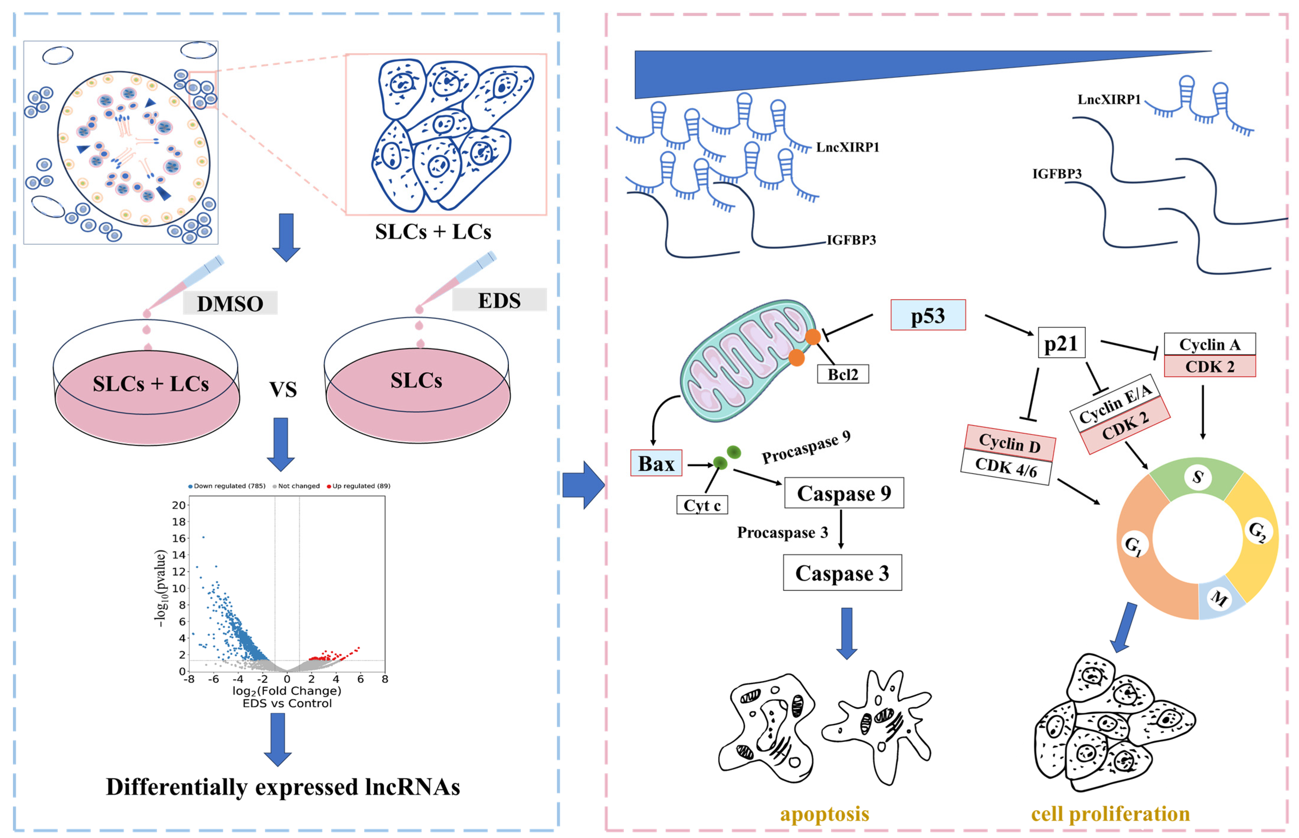
Long Non-Coding RNA lncXIRP1 Regulates the Proliferation and Apoptosis of Pig Leydig Cells

1
Key Laboratory of Animal Genetics, Breeding and Reproduction of Shaanxi Province, College of Animal Science and Technology, Northwest A&F University, Yangling 712100, China
2
College of Science, Northwest A&F University, Yangling 712100, China
3
Henan Key Laboratory of Farm Animal Breeding and Nutritional Regulation, Henan Pig Breeding Engineering Research Centre, Institute of Animal Husbandry, Henan Academy of Agricultural Sciences, Number 116, Hua Yuan Road, Zhengzhou 450002, China
*
Authors to whom correspondence should be addressed.
These authors contributed equally to this work.
Agriculture 2025, 15(8), 802; https://doi.org/10.3390/agriculture15080802
Submission received: 2 March 2025 / Revised: 30 March 2025 / Accepted: 7 April 2025 / Published: 8 April 2025
(This article belongs to the Section Farm Animal Production)

Abstract

:
Leydig cells (LCs) originate from stem Leydig cells (SLCs) and synthesize testosterone, a hormone indispensable for the development, sustenance, and functionality of the male reproductive system. Accumulating evidence suggests that long non-coding RNAs (lncRNAs) play pivotal roles in animal reproductive processes, yet the functional contributions of lncRNAs in pig LCs remain largely uncharacterized. The aim of this study was to examine how lncRNAs influence the function of LCs and their underlying molecular regulatory mechanisms. To achieve this, RNA-seq was conducted on cells before ethane dimethane sulfonate (EDS) treatment (SLCs and LCs) and after EDS treatment (SLCs), identifying 887 significantly downregulated lncRNAs and 30 upregulated lncRNAs after EDS treatment. Bioinformatics analysis identified lncXIRP1 for further investigation. The effects of lncXIRP1 on LCs proliferation, apoptosis, and expression of genes related to testosterone synthesis were investigated by using RT-qPCR, Western blot, CCK-8 and other methods. Bioinformatics predictions have unveiled the existence of a binding site between lncXIRP1 and IGFBP3. Through RT-qPCR experiments and a dual-luciferase reporter system, it was conclusively demonstrated that lncXIRP1 has the capacity to repress the expression of IGFBP3 mRNA, thereby inhibiting the proliferation and transcription activity of genes associated with testosterone synthesis in LCs and promoting their apoptosis. These results provide a theoretical foundation for further exploration of the impact of lncRNAs on LCs function and improving pig reproductive performance.

1. Introduction

Leydig cells (LCs), resident within the interstitial compartment amidst the seminiferous tubules, exhibit pivotal roles in male reproductive physiology. These cells are endowed with distinctive morphological features and specialized physiological functions, playing a fundamental part in sustaining male reproductive health [1]. The testosterone synthesized and secreted by LCs promotes male reproductive development and maintains secondary sexual characteristics [2]. In rats, the lineage development of adult Leydig cells (ALCs) is divided into four stages: stem Leydig cells (SLCs), progenitor Leydig cells (PLCs), immature Leydig cells (ILCs), and ALCs [3]. In these four stages, SLCs, which are located in the seminiferous tubules and peritubular region of the testes in male animals, play a particularly important role. They represent a unique category of adult stem cells that possess multipotent differentiation capabilities, enabling self-renewal and differentiation, thereby ensuring the stability of the LC population within the body [4]. Due to the relatively simple structure of rodent testes, it is now possible to directly isolate complete seminiferous tubules. However, this method is not applicable to humans and large domestic animals. To study SLCs, researchers used ethane dimethane sulphonate (EDS), and an alkylating agent that specifically eliminates differentiated interstitial cells, thereby obtaining SLCs for subsequent research [5]. Existing studies have shown that LC function involves numerous receptors and hormones. Nonetheless, studies examining the regulatory role of non-coding RNAs in pig reproductive cells (LCs) are still comparatively scant.
Long non-coding RNAs (lncRNAs) generally exceed 200 nucleotides in length. LncRNAs possess the capacity to modulate transcription, epigenetic alterations, and RNA and protein stability, as well as translation and post-translational modifications through their interactions with DNA, various RNA species (such as miRNAs and mRNAs), and proteins. Consequently, they play a role in regulating diverse biological processes, encompassing cell cycle progression and cellular differentiation [6]. LncRNA-412.25 regulates the proliferative and apoptotic activities of Hu sheep granulosa cells through its interaction with miR-346, leading to the activation of the leukemia inhibitory factor (LIF)/signal transducer and activator of transcription 3 (STAT3) signaling cascade [7]. During the initial stages of human muscle development, lncFAM71E1-2:2 (lncFAM) undergoes notable upregulation. Enhanced expression of lncFAM accelerates the differentiation of myoblasts into myotubes, whereas its knockdown impairs this differentiation. Positioned within the cell nucleus, lncFAM interacts with HNRNPL, as evidenced by ChIRP-MS studies, thereby facilitating the transcription and synthesis of MYBPC2 [8]. The MUNC lncRNA, also known as DRReRNA, serves as a cis-regulatory enhancer RNA for the Myod1 gene and enhances the expression of other myogenic genes in trans through its mediation in the recruitment of the Cohesin complex [9]. A study identified an evolutionarily conserved peptide of 46 amino acids from lncRNA. This peptide, called Myoregulin (MLN), interacts with the sarcoplasmic reticulum Ca2+-ATPase (SERCA), inhibiting Ca2+ reuptake into the sarcoplasmic reticulum [10]. Studies have shown that abnormal expression of lncRNAs and their corresponding pathogenic roles in male reproduction have been illuminated in recent years [11]. In mice, the overexpression of lncRNA-Gm2044 has been shown to partially impair spermatogenesis. It also participates in regulating multiple signaling pathways related to spermatogenesis [12]. The competitive endogenous RNA (ceRNA) regulatory network of LOC102549726/miR-760-3p/Atf6 may be involved in nickel-induced disruption of steroidogenesis in rat LCs [13]. Overall, these studies suggest that lncRNAs play critical roles in regulating animal reproduction. Despite pigs being economic and model animals, there are relatively few reports on the regulatory role of lncRNAs in pig LCs lineage development.
In our investigation, we conducted RNA sequencing on freshly isolated primary Leydig cells from pig including SLCs and LCs. Additionally, we analyzed SLCs from LCs treated with EDS. Bioinformatics analysis of the RNA-seq data identified a total of 887 lncRNAs that exhibited significant downregulation and 30 lncRNAs that showed upregulation following EDS treatment. Among the downregulated lncRNAs (Top 5), lncXIRP1 was selected for further investigation due to its potential role in regulating pig LCs. Its coding potential was predicted and subsequently confirmed as a non-coding RNA. Through overexpression and interference experiments, we explored the effects lncXIRP1 on the proliferation, apoptosis, and expression of testosterone synthesis-related genes in pig LCs. Therefore, the purpose of this study was to investigate the effect of lncRNAs on the function of LCs and their underlying molecular regulatory mechanisms. These explorations aim to understand the impacts of lncRNA on the function of testicular interstitial cells and enhance the reproductive efficiency of pigs.

2. Material and Methods

2.1. Sample Collection

In this study, 15 healthy 7-day-old male piglets were selected from the Yangling Benxiang breed cohort. The animals were subjected to electrical stunning followed by immediate exsanguination to ensure death prior to tissue sampling. Tissue samples—kidney, liver, spleen, brain, testis, epididymis—were collected from sampled piglets and preserved in liquid nitrogen. Fresh testes samples from these sampled piglets were stored in phosphate-buffered saline (PBS) (Invitrogen, Carlsbad, CA, USA). Pig LCs from the fresh testicular samples were extracted using enzymatic digestion and differential adhesion techniques [14].

2.2. RNA-Seq Analysis

EDS was dissolved in a solution comprising dimethyl sulfoxide (DMSO) and sterile water in a 1:3 volume ratio. The treated cells were divided into the following experimental groups (n = 3 per group): 1.0 mg/mL EDS (representing the final concentration) and blank control group (0 mg/mL EDS which received an equivalent volume of DMSO as that used in the 1.0 mg/mL EDS group). After 24 h of treatment, both the blank control group cells (labeled as control group) and EDS-treated group cells (labeled as EDS-treated group) were collected. RNA was extracted using Trizol for sequencing. The raw image data acquired by the Illumina Sequencing Analyzer underwent automated base recognition to convert them into initial sequence reads. Adapters, low-quality reads, and sequences with poly-N structures were removed to obtain clean sequencing data for further analysis. The cleaned data were then aligned to the pig reference genome using Top Hat (version 2.1.0). Due to the consistency of sequencing results, we excluded a set of data before proceeding with bioinformatics analysis.

2.3. Cell Culture and Transfection

Pig LCs were cultured in high-glucose DMEM (BBI, Shanghai, China) supplemented with 10% fetal bovine serum (FBS; Zetalife, Menlo Park, CA, USA) and 1% penicillin–streptomycin (Solarbio, Beijing, China) at 37 °C in a humidified 5% CO2 incubator. Upon reaching approximately 90% confluence, the cells were detached using 0.25% trypsin-EDTA (Solarbio, Beijing, China), neutralized with complete medium, and centrifuged at 1000× g for 5 min. Cell density was adjusted to 2 × 10⁵ cells/mL using a hemocytometer, and 1 mL of cell suspension was seeded into each well of the 12-well plate.
Transfection was initiated when cells reached 60–80% confluence (typically 12 h~24 post-seeding). The lncRNA XIRP1 overexpression vector or si-XIRP1 was complexed with Liposomal Transfection Reagent (Yeasen, Shanghai, China) at a 1:3 (μg:μL) plasmid-to-reagent ratio or 40 pmol siRNA concentration, according to manufacturer protocols. Medium was replaced with fresh complete DMEM 6 h after transfection to minimize cytotoxicity. Cells were harvested for total RNA extraction (TRIzol; Takara, Osaka, Japan) or protein lysate preparation (RIPA buffer, Beyotime, Shanghai, China) at 48 h post-transfection to assess overexpression/knockdown efficiency and downstream effects.

2.4. Isolation and Conversion of RNA into cDNA

RNA was isolated from pig tissue and cellular samples utilizing the Trizol reagent (Takara, Japan) method. The quality, quantity, and intactness of the isolated RNA were assessed via a Nanodrop spectrophotometer (Thermo Scientific, Waltham, MA, USA) and agarose gel electrophoresis, respectively. Following this, Hifair® III 1st Strand cDNA Synthesis SuperMix for qPCR (including gDNA digestion; Yeasen, Shanghai, China) was employed to reverse transcribe the RNA into cDNA.

2.5. Quantitative Real-Time PCR (qRT-PCR)

Quantitative primers were designed for the candidate lncRNAs, marker genes related to proliferation, apoptosis, and testosterone synthesis, as well as stem-loop primers for the candidate miRNAs. Subsequently, RT-qPCR was performed using ChamQ SYBR qPCR Master Mix (Vazyme, Nanjing, China). The primer sequences are listed in Table 1 and Table 2.

2.6. Protein Extraction and Western Blot

RIPA and PMSF (Shaanxi Zhonghui Hecai Biomedical Technology Co., Ltd., Shaanxi, China) were mixed in a ratio of 100:1 and added to LCs after the culture medium was completely removed. The mixture was left to stand on ice for 10–15 min to ensure complete cell lysis. The cells were detached using a cell scraper and the cell suspension was collected in a centrifuge tube. Total protein concentration was quantitated using a bicinchoninic acid (BCA) protein assay kit (Solarbio, Beijing, China) following the manufacturer’s instructions. Subsequently, 5× SDS-PAGE loading buffer (containing DTT) was added to the protein lysate at a 4:1 ratio, thoroughly mixed, and incubated in a water bath at 100 °C for 10 min. It was then stored at −80 °C. Next, 15% SDS-PAGE gel was prepared and the prepared protein sample was added to the lane 15 μg/well, followed by electrophoresis. Once the bromophenol blue indicator band had reached the bottom of the separation gel, the power was turned off and the proteins were transferred onto a PVDF membrane with a pore size of 0.22 μm. After transfer, the membrane was blocked with 5% skimmed milk powder for 2 h, followed by three washes with TBST. The membrane was then incubated with the primary antibody (proliferation-related proteins: PCNA, CDK2, Cyclin D1; apoptosis-related proteins: P53, Bax; steroid hormone synthesis-related proteins: LHR and β-actin) overnight at 4 °C (antibody information available in Table 3). After another three washes with TBST, the membrane was incubated with the secondary antibody at room temperature for 2 h. Finally, a chemiluminescent imaging system was used for detection (Sagecapture, Beijing, China).

2.7. Cell Counting Kit 8 (CCK-8)

After cell transfection for 24 h/48 h, 10 μL of CCK-8 reagent was added to each well. Following incubation at 37 °C for 3 h, the absorbance at 450 nm was measured using a microplate reader to assess cell viability.

2.8. Statistical Analysis

The results were presented as the arithmetic mean ± standard deviation of at least three biological replicates. Statistical analysis was executed employing SPSS software, version 23.0 (SPSS, Chicago, IL, USA), and GraphPad Prism, version 8.0 (GraphPad Software, San Diego, CA, USA). An independent-sample t-test was used to compare two groups, and one-way analysis of variance (ANOVA) was used to compare three or more groups. * p < 0.05 was considered statistically significant and ** p < 0.01 was considered highly significant.

3. Results

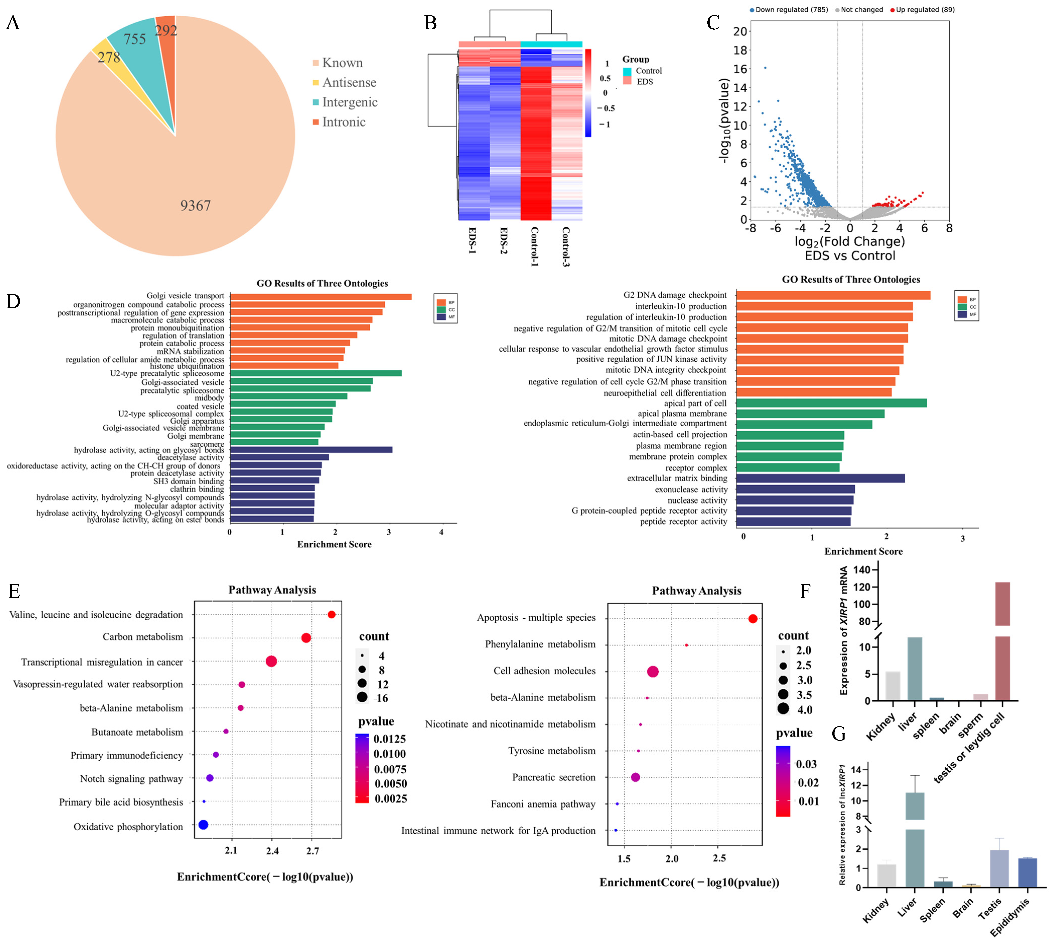
3.1. RNA-Seq Data Analysis and Differential Expression lncRNAs Screening

To identify key lncRNAs associated with maintaining the normal function of LCs, we performed high-throughput RNA-seq profiling of EDS-treated and control groups. A total of 10,692 lncRNAs were identified in pig LCs, including 9367 known lncRNAs, 292 novel lncRNAs, 755 intergenic lncRNAs, and 278 antisense lncRNAs (Figure 1A). There were 30 upregulated and 887 downregulated differentially expressed lncRNAs identified in the EDS treated group versus control groups (Figure 1B,C). Gene ontology (GO) and Kyoto Encyclopedia of Genes and Genomes (KEGG) pathway analyses were conducted on differentially expressed lncRNAs, resulting in the identification of 106 target genes associated with these lncRNAs through cis-regulatory interactions. According to GO analysis, these genes were related to transmembrane transport, apoptosis, calmodulin-dependent protein kinase activity, adenine nucleotide receptor activity, and other related functions (Figure 1D). Trans-acting analysis yielded a total of 3018 target genes linked to the differentially expressed lncRNAs which are essential for tubulogenesis and development, estrogen signaling, oxidoreductase activity, and aromatase function (Figure 1D). Analysis using KEGG indicated enrichment of these genes in pathways including protein digestion and absorption, ECM-receptor interaction, thyroid hormone signaling, and Notch signaling (Figure 1E).
The top five lncRNA downregulated candidates including lncXIRP1 were identified. We analyzed the expression patterns of the maternal gene XIRP1 (data retrieved from http://iswine.iomics.pro/, accessed on 12 July 2023) and revealed high expression levels of XIRP1 in testicular tissues and LCs (Figure 1F). In addition, lncXIRP1 expression was found to be higher in liver and testes (Figure 1G). The coding potential of lncXIRP1 was predicted by using online tools (http://cpc2.gao-lab.org/, accessed on 15 December 2023), revealing it to be a non-coding RNA. These findings suggest that lncXIRP1 may have a crucial role in regulating the function of LCs.

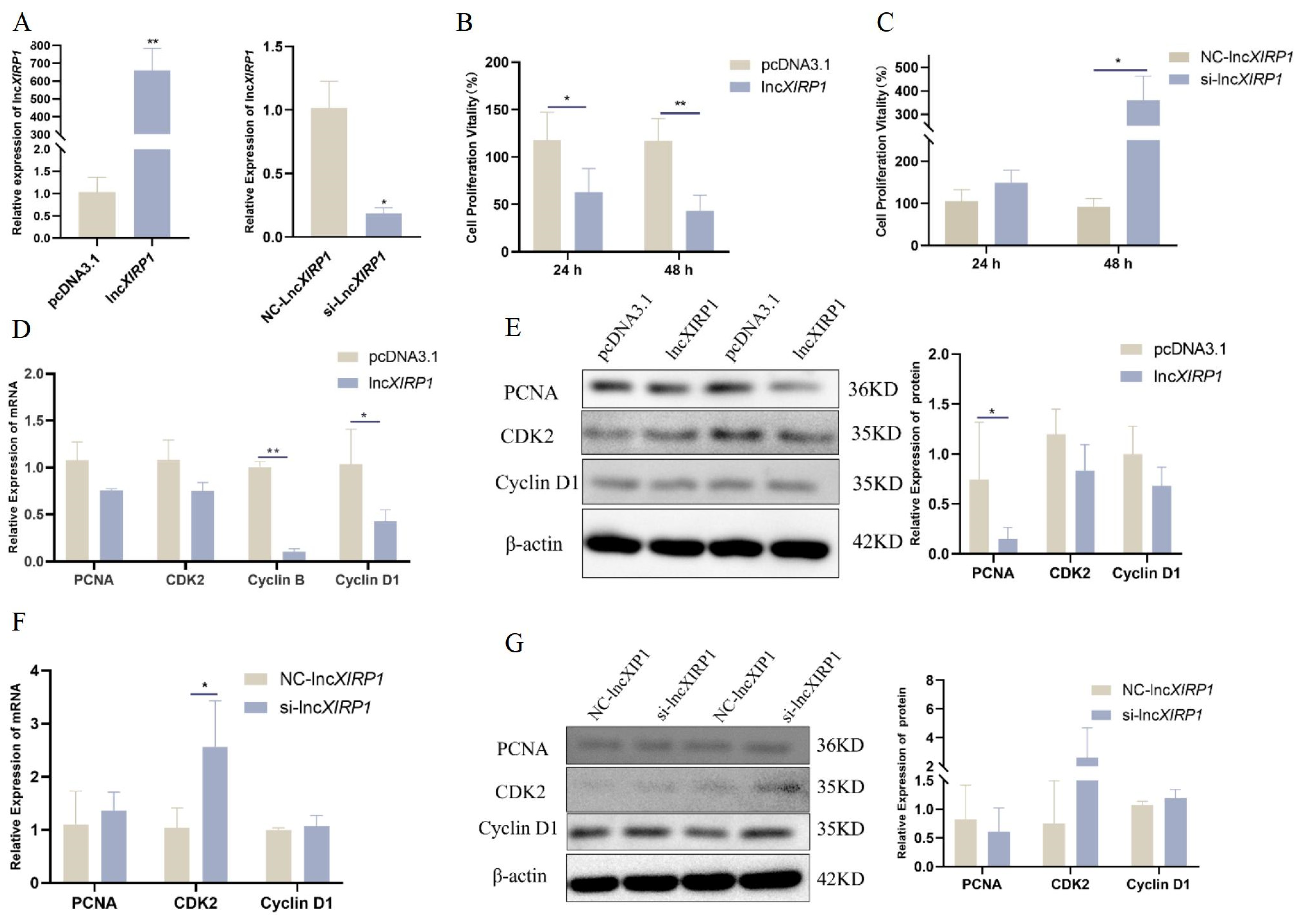
3.2. The Effect of lncXIRP1 on the Proliferation of Pig LCs

The main function of LCs is to produce testosterone, a sex hormone critical for spermatogenesis and sperm maturation, directly affecting sperm quantity and quality. The investigation of the influence of lncRNA on the proliferation and apoptosis of porcine LCs can serve as a stepping stone for elucidating its regulatory function in spermatogenesis in male animals.
To further explore the function of lncXIRP1, overexpression and interference strategies were used to explore its effects on LC. First, it was transfected into pig LCs. RT-qPCR analysis showed that after transfecting lncXIRP1 expression was significantly higher than the pcDNA3.1 group. Conversely, the expression level of lncXIRP1 was significantly lower in the si-lncXIRP1 group compared to the NC group, confirming the feasibility of proceeding with subsequent experiments (Figure 2A).
Subsequently, the CCK-8 assay was employed to assess the impact of lncXIRP1 on the cell viability of LCs, revealing that overexpression of lncXIRP1 markedly decreased the proliferation rate of LCs (Figure 2B,C). However, after transfection with si-lncXIRP1 for 48 h, the proliferation activity of LCs significantly increased. Therefore, we chose to perform subsequent experiments 48 h after transfection. RT-qPCR and Western blotting were utilized to investigate the influence of lncXIRP1 on the expression levels of proliferation-associated marker genes. It was revealed that the mRNA expressions of Cyclin B and Cyclin D1, and the protein expression of PCNA were significantly downregulated after overexpressing lncXIRP1 (Figure 2D,E). Conversely, the expression level of CDK2 mRNA increased after transfection with si-lncXIRP1 (Figure 2F,G). These findings suggest that lncXIRP1 can impair the proliferative activity of LCs and downregulate the expression of proliferation-related marker genes, effectively inhibiting the growth of LCs.

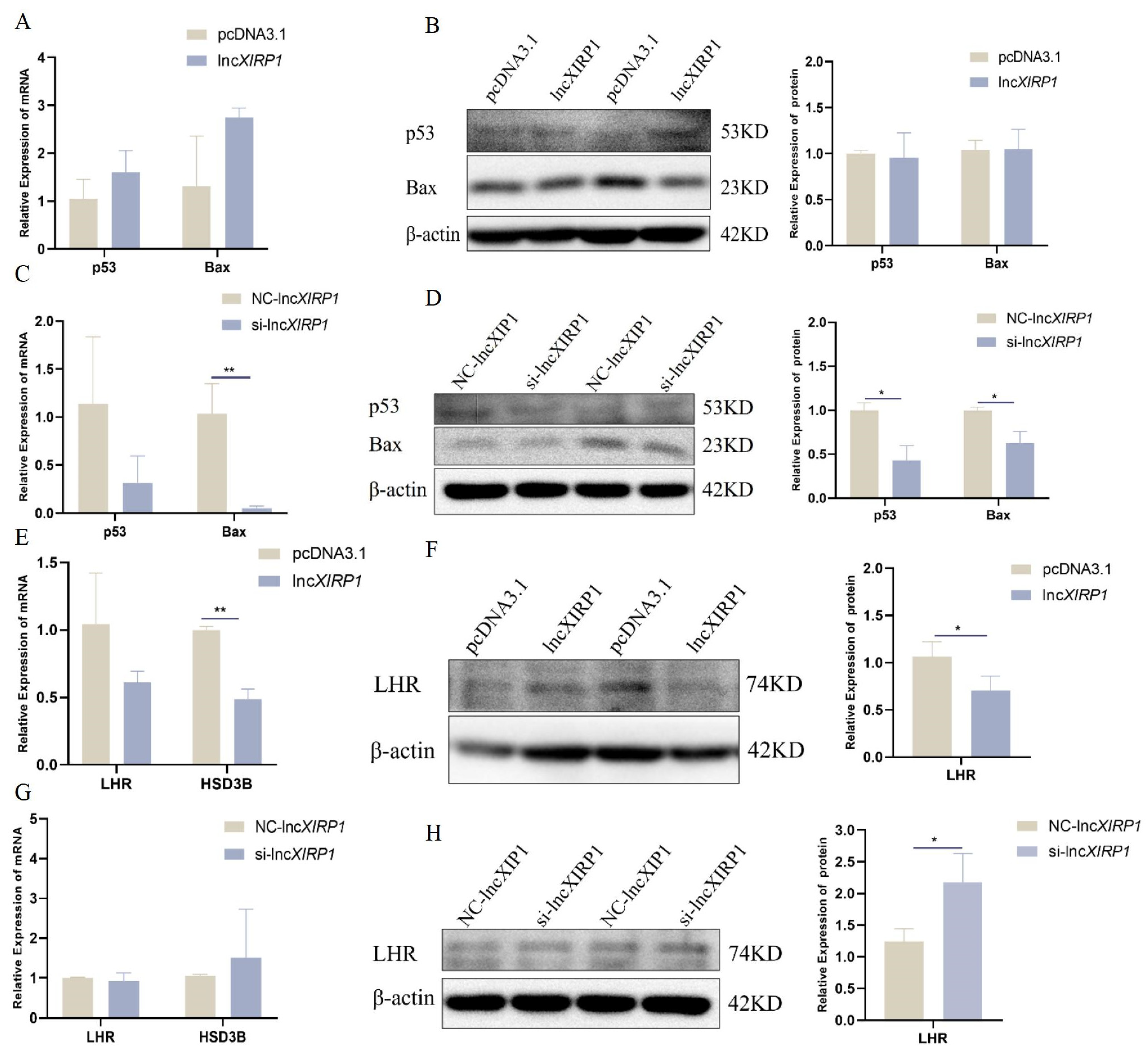
3.3. The Effect of lncXIRP1 on Apoptosis and Testosterone Synthesis in LCs

To gain deeper insights into the impact of lncXIRP1 on interstitial cell function, we employed qRT-PCR and Western blotting to examine its effect on apoptosis in pig testicular LCs. The results indicated that overexpression of lncXIRP1 did not significantly alter the expression of mRNA and protein levels of apoptosis-related marker genes (Figure 3A,B). After lncXIRP1 knockdown, the expression level of the apoptosis-related marker genes Bax mRNA, p53, and Bax protein were significantly decreased (Figure 3C,D). Simultaneously, RT-qPCR and Western blotting were used to evaluate the effect of lncXIRP1 on marker genes associated with testosterone synthesis, confirming that lncXIRP1 significantly inhibited the expression levels of HSD3B mRNA and LHR protein (Figure 3E–H). The above results indicate that lncXIRP1 may affect LCs function and further affect the process of spermatogenesis by affecting apoptosis and testosterone synthesis in LCs.

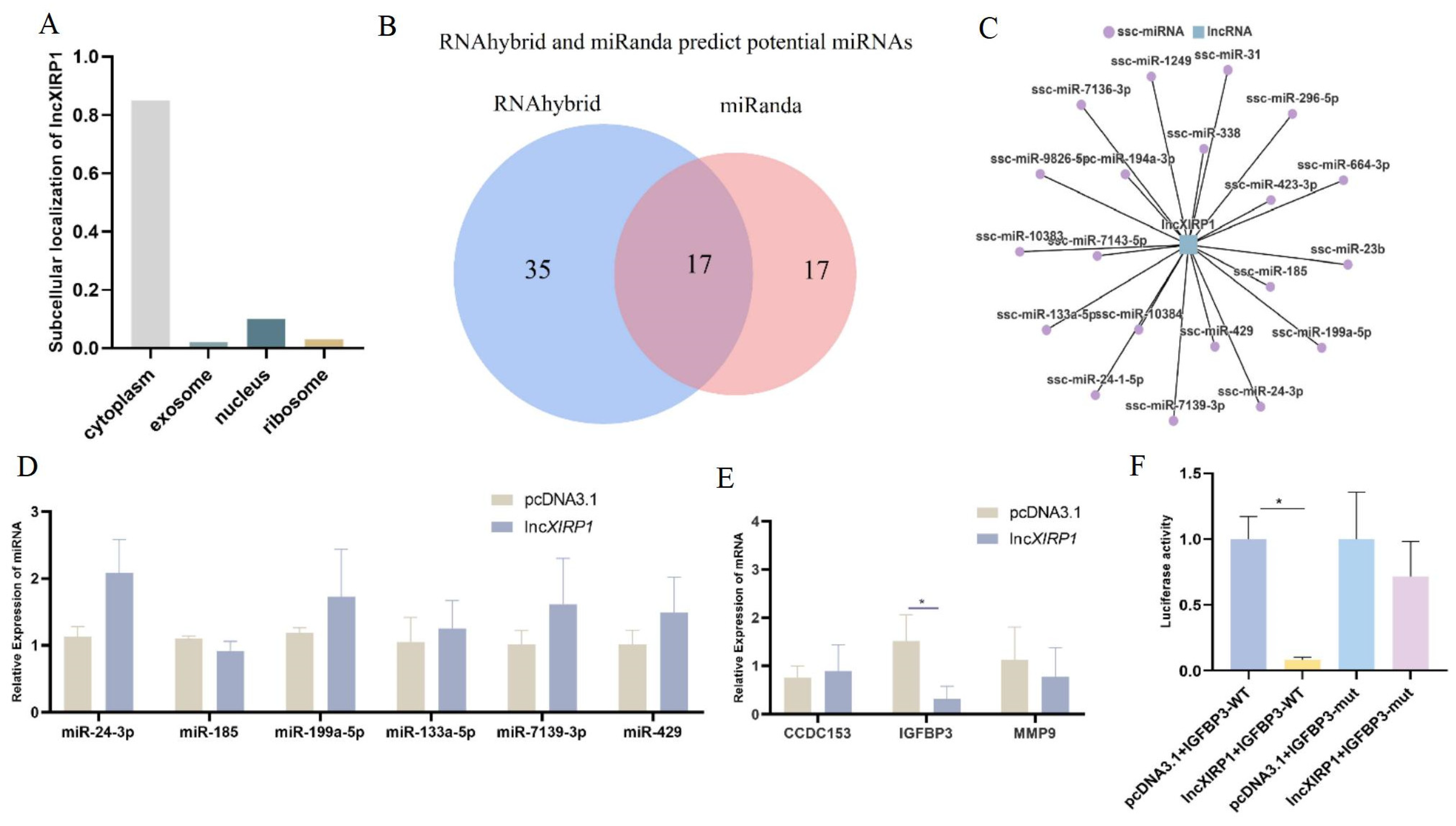
3.4. The Molecular Regulatory Mechanism of lncXIRP1

To further investigate the molecular regulatory mechanism of lncXIRP1, we first investigated the subcellular localization of lncXIRP1 in cells using the online tool (http://locate-r.azurewebsites.net, accessed on 12 July 2023), and predicted that lncXIRP1 is primarily expressed in the cytoplasm (Figure 4A). Initially, we focused on the molecular regulatory mechanism of lncRNA binding to miRNA, which is currently widely studied. We employed RNAhybrid and miRanda to predict miRNAs that may bind to lncXIRP1 and identified 17 overlapping miRNAs through a Venn diagram analysis (Figure 4B). Additionally, we predicted the binding relationships between the miRNAs differentially expressed after EDS treatment [15] and lncXIRP1, screening out those miRNAs potentially interacting with lncXIRP1. Subsequently, we constructed a visual network diagram of these potential lncXIRP1–miRNA interactions (Figure 4C). We selected candidate miRNAs using RT-qPCR; however, no significant inhibitory effect was observed with the overexpression of lncXIRP1 (Figure 4D). Therefore, further investigation is needed to identify the miRNAs potentially bound by lncXIRP1.
Previous research has demonstrated that cytoplasmic lncRNAs can regulate cellular functions through interactions with proteins, alterations in mRNA stability, or modulations in translation efficiency. Therefore, we further investigated whether lncXIRP1 can directly bind to mRNA to exert its regulatory effects. Furthermore, we have chosen three mRNAs based on previous research output (CCDC153, IGFBP3, and MMP9) to investigate the regulatory of lncXIRP1 to spermatogenesis and hormone synthesis. Using the online tool (https://rna.informatik.uni-freiburg.de/IntaRNA/Input.jsp, accessed on 3 April 2024), we predicted the targeting relationships between these candidate mRNAs and lncXIRP1, revealing binding sites for all of them. Subsequently, we designed and synthesized RT-qPCR primers for the three candidate mRNAs and examined the expression levels of these mRNAs after overexpressing lncXIRP1. Significantly, our results indicated a notable downregulation, specifically in the expression of IGFBP3 mRNA (Figure 4E), indicating that lncXIRP1 might regulate the function of LCs through its interaction with IGFBP3. Subsequently, we constructed psi-check2 vectors for both the wild-type (WT) and mutant (mut) forms of IGFBP3 and employed a dual-luciferase reporter system to validate the targeting relationship between lncXIRP1 and IGFBP3 (Figure 4F). The results showed that lncXIRP1 markedly suppressed the luciferase activity of the wild-type IGFBP3 (IGFBP3-WT), whereas it exerted no significant effect on the luciferase activity of the mutated IGFBP3 (IGFBP3-mut). These results suggest the existence of a targeting relationship between lncXIRP1 and IGFBP3.

4. Discussion

The normal function of LCs in the testes is crucial for spermatogenesis, which is a complex and sophisticated biological regulation process [16]. lncRNAs belonging to the class of non-coding RNAs exert crucial functions in post-transcriptional regulatory mechanisms and are extensively implicated in the modulation of diverse biological pathways. Therefore, exploring the impact of lncRNA on LCs function is conducive to a deeper understanding of its role in the regulation of LCs function, thus providing a theoretical basis for improving pig reproductive performance. In this study, based on RNA-seq, we identified five lncRNAs that were significantly downregulated in the EDS treatment group compared to the control group, meaning they were highly expressed in the control group. Among these, lncXIRP1 is a transcript produced from the antisense strand of XIRP1. Using the online bioinformatics tool (http://iswine.iomics.pro/, accessed on 12 July 2023), we found that XIRP1 mRNA is highly expressed in pig testes and LCs, leading us to hypothesize that lncXIRP1 might have a regulatory role in LCs. To understand the function of lncXIRP1, we constructed expression profiles across different tissues using RT-qPCR. The results showed that lncXIRP1 was widely expressed in various pig tissues, suggesting it may have broad functions.
Current research indicates that lncRNAs play significant roles in cell proliferation, apoptosis, pyroptosis, and differentiation. Meanwhile, some studies have discovered that lncRNAs can affect cell function by regulating hormone synthesis [17]. For instance, lncRNA-m18as1 competitively binds miR-18a-5p to regulate the secretion of a follicle-stimulating hormone in rat primary pituitary cells through the Smad2/3 pathway [18]. Additionally, lncGSAR acts as a ceRNA to activate SCAP/SREBP signaling by sequestering miR-125b, thereby regulating steroid hormone secretion in granulosa cells [19]. In this study, we investigated the effects of lncXIRP1 on LC proliferation and apoptosis using overexpression and interference strategies. We found that lncXIRP1 significantly inhibits LC proliferation. However, it only significantly inhibited LCs apoptosis when lncXIRP1 was interfered with, which we speculate is due to its high endogenous expression in LCs. Therefore, overexpression of lncXIRP1 did not have a significant impact on cell apoptosis, while interference with lncXIRP1 markedly inhibited LC apoptosis. Testosterone, an important male hormone, has a critical impact on the male reproductive system. It is primarily synthesized by LCs and plays various crucial roles in the testes. Testosterone is essential for sperm formation, testicular growth and development, libido regulation, and the maintenance of muscle mass and bone density. It also affects the development and function of reproductive organs such as the testes and prostate. This study explored the impact of lncXIRP1 on testosterone synthesis and found that lncXIRP1 significantly inhibits the transcription levels and translation efficiency of genes associated with testosterone synthesis (e.g., StAR, CYP11A1, and HSD3B). While our current data focus on molecular determinants of steroidogenesis, it is noteworthy that transcriptional regulation of these rate-limiting enzymes has been shown to directly correlate with testosterone output in Leydig cells [20]. This discovery reveals that lncXIRP1 may play a regulatory role in pig testicular development and spermatogenesis through modulating the steroidogenic machinery.
Based on the influence of lncXIRP1 on the expression levels of genes related to proliferation, apoptosis, and testosterone synthesis in porcine Leydig cells, we propose that lncXIRP1 could potentially be utilized as a marker for marker-assisted selection (MAS) when screening for high-reproductive-performance boars. The expression level of lncXIRP1 could serve as a biomarker for screening boar fertility. qRT-PCR detection can be used to identify pig with relatively low expression levels of lncXIRP1. This is negatively correlated with changes in the expression levels of marker genes associated with Leydig cell proliferation and testosterone synthesis, thereby improving semen quality. Of course, this requires further validation in pig populations and correlation analysis with semen quality to enhance the accuracy of lncXIRP1 as a biomarker.
Most studies on lncRNA have focused on their ceRNA effect, which is achieved by the competitive binding of lncRNA with miRNA, thus inducing the degradation or translation inhibition of other targets [21]. In this study, we used online databases along with previous miRNA sequencing data before and after EDS treatment to identify potential miRNAs that may bind to lncXIRP1. RT-qPCR analysis of miRNA changes revealed that overexpression of lncXIRP1 did not significantly downregulate any miRNAs. Therefore, we speculate that lncXIRP1 may not exert its regulatory function through miRNA sequestration. Previous studies have indicated that besides interacting with miRNAs, lncRNAs can also regulate processes by interacting with mRNAs or proteins. For example, lncRNA LBCS directly interacts with AR mRNA, guiding hnRNPK to interact with the 5′-UTR of AR mRNA, thereby reducing the translation efficiency of AR [22]. LINC00665 is capable of engaging with the 3′-UTR of both MTF1 and YY2 mRNA, thereby facilitating the degradation of these mRNAs [23]. Thus, we initially screened several mRNAs that might influence testicular development and spermatogenesis and found that lncXIRP1 significantly reduced the expression of IGFBP3. This indicates that lncXIRP1 could potentially modulate the proliferation, apoptotic processes, and expression of testosterone synthesis-related genes in LCs via its interaction with IGFBP3 (Figure 5). Despite the fact that we employed more direct research approaches, such as RIP, in this study to verify the targeting relationship between lncXIRP1 and IGFBP3, qRT-PCR and a dual-luciferase reporter system were also capable of demonstrating the existence of a targeting relationship between the two. In previous studies, researchers have also utilized these two methods to verify the targeting relationship between lncRNA and its downstream targets. For instance, Lu et al. demonstrated the targeting relationship between Linc00673 and miR-150-5p by bioinformatics prediction, qRT-PCR, and dual-luciferase reporter system [24]; similarly, Liu et al. also employed analogous methods to confirm that miR-34a is a potential downstream miRNA of the lncRNA MALAT1 [25].
This study offers a crucial theoretical foundation for deeply exploring the function of lncRNA in regulating LCs within the testes. Additionally, it offers scientific evidence for understanding and improving the reproductive performance of pig, promoting further research in the field of reproductive biology and the development of the pig industry. In this study, we have preliminarily verified the effect of lncXIRP1 on the function of pig LCs through in vitro experiments. It is noteworthy that while the analysis of transcription and translation of key steroidogenic enzymes provides mechanistic insights, direct quantification of testosterone secretion dynamics through techniques such as LC-MS/MS or in vitro hormone accumulation assays would strengthen the physiological relevance of these findings. However, we observed a concerted downregulation of mRNA and protein levels of StAR and CYP11A1, the primary gatekeepers for cholesterol transport and side-chain cleavage, strongly indicating impaired testosterone biosynthetic capacity [26]. Future studies incorporating in vitro hormone accumulation assays will further elucidate the kinetic effects of lncXIRP1 on androgen production. To gain a deeper understanding of the mechanism of action of lncXIRP1 and its specific contributions in physiological processes, we also consider further exploring the molecular regulatory mechanism of lncXIRP1 in the future, including how it interacts with DNA, RNA, or other proteins, and how these interactions jointly regulate gene expression and cellular functions. Our findings are essential to offer both theoretical insights and experimental substantiation for exploring the potential of lncRNA in enhancing pig reproductive performance.

5. Conclusions

In conclusion, our research utilized RNA-seq to pinpoint lncXIRP1 as a potential regulator of LC function. Furthermore, through the application of overexpression and knockdown techniques, we discovered that lncXIRP1 markedly inhibits the proliferative capacity and testosterone production of pig LCs while concurrently enhancing their apoptotic rate. These findings establish a robust theoretical foundation for advancing the comprehension of lncRNA’s regulatory role in LC function, while also offering insights into its effects on LC activity and strategies to enhance pig reproductive performance.

Author Contributions

Data curation, X.C.; funding acquisition, C.P.; resources, C.P.; validation, H.Y., X.L. (Xianzhao Lu) and S.Z.; visualization, H.Y. and X.L. (Xianzhao Lu); writing—original draft, H.Y.; writing—review and editing, Q.T., X.L. (Xianyong Lan), J.W. and X.C. All authors have read and agreed to the published version of the manuscript.

Funding

This work was funded by the Key Research and Development Program of Henan Province (251111110200).

Institutional Review Board Statement

The experimental animals and procedures used in this study comply with the animal policy of Northwest A&F University (protocol number NWAFAC1008, approved on 22 March 2022), and the handling of experimental animals fully complies with animal welfare policies.

Data Availability Statement

All relevant data are clearly mentioned within the article and are publicly available in appropriate data repositories.

Conflicts of Interest

The authors declare no financial or other conflicts of interest related to this study.

References

  1. Zhou, R.; Wu, J.; Liu, B.; Jiang, Y.; Chen, W.; Li, J.; He, Q.; He, Z. The roles and mechanisms of Leydig cells and myoid cells in regulating spermatogenesis. Cell Mol. Life Sci. 2019, 76, 2681–2695. [Google Scholar] [CrossRef] [PubMed]
  2. Jiang, M.H.; Cai, B.; Tuo, Y.; Wang, J.; Zang, Z.J.; Tu, X.; Gao, Y.; Su, Z.; Li, W.; Li, G.; et al. Characterization of Nestin-positive stem Leydig cells as a potential source for the treatment of testicular Leydig cell dysfunction. Cell Res. 2014, 24, 1466–1485. [Google Scholar] [CrossRef] [PubMed]
  3. Chen, P.; Zirkin, B.R.; Chen, H. Stem Leydig Cells in the Adult Testis: Characterization, Regulation and Potential Applications. Endocr. Rev. 2020, 41, 22–32. [Google Scholar] [CrossRef] [PubMed]
  4. Chen, H.; Wang, Y.; Ge, R.; Zirkin, B.R. Leydig cell stem cells: Identification, proliferation and differentiation. Mol. Cell Endocrinol. 2017, 445, 65–73. [Google Scholar] [CrossRef]
  5. Guan, X.; Chen, F.; Chen, P.; Zhao, X.; Mei, H.; Liu, J.; Lian, Q.; Zirkin, B.R.; Chen, H. Effects of spermatogenic cycle on Stem Leydig cell proliferation and differentiation. Mol. Cell Endocrinol. 2019, 481, 35–43. [Google Scholar] [CrossRef]
  6. Begolli, R.; Sideris, N.; Giakountis, A. LncRNAs as Chromatin Regulators in Cancer: From Molecular Function to Clinical Potential. Cancers 2019, 11, 1524. [Google Scholar] [CrossRef]
  7. Yao, X.; El-Samahy, M.A.; Li, X.; Bao, Y.; Guo, J.; Yang, F.; Wang, Z.; Li, K.; Zhang, Y.; Wang, F. LncRNA-412.25 activates the LIF/STAT3 signaling pathway in ovarian granulosa cells of Hu sheep by sponging miR-346. FASEB J. 2022, 36, e22467. [Google Scholar] [CrossRef]
  8. Chang, M.W.; Yang, J.H.; Tsitsipatis, D.; Yang, X.; Martindale, J.L.; Munk, R.; Pandey, P.R.; Banskota, N.; Romero, B.; Batish, M.; et al. Enhanced myogenesis through lncFAM-mediated recruitment of HNRNPL to the MYBPC2 promoter. Nucleic Acids Res. 2022, 50, 13026–13044. [Google Scholar] [CrossRef]
  9. Przanowska, R.K.; Weidmann, C.A.; Saha, S.; Cichewicz, M.A.; Jensen, K.N.; Przanowski, P.; Irving, P.S.; Janes, K.A.; Guertin, M.J.; Weeks, K.M.; et al. Distinct MUNC lncRNA structural domains regulate transcription of different promyogenic factors. Cell Rep. 2022, 38, 110361. [Google Scholar] [CrossRef]
  10. Anderson, D.M.; Anderson, K.M.; Chang, C.L.; Makarewich, C.A.; Nelson, B.R.; McAnally, J.R.; Kasaragod, P.; Shelton, J.M.; Liou, J.; Bassel-Duby, R.; et al. A micropeptide encoded by a putative long noncoding RNA regulates muscle performance. Cell 2015, 160, 595–606. [Google Scholar] [CrossRef]
  11. Hashemi Karoii, D.; Azizi, H. Functions and mechanism of noncoding RNA in regulation and differentiation of male mammalian reproduction. Cell Biochem. Funct. 2023, 41, 767–778. [Google Scholar] [CrossRef] [PubMed]
  12. Hu, K.; Gao, Y.; Xu, Y.; He, C.; Wang, K.; Li, L.; Liao, Y.; Liu, X.; Liang, M. Overexpression of lncRNA-Gm2044 in spermatogonia impairs spermatogenesis in partial seminiferous tubules. Poult. Sci. 2022, 101, 101930. [Google Scholar] [CrossRef] [PubMed]
  13. Chen, B.; Tan, L.; Wang, Y.; Yang, L.; Liu, J.; Chen, D.; Huang, S.; Mao, F.; Lian, J. LOC102549726/miR-760-3p network is involved in the progression of ISO-induced pathological cardiomyocyte hypertrophy via endoplasmic reticulum stress. J. Mol. Histol. 2023, 54, 675–687. [Google Scholar] [CrossRef]
  14. Yu, S.; Zhang, P.; Dong, W.; Zeng, W.; Pan, C. Identification of Stem Leydig Cells Derived from Pig Testicular Interstitium. Stem. Cells Int. 2017, 2017, 2740272. [Google Scholar] [CrossRef] [PubMed]
  15. Cui, Y.; Chen, R.; Ma, L.; Yang, W.; Chen, M.; Zhang, Y.; Yu, S.; Dong, W.; Zeng, W.; Lan, X.; et al. miR-205 Expression Elevated With EDS Treatment and Induced Leydig Cell Apoptosis by Targeting RAP2B via the PI3K/AKT Signaling Pathway. Front. Cell Dev. Biol. 2020, 8, 448. [Google Scholar] [CrossRef]
  16. Barbagallo, F.; Condorelli, R.A.; Mongioi, L.M.; Cannarella, R.; Aversa, A.; Calogero, A.E.; La Vignera, S. Effects of Bisphenols on Testicular Steroidogenesis. Front. Endocrinol. 2020, 11, 373. [Google Scholar] [CrossRef]
  17. Taylor, D.H.; Chu, E.T.; Spektor, R.; Soloway, P.D. Long non-coding RNA regulation of reproduction and development. Mol. Reprod. Dev. 2015, 82, 932–956. [Google Scholar] [CrossRef]
  18. Zhang, W.; Ren, W.; Han, D.; Zhao, G.; Wang, H.; Guo, H.; Zheng, Y.; Ji, Z.; Gao, W.; Yuan, B. LncRNA-m18as1 competitively binds with miR-18a-5p to regulate follicle-stimulating hormone secretion through the Smad2/3 pathway in rat primary pituitary cells. J. Zhejiang Univ. Sci. B 2022, 23, 502–514. [Google Scholar] [CrossRef]
  19. Wang, Y.; Guo, Y.; Duan, C.; Li, J.; Ji, S.; Yan, H.; Liu, Y.; Zhang, Y. LncGSAR Controls Ovarian Granulosa Cell Steroidogenesis via Sponging MiR-125b to Activate SCAP/SREBP Pathway. Int. J. Mol. Sci. 2022, 23, 12132. [Google Scholar] [CrossRef]
  20. Payne, A.H.; Youngblood, G.L. Regulation of expression of steroidogenic enzymes in Leydig cells. Biol. Reprod. 1995, 52, 217–225. [Google Scholar] [CrossRef]
  21. Chan, J.J.; Tay, Y. Noncoding RNA:RNA Regulatory Networks in Cancer. Int. J. Mol. Sci. 2018, 19, 1310. [Google Scholar] [CrossRef] [PubMed]
  22. Gu, P.; Chen, X.; Xie, R.; Xie, W.; Huang, L.; Dong, W.; Han, J.; Liu, X.; Shen, J.; Huang, J.; et al. A novel AR translational regulator lncRNA LBCS inhibits castration resistance of prostate cancer. Mol. Cancer 2019, 18, 109. [Google Scholar] [CrossRef] [PubMed]
  23. Ruan, X.; Zheng, J.; Liu, X.; Liu, Y.; Liu, L.; Ma, J.; He, Q.; Yang, C.; Wang, D.; Cai, H.; et al. lncRNA LINC00665 Stabilized by TAF15 Impeded the Malignant Biological Behaviors of Glioma Cells via STAU1-Mediated mRNA Degradation. Mol. Ther. Nucleic Acids 2020, 20, 823–840. [Google Scholar] [CrossRef] [PubMed]
  24. Lu, W.; Zhang, H.; Niu, Y.; Wu, Y.; Sun, W.; Li, H.; Kong, J.; Ding, K.; Shen, H.M.; Wu, H.; et al. Long non-coding RNA linc00673 regulated non-small cell lung cancer proliferation, migration, invasion and epithelial mesenchymal transition by sponging miR-150-5p. Mol Cancer 2017, 16, 118. [Google Scholar] [CrossRef]
  25. Liu, Y.; Gao, S.; Du, Q.; Zhao, Q. Knockdown of long non-coding RNA metastasis associated lung adenocarcinoma transcript 1 inhibits the proliferation and migration of bladder cancer cells by modulating the microRNA-34a/cyclin D1 axis. Int. J. Mol. Med. 2019, 43, 547–556. [Google Scholar] [CrossRef]
  26. Miller, W.L. Steroid hormone synthesis in mitochondria. Mol. Cell Endocrinol. 2013, 379, 62–73. [Google Scholar] [CrossRef]
Figure 1. RNA seq data analysis of pig LCs treated with EDS and untreated groups. (A) The lncRNAs and their types present in pig LCs. (B,C) lncRNAs clustering heatmaps and differentially expressed lncRNAs volcano maps. (D,E) GO and KEGG pathways for target genes of differentially expressed lncRNAs. (F) Expression levels of XIRP1 mRNA in different tissues of pigs (http://iswine.iomics.pro/, accessed on 12 July 2023). (G) The expression levels of lncXIRP1 in different tissues of pigs.
Agriculture 15 00802 g001
Figure 2. lncXIRP1 inhibits the proliferation of LCs. (A) Detection of lncXIRP1 overexpression efficiency and interference efficiency. (B,C) CCK-8 detection of the effects of lncXIRP1 and si-lncXIRP1 on the proliferation activity of LCs. (DG) RT-qPCR and Western blot were used to detect the effects of lncXIRP1 or si-lncXIRP1 on the expression levels of marker genes related to the proliferation of LCs. * p < 0.05 was considered statistically significant and ** p < 0.01 was considered highly significant.
Agriculture 15 00802 g002
Figure 3. lncXIRP1 promotes the apoptosis of LCs and inhibits the synthesis of testosterone in LCs. (A,B) RT-qPCR and Western blot were used to detect the effects of lncXIRP1 on the apoptosis of LCs. (C,D) The effects of lncXIRP1 and si-lncXIRP1 on LC apoptosis were examined using RT-qPCR and Western blot analyses. (E,F) RT-qPCR and Western blot were used to detect the effects of lncXIRP1 overexpression or si-lncXIRP1 on the synthesis of testosterone in LCs. (G,H) RT-qPCR and Western blot were conducted to investigate the effects of lncXIRP1 overexpression and lncXIRP1 knockdown (si-lncXIRP1) on testosterone synthesis in LCs. * p < 0.05 was considered statistically significant and ** p < 0.01 was considered highly significant.
Agriculture 15 00802 g003
Figure 4. lncXIRP1 may regulate the function of LCs through IGFBP3. (A) Subcellular localization of lncXIRP1. (B) RNAhybrid and miRanda predicted the possible binding miRNA Venn diagram of lncXIRP1. (C) The constructed RNAhybrid and miRanda prediction and the potential miRNA visualization network diagram of differential expression before and after EDS treatment that may bind to lncXIRP1. (D) Changes in the expression levels of miRNAs that may bind after overexpression of lncXIRP1. (E) Changes in the expression levels of mRNAs that may bind after overexpression of lncXIRP1. (F) Verification of the targeting relationship between lncXIRP1 and IGFBP3 using a dual-luciferase reporter system. * p < 0.05 was considered statistically significant.
Agriculture 15 00802 g004
Figure 5. Molecular regulatory mechanism of lncXIRP1 in pig LCs. The increase in lncXIRP1 was able to sequester IGFBP3, thereby promoting the expression of P53 and Bax, and subsequently facilitating apoptosis in Leydig cells. Following the interference with lncXIRP1, the expression level of IGFBP3 mRNA was elevated, resulting in increased expression of Cyclin D and CDK2, and ultimately stimulating proliferation of Leydig cells.
Agriculture 15 00802 g005
Table 1. Primer sequences of marker genes and primers for mRNAs that may be bound by lncXIRP1.
Primer NamePrimer SequenceTm ValueGC%PCR Product Length
PCNAF: AATGCAGACACCTTGGCACT60.1850.00154
R: GTGCAAATTCACCAGAAGGCA59.6647.62
CDK2F: GAGCCTTTGGAGTCCCTGTTC60.6157.14148
R: CGGGTCACCATCTCAGCAAAG61.2857.14
Cyclin BF: AATCCCTTCTTGTGGTTA49.9438.89104
R: CTTAGATGTGGCATACTTG50.6042.11
Cyclin DF: TACACCGACAACTCCATCCG59.4755.00224
R: GAGGGCGGGTTGGAAATGAA60.6155.00
BaxF: GCTGACGGCAACTTCAACTG60.0455.00202
R: GCGTCCCAAAGTAGGAGAGG59.8260.00
Bcl-2F: CATGTGTGTGGAGAGCGTCA60.3255.00135
R: TCTACCATGGACTTCCCCCA59.5855.00
Caspase 3F: TCAGAGGGGACTGCTGTAGA59.3055.0081
R: AGTCCAATTCTGTGCCTCGG60.0455.00
GAPDHF: TCGGAGTGAACGGATTTGGC60.6755.0095
R: GAAGGGGTCATTGATGGCGA60.1155.00
LHRF: TCTCCCTATCAAAGTAATCC 51.0040.00148
R: GTTCTGGATCAGTATTTCAG50.9940.00
HSD3BF: GCTGGAGGAGAAGGATCTGC59.8960.0089
R: TGCTCTGGAGCTTAGAAAATTCC58.7343.48
CCDC153F: TCATCCCCTGGAAGTGTGTGG61.7357.14205
R: GGAAAGAGCGGTGGAGAATGG61.0157.14
IGFBP3F: TGCCTGACTCCAAACTCCAC59.8955.00199
R: AACTTGAGGTGGTTCAGCGT59.8250.00
MMP9F: CGCCGACATCGTTATCCAGT60.2555.00153
R: TTGCCCAGAGACCACAACTC59.8955.00
Table 2. Candidate lncRNA and miRNA primers.
Primer Name Primer Sequence
lncXIRP1F: TGAAAGTGGAGGGTGACTCAA
R: CTTATGGCTACCAGTGGAGGCTAT
miR-24-3pRT: GTCGTATCCAGTGCAGGGTCCGAGGTATTCGCACTGGATACGACCTGTTC
F: TGCGTCCGTGGCTCAGTTCAGCAG
miR-185RT: GTCGTATCCAGTGCAGGGTCCGAGGTATTCGCACTGGATACGACTCAGGA
F: TGCGTCTTGGAGAGAAAGGCAGT
miR-199a-5pRT: GTCGTATCCAGTGCAGGGTCCGAGGTATTCGCACTGGATACGACGAACAG
F: TGCGTCCGCCCAGTGTTCAGACTAC
miR-133a-5pRT: GTCGTATCCAGTGCAGGGTCCGAGGTATTCGCACTGGATACGACATTTGG
F: TGCGTCCGAGCTGGTAAAATGGAA
miR-7139-3pRT: GTCGTATCCAGTGCAGGGTCCGAGGTATTCGCACTGGATACGACCCTCAT
F: TCGAGATAGGGCACAGGATGGG
miR-429RT: GTCGTATCCAGTGCAGGGTCCGAGGTATTCGCACTGGATACGACACGGCA
F: GCGGTCGCTAATACTGTCTGGTAA
GPRGTGCAGGGTCCGAGGT
Table 3. Information on primary protein antibody.
Antibody NameDilution RatioProtein Molecular WeightBrand NameSerial Number
PCNA1:50036 KDWanleiBio, Shenyang, ChinaWL02208
CDK21:50035 KDWanleiBio, Shenyang, ChinaWL02028
Cyclin D11:50035 KDWanleiBio, Shenyang, ChinaWL01435a
P531:50053 KDWanleiBio, Shenyang, ChinaWL01919
Bax1:50023 KDWanleiBio, Shenyang, ChinaWL01637
LHR1:50074 KDBioss, Beijing ChinaBS-6431R
β-actin1:500042 KDProteintech, Rosemont, Illinois, USA66009-Hg
Disclaimer/Publisher’s Note: The statements, opinions and data contained in all publications are solely those of the individual author(s) and contributor(s) and not of MDPI and/or the editor(s). MDPI and/or the editor(s) disclaim responsibility for any injury to people or property resulting from any ideas, methods, instructions or products referred to in the content.

