Article

Diversity and Spatial Distribution of Phytopathogenic Fungi as Biological Control Agents for Goosegrass (Eleusine indica)

by
Claudia Fabbris
,
Monara Nogueira Silva
,
Leticia Alves da Silva
,
Victor Humberto Ribeiro de Oliveira
,
Marcia Ferreira Queiroz
,
Eliane Mayumi Inokuti
,
Bruno Sérgio Vieira
* and
André Luiz Firmino
Instituto de Ciências Agrárias, Universidade Federal de Uberlândia, Monte Carmelo 38500-000, Brazil
*
Author to whom correspondence should be addressed.
Agriculture 2025, 15(16), 1721; https://doi.org/10.3390/agriculture15161721 (registering DOI)
Submission received: 17 June 2025 / Revised: 28 July 2025 / Accepted: 2 August 2025 / Published: 9 August 2025
(This article belongs to the Section Crop Protection, Diseases, Pests and Weeds)

Abstract

This study investigated the diversity and distribution of phytopathogenic fungi associated with goosegrass (Eleusine indica), an aggressive weed in agriculture, and bioprospected fungi isolates with potential for biological control of this species. Samples showing disease symptoms were collected from Goias, Minas Gerais, and São Paulo (Brazilian states), resulting in 88 isolates, of which 50 were phytopathogenic to E. indica. A total of 26 isolates were considered more aggressive based on visual analysis and were preliminarily identified at the genus level, with an emphasis on Bipolaris, Fusarium, Curvularia, Exserohilum, and Alternaria. The influence of climatic factors, such as sunny days (UV radiation), temperature, and precipitation on fungal occurrence was analyzed. These climatic factors are critical to the presence of fungi, providing insights into their potential as biological control agents and guiding future surveys of specific genera. The number of sunny days during surveys influenced the occurrence of fungi associated with E. indica, depending on the genera of the fungi. In addition, precipitation was also a determining factor for a higher incidence of fungal isolates during periods of increased rainfall, suggesting a positive relationship between relative humidity and the dispersal or infection of phytopathogenic fungi. New tests will be conducted to confirm the potential of the identified plant phytopathogenic fungi as biological control agents against E. indica.

1. Introduction

Weed management is one of the main challenges for farmers. Weeds are defined as any plant species that negatively affects crop production. Weeds are characterized by their high seed production capacity, dormancy, and strong competitive ability [1]. Several factors limit agricultural productivity worldwide, among which weeds are highlighted because of their competition for water, light, nutrients, and space [2]. Additionally, weeds can serve as hosts for pests and pathogens [3], hinder harvesting, and exert allelopathic effects on cultivated plants [4], thereby causing significant losses in the quality and quantity of various crop products.
The use of chemical herbicides is the main control method used in large-scale agriculture. In Brazil alone, the volume of herbicides sold in 2023 was around 422 thousand tons, which represented 55% of the total volume of active ingredients of pesticides sold in the country [5]. However, the almost exclusive and intensive use of herbicides has led to a considerable increase in the number of biotypes of species resistant to herbicide molecules. Currently, 273 species of weeds are registered with biotypes resistant to one or more herbicides [6], while few molecules with new mechanisms of action have emerged in recent times [7].
In the first 40 years of the chemical herbicide era, a new Mode of Action (MOA) was introduced every two to three years. However, this initial phase of discovery and release of new MOAs did not persist, leading to stagnation in innovation within the chemical industry. Over the past 30 years, no herbicides with new molecular targets or MOAs have been introduced [8,9]. In 2021, the FMC Company achieved a new mode of action classification for its proprietary herbicidal molecule tetflupyrolimet. This compound was recognized as the first active ingredient belonging to Group 28 of the Herbicide Resistance Action Committee (HRAC) and the Weed Science Society of America (WSSA). Furthermore, it is the first herbicide with a new mechanism of action that has been introduced to the crop protection industry for more than three decades.
Resistance to chemical herbicides has increased owing to the repeated use of the same mode of action, with many weeds developing multiple resistance to several herbicides, each with a different MOA [10]. Several factors contribute to this scenario, including the consolidation of the agrochemical industry into a few major players, lack of support for new herbicide discovery programs, and increasingly stringent regulations that make registering new products more difficult [11,12]. Currently, there are 273 herbicide-resistant weed species worldwide, including 156 eudicots and 117 monocots and reported in 101 crops in 74 countries. In Brazil, 50 biotypes of various species are resistant to chemical herbicides [10].
In this context and related to resistance to chemical compounds, goosegrass (Eleusine indica) (L.) Gaertn. is an interesting target for biological control programs, as it has developed multiple resistance to chemical herbicides. As reported in Brazil in 2017, goosegrass is resistant to Acetyl-CoA Carboxylase (ACCase) inhibitors and Enolpyruvyl Shikimate Phosphate Synthase (EPSP) inhibitors [10]. While biological control is increasingly consolidated for the management of insect pests, fungal pathogens, and nematodes, little attention has been given to this management strategy for weeds. Two main biological control strategies for weeds are recognized: classical (CBC)—involving the search for natural enemies in the center of the origin of the weed and release of the selected organism in the area where the problem exists—and inundative (CBI) or bioherbicide—involving the mass production of the natural enemy and its release in crop fields as an alternative to the use of synthetic herbicides for weed control in agroecosystems.
Eleusine indica belongs to the Poaceae family and can be annual or perennial, tufted, and upright, reaching 30 to 50 cm in height, with glabrous stems and a growth habit of forming dense clumps [13]. It is a monocot species with a life cycle of approximately 120 days that occurs between October and April [1]. This weed thrives in compact soils with low fertility and high acidity [14]. It is considered a summer weed; the seed dormancy phase occurs during the coldest period of the year [1]. Although native to Asia and primarily spread through seeds [15], it is distributed across tropical, subtropical, and temperate regions of the world, being found in nearly all of the producing regions of Brazil [16]. It is a weed present in areas cultivated with various agricultural crops, both annual and perennial. Among the many negative effects of its presence are: reduced productivity, loss of agricultural product quality, and the spread of pests and diseases [17]. Eleusine indica hosts several pathogens that are harmful to crops, including Meloidogyne incognita, Rotylenchulus reniformis, and Pratylenchus pratensis [18].
To develop a bioherbicide for E. indica, it is necessary to collect and identify phytopathogenic fungi associated with this species that exhibit desirable characteristics, such as ease of cultivation, efficient production of infectious structures, host specificity, high virulence, genetic stability, rapid action in weed control, and adequate shelf life [19]. This bioprospecting process involves collecting infected plants directly from the field followed by laboratory analysis to obtain pure cultures or isolates. It is essential to record geographic coordinates during collection to map the origins of plant materials.
Given this context, the present study aimed to demonstrate the diversity and spatial distribution of fungal isolates associated with E. indica collected during a biological control program for this target, make inferences about climatic aspects that influence the occurrence of these fungi and confirm the pathogenicity of these isolates to E. indica.

2. Materials and Methods

2.1. Surveys

Plant tissues of E. indica showing necrotic lesions were collected, placed in a botanical press between newspaper sheets, and sent to the Laboratório de Microbiologia e Fitopatologia (LAMIF) at the Universidade Federal de Uberlândia-UFU/Campus Monte Carmelo (18°43′21.1″ S, 47°31′27.4″ W), Brazil. The surveys were made randomly via stops at the edges of the highways when the target plant was sighted. Geographic coordinates were collected using a global positioning system (GPS) receiver to create spatial distribution maps of the phytopathogenic fungi associated with the target species using QGIS 3.32.3 software. Surveys were carried out in three Brazilian states (São Paulo, Goias, and Minas Gerais) on different dates: 25 March, 6 July, 13 July, 11 August, and 3 December, 2023, totaling five collections.

2.2. Obtaining and Identifying the Isolates

Plant tissues of goosegrass (E. indica) with necrotic lesions were observed under a stereomicroscope to check for the presence (direct isolation) or absence (indirect isolation) of pathogen structures, following the methodology described by [20].
Preliminary identification of fungal isolates at the genus level was performed based on the observation of fungal structures such as conidia and conidiophores. Thus, these isolates were assigned the BEI code (Biocontrol Eleusine indica). When identification was not possible, they were assigned the BEINI code (Biocontrol Eleusine indica Non-identified). Subsequently, the unidentified fungal isolates were subcultured on petri dishes containing vegetable broth agar (VBA) medium to induce sporulation, allowing identification based on morphology.
The fungal isolates were subcultured on potato dextrose agar (PDA) and preserved after 15 days of fungal colony growth. The fungal isolates were stored using Castellani’s method [21] and cryopreserved at −80 °C in an ultra-freezer [20]. The fungal isolates were deposited in the collection of the Laboratório de Microbiologia e Fitopatologia.

2.3. Pathogenicity Test

Goosegrass (E. indica) seeds were obtained from AgroCosmos® and stored at 5 °C. A mixture of soil, sand, and manure in a 2:1:1 ratio was sterilized in an autoclave for 1 h. Seeds were sown in trays, and after 10 days the seedlings were individually transplanted into 240 mL disposable cups containing the described substrate. During the early stages of seedling development, the trays were exposed to sunlight to favor germination and irrigated once a day. After transplantation, the seedlings were transferred to a greenhouse with a micro-sprinkler irrigation system programmed to irrigate three times a day at 8 a.m., 2 p.m., and 5 p.m. for 10 min.
Healthy goosegrass seedlings at the two permanent leaf pair stage were inoculated with mycelium discs (5.0 mm in diameter) collected from fungal colonies grown on PDA medium. Inoculation was performed by placing a mycelium disc on the adaxial surface of each fully developed leaf and applying pressure to ensure adherence to the leaf. After inoculation, the plants were incubated for 48 h in a humid chamber made of moistened plastic bags. At the end of this period, the first evaluation was conducted, and the inoculated plants were transferred back to the greenhouse for daily observation of symptom development. The control treatment involved inoculating the plants with mycelium discs containing only the PDA culture medium.
Severity assessment was performed using a rating scale, in which: 0 = absence of symptoms; 1 = discrete symptoms, with slightly severe leaf spots; 2 = evident symptoms, with moderately severe leaf spots; 3 = generalized symptoms, with severe leaf spots; 4 = dead plants.

2.4. DNA Extraction

In the pathogenicity test, 26 fungal isolates were selected as the most aggressive based on visual analysis of symptom manifestation. The isolates were cultured on potato dextrose agar (PDA) and incubated in a BOD incubator at 25 °C for 7 days. The Wizard Genomic DNA Purification Kit (Promega) was used for DNA extraction following the manufacturer’s instructions.
Molecular identification of the fungal isolates was performed using PCR (Polymerase Chain Reaction (PCR), which amplifies specific regions of DNA. For isolates that could not be identified preliminarily at the genus level by morphology, PCR of the Internal Transcribed Spacer (ITS) region (ITS1/ITS4) was performed, as described by [22].
Each PCR reaction had a total volume of 25 µL, composed of 0.3 µL of Taq DNA polymerase, 0.5 µL of dNTP (10 mM), 2.5 µL of 10× buffer, 1.25 µL of each primer (10 µM), 1 µL of DNA (10–15 ng/µL), and 18.20 µL of sterilized ultrapure water. The thermocycler program for the PCR reaction consisted of an initial denaturation at 95 °C for 3 min, followed by 35 cycles, each including three steps: denaturation at 95 °C for 30 s, annealing at 50.8 °C for 1 min, and extension at 72 °C for 90 s. After the cycles, a final extension step at 72 °C for 10 min was performed to ensure complete DNA synthesis. The PCR products were analyzed on 1% agarose gel and visualized using a transilluminator. The amplified fragments were purified and sent to the company ACTGene (Alvorada, Rio Grande do Sul, Brazil) for sequencing.

2.5. Phylogenetic Analysis

After the sequencing phase, the electropherograms and sequences of each isolate were analyzed using the SeqAssem v07/2008 software. The generated sequences, along with those obtained from GenBank (https://www.ncbi.nlm.nih.gov/genbank/) (accessed on 15 February 2025), a comprehensive public genomic and DNA sequence database maintained by the National Center for Biotechnology Information (NCBI), part of the National Institutes of Health (NIH) of the United States, were aligned using MEGA 7.0. The sequences were then optimized using the MAFFT and Gblocks tools on NGPhylogeny (https://ngphylogeny.fr/) (accessed on 18 February 2025) using the default settings. In SequenceMatrix 1.7.8 [23], phy files were generated for analysis in the CIPRES Science Gateway v3.1 [24] using the Randomized Axelerated Maximum Likelihood (RAxML) tool for phylogenetic inference based on the Maximum Likelihood (ML) method. The tree was visualized with FigTree v1.4.4 [25] and edited using Foxit PDF Editor v.2.2.1.1119 and Canva.

2.6. Analysis of Spatial Distribution of Phytopathogenic Fungi on Eleusine indica and Associated Climatic Conditions

For the development of the collection map, the georeferenced collection points were organized in a spreadsheet and imported into QGIS 3.32.3 as a delimited text layer. Subsequently, these points were converted into a shapefile file in point format. Shapefile files containing the municipal and state boundaries of Brazil were added and the municipalities where the fungal collections took place were selected and exported as a new shapefile layer. This layer was then re-imported to visually isolate these municipalities from others, allowing them to be highlighted on the map. The symbology tool was used to classify the collection points according to the order of the surveys performed. Finally, the map was created using the Brazilian state layers, selected municipalities, and collection points identified by their decimal coordinates.
For the creation of climatological maps, historical data obtained from the Meteoblue platform (ISO 9001) were used. The data were manually entered into specific fields of the attribute table for the collection municipalities. The symbology tool allowed each map to be classified according to its respective climatological theme. During the creation of these maps, the state cartographic base of Brazil, collection municipalities, and climatological data classified for each municipality were used, applying color gradients to represent each theme in a differentiated way.
A shapefile layer was created for the spatial distribution map. During the editing of this layer, points were inserted into the municipalities to indicate the presence of collected fungi. A total of 50 fungi were recorded, corresponding to 50 points on the map. Classification was performed according to the names of the fungi, and different geometric shapes were assigned to each one using the symbology tool. The final map consisted of the state layers of Brazil, collection municipalities, and points indicating the fungi collected in each municipality.

3. Results

3.1. Surveys and Pathogenicity Test

The surveys were conducted in São Paulo (8 samples), Goias (3 samples), and Minas Gerais (7 samples), totaling 18 cities (Brazil): 3 in Goias (GO), 7 in Minas Gerais (MG), and 8 in São Paulo (SP) (Table 1). Cities where E. indica Samples Were Collected: Cristalina-GO, Rio Verde-GO, Santa Helena de Goias-GO, Delta-MG, Ibiá-MG, Monte Carmelo-MG, Patrocínio-MG, Romaria-MG, São Gotardo-MG, São Jerônimo dos Poções-MG, Araras-SP, Guatapará-SP, Holambrá-SP, Iracemópolis-SP, Piracicaba-SP, Pirassununga-SP, Sales Oliveira-SP, São Carlos-SP.
The geographic coordinates obtained using the GPS receiver were mapped to evaluate the distribution of the collected samples, totaling 27 points. Subsequently, to understand the spatial distribution of fungi associated with the diseases, thematic maps were generated from these data, enabling a more detailed analysis of the occurrences. Thus, Figure 1 shows the number of collection points and which collection each sample was obtained from: Collection 1 (12 sampling points), Collection 2 (2 sampling points), Collection 3 (8 sampling points), Collection 4 (3 sampling points), and Collection 5 (2 sampling points).
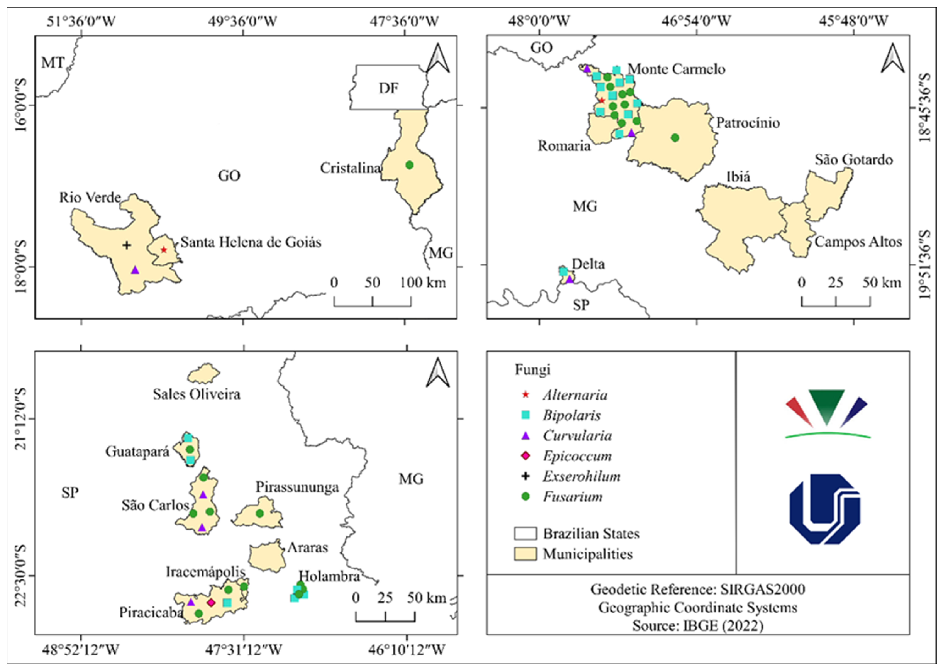
After processing the E. indica samples, 43 isolates were obtained through direct isolation, which were assigned the BEI code, and 45 isolates were obtained through indirect isolation, which were coded as BEINI, totaling 88 fungal isolates. Based on the pathogenicity test, the 50 fungal isolates were phytopathogenic to E. indica. At the genus level, the following phytopathogenic isolates were found: Alternaria (2), Bipolaris (17), Epicoccum (1), Curvularia (7), Exserohilum (1), and Fusarium (22). The cities that presented phytopathogenic isolates and the number of occurrences were of great importance in evaluating the biotic and abiotic factors that may have a direct influence on the pathogenicity of the fungal isolates (Table 2) and their spatial distribution (Figure 2).
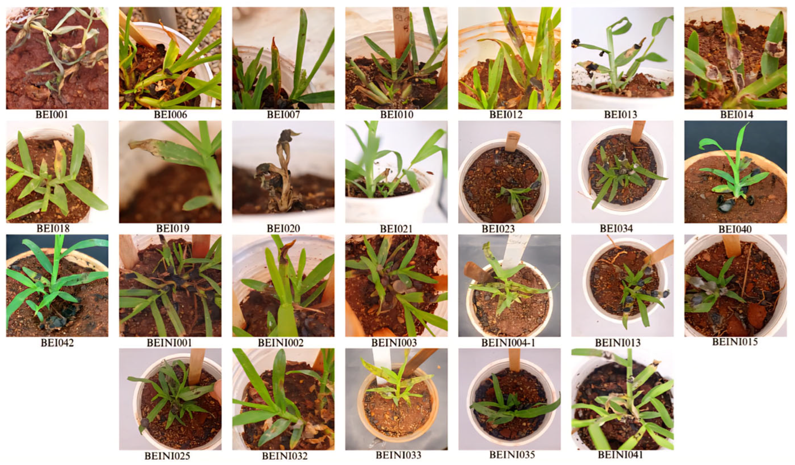
Based on their severity assessments, 26 aggressive phytopathogenic isolates were selected as potential biological control agents for future bioprospecting steps (Figure 3) and (Table 1).

3.2. Analysis of Spatial Distribution of Phytopathogenic Fungi on Eleusine indica and Associated Climatic Conditions

Bipolaris, Fusarium, and Curvularia were the most commonly found fungal genera, with the latter two found in all three Brazilian states where collections took place. The genus Bipolaris was found in the cities of Delta (MG), Guatapará (SP), Monte Carmelo (MG), Holambra (SP), and Piracicaba (SP). Fusarium was found in Cristalina (GO), Guatapará (SP), Holambra (SP), Iracemápolis (SP), Monte Carmelo (MG), Patrocínio (MG), Piracicaba (SP), Pirassununga (SP), and São Carlos (SP). Alternaria was found in Monte Carmelo (MG) and Santa Helena de Goias (GO). Curvularia was found in Delta (MG), Monte Carmelo (MG), Piracicaba (SP), Rio Verde (GO), and São Carlos (SP). Epicoccum was found in Piracicaba (SP) and Exserohilum in Rio Verde (GO).
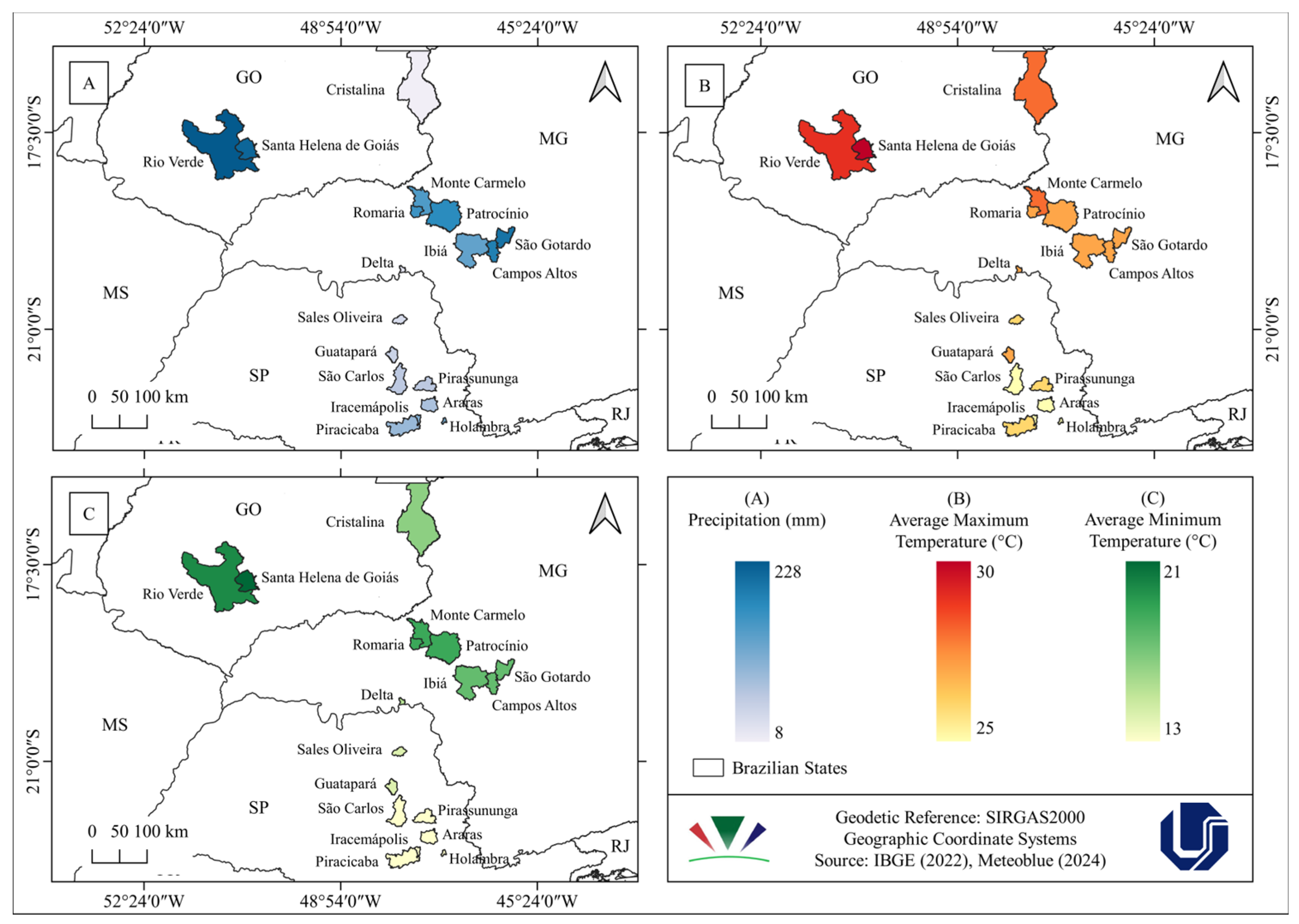
The surveys were made at different times of the year; that is, a mapping of the average precipitation, maximum average temperature, and minimum average temperature (Figure 4) was created using historical data from 1940 for the cities where the fungal isolates were obtained, considering the month in which the E. indica samples were collected.
According to the map colorations, it can be observed that there is a variation of 5 °C in the average maximum temperatures, 8 °C in the average temperatures, and 220 mm in precipitation. As mentioned earlier, 26 aggressive phytopathogenic isolates were chosen for discussion based on the climatological data and survey periods (Table 3).
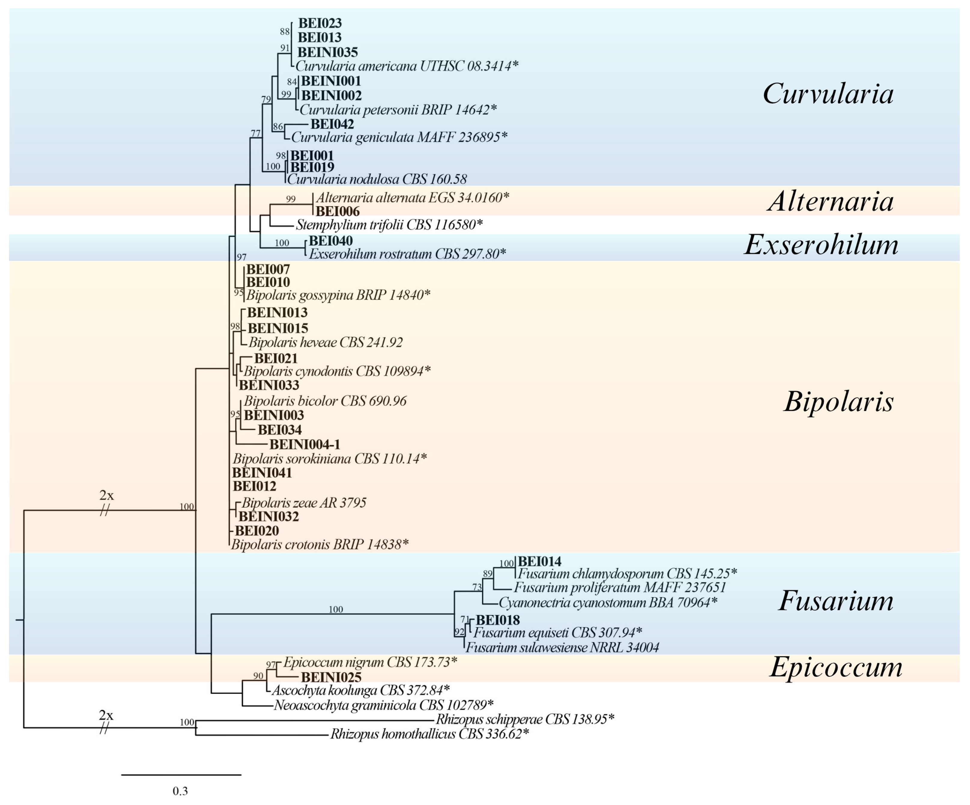
Additionally, a phylogenetic tree of these isolates was constructed to confirm their identification at the genus level (Figure 5). Fusarium isolates exhibiting sporulation were not included in the tree. The 26 most aggressive fungal isolates to E. indica were distributed into 6 genera: Bipolaris, Fusarium, Alternaria, Exserohilum, Curvularia, and Epicoccum (Figure 6).
Figure 7 shows the characteristics of sunny, partially cloudy, cloudy, and rainy days. Few sunny days were observed during the survey months, but there was a predominance in July in the cities of Guatapará, Holambra, and Piracicaba, with approximately 10, 12, and 14 sunny days, respectively.
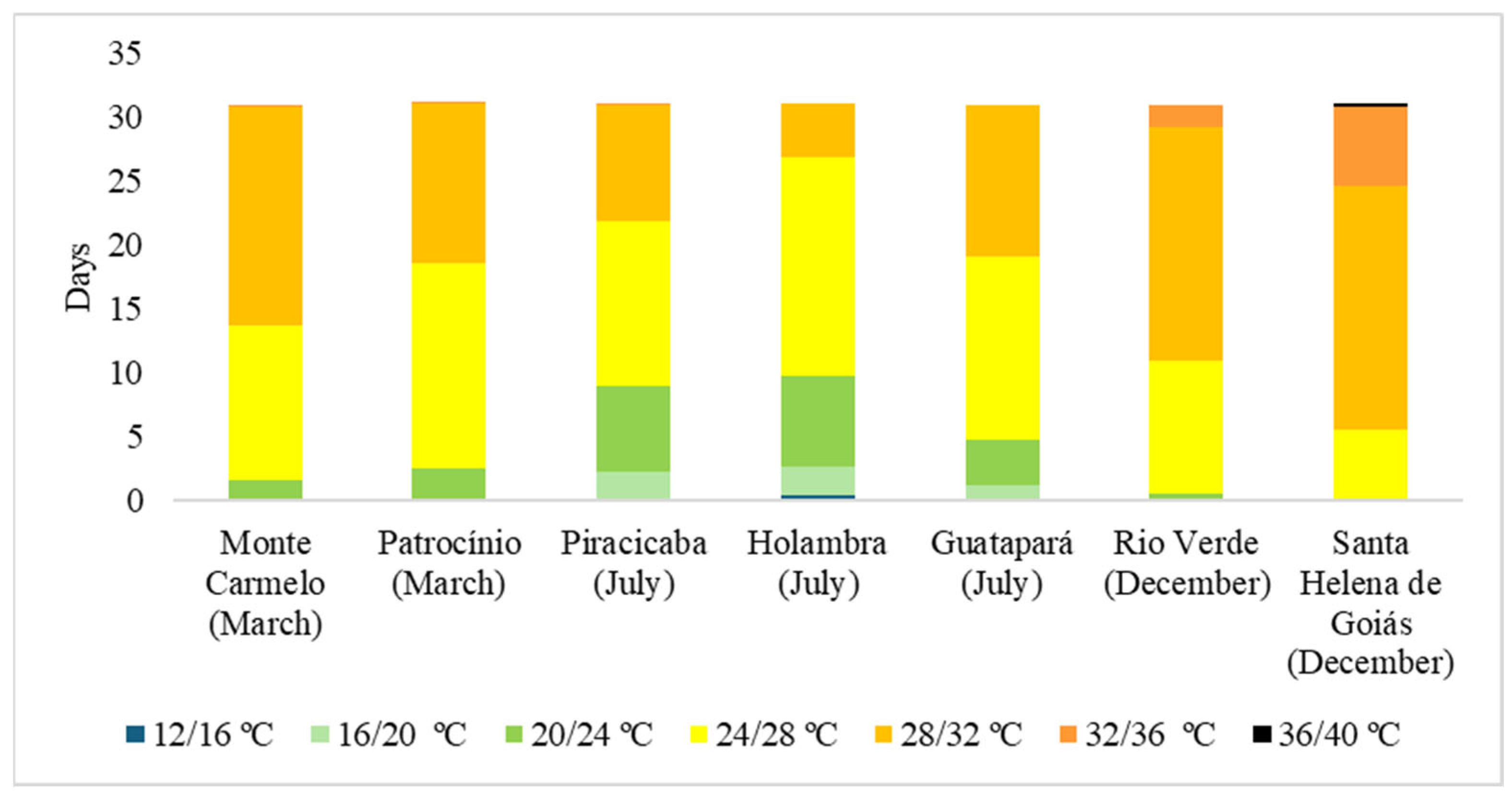
Temperature is another important abiotic factor (Figure 6) that is one of the critical factors affecting the physiology and phytopathogenic capacity of fungi [26,27]. The months mostly had temperatures between 24/28 and 28/32 °C, even in July, which is part of the winter season.
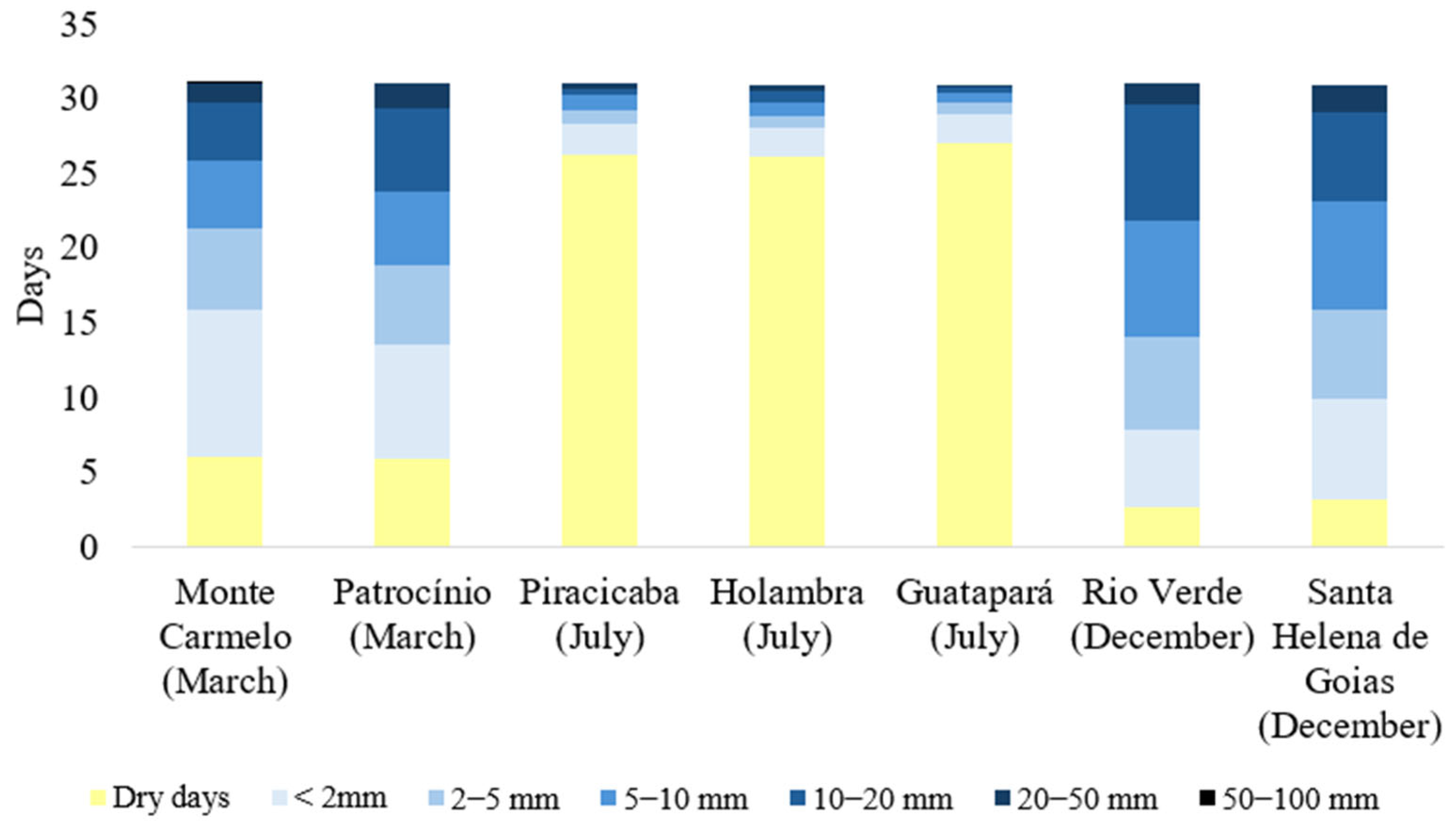
Figure 8 shows the number of precipitation days in millimeters (mm) during the collection months. Another abiotic factor that influences the interaction between phytopathogenic fungi and plants is humidity and precipitation [28,29]. In Brazil, summer is considered to be rainy, whereas winter is dry. The presented data show that in March and December, there were approximately 6 and 3 dry days, respectively, whereas in July, there were approximately 27 dry days.

4. Discussion

This study presents the first stage of an unprecedented program in Brazil aimed at developing a bioherbicide-containing fungal propagule for the management of E. indica, which is one of the most important weed species in global agriculture. Surveys carried out in three Brazilian states have revealed diverse mycobiota associated with this target species.
After processing the E. indica samples, 88 fungal isolates were found and preserved in pure culture. Based on the pathogenicity test, 50 fungal isolates were phytopathogenic to E. indica. At the genus level, the following phytopathogenic isolates were found: Alternaria (2), Bipolaris (17), Epicoccum (1), Curvularia (7), Exserohilum (1), and Fusarium (22). Based on their severity assessments, 26 aggressive phytopathogenic isolates were selected as potential biological control agents for future bioprospecting steps. The next experiments will be to inoculate, under greenhouse conditions, the 26 isolates of phytopathogenic fungi most aggressive to E. indica with spore suspensions in populations of goosegrass (e.g., 40 plants per pot) and evaluate the potential of these fungi for controlling this target species according to the scale of the Brazilian Weed Society. One or two fungal isolates that demonstrate high efficiency in controlling E. indica by causing high mortality in post-emergence goosegrass plants will be selected as elite isolates. The specificity of fungi in not causing diseases in economically important crops will be evaluated. Subsequently, a mass production protocol for the fungus will be developed, a prototype formulated, its shelf life evaluated, and finally, field testing will be conducted to confirm its bioherbicidal potential for the management of goosegrass weed.
Below, some biotic and abiotic aspects that favored the occurrence of the genera of phytopathogenic fungi that were most aggressive to E. indica are discussed.
Bipolaris isolates were found in four cities: Monte Carmelo (MG), Holambra (SP), Piracicaba (SP), and Guatapará (SP). The impact of biotic and abiotic stressors on the diversity and functionality of Bipolaris species was investigated, highlighting that high temperatures (20–30 °C) favor growth and secondary metabolite production, increasing their pathogenicity [30]. Temperatures between 25 °C and 30 °C are ideal for fungal pathogen infection and intensifying disease occurrence [31,32]. The variation in the pathogenicity of Bipolaris isolates under different precipitation levels was analyzed, concluding that precipitation favors their dispersal and severity in weedy grasses such as Microstegium vimineum [33]. In Urochloa decumbens seeds, [34] it was observed that the incidence of Bipolaris was higher in areas with cumulative pre-harvest precipitation between 40 and 115 mm. In [32,35] was associated that relative humidity above 80% increased Bipolaris spp. infection, including B. maydis, and [36] highlighted that frequent rainfall and high humidity create ideal conditions for the spread of Bipolaris spp. The influence of light on the germination and development of the Bipolaris was also studied. It was demonstrated that Bipolaris euphorbiae is highly tolerant to sunlight and ultraviolet radiation and maintains conidial viability even after prolonged exposure [37]. The germination of six Bipolaris bicolor isolates was analyzed, verifying that light affects germ tube growth but not the overall germination rate [38]. The climatic conditions in the collection areas were within the optimal range for phytopathogenic fungi of the genus Bipolaris; however, precipitation varied. In São Paulo, there were more dry days than rainy days, whereas Monte Carmelo had a higher incidence of Bipolaris, with temperatures between 24 °C and 32 °C and 25 rainy days (2–100 mm) during the collection period, favoring fungal occurrence.
Fusarium spp. were identified in Monte Carmelo (MG), Patrocínio (MG), Holambra (SP), and Piracicaba (SP). The growth of Fusarium spp. and mycotoxin production are directly related to temperature, with pathogenicity increasing at 30 °C, especially in interactions with humidity [39]. In [40], it was demonstrated that irrigation and the presence of infected grasses promote the spread of Fusarium spp. and the contamination of neighboring crops, including weeds. Soil moisture and precipitation are determining factors for the germination of Fusarium conidia and the development of infections in various plant species [41]. Precipitation, measured in millimeters (mm), directly influences the germination of conidia and the development of phytopathogenic fungi of the Fusarium genus, which affects various species, including weeds. For example, in Fusarium verticillioides, the presence of water is essential for conidial germination and infection in susceptible hosts [42] Although the exact amount of precipitation required varies among Fusarium spp. and environmental conditions, free water on leaves is a critical factor for germination [43]. A relative humidity above 70% accelerates mycelial growth and increases mycotoxin production [44,45]. Sunlight, particularly UV radiation, influences the pathogenicity of Fusarium spp. Studies have indicated that exposure to UV radiation can increase its virulence, affecting its infectious capacity and the production of secondary metabolites essential for its pathogenicity [46,47]. However, prolonged exposure to UV-C radiation can reduce fungal viability and inhibit conidial germination [48]. While these studies focused on UV-C radiation, they suggested that direct sunlight exposure, which is rich in UV radiation, can affect the development of Fusarium spp., varying according to intensity and spectral composition throughout the day and seasons. In this study, humidity was the only climatic factor that did not align with the observed patterns, as there was little rainfall in São Paulo. The highest incidence of Fusarium spp. was recorded in Monte Carmelo, where climatic conditions favored its proliferation: temperatures between 24 °C and 32 °C for over 30 days and 25 days of rain, with precipitation ranging from 2 to 100 mm. This suggests that the combination of high temperature and humidity is critical for the occurrence of the fungus.
Curvularia isolates were identified in Monte Carmelo (MG) and Piracicaba (SP). Studies have indicated that environmental factors such as temperature and humidity influence toxicity and pathogenicity. It was shown that elevated temperatures increase the phytotoxic activity of metabolites from Curvularia intermedia, suggesting its potential in the biological control of Pandanus amaryllifolius [49]. Temperatures above 30 °C can favor some Curvularia species. However, thermal extremes can inhibit their growth [30]. Humidity and precipitation are essential for the germination of Curvularia spp. conidia [50]. The severity of infection in maize hybrids increased 30 days after flowering, a period when climatic conditions, such as precipitation, favored the development of Curvularia spp. [51]. The pathogenicity of Curvularia lunata in grasses is also directly linked to humidity, with infections intensifying under high humidity [52]. It was confirmed that adequate humidity can increase the virulence and proliferation of Curvularia lunata in crops and weeds [53]. Relative humidity levels between 70% and 80% are ideal for growth and pathogenicity, particularly in tropical and subtropical climates [54]. In [55], the authors indicated that climatic factors such as sunlight and wind can increase the atmospheric distribution of fungal spores, including Curvularia spp. Additionally, research on the influence of light quality on the growth and sporulation of phytopathogenic fungi suggests that different wavelengths of light can affect these processes, although the specific effect on Curvularia has not been detailed [56]. Therefore, while there are no specific studies on the direct influence of sunlight on the growth of Curvularia spp., it is plausible that sunlight plays a role in the dispersal and possibly in the development of these fungi. Some studies have linked UV light exposure to the increased production of secondary metabolites, which can influence interactions with host plants and pathogenicity [30]. In [57], the authors reported that UV radiation increases resistance to environmental stress in Curvularia coicis, suggesting that this adaptation may be related to its pathogenicity in weeds. Piracicaba-SP was the only city that did not align with the humidity conditions because of the number of dry days during the collection period. The highest occurrence of Curvularia spp. isolates was in Monte Carmelo, which is directly related to climatic conditions, as the collection took place in the summer, with temperatures ranging from 24 °C to 32 °C over 30 days and 25 days of rain, with precipitation varying from 2 to 100 mm.
Exserohilum spp. were found in Rio Verde (GO). In [58], the authors evaluated the germination of Exserohilum turcicum conidia under varying temperature and light conditions and concluded that 23 °C is the ideal temperature, with darkness promoting this process. In [59], the authors indicated that exposure to light can inhibit the development of Exserohilum turcicum conidia, especially at temperatures between 20 °C and 26 °C. It is important to note that precipitation not only facilitates the germination of Exserohilum turcicum conidia but also contributes to the spread of the pathogen, as conidia can be transported by wind and rain splashes [60]. The relative humidity of the air, close to 93%, is essential for the sporulation and development of E. turcicum [59]. The optimal growth temperature for the fungus is between 18 °C and 27 °C, with the ideal range for conidial development being 20 °C to 26 °C, with light inhibiting this process. The ideal temperature for the sporulation of Exserohilum spp. is 25 °C [61,62,63]. Exserohilum sp. occurred in Rio Verde (GO), with temperatures ranging from 24 °C to 36 °C over 29 days and 28 days of rain, varying from 2 to 50 mm, favoring its development.
Alternaria spp. were found in Monte Carmelo (MG) and Santa Helena de Goiás (GO). In [64], the authors discussed biocontrol strategies for plant phytopathogenic fungi, including Alternaria, highlighting how environmental conditions, particularly temperature, influence the pathogenicity of these fungi in various crops. This study revealed that elevated temperatures promote the growth and virulence of Alternaria spp. in host plants. In [65], the authors addressed the interaction between variable temperatures and the pathogenicity of different Alternaria species, indicating that increased temperatures enhanced the growth of the fungus and its ability to cause diseases in weeds. Studies have also shown that high temperatures combined with low humidity significantly reduce its occurrence [66,67]. In [68], the authors observed that precipitation indices of 420.5 mm and 397.4 mm in two periods of sunflower cultivation were below the ideal index of 650 mm [69]. Despite this, constant precipitation throughout the sunflower cycle favored the development of Alternaria sp. leaf spots. A study with Alternaria solani in tomatoes revealed that an average relative humidity of 79.4% and accumulated precipitation of 717.4 mm during the evaluation period favored the development of the pathogen [70]. Ref. [71] investigated Alternaria alternata, a pathogen found in Cuscuta japonica, suggesting that humidity and precipitation affect the fungus’s ability to cause diseases in this weed, with rain increasing the severity of infections and aiding the dispersal of conidia. Relative humidity above 70% has been associated with a higher prevalence of Alternaria spp., as it is crucial for conidial germination and growth [67,72]. It was noted that the A. cassiae isolate, registered as a bioherbicide under the name CASST in the United States, sporulates only when exposed to direct sunlight or incandescent light radiation [73]. It was suggested that sporulation of A. cassiae is favored by incubating the mycelial mass at 25 °C in continuous darkness [74]. Although UV light is harmful to many fungi, Alternaria spp. have shown some adaptability, with moderate exposure to UV radiation potentially inducing stress responses that affect virulence and survival [66]. In [75], the authors suggested that Alternaria spp., especially A. brassicae, possess genetic adaptations that allow for better survival and pathogenicity under various light and radiation conditions. Exposure to UV radiation can affect the expression of genes related to pathogenicity and toxin production by A. alternata, thereby altering its ability to infect hosts. Additionally, UV radiation can induce resistance in some plants, thereby affecting the dynamics of Alternaria infection [76]. The highest occurrence of Alternaria spp. was in Monte Carmelo, directly linked to climatic conditions, as the collection was made in the summer, with temperatures ranging from 24 °C to 32 °C over 30 days and 25 days of rain varying from 2 to 100 mm. In Santa Helena de Goias, temperatures ranged from 24 °C to 36 °C over 30 days, with 28 days of rain varying from 2 mm to 50 mm favoring the development of the fungus and thus the occurrence of Alternaria spp.
Thus, it is crucial to assess the abiotic factors that might have had a direct influence on the pathogenicity of the fungi, considering that the surveys were mainly carried out during two different seasons: summer and winter. Summers in Brazil are typically characterized by high temperatures, with 2023 being recorded as the hottest year in the country’s history. The average temperature in Brazil is significantly higher than the historical average, with heatwaves and extreme weather events becoming more frequent. Precipitation patterns also changed during these summers, particularly under the influence of phenomena such as El Niño, which amplified temperatures and altered the distribution of rainfall [77,78]. Humidity and precipitation play significant roles in the interactions between plant phytopathogenic fungi and plants. Adequate humidity can facilitate infection, as many plant phytopathogenic fungi rely on humid environments for germination, penetration, and colonization [29,30]. The work cited next describes the winter conditions in different regions of Brazil. In the South, temperatures are colder, while in the Northeast and North, winters are more humid and rainier. Climate conditions in Brazilian winters are changing, highlighting the interactions between climatic phenomena such as El Niño and cold weather and underscoring the importance of exploring how this affects agriculture and the environment [79].
Biotic factors such as interactions between plant phytopathogenic fungi and the host microbiota are essential for determining the severity of fungal infections. Studies show that the presence of other microorganisms in the environment can alter the success of fungal infections [80,81]. Abiotic factors such as temperature variation, water availability, nutrient concentration, and heavy metals exert selective pressure on fungi, influencing their ability to survive and adapt. Factors such as water stress, salinity, and temperature change alter the pathogenic behavior of many fungi, either by increasing their aggressiveness or restricting their growth. Furthermore, fungal endophytes, microorganisms that live within plants without causing harm, help plants cope with biotic and abiotic stresses by promoting growth and disease resistance [82,83].
A study primarily focused on the use of plant phytopathogenic fungi as biological control agents has been published [28]. The authors also discussed aspects of their distribution and the importance of understanding the biogeography of these fungi for their effective use in agriculture. The study analyzed the geographic distribution of various plant pathogens in the U.S., highlighting how environmental factors influence the distribution of plant phytopathogenic fungi [84]. This study mapped areas of susceptibility to different pathogens and provided detailed data on the relative abundance of species in different regions. Another study investigated how the composition of plant phytopathogenic fungal communities influences other fungal communities in subtropical forests, presenting data on the spatial distribution of fungi at different stages of forest succession [85]. Additionally, the VHFF–Virtual Herbarium of Flora and Fungi (2024), in partnership with the Reference Center for Environmental Information (RCEI)), has been using modeling techniques to predict the potential distribution of fungi in various regions of Brazil, expanding the understanding of how different species of fungi are distributed and how environmental and climatic factors can influence this distribution over time.

5. Conclusions

This pioneering study in Brazil revealed a significant diversity of plant phytopathogenic fungi associated with goosegrass (Eleusine indica) in the three states where surveys were conducted in Brazil. This research project is part of a partnership between the Universidade Federal de Uberlândia-UFU/Campus Monte Carmelo and the company Nitro-Biocontrol. This initial prospecting phase is essential for the next steps, aimed at selecting a biological control agent for E. indica. The same team will conduct new tests to confirm the potential of the identified plant phytopathogenic fungi as biological control agents against E. indica using inoculations with spore suspensions of the 26 most aggressive isolates. The new tests will include measurements of the specificity of the isolate selected, the development of a prototype, application technology tests, and finally field tests aimed at developing an innovative bioherbicide for the management of goosegrass weed, which could provide a sustainable alternative to chemical herbicides.

Author Contributions

Conceptualization, C.F., B.S.V. and A.L.F.; methodology, C.F., M.N.S., L.A.d.S. and V.H.R.d.O.; preparation of maps, M.N.S. and C.F.; validation, M.F.Q. and E.M.I.; investigation, C.F.; data curation, C.F. and V.H.R.d.O.; writing—original draft preparation, C.F. and B.S.V.; writing—review and editing, C.F., M.F.Q., E.M.I. and B.S.V.; supervision, B.S.V. and A.L.F.; project administration, B.S.V. and A.L.F.; funding acquisition, A.L.F. and B.S.V. All authors have read and agreed to the published version of the manuscript.

Funding

This work was supported by Nitro|Biocontrol (23117.076352/2022-48), and FINEP (Financier of Studies and Projects)—Brazilian public company linked to the Ministry of Science, Technology and Innovation (MCTI) Protocolo: 589B149E-B735-46FA-8242-D3F9FB9CE939, Bioeconomy Production Chains Program: Promotion of ICT-01/2022.

Institutional Review Board Statement

Not applicable.

Data Availability Statement

The original contributions presented in the study are included in the article. Further inquiries can be directed to the corresponding author.

Conflicts of Interest

The authors declare no conflicts of interest.

References

  1. Fontes, J.R.A.; Shiratsuchi, L.S.; Neves, J.L.; Júlio, L.D.; Sodré Filho, J. Manejo Integrado de Plantas Daninhas; Embrapa Cerrados: Planaltina, Brazil, 2003. [Google Scholar]
  2. Dias, A.; Carvalho, S.; Marcolini, L.; Melo, M.; Christoffoleti, P. Competitiveness of alexandergrass or Bengal dayflower with soybean. Planta Daninha 2010, 28, 515–522. [Google Scholar] [CrossRef]
  3. Dalazen, G.; Curioletti, L.; Cagliari, D.; Stacke, R.; Guedes, J. Hairy fleabane as a source of major insect pests of soybean. Planta Daninha 2016, 34, 403–409. [Google Scholar] [CrossRef]
  4. Zanatta, J.F.; Figueiredo, S.; Fontana, L.C.; Procópio, S.O. Interferência de plantas daninhas em culturas olerícolas. Rev. da FZVA 2006, 13, 39–57. [Google Scholar]
  5. IBAMA. Instituto Brasileiro do Meio Ambiente e dos Recursos Naturais Renováveis. Relatórios de Comercialização de Agrotóxicos—Boletim Anual de Produção, Importação, Exportação e Vendas de Agrotóxicos. Boletim 2023. Available online: https://www.gov.br/ibama/pt-br (accessed on 15 February 2025).
  6. Heap, I. The International Survey of Herbicide Resistant Weeds. Online. Internet. Available online: www.weedscience.org (accessed on 10 April 2025).
  7. Riechers, D.E.; Soltani, N.; Chauhan, B.S.; Concepcion, J.C.T.; Geddes, C.M.; Jugulam, M.; Kaundun, S.S.; Preston, C.; Wuerrfel, R.J.; Sikkema, P.H. Herbicide resistance is complex: A global review of cross resistance in weeds within herbicide groups. Weed Sci. 2024, 72, 465–486. [Google Scholar] [CrossRef]
  8. Gerwick, B.C. Thirty Years of Herbicide Discovery: Surveying the Past and Contemplating the Future; Informa: London, UK, 2010. [Google Scholar]
  9. Duke, S.O. Why have no new herbicide modes of action appeared in recent years? Pest Manag. Sci. 2012, 68, 505–512. [Google Scholar] [CrossRef]
  10. IM, H. International Survey of Herbicide Resistant Weeds. Acessed em 20 December de 2023; Cambridge University Press: Cambridge, UK, 2003. [Google Scholar]
  11. Davis, A.S.; Frisvold, G.B. Are herbicides a once in a century method of weed control? Pest Manag. Sci. 2017, 73, 2209–2220. [Google Scholar] [CrossRef]
  12. Copping, L.G. The evolution of crop protection companies. Outlooks Pest Manag. 2018, 29, 25–37. [Google Scholar] [CrossRef]
  13. Lorenzi, H. Manual de Identificação e Controle de Plantas Daninhas: Plantio Direto e Convencional; Instituto Plantarum: Nova Odessa, Brazil, 2014. [Google Scholar]
  14. Vidal, R.A.; Lamego, F.P.; Portes, E.D.S.; Trezzi, M.M. Resistência de Eleusine indica aos inibidores de ACCase. Planta Daninha 2006, 24, 163–171. [Google Scholar] [CrossRef]
  15. Lorenzi, H. Plantas daninhas do Brasil: Terrestres, aquáticas, parasitas e tóxicas; Instituto Plantarum: Nova Odessa, Brazil, 2008. [Google Scholar]
  16. Silva, W.L. Herbicidas Residuais no Controle de Eleusine indica e na Seletividade da Cultura da Soja. Dissertação (Master’s thesis), Instituto Federal Goiano, Goias, Brazil, 2020. [Google Scholar]
  17. Carvalho, L.B. Plantas Daninhas; Editado Pelo Autor: Lages, Brazil, 2013. [Google Scholar]
  18. Andrade Junior, E.R.D. Capim pé-de-galinha (Eleusine indica) resistente a herbicidas inibidores da ACCase em áreas algodoeiras de mato grosso. Ph.D. Thesis, Universidade Federal de Mato Grosso, Cuiabá, Brazil, 2018. [Google Scholar]
  19. Kumar, V.; Aggarwal, N.K.; Malik, A. Bioherbicidal Concept: A novel strategy to control weeds. In Microbial Bioprospecting for Sustainable Development; Springer: Singapore, 2018; pp. 29–40. [Google Scholar] [CrossRef]
  20. Alfenas, A.C.; Mafia, R.G. Métodos em Fitopatologia; Editora UFV: Viçosa, Brazil, 2016. [Google Scholar]
  21. Castellani, A. Viability of some pathogenic fungi in distilled water. J. Trp. Med. Hyg. 1939, 42, 225. [Google Scholar]
  22. White, T.J. Amplification and Direct Sequencing of Fungal Ribosomal RNA Genes for Phylogenetics. In PCR Protocols, a Guide to Methods and Applications; Academic Press: New York, NY, USA, 1990; pp. 315–322. [Google Scholar]
  23. Vaidya, G.; Lohman, D.J.; Meier, R. SequenceMatrix: Concatenation software for the fast assembly of multi-gene datasets with character set and codon information. Cladistics 2011, 27, 171–180. [Google Scholar] [CrossRef]
  24. Miller, M.A.; Pfeiffer, W.; Schwartz, T. The CIPRES science gateway: Enabling high-impact science for phylogenetics researchers with limited resources. In Proceedings of the 1st Conference of the Extreme Science and Engineering Discovery Environment: Bridging from the Extreme to the Campus and Beyond, Anais, NY, USA, 16–20 July 2012; pp. 1–8. [Google Scholar] [CrossRef]
  25. Rambaut, A. FigTree 2014, v1.4.4: A Graphical Viewer of Phylogenetic Trees. Available online: http://tree.bio.ed.ac.uk/software/figtree/ (accessed on 23 January 2025).
  26. Leach, M.D.; Cowen, L.E.; Heitman, J. Surviving the Heat of the Moment: A fungal pathogens perspective. PLoS Pathog. 2013, 8, e1003163. [Google Scholar] [CrossRef] [PubMed]
  27. Almiman, B. Identifying the optimal temperature and water activity conditions of phytopathogenic fungi recovered from Al-Baha province. J. Umm Al-Qura Univ. Appl. Sci. 2024, 10, 640–651. [Google Scholar] [CrossRef]
  28. Peng, Y.; Li, S.J.; Yan, J.; Tang, Y.; Cheng, J.P.; Gao, A.J.; Yao, X.; Ruan, J.J.; Xu, B.L. Research Progress on Phytopathogenic Fungi and Their Role as Biocontrol Agents. Front. Microbiol. 2021, 12, 670135. [Google Scholar] [CrossRef] [PubMed]
  29. Ezziyyani, M.; Hamdache, A.; Asraoui, M.; Requena, M.E.; Egea-Gilabert, C.; Castillo, M.E.C. Effect of Climate Change on Growth, Development and Pathogenicity of Phytopathogenic Telluric Fungi. Adv. Intell. Syst. Comput. 2019, 911, 14–21. [Google Scholar] [CrossRef]
  30. Khan, N.A.; Asaf, S.; Ahmad, W.; Jan, R.; Bilal, S.; Khan, I.; Khan, A.L.; Kim, K.-M.; Al-Harrasi, A. Diversity, Lifestyle, Genomics, and Their Functional Role of Cochliobolus, Bipolaris, and Curvularia Species in Environmental Remediation and Plant Growth Promotion under Biotic and Abiotic Stressors. J. Fungi 2023, 9, 254. [Google Scholar] [CrossRef]
  31. Muniz, P.H.P.C.; de Oliveira, T.A.S.; Duarte, E.A.A.; Rodrigues, F.; Carvalho, D.D.C. Characterization of Bipolaris bicolor germination: Effects of a physical factor on fungal adaptability. Braz. J. Microbiol. 2024, 55, 3521–3528. [Google Scholar] [CrossRef]
  32. Ferreira, C.M.; Saluci, J.C.G.; Vivas, M.; Santos, J.S.; de Andrade Junior, M.S.; Vivas, J.M.S.; Ramos, G.K.S.; Graviana, G.A. Characterization of the Bipolaris maydis: Symptoms and pathogenicity in popcorn genotypes (Zea mays L.). Braz. J. Biol. 2024, 84, e256799. [Google Scholar] [CrossRef] [PubMed]
  33. Kleczewski, N.M.; Flory, S.L.; Clay, K. Variation in Pathogenicity and Host Range of Bipolaris sp. Causing Leaf Blight Disease on the Invasive GrassMicrostegium vimineum. Weed Sci. 2012, 60, 486–493. [Google Scholar] [CrossRef]
  34. Barbosa ASV. Locais de Produção na Qualidade Fisiológica e Sanitária de Sementes de Urochloa decumbens. Dissertação, Universidade Estadual Paulista, São Paulo, Brazil, 2023. [Google Scholar]
  35. Sun, X.; Qi, X.; Wang, W.; Liu, X.; Zhao, H.; Wu, C.; Chang, X.; Zhang, M.; Chen, H.; Gong, G. Etiology and Symptoms of Maize Leaf Spot Caused by Bipolaris spp. in Sichuan, China. Pathogens 2020, 9, 229. [Google Scholar] [CrossRef]
  36. Adhikari, A. Identification, Epidemiology, Genomics and Biocontrol of Bipolaris Species, Pathogens of Crops and Invasive Grasses. Dissertação, University of Florida, Gainesville, FL, USA, 2022. [Google Scholar]
  37. Moraes, C. Produção Massal e Influência de Fatores Físicos no Cultivo e Viabilidade de Bipolaris Euphorbiae. Dissertação, Universidade Estadual Paulista, São Paulo, Brazil, 2009. [Google Scholar]
  38. Muniz PHPC. Fotomorfogênese e Patogenicidade de Bipolaris bicolor em Trigo, Milho e Sorgo. Dissertação (Master’s thesis), Universidade Estadual de Goias, Goias, Brazil, 2022. [Google Scholar]
  39. Mshelia, L.P.; Selamat, J.; Samsudin, N.I.P.; Rafii, M.Y.; Abdul Mutalib, N.-A.; Nordin, N.; Berthiller, F. Effect of temperature, water activity and carbon dioxide on fungal growth and mycotoxin production of acclimatised isolates of Fusarium verticillioides and F. graminearum. Toxins 2020, 12, 478. [Google Scholar] [CrossRef] [PubMed]
  40. Gerling, M.; Petry, L.; Barkusky, D.; Büttner, C.; Müller, M.E.H. Infected grasses as inoculum for Fusarium infestation and mycotoxin accumulation in wheat with and without irrigation. Mycotoxin Res. 2023, 39, 19–31. [Google Scholar] [CrossRef]
  41. Bencheikh, A.; Belabed, I.; Rouag, N. Fusarium head blight of wheat: Current knowledge on associated species and their mycotoxins, pathogenicity diversity, and management strategies. Australas. Plant Pathol. 2024, 53, 457–471. [Google Scholar] [CrossRef]
  42. Sousa, R.R. Incidência de Fusarium verticillioides em Sementes de Milho e Métodos de Inoculação em Diferentes Genótipos e Estágios Fenológicos. Dissertação, Universidade Federal do Tocantins, Palmas, Brazil, 2017. [Google Scholar]
  43. Viana, F.M.P.; dos SANTOS, A.A.; Freire, F.; Cardoso, J.E.; Vidal, J.C. Recomendações para o Controle das Principais Doenças que Afetam a Cultura do Melão na Região Nordeste; Embrapa: Fortaleza, Brazil, 2001. [Google Scholar]
  44. Desai, S.; Dubey, S.C.; Prasad, R.D. Impacts of climate change on Fusarium species vis-à-vis adaptation strategies. Indian Phytopathol. 2020, 73, 593–603. [Google Scholar] [CrossRef]
  45. Gutiérrez-Pozo, M.; Verheecke-Vaessen, C.; Kourmpetli, S.; Terry, L.A.; Medina, A. Effect of Temperature, Relative Humidity, and Incubation Time on the Mycotoxin Production by Fusarium spp. Responsible for Dry Rot in Potato Tubers. Toxins 2024, 16, 414. [Google Scholar] [CrossRef]
  46. Beccari, G.; Hao, G.; Liu, H. Fusarium pathogenesis: Infection mechanisms and disease progression in host plants. Front. Plant Sci. 2022, 13, 1020404. [Google Scholar] [CrossRef]
  47. Wang, Y.; Wang, R.; Sha, Y. Distribution, pathogenicity and disease control of Fusarium tricinctum. Front. Microbiol. 2022, 13, 939927. [Google Scholar] [CrossRef]
  48. Rocha, A.B.O. Efeito da luz fluorescente e da radiação UV-C no controle da podridão seca e rhizoctoniose em batata-semente após a colheita. Rev. Científica Multidiscip. Núcleo Do Conhecimento 2019, 4, 155–169. [Google Scholar]
  49. Meepagala, K.M.; Johnson, R.D.; Duke, S.O. Curvularin and Dehydrocurvularin as Phytotoxic Constituents from Curvularia intermedia Infecting Pandanus amaryllifolius. J. Agric. Chem. Env. 2016, 5, 12–22. [Google Scholar] [CrossRef]
  50. Torres, D.P. Etiologia da Mancha de Curvularia em Gladíolo e Aspectos da Interação Planta-Patógeno. Dissertação, Universidade Federal de Viçosa, Viçosa, Brazil, 2013. [Google Scholar]
  51. Vaz-de-Melo, A.; Afferri, F.S.; Dotto, M.A.; Peluzio, J.M.; dos Santos, G.R.; de Carvalho, E.V. Reação de híbridos de milho à Curvularia spp., sob dois níveis de adubação com nitrogênio, no sul do Tocantins. Sci. Agrar. 2010, 11, 149–154. [Google Scholar] [CrossRef]
  52. Marin-Felix, Y.; Hernández-Restrepo, M.; Crous, P.W. Multi-locus phylogeny of the genus Curvularia and description of ten new species. Mycol. Prog. 2020, 19, 559–588. [Google Scholar] [CrossRef]
  53. dos Santos, P.R.R.; Leão, E.U.; Aguiar, R.W.d.S.; de Melo, M.P.; dos Santos, G.R. Morphological and molecular characterization of Curvularia lunata pathogenic to andropogon grass. Bragantia 2018, 77, 326–332. [Google Scholar] [CrossRef]
  54. Mehta, T.; Meena, M.; Nagda, A. Bioactive compounds of Curvularia species as a source of various biological activities and biotechnological applications. Front. Microbiol. 2022, 13, 1069095. [Google Scholar] [CrossRef]
  55. Suehara, M.B.; da Silva, M.C.P. Prevalência de fungos anemófilos no Brasil e a correlação com doenças respiratórias e infecções fúngicas. Cienc. Saude Coletiva 2023, 28, 3289–3300. [Google Scholar] [CrossRef]
  56. Valle, T.L. Influência da Qualidade da luz no Crescimento e Esporulação do Metarhizium Anisopliae (Metch.) Sorokin. Dissertação, Universidade de São Paulo, São Paulo, Brazil, 1984. [Google Scholar]
  57. Liu, B.; Xiao, X.; Zhou, X.; Zhou, J.; Lan, L.; Long, X.; Pan, Y.; Du, M.; Zhao, X. Effects of Lactobacillus plantarum CQPC01-fermented soybean milk on activated carbon-induced constipation through its antioxidant activity in mice. Food Sci. Nutr. 2019, 7, 2068–2082. [Google Scholar] [CrossRef]
  58. Camera, J.N.; Deuner, C.C. Limiar térmico para a germinação de conídios de Exserohilum turcicum. Rev. Ceres 2017, 67, 478–487. [Google Scholar] [CrossRef]
  59. Machado, M.R.F. Interactions Between Exserohilum Turcicum and Maize Seeds: Detection, Effects and Transmission of the Pathogen. Ph.D. Thesis, Universidade Federal de Lavras, Lavras, Brazil, 2021. [Google Scholar]
  60. Casela, C.R.; Ferreira, A.D.S.; Fernandes, F.; Pinto, N.D.A. Cultivo do Milho: Doenças Foliares; Embrapa: Sete Lagoas, Brazil, 2002. [Google Scholar]
  61. Leach, C.M.; Fullerton, R.A.; Young, K. Northern leaf blight of maize in New Zealand: Release and dispersal of conidia of Drechslera turcica. Phytopathology 1977, 67, 380–387. [Google Scholar] [CrossRef]
  62. White, D.G. Compendium of Corn Diseases; The American Phytopathological Society: Saint Paul, MN, USA, 1999. [Google Scholar]
  63. Bhowmik, T.P.; Prasada, R. Physiologic specialization in Helminthosporium turcicum Pass. From India. Phytopathol. Zeits-Chrift 1970, 68, 84–87. [Google Scholar] [CrossRef]
  64. Carrascal-Hernández, D.C.; Flórez-López, E.; Peralta-Ruiz, Y.; Chaves-López, C.; Grande-Tovar, C.D. Eco-Friendly Biocontrol Strategies of Alternaria Phytopathogen Fungus: A focus on gene-editing techniques. Agriculture 2022, 12, 1722. [Google Scholar] [CrossRef]
  65. Fernandes, C.; Casadevall, A.; Gonçalves, T. Mechanisms of Alternaria pathogenesis in animals and plants. Fems Microbiol. Rev. 2023, 47, fuad061. [Google Scholar] [CrossRef]
  66. Giorni, P.; Bulla, G.; Bellotti, G.; Antinori, M.E.; Guerrieri, M.C.; Fiorini, A.; Bertuzzi, T.; Puglisi, E. In planta evaluation of different bacterial consortia for the protection of tomato plants against Alternaria spp. infection and Alternaria toxins presence in fruits. Front. Hortic. 2024, 3, 1447425. [Google Scholar] [CrossRef]
  67. Balodi, R.; Raghavendra, K.V.; Singh, P.K.; Hussain, Z.; Suroshe, S.S.; Kumar, P.; Chander, S. Alternaria alternata species complex impairing solanaceous vegetables in Northern parts of India: An emerging problem in Solanum lycopersicum L. 3 Biotech 2024, 14, 218. [Google Scholar] [CrossRef] [PubMed]
  68. Amabile, R.F.; Vasconcelos, C.M.; Gomes, A.C. Severidade da mancha-de-alternária em cultivares de girassol na região do Cerrado do Distrito Federal. Pesqui. Agropecu. Bras. 2002, 37, 251–257. [Google Scholar] [CrossRef]
  69. Siqueira IF Girassol; Fundação Instituto Agronômico do Paraná: Londrina, Brazil, 1980.
  70. Coelho, F.S.; Alvarenga, M.A.R.; Leão, A.B.; Rodrigues, L. Controle de Alternaria solani com fungicidas na cultura do tomateiro. Rev. Fac. Nac. Agron. Medellín 2011, 64, 5845–5851. [Google Scholar]
  71. Liu, Y.; Ahmed, A.; Munir, S.; Chen, L.; He, P.; He, Y.; Tang, P.; Kong, B.; Wu, Y.; He, P. Alternaria alternata Pathogen from Cuscuta japonica Could Serve as a Potential Bioherbicide. J. Fungi 2024, 10, 494. [Google Scholar] [CrossRef]
  72. Pyrri, I.; Bruffaerts, N.; Radovic, M.; D’hOoge, E.; Janjusevic, L.; Sikoparija, B. Variability in Alternaria alternata spore characteristics under different culture conditions: Implications for automatic detection using air flow cytometry. Aerobiologia 2024, 40, 437–446. [Google Scholar] [CrossRef]
  73. Walker, H.L. Control of Sicklepod, Showy Crotalaria and Coffee Senna with Fungal Pathogen. U.S. Patent 4,390,360, 28 June 1983. [Google Scholar]
  74. DE Ávila, Z.R.; DE Mello, S.C.M.; Ribeiro, Z.M.D.A.; Fontes, E.M.G. Produção de inóculo de Alternaria cassie. Pesqui. Agropecu. Bras. 2000, 35, 533–541. [Google Scholar] [CrossRef]
  75. Rajarammohan, S.; Paritosh, K.; Pental, D.; Kaur, J. Comparative genomics of Alternaria species provides insights into the pathogenic lifestyle of Alternaria brassicae—A pathogen of the Brassicaceae family. BMC Genom. 2019, 20, 1036. [Google Scholar] [CrossRef]
  76. Cao, C.; Gong, S.; Li, Y.; Tang, J.; Li, T.; Zhang, Q. Pathogenic Factors and Mechanisms of the Alternaria Leaf Spot Pathogen in Apple. Horticulturae 2024, 10, 212. [Google Scholar] [CrossRef]
  77. The World Bank Group. Explore Historical and Projected Climate Data, Climate Data by Sector, Impacts, Key Vulnerabilities and What Adaptation Measures Are Being Taken. Explore the Overview for a General Context of How Climate Change Is Affecting Brazil. 2021. Available online: https://climateknowledgeportal.worldbank.org/country/brazil/climate-data-historical (accessed on 13 October 2024).
  78. Ballarin, A.S.; Sone, J.S.; Gesualdo, G.C.; Schwamback, D.; Reis, A.; Almagro, A.; Wendland, E.C. CLIMBra-climate change dataset for Brazil. Sci. Data 2023, 10, 47. [Google Scholar] [CrossRef]
  79. Marengo, J.; Espinoza, J.C.; Bettolli, L.; Cunha, A.P.; Molina-Carpio, J.; Skansi, M.; Correa, K.; Ramos, A.M.; Salinas, R.; Sierra, J.-P. A cold wave of winter 2021 in central South America: Characteristics and impacts. Clim. Dyn. 2023, 61, 2599–2621. [Google Scholar] [CrossRef]
  80. Yadav, U.; Singh, P.C. Biotic Stress to Plants: Fungal pathogen as a major biotic stress. In Plant-Microbe Interaction and Stress Management; Chauhan, P.S., Tewari, S.K., Misra, S., Eds.; Springer: New York, NY, USA, 2024; pp. 1–305. [Google Scholar] [CrossRef]
  81. Flores-Nunez, V.M.; Stukenbrock, E.H. The impact of filamentous plant pathogens on the host microbiota. BMC Biol. 2024, 22, 175. [Google Scholar] [CrossRef] [PubMed]
  82. Verma, A.; Shameem, N.; Jatav, H.S.; Sathyanarayana, E.; Parray, J.A.; Poczai, P.; Sayyed, R.Z. Fungal endophytes to combat biotic and abiotic stresses for climate-smart and sustainable agriculture. Front. Plant Sci. 2022, 13, 953836. [Google Scholar] [CrossRef] [PubMed]
  83. Gowtham, H.G.; Hema, P.; Murali, M.; Shilpa, N.; Nataraj, K.; Basavaraj, G.L.; Singh, S.B.; Aiyaz, M.; Udayashankar, A.C.; Amruthesh, K.N. Fungal Endophytes as Mitigators against Biotic and Abiotic Stresses in Crop Plants. J. Fungi 2024, 10, 116. [Google Scholar] [CrossRef] [PubMed]
  84. Dietzel, K.; Valle, D.; Fierer, N.; U’Ren, J.M.; Barberán, A. Geographical Distribution of Fungal Plant Pathogens in Dust Across the United States. Front. Ecol. Evol. 2019, 7, 304. [Google Scholar] [CrossRef]
  85. Chen, M.; Yang, J.; Xue, C.; Tu, T.; Su, Z.; Feng, H.; Shi, M.; Zeng, G.; Zhang, D.; Qian, X. Community composition of phytopathogenic fungi significantly influences ectomycorrhizal fungal communities during subtropical forest succession. Appl. Microbiol. Biotechnol. 2024, 108, 99. [Google Scholar] [CrossRef]
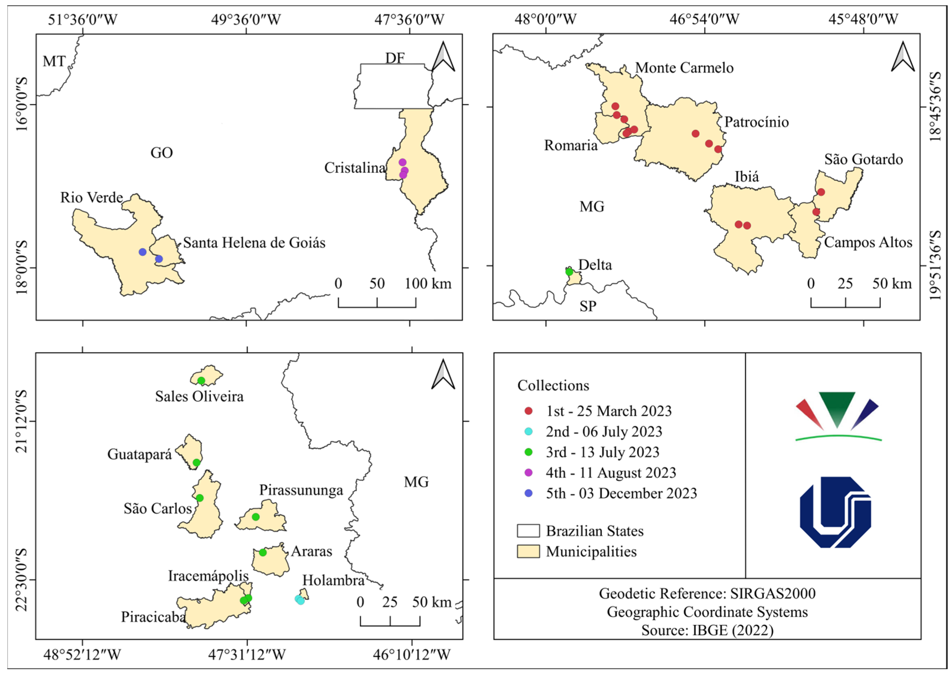
Figure 1. Distribution map of fungal surveys associated with E. indica.
Agriculture 15 01721 g001
Figure 2. Spatial distribution of fungal phytopathogenic isolates to E. indica.
Agriculture 15 01721 g002
Figure 3. Potential isolates of phytopathogenic fungi as candidates for biocontrol agents of E. indica.
Agriculture 15 01721 g003
Figure 4. Average precipitation, maximum temperature, and minimum temperature in the surveys cities since 1940.
Agriculture 15 01721 g004
Figure 5. Phylogenetic analysis inferred by maximum likelihood based on alignment of the ITS region of phytopathogenic fungal isolates associated with Eleusine indica (goosegrass) from various families and an outgroup. Only RAxML support values greater than 70% are shown on the nodes. The genera of the isolates identified in the analysis are outlined by colored boxes, with the genus name indicated to the right. Isolates marked with “*” at the end of isolate identification were identified as ex-type or representative isolates. Phylogenetic analysis was rooted with the isolates Rhizopus schipperae CBS 138.95 and Rhizopus homothallicus CBS 336.62.
Agriculture 15 01721 g005
Figure 6. Temperatures throughout the collection months.
Agriculture 15 01721 g006
Figure 7. Cloudy sky, sun, and precipitation days.
Agriculture 15 01721 g007
Figure 8. Precipitation during the collection.
Agriculture 15 01721 g008
Table 1. Most aggressive phytopathogenic isolates and their respective severity scores.
Selected IsolatesScoreSelected IsolatesScore
BEI0014BEI0402
BEI0062BEI0421
BEI0072BEINI0013
BEI0103BEINI0023
BEI0123BEINI0031
BEI0133BEINI004-13
BEI0143BEINI0133
BEI0182BEINI0153
BEI0192BEINI0251
BEI0204BEINI0321
BEI0211BEINI0333
BEI0231BEINI0351
BEI0342BEINI0413
Table 2. Amount of phytopathogenic isolates and their respective cities of occurrence.
CollectionCityQuantity of Phytopathogenic Isolates
1Monte Carmelo (MG)22
Patrocínio (MG)1
2Holambra (SP)6
3Delta (MG)5
Guatapará (SP)5
Iracemápolis (SP)3
Piracicaba (SP)1
Pirassununga (SP)2
São Carlos (SP)1
4Cristalina (GO)1
5Rio Verde (GO)2
Santa Helena de Goiás (GO)1
Table 3. Most aggressive fungal phytopathogenic isolates to E. indica and their locations.
GenusCityCollectionQuantity
BipolarisMonte Carmelo (MG)1 (March)8
Holambra (SP)2 (July)3
Piracicaba (SP)3 (July)1
Guatapará (SP)3 (July)2
FusariumMonte Carmelo (MG)1 (March)3
Patrocínio (MG)1 (March)1
Holambra (SP)2 (July)1
Piracicaba (SP)3 (July)2
CurvulariaMonte Carmelo (MG)1 (March)1
Piracicaba (SP)3 (July)1
ExserohilumRio Verde (GO)5 (December)1
AlternariaMonte Carmelo (MG)1 (March)1
Santa Helena de Goiás (GO)5 (December)1
Disclaimer/Publisher’s Note: The statements, opinions and data contained in all publications are solely those of the individual author(s) and contributor(s) and not of MDPI and/or the editor(s). MDPI and/or the editor(s) disclaim responsibility for any injury to people or property resulting from any ideas, methods, instructions or products referred to in the content.