Article

Development of a Selective Agar for the Detection of Probiotic Strain Ligilactobacillus animalis NP51 and Other Lactic Acid Bacteria in Cattle Feed

1
Poultry Diagnostic and Research Center, The University of Georgia, Athens, GA 30602, USA
2
Department of Biomedical Sciences and Pathobiology, Virginia Polytechnic Institute and State University, Blacksburg, VA 24060, USA
3
School of Animal Sciences, Virginia Polytechnic Institute and State University, Blacksburg, VA 24060, USA
*
Author to whom correspondence should be addressed.
Current address: USDA, FSIS, Athens, GA 30602, USA.
Current address: Bioinformatics and Computational Biology, George Mason University, Fairfax, VA 22030, USA.
Agriculture 2025, 15(12), 1284; https://doi.org/10.3390/agriculture15121284
Submission received: 22 April 2025 / Revised: 21 May 2025 / Accepted: 4 June 2025 / Published: 13 June 2025
(This article belongs to the Section Farm Animal Production)

Abstract

The enormous potential of bacteriotherapy in disease treatment and prevention has created a large probiotic market. Significant challenges exist in assessing probiotic quality, efficacy and viability. Lactic acid bacteria (LAB) are commonly used probiotics and the most abundant of the vertebrate microbiota. The goal of this study was to make MRS agar specific for probiotic Ligilactobacillus animalis NP51, since the current formulation is not sufficiently selective. Here, 53 chemicals were screened to identify compound(s) that reduced the growth of non-LAB and fungi on de Mann, Rogosa, and Sharpe (MRS) agar, and which were selective for LAB and specifically the probiotic strain NP51. Cattle feed was selected as the sample type, as it is commonly amended with Lactobacillus or yeast probiotics and often includes silage, a diverse microbial consortium of fungi and LAB. Modified MRS was evaluated for its effectiveness in determining probiotic viability and the detection of L. animalis NP51 in cattle feed, amended with this probiotic. qPCR was used to specifically detect and enumerate NP51 in commercial and experimental feed samples. For four selective agents, nystatin, guanidine hydrochloride, CuSO4, and ZnCl, it was identified that when used together, they reduced the growth of bacteria and fungi, but did not inhibit the Lactobacillus probiotic NP51 and other LAB. Metagenomic analysis revealed LAB as the major group cultivated on modified MRS agar from the plating of cattle feed amended with silage. As an enrichment, modified MRS broth improved the qPCR detection of probiotic strain NP51. This study illustrated that improvements can be made to existing bacteriological media for enumerating probiotic NP51 and determining the product’s viability.

1. Introduction

In the early 20th century, Russian-born biologist Elie Metchnikoff pioneered the concept of using microorganisms to promote good health. Through observation of Bulgarian peasants who ingested soured milk regularly, Metchnikoff concluded that drinking fermented milk products in large quantities would result in a healthier and prolonged life [1]. His observations laid the foundation for the concept of beneficial microbes. In 1965, Lilly and Stillwell introduced the term probiotic to describe microbial products with growth-promoting activities [2]. Later, Parker expanded on this by defining probiotics as microbes and their end-products that contribute to intestinal microbial balance and improve resistance to infection [3]. Fuller further emphasized the health benefits of live microbes by using feed supplements to modulate an animal’s indigenous microbiota [4]. Today, probiotics are defined as “live microorganisms which, when administered in adequate amounts, confer a health benefit on the host” [5]. Products containing viable probiotic microorganisms that are intended for consumption are referred to as direct-fed microbials [6].
Lactic acid bacteria (LAB) were recognized early as having probiotic properties [7]. Some of the LAB found in today’s probiotic supplements include Lactobacillus, Bifidobacterium, Leuconostoc, Pediococcus, Enterococcus, and Streptococcus spp. [8,9]. Most probiotic genera have “generally recognized as safe (GRAS)” status awarded by the Food and Drug Administration (FDA), enabling broad applications as nutritional supplements. Exceptions to this status include some Enterococcus and Streptococcus spp., which are considered pathogens [6]. According to the 2016 Food and Agriculture Organization (FAO) report on Probiotics in Animal Nutrition, 45 microbial species have been used as probiotics in animal diets, including 26 that have been commercialized [8]. Of these, 27 are lactic acid bacteria.
Among the positive impacts of probiotic ingestion, Lactobacillus, Bacillus, and Bifidobacterium have been shown to reduce pathogenic bacteria in the intestine, promote intestinal barrier function, and modulate immune responses [10,11,12,13,14]. In animal production, direct-fed microbials are added to diets to improve live weight gain and feed conversion rate, and provide protection from intestinal pathogens, thus reducing morbidity and mortality. Pre-ruminant calves, chickens and pigs benefit significantly from bacterial probiotics while adult ruminants have been found to benefit more from yeast probiotics such as Saccharomyces cerevisiae [15]. In dairy cattle, the addition of S. cerevisiae to the diet has been reported to reduce lactic acid and increase the volatile fatty acids and rumen pH [16], leading to improved milk yield and milk fat content [16,17]. Similarly, Lan et al. demonstrated that feeding 15-day old chicks two probiotic strains of chicken intestinal Lactobacillus resulted in an improved abundance of lactobacilli over Enterobacteriaceae in the fecal microbiota, and an increase in weight gain of 10.7% compared to the control group [18]. The benefits of probiotics have been shown among many different studies and have resulted in a growth of the direct-fed microbial market in animal production.
There has been significant commercial application of direct-fed microbials in aquaculture [19], meat and poultry production [20], especially as farmers have pivoted to antibiotic-free production [8,21]. Probiotics generate USD 45.6 billion globally. In the U.S. alone, this market is expected to double in growth from USD 1.7 billion in 2016 to USD 3.56 billion by 2025 [22]. Challenges associated with regulatory approval or customer acceptance for any probiotic product include product efficacy and safety [8,22]; probiotic survival [20,23]; product concentration and viability [20,22]; product purity [22]; and probiotic strain verification [22]. Regarding its use as an additive, the end-user may also want to verify the concentration of viable probiotic after it is mixed in products at the processing facility. Many of these probiotics are the same member species as those present in the vertebrate intestine [24,25,26,27,28], the environment [29], or feed components (ex. silage) used in some diets [30,31], and therefore it is difficult to distinguish the probiotic from members of the normal microbiota.
Ligilactobacillus animalis strain NP51, formerly known as Lactobacillus acidophilus NP51 and Lactobacillus animalis NP51 [32], is a LAB probiotic used in cattle feed under the product name BovaminTM. This strain was originally isolated from cattle and screened for its ability to inhibit the growth of Escherichia coli O157:H7 in vitro [33]. It has been shown to reduce the prevalence and abundance of E. coli O157:H7 and Salmonella enterica in beef cattle [34,35]. Additionally, this probiotic strain has demonstrated the ability to reduce chronic inflammation associated with Mycobacterium avium subspecies paratuberculosis, the etiological agent responsible for Johne’s disease in cattle [36]. Like Lactobacillus acidophilus, Ligilactobacillus animalis is found in many animal species [37,38,39,40,41]. A common challenge with any LAB probiotic is distinguishing it from the same or closely related genera and species that naturally inhabit the gastrointestinal tract.
In 1960, de Mann reported on a medium for the cultivation of lactobacilli, focusing on organisms present in dairy products. de Mann’s goal was to develop “a non-selective medium which would support good growth of lactobacilli in general” [42]. MRS, named for de Mann, Rogosa, and Sharpe, who described the first formulation, consists of glucose, peptone, yeast extract, Tween 80 and salts, and supports the growth of most lactobacilli, including many auxotrophs exhibiting profound amino acid and vitamin requirements [43,44]. The medium is by no means selective, as it will support the growth of many other microbes, including member species of the phyla Firmicutes, Proteobacter, Actinobacteria, Ascomycota, or Basidiomycota, which inhabit the same environments as the lactobacilli. It is the inclusion of inhibitory chemicals such as antibiotics [45,46,47], dyes [48,49], detergents [50,51], or alcohols [52] that improves the selectivity of base media.
There are significant deficiencies in the current methodologies used to standardize commercial probiotic products, largely because of the deficiency in cultivation media specific for these organisms. Modifying existing selective and differential culture media is necessary to improve the isolation of direct-fed microbials and support quality control measures. This study focused on the modification of MRS [42] to enhance its selectivity for the isolation of commercial probiotic Ligilactobacillus animalis NP51 added in cattle feed. Nutritionally balanced cattle feed often contains silage (fermented corn husks and stems), which contains a variety of lactic acid bacteria, making it difficult to assay for the presence of specific probiotic products. To validate the presence of the probiotic strain L. animalis NP51, a real-time quantitative PCR was applied to detect and enumerate the strain amongst a background of other lactic acid bacteria. The combination of a selective isolation medium and a strain-specific PCR is a significant step forward in improving the quality control methodology for probiotics.

2. Materials and Methods

2.1. Bacteria, Culture, and Growth Parameters

Commercially available Lactobacilli MRS (deMann, Rogosa, and Sharpe) agar (BD 288210; Becton, Dickinson and Company, Franklin Lakes, NJ, USA) was used to determine viable cell counts and observe colony morphology. MRS was the base medium modified in this study to enhance selectivity for lactic acid bacteria (LAB) and the probiotic strain Ligilactobacillus animalis NP51. Unless stated, all plates were incubated in a microaerophilic atmosphere of 5% O2, 10% CO2, and 85% nitrogen, conditions under which the probiotic strain NP51 grows best.
For this study, four lots (3192398; 3191509; 3183177; 3183176) of lyophilized culture of the strain Ligilactobacillus animalis (NP51) were obtained from ProbioFerm, Ltd. (Des Moines, IA, USA) and stored at −20 °C during use. ProbioFerm manufactures the lyophilized NP51 used to formulate the cattle feed supplement BovamineTM (Nutrition Physiology Co. LLC, Kansas City, MO, USA). Lyophilized NP51 cells were suspended in phosphate-buffered saline (PBS, pH 7) and serial dilutions were spread-plated onto MRS agar and incubated at 37 °C to determine viability and observe colony morphology. Cell counts of lyophilized product were determined microscopically using a Neubauer counting chamber. In some studies, silage, obtained from a local feed mill (Godfrey, Madison, GA, USA) and kefir (Lifeway Foods, Morton Grove, IL, USA) were used as a LAB control.

2.2. Samples and Analysis of Microorganisms in Cattle Feed

As NP51 is a commercial cattle probiotic strain, cattle feed was used as the sample type for development of the LAB-selective agar. Over a 6-month period, corn, rye, and sorghum silages were obtained from a private feed mill (Godfrey, Madison, GA, USA) and used as sources of naturally occurring microorganisms found in cattle feeds. In addition, we acquired samples of steer finish ration from the private feed mill to obtain feed samples that were antibiotic-free. These samples were used as quality control for NP51 performance in culture and qPCR.
Twenty-seven samples (400 g/sample) of commercial steer finish ration, supplemented with BovamineTM (Nutrition Physiology Co. LLC, Snellville GA, USA), were received from a commercial feed yard (Amarillo, Texas). An additional 156 BovamineTM-containing samples (50 g/sample), obtained from 26 lots (6 samples per lot) of commercial steer finish ration were used to determine the sensitivity and specificity of the assays. It was not determined whether these commercial feed samples contained feed-grade antimicrobials.
For culture analysis, 5 g of silage was placed into an individual Filtra-bag stomacher bag (LabPlas, Sainte-Julie, QC, Canada), and phosphate-buffered saline (PBS, pH = 7) + 0.1% Tween 80 was added at a 1:10 ratio and stomached for 2 min at high speed in a Seward stomacher 80 (Seward Laboratory Systems, Inc., Davie, FL, USA). The liquid suspension was placed into a 50 mL conical tube and vortexed for 20 min. Serial 1:10 dilutions were prepared in PBS and 0.1 ml of each dilution was spread-plated onto MRS at final dilutions of 10−2 to 10−9. Plates were incubated at 37 °C for 24 h. Gram stain and phenotypic observations were performed on a portion of colonies from each sample selected based on the diversity of colony morphologies.
Feed samples were homogenized in Ziploc bags by inverting 10 times, then a 200 g aliquot of each was placed into a Filtra-bag. Buffered peptone water (BPW; BD 218105; Becton, Dickinson and Company, Franklin Lakes, NJ, USA) was added to each Filtra-bag (1:5 ratio of feed to BPW) and stomached for 2 min in a Stomach 400 series (Seward Laboratory Systems, Inc., Nottingham, UK). Serial dilutions were prepared in BPW and 0.L ml was spread onto MRS agar at final dilutions of 10−4 to 10−7. Plates were incubated for 24 h at 37 °C, and cultured organisms were observed by Gram stain and colony morphology. To archive samples for further study, glycerol was added to the suspensions to a final concentration of 15% and stored at −20 °C.
To determine the ability of modified MRS to enrich samples for lactic acid bacteria, 50 g from each of 156 feed samples was placed in a Filtra-bag containing 450 mL of Modified MRS broth (nystatin 50 U/mL, guanidine HCL 1 mg/mL, ZnCl 0.6 mg/mL, and CuSO4 6 μg/mL) and stomached for 2 min. Two 10 μL aliquots from each sample were immediately spotted onto Modified MRS agar, resulting in a final dilution of 10−5. Then, 1 mL of each sample was collected at the 0 h time point for DNA extraction, placed in 1.5 mL microfuge tube, and stored at −20 °C. The feed samples were incubated in Modified MRS broth aerobically at 37 °C by positioning the Filtra-bags upright in a neoprene tub. Then, 1 mL samples were collected for DNA extraction every 8 h during a 24 h incubation and stored at −20 °C. DNA was extracted from suspensions using a Qiagen QIAamp cador Pathogen Mini Kit (Qiagen Inc., Hilden, Germany) after Pretreatment B1 for Difficult-to-Lyse Bacteria (Qiagen Lysis tubes L and ATL buffer) as described in the manufacturer’s instructions.

2.3. Screening for Selective Chemical Agents

Using an approach like the one described by Krumwiede and Pratt [48], fifty-three chemicals (Supplemental Table S1), at a concentration of 1.0 mg/mL and 0.01 mg/mL, were added to MRS agar to test for the inhibition of undesirable microbial growth while supporting the growth of NP51 and lactic acid bacteria present in silage. BovamineTM-supplemented cattle feed samples 2, 6 and 12 were selected due to their diversity of bacterial and fungal colonies on MRS. Serial dilutions were prepared in BPW and 0.1 mL was spread onto MRS agar at final dilutions of 10−4–10−7. Chemicals that exhibited inhibitory effects against undesirable organisms were used in gradient plating to identify effective concentrations [53]. Final concentrations were determined by evaluating ranges that produced effective inhibition but did not reduce NP51 colony size or plate counts.

2.4. Fermentation Profile of Ligilactobacillus animalis Probiotic Strain NP51

To add a differential phenotype to Modified MRS, API 50 CH test strips and API CHL medium (bioMérieux, Marcy l’Etoile, France) were used to identify NP51’s carbohydrate utilization profile. A suspension of NP51 (2.0 McFarland) in MRS broth was made by culturing in a microaerophilic atmosphere at 37 °C for 24 h. This culture was used as inoculum for API CHL medium and applied to the 50CH strip according to the manufacturer’s instructions. Mineral oil was added to one set of strips to create an anaerobic atmosphere. Strips were incubated at 37 °C and observed for acid production, and the results were documented every 24 h for 72 h. Carbohydrates identified by the 50CH assay were used to replace the dextrose in MRS medium containing 0.017% bromocresol purple (pH indicator) to determine whether any would be effective in the differentiation of NP51 from the naturally occurring lactic acid bacteria found in silage and cattle feed.

2.5. Bile Salt Hydrolase Activity Assay

Bile acids taurodeoxycholic and taurocholic (Sigma Aldrich, St. Louis, MO, USA) were used to determine if a bile acid hydrolase phenotype would be effective in the differentiation of NP51 from the naturally occurring lactic acid bacteria found in silage and cattle feed grown on MRS medium. Four concentrations—0.125%, 0.25%, 0.5%, and 0.625%—of bile acids were added to MRS medium to detect the precipitate formation that would result from hydrolase activity.

2.6. Real-Time PCR Detection and Enumeration of Probiotic Strain NP51

Primers for L. animalis NP51, targeting the 16S-23S intergenic spacer region (ISR) as described by Randhawa et al. [54], were used to detect and enumerate this strain in cattle feed. A fluorescein amidite (FAM)/black hole quencher (BHQ) dual-labeled probe was synthesized 5′-/6-FAM/cgtgcaaagcaggcgctctc/3-BHQ-1/-3′. This oligonucleotide is internal to primers for amplifying 16S-23S ISR [54] and was designed using Primer3Plus (https://www.primer3plus.com/index.html; Accessed on 9 February 2015). Parameters for NP51 qPCR were set by empirically varying MgCl2 concentrations, annealing temperature, and primer/probe concentrations, using competing DNA extracted from feed samples, silage, or kefir (Lifeway Foods, Inc.; Morton Grove, IL, USA) spiked with L. animalis NP51 DNA. Kefir and silage were also used as a diverse source of lactobacilli species to assess the specificity of the PCR. Lactobacillus gallinarum, a close phylogenetic relative of L. animalis, served as a negative control. DNA was extracted from samples using Qiagen QIAamp cador Pathogen Mini Kit, as described previously. Once optimized, qPCR was performed using the Quantifast RT-PCR + ROX Vial Kit (Qiagen Inc) in a 25 μL volume reaction containing 5.0 μL DNA template, 0.5 μL probe (12.5 μM), and 0.5 μL per primer (12.5 μM). The PCR program parameters were as follows: 94 °C hot start for 5 min, 94 °C denaturation for 10 s, 53 °C annealing for 30 s and 72 °C elongation for 15 s in an Applied Biosystems 7500 Real-Time PCR System.

2.7. Determining the Sensitivity of L. animalis Probiotic Strain NP51 Real-Time qPCR

A suspension (OD 0.45 600 nm) of NP51 lyophilized product (Lot#3192398; Nutrition Physiology Co. LLC; Snellville, GA, USA) was serially diluted 10−2–10−5 to provide plate count controls for the assays. LAB colony counts from feed/kefir samples were determined by spread-plating serial dilutions onto MRS plates then incubating aerobically at 37 °C for 24 h. Once the cell densities of the suspensions were known, mixed ratios of NP51:LAB of 1:10 to 1:10,000 and up to 1000:10,000 were plated onto MRS agar and incubated aerobically at 37 °C for 24 h. Colonies were collected from each plate using a sterile swab, which was placed in 1 mL phosphate-buffered saline, vortexed for 1 min to suspend cells in PBS, then frozen at −20 °C. DNA was extracted from suspensions using the Qiagen QIAamp cador Pathogen Mini Kit as previously described. These samples served as the template for the Real-Time qPCR, as described in a previous section.

2.8. Whole Genome Sequencing to Confirm Media Selectivity for LAB

Whole genome sequencing (WGS) was used to determine which organisms were inhibited by the chemical agents used to enhance the selectivity of MRS agar. Microbial suspensions from 4 steer ration feed samples with L. animalis NP51 supplementation were selected based on the diversity of yeast, mold, and bacteria colony phenotypes. Then, 100 μL of suspension (10−2, 10−3 dilutions) was spread-plated onto MRS, MRS plus nystatin (50 U/mL), and modified MRS, and incubated overnight at 37 °C. Growth was scraped from each plate with a sterile cotton swab and suspended in PBS. DNA was extracted using Promega Wizard Whole Genomic DNA Extraction kit (Promega; Madison, WI, USA) with the addition of bead beating prior to the standard protocol provided by the manufacturer. Sequencing was performed by Georgia Genomics and Bioinformatics Core (https://dna.uga.edu/). The Illumina sequencing run type NextSeq PE150 Mid Output flow cell generated the WGS data. WGS raw data sequences for microorganisms were annotated by Phylum, Order, Family and Genus categories using the MG-RAST Metagenomics Analysis Server (www.mg-rast.org) [55]. The metagenome libraries are publicly available on the MG-RAST Metagenomics Analysis Server (www.mg-rast.org) (Table 1).
Sequences from raw data were counted by Genus or Order to quantitate abundances for each category and subtracted from the total microbe sequences to identify missing species. Each dataset except MRS2 was split into eight datasets in MG-RAST for analysis due to the large size of data. At first all split datasets were downloaded and edited, and microbial names in the Order category for Lactobacillales were extracted using Python version 3.7 programming [56] in the Jupyter notebook, a web-based interactive computing platform [57], then species of lactic acid bacteria genera were extracted in one script. The extracted species of microbes in each genus were mapped to media type. The data with read counts for each species depicted the abundances of each and revealed if any species was not detected on a media formulation.
Diversity analysis was performed by calculating individual species counts, which were normalized by the following steps: (1) the individual counts of each species were divided by the total MG-RAST read counts for each media formulation for each sample; (2) the result was multiplied by a gated number, which was the lowest total sample read count. The diversity analysis was performed with R using the Vegan version 2.5-4 package. Shannon and Inverse Simpson diversity were calculated for each media formulation and sample. The individual diversity values for each sample were summed up and the average values for each media formulation (MRS, MRS-Nystatin, and modified MRS) were calculated. Bar plots of average diversity were created for the Shannon and Inverse Simpson diversity of each media formulation. The boxplots of Shannon and Inverse Simpson were also calculated for each media formulation using R.

2.9. Statistical Analysis

The reads of individual species for seven Lactobacillales genera were counted—Pediococcus, Lactobacillus, Enterococcus, Lactococcus, Leuconostoc, Streptococcus and Weissella. Then, total read counts of each genus were normalized by dividing the total read counts of each media and multiplying it by 100 to generate average counts for each media formulation (MRS, modified MRS, and MRS-Nys). As the data were not normally distributed, the Wilcoxon test, a nonparametric test method for matched-pair data analysis based on differences, was performed in R to detect statistical significance.

3. Results

3.1. Growth Conditions for Cultivating Ligilactobacillus animalis Probiotic Strain NP51

To determine optimum growth conditions for detecting probiotic L. animalis NP51 using culture, suspensions of diluted commercial product were incubated in aerobic or microaerophilic (5% O2, 10% CO2, 85% N2) atmospheric conditions. Probiotic strain NP51 grew in both atmospheres to comparable numbers on traditional MRS, and there was no visible difference in colony size in the two atmospheric conditions following 24-hour incubation. As some lactobacilli grow better at 42 °C [58], growth at 37 °C and 42 °C was assessed to determine if temperature could be a selective feature for improving the selection of probiotic strain NP51 amongst contaminating bacterial growth. NP51 colony size was larger at 37 °C (≥1 mm compared to <1 mm at 42 °C) after 24 h incubation, indicating no growth advantage at higher temperatures. Therefore, subsequent experiments were performed aerobically at 37 °C.

3.2. Microorganisms Isolated from Cattle Feed on Commercial MRS Agar

The diversity and identity of abundant microbes in cattle feed was assessed by plating cattle feed and silage samples on MRS agar. Some of the cattle feed samples were formulated with silages, which naturally contain an abundance of LAB [30]. Examples of the resulting growth are shown in Figure 1. Plated samples varied in appearance ranging from the confluent growth of mucoid colonies to colony types with irregular thick rugose phenotypes, and yeast and molds. Other samples contained colony types with circular smooth opaque white or yellow phenotypes, or circular rough translucent to opaque phenotypes. Gram staining and microscopy revealed that yeast was an abundant component of many plated samples. In fact, some of the commercial feed samples were later found to have been amended with a yeast feed supplement. Microbial cell types found in abundance in cattle feed were Gram-positive rods and cocci, with Gram-positive cocci arranged in clusters or chains. Some Gram-positive rods contained refractile-staining endospores, as was verified by endospore staining [59].

3.3. Identification of Inhibitors That Improve Selectivity of MRS Agar for LAB and Specifically Probiotic Strain NP51 in Cattle Feed and Silage

Several dyes, detergents, and heavy metals were assessed for inhibitory properties for fungal and bacterial growth, without interfering with LAB growth. Since yeasts were the most abundant group in some cattle feed samples, the anti-fungal nystatin was evaluated in this screen. As nystatin is volatile and its decomposition is accelerated when exposed to heat, oxygen and light [60], a second fungal inhibitor was sought that could extend the shelf life for a selective MRS formulation. Of the 53 chemicals screened (see Supplemental Table S1), guanidine hydrochloride, guanidine thiocyanate and phenyl ethyl alcohol were found to be inhibitory for yeasts and molds (Table S1; Figure 2). Nystatin and these additional fungal inhibitors were screened using cattle feed, silage, and commercial yeast feed additives such as XPC (Diamond V, Cedar Rapids, Iowa, USA), Yea-Sacc (Alltech, Nicholasville, KY, USA) and Biosaf (PhiBro, Teaneck, NJ, USA) to identify effective concentrations. Dyes, detergents, and heavy metals were selected and screened against non-lactic acid bacteria based on previous work [61,62,63,64]. Chemicals screened in this study were selected based upon their commercial availability, and the effects of these chemicals as growth inhibitors are shown in Supplemental Table S1. Potentially useful inhibitors were selected based on their ability to reduce fungal and bacterial growth without inhibiting probiotic strain NP51. Nystatin, guanidine hydrochloride, cupric sulfate (CuSO4), and zinc chloride (ZnCl) were most effective at reducing the fungi and bacteria present in feed while allowing the growth of probiotic strain NP51 (Table S1; Figure 2). Nystatin (50 U/mL) and guanidine hydrochloride (0.1 mg/mL) effectively reduced the growth of all yeast and molds in feeds without adversely affecting the culture of the probiotic strain. ZnCl inhibited mucoid, irregular thick rugose, swarming and circular rough translucent colony types in feeds. Copper sulfate inhibited irregular thick rugose and swarming colony types, as well as reducing the size of the mucoid colonies on MRS agar. ZnCl or CuSO4 alone provided the most complete inhibition of these bacterial colony types. However, when these inhibitors were combined, the growth of NP51 was also inhibited.

3.4. Effort to Improve Probiotic Strain Specific Detection

The ideal selective medium for an LAB probiotic strain would inhibit all other bacteria, enabling the quantitation of the product itself. Therefore, we sought additional inhibitors to improve strain-specific detection. A previous unpublished study (communicated to us by the manufacturer) indicated that probiotic strain NP51 grew in the presence of gentamicin (10 μg/mL), streptomycin (40 μg/mL) or oxacillin (2 μg/mL). Therefore, these antibiotics were evaluated in gradient plating with nystatin (50 U/mL), guanidine hydrochloride (1 mg/mL), CuSO4 (4 μg/mL to 8 μg/mL) and ZnCl (0.4 mg/mL to 0.8 mg/mL) to determine their ability to reduce the growth of other LAB present in feeds. Unfortunately, these antimicrobial combinations reduced LAB growth from silage only by 1–2 Log10 (Table 2), and there was no combination of inhibitors that resulted in a homogeneous population of probiotic strain NP51. Furthermore, the addition of gentamicin or streptomycin also decreased NP51 colony size, indicating inhibitory effects.
In addition, we sought to determine if there was a unique biochemical feature of probiotic strain NP51 that could be exploited to provide a differential quality to the Modified MRS medium. The carbohydrate API-CH50 test was used to determine the carbohydrate fermentation profile for NP51, so as to identify sugar(s) utilization that may be unique. However, strain NP51 was not unique in terms of its carbohydrate metabolism in relation to other LAB in silage and kefir. Bile salt hydrolase activity was also assessed, but there was no phenotype (bile salt precipitation or colony morphology) that differentiated NP51 from other LAB. Therefore, the final formulation of the Modified MRS medium, used in subsequent experiments, was made by supplementing commercial MRS medium with nystatin (50 U/mL), guanidine HCL (1 mg/mL), ZnCl (0.6 mg/mL), and CuSO4 (6 μg/mL).

3.5. Modified MRS Medium Selects for LAB Present in Cattle Feed

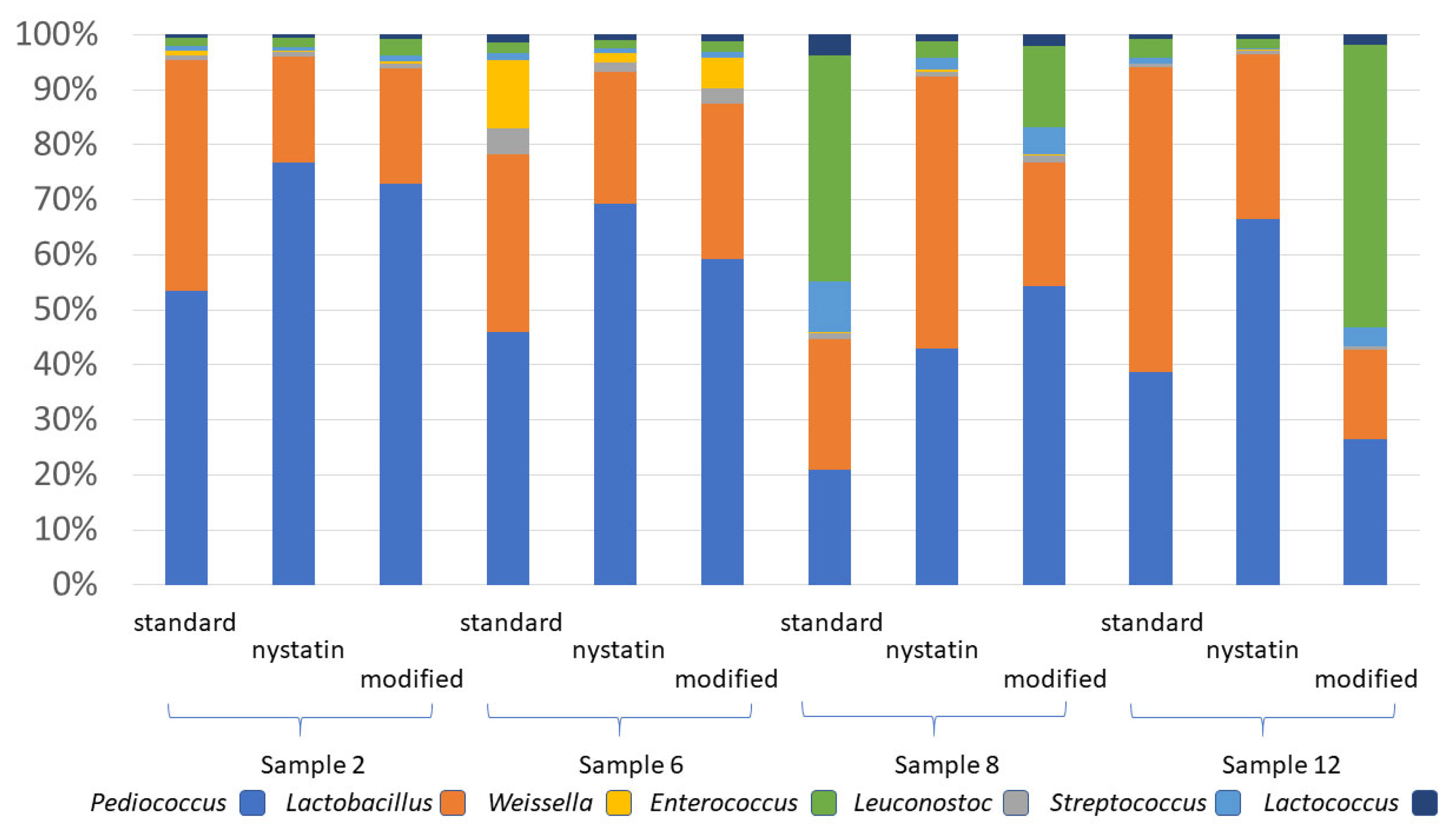
High throughput sequencing and metagenome analysis was used to characterize the microbial community of cattle feed, after overnight culturing on MRS, MRS with nystatin or modified MRS (nystatin, guanidine HCl, ZnCl, and CuSO4), to identify the organisms enriched by each. The sequencing results reveal that two of the four feed samples had a high abundance of Lactobacillales (>80%; Figure 3) when cultured on MRS agar. Except for sample 2 plated on standard MRS, Saccharomycetales species were a minor component, representing <2% of the total microbial species detected on MRS agar (Figure 3). However, nystatin repressed the growth of Saccharomycetales (yeast) and increased Lactobacillales abundance from 84 to 95% (Figure 3). The more selective modified MRS greatly improved Lactobacillales detection in samples with low LAB abundance and repressed the growth of competing bacteria (Figure 3). Five genera of Lactobacillales were detected, with Pediococcus and Lactobacillus accounting for >75% of the total LAB cultured, and they made up >25% of the microbial community cultivated on MRS agar (Figure 4). However, Enterococcus was the dominant Lactobacillales genus in feed samples with a low prevalence of LAB. The Wilcoxon signed-rank test detected no difference between MRS and Modified MRS (nystatin, guanidine HCl, ZnCl, and CuSO4) in terms of the genus total count including all species (p-value = 0.4688). This test has also confirmed that MRS containing nystatin contained a similar LAB abundance to unmodified MRS (p-value = 0.5781), indicating that it did not suppress the growth of LAB (Table S3).
The mean Shannon and Inverse Simpson diversity analyses of the three formulations are shown in Figure 5. Tables S4 and S5 show the values for individual feed samples on each media. These analyses have revealed that feed cultured on unmodified MRS exhibited higher diversity indices (Shannon 2.74, Inverse Simpson 8.61) compared to the other formulations (modified MRS with nystatin, guanidine HCl, ZnCl, and CuSO4—Shannon 2.21; MRS with nystatin,—Shannon 2.40; modified MRS with nystatin, guanidine HCl, ZnCl, and CuSO4—Inverse Simpson 4.88; and modified MRS with nystatin—Inverse Simpson 5.63). However, these differences were not statistically significant according to Tukey HSD or t-test (p >0.05). Therefore, modified MRS with nystatin, guanidine HCl, ZnCl, and CuSO4, and MRS containing nystatin exhibit, selective properties against non-LAB species, but the components of modified MRS with nystatin, guanidine HCl, ZnCl, and CuSO4 did not suppress the diversity of Lactobacillales species.

3.6. Detection of Ligilactobacillus NP51 Abundance by qPCR

While culture-based detection is preferable to validate viable numbers, PCR offers the option of the direct detection of probiotics in feedstuffs if the organism is present at high enough levels, and if PCR inhibitors can be managed. We modified the PCR specific to the existing Ligilactobacillus animalis strain NP51 [44] by including a fluorescent internal probe to increase the PCR’s specificity and sensitivity and enable quantitative detection of the strain. The molecular specificity of the qPCR was determined using silage and kefir, while Lactobacillus gallinarum, a close phylogenetic relative of L. animalis, served as a negative control [65]. Silage was included as it is a common ingredient in cattle feed and naturally contains abundant LAB [66]. Kefir was chosen since this fermented product contains a high diversity of LAB species and strains [67]. All these samples were negative for strain NP51 by qPCR. Figure 6 shows the molecular sensitivity of the qPCR for detecting strain NP51 alone or mixed with other lactic acid bacteria. As few as ten NP51 genomes were detected in samples from pure culture (Ct value: 33.37 ± 0.289). The sensitivity of the qPCR was evaluated by extracting DNA from a mixed LAB population consisting of the probiotic strain NP51 mixed in kefir at NP51:kefir ratios ranging from 1:10 to 1:10,000 cells, and 1000:10,000 cells determined by plating on MRS agar. The NP51 qPCR assay detected a single colony of NP51 irrespective of the amount of competing LAB. For example, one NP51 colony could be detected amongst 10,000 LAB colonies (Ct value—20.40 ± 0.44).
We next applied the qPCR to control feed samples that were amended with 100–107 NP51 cells per gram to demonstrate that the assay could be directly applied to feed samples. The NP51 qPCR successfully detected 100 cells (Ct value: 33.37 ± 0.29) to 107 cells (Ct value: 20.07 ± 0.05) per gram of feed. We acquired 26 feed samples that were not amended with NP51, and these samples were used to determine the specificity and sensitivity of the qPCR. Statistical analysis of the data revealed that the qPCR assay was 100% sensitive (95% confidence interval from 1.00 to 0.891) and 100% specific (95% confidence interval from 0.00 to 0.108). As the sample size was small (n = 26), a proportion as low as 90% could exist for true positives due to random variability.
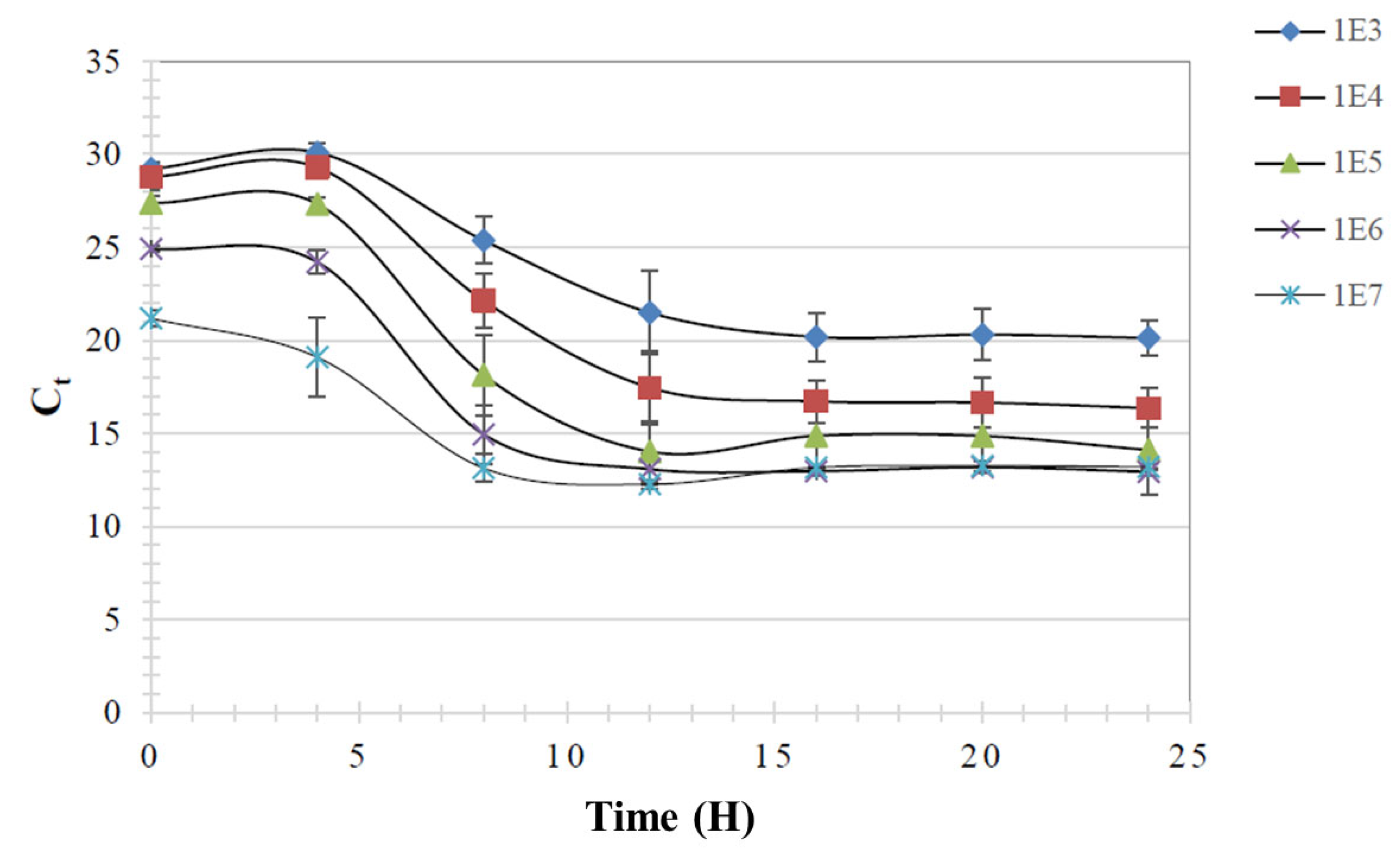
Culture enrichment was applied to determine if the detection of strain NP51 in cattle feed could be improved. The probiotic was mixed into feed at 103 to 107 cells/gram, and then feed samples were incubated in modified MRS broth with nystatin, guanidine HCl, ZnCl, and CuSO4 for 4, 8, 12, 16 and 24 h at 37 °C (Figure 7). The 8- and 12-hour incubations demonstrated decreased qPCR Ct values, which plateaued following 16-hour incubation, indicating the enrichment of the probiotic strain.
Enrichment and qPCR was then used to determine product viability and concentration in NP51-amended commercial feed. Twenty-six commercial feed lots containing NP51 were sampled before (0 h) and after incubation in modified MRS broth with nystatin, guanidine HCl, ZnCl, and CuSO4 (24 h) (Table S2). Each lot consisted of six samples collected from each commercial feed bin (n = 156). Enrichment and DNA isolation was performed on each sample to determine the mean and standard deviation for each lot. The average Ct value for the 26 NP51-amended lots was 31.45 ± 0.526 at 0-hour and 31.83 ± 0.455 following 24-hour enrichment, indicating no growth of the probiotic. Probiotic strain NP51 concentration, as determined by qPCR, ranged from 3.9 × 10² ± 145.559 to 1.0 × 10³ ± 87.678 per gram in the cattle feed, which is significantly below the manufacturer’s recommendations.
The qPCR was applied to twenty-seven additional samples of commercial steer finish ration, and we estimated the NP51 concentration at 460–980 cells per gram. However, the manufacturer’s recommendation is 106 NP51 cells/gram feed. These results suggest that the probiotic may have been mixed incorrectly in some feed mills, or that another variable was influencing quantity. To determine if qPCR inhibitors were responsible for the low estimates of NP51 in commercial feed, cattle feed DNA was isolated and added (10 or 100 ng) to a PCR reaction containing 1 ng NP 51 DNA. There were no differences in Ct values in samples containing cattle feed DNA compared to NP51 DNA alone, indicating an absence of PCR inhibitors.

3.7. Assessing the Antimicrobial Activity of Cattle Feed in Culture of LAB Strain NP51

The results indicate that the enrichment did not decrease the qPCR Ct, suggesting that NP51 did not grow well. Whether the commercial cattle feed samples used in this study contained antibiotics was unknown; however, antimicrobials are FDA approved for use in cattle feed to treat or prevent bacterial infections such as shipping fever, pneumonia and salmonellosis [68]. Therefore, samples were plated onto modified MRS agar with nystatin, guanidine HCl, ZnCl, and CuSO4 to assess bacterial viability. Plate counts ranged from 0 to 7 CFUs/g. Each plate was scraped to prepare the template for NP51 qPCR. The qPCR results indicate that 25/26 plates did not contain NP51 colonies, but one sample was positive, with a Ct value of 27.06. The qPCR performed prior to plating estimated 100 to 1000 NP51 cells per gram cattle feed; however, the absence of growth on the plates suggests the presence of antimicrobials in the feed.
To test this hypothesis, the eluate from seven commercial feed samples was evaluated for growth inhibition by adding it to modified MRS broth (1:1) inoculated with 105 viable NP51 cells/mL and incubated for 24 h. The average Ct value was essentially unchanged after enrichment (32.40 ± 0.481 vs. 32.06 ± 0.626), while NP51 control enrichments had average Ct values of 23.99 ± 0.119 and 21.96 ± 1.273. These results indicate that antimicrobials were likely present in these commercial cattle feed samples.

4. Discussion

The purpose of this study was to improve the selectivity of a commercial medium for the detection of Ligilactobacillus animalis NP51 cattle feed. Fermented grains containing abundant LAB are often used in cattle feed [30,31] and therefore pose a challenge for quantifying probiotic lactobacilli used to amend feed. In reviewing the literature, it became clear that there was no available selective culture media for LAB that could be modified to detect a particular probiotic strain. By enhancing the selectivity of a commercial culture medium to reduce non-lactobacillus growth, we could focus on detecting the probiotic strain within the lactic acid bacterial population. Each of the chemicals added to MRS medium have different modes of action for repressing background organisms. While the fermentation of different plant materials, to make silage for cattle feed, favors lactic acid bacteria populations [69,70,71,72], it also promotes the growth of many other microbes including fungi [70,71]. Nystatin is a polyene antimicrobial that causes damage to the fungal cellular membrane by its affinity for membrane sterols [73]. The inclusion of nystatin in MRS media was effective at repressing fungal growth, and antifungal agents have been included in several MRS formulations for improving LAB detection in fermented products that naturally contain fungi [74,75,76,77]. The mechanism of action of guanidine hydrochloride, added as an additional inhibitor of yeast, mold, and bacteria, appears to be attributed to the microbial membrane [78].
While probiotic strain NP51 was resistant to several antibiotics, no combination was found that supported its growth but inhibited all other LAB. Antibiotics have been included in MRS formulations for the detection of specific LAB in fermented products including silage, but their success has varied depending on antibiotic(s) selected, Lactobacillales detected, or the application [74,75,79,80,81,82]. In this study, the addition of guanidine HCl, ZnCl, and CuSO4 did not affect Lactobacillales abundance in feed samples tested. In fact, guanidine, zinc and copper salts appeared to promote the growth of lactic acid bacteria in two of these samples, while repressing the Bacillales. Zinc chloride and cupric sulfate bind iron–sulfur clusters of dehydratases in bacteria, generating a bacteriostatic effect [63,64] and a combination of these inhibitors was effective in selecting for LAB. The feed samples varied in Lactobacillales and Bacillales abundance on MRS and the different MRS formulations described in this study. These differences may be attributed to the diversity and sources of silage samples used in the cattle feed formulation; for example, Bacillales abundance can vary depending on the plant material used [72]. Most recent studies describing the microbial composition of silage involve non-culture-based molecular methods targeting the bacterial 16S rDNA or fungal ribosomal–internal transcribed spacer (ITS) region [70]. These PCR-based approaches can introduce bias that misrepresents the distribution of microbial taxa present [83,84]. In this study, the microbial composition was determined by profiling cultured samples on MRS and MRS formulations and sequencing the total metagenomic DNA. The fungi identified from this metagenomic analysis were a minor component of most samples, even on a nutrient-rich medium like MRS. However, this may have occurred as 37 °C incubation favors bacteria and yeast over molds. The addition of nystatin alone or in combination with guanidine, zinc and copper salts did alter the Lactobacillales composition, favoring Pediococcus and Lactobacillus over the other lactic acid bacteria Weissella, Leuconostoc, Lactococcus, Streptococcus and Enterococcus. And there were differences among feed samples in terms of the abundance of specific LAB genera isolated on MRS or modified MRS. This sample variability may reflect differences in plant material, the inclusion of the NP51 probiotic or the variation in fermentation conditions to produce the silage [69,70,71,85,86,87].
Modified MRS with nystatin, guanidine HCl, ZnCl, and CuSO4 worked as both an enrichment broth and an agar for enumeration by direct plating, allowing the probiotic’s viability to be verified. However, the challenge was in removing antimicrobials contained in commercial feed prior to enrichment. Antimicrobials currently approved for use in cattle feed by the U.S. Food and Drug Administration include monensin, tylosin, bacitracin, oxytetracycline, erythromycin and chlortetracycline [68]. And some lactobacilli are quite sensitive to monensin [88], including this strain of Ligilactobacillus animalis NP51, which was also susceptible to oxytetracycline, bacitracin and erythromycin. Therefore, when antimicrobials are present in feed, the viability of the probiotic can become compromised, and may affect the product’s efficacy. This is likely to be true for all foodstuffs in which preservatives are used to improve shelf life.

5. Conclusions

Using modified MRS with nystatin (50 U/mL), guanidine HCl (1 mg/mL), ZnCl (0.6 mg/mL), and CuSO4 (6 μg/mL) as an enrichment, coupled with a probiotic-specific PCR, was effective if the feed sample did not contain antimicrobials. This formulation was empirically derived from agar screens of 53 chemicals, the gradient plating of 5 pre-screened chemicals, and varying concentrations of select antimicrobials of cattle feed microbiota and lyophilized probiotic Ligilactobacillus animalis strain NP51. The modified MRS suppressed non-LAB and fungi present in cattle feed. Since cultivation may be problematic, a noncultivation-based method such as PCR can increase the specificity and sensitivity needed to detect the probiotic in foodstuffs. Real-time PCR was successfully used to confirm the presence of probiotic NP51 and estimate its abundance in cattle feed. PCR can become an important component in differentiating probiotic strains from the same bacterial species that inhabit the animal intestine, environment, and fermented foods. Quality control also necessitates the accurate enumeration of the probiotic, ensuring proper concentrations in amended cattle feed. The selective Modified MRS formulation combined with a strain-specific PCR may also be useful in probiotic detection and enumeration in feed and animals fed the bacterial product.

Supplementary Materials

The following supporting information can be downloaded at: https://www.mdpi.com/article/10.3390/agriculture15121284/s1, Table S1: Identification of compound(s) selective for Lactobacillus animalis probiotic NP51 in MRS. Table S2: qPCR detection and enumeration of Lactobacillus animalis NP51 in commercial feed pre- and post-enrichment in MRS with nystatin (50 U/mL), guanidine HCL (1 mg/mL), ZnCl (0.6 mg/mL), and CuSO4 (6 μg/mL). Table S3: Differences in Lactobacillales composition at the genus level resulting from aerobic growth on MRS formulations at 37 °C as determined using the nonparametric Wilcoxon test. Table S4: Community diversity of animal feed following aerobic growth on MRS formulations at 37 °C. Table S5: Average community diversity of animal feed aerobically grown on MRS formulation at 37 °C.

Author Contributions

Conceptualization, M.D.L.; methodology, K.T., S.A.; software, N.F.-N., S.A.; validation, J.J.M., M.D.L., K.T., S.A.; formal analysis, K.T., S.A.; investigation, M.D.L., K.T.; resources, M.D.L., K.T.; data curation, K.T., S.A.; writing—original draft preparation, K.T.; writing—review and editing, M.D.L., J.J.M., K.T., S.A., N.F.-N.; visualization, K.T., S.A.; supervision, M.D.L.; project administration, M.D.L.; funding acquisition, M.D.L. All authors have read and agreed to the published version of the manuscript.

Funding

Bovamine (containing probiotic strain NP51) and funding for KT was provided by Nutrition Physiology Co. LLC; Snellville, GA. J.J.M. was supported by USDA HATCH Fund VA-160130. The funders had no role in the study design, data collection and interpretation, or the decision to submit the work for publication.

Institutional Review Board Statement

This study did not involve animals or humans. The research described in this publication was approved by The University of Georgia Biosafety Committee.

Data Availability Statement

Data can be accessed via https address www.mg-rast.org/mgmain.html?mgpage=project&project= plus the project ID descriptor, as described in Table 1.

Conflicts of Interest

The authors declare there were no commercial or financial relationships associated with any of the research described in this publication.

References

  1. Metchnikoff, E. The Prolongation of Life: Optimistic Studies; G. P. Putnam’s Sons: New York, NY, USA; London, UK, 1908. [Google Scholar]
  2. Lilly, D.M.; Stillwell, R.H. Probiotics: Growth-Promoting Factors Produced by Microorganisms. Science 1965, 147, 747–748. [Google Scholar] [CrossRef] [PubMed]
  3. Parker, R.B. Probiotics, the other half of the antibiotic story. Anim. Nutr. Health 1974, 29, 4–8. [Google Scholar]
  4. Fuller, R. Probiotics in man and animals. J. Appl. Bacteriol. 1989, 66, 365–378. [Google Scholar] [PubMed]
  5. Mack, D.R. Probiotics-mixed messages. Can. Fam. Physician 2005, 51, 1455–1457, 1462–1464. [Google Scholar] [PubMed Central]
  6. Anonymous. Direct Fed Microbial Products. Available online: https://www.fda.gov/regulatory-information/search-fda-guidance-documents/cpg-sec-689100-direct-fed-microbial-products (accessed on 23 October 2015).
  7. Gilliland, S.E. Health and nutritional benefits from lactic acid bacteria. FEMS Microbiol. Rev. 1990, 7, 175–188. [Google Scholar] [CrossRef]
  8. Bajagai, Y.S.; Klieve, A.V.; Dart, P.J.; Bryden, W.L. Probiotics in Animal Nutrition: Production, Impact and Regulation; FAO: Rome, Italy, 2016.
  9. Silva, D.R.; Sardi, J.d.C.O.; de Souza Pitangui, N.; Roque, S.M.; da Silva, A.C.B.; Rosalen, P.L. Probiotics as an alternative antimicrobial therapy: Current reality and future directions. J. Funct. Foods 2020, 73, 104080. [Google Scholar] [CrossRef]
  10. Chen, X.; Xu, J.; Shuai, J.; Chen, J.; Zhang, Z.; Fang, W. The S-layer proteins of Lactobacillus crispatus strain ZJ001 is responsible for competitive exclusion against Escherichia coli O157:H7 and Salmonella typhimurium. Int. J. Food Microbiol. 2007, 115, 307–312. [Google Scholar] [CrossRef] [PubMed]
  11. Klaenhammer, T.R. Genetics of bacteriocins produced by lactic acid bacteria. FEMS Microbiol. Rev. 1993, 12, 39–85. [Google Scholar] [CrossRef]
  12. Ramiah, K.; van Reenen, C.A.; Dicks, L.M. Surface-bound proteins of Lactobacillus plantarum 423 that contribute to adhesion of Caco-2 cells and their role in competitive exclusion and displacement of Clostridium sporogenes and Enterococcus faecalis. Res. Microbiol. 2008, 159, 470–475. [Google Scholar] [CrossRef]
  13. Sakai, F.; Hosoya, T.; Ono-Ohmachi, A.; Ukibe, K.; Ogawa, A.; Moriya, T.; Kadooka, Y.; Shiozaki, T.; Nakagawa, H.; Nakayama, Y.; et al. Lactobacillus gasseri SBT2055 induces TGF-beta expression in dendritic cells and activates TLR2 signal to produce IgA in the small intestine. PLoS ONE 2014, 9, e105370. [Google Scholar] [CrossRef]
  14. Elshaghabee, F.M.; Rokana, N.; Gulhane, R.D.; Sharma, C.; Panwar, H. Bacillus as potential probiotics: Status, concerns, and future perspectives. Front. Microbiol. 2017, 8, 1490. [Google Scholar] [CrossRef] [PubMed]
  15. Fuller, R.; Tannock, G.W. Probiotics from Farm Animals. In Probiotics: A Critical Review; Horizon Scientific Press Ltd.: Poole, UK, 1999. [Google Scholar]
  16. Desnoyers, M.; Giger-Reverdin, S.; Bertin, G.; Duvaux-Ponter, C.; Sauvant, D. Meta-analysis of the influence of Saccharomyces cerevisiae supplementation on ruminal parameters and milk production of ruminants. J. Dairy Sci. 2009, 92, 1620–1632. [Google Scholar] [CrossRef]
  17. Kung, L., Jr.; Kreck, E.M.; Tung, R.S.; Hession, A.O.; Sheperd, A.C.; Cohen, M.A.; Swain, H.E.; Leedle, J.A. Effects of a live yeast culture and enzymes on in vitro ruminal fermentation and milk production of dairy cows. J. Dairy Sci. 1997, 80, 2045–2051. [Google Scholar] [CrossRef] [PubMed]
  18. Lan, P.T.; Binh le, T.; Benno, Y. Impact of two probiotic Lactobacillus strains feeding on fecal lactobacilli and weight gains in chicken. J. Gen. Appl. Microbiol. 2003, 49, 29–36. [Google Scholar] [CrossRef]
  19. Wang, A.; Ran, C.; Wang, Y.; Zhang, Z.; Ding, Q.; Yang, Y.; Olsen, R.E.; Ringo, E.; Bindelle, J.; Zhou, Z. Use of probiotics in aquaculture of China-a review of the past decade. Fish Shellfish Immunol. 2019, 86, 734–755. [Google Scholar] [CrossRef]
  20. Markowiak, P.; Slizewska, K. The role of probiotics, prebiotics and synbiotics in animal nutrition. Gut Pathog. 2018, 10, 21. [Google Scholar] [CrossRef]
  21. Al-Khalaifah, H.S. Benefits of probiotics and/or prebiotics for antibiotic-reduced poultry. Poult. Sci. 2018, 97, 3807–3815. [Google Scholar] [CrossRef]
  22. Jackson, S.A.; Schoeni, J.L.; Vegge, C.; Pane, M.; Stahl, B.; Bradley, M.; Goldman, V.S.; Burguiere, P.; Atwater, J.B.; Sanders, M.E. Improving End-User Trust in the Quality of Commercial Probiotic Products. Front. Microbiol. 2019, 10, 739. [Google Scholar] [CrossRef] [PubMed]
  23. Afgan, E.; Baker, D.; Batut, B.; van den Beek, M.; Bouvier, D.; Cech, M.; Chilton, J.; Clements, D.; Coraor, N.; Gruning, B.A.; et al. The Galaxy platform for accessible, reproducible and collaborative biomedical analyses: 2018 update. Nucleic Acids Res. 2018, 46, W537–W544. [Google Scholar] [CrossRef]
  24. Lu, J.; Idris, U.; Harmon, B.; Hofacre, C.; Maurer, J.J.; Lee, M.D. Diversity and succession of the intestinal bacterial community of the maturing broiler chicken. Appl. Environ. Microbiol. 2003, 69, 6816–6824. [Google Scholar] [CrossRef]
  25. Pryde, S.E.; Richardson, A.J.; Stewart, C.S.; Flint, H.J. Molecular analysis of the microbial diversity present in the colonic wall, colonic lumen, and cecal lumen of a pig. Appl. Environ. Microbiol. 1999, 65, 5372–5377. [Google Scholar] [CrossRef] [PubMed]
  26. Busconi, M.; Reggi, S.; Fogher, C. Evaluation of biodiversity of lactic acid bacteria microbiota in the calf intestinal tracts. Antonie Van Leeuwenhoek 2008, 94, 145–155. [Google Scholar] [CrossRef] [PubMed]
  27. Khan, A.A.; Nawaz, M.S.; Robertson, L.; Khan, S.A.; Cerniglia, C.E. Identification of predominant human and animal anaerobic intestinal bacterial species by terminal restriction fragment patterns (TRFPs): A rapid, PCR-based method. Mol. Cell. Probes 2001, 15, 349–355. [Google Scholar] [CrossRef]
  28. Balcazar, J.L.; de Blas, I.; Ruiz-Zarzuela, I.; Vendrell, D.; Girones, O.; Muzquiz, J.L. Sequencing of variable regions of the 16S rRNA gene for identification of lactic acid bacteria isolated from the intestinal microbiota of healthy salmonids. Comp. Immunol. Microbiol. Infect. Dis. 2007, 30, 111–118. [Google Scholar] [CrossRef]
  29. George, F.; Daniel, C.; Thomas, M.; Singer, E.; Guilbaud, A.; Tessier, F.J.; Revol-Junelles, A.M.; Borges, F.; Foligne, B. Occurrence and Dynamism of Lactic Acid Bacteria in Distinct Ecological Niches: A Multifaceted Functional Health Perspective. Front. Microbiol. 2018, 9, 2899. [Google Scholar] [CrossRef]
  30. Tannock, G.W.; Tilsala-Timisjarvi, A.; Rodtong, S.; Ng, J.; Munro, K.; Alatossava, T. Identification of Lactobacillus isolates from the gastrointestinal tract, silage, and yoghurt by 16S-23S rRNA gene intergenic spacer region sequence comparisons. Appl. Environ. Microbiol. 1999, 65, 4264–4267. [Google Scholar] [CrossRef] [PubMed]
  31. Jin, W.; Zhang, Z.; Zhu, K.; Xue, Y.; Xie, F.; Mao, S. Comprehensive Understanding of the Bacterial Populations and Metabolites Profile of Fermented Feed by 16S rRNA Gene Sequencing and Liquid Chromatography-Mass Spectrometry. Metabolites 2019, 9, 239. [Google Scholar] [CrossRef]
  32. Zheng, J.; Wittouck, S.; Salvetti, E.; Franz, C.; Harris, H.M.B.; Mattarelli, P.; O’Toole, P.W.; Pot, B.; Vandamme, P.; Walter, J.; et al. A taxonomic note on the genus Lactobacillus: Description of 23 novel genera, emended description of the genus Lactobacillus Beijerinck 1901, and union of Lactobacillaceae and Leuconostocaceae. Int. J. Syst. Evol. Microbiol. 2020, 70, 2782–2858. [Google Scholar] [CrossRef]
  33. Brashears, M.M.; Jaroni, D.; Trimble, J. Isolation, Selection, and Characterization of Lactic Acid Bacteria for a Competitive Exclusion Product to Reduce Shedding of Escherichia coli O157:H7 in Cattle. J. Food Prot. 2003, 66, 355–363. [Google Scholar] [CrossRef]
  34. Peterson, R.E.; Klopfenstein, T.J.; Erickson, G.E.; Folmer, J.; Hinkley, S.; Moxley, R.A.; Smith, D.R. Effect of Lactobacillus acidophilus strain NP51 on Escherichia coli O157:H7 fecal shedding and finishing performance in beef feedlot cattle. J. Food Prot. 2007, 70, 287–291. [Google Scholar] [CrossRef]
  35. Flach, M.G.; Dogan, O.B.; Kreikemeier, W.M.; Nightingale, K.K.; Brashears, M.M. Reduction of Pathogens in Feces and Lymph Nodes Collected from Beef Cattle Fed Lactobacillus salivarius (L28), Lactobacillus acidophilus (NP51) and Propionibacterium freudenreichii (NP28), Commercially Available Direct-Fed Microbials. Foods 2022, 11, 3834. [Google Scholar] [CrossRef] [PubMed]
  36. Karunasena, E.; Kurkure, P.C.; Lackey, R.D.; McMahon, K.W.; Kiernan, E.P.; Graham, S.; Alabady, M.S.; Campos, D.L.; Tatum, O.L.; Brashears, M.M. Effects of the probiotic Lactobacillus animalis in murine Mycobacterium avium subspecies paratuberculosis infection. BMC Microbiol. 2013, 13, 8. [Google Scholar] [CrossRef] [PubMed]
  37. Dewhirst, F.E.; Chien, C.C.; Paster, B.J.; Ericson, R.L.; Orcutt, R.P.; Schauer, D.B.; Fox, J.G. Phylogeny of the defined murine microbiota: Altered Schaedler flora. Appl. Environ. Microbiol. 1999, 65, 3287–3292. [Google Scholar] [CrossRef] [PubMed]
  38. Ripamonti, B.; Agazzi, A.; Bersani, C.; De Dea, P.; Pecorini, C.; Pirani, S.; Rebucci, R.; Savoini, G.; Stella, S.; Stenico, A.; et al. Screening of species-specific lactic acid bacteria for veal calves multi-strain probiotic adjuncts. Anaerobe 2011, 17, 97–105. [Google Scholar] [CrossRef]
  39. Rezvani, M.; Mendoza, M.; Koci, M.D.; Daron, C.; Levy, J.; Hassan, H.M. Draft Genome Sequences of Lactobacillus animalis Strain P38 and Lactobacillus reuteri Strain P43 Isolated from Chicken Cecum. Genome Announc. 2016, 4, e01229-16. [Google Scholar] [CrossRef]
  40. Ruiz-Moyano, S.; Martín, A.; Benito, M.J.; Nevado, F.P.; de Guía Córdoba, M. Screening of lactic acid bacteria and bifidobacteria for potential probiotic use in Iberian dry fermented sausages. Meat Sci. 2008, 80, 715–721. [Google Scholar] [CrossRef]
  41. Lee, H.J.; Lee, J.B.; Park, S.Y.; Choi, I.S.; Lee, S.W. Antimicrobial activity of dominant Ligilactobacillus animalis strains in healthy canine feces and their probiotic potential. FEMS Microbiol. Lett. 2022, 369, fnac115. [Google Scholar] [CrossRef]
  42. De Man, J.C.; Rogosa, M.; Sharpe, M.E. A medium for the cultivation of lactobacilli. J. Appl. Bacteriol. 1960, 23, 130–135. [Google Scholar] [CrossRef]
  43. Chopin, A. Organization and regulation of genes for amino acid biosynthesis in lactic acid bacteria. FEMS Microbiol. Rev. 1993, 12, 21–37. [Google Scholar] [CrossRef]
  44. Makarova, K.; Slesarev, A.; Wolf, Y.; Sorokin, A.; Mirkin, B.; Koonin, E.; Pavlov, A.; Pavlova, N.; Karamychev, V.; Polouchine, N.; et al. Comparative genomics of the lactic acid bacteria. Proc. Natl. Acad. Sci. USA 2006, 103, 15611–15616. [Google Scholar] [CrossRef]
  45. Jeffries, L. Novobiocin-tetrathionate broth: A medium of improved selectivity for the isolation of Salmonellae from faeces. J. Clin. Pathol. 1959, 12, 568–571. [Google Scholar] [CrossRef] [PubMed]
  46. Goossens, H.; De Boeck, M.; Butzler, J.P. A new selective medium for the isolation of Campylobacter jejuni from human faeces. Eur. J. Clin. Microbiol. 1983, 2, 389–393. [Google Scholar] [CrossRef] [PubMed]
  47. Livingston, S.J.; Kominos, S.D.; Yee, R.B. New medium for selection and presumptive identification of the Bacteroides fragilis group. J. Clin. Microbiol. 1978, 7, 448–453. [Google Scholar] [CrossRef]
  48. Krumwiede, C.; Pratt, J.S. Observations on the Growth of Bacteria on Media Containing Various Anilin Dyes. J. Exp. Med. 1914, 19, 20–27. [Google Scholar] [CrossRef]
  49. Stokes, J.L.; Osborne, W.W. A selenite brilliant green medium for the isolation of Salmonella. Appl. Microbiol. 1955, 3, 217–220. [Google Scholar] [CrossRef] [PubMed]
  50. Macconkey, A.T. Bile Salt Media and their advantages in some Bacteriological Examinations. J. Hyg. 1908, 8, 322–334. [Google Scholar] [CrossRef]
  51. Miller, R.G.; Tate, C.R.; Mallinson, E.T.; Scherrer, J.A. Xylose-lysine-tergitol 4: An improved selective agar medium for the isolation of Salmonella. Poult. Sci. 1991, 70, 2429–2432. [Google Scholar] [CrossRef]
  52. Berrah, G.; Konetzka, W.A. Selective and reversible inhibition of the synthesis of bacterial deoxyribonucleic acid by phenethyl alcohol. J. Bacteriol. 1962, 83, 738–744. [Google Scholar] [CrossRef]
  53. Hunt, D.E.; Sandham, H.J. Improved agar gradient-plate technique. Appl. Microbiol. 1969, 17, 329–330. [Google Scholar] [CrossRef]
  54. Randhawa, S.; Brashears, M.; McMahon, K.; Fokar, M.; Karunasena, E. Comparison of Phenotypic and Genotypic Methods Used for the Species Identification of Lactobacillus NP51 and Development of a Strain-Specific PCR Assay. Probiotics Antimicrob. Proteins 2010, 2, 274–283. [Google Scholar] [CrossRef]
  55. Keegan, K.P.; Glass, E.M.; Meyer, F. MG-RAST, a Metagenomics Service for Analysis of Microbial Community Structure and Function. Methods Mol. Biol. 2016, 1399, 207–233. [Google Scholar] [CrossRef]
  56. Ekmekci, B.; McAnany, C.E.; Mura, C. An Introduction to Programming for Bioscientists: A Python-Based Primer. PLoS Comput. Biol. 2016, 12, e1004867. [Google Scholar] [CrossRef]
  57. Kluyver, T.; Ragan-Kelley, B.; Pérez, F.; Granger, B.E.; Bussonnier, M.; Frederic, J.; Kelley, K.; Hamrick, J.B.; Grout, J.; Corlay, S. Jupyter Notebooks-a publishing format for reproducible computational workflows. Elpub 2016, 2016, 87–90. [Google Scholar]
  58. Hammes, W.P.; Hertel, C. Bergey’s manual of systematic bacteriology. Volume 3, The firmicutes; In Genus I. Lactobacillus, 2nd ed.; online resource; Springer: Berlin/Heidelberg, Germany, 2009; Volume 2, p. 1. [Google Scholar]
  59. Schaeffer, A.B.; Fulton, M.D. A Simplified Method of Staining Endospores. Science 1933, 77, 194. [Google Scholar] [CrossRef]
  60. Sigma. Nystatin dihydrate Cell Culture Tested. 2007. Available online: https://www.sigmaaldrich.com/deepweb/assets/sigmaaldrich/product/documents/305/915/n4014pis.pdf?srsltid=AfmBOoqbeVgCF1wtEJ0gJLdlHL55suonZq2xL5u1vC2yUVWFHjS3ReZ4 (accessed on 3 July 2014).
  61. Fung, D.Y.; Miller, R.D. Effect of dyes on bacterial growth. Appl. Microbiol. 1973, 25, 793–799. [Google Scholar] [CrossRef]
  62. Baker, Z.; Harrison, R.; Miller, B.F. The bactericidal action of synthetic detergents. J. Exp. Med. 1941, 74, 611. [Google Scholar] [CrossRef]
  63. Xu, F.F.; Imlay, J.A. Silver (I), mercury (II), cadmium (II), and zinc (II) target exposed enzymic iron-sulfur clusters when they toxify Escherichia coli. Appl. Environ. Microbiol. 2012, 78, 3614–3621. [Google Scholar] [CrossRef]
  64. Macomber, L.; Imlay, J.A. The iron-sulfur clusters of dehydratases are primary intracellular targets of copper toxicity. Proc. Natl. Acad. Sci. USA 2009, 106, 8344–8349. [Google Scholar] [CrossRef]
  65. Whitman, W.B.; Parte, A.C. Systematic Bacteriology; Springer: Berlin/Heidelberg, Germany, 2009. [Google Scholar]
  66. Cunningham, A.; Smith, A. The microbiology of silage made by the addition of mineral acids to crops rich in protein. II. The microflora. J. Dairy. Res. 1940, 2, 243. [Google Scholar] [CrossRef]
  67. Arslan, S. A review: Chemical, microbiological and nutritional characteristics of kefir. CyTA J. Food 2015, 13, 340–345. [Google Scholar] [CrossRef]
  68. FDA. Approved Animal Drug Products (Green Book); US Food and Drug Administration: Silver Spring, MD, USA, 2018.
  69. Zhang, Y.; Tao, X.; Liu, Q.; Zhang, Y.J.; Xu, J.; Zhang, W.; Wang, J.; Zhang, D.; Li, B.; Wang, L.; et al. Succession changes of fermentation parameters, nutrient components and bacterial community of sorghum stalk silage. Front. Microbiol. 2022, 13, 982489. [Google Scholar] [CrossRef] [PubMed]
  70. Agarussi, M.C.N.; Pereira, O.G.; Pimentel, F.E.; Azevedo, C.F.; da Silva, V.P.; FF, E.S. Microbiome of rehydrated corn and sorghum grain silages treated with microbial inoculants in different fermentation periods. Sci. Rep. 2022, 12, 16864. [Google Scholar] [CrossRef]
  71. Drouin, P.; Tremblay, J.; da Silva É., B.; Apper, E. Changes to the microbiome of alfalfa during the growing season and after ensiling with Lentilactobacillus buchneri and Lentilactobacillus hilgardii inoculant. J. Appl. Microbiol. 2022, 133, 2331–2347. [Google Scholar] [CrossRef]
  72. Wang, T.; Zhang, J.; Shi, W.; Sun, J.; Xia, T.; Huang, F.; Liu, Y.; Li, H.; Teng, K.; Zhong, J. Dynamic Changes in Fermentation Quality and Structure and Function of the Microbiome during Mixed Silage of Sesbania cannabina and Sweet Sorghum Grown on Saline-Alkaline Land. Microbiol. Spectr. 2022, 10, e0248322. [Google Scholar] [CrossRef] [PubMed]
  73. Ghannoum, M.A.; Rice, L.B. Antifungal agents: Mode of action, mechanisms of resistance, and correlation of these mechanisms with bacterial resistance. Clin. Microbiol. Rev. 1999, 12, 501–517. [Google Scholar] [CrossRef]
  74. Simpson, P.J.; Fitzgerald, G.F.; Stanton, C.; Ross, R.P. Enumeration and identification of pediococci in powder-based products using selective media and rapid PFGE. J. Microbiol. Methods 2006, 64, 120–125. [Google Scholar] [CrossRef]
  75. Leuschner, R.G.; Bew, J.; Simpson, P.J.; Ross, P.R.; Stanton, C. Enumeration of probiotic pediococci in animal feed: Interlaboratory study. J. AOAC Int. 2003, 86, 791–801. [Google Scholar] [CrossRef] [PubMed]
  76. Vera, A.; Rigobello, V.; Demarigny, Y. Comparative study of culture media used for sourdough lactobacilli. Food Microbiol. 2009, 26, 728–733. [Google Scholar] [CrossRef]
  77. Duniere, L.; Jin, L.; Smiley, B.; Qi, M.; Rutherford, W.; Wang, Y.; McAllister, T. Impact of adding Saccharomyces strains on fermentation, aerobic stability, nutritive value, and select lactobacilli populations in corn silage. J. Anim. Sci. 2015, 93, 2322–2335. [Google Scholar] [CrossRef]
  78. Gomes, A.R.; Varela, C.L.; Pires, A.S.; Tavares-da-Silva, E.J.; Roleira, F.M.F. Synthetic and natural guanidine derivatives as antitumor and antimicrobial agents: A review. Bioorganic Chem. 2023, 138, 106600. [Google Scholar] [CrossRef]
  79. Oberg, C.J.; Moyes, L.V.; Domek, M.J.; Brothersen, C.; McMahon, D.J. Survival of probiotic adjunct cultures in cheese and challenges in their enumeration using selective media. J. Dairy Sci. 2011, 94, 2220–2230. [Google Scholar] [CrossRef]
  80. Farahmand, N.; Ouoba, L.I.I.; Naghizadeh Raeisi, S.; Sutherland, J.; Ghoddusi, H.B. Probiotic Lactobacilli in Fermented Dairy Products: Selective Detection, Enumeration and Identification Scheme. Microorganisms 2021, 9, 1600. [Google Scholar] [CrossRef]
  81. Colombo, M.; de Oliveira, A.E.; de Carvalho, A.F.; Nero, L.A. Development of an alternative culture medium for the selective enumeration of Lactobacillus casei in fermented milk. Food Microbiol. 2014, 39, 89–95. [Google Scholar] [CrossRef]
  82. Ingham, S.C. Use of modified Lactobacillus selective medium and Bifidobacterium iodoacetate medium for differential enumeration of Lactobacillus acidophilus and Bifidobacterium spp. in powdered nutritional products. J. Food Prot. 1999, 62, 77–80. [Google Scholar] [CrossRef]
  83. Suzuki, M.T.; Taylor, L.T.; DeLong, E.F. Quantitative analysis of small-subunit rRNA genes in mixed microbial populations via 5′-nuclease assays. Appl. Environ. Microbiol. 2000, 66, 4605–4614. [Google Scholar] [CrossRef]
  84. Polz, M.F.; Cavanaugh, C.M. Bias in template-to-product ratios in multitemplate PCR. Appl. Environ. Microbiol. 1998, 64, 3724–3730. [Google Scholar] [CrossRef]
  85. Wali, A.; Hou, J.; Tsuruta, T.; Nishino, N. Bacterial and fungal microbiota of total mixed ration silage stored at various temperatures. J. Appl. Microbiol. 2022, 133, 579–590. [Google Scholar] [CrossRef]
  86. Ridwan, R.; Abdelbagi, M.; Sofyan, A.; Fidriyanto, R.; Astuti, W.D.; Fitri, A.; Sholikin, M.M.; Rohmatussolihat; Sarwono, K.A.; Jayanegara, A.; et al. A meta-analysis to observe silage microbiome differentiated by the use of inoculant and type of raw material. Front. Microbiol. 2023, 14, 1063333. [Google Scholar] [CrossRef]
  87. Xu, D.; Ding, W.; Ke, W.; Li, F.; Zhang, P.; Guo, X. Modulation of Metabolome and Bacterial Community in Whole Crop Corn Silage by Inoculating Homofermentative Lactobacillus plantarum and Heterofermentative Lactobacillus buchneri. Front. Microbiol. 2018, 9, 3299. [Google Scholar] [CrossRef]
  88. Dennis, S.M.; Nagaraja, T.G.; Bartley, E.E. Effects of lasalocid or monensin on lactate-producing or -using rumen bacteria. J. Anim. Sci. 1981, 52, 418–426. [Google Scholar] [CrossRef]
Figure 1. Colony and cell morphology of bacteria and fungi from cattle feed and silage cultured on commercial MRS agar. (A) Colony type and cellular morphology of a mold. (B) Gram-positive cocci (1) or bacilli (2) with smooth colony margins. (C) Gram-positive bacilli associated with mucoid colonies with irregular, lobate margins. (D) Gram-positive bacilli with fuzzy, rugose colony types. (E) Gram-positive bacilli from a swarming colony type. (F) Budding yeast with colonies with smooth margins.
Agriculture 15 01284 g001
Figure 2. MRS formulations with improved selectivity for lactic acid bacteria (LAB) and probiotic strain Ligilactobacillus animalis NP51. Gradient plates were used to identify chemical combinations in MRS that were selective for NP51 but inhibitory for non-LAB bacteria and fungi. Commercial MRS served as control (A). The chemical combinations examined were (B) nystatin, guanidine HCl; (C) nystatin, guanidine HCl, ZnCl, CuSO4; (D) guanidine HCl, ZnCl, CuSO4, gentamicin; and (E) guanidine HCl, ZnCl, CuSO4, streptomycin.
Agriculture 15 01284 g002
Figure 3. Abundance of bacterial orders in feed samples 2, 6, 8, and 12 cultured on commercial MRS, MRS with nystatin, or modified MRS (nystatin, guanidine HCl, ZnCl, and CuSO4). Whole genome sequencing was used to characterize the microbial population in feed samples following culture on the different media formulations. Sequence identities were determined using the MG-RAST Metagenomic Analysis Server.
Agriculture 15 01284 g003
Figure 4. Lactobacillales composition of feed samples cultured on standard MRS, MRS with nystatin, or modified MRS (nystatin, guanidine HCl, ZnCl, and CuSO4). Whole genome sequencing was used to characterize the microbial population cultured from feed samples (2, 6, 8, 12) plated on the different media. Data were analyzed using MG-RAST Metagenomic Analysis Server and focused on genera belonging to the order Lactobacillales (Enterococcus, Lactobacillus, Lactococcus, Leuconostoc, Streptococcus and Weissella).
Agriculture 15 01284 g004
Figure 5. Community diversity of animal feed following aerobic growth on MRS medium alone or supplemented with nystatin (MRS-Nys) or guanidine HCl, ZnCl, CuSO4 and nystatin (ModMRS) at 37 °C. Whole genome sequencing was used to characterize the microbial community following growth on MRS formulations. (A) Shannon Diversity. (B) Inverse Simpson.
Agriculture 15 01284 g005
Figure 6. qPCR detection of Ligilactobacillus animalis LP51 in pure broth culture (blue squares), colonies in mixed lactic acid bacteria culture (red triangles) and cells mixed with feed (green circles) to quantify NP51 cell concentration.
Agriculture 15 01284 g006
Figure 7. qPCR detection of Ligilactobacillus LP51 (103–107 cells/gram feed) after culture in modified MRS broth (nystatin, guanidine HCl, ZnCl, and CuSO4) at 37 °C.
Agriculture 15 01284 g007
Table 1. Animal feed metagenomes following growth on MRS formulation.
Sample Name 1Project ID 2Sample Name 1Project ID 2
MRS 2mgp83932MRS 6 mgp84598
MRS 8mgp84599MRS 12mgp86311
MRS plus Nys 2mgp83479MRS plus Nys 6mgp83478
MRS plus Nys 8mgp83480MRS plus Nys 12mgp84597
Modified MRS 2mgp83308Modified MRS 6mgp83442
Modified MRS 8mgp83443Modified MRS 12mgp83477
1 Animal feed samples 2, 6, 8, and 12 grown aerobically at 37 °C on MRS alone, MRS with nystatin (MRS plus Nys), or MRS with guanidine, ZnCl, CuSO4 and nystatin (Modified MRS). 2 MG-RAST Data Sets. Data can be accessed via https address www.mg-rast.org/mgmain.html?mgpage=project&project= plus the project ID descriptor. For example, to access the MRS 2 data set, go to www.mg-rast.org/mgmain.html?mgpage=project&project=mgp83932.
Table 2. Enhanced selective MRS medium formulation for improved isolation of the lactic acid bacteria and probiotic strain NP51.
Selective Agents 2Plate Counts (Log10, CFU/g) 1
L. animalis NP51Cattle FeedSilage
123424681012141812
None7.647.808.007.91TNTC7.50TNTCTNTC5.315.505.485.924.316.96
A7.577.818.087.99TNTC7.33TNTCTNTC5.305.525.486.034.336.79
B7.657.777.967.86TNTC7.16TNTC5.485.315.67NG5.794.326.52
C7.557.787.977.825.485.95TNTCNGNGNGNG4.482.905.48
D7.537.758.997.605.706.56TNTC5.323.484.65NG5.184.176.26
E7.617.857.967.88TNTC6.97TNTC5.003.484.08NG5.043.596.61
F7.557.838.007.705.185.78TNTCNGNGNGNGNG3.155.60
G7.437.667.907.245.185.90TNTC3.483.30NGNG4.603.236.00
H7.457.817.987.84TNTC6.89TNTC3.783.60NGNG4.853.576.54
I7.277.597.707.615.005.60TNTCNGNGNGNGNG3.086.04
J7.217.627.667.054.905.95TNTCNGNGNGNG4.703.116.28
1 Four different, commercial lots of L. animalis NP51, as well as eluates from 2 silage and 9 cattle feed samples, were plated and incubated overnight at 37 °C. The results are expressed as Log10 transformed CFU except when counts were greater than 1 × 10⁹ at the highest dilution (TNTC) or there was no growth (NG, <1 × 10²) at the lowest dilution. These feed samples were selected based on high levels of swarming, mucoid, yeast or mold colony types. 2 (A) Nystatin 50/mL, guanidine HCl 1 mg/mL; (B) ZnCl 0.4 mg/mL, CuSO4 4 μg/mL; (C) ZnCl 0.4 mg/mL, CuSO4 4 μg/mL, gentamicin 10 μg/mL; (D) ZnCl 0.4 mg/mL, CuSO4 4 μg/mL, streptomycin 30 μg/mL; (E) ZnCl 0.6 mg/mL, CuSO4 6 μg/mL; (F) ZnCl 0.6 mg/mL, CuSO4 6 μg/mL, gentamicin 10 μg/mL; (G) ZnCl 0.6 mg/mL, CuSO4 6 μg/mL, streptomycin 30 μg/mL; (H) ZnCl 0.8 mg/mL, CuSO4 8 μg/mL; (I) ZnCl 0.8 mg/mL, CuSO4 8 μg/mL, gentamicin 10 μg/mL; (J) ZnCl 0.8 mg/mL, CuSO4 8 μg/mL, streptomycin 30 μg/mL.
Disclaimer/Publisher’s Note: The statements, opinions and data contained in all publications are solely those of the individual author(s) and contributor(s) and not of MDPI and/or the editor(s). MDPI and/or the editor(s) disclaim responsibility for any injury to people or property resulting from any ideas, methods, instructions or products referred to in the content.