Article

Effect of Exogenous Plant Growth Regulators on Antioxidant Defense in Zucchini Cotyledons Under Different Light Regimes

Institute of Plant Physiology and Genetics, Bulgarian Academy of Sciences, Acad. Georgi Bonchev Street, Bldg. 21, 1113 Sofia, Bulgaria
*
Author to whom correspondence should be addressed.
Agriculture 2025, 15(12), 1258; https://doi.org/10.3390/agriculture15121258
Submission received: 25 April 2025 / Revised: 4 June 2025 / Accepted: 6 June 2025 / Published: 10 June 2025

Abstract

Epigeal cotyledons with excised embryonic axes are often used as a model system to study the processes of cell division and expansion. These processes are regulated by diverse phytohormones and signaling molecules. Phytohormones modulate antioxidant defense systems and interact with reactive oxygen species (ROS) to synchronize normal plant cell growth. This study provides new information concerning alterations in enzymatic antioxidants linked to the production and scavenging of ROS in excised epigeal cotyledons of zucchini grown on solutions of methyl jasmonate (MeJA) and cytokinins (CKs)—N6-benzyl adenine and N1-(2-chloropyridin-4-yl)-N2-phenylurea—in the presence or absence of light under laboratory conditions. The cotyledon material was used to determine the dynamics of selected biochemical parameters starting from the 2nd to the 6th day of incubation. In general, our results revealed that exogenous MeJA caused a reduction in the content of hydrogen peroxide (H2O2) and free proline, as well as in the activity of superoxide dismutase (SOD), guaiacol peroxidase (POX) and catalase (CAT) in dark-grown cotyledons. Applied alone, both cytokinins increased most of the parameters studied, except proline and protein levels. However, when MeJA was combined with CKs, it acted in a diverse manner, ranging from antagonistic to synergistic depending on the cytokinin type, parameter measured and light regime. Similar alterations were also found in the levels of leaf pigments in the cotyledons grown under light conditions. In general, the changes in the antioxidant enzyme activities due to light were more intense than those observed in dark-grown cotyledons. The data obtained show, for the first time, the involvement of the hormonal interplay between MeJA and CKs on the biochemical changes in antioxidant defense during cotyledon growth under different light conditions.

1. Introduction

Plants are highly sensitive to the environmental conditions during all growth and development stages due to their sessile origin. The presence or absence of light acts as an environmental stimulus that regulates plant growth and development during the entirety of plant ontogenesis. The perception and transmission of light via specific receptors modulate hormone levels and their interplay, which trigger signals that alter plant molecular, biochemical and physiological processes including cell growth and division. In addition, it is suggested that the dark also controls the above-mentioned interactions in a specific way [1,2]. A better understanding of these mechanisms will allow a clearer assessment of the role of light/dark conditions and different phytohormones in controlling plant growth and development.
The epigeal cotyledon is a useful model system for studying the control of cell expansion and division under dark and senescence-related events following germination, because after the excision of the embryonic axis, active growth happens without interaction with the remaining seedling parts [3]. Exogenous applications of plant growth regulators and phytohormones have been used to study different aspects of cell growth and division in etiolated zucchini cotyledons in the aforementioned model system [4,5,6,7].
Cytokinins (CKs) are the main class of phytohormones that play a key role in numerous growth and development processes of plants, such as cell division, overcoming seed dormancy, stimulation of stem growth and differentiation, bud formation and senescence retardation [8,9,10]. They take part in the activation and utilization of nutritional reserves during seed germination and water imbibition. These phytohormones can also boost fresh biomass accumulation and chlorophyll synthesis and influence cell permeability. The cytokinin ability to regulate stomatal opening, chloroplast differentiation, chlorophyll biosynthesis and many photosynthesis-related transcripts has been noted, as well as the ability to protect the photosynthetic apparatus and chloroplast structure under stress [11,12,13,14,15,16]. CKs also take part in transcription and translation processes because of their possible interaction with different secondary messengers, for example, Ca2+ and reactive oxygen species (ROS) [10,17,18]. Their exogenous application enhances epigeal zucchini cotyledon growth [3]. In addition to adenine-type CKs, including N6-benzyl adenine (BA), there are urea-based synthetic compounds with strong cytokinin-like phytohormone action [19]. One of the most highly active synthetic CKs is N1-(2-chloropyridin-4-yl)-N2-phenylurea (4PU-30) [20].
Methyl jasmonate (MeJA) is a natural volatile metabolite that belongs to lipid-based phytohormones from the class of jasmonates, originating its synthesis in the plastids through the octadecanoid pathway [21]. In general, jasmonates demonstrate action opposite to CKs, with inhibitory effects on seed germination, root growth and stomata opening, leading to chlorophyll structural damage [22]. Exogenous MeJA triggers the generation of ROS, inducing signal transduction and various defense mechanisms in different cell and organ cultures [21,23]. Stoynova-Bakalova et al. [3] reported that in dark conditions, MeJA showed an antagonistic effect on the BA cytokinin regarding the cellular division of epigeal zucchini cotyledons in the excised condition but worked mutually in increasing cellular expansion. There is limited information about biochemical alterations in epigeal zucchini cotyledons cultivated under light conditions [24,25,26]. Thus, Damyanova et al. [24,25] reported the effects of CKs and MeJA on endogenous polyamines in zucchini cotyledons grown in the presence of Cu2+, while Stoynova-Bakalova et al. [26] showed changes in some enzymatic antioxidants due to the presence of CKs and/or MeJA and high temperatures or excess of Cu2+.
Reactive oxygen species (ROS: O2●−, OH, 1O2, and H2O2) are a natural outcome of the aerobic metabolism in plant cells. The result of ROS formation mostly depends on their cellular concentration [18,27,28,29]. A negative outcome is reported at high ROS levels mainly due to their avalanche effect, which boosts oxidative stress. Positive ROS influence is observed at low or moderate levels because of their signaling functions. According to the “oxidative window” model, ROS are key players in the regulation of seed germination and development [18,30,31]. Enzymatic (superoxide dismutase, SOD; catalase, CAT; peroxidases, POX, etc.) and non-enzymatic (ascorbate, glutathione, α-tocopherol, carotenoids, proline, and phenolic compounds) antioxidants are essential players in ROS detoxification, keeping the cellular redox status in plants grown under both normal and stress conditions in balance. ROS are rigorously controlled by an antioxidant enzymatic system together with hormone signaling pathways [18,21,32]. Phytohormones have an ability to modulate antioxidant defense systems, which is prerequisite to help plants coping with oxidative stress damages ensuring further plant growth under stress conditions [33].
Studies concerning the interaction of ROS and plant growth regulators during cell cycle regulation and seed germination, cell and organ cultures are focused mainly on abscisic acid, gibberellins, ethylene, and auxin, but the information about cytokinins and jasmonates, and especially their interaction, is rather scarce [17,21,30,31,34,35,36,37]. The engagement of antioxidant systems and ROS in excised cotyledons with vigorous growth is underestimated under conditions without stress [3,26] but seems an important aspect of cell expansion and division. Thus, the physiological study of enzymatic antioxidants linked with the production and scavenging of ROS in excised epigeal cotyledons of zucchini grown on MeJA and cytokinins (BA and 4PU-30) solutions applied alone or in combination will broaden the knowledge about phytohormones and ROS interaction in the presence and absence of light.
The aim of our study is to explore the interplay between adenine and phenylurea types of cytokinins and MeJA—alone and in combination—in order to better understand their role in the antioxidant defense system and ROS generation in the presence or lack of light in a model system which enables us to follow biochemical and signaling alterations connected only with cell division and expansion in the early stage of isolated zucchini cotyledon growth. The use of this experimental design provides the possibility to analyze the changes in the activity of ROS-detoxifying enzymes during cell division and elongation without any other players that could influence cell protective mechanisms. Our research will give new insights into how specific plant hormones control the processes that keep ROS levels balanced in the cotyledon cells. This will help understanding of the physiological and biochemical changes that accompany cell division and enlargement and contribute for developing of strategies for optimizing plant growth and productivity.

2. Materials and Methods

2.1. Plant Material and Growth Conditions

The data were obtained from two experiments performed in the years 2024 and 2025 under laboratory conditions in the Institute of Plant Physiology and Genetics (Sofia, Bulgaria). Seeds of Cucurbita pepo L., cv. Cocozelle var. Tripolis (zucchini), were stripped of their testa. The cotyledons (10 per Petri) isolated from the embryonic axes of the dry seed were transferred in covered Petri dishes (10 cm) on two-layer filter paper drenched with 10 mL distilled water, supplemented with methyl jasmonate, cytokinin, or their combination. The following treatment groups were used:
(1)
Control (on distilled water),
(2)
10−4 M MeJA,
(3)
10−5 M BA,
(4)
10−5 M 4PU-30,
(5)
MeJA+BA, containing combination of 10−4 M MeJA and 10−5 M BA, and
(6)
MeJA+4PU-30, containing combination of 10−4 M MeJA and 10−5 M 4PU-30.
Furthermore, these six treatments were divided into two experimental subgroups depending on lighting conditions. The dark-grown cotyledons were cultivated in a laboratory thermostatic chamber at 28 °C without a light source. The light-incubated cotyledons were grown in a growth chamber under a photoperiod of 16/8 h (photon flux density 200 µmol m−2 s−1) at 28 °C. Samples for the biochemical analyses were collected on the 2nd, 3rd, 4th, 5th, and 6th days of treatment, immediately frozen in liquid nitrogen, and stored at −80 °C. The choice of sampling days was based on the experimental evidence for the beginning and ending of new cell divisions (dark-grown subgroup) [4,5], which showed that the rate of expansion starts at least from day 2 (for BA and 4PU-30) but ends on day 6 when no new cell division processes were detected in control cotyledons.

2.2. Biochemical Analyses

2.2.1. Activity of Antioxidant Enzymes

Approximately 200 mg of cotyledon material was ground in a cold mortar with a pestle using 100 mM potassium phosphate buffer (3 mL), pH 7.0 (containing 1 mM EDTA and 1% polyvinylpyrolidone), and then centrifuged at 15,000× g (4 °C) to obtain the supernatant for measurements of the antioxidant enzymatic activities. Guaiacol peroxidase activity was determined using guaiacol (1%) as an electron donor and H2O2 (15%) as a substrate. The reaction mixture comprised 20 µL supernatant, 1.1 mL reaction buffer containing 0.05 M K2HPO4/KH2PO4 (pH 7.0), 360 µL guaiacol, and 20 µL H2O2. The difference in absorbance was examined at 470 nm for 1 min [38]. Catalase activity was determined in a total volume of 3 mL reaction mixture via monitoring the degradation of 20 µL of 6% H2O2 for 1 min at 240 nm [39]. The rate of inhibition of the photochemical reduction of nitroblue tetrazolium was monitored at 560 nm and used to determine the activity of SOD. One unit of superoxide dismutase defined the enzymatic amount needed to result in 50% inhibition [40]. The content of soluble protein was evaluated with Bradford’s reagent [41].

2.2.2. Content of Hydrogen Peroxide and Proline

The supernatant obtained from approximately 250 mg cotyledon material ground in 4 mL 0.1% cold trichloroacetic acid and centrifuged for 30 min at 15,000× g (4 °C) was used to analyze the content of hydrogen peroxide and proline. The protocol of Bates et al. [42] was applied to evaluate free proline. An aliquot of 0.5 mL supernatant was incubated with ninhydrin reagent for 1 h at 100 °C and cooled on ice. The absorbance was read at 520 nm and the amount of proline was calculated by a standard curve. The content of hydrogen peroxide was determined as described by Frew et al. [43]. Following a 1 h incubation of 75 µL supernatant with 75 µL of 1 M KI, the absorbance was read at 352 nm, and the concentration of H2O2 was calculated using a molar extinction coefficient of 23 mM−1 cm−1.

2.2.3. Leaf Pigment Content

The cotyledons grown in light were used for that assay. Leaf pigment measurements were performed following the method of Arnon [44]. Chlorophylls and carotenoids were extracted from two cotyledon discs (5 mm diameter) in 5 mL of 80% acetone. The homogenates obtained were centrifuged at 5000× g for 5 min, and the resulting supernatants were adjusted to 5 mL with 80% acetone. The absorbance was measured spectrophotometrically at 460, 645, and 663 nm. The results were calculated on the basis of dm2 area of material.

2.2.4. Chemicals and Equipment Used

All chemicals used in the analyses were purchased from a local representative of Sigma-Aldrich (Saint Louis, MO, USA). Multiskan Spectrum (Thermo Electron Corporation, Uusimaa, Finland) and Shimadzu UV-1601 (Shimadzu, Kyoto, Japan) spectrophotometers were used to perform spectrophotometric measurements. A refrigerated Sigma 2-16K centrifuge (SciQuip, Newtown, UK) was used to obtain the supernatants.

2.3. Statistics

The presented results were obtained from two independent experiments. The separate biochemical analyses were performed in three replicates. The data are the mean values ± SE. One-way ANOVA with Duncan’s post hoc multiple-range test was used to assess the significant differences between treatments at p < 0.05. Statistical analyses were performed on the respective treatment groups for each experimental day. All calculations were performed with Microsoft Office Excel 2016.

3. Results

3.1. H2O2 Content

In general, the basal control levels of H2O2 (Figure 1) in dark-grown cotyledons were lower than those measured in the light-grown ones. The changes in H2O2 were intensive at the 2nd and 3rd days in dark-grown cotyledons (Figure 1A). Then, from the 4th day, the level within each single treatment group became relatively stable. The treatment with MeJA reduced the content of H2O2 in dark-grown cotyledons, reaching 70% inhibition at the 4th and 6th days of treatment. In contrast to MeJA, CK-treated cotyledons experienced an increase in H2O2. At the early stage of treatment (second day), BA and 4PU-30 raised H2O2 levels by 846% and by 558%, respectively, as compared to the control. Later, 4PU-30 applied alone or in combination caused a higher increase in hydrogen peroxide concentration as compared to BA. The dark (Figure 1A) induced a more pronounced stimulatory effect on the H2O2 content in cotyledons treated with CKs than light (Figure 1B). However, in cotyledons grown in light, BA influenced H2O2 content more intensively than 4PU-30, and it reached up to a 330% increase as compared to the control on the 3rd day of treatment. No H2O2 was detected after MeJA treatment on the 2nd day in cotyledons grown in light, but later on its level was near the control (Figure 1B).

3.2. SOD Activity

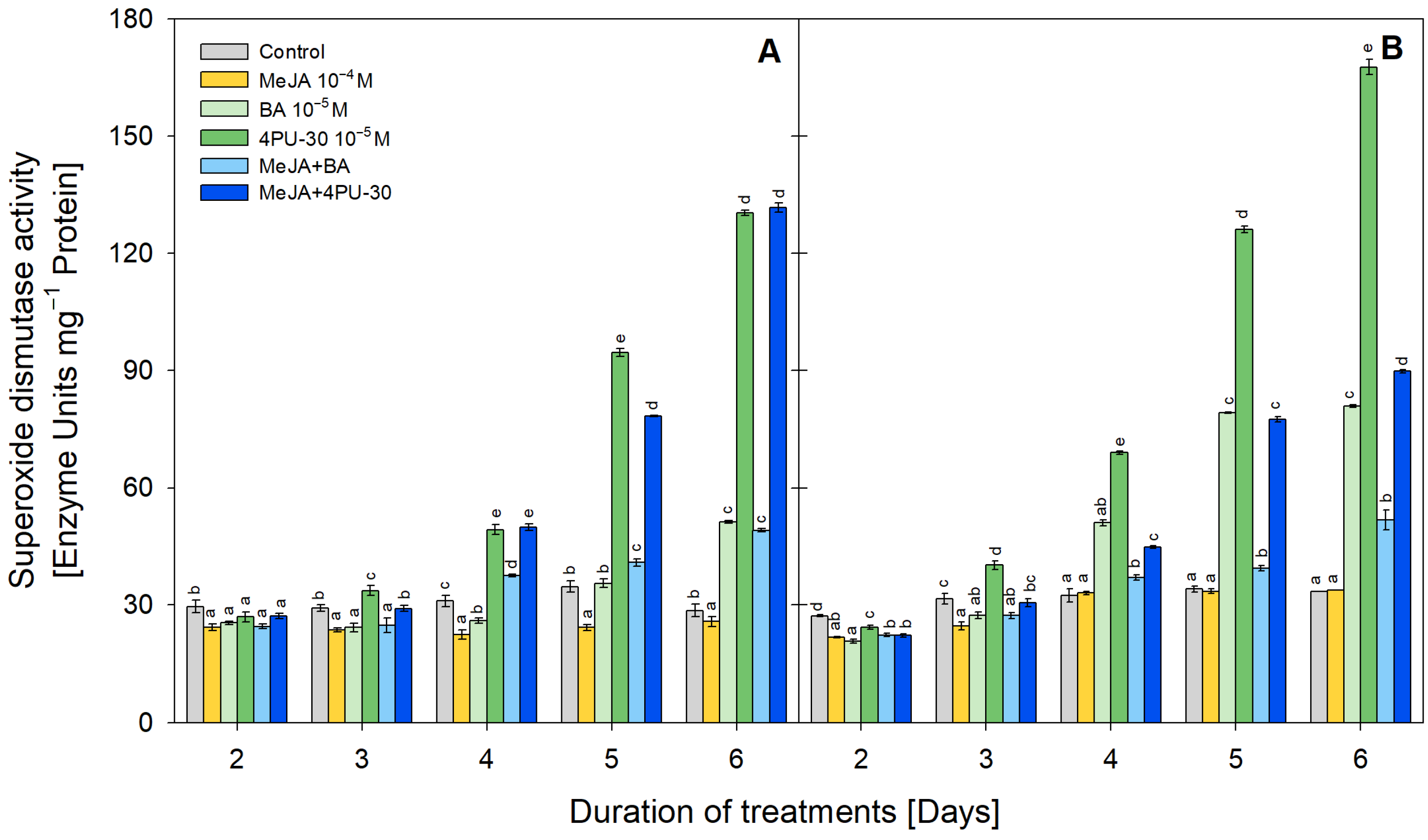
On the second day under dark conditions (Figure 2A), the highest SOD activity was detected in control cotyledons. Application of MeJA was found to inhibit SOD activity during the entire experiment, with a maximal reduction with 30% on the fifth day. At first three measurement points, BA reduced this parameter by approximately 17%, but at the end of the experimental period, an induction with 80% was found. Initially, 4PU-30 also caused slight inhibition (10%), but from the 3rd day onward an opposite tendency was observed, and SOD activity increased with time up to 356% above the respective control. MeJA did not provoke a strong reduction in the SOD activity enhanced by both CKs. Even more, at the end of the experimental period, there were no statistical differences between cotyledons individually treated with CKs and in combination with MeJA.
At the early stage of treatment (second day) under light conditions (Figure 2B), the highest SOD activity was found in the control zucchini cotyledons. On the third day, only the 4PU-30 application raised enzymatic activity (with 27%), while the mixture solution (MeJA+4PU-30) maintained the SOD activity close to the control. During the last three points of the reported period (at the fourth, fifth, and sixth days), a steadily increasing SOD activity was observed in all cotyledons treated with CKs (individually and in combination), with maximal effectiveness due to the application of 4PU-30 (up to 402% higher levels above the control on the 6th day). At the same time, application of MeJA alone did not influence SOD activity but notably reduced the stimulatory effect of both CKs.

3.3. POX Activity

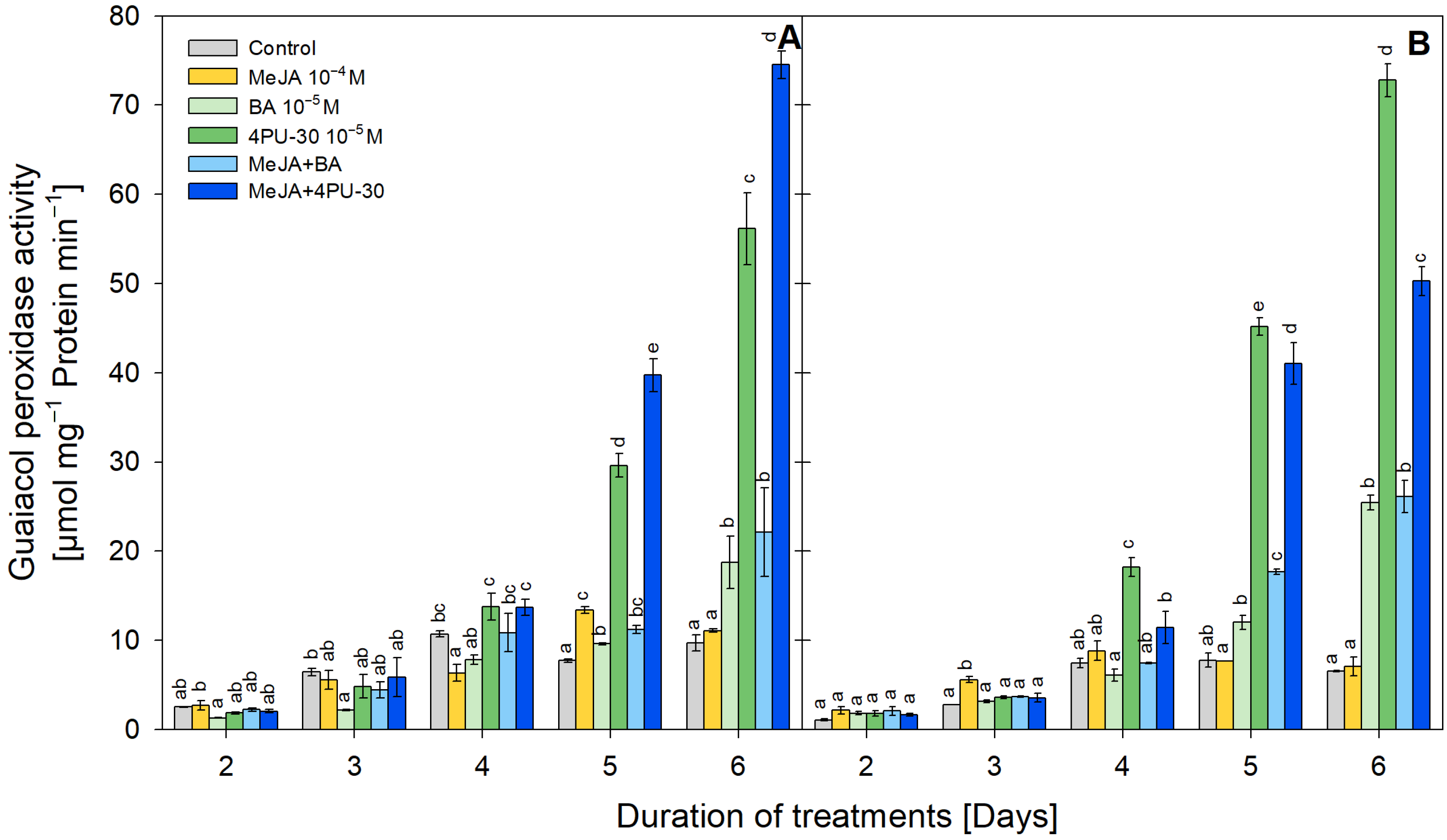
An inhibited POX activity with 66% due to BA was detected (3rd day) and with 40% due to MeJA (4th day) in dark-grown cotyledons (Figure 3A). The application of MeJA did not cause any significant effect in combined treatment as compared to BA. The treatment with 4PU-30 gradually increased POX activity with time, which peaked at 479% above the control (6th day). The application of MeJA led to an additional rise in POX activity starting from the 5th day of treatment in the combined treatment MeJA+4PU-30.
All treatments did not influence POX activity in light-grown cotyledons on the second day (Figure 3B). On the third day only MeJA enhanced this parameter meaningfully (101% increment), but in the rest of the measurements this effect was not detected. A very sharp and huge increase in POX activity was recorded due to 4PU-30 application, and the maximal level (1020% higher than the control) was measured on the 6th day. Application of MeJA diminished the 4PU-30-induced stimulation of POX activity starting from the 4th day and finally reached 673% induction above the control. As compared to 4PU-30, BA also increased the activity of POX but to a lower degree: with 54% on the 5th day and with 290% on the 6th day. MeJA led to incidental additional stimulation of BA-induced increase in POX on the 5th day of treatment, but at the end of the experiment, it did not alter the CK effect.

3.4. CAT Activity

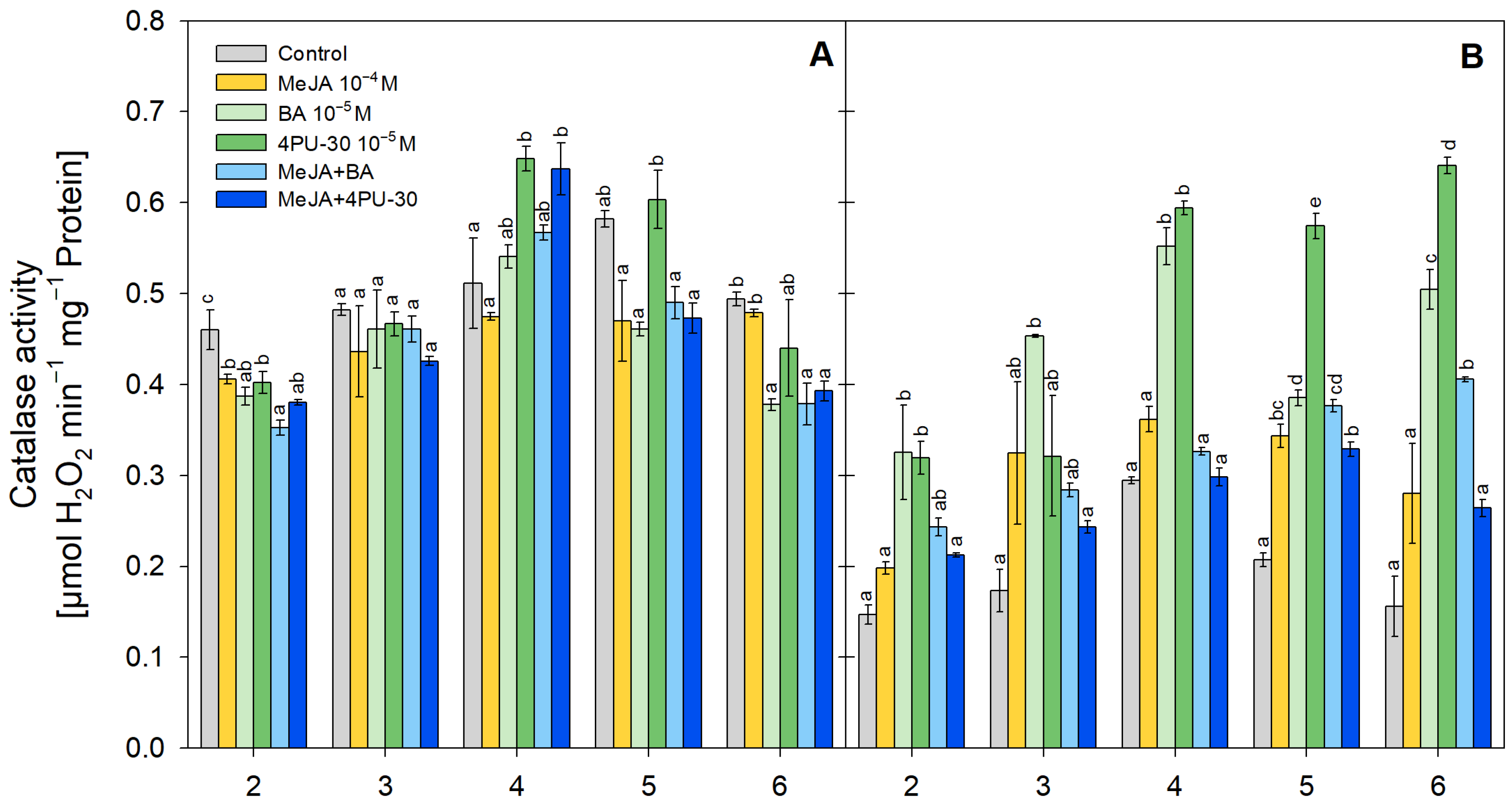
The constitutive CAT activity in the dark-grown cotyledons was higher than that measured in the light-grown ones (Figure 4). The effects of the treatments on CAT activity were also related to the presence of light. Under dark conditions (Figure 4A), MeJA alone treatment provoked a decrease in CAT activity during the entire experimental period. At the first measurement point, the activity of CAT was the highest in control cotyledons, while it was the most reduced (with 23%) in MeJA+BA treatment group. On the third day, all compounds did not significantly influence CAT activity in dark-grown treatment subgroups. Only on the 4th day of treatment 4PU-30 applied alone and in combination with MeJA increased CAT activity up to 26% and 24% above the respective control. At the next measurement point, except for 4PU-30 applied alone, all treatments caused a decrease in CAT activity, although the changes were not significant. At the end of the testing period, an inhibition of CAT activity became significant as compared to the control for BA, MeJA+BA, and MeJA+4PU-30 treatment groups.
Under light conditions (Figure 4B), the values of enzymatic activity stayed higher in MeJA-only-treated cotyledons during the entire experiment, although the increase was not significant. At first, the individual application of both CKs—BA and 4PU-30 enhanced the enzymatic activity by 120%. This tendency continued till the end of the testing period, when the highest activity was caused by 4PU-30 (311%). Starting from the 4th day, MeJA significantly decreased the cytokinin-induced CAT activity, especially in combination with the phenylurea type of cytokinin.

3.5. Protein Content

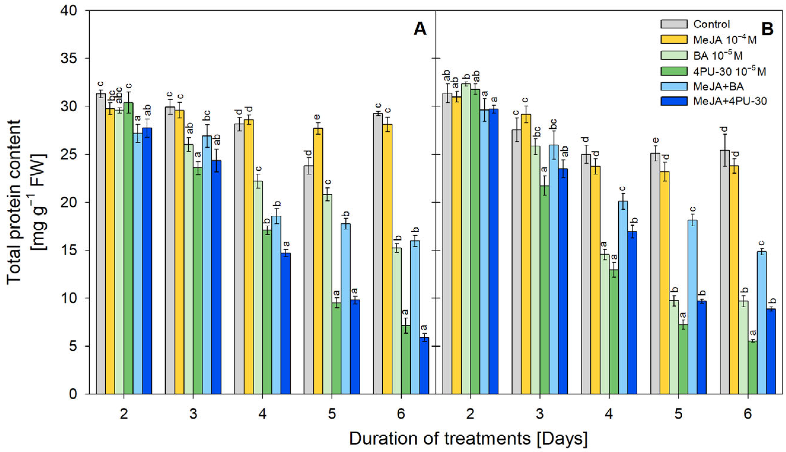
Initially, there were no substantial deviations from the respective controls in total protein content after all treatments under both light regimes (Figure 5). Starting from the 3rd day, MeJA applied alone did not cause significant alterations in the protein content under both light and dark conditions, while there was a decreasing tendency due to CK application, especially under light conditions (Figure 5B). The phenylurea type of cytokinin 4PU-30 had a stronger effect (up to 80% below the control on the 6th day) as compared to BA. MeJA did not meaningfully influence the protein content reduced by cytokinins under dark conditions (Figure 5A), with the exception of day 4 where additional reduction was found. On the opposite, under light conditions, MeJA mitigated the CK-induced protein content reduction (Figure 5B).

3.6. Free Proline Content

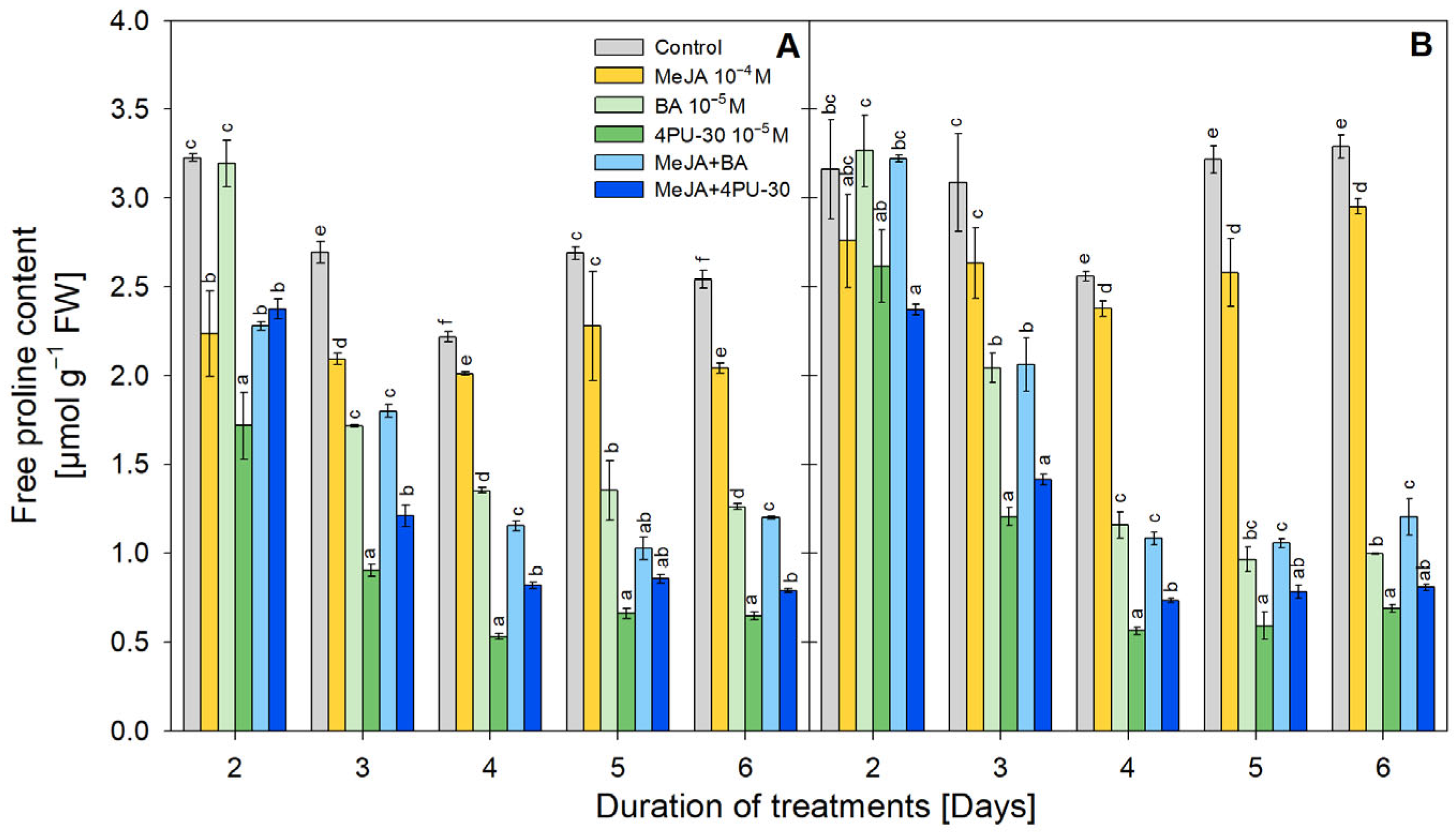
All treatments, with the exception of BA-treated cotyledons on the second day, resulted in a reduction in free proline content (Figure 6). Starting from the 4th day, a relatively stable decrease in proline was detected until the end of the experimental period. This decrease in proline did not depend on the presence or absence of light, and the changes due to the treatments in cotyledons grown under both light regimes were comparable. The alone application of MeJA minimally affected this parameter, while the strongest effect was detected in 4PU-30 treatment, leading to a maximal decrement of proline concentration down to 80% as compared to the respective controls after the third day of the experiment. The effect of MeJA in combined MeJA+4PU-30 treatment provoked a significant increase in proline as compared to 4PU-30 alone application under dark conditions.

3.7. Chlorophyll and Carotenoids Content

MeJA application negatively influenced leaf pigment content when applied alone (Figure 7). Both BA and 4PU-30 provoked a substantial increase in the leaf pigment contents during the first two measurement points. On the 2nd day, BA led to 118%, and 4PU-30 caused a 293% rise in chlorophyll a content. Additional increases of up to 371% (BA) and 378% (4PU-30) in chlorophyll a levels were detected on the 3rd day of treatment. A similar positive effect was detected in chlorophyll b and carotenoid accumulation. Starting from the 4th day of the experiment, there were no significant differences between the values measured in the control and CK-treated cotyledons. The negative effect of MeJA was partly compensated due to the CKs action in the combined treatments. The mix of MeJA and CK’s increased the leaf pigment content at the early stage (significant only for chlorophyll a) and later on, starting from the 4th day, it maintained the pigments near the control.

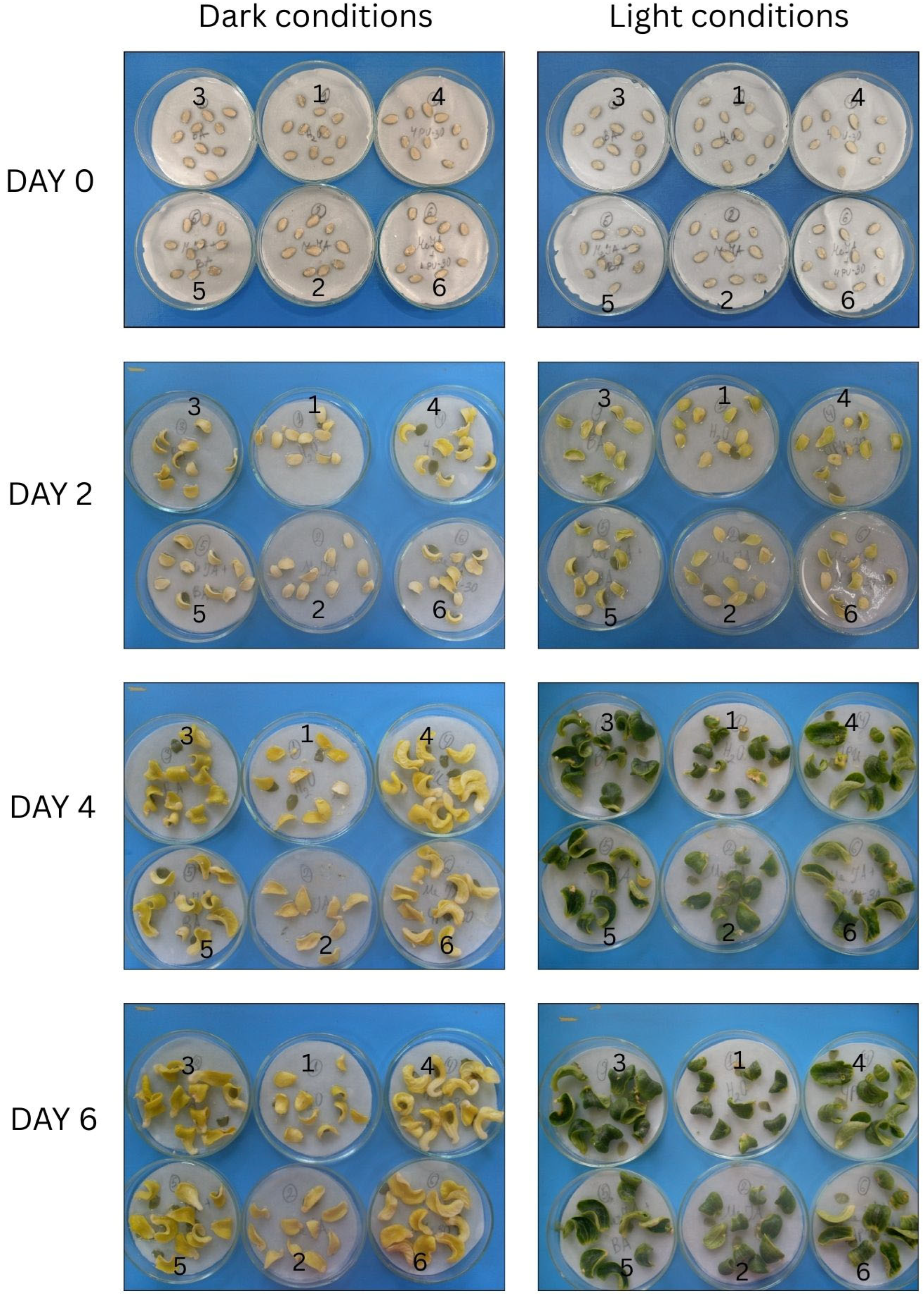
3.8. Dynamics of Phenotypic Alterations

Images of cotyledons grown under dark and light conditions are presented in Figure 8. The comparison between treatments on each day revealed that starting from the 2nd day, both CKs applied alone enlarged the size of the cotyledons grown in dark and light regimes. Cotyledons treated with MeJA only did not have visible differences from the control ones. This tendency was valid till the end of the experiment, with the exception of the 6th day, when MeJA caused a slight cotyledon enlargement. In addition, starting from the 2nd day, greening of cotyledons cultivated under light condition was visually observed and corresponded to the alterations of photosynthetic pigments shown in Figure 7. MeJA in combined variants slightly retarded the cotyledon growth induced by CKs.

4. Discussion

4.1. Alteration in Antioxidant Defense System in Dark-Grown Cotyledons

Cytokinins and jasmonates are phytohormones that are intensively studied in relation to plant cell growth and development. A relatively simple experimental system of excised zucchini cotyledons grown under dark conditions provides a physiologically homogenous material derived from a single organ [4]. The same authors established that both CKs (4PU-30 and BA) raised cell expansion and division rate and enlarged cotyledon area from day two onward [3,5]. Although MeJA suppressed cell division [45], it evoked cotyledon expansion [3]. Because of the tight interaction between phytohormones and ROS, the usage of the model system of dark-grown excised zucchini cotyledons offers an opportunity to study the effects of CK and JA on the antioxidant system through monitoring of selected ROS (H2O2), non-enzymatic (proline), and enzymatic antioxidants (activities of CAT, POX, and SOD). Usually, H2O2 is naturally generated during diverse physiological processes as a product of multiple enzymatic or non-enzymatic reactions [46,47]. Hydrogen peroxide in low concentrations is designated as the steadiest ROS, having enough lifetime to be able to migrate between the cells and act as a signaling molecule [28,32,48]. The MeJA-induced augmentation of H2O2 involving signaling function was reported to be an early response to the jasmonate exposure, consequently influencing antioxidant defense enzymes in plants [27,49]. Our results showed an increase in H2O2 due to MeJA only on the 2nd day, which is in agreement with the previous studies and provides additional evidence valid for cotyledon organ culture. In intact seeds, many questions regarding the regulation and generation of ROS in space and time still remain unanswered, including these for ROS and phytohormonal cross-talk [31]. The observed consistent further suppression of H2O2 in cotyledons due to MeJA under dark could be connected to the substitution of its signaling functions with that of the exogenously applied MeJA, which is also acknowledged to participate in different signaling pathways [50]. Changes obtained for the enzymatic activities connected with production (SOD) or decomposition (CAT, POX) of H2O2 evidenced that they could be at least partially responsible for the observed suppression of hydrogen peroxide content under dark conditions. Our results are in line with the suggestion of Stoynova-Bakalova et al. [3] that zucchini cotyledons responded to treatment with 100 µM MeJA under dark conditions by altering induction and accumulation of different polypeptides. On the other side, cotyledons with separated embryonal axes are known to be CK deficient, so supplementation of 4PU-30 and BA clarified the intensified cell division reported from day 2 to 4 (2 days earlier than the control) [5]. The higher division rate due to 4PU-30 and BA implies intensified energy consumption and the corresponding electron transfer demands in excised cotyledons under dark. Then, the substantial increase in H2O2 observed due to CKs, especially by 4PU-30, could be derived from mitochondria (inner membrane electron transport chain), glyoxysomes and/or peroxisomes (fatty acid utilization after β-oxidation), and endoplasmic reticulum [31,32,48,51,52]. The minor alteration of SOD, CAT, and POX along with the accumulation of H2O2 during intensive cell proliferation and growth could be related to rigorous cellular respiration, since under dark conditions mitochondria are a major source of ROS/H2O2 [46,53]. Later on, starting from the 4th day when intensive cell division was terminated in CK-treated cotyledons until the end of the monitored period, H2O2 maintained its levels almost at a plateau, probably because of the fine-tuning between the activity of generating and detoxifying enzymes, as supported by the obtained data. In combined treatments, the effect of MeJA on H2O2 levels was found to be antagonistic to these after exogenous CKs application, and the cumulative outcome shifts the content of ROS closer to the control. However, a specific antagonistic/synergistic effect of MeJA was detected for the detoxifying enzymatic activities of CAT/POX, respectively, in cotyledons due to combined treatment with 4PU-30. This implies possible involvement of other not yet completely known mechanisms of cross-interaction between MeJA and phenylurea-type CK in modulation of the activity of antioxidant players during cell division.
The proline imino acid is considered to have a myriad of roles in plant cells: as a signaling molecule, a non-enzymatic antioxidant, a repository function as proline-rich storage proteins, a role in protein synthesis and cell cycle transition, etc. [54,55]. Exogenous MeJA and CKs increased proline content in intact plants grown in different experimental conditions [56,57,58,59,60,61,62], while proline was found to decrease in our model system. The excised embryonic axes of cotyledons could elucidate this discrepancy because the ability of zucchini cotyledons to accomplish key ontogeny transitions is blocked in our experiments. However, the various functions of proline in plant development and during key transitions are very important [55]. For example, proline itself could effectively scavenge ROS overproduced during either different stress reactions or normal plant development [63]. In addition, proline metabolism is closely connected with that of polyamines (PAs), which are plant growth regulators that participate in numerous physiological processes, including cell proliferation and growth, organogenesis, plant stress responses, and senescence. Both proline and PAs have a common precursor, and they could regulate a biosynthesis or metabolism of each other [64,65,66]. In general, PA biosynthesis is positively regulated by CKs and MeJA [67,68,69,70]. The decreased proline content observed after phytohormone applications could be explained by either a biosynthesis shift toward PAs or its incorporation in cell-wall proteins, as it could be used as a resource for cell division and/or enlargement [52,53,66]. In addition, the similar alteration detected in total protein content supports this suggestion since the decrease in protein signifies for its utilization for cotyledon growth. This observation is valid especially for CKs (applied alone or in combination with MeJA), since they are important players in nutrient reserve utilization. The storage role of proline fits better also with the huge decrease observed due to CK application because the alteration intensifies with time and is in line with the ability of CKs to induce both cell division and expansion [3,5] and the cotyledon growth documented by us. Although the suggestion concerning the decrease in proline due to PA synthesis shift could not be excluded and should be further explored.
Although MeJA and CKs applied alone caused a decrease in proline content, in combined treatments MeJA exhibited specific action that depends on the CKs type. MeJA acted synergistically with BA, while with 4PU-30 the effect was antagonistic. In addition, previous studies [3,6] also imply complicated regulation of proteins in dark-grown zucchini cotyledons, particularly after exogenous application of MeJA and BA. Taken together, our data suggest one more time that the different types of CK interact in diverse mechanisms in plant metabolism during cell division and growth under dark.

4.2. Alteration in Antioxidant Defense System and Leaf Pigments in Light-Grown Cotyledons

Along with phytohormones, light is also an important modulator of plant development, especially during early initial stages [71,72,73]. The effects of light on cell enlargement are similar to those triggered by CKs, and obtaining a larger area for catching light is known as the main task of the epigeal cotyledons post-germination. In addition, the effect of MeJA strongly depends on the presence of light [74]. Therefore, comparing the alterations in the selected components of the antioxidant network in cotyledons grown under dark and light conditions could be useful also to broaden knowledge about biochemical processes in developing cotyledon organs and could serve as a base for further progress of investigations on optimization of plant growth for sustainable agriculture.
Another important reason to compare light- and dark-grown zucchini cotyledons in excised conditions is that the light reactions of photosynthesis are the major source of ROS, which are produced in the reaction centers of both photosystems in thylakoids of the chloroplasts [18,32,75]. As a result, we also detected a higher basal level of H2O2 in light-grown cotyledons as compared to dark-grown ones. In addition, the presence of light stimulated H2O2 synthesis in MeJA-treated cotyledons, and its level was steadily higher than that detected in dark-grown cotyledons at each respective measurement point except on the 2nd day. The non-detected H2O2 content after MeJA application on the 2nd day of light treatment implies that there is still an insufficient amount of photosynthetic pigments, which could ensure enough electron transport chains that produce ROS/H2O2. This suggestion fits well with the acknowledged ability of MeJA to affect the photosynthetic pigments [76,77,78], which was also observed in the current study for the cotyledon organ culture of zucchini. In addition, MeJA enhanced the activity of antioxidant enzymes POX and CAT, which could also contribute to the not markedly high H2O2 levels as compared to the controls.
In contrast to MeJA, the highest CK-induced content of leaf pigments was found to be in parallel with the quantity of H2O2. Although SOD activity was enhanced due to CK alone application, the enzymatic activities of CAT and POX were also increased, which prevented the over-generation of H2O2. This indicated that CKs take part in the regulation of maintenance of the antioxidant network homeostasis not only in the dark but also under light conditions [37]. Leaf pigments are vital components for plant photosynthesis—the chief process that guarantees high crop yield. Their content could be modulated by various factors, including phytohormones [16,73]. MeJA reduced pigments in zucchini cotyledons, and logically this might lead to reduced photosynthetic efficiency [77]. On the opposite, both CKs caused an increase in leaf pigments, which corresponds to better photosynthetic efficiency that in turn implicates improved crop production and quality [73,79]. In addition to chlorophylls, CKs also increased carotenoids, which are important components of antioxidant defense that prevent ROS over-accumulation [80].
The dynamics of alterations in the amount of proline and protein after MeJA treatment could be attributed to the possibility of cell and cotyledon enlargement only, not to cell proliferation [81]. Similarly to dark-grown cotyledons, in light-grown ones there was a minor decrease in total protein and proline content accompanied by a significant reduction in leaf pigments due to MeJA. It is worth noting that, in the cotyledon organ studied, the coordination between carbon and nitrogen source–sink dynamics must be tightly regulated, as both processes occur in the same tissue. Thus, the detected negative effect of MeJA to photosynthetic pigments in our study is in accordance with that already reported [76,77,78] and correlated well with the slight decrease in total protein and proline content identified by us. Unlike MeJA, CKs induced a strong reduction in proline and protein content. This could be attributed to the different effects of MeJA and CKs on cell division. Furthermore, in contrast to MeJA, we found that from the 4th day onwards, the total protein content was reduced even more in light than in the dark due to CK alone application, particularly by BA. The light-induced synthesis of photosynthetic pigments that is accelerated by CKs undoubtedly requires additional resources, and proteins and proline are rational opportunities. The stimulation of chlorophyll synthesis and also fine regulation of various photosynthesis-related proteins and transcripts by CKs was established in a study of CK-deficient tobacco leaves [82]. The chloroplast biogenesis was documented to correlate with cell size but proceed even when cell division is ceased [73,83,84]. So, the addition of CKs stimulates the formation of the leaf pigments and also could modulate the content of the proteins that take part in photosynthesis and contribute to the build-up of light-harvesting complexes in light-grown cotyledons. As a result, the total protein content declined after CK alone application, distinctly observed in light. Stoynova-Bakalova et al. [6] also showed that BA increased RUBISCO large subunit quantity simultaneously with the decrease in the amount of reserve proteins under dark conditions as well.
The exogenous application of MeJA in the combined treatments (MeJA+CKs) counteracted the CK-induced changes in most of the parameters studied under light conditions. The interaction between MeJA and CKs could be characterized as antagonistic in relation to total protein, hydrogen peroxide, photosynthetic pigments content, and also the activities of CAT and SOD. The antagonistic effect of MeJA on CK action was obvious in light-grown cotyledons, unlike the dark-grown ones in which this tendency was not clearly observed.

5. Conclusions

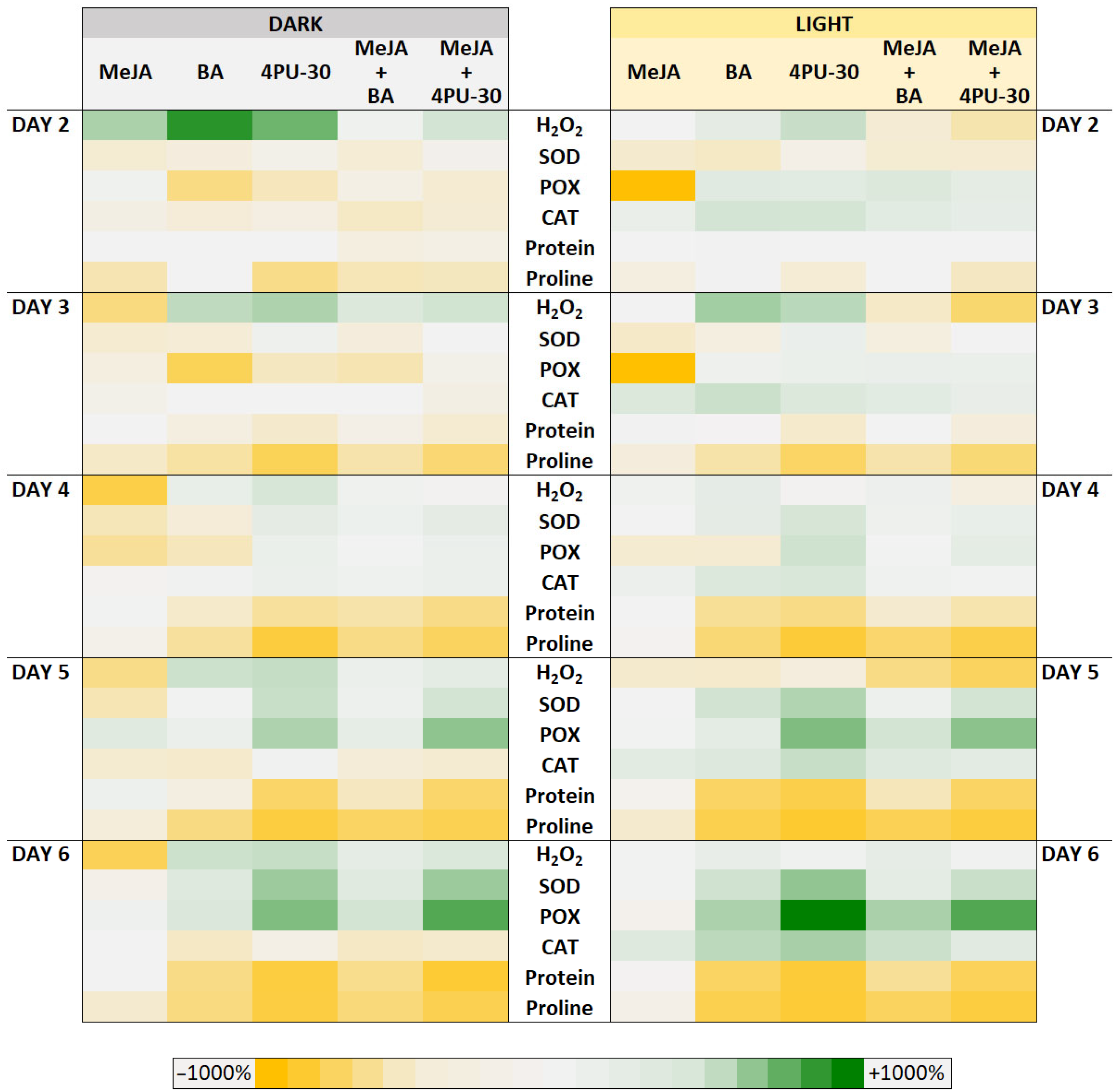
Although our study is fundamental and it is not directly applicable to agricultural practice, the estimation of the combined action of different CK types and MeJA in relation to important biochemical parameters could give a new approach for obtaining unique knowledge for the phytohormone interactions. Moreover, the control of cell division as a basis of the development of organs and tissues is essential for further optimization of plant growth. Our results provide new information regarding the effect of two diverse classes of phytohormones on the biochemical alterations of the antioxidant defense system and ROS that accompany organ growth, and they might involve different routes of metabolic pathways under dark and light conditions. Our study supposes that the presence of light could additionally alter the interaction between phytohormones, which reflects in more intense changes in biochemical responses of antioxidant defense in developing cotyledons (Figure 9). However, other components of biochemical mechanisms must not be excluded, and the cell antioxidant machinery should be considered as a part of a wider plant physiological response during cell division and growth in isolated cotyledons.
Future research on the relationship between MeJA and CK and their cross-talk with other key plant growth regulators such as polyamines that also modulate plant antioxidant defense, cell growth, and development could supplement the knowledge on the physiological alterations during the early growth of cotyledons under light and dark conditions. Further investigations will provide more complex information on phytohormone interplay in the early stage of cotyledon growth, focusing on the content of polyamines and non-enzymatic antioxidants, the level of gene expression of key biosynthetic and catabolic enzymes of the polyamine pathway, and major antioxidant enzymes. The role of plant growth regulators in these processes using a relatively simple model system would be a prerequisite for better agricultural practices and allow the development of sustainable agriculture.

Author Contributions

Conceptualization, D.T.; methodology, D.T.; formal analysis, A.P., D.T., Z.K. and I.S.; data curation, D.T. and I.S.; writing—original draft preparation, Z.K. and A.P.; writing—review and editing, D.T. and I.S.; visualization, I.S. and Z.K.; supervision, D.T. All authors have read and agreed to the published version of the manuscript.

Funding

This research received no external funding.

Institutional Review Board Statement

Not applicable.

Informed Consent Statement

Not applicable.

Data Availability Statement

The data presented in this study are available on request from the corresponding author. The data are not publicly available due to privacy.

Conflicts of Interest

The authors declare no conflicts of interest.

Abbreviations

BAN6-benzyl adenine
CATcatalase
CKcytokinin
MeJAmethyl jasmonate
POXguaiacol peroxidase
ROSreactive oxygen species
4PU-30N1-(2-chloropyridin-4-yl)-N2-phenylurea
SODsuperoxide dismutase
PAspolyamines

References

  1. Deepika; Ankit; Sagar, S.; Singh, A. Dark-induced hormonal regulation of plant growth and development. Front. Plant Sci. 2020, 11, 581666. [Google Scholar]
  2. Shimotohno, A.; Aki, S.S.; Takahashi, N.; Umeda, M. Regulation of the plant cell cycle in response to hormones and the environment. Ann. Rev. Plant Biol. 2021, 72, 273–296. [Google Scholar] [CrossRef]
  3. Stoynova-Bakalova, E.; Petrov, P.I.; Gigova, L.; Baskin, T.I. Differential effects of methyl jasmonate on growth and division of etiolated zucchini cotyledons. Plant Biol. 2008, 10, 476–484. [Google Scholar] [CrossRef]
  4. Stoynova-Bakalova, E. Properties of plate meristem of growing epigeal cotyledons in an experimental system. Environ. Exp. Bot. 2007, 59, 76–83. [Google Scholar] [CrossRef]
  5. Stoynova-Bakalova, E.; Petrov, P.I. Control by cytokinins of the cellular behavior in the plate meristem of zucchini cotyledons. Planta 2006, 223, 1256–1262. [Google Scholar] [CrossRef] [PubMed]
  6. Stoynova-Bakalova, E.; Petrov, P.; Gigova, L.; Ivanova, N. Differences in cytokinin control on cellular dynamics of zucchini cotyledons cultivated in two experimental systems. Plant Biol. 2011, 13, 22–27. [Google Scholar] [CrossRef]
  7. Stoynova-Bakalova, E.Z.; Petrov, P.I.; Karanov, E.N. Effects of benzylaminopurine and abscisic acid on distribution of rRNA in the palisade cells of excised Cucurbita pepo cotyledons. Biol. Plant. 2001, 44, 355–360. [Google Scholar] [CrossRef]
  8. Roitsch, T.; Ehneß, R. Regulation of source/sink relations by cytokinins. Plant Growth Regul. 2000, 32, 359–367. [Google Scholar] [CrossRef]
  9. Sergiev, I.; Todorova, D.; Somleva, M.; Alexieva, V.; Karanov, E.; Stanoeva, E.; Lachkova, V.; Smith, A.; Hall, M. Influence of cytokinins and novel cytokinin antagonists on the senescence of detached leaves of Arabidopsis thaliana. Biol. Plant. 2007, 51, 377–380. [Google Scholar] [CrossRef]
  10. Gao, S. Function and mechanism study of plant cytokinins. In Proceedings of the 2020 10th International Conference on Biomedical Engineering and Technology, Tokyo, Japan, 15–18 September 2020; pp. 80–84. [Google Scholar]
  11. Parthier, B. Hormone-induced alterations in plant gene expression. Biochem. Physiol. Pfl 1989, 185, 289–314. [Google Scholar] [CrossRef]
  12. Mok, M.C.; Mok, D.W.; Turner, J.E.; Mujer, C.V. Biological and biochemical effects of cytokinin-active phenylurea derivatives in tissue culture systems. HortScience 1987, 22, 1194–1197. [Google Scholar] [CrossRef]
  13. Houssa, C.; Bernier, G.; Pieltain, A.; Kinet, J.M.; Jacqmard, A. Activation of latent DNA-replication origins: A universal effect of cytokinins. Planta 1994, 193, 247–250. [Google Scholar] [CrossRef]
  14. Davies, P.J. The plant hormones: Their nature, occurrence, and functions. In Plant Hormones: Physiology, Biochemistry and Molecular Biology; Davies, P.J., Ed.; Springer: Dordrecht, The Netherlands, 1995; pp. 1–12. [Google Scholar]
  15. Hare, P.D.; Cress, W.A.; Van Staden, J. The involvement of cytokinins in plant responses to environmental stress. Plant Growth Regul. 1997, 23, 79–103. [Google Scholar] [CrossRef]
  16. Cortleven, A.; Schmülling, T. Regulation of chloroplast development and function by cytokinin. J. Exp. Bot. 2015, 66, 4999–5013. [Google Scholar] [CrossRef]
  17. Huang, H.; Ullah, F.; Zhou, D.-X.; Yi, M.; Zhao, Y. Mechanisms of ROS regulation of plant development and stress responses. Front. Plant Sci. 2019, 10, 800. [Google Scholar] [CrossRef]
  18. Su, J.; Liu, Y.; Han, F.; Gao, F.; Gan, F.; Huang, K.; Li, Z. ROS, an important plant growth regulator in root growth and development: Functional genes and mechanism. Biology 2024, 13, 1033. [Google Scholar] [CrossRef]
  19. Hönig, M.; Plíhalová, L.; Husičková, A.; Nisler, J.; Doležal, K. Role of cytokinins in senescence, antioxidant defence and photosynthesis. Int. J. Mol. Sci. 2018, 19, 4045. [Google Scholar] [CrossRef] [PubMed]
  20. Shudo, K. Chemistry of phenylurea cytokinins. In Cytokynins: Chemistry, Activity and Function; Mok, D.W.S., Mok, M.C., Eds.; CRC Press: Boca Raton, FL, USA; Ann Arbor, MI, USA; London, UK; Tokyo, Japan, 1994; Chapter 3; pp. 35–42. [Google Scholar]
  21. Pan, J.; Wang, H.; You, Q.; Cao, R.; Sun, G.; Yu, D. Jasmonate-regulated seed germination and crosstalk with other phytohormones. J. Exp. Bot. 2023, 74, 1162–1175. [Google Scholar] [CrossRef]
  22. Wang, C.Y.; Buta, J.G. Methyl jasmonate reduces chilling injury in Cucurbita pepo through its regulation of abscisic acid and polyamine levels. Environ. Exp. Bot. 1994, 34, 427–432. [Google Scholar] [CrossRef]
  23. Ho, T.-T.; Murthy, H.N.; Park, S.-Y. Methyl jasmonate induced oxidative stress and accumulation of secondary metabolites in plant cell and organ cultures. Int. J. Mol. Sci. 2020, 21, 716. [Google Scholar] [CrossRef]
  24. Damyanova, M.; Todorova, D.; Sergiev, I. Polyamine alterations in isolated zucchini cotyledons grown in presence of cytokinins and Cu2+. Am. J. Plant Sci. 2014, 5, 2141–2147. [Google Scholar] [CrossRef]
  25. Damyanova, M.; Todorova, D.; Sergiev, I. Endogenous polyamine profiles of isolated zucchini cotyledons incubated on solutions of Cu2+ and methyl jasmonate. Oxid. Commun. 2015, 38, 104–113. [Google Scholar]
  26. Stoynova-Bakalova, E.; Ivanova, N.; Bakalov, D.; Gigova, L. Modifying effects of some plant hormones on zucchini cotyledons subjected to high temperature and excess copper. Bot. Lith. 2020, 26, 28–39. [Google Scholar] [CrossRef]
  27. Soares, C.; Carvalho, M.E.; Azevedo, R.A.; Fidalgo, F. Plants facing oxidative challenges—A little help from the antioxidant networks. Environ. Exp. Bot. 2019, 161, 4–25. [Google Scholar] [CrossRef]
  28. Mittler, R.; Vanderauwera, S.; Suzuki, N.; Miller, G.A.D.; Tognetti, V.B.; Vandepoele, K.; Gollery, M.; Shulaev, V.; Van Breusegem, F. ROS signaling: The new wave? Trends Plant Sci. 2011, 16, 300–309. [Google Scholar] [CrossRef] [PubMed]
  29. Mittler, R. ROS are good. Trends Plant Sci. 2017, 22, 11–19. [Google Scholar] [CrossRef]
  30. Wojtyla, Ł.; Lechowska, K.; Kubala, S.; Garnczarska, M. Different modes of hydrogen peroxide action during seed germination. Front. Plant Sci. 2016, 7, 66. [Google Scholar] [CrossRef]
  31. Bailly, C. The signalling role of ROS in the regulation of seed germination and dormancy. Biochem. J. 2019, 476, 3019–3032. [Google Scholar] [CrossRef]
  32. Phua, S.Y.; De Smet, B.; Remacle, C.; Chan, K.X.; Van Breusegem, F. Reactive oxygen species and organellar signaling. J. Exp. Bot. 2021, 72, 5807–5824. [Google Scholar] [CrossRef]
  33. Raza, A.; Salehi, H.; Rahman, M.A.; Zahid, Z.; Madadkar Haghjou, M.; Najafi-Kakavand, S.; Zhuang, W. Plant hormones and neurotransmitter interactions mediate antioxidant defenses under induced oxidative stress in plants. Front. Plant Sci. 2022, 13, 961872. [Google Scholar] [CrossRef]
  34. Oracz, K.; Karpinski, S. Phytohormones signaling pathways and ROS involvement in seed germination. Front. Plant Sci. 2016, 7, 864. [Google Scholar] [CrossRef] [PubMed]
  35. Tognetti, V.B.; Bielach, A.; Hrtyan, M. Redox regulation at the site of primary growth: Auxin, cytokinin and ROS crosstalk. Plant Cell Environ. 2017, 40, 2586–2605. [Google Scholar] [CrossRef]
  36. Ochatt, S.J. Less frequently used growth regulators in plant tissue culture. Methods Mol. Biol. 2024, 2827, 109–143. [Google Scholar]
  37. Nowakowska, K.; Pińkowska, A.; Siedlecka, E.; Pacholczak, A. The effect of cytokinins on shoot proliferation, biochemical changes and genetic stability of Rhododendron ‘Kazimierz Odnowiciel’ in the in vitro cultures. Plant Cell Tissue Organ Cult. 2022, 149, 675–684. [Google Scholar] [CrossRef]
  38. Dias, I.; Costa, M. Effect of low salt concentration on nitrate reductase and peroxidase of sugar beet leaves. J. Exp. Bot. 1983, 34, 537–543. [Google Scholar] [CrossRef]
  39. Aebi, M. Catalase in vitro. Methods Enzym. 1984, 105, 121–126. [Google Scholar]
  40. Beauchamp, C.; Fridovich, I. Superoxide dismutase. Improved assay and an assay applicable to acrylamide gels. Anal. Biochem. 1971, 44, 276–287. [Google Scholar] [CrossRef]
  41. Bradford, M. A rapid and sensitive method for the quantification of microgram quantities of protein utilizing the principle of protein-dye binding. Anal. Biochem. 1976, 72, 248–254. [Google Scholar] [CrossRef]
  42. Bates, L.; Waldren, R.; Teare, I. Rapid determination of free proline for water-stress studies. Plant Soil 1973, 39, 205–207. [Google Scholar] [CrossRef]
  43. Frew, J.E.; Jones, P.; Scholes, F. Spectrophotometric determination of hydrogen peroxide and organic hydroperoxide at low concentrations in aqueous solution. Anal. Chim. Acta 1983, 155, 139–150. [Google Scholar] [CrossRef]
  44. Arnon, D.I. Copper enzymes in isolated chloroplasts. Polyphenoloxidase in Beta vulgaris. Plant Physiol. 1949, 24, 1–15. [Google Scholar] [CrossRef] [PubMed]
  45. Kamińska, M. Role and activity of jasmonates in plants under in vitro conditions. Plant Cell Tissue Organ Cult. 2021, 146, 425–447. [Google Scholar] [CrossRef]
  46. Ślesak, I.; Libik, M.; Karpinska, B.; Karpinski, S.; Miszalski, Z. The role of hydrogen peroxide in regulation of plant metabolism and cellular signalling in response to environmental stresses. Acta Biochim. Pol. 2007, 54, 39–50. [Google Scholar] [CrossRef]
  47. Kärkönen, A.; Dewhirst, R.A.; Mackay, C.L.; Fry, S.C. Metabolites of 2,3-diketogulonate delay peroxidase action and induce non-enzymic H2O2 generation: Potential roles in the plant cell wall. Arch. Biochem. Biophys. 2017, 620, 12–22. [Google Scholar] [CrossRef]
  48. Smirnoff, N.; Arnaud, D. Hydrogen peroxide metabolism and functions in plants. New Phytol. 2019, 221, 1197–1214. [Google Scholar] [CrossRef]
  49. Parra-Lobato, M.C.; Fernandez-Garcia, N.; Olmos, E.; Alvarez-Tinaut, M.C.; Gómez-Jiménez, M.C. Methyl jasmonate-induced antioxidant defence in root apoplast from sunflower seedlings. Environ. Exp. Bot. 2009, 66, 9–17. [Google Scholar] [CrossRef]
  50. Ghorbel, M.; Brini, F.; Sharma, A.; Landi, M. Role of jasmonic acid in plants: The molecular point of view. Plant Cell Rep. 2021, 40, 1471–1494. [Google Scholar] [CrossRef] [PubMed]
  51. Bailly, C. Active oxygen species and antioxidants in seed biology. Seed Sci. Res. 2004, 14, 93–107. [Google Scholar] [CrossRef]
  52. Černý, M.; Habánová, H.; Berka, M.; Luklová, M.; Brzobohatý, B. Hydrogen Peroxide: Its role in plant biology and crosstalk with signalling networks. Int. J. Mol. Sci. 2018, 19, 2812. [Google Scholar] [CrossRef]
  53. Rhoads, D.M.; Umbach, A.L.; Subbaiah, C.C.; Siedow, J.N. Mitochondrial reactive oxygen species. Contribution to oxidative stress and interorganellar signaling. Plant Physiol. 2006, 141, 357–366. [Google Scholar] [CrossRef]
  54. Kavi Kishor, P.B.; Sreenivasulu, N.E.S.E. Is proline accumulation per se correlated with stress tolerance or is proline homeostasis a more critical issue? Plant Cell Environ. 2014, 37, 300–311. [Google Scholar] [CrossRef]
  55. Kavi Kishor, P.B.; Hima Kumari, P.; Sunita, M.S.L.; Sreenivasulu, N. Role of proline in cell wall synthesis and plant development and its implications in plant ontogeny. Front. Plant Sci. 2015, 6, 544. [Google Scholar] [CrossRef]
  56. Thomas, J.C.; McElwain, E.F.; Bohnert, H.J. Convergent induction of osmotic stress-responses: Abscisic acid, cytokinin, and the effects of NaCl. Plant Physiol. 1992, 100, 416–423. [Google Scholar] [CrossRef] [PubMed]
  57. Thomas, J.C.; Smigocki, A.C.; Bohnert, H.J. Light-induced expression of ipt from Agrobacterium tumefaciens results in cytokinin accumulation and osmotic stress symptoms in transgenic tobacco. Plant Mol. Biol. 1995, 27, 225–235. [Google Scholar] [CrossRef] [PubMed]
  58. Chen, Y.L.; Zou, Z.R.; Yang, S.L. Effect of exogenous methyl jasmonate on osmotic adjustment capacity and proline metabolism of Jatropha curcas seedlings under salt stress. Acta Bot. Boreal-Occid Sin. 2023, 43, 794–804. [Google Scholar]
  59. Javadipour, Z.; Balouchi, H.; Movahhedi Dehnavi, M.; Yadavi, A. Physiological responses of bread wheat (Triticum aestivum) cultivars to drought stress and exogenous methyl jasmonate. J. Plant Growth Regul. 2022, 41, 3433–3448. [Google Scholar] [CrossRef]
  60. Ahmadi, F.I.; Karimi, K.; Struik, P.C. Effect of exogenous application of methyl jasmonate on physiological and biochemical characteristics of Brassica napus L. cv. Talaye under salinity stress. S. Afr. J. Bot. 2018, 115, 5–11. [Google Scholar] [CrossRef]
  61. Tayyab, N.; Naz, R.; Yasmin, H.; Nosheen, A.; Keyani, R.; Sajjad, M.; Hassan, M.N.; Roberts, T.H. Combined seed and foliar pre-treatments with exogenous methyl jasmonate and salicylic acid mitigate droughtinduced stress in maize. PLoS ONE 2020, 15, e0232269. [Google Scholar] [CrossRef]
  62. Repkina, N.; Ignatenko, A.; Holoptseva, E.; MiszalskI, Z.; Kaszycki, P.; Talanova, V. Exogenous methyl jasmonate improves cold tolerance with parallel induction of two cold-regulated (COR) genes expression in Triticum aestivum L. Plants 2021, 10, 1421. Plants 2021, 10, 1421. [Google Scholar] [CrossRef]
  63. Renzetti, M.; Funck, D.; Trovato, M. Proline and ROS: A unified mechanism in plant development and stress response? Plants 2025, 14, 2. [Google Scholar] [CrossRef]
  64. Galili, G.; Tang, G.; Zhu, X.; Gakiere, B. Lysine catabolism: A stress and development super-regulated metabolic pathway. Curr. Opin. Plant Biol. 2001, 4, 261–266. [Google Scholar] [CrossRef] [PubMed]
  65. Maurya, A.K.; Agarwal, R.; Gupta, R. Unraveling the crosstalk among ethylene, nitric oxide, and polyamines in tailoring the abiotic stress resilience in plants. Stress Biol. 2025, 5, 20. [Google Scholar] [CrossRef]
  66. Sergiev, I.; Todorova, D.; Katerova, Z.; Brambilla, I.; Mapelli, S.; Simova, S. Polyamines and amino acids in triticale plants grown on humic acids enriched nutrient solution and treated with UV-B irradiation. Theor. Exp. Plant Physiol. 2018, 30, 153–163. [Google Scholar] [CrossRef]
  67. Legocka, J.; Żarnowska, A. Role of polyamines in the cytokinindependent physiological processes II. Modulation of polyamine levels during cytokinin-stimulated expansion of cucumber cotyledons. Acta Physiol. Plant 2000, 22, 395–401. [Google Scholar] [CrossRef]
  68. Biondi, S.; Scoccianti, V.; Scaramagli, S.; Ziosi, V.; Torrigani, P. Auxin and cytokinin modify methyl jasmonate effects on polyamine metabolism and ethylene biosynthesis in tobacco leaf discs. Plant Sci. 2003, 165, 95–101. [Google Scholar] [CrossRef]
  69. Haggag, W.M.; Abd-El-Kareem, F. Methyl jasmonate stimulates polyamines biosynthesis and resistance against leaf rust in wheat plants. Arch. Phytopathol. Plant Prot. 2009, 42, 16–31. [Google Scholar] [CrossRef]
  70. Todorova, D.; Kamenova, S.; Ananieva, K. Changes of endogenous polyamines in excised Cucurbita pepo L. (zucchini) cotyledons cultivated in the presence of benzyladenine and methyl jasmonate. Genet. Plant Physiol. 2015, 5, 39–47. [Google Scholar]
  71. Kusnetsov, V.V.; Doroshenko, A.S.; Kudryakova, N.V.; Danilova, M.N. Role of phytohormones and light in de-etiolation. Russ. J. Plant Physiol. 2020, 67, 971–984. [Google Scholar] [CrossRef]
  72. Armarego-Marriott, T.; Sandoval-Ibañez, O.; Kowalewska, Ł. Beyond the darkness: Recent lessons from etiolation and de-etiolation studies. J. Exp. Bot. 2020, 71, 1215–1225. [Google Scholar] [CrossRef]
  73. Cackett, L.; Luginbuehl, L.H.; Schreier, T.B.; Lopez-Juez, E.; Hibberd, J.M. Chloroplast development in green plant tissues: The interplay between light, hormone, and transcriptional regulation. New Phytol. 2022, 233, 2000–2016. [Google Scholar] [CrossRef]
  74. Zhang, L.; Xing, D. Methyl jasmonate induces production of reactive oxygen species and alterations in mitochondrial dynamics that precede photosynthetic dysfunction and subsequent cell death. Plant Cell Physiol. 2008, 49, 1092–1111. [Google Scholar] [CrossRef] [PubMed]
  75. Asada, K. Production and scavenging of reactive oxygen species in chloroplasts and their functions. Plant Physiol. 2006, 141, 391–396. [Google Scholar] [CrossRef] [PubMed]
  76. Hanaka, A.; Nowak, A.; Ozimek, E.; Dresler, S.; Plak, A.; Sujak, A.; Reszczyńska, E.; Strzemski, M. Effect of copper stress on Phaseolus coccineus in the presence of exogenous methyl jasmonate and/or Serratia plymuthica from the Spitsbergen soil. J. Hazard. Mater. 2022, 436, 129232. [Google Scholar] [CrossRef] [PubMed]
  77. Ananieva, K.; Ananiev, E.D.; Mishev, K.; Georgieva, K.; Malbeck, J.; Kamínek, M.; Van Staden, J. Methyl jasmonate is a more effective senescence-promoting factor in Cucurbita pepo (zucchini) cotyledons when compared with darkness at the early stage of senescence. J. Plant Physiol. 2007, 164, 1179–1187. [Google Scholar] [CrossRef]
  78. Kim, S.J.; Tran, B.Q.; Jung, S. Methyl jasmonate-induced senescence results in alterations in the status of chlorophyll precursors and enzymatic antioxidants in rice plants. Biochem. Biophys. Res. Commun. 2023, 671, 38–45. [Google Scholar] [CrossRef]
  79. Hudeček, M.; Nožková, V.; Plíhalová, L.; Plíhal, O. Plant hormone cytokinin at the crossroads of stress priming and control of photosynthesis. Front. Plant Sci. 2023, 13, 1103088. [Google Scholar] [CrossRef]
  80. Ashraf, M.H.P.J.C.; Harris, P.J. Photosynthesis under stressful environments: An overview. Photosynthetica 2013, 51, 163–190. [Google Scholar] [CrossRef]
  81. Stoynova-Bakalova, E.; Petrov, P.; Tsukaya, H. Cell division and cell enlargement in isolated Cucurbita cotyledons grown in darkness and in light. J. Plant Res. 2002, 115, 375–380. [Google Scholar] [CrossRef]
  82. Werner, T.; Holst, K.; Pörs, Y.; Guivarc’h, A.; Mustroph, A.; Chriqui, D.; Grimm, B.; Schmülling, T. Cytokinin deficiency causes distinct changes of sink and source parameters in tobacco shoots and roots. J. Exp. Bot. 2008, 59, 2659–2672. [Google Scholar] [CrossRef]
  83. Loudya, N.; Mishra, P.; Takahagi, K.; Uehara-Yamaguchi, Y.; Inoue, K.; Bogre, L.; Mochida, K.; Lopez-Juez, E. Cellular and transcriptomic analyses reveal two staged chloroplast biogenesis underpinning photosynthesis build-up in the wheat leaf. Genome Biol. 2021, 22, 151. [Google Scholar] [CrossRef]
  84. Lopez-Juez, E.; Pyke, K.A. Plastids unleashed: Their development and their integration in plant development. Int. J. Dev. Biol. 2005, 49, 557–577. [Google Scholar] [CrossRef] [PubMed]
Figure 1. Alterations of hydrogen peroxide content in zucchini cotyledons grown on solutions of methyl jasmonate (MeJA) and cytokinins (BA, 4PU-30) alone or in combination under dark (A) and light (B) conditions. Different small letters indicate significant differences between treatments within each experimental group for each experimental day.
Agriculture 15 01258 g001
Figure 2. Alterations of superoxide dismutase activity in zucchini cotyledons grown on solutions of methyl jasmonate (MeJA) and cytokinins (BA, 4PU-30) alone or in combination under dark (A) and light (B) conditions. Different small letters indicate significant differences between treatments within each experimental group for each experimental day.
Agriculture 15 01258 g002
Figure 3. Alterations of guaiacol peroxidase activity in zucchini cotyledons grown on solutions of methyl jasmonate (MeJA) and cytokinins (BA, 4PU-30) alone or in combination under dark (A) and light (B) conditions. Different small letters indicate significant differences between treatments within each experimental group for each experimental day.
Agriculture 15 01258 g003
Figure 4. Alterations of catalase activity in zucchini cotyledons grown on solutions of methyl jasmonate (MeJA) and cytokinins (BA, 4PU-30) alone or in combination under dark (A) and light (B) conditions. Different small letters indicate significant differences between treatments within each experimental group for each experimental day.
Agriculture 15 01258 g004
Figure 5. Alterations of total protein content in zucchini cotyledons grown on solutions of methyl jasmonate (MeJA) and cytokinins (BA, 4PU-30) alone or in combination under dark (A) and light (B) conditions. Different small letters indicate significant differences between treatments within each experimental group for each experimental day.
Agriculture 15 01258 g005
Figure 6. Alterations of free proline content in zucchini cotyledons grown on solutions of methyl jasmonate (MeJA) and cytokinins (BA, 4PU-30) alone or in combination under dark (A) and light (B) conditions. Different small letters indicate significant differences between treatments within each experimental group for each experimental day.
Agriculture 15 01258 g006
Figure 7. Alterations of chlorophyll a (A), chlorophyll b (B) and carotenoids (C) content in zucchini cotyledons grown on solutions of methyl jasmonate (MeJA) and cytokinins (BA, 4PU-30) alone or in combination under light. Different small letters indicate significant differences between treatments within each experimental group for each experimental day.
Agriculture 15 01258 g007
Figure 8. Phenotype alterations of zucchini cotyledons incubated for 0, 2, 4, and 6 days on water (Control, (1)), MeJA (2), BA (3), 4PU-30 (4), MeJA+BA (5) or MeJA+4PU-30 (6), grown under dark (left) and light (right) conditions.
Agriculture 15 01258 g008
Figure 9. Comparative summary of the alterations between the parameters analyzed under dark (left) and light (right) conditions in zucchini cotyledons presented as heatmap. The scalebar indicates the variation in colors intensity depending on parameter changes from dark yellow to dark green, as percentage normalized to the respective controls.
Agriculture 15 01258 g009
Disclaimer/Publisher’s Note: The statements, opinions and data contained in all publications are solely those of the individual author(s) and contributor(s) and not of MDPI and/or the editor(s). MDPI and/or the editor(s) disclaim responsibility for any injury to people or property resulting from any ideas, methods, instructions or products referred to in the content.