Article

A Comparative Study on the Interaction Performance of the Striped Flea Beetle with Different Fungal Entomopathogens

1
National Key Lab of Green Pesticide, College of Plant Protection, South China Agricultural University, Guangzhou 510642, China
2
All-Russian Research Institute of Plant Protection, Saint-Petersburg 196608, Russia
*
Author to whom correspondence should be addressed.
These authors contributed equally to this work.
Agriculture 2025, 15(11), 1188; https://doi.org/10.3390/agriculture15111188
Submission received: 10 April 2025 / Revised: 25 May 2025 / Accepted: 28 May 2025 / Published: 30 May 2025
(This article belongs to the Section Crop Protection, Diseases, Pests and Weeds)

Abstract

The striped flea beetle (SFB), Phyllotreta striolata, is a significant pest of cruciferous crops. Entomopathogenic fungi (EPF) hold great promise for the integrated pest management (IPM) of the SFB. However, the lack of understanding of the different interactions between the SFB and EPF restricts the development of mycoinsecticides. This study aims to elucidate the interaction performance of the SFB with three EPF—Beauveria bassiana BbPs01 (Bb), Metarhizium robertsii MrCb01 (Mr), and Cordyceps javanica IjH6102 (Cj). The bioassay results indicated that the virulences of EPF to the SFB adults were recorded as Bb > Mr > Cj. Then, the EPF with distinct infection pathways were observed, in which Bb penetrated the SFB cuticle via germ-tubes and appressoria, Mr typically invaded using appressoria, while Cj employed germ-tubes. Moreover, the SFB with different symptoms following infection by the EPF species were recorded. Bb primarily caused SFB adults to lose their appetite, become sluggish, and die rapidly. In contrast, SFB adults infected with Mr often experienced shivering, uncoordinated movement, and slower death. Cj-infected larvae frequently displayed dendrite-like melanization originating from the spiracles, while infected adults exhibited weak shivering and slow death, which seems similar to Mr. Our findings provide novel insights into the interactions between EPF and insects and offer valuable materials for enhancing the application of mycoinsecticides in the control of the SFB.

1. Introduction

The striped flea beetle (SFB), Phyllotreta striolata (Fabricius), a destructive chrysomelid pest, poses significant threats to global cruciferous crop production including cabbage, kale, and radish. With established populations across North America, Europe, and Asia, the SFB causes substantial economic losses, notably CAD 300 million annually in Canadian canola fields [1,2,3], and triggers heavy insecticide reliance in Indian radish cultivation [4].
In China, cruciferous vegetables are the most widely cultivated and consumed vegetables, with a planting area accounting for approximately 25% of the total vegetable area. The mild climate in southern China is conducive to year-round cultivation of cruciferous vegetables, which in turn provides an ideal environment for flea beetles to thrive, resulting in severe damage [2]. Control of the SFB has traditionally relied on chemical insecticides, primarily targeting adult beetles. However, foliar pesticide sprays cannot reach the eggs, larvae, and pupae in the soil. Once the pesticide efficacy diminishes, pupae continue to emerge and cause damage. Moreover, adults’ ability to fly and jump allows them to easily evade sprayed chemicals, and their cuticles make it difficult for pesticides to adhere and penetrate, resulting in poor control effectiveness. To manage outbreaks, the dosage and frequency of pesticide applications are often increased, leading to issues such as increased resistance, reduced natural enemies, environmental pollution, and excessive pesticide residues in agricultural products [2,5]. Therefore, finding effective biological control methods has become a practical necessity for SFB management [5].
Entomopathogenic fungi (EPF) are crucial biocontrol agents of insect populations and represent an important natural resource that has long been recognized and studied. The use of EPF to control agricultural and forestry pests dates back over 200 years. Especially since the 21st century, in pursuit of sustainable economic and social development and to reduce the use of chemical pesticides, EPF have been increasingly emphasized [6,7]. Currently, there are 48 registered fungal insecticide species in China (http://www.chinapesticide.org.cn/, accessed on 6 May 2025), mainly including Beauveria bassiana, Metarhizium anisopliae, and Cordyceps javanica (formerly Isaria javanica, Paecilomyces javanicus), which are usually used to control Lepidopteran, Hemipteran, and Coleopteran pests, such as corn borers, grubs, aphids, and whiteflies, etc. More EPF are used in other countries and regions [8]. However, the application of mycoinsecticides is still limited in scale compared to chemical insecticides, primarily due to their sensitivity to environmental conditions and the instability of their control effects. Only by understanding the pathogenic mechanisms of EPF can we address the challenges of fungal insecticides and elevate their application [9].
At the microinteraction level, the pathogenic dynamics between EPF and their insect hosts unfold through a sequential infection cascade. This process initiates with cuticular adhesion, followed by penetration peg formation to breach the hemocoel, subsequent generation of invasive hyphal bodies, and systemic proliferation within the host. Crucially, this biological arms race involves dual adaptation mechanisms: the insect mobilizes cellular and humoral defenses to counter fungal invasion, while EPF deploy sophisticated countermeasures including effector protein secretion and biosynthesis of immunomodulatory secondary metabolites to subvert host resistance [10]. However, our current understanding of the insecticidal mechanisms of EPF is limited, and there is a lack of in-depth research on the specific details of the interaction between EPF and host insects during the infection process, especially the diversity in interactions between different EPF and insects. Undoubtedly, this is a significant reason limiting the development of EPF insecticides [11].
Previously, our research group conducted studies on SFB biology and the insecticidal activity of EPF. We discovered bioactive strains of Beauveria bassiana, Metarhizium robertsii, M. anisopliae, and Cordyceps javanica and developed application technologies such as vegetable seed pelletization [12,13]. In this study, we aim to explore the interaction between the SFB and three EPF species from the perspectives of pathogenesis, revealing new insights into the infection characteristics of EPF and the response modes of the SFB.

2. Methods and Materials

2.1. Strain of Entomopathogenic Fungi and Cultivation

This study employed three entomopathogenic fungal strains deposited in the Guangdong Microbial Culture Collection Center (GDMCC): Beauveria bassiana strain BbPs01 (GDMCC 61494), originally isolated from SFB; Metarhizium robertsii strain MrCb01 (GDMCC 62317), obtained from larvae of Clanis bilineata tingtauica (soybean moth); and Cordyceps javanica strain IjH6102 (GDMCC 60936), recovered from agricultural soil samples.
Fungal inoculum preparation was conducted as follows: EPF slant cultures were inoculated onto PDA (Potato Dextrose Agar) plates and incubated for 2 weeks in an incubator (LRH-250-Gb, Guangdong Taihongjun Scientific Instrument Co., Ltd., Shaoguan, China). Mature conidia were harvested by gentle scraping using sterile inoculating loops and transferred into 50 mL conical tubes containing 0.02% sterile Tween-80 solution. The suspensions were vortex-mixed to homogeneity and then filtered through sterile miracloth to remove mycelial debris. Conidial concentration was adjusted to 1 × 10⁸ spores/mL using a hemocytometer, with viability confirmed (>95% germination rate through microscopic examination on PDA after 24 h incubation). The standardized suspensions were centrifuged (3000× g, 10 min) and resuspended in fresh 0.02% Tween-80 solution before being prepared as stock suspensions stored at 4 °C for subsequent bioassays [13].

2.2. Striped Flea Beetle and Rearing

The reported method was sourced from the studies in [14,15]. In summary, the adults were fed with the Chinese flowering cabbage, Brassica campestris L. ssp. chinensis var. utilis Tsen et in a cage (20 × 20 × 20 cm) placed in a climatic chamber (RLH-400A-GSI-L3, Guangdong Taihongjun Scientific Instrument Co., Ltd., Shaoguan, China) at 26 °C, 70% RH, and 14L:10D. The population for use was established for twenty generations.

2.3. Bioassay of Entomopathogenic Fungi Bioactivity to Striped Flea Beetle Adult

The immersion method was obtained from the study in [13]. First, serial suspensions of EPF conidia (1 × 10⁸, 1 × 10⁷, 1 × 10⁶ and 1 × 10⁵ spores/mL) were prepared from the stocks in 0.02% Tween-80 solution. SFP adults were collected from colony cages using an insect aspirator and briefly anesthetized with CO2. Following anesthesia, individuals were immersed in EPF conidial suspensions for 10 s using fine-tipped forceps, air-dried under laminar flow for 5 min, and transferred to 7 cm Petri dishes lined with sterile filter paper. Each dish contained 20 beetles and was provisioned daily with fresh Chinese flowering cabbage leaves. Three biological replicates (20 beetles per replicate) were implemented per treatment. Negative controls received 0.02% Tween-80 solution following identical protocols. The effects were checked daily: the insect was considered dead if it did not move when its body (feet or antennae) was touched with a fine brush. The dead beetles were transferred into a Petri dish keeping high moisture to observe mycosis by EPF. The same experiment was repeated two times.
Corrected mortality was calculated according to Abbott’s formula. The values of LC50 and LT50 were evaluated based on Probit regression analysis. The DPS software (V9.01) was used to complete the statistical analyses [16].

2.4. Observation on the Performance of Striped Flea Beetle Infected by Entomopathogenic Fungi

2.4.1. Adults Infected by Entomopathogenic Fungi

After immersion in the EPF conidial suspension, the SFB adults were placed in Petri dishes containing leaves of Chinese flowering cabbage and reared in the climatic chamber. The 0.02% Tween-80 solution was used as the control, and the experiments were replicated three times.
Behavioral monitoring: EPF-induced behavioral modifications were documented using a digital imaging system (XTL-165-MT, Phoenix Optics Co., Ltd., Shangrao, China).
About ten adults were selected at 6/12/18/24/30/36 h post-treatment, for observation through the scanning electron microscopic (SEM) (EVO MA 15, ZEISS Group, Oberkochen, Germany), according to a published protocol [17].
For anatomical observation, 10 μL of phosphate buffer solution (PBS) was dropped in the center of the slide. Then, the adults were selected and dissected with a dissecting needle, covered with a coverslip, and transferred to a microscope (MC-D500U(I)/TP, Phoenix Optics Co., Ltd., Shangrao, China) for observation and photography.

2.4.2. Larvae Infected by Entomopathogenic Fungi

The third instar SFB larvae were immersed in the EPF conidia suspension of 1×10⁸ spores/mL for 10 s; after dryness, they were introduced in the Petri dish and reared with the pieces of Raphanus sativus L. (treated with 200–400 μg/mL ciprofloxacin to inhibit the bacterium, Serratia marcescens, and 50 μg/mL kanamycin to inhibit mold). Then, they were placed in the climatic chamber for culture as above. The solution of 0.02% Tween-80 served as both the control and mock immersion treatment. The experiments were replicated three times. The observation of symptoms and anatomy were the same as described above.

3. Results

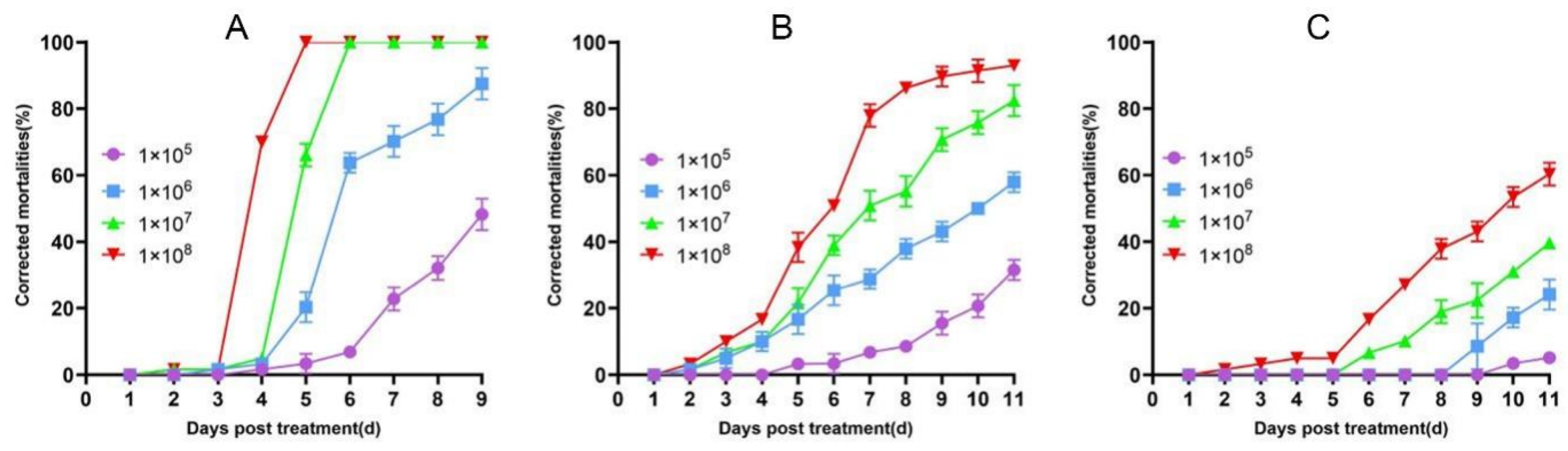
3.1. Virulence of the Entomopathogenic Fungi to the Striped Flea Beetle Adults

The mortality of the SFB showed a dose/time-dependent response to the EPF strains BbPs01, MrCb01, and IjH6102 (Figure 1). At concentrations ranging from 105 to 108 spores/mL, mortality rates increased rapidly from 4 to 6 days post-treatment (dpt). However, the strains exhibited distinct patterns of efficacy; particularly, BbPs01 had the quickest and the highest impact on the SFB, followed by MrCb01 and IjH6102.
LC-P regression analysis indicated that the equations were confident and the LC50 values could be evaluated (Table 1). At 6 dpt, the LC50 of BbPs01 against adults was 6.14 × 10⁶ spores/mL, while at 11 dpt, the LC50 values of MrCb01 and IjH6102 were 3.69 × 10⁶ and 76.46 × 10⁶ spores/mL, respectively. Similarly, the LT-P equations and LT50 values were evaluated (Table 2). The LT50 values were recorded as 8.93 d for BbPs01 at a concentration of 1 × 10⁵ spores/mL, 8.53 d for MrCb01 at 1 × 10⁶ spores/mL, while LT50 values could not be determined for IjH6102 because at the highest dose (1 × 10⁸ spores/mL) 50% mortality was not reached throughout the trial.

3.2. Morphology of the Infection Process of Entomopathogenic Fungi to Striped Flea Beetle

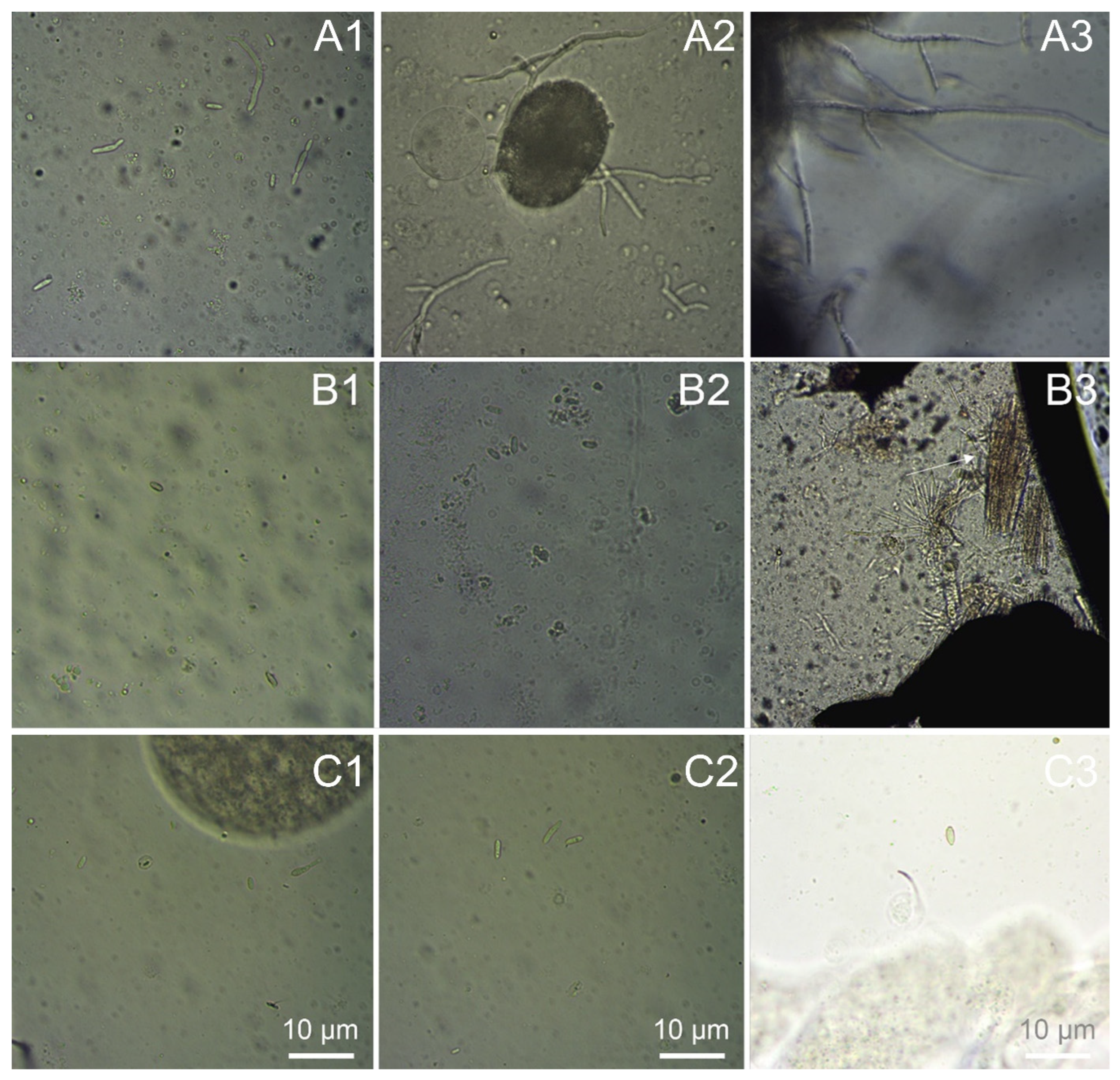
3.2.1. Attachment and Penetration of Entomopathogenic Fungi on the Striped Flea Beetle Adult’s Cuticle

In general, the EPF adhered to the SFB body surface at 6 h of post-treatment (hpt), germinated at 12–18 hpt, and penetrated at 12–30 hpt (Figure 2, Table 3). However, Bb demonstrated a significant temporal advantage in the pathogenic progression compared to Mr and Cj. Respectively, the conidia germination started at 6–12, 12–18, and 12–18 hpt for Bb, Mr, and Cj, while the penetration began at 12–18, 30, and 18–30 hpt for Bb, Mr and Cj (Figure 2, Table 3). Furthermore, mucus between the conidia and the body surface was observed at 12 hpt for Bb and Mr, but not for Cj. However, in the case of Bb and Cj, other than Mr, direct penetration with hyphae was observed, while the appressoria were detected in Bb and Mr, but not in Cj (Figure 2, Table 3).

3.2.2. Symptoms of Striped Flea Beetle Adults Infected by Entomopathogenic Fungi

The normal adults (control group) were highly active, moving and feeding freely, smoothly, and rapidly (Video S1, Table 3). In the early and middle stages after treatment with EPF, SFB adults generally exhibited no obvious external morphological abnormalities; the growth of fungal hyphae and conidia on their bodies could be observed 3–5 days post-death. However, numerous abnormal behaviors were observed, including increased self-grooming actions using their antennae and mouthparts, in terms of higher frequency and duration, reduced appetite, and slower movement. Bb-infected SFB displayed these abnormal behaviors at 3 dpt (Table 3, Video S1). Meanwhile, Mr-infected beetles not only showed the same behavior at 3–4 dpt but also exhibited more pronounced symptoms such as localized shaking, generalized convulsions, stiff legs, and physical imbalance (Table 3, Video S1). Similarly, Cj-infected SFB showed these symptoms at 4–5 dpt, along with mild body convulsions akin to those seen in Mr-infected SFB. Notably, unlike Bb-infected SFB, Mr- and Cj-infected SFB died with their abdomens facing upward (Table 3, Video S1).

3.2.3. Anatomical Hemocoel of Striped Flea Beetle Adults Infected by Entomopathogenic Fungi

Generally, Bb-infected adults exhibited typical behavioral changes and harbored a significant amount of EPF (both yeast-like blastospores and hyphae) within their hemocoel. The hyphae invaded the tissues, and by the time the adults died, all tissues and organs had been extensively colonized. The tissues had lost their toughness and were easily disrupted by the touch of a dissecting needle (Table 3, Figure 3A). In contrast, Mr-infected and Cj-infected adults had only a small number of blastospores in the hemocoel, with no detectable hyphae (Table 3, Figure 3B,C). In Mr-infected adults, the muscle fiber bundles appeared loose and free. However, Cj-infected adults had no abnormalities in the internal tissues.

3.2.4. Characteristics of Striped Flea Beetle Larvae Infected by Entomopathogenic Fungi

Overall, EPF-infected larvae exhibited melanized spots on their cuticle, but different melanization patterns were observed in the different EPF infections. Bb-infected larvae displayed a low rate of melanization, with small black spots and, in a minority of cases, a pinkish body color after death (Table 3, Figure 4A). Similarly, Mr-infected larvae had a low melanization rate, but the melanized spots were larger, and most failed to form conidia (Table 3, Figure 4B). In contrast, Cj-infected larvae more frequently exhibited “dendritic” melanized patterns, particularly at the inward extensions of the epidermis and spiracles (Table 3, Figure 4C). Notably, when the larvae were treated with the same EPF, those that died more rapidly had no or smaller melanized spots, whereas the larvae that died more slowly exhibited more pronounced melanized spots.

3.2.5. Anatomical Hemocoel of Striped Flea Beetle Larvae

Overall, following the emergence of typical infection characteristics, Bb-infected larvae exhibited the presence of hyphal bodies, hyphae, and melanized clumps in the hemocoel (Table 3, Figure 5A). In contrast, Mr-infected larvae harbored a small number of blastospores within their hemocoel. Partial melanization was observed in the epidermis, spiracles, and tracheae, but no hyphae or abnormal muscle bundles were detected (Table 3, Figure 5B). Cj-infected larvae displayed “dendritic-like melanization” in their tracheae, with hyphae penetrating through the tracheal wall into the body cavity, leading to the accumulation of hemocytes around the hyphae and the formation of nodules (Table 3, Figure 5C).
Dissection of freshly deceased larvae revealed distinct patterns of fungal colonization. In Bb-infected larvae, the hyphae had extensively colonized the tissues (Table 3, Figure 5A). For Mr-infected larvae, although only a few blastospores were initially present, their tissues became densely filled with hyphae 1–2 days post-mortem (Table 3, Figure 5B). In Cj-infected larvae, hyphae penetrated the tracheae, causing hemocyte aggregation and blackening, with a small number of free blastospores remaining in the hemocoel (Table 3, Figure 5C).

4. Discussion

The SFB is a key pest seriously damaging cruciferous vegetable production in the world, which requires the urgent development of a sustainable control technology [2]. Despite the extensive research on EPF such as B. bassiana, M. robertsii, and C. javanica for pest control, no mycoinsecticides targeting the SFB have been commercially registered in the world to date [8]. This gap stems primarily from inconsistent field efficacy [18]. The lower efficacy of EPF to control the SFB is the main reason for limiting the application. Different EPF strains exhibit diverse bioactivities against the SFB, which is supported by the results of this research. Among the three selected strains, BbPs01 shows higher efficacy than MrCb01 and IjH6102, which suggests that BbPs01 might have better potential for field application.
In this study, we compared the interaction performance between three EPF—B. bassiana, M. robertsii, and C. javanica—and the striped flea beetle, which yielded novel insights, materials, and methodologies, as well as several intriguing data. In fact, this study introduces a new model for examining the interactions between EPF and insects. The SFB, a coleopteran insect with a highly complex life history, spends its egg–larval–pupal stages in the soil, while adults live above ground and are exposed to a more complex environment throughout their lives, particularly due to the vast array of microorganisms in the soil [19]. Consequently, their adaptation and response to fungal pathogens are distinct from those of insects that live entirely above ground. Additionally, the SFB is an economically significant pest that has garnered considerable attention from researchers. The genome of the SFB has been sequenced and published, and its artificial rearing method is relatively straightforward, ensuring a stable supply of insects for experimentation [20].
Each of the three EPF used in this study has unique characteristics. Beauveria bassiana is one of the earliest discovered and most widely applied EPF in pest control. Its “resource siege” mechanism makes it particularly effective in coordinated IPM programs [8]. As a cornerstone of fungal biocontrol since its first documented use in 1835, it operates through a nutrient depletion strategy. This cosmopolitan pathogen secretes chitinolytic enzymes (CHI1, CHI2) and lipases (LipA/B) to degrade cuticular polymers, systematically exhausting host nutrient reserves while suppressing immune responses via metalloproteases (Pr1) [21,22]. This investigation yields substantial empirical evidence corroborating the aforementioned theoretical framework. Specifically, Bb demonstrates accelerated germination and penetration rates, coupled with extensive hyphal proliferation within the SFB’s hemocoel. These observations collectively indicate that Bb strategically appropriates host nutritional resources to sustain its rapid vegetative growth and pathogenic development. In contrast, the generalist Metarhizium species, such as M. anisopliae and M. robertsii, primarily employ a toxin strategy by producing non-ribosomal peptide toxins (e.g., destruxins) [23] to outcompete the host insects [24]. The experimental data corroborate the toxin-mediated pathogenic strategy hypothesis. Notably, Mr exhibits delayed hyphal proliferation within the SFB, with host mortality occurring prior to fungal myceliation—a phenotypic signature consistent with toxin-driven pathogenesis rather than nutrient-exhaustion mortality.
Cordyceps javanica, although commonly used for pest control in China (e.g., against whiteflies and Spodoptera litura), has fewer studies on its mechanism of action [25]. Interestingly, this study has just discovered some interesting new results. Firstly, in the process of infecting SFB adults, although Cj did not produce so many spores attached to the cuticle as Bb and Mr did, the spore germination time is as early as Bb (germination began at 12 h) and earlier than that of Mr. Cj directly invades through the germ-tubes, rather than Bb and Mr forming appressoria. In addition, Cj infected-adults appear to have stiff and weak legs, imbalance, and slight body convulsions, similar to the symptoms caused by Mr, which recall neurotoxic symptoms [26]. Secondly, Cj-infected larvae often invade from the spiracles and form “dendritic” melanization. In addition, Cj only produces a small number of single-celled yeast-like blastospores in the SFB hemocoel, which is similar to Mr. Obviously, compared with Bb and Mr, Cj has a different mode of interaction with the SFB. On the one hand, Cj has relatively weak pathogenicity against the SFB, which is manifested by fewer spores attaching to the host, no appressorium formation, and no hyphae developing in the hemocoel. On the other hand, when infected with Cj, the SFB exhibits mild shivering symptoms, which seems to suggest that it employs a toxin-based strategy to attack insects, similar to Mr. Moreover, it has been reported that Cj produces an alkaloid, heteratisine, which has high insecticidal and acetylcholinesterase (AChE) inhibitory activities [27]. Collectively, these findings postulate that Cj may employ a toxin-mediated pathogenic strategy analogous to Mr during host insect colonization. This provisional hypothesis, however, mandates systematic validation through molecular characterization of the candidate fungal effector proteins and cellular-level interrogation of the toxin biosynthesis pathways to elucidate the real mechanisms of action of these entomopathogens.

5. Conclusions

In conclusion, this research provides extensive data on the three distinct interaction modes between EPF and the SFB. Our findings offer new insights into the EPF–insect interaction and will enhance the application of mycoinsecticides for SFB control.

Supplementary Materials

The following supporting information can be downloaded at: https://www.mdpi.com/article/10.3390/agriculture15111188/s1, Video S1: SFB infected by EPF.mp4.

Author Contributions

Conceptualization, Q.W. and Q.H.; methodology, X.P., X.H. and V.D.; software, X.P., K.Z. and X.H.; validation, K.Z. and V.D.; resources, Q.W., A.B. and Q.H.; writing—original draft preparation, X.P., X.H. and Q.H.; writing—review and editing, K.Z., A.B. and Q.W.; supervision, project administration and funding acquisition, Q.H. and Q.W. All authors have read and agreed to the published version of the manuscript.

Funding

This research was funded by the National Natural Science Foundation of China (grant number 32361133546) and the Russian Science Foundation (grant number 24-46-00005).

Data Availability Statement

All relevant data are within the manuscript.

Conflicts of Interest

The authors declare no conflicts of interest.

References

  1. Rousseau, M.; LeSage, L. Earliest North American occurrence of Phyllotreta striolata (Coleoptera: Chrysomelidae) from Québec, Canada. Can. Entomol. 2016, 148, 476–478. [Google Scholar] [CrossRef]
  2. Li, Z.Y.; Costamagna, A.C.; Beran, F.; You, M.S. Biology, ecology, and management of flea beetles in Brassica crops. Annu. Rev. Entomol. 2024, 69, 199–217. [Google Scholar] [CrossRef] [PubMed]
  3. Mittapelly, P.; Guelly, K.N.; Hussain, A.; Cárcamo, H.A.; Soroka, J.J.; Vankosky, M.A.; Hegedus, D.D.; Tansey, J.A.; Costamagna, A.C.; Gavloski, J.; et al. Flea beetle (Phyllotreta spp.) management in spring-planted canola (Brassica napus L.) on the northern Great Plains of North America. Glob. Change Biol. Bioenergy 2024, 16, e13178. [Google Scholar] [CrossRef]
  4. Raghavendra, K.V.; Rekha, B.; Ramesh, K.B.; Felix, K.T.; Chander, S. Integrated pest management strategy for striped flea beetle, Phyllotreta striolata infesting radish (Raphanus sativus). Indian J. Agric. Sci. 2023, 93, 1308–1313. [Google Scholar] [CrossRef]
  5. Kong, L.; Xu, J.; Shen, W.; Zhang, S.; Xu, Z.; Zhu, K.Y. Development and evaluation of RNA microsphere-based RNAi approaches for managing the striped flea beetle (Phyllotreta striolata), a globally destructive pest of Cruciferae crops. Pest. Manag. Sci. 2025, 81, 1529–1538. [Google Scholar] [CrossRef]
  6. Gielen, R.; Ude, K.; Kaasik, A.; Poldmaa, K.; Teder, T.; Tammaru, T. Entomopathogenic fungi as mortality agents in insect populations: A review. Ecol. Evol. 2024, 14, e70666. [Google Scholar] [CrossRef]
  7. Panwar, N.; Szczepaniec, A. Endophytic entomopathogenic fungi as biological control agents of insect pests. Pest. Manag. Sci 2024, 80, 6033–6040. [Google Scholar] [CrossRef]
  8. Jiang, Y.L.; Wang, J.J. The registration situation and use of mycopesticides in the world. J. Fungi 2023, 9, 940. [Google Scholar] [CrossRef]
  9. Rajput, M.; Sajid, M.S.; Rajput, N.A.; George, D.R.; Usman, M.; Zeeshan, M.; Iqbal, O.; Bhutto, B.; Atiq, M.; Rizwan, H.M.; et al. Entomopathogenic fungi as alternatives to chemical acaricides: Challenges, opportunities and prospects for sustainable tick control. Insects 2024, 15, 1017. [Google Scholar] [CrossRef]
  10. Wang, B.; Kang, Q.J.; Lu, Y.Z.; Bai, L.Q.; Wang, C.S. Unveiling the biosynthetic puzzle of destruxins in Metarhizium species. Proc. Natl. Acad. Sci. USA 2016, 113, E4756. [Google Scholar] [CrossRef]
  11. Ying, S.H. Subcellular biochemistry and biology of filamentous entomopathogenic fungi. Adv. Appl. Microbiol. 2024, 129, 35–58. [Google Scholar] [CrossRef] [PubMed]
  12. Chen, W.; Yuan, W.; Guan, X.; Hu, Q. Biodiversity of Isaria in soil and its activity against Phyllotreta striolata. J. South China Agric. Univ. 2021, 42, 75–82. [Google Scholar] [CrossRef]
  13. Chen, W.; Yuan, W.J.; He, R.K.; Pu, X.H.; Hu, Q.B.; Weng, Q.F. Screening of fungal strains and formulations of Metarhizium anisopliae to control Phyllotreta striolata in Chinese flowering cabbage. Insects 2023, 14, 567. [Google Scholar] [CrossRef]
  14. Nagalingam, T.; Costamagna, A.C. Two methods for rearing the striped flea beetle Phyllotreta striolata (Coleoptera: Chrysomelidae) under laboratory conditions. Can. Entomol. 2019, 151, 677–683. [Google Scholar] [CrossRef]
  15. Yao, L.; Pu, X.; Wu, Y.; Zhang, K.; Berestetskiy, A.; Hu, Q.; Weng, Q. An optimized bioassay system for the striped flea beetle, Phyllotreta striolata. Insects 2025, 16, 510. [Google Scholar] [CrossRef]
  16. Tang, Q.Y.; Zhang, C.X. Data processing system (DPS) software with experimental design, statistical analysis and data mining developed for use in entomological research. Insect Sci. 2013, 20, 254–260. [Google Scholar] [CrossRef]
  17. Abrar, A.; Abbas, M.; Mehmood, S.; Ghani, N.; Fatima, A.; Shahzadi, R. Scanning electron microscopy for identification of local strain of Aspergillus parasiticus and its larvicidal efficacy against Aedes aegypti and non-target toxicity testing on fingerlings of Hypophthalmichthys molitrix. Microsc. Res. Tech. 2022, 85, 3187–3192. [Google Scholar] [CrossRef]
  18. Price, C.S.V.; Campbell, H.; Pope, T.W. Assessing the potential of biopesticides to control the cabbage stem flea beetle Psylliodes chrysocephala. Pest. Manag. Sci. 2024, 80, 2471–2479. [Google Scholar] [CrossRef]
  19. Reddy, G.V.; Tangtrakulwanich, K.; Miller, J.H.; Ophus, V.L.; Prewett, J. Sustainable management tactics for control of Phyllotreta cruciferae (Coleoptera: Chrysomelidae) on Canola in Montana. J. Econ. Entomol. 2014, 107, 661–666. [Google Scholar] [CrossRef]
  20. King, R.; Buer, B.; Davies, T.G.E.; Ganko, E.; Guest, M.; Hassani-Pak, K.; Hughes, D.; Raming, K.; Rawlings, C.; Williamson, M.; et al. The complete genome assemblies of 19 insect pests of worldwide importance to agriculture. Pestic. Biochem. Physiol. 2023, 191, 105339. [Google Scholar] [CrossRef]
  21. Rohlfs, M.; Churchill, A.C.L. Fungal secondary metabolites as modulators of interactions with insects and other arthropods. Fungal Genet. Biol. 2011, 48, 23–34. [Google Scholar] [CrossRef]
  22. Tong, S.; Li, M.L.; Keyhani, N.O.; Liu, Y.; Yuan, M.; Lin, D.M.; Jin, D.; Li, X.B.; Pei, Y.; Fan, Y.H. Characterization of a fungal competition factor: Production of a conidial cell-wall associated antifungal peptide. PLoS Pathog. 2020, 16, e1008518. [Google Scholar] [CrossRef] [PubMed]
  23. Wang, J.; Hu, H.; Pang, s.; Yin, X.; Cao, B.; Huang, J.; Xu, X.; Weng, Q.; Hu, Q. Destruxin a inhibits the hemocytin-mediated hemolymph immunity of host insects to facilitate Metarhizium infection infection. Cell Rep. 2024, 43, 113686. [Google Scholar] [CrossRef] [PubMed]
  24. Shang, J.M.; Song, S.X.; Wang, C.S. Metarhizium robertsi. Trends Parasitol. 2024, 4, 192–193. [Google Scholar] [CrossRef]
  25. Wang, D.; Xing, P.X.; Diao, H.L.; Zhou, W.W.; Li, X.W.; Zhang, L.J.; Ma, R.Y. Pathogenicity characteristics of the entomopathogenic fungus Cordyceps javanica ij-tg19 to Acyrthosiphon pisum. Biocontrol 2023, 68, 447–458. [Google Scholar] [CrossRef]
  26. Yin, F.; Hu, L.; Li, Z.; Yang, X.; Kendra, P.E.; Hu, Q. Effects of destruxin a on hemocytes of the domestic silkworm, Bombyx mori. Front. Microbiol. 2023, 14, 1210647. [Google Scholar] [CrossRef]
  27. Xing, P.; Mao, R.; Zhang, G.; Li, Y.; Zhou, W.; Diao, H.; Ma, R. Secondary metabolites in Cordyceps javanica with insecticidal potential. Pestic. Biochem. Physiol. 2024, 204, 106076. [Google Scholar] [CrossRef]
Figure 1. Mortality of P. striolata adults treated with B. bassiana BbPs01 (A), M. robertsii MrCb01 (B), and C. javanica IjH6102 (C). Abbott’s formula was used to perform natural correction for the cumulative mortality rate in the control group.
Agriculture 15 01188 g001
Figure 2. SEM profiles of EPF-treated SFB adults. (A), Treatment with BbPs01: (A1), conidia adhered to the ventral protrusions of prothorax (6 hpt); (A2), conidia attached to the elytra bristle fossa (6 hpt); (A3/4), conidia began to germinate, forming a germ-tube (Gt) (12/18 hpt), with mucus (red arrow) between the conidia and the epidermis; Gt directly penetrated the epidermis (white arrow); and (A5), the germinated conidia differentiated to form appressoria (Ap) (30 hpt) (black arrows). (B), Treatment with MrCb01: (B1), conidia on the anterior thoracic sternum (6 hpt); (B2), conidia in tarsus (6 hpt); (B3), mucus between the conidia and the epidermis (red arrow) (12 hpt); (B4), conidia germination to form Gt (18 hpt); and (B5), germinated conidia differentiate Ap (black arrow) (30 hpt). (C), Treatment with IjH6102: (C1/2), conidia adhered to the bristle fossa of elytra or the sternum of the anterior thorax (6 hpt); (C3), conidia forming Gt (12 hpt); and (C4/5), Gt directly penetrate (white arrow) and degrade the insect epidermis (black arrow) (30/48 hpt).
Agriculture 15 01188 g002
Figure 3. Anatomical changes in the hemocoel of EPF-infected SFB adults. (A), Adults treated with BbPs01: (A1), dpt blastospores and hyphal bodies in hemolymph at 3 dpt; (A2), at 4 dpt, mycelia invaded tissues; and (A3), at 5 dpt, mycelia invaded the body of the just-deceased insect. (B), Adults treated with MrCb01: (B1, at 4 dpt, blastospores in hemolymph; (B2), at 5 dpt, blastospores in the hemocoel of the just-deceased insect; and (B3), at 6 dpt, there were loose and free bundles of muscle fibers (white arrows). (C), Adults treated with IjH6102: (C1), at 5 dpt, blastospores in the hemolymph; (C2), at 6 dpt, blastospores in the just-deceased insect; and (C3), at 8 dpt, the SFB hemocoel.
Agriculture 15 01188 g003
Figure 4. Symptoms of EPF-infected SFB larvae. (A), Larvae treated with BbPs01: (A1), at 2 dpt, melanized spots on the cuticle; (A2), at 5 dpt, dead larvae; (A3), at 7 dpt, hyphae grew on the surface of the dead insects; and (A4), at 8 dpt, conidia formed. (B), Larvae treated with MrCb01: (B1), at 4 dpt, melanized live larvae; (B2), at 6 dpt, newly dead larvae; and (B3/4), at 8/10 dpt, dead larvae covered by hyphae/conidia. (C), Larvae treated with IjH6102: (C1), at 5 dpt, the larvae with “dendritic” melanization; (C2), at 7 dpt, the pre-pupa with “dendritic” melanization; (C3), at 9 dpt, the just-deceased larvae; and (C4), at 10 dpt, hyphae formed on the body surface.
Agriculture 15 01188 g004
Figure 5. Anatomical observation of the EPF-infected SFB larvae. (A), Larvae treated with BbPs01: (A1/2), at 3–4 dpt, blastospores in hemolymph; (A3), at 6 dpt, melanization and mycelia in hemocoel; and (A4), at 8 dpt, melanization and mycelia in hemocoel of just-deceased larvae. (B), Larvae treated with MrCb01: (B1), at 4 dpt, blastospores in the hemolymph; (B2/3), at 5–6 dpt, melanization in the cuticle and tracheae; and (B4), at 8 dpt, the larvae just died, and the hyphae invaded the internal tissues. (C), Larvae treated with IjH6102: (C1), at 3 dpt, a small number of blastospores in the hemolymph; (C2), at 5 dpt, the hyphae penetrated the tracheae with melanization; (C3), at 6 dpt, the tracheae show “dendritic” melanization; and (C4), at 9 dpt, the larvae just died, the tracheae were melanized, the hyphae penetrated the tracheae, and the hemocytes were encapsulated (black arrow).
Agriculture 15 01188 g005
Table 1. LC-P equation and LC50 of EPF against SFB adults.
EPFLC-P Equation (y = A + Bx) and Significant TestLC50 (95% Confidence Interval, ×106 Spores/mL)
Intercept (A)Slope (B)SERχ2DFp
BbPs015 dpt−4.34381.4160.16820.94443.307520.191339.68(27.07–59.69)
6 dpt−7.30982.12670.28710.98431.290920.52446.14(3.64–8.83)
MrCb016 dpt0.10770.68830.13270.94725.140720.0765128.115 (9.81–529.55)
10 dpt−0.21440.88780.11800.95535.402120.06717.47(4.04–12.18)
11 dpt0.44700.81790.11800.96412.998520.22333.691 (0.54–6.72)
IjH610210 dpt0.37740.64220.12240.93536.143020.0464157.61(70.01–719.66)
11 dpt0.44990.66100.11580.94096.112720.047176.46(39.65–217.80)
The regression parameters of LC50 were calculated through Probit analysis using DPS. LC50 values were calculated based on infection-induced mortality data.
Table 2. LT-P equation and LT50 of EPF against SFB adults.
EPF Concentration (spores/mL)LT-P Equation (y = A + Bx) and Significant TestLT50 (95% Confidence Interval, ×106 Spores/mL)
Intercept (A)Slope (B)SERχ2DFp
BbPs011 × 105−1.24466.56690.90460.99340.823840.93528.93(8.16–10.34)
MrCb011 × 1061.77133.46760.76060.99270.311240.98918.53(7.78–9.46)
1 × 1071.11814.61830.78900.99050.742340.94606.93(6.12–7.48)
IjH61021 × 108///////>11 d
The regression parameters of LT50 were calculated through Probit analysis using DPS. LT50 values were calculated based on infection-induced mortality data.
Table 3. The interaction features of EPF and the SFB in the infection progress.
Observed ItemBbPs01MrCb01IjH6102
AdultsEPF adhesionGermination of conidia12 hpt18 hpt12 hpt
Mucus secretion12 hpt12 hpt12 hpt
Penetration of germ-tube12–18 hpt/18 hpt
Appressoria formation24 hpt24 hpt/
SymptomsSelf-groomingIncreaseIncreaseIncrease
Appetite decrease3 dpt3–4 dpt4–5 dpt
Typical symptomsInactivity, slow movement with a balanced styleTremors of antennae and feet, generalized convulsions, inactivity, and body imbalance with leg stiffnessLameness and leg weakness, imbalance, slight body convulsions
HemocoelEarly stageIn 1–3 dpt, massive hyphal bodiesIn 2–4 dpt, a few blastospores; no hyphal bodiesIn 2–4 dpt, a few blastospores; no hyphal bodies
Mid-stageIn 2–4 dpt, mycelia invade tissues and organsIn 3–5 dpt, no myceliaIn 4–6 dpt, no mycelia
Late stageIn 4 dpt, massive mycelia, tissues, and organs are easy to break and deconstructIn 5- dpt, a few blastospores, muscles loose, and dissociationIn 6- dpt, a few blastospores but no other abnormalities
LarvaeSymptomsTypicalPink bodyMelanization in cuticle and spiracle“dendritic” melanization
Melanization~10% insects melanized; small melanized spots on the cuticle and spiracle; the melanized area is small and unchanged~30% larvae melanized; ~20% larvae melanized on spiracle; and the melanized area obviously increases gradually~15% larvae with “dendritic” melanization; the melanized area obviously increases till 2–3 days before death
Movement before deathSlowly moving since an early stageSlowly moving, no other abnormal movementSlowly moving; no other abnormal movement
HemocoelEarly stageAt 1–3 dpt, massive hyphal bodies and no melanizationAt 1–4 dpt, a few blastospores and no melanizationAt 1–5 dpt, mycelia penetrating tracheae and forming a melanized capsule
Mid-stageAt 4–6 dpt, massive mycelia and melanized clumpsAt 4–7 dpt, melanization occurred but no myceliaAt 5–9 dpt, “dendritic” melanization in tracheae; no mycelia
Late stageAt 6 dpt, mycelia invade tissues and organs and grow on the body surfaceAt 7 dpt, a few blastospores, but no mycelia in the hemocoelAt 9 dpt, mycelia penetrating tracheae
Disclaimer/Publisher’s Note: The statements, opinions and data contained in all publications are solely those of the individual author(s) and contributor(s) and not of MDPI and/or the editor(s). MDPI and/or the editor(s) disclaim responsibility for any injury to people or property resulting from any ideas, methods, instructions or products referred to in the content.

