Article

Microbial Population in Curcuma Species at Different Growth Stages

by
Neptu Islamy Raharja
1,
Mohammad Amzad Hossain
1,2,* and
Hikaru Akamine
1,2
1
The United Graduate School of Agricultural Sciences, Kagoshima University, Kagoshima 890-0065, Japan
2
Faculty of Agriculture, University of The Ryukyus, Nishihara, Okinawa 903-0213, Japan
*
Author to whom correspondence should be addressed.
Agriculture 2025, 15(10), 1092; https://doi.org/10.3390/agriculture15101092
Submission received: 3 April 2025 / Revised: 2 May 2025 / Accepted: 15 May 2025 / Published: 19 May 2025
(This article belongs to the Special Issue Beneficial Microbes for Sustainable Crop Production)

Abstract

:
Turmeric (Curcuma spp.) is widely cultivated in tropical regions for its use in traditional medicine and culinary purposes. This study investigated the bacterial populations in the rhizosphere, stems, and leaves of the Curcuma species and strains at different growth stages. Bacterial population cultivated in the field and plastic house showed variations across growth stages. The rhizosphere possessed the highest bacterial populations in both experiments (1.8 to 11.9 × 10⁶ CFU/g and 1.7 to 24.3 × 10⁶ CFU/g, respectively), with C. amada and Ryudai gold as the highest. Endophytic bacteria in stems and leaves also peaked at the middle growth stage. Principal Component Analysis (PCA) revealed distinct separations among Curcuma species planted in the field and plastic house at different growth stages. C. aromatica and C. longa strain L2 clustered differently under field conditions, while C. zedoaria and C. xanthorrhiza were distinct under plastic house conditions. Combined PCA revealed a clear separation between the field and plastic house, with tighter clustering observed in the plastic house. Leaf-associated bacterial populations were compositionally distinct from those in the rhizosphere and stems. These findings suggest that the Curcuma growth stage and species significantly affect bacterial community structure, supporting the development of targeted cultivation strategies and microbial applications to enhance productivity and sustainability in turmeric farming.

1. Introduction

Curcuma, a diverse genus of Zingiberaceae, encompasses several species of significant economic, medicinal, and agricultural importance [1]. Among them, Curcuma longa L., commonly known as turmeric, is highly valued for its culinary applications and bioactive compounds, particularly curcumin [2]. This polyphenolic compound is renowned for its antioxidant, anti-inflammatory, and antimicrobial properties, making turmeric a vital component in traditional medicine, functional foods, and pharmaceutical research [2,3]. The widespread cultivation of Curcuma species, particularly in tropical and subtropical regions, underscores the need for a deeper understanding of the biological and ecological factors influencing their growth and productivity. One of the most critical yet often overlooked factors in plant development is the role of microbial communities associated with plant roots, leaves, and internal tissues [4]. These microorganisms contribute to plant health, nutrient cycling, disease resistance, and overall productivity [4,5]. In Curcuma species, the microbial populations present in the rhizosphere, phyllosphere, and endosphere undergo significant shifts across different growth stages, impacting plant physiology and resilience [6]. However, despite their importance, the dynamic interactions between Curcuma plants and their associated microbes throughout the growth cycle remain largely unexplored.
The rhizosphere, a biologically active soil zone around roots, is a key site of plant–microbe interaction [7]. Root exudates, including sugars, amino acids, and secondary metabolites, attract beneficial microbes and deter pathogens. During early growth, these microbes enhance seedling vigor, nutrient uptake, and disease suppression, laying the foundation for healthy root development and improved plant performance [6,7,8].
As Curcuma plants progress into the vegetative growth stage, characterized by rapid foliage expansion and root elongation, microbial populations in the rhizosphere shift in response to changing root exudation patterns and nutrient demands [6]. Beneficial microorganisms such as plant growth-promoting rhizobacteria (PGPR) and mycorrhizal fungi contribute to enhanced nutrient availability, improved soil structure, and increased resistance to environmental stressors [9]. These microbial communities assist in nitrogen fixation, phosphate solubilization, and the production of plant hormones such as auxins and cytokinins, all of which are essential for robust vegetative growth [9,10]. Beyond the rhizosphere, the endosphere represents a specialized niche within plant tissues where endophytic microorganisms reside. These microbes establish symbiotic relationships with their host plants, contributing to nutrient uptake, hormone regulation, and stress tolerance [7,11]. Previous studies have reported that endophytic bacteria in Curcuma species may enhance curcumin biosynthesis, exhibit antimicrobial activity, and serve as biocontrol agents against phytopathogens [12]. Exploring the functional roles of endophytic microbes at different growth stages offers valuable insights into plant–microbe interactions and their potential for sustainable agriculture. The phyllosphere microbiota, though often overlooked, also supports plant health by suppressing pathogens through competitive exclusion and antimicrobial production [13]. Environmental factors, such as humidity, temperature, and UV exposure, shape these microbial communities, influencing plant development throughout the Curcuma growth cycle [6,14]. Additionally, cultivation conditions (e.g., field vs. plastic house) can significantly alter microbial diversity and abundance [15,16], highlighting the importance of environmental context in optimizing Curcuma cultivation for better health and productivity.
Previous studies have predominantly focused on a single Curcuma species to analyze microbial diversity or identify specific functional microbial groups. It is believed that bacterial populations and their functions vary depending on plant species, plant growth stages, plant parts, and cultivation places. In response, this study introduces a novel approach by quantifying and comparing total bacterial populations across different growth stages and plant tissues (rhizosphere, stem, and leaves) in multiple Curcuma species cultivated in the field and plastic house. By elucidating the temporal shifts in microbial communities from germination to maturity and comparing microbial dynamics between plant species, this research seeks to advance our understanding of plant–microbe interactions. Understanding these microbial dynamics will provide valuable insights into optimizing microbial populations and functions as biofertilizer and stimulant, improving yields, and promoting environmentally sustainable farming systems for species. These findings also offer foundational information that may support future microbiome research related to plant cultivation strategies. To the best of our knowledge, this study is one of the first to compare microbial load dynamics across different growth stages in multiple Curcuma species cultivated in both a field and plastic house.

2. Materials and Methods

2.1. Curcuma Species or Strains

This study used five turmeric species for plastic house experiments: Curcuma longa (cultivar Ryudai gold and five strains), C. zedoaria (ZE), C. xanthorrhiza (ZA), C. aromatica (AR), and C. amada (AM). The cultivar Ryudai gold (RG) and five strains belong to the Curcuma longa species; the five strains are known as C. longa strain 1 (L1), C. longa strain 2 (L2), C. longa strain 3 (L3), C. longa strain 4 (L4), and C. longa strain 5. Four turmeric species were used in the field experiment: Curcuma longa (cultivar Ryudai gold, C. longa strain 1 (L1), C. longa strain 2 (L2), C. longa strain 3 (L3), C. zedoaria (ZE), C. xanthorrhiza (ZA), and C. aromatica (AR). The various species, cultivars, and strains of turmeric differ in rhizome dimensions, shapes, and colors. They also possess distinct chemical properties, flavors, tastes, and physiological as well as morphological traits of the shoot (data not yet published). Given their yield performance, these turmeric varieties have the potential for commercial production.

2.2. Curcuma Cultivation

The pot experiment was conducted in a plastic house from 2 May 2022 to 6 February 2023. Air-dried dark-red soil of 3.5 kg and cultured soil (commercial name: Hanasakimonogatari) of 2.5 kg were mixed properly and placed in each Wagner pot (0.05 m2). As the rhizome sizes were different from the Curcuma species and strains, the best seed-rhizomes were selected for each species and strains. One seed-rhizome per pot was planted at the depth of 6 cm [17]. The pots were placed in the house randomly. The outdoor environment was maintained in the house by keeping the windows opened, but the windows were closed during typhoons. Water was applied regularly as required to maintain the optimum soil moisture level for proper seedling emergence and plant growth.
The field experiment was conducted from May 2022 to February 2023 at the Subtropical Field Science Center of the University of the Ryukyus Okinawa. Th experiment was conducted on fields of dark red soil (Shimajiri Maji, Chromic Luvisol) [18].

2.3. Sample Collection

We collected both the plant (leaf and stem) and rhizosphere samples three times at the early growth stage (3- to 4-leaf stage, July), the middle growth stage (7- to 9-leaf stage, October), and the late growth stage (plant maturing stage, December) when the plants were still green. The plants were cut at the soil surface, and the leaves were separated from the stems. The rhizosphere soil was collected from each plant, and a composite soil sample was prepared for each Curcuma species or strains.

2.4. Isolation and Enumeration of Endophytic Bacteria

Plant samples (stems and leaves) were cleaned by washing in running water and cut into pieces. The surface of the sample pieces was washed and sterilized to free from microbes as follows. The leaves or stems were washed with sterile distilled water and then with 70% ethanol solution for one minute. The leaves or stems were then washed with 3% sodium hypochlorite solution (Nacalai Tesque, Kyoto, Japan) for 1.5 min for leaves and 3 min for stems. Finally, they were rinsed with sterile distilled water three times [19]. The samples were then dried using sterile tissue paper. Surface sterilization of the samples were performed by spreading 0.1 mL of distilled water on Nutrient Agar media (Difco; BD, Franklin Lakes, NJ, USA), then the petri dishes were incubated (Sanyo MIR-152; Sanyo, Osaka, Japan) at 28 °C for 14 days, and it was ensured that no colonies appeared. Surface sterilized leaves or stems were crushed using a sterile mortar and pestle. The crushed sample was put into a test tube containing 9 mL of sterile physiological NaCl solution (0.85%) then serially diluted, and 0.1 mL of each dilution was plated on a Nutrient Agar media that had been added with 50 mg/L nystatin (Nacalai Tesque, Kyoto, Japan) to inhibit the growth of the fungus [19]. The diluted samples were then plated onto petri dishes and incubated at 28 °C for 14 days. Colonies that appeared on petri dishes were counted every day for 14 days. All analyses were conducted in triplicate.

2.5. Isolation and Enumeration of Rhizosphere Heterotrophic Bacteria

Rhizosphere soil samples were taken by removing the Curcuma plant from the soil and carefully shaking the rhizome to remove loose and non-adherent soil. The soil that was still attached to the roots and rhizome was then collected using a sterile spatula, and the rhizosphere soil obtained was then put into a sterile plastic bag. Each soil composite sample of 10 g was taken and then put into 90 mL of physiological NaCl solution (0.85%). It was homogenized using an orbital shaker for 30 min at a speed of 150 rpm, followed by serial dilutions. A total of 0.1 mL of soil suspension from each dilution was spread on Nutrient Agar media. The diluted samples were then plated onto petri dishes and incubated at 28 °C for 7 days using a Sanyo MIR-152 incubator. Bacterial colonies that appeared were counted every day. All analyses were conducted in triplicate.

2.6. Data Analysis

The data of this study were analyzed qualitatively and quantitatively. The data were expressed as mean ± standard deviation (SD). Statistical analysis was conducted using the one-way Analysis of Variance (ANOVA) then followed up with the Tukey’s test using the IBM SPSS (Statistical Program Software System) program version 26.0 (IBM Corp., Armonk, NY, USA). Significant differences were those in which p < 0.05. Principal component analysis (PCA) was constructed using SIMCA Version 17 (Sartorius, Gottingen, Germany).

3. Results

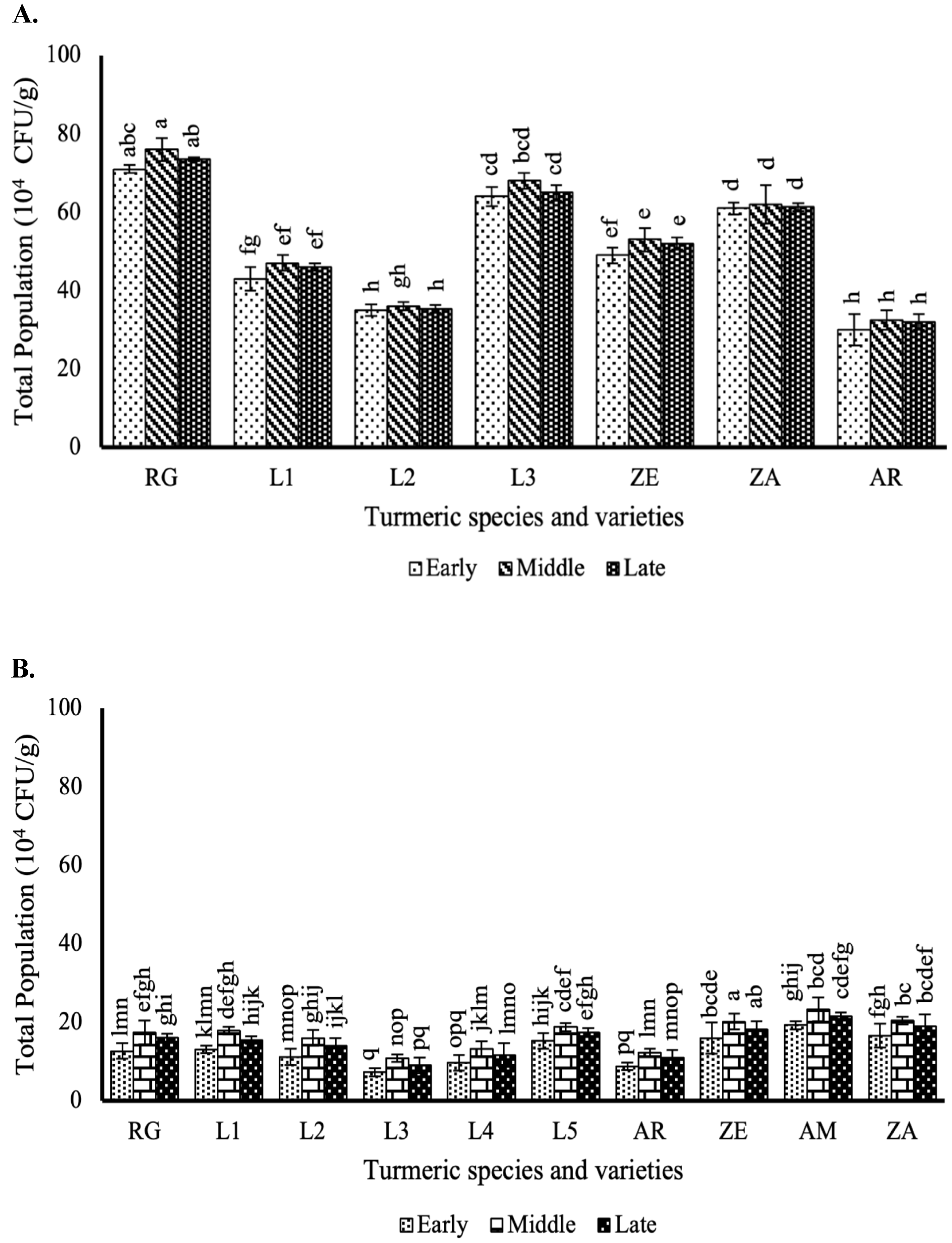
3.1. Effect of Different Curcuma Growth Stages on Rhizosphere Heterotrophic Bacteria Population

The bacterial population in the rhizosphere soil of Curcuma species or strains varied across growth stages in both experiments (Figure 1). In field-grown samples, the bacterial counts ranged from 1.8 to 9.1 × 10⁶ CFU/g during the early stage, 2.0 to 11.9 × 10⁶ CFU/g in the middle stage, and 1.9 to 10.7 × 10⁶ CFU/g at the late stage. In contrast, rhizosphere soil from Curcuma cultivated in a plastic house exhibited higher bacterial populations, ranging from 1.7 to 21.8 × 10⁶ CFU/g in the early stage, 1.8 to 24.3 × 10⁶ CFU/g in the middle stage, and 1.7 to 23.1 × 10⁶ CFU/g in the late stage.
Significant differences were observed in the bacterial populations depending on the Curcuma species or strain and their growth stage. The highest bacterial population was recorded in the rhizosphere soil of the RG turmeric strain grown in the field during the middle growth stage (11.9 × 10⁶ CFU/g) (p < 0.05), followed by C. amada cultivated in the plastic house at the same stage (24.3 × 10⁶ CFU/g).

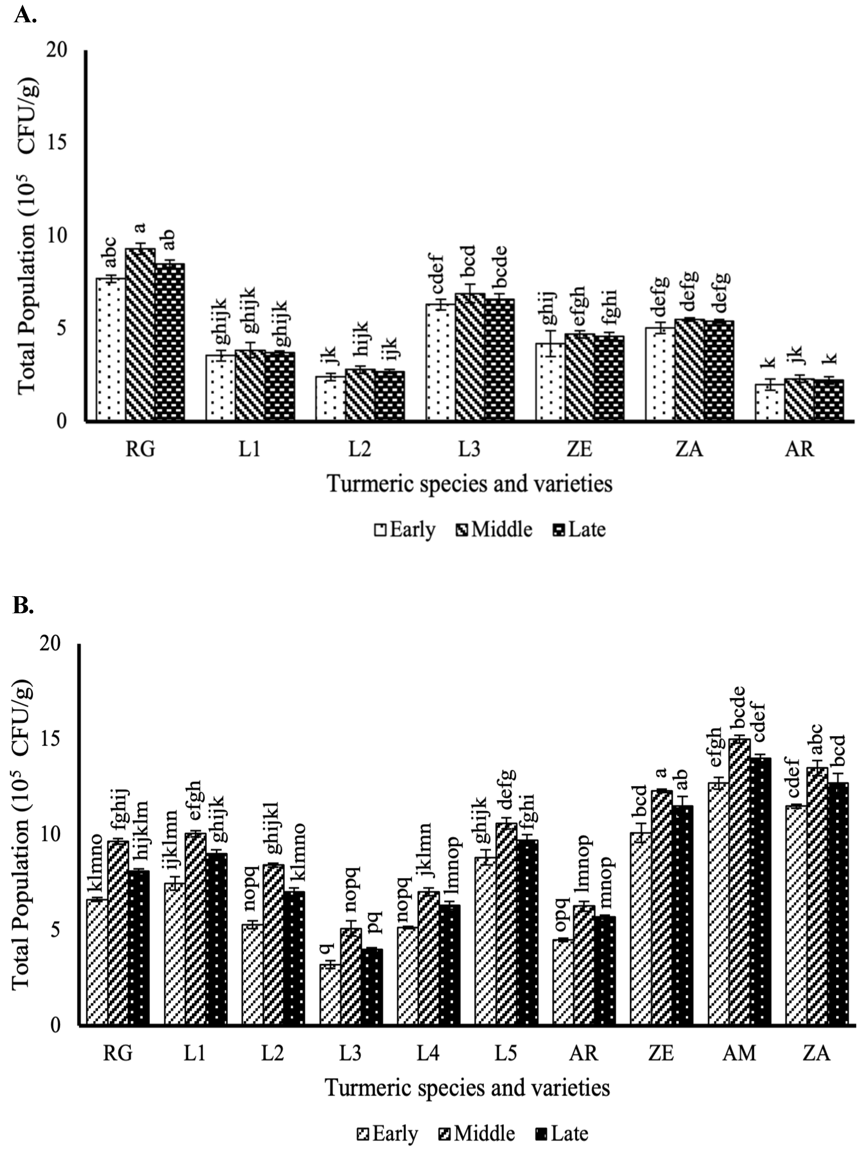
3.2. Effect of the Different Curcuma Growth Stages on Endophytic Bacteria Population in the Stems

The population of endophytic bacteria in Curcuma stems varied significantly across growth stages and among species or strains (Figure 2). In field-grown plants, stem bacterial counts ranged from 2.0 to 9.3 × 10⁵ CFU/g, while those cultivated in a plastic house ranged from 3.2 to 15.0 × 10⁵ CFU/g. The RG strain exhibited the highest bacterial population during the middle growth stage, reaching 9.3 × 10⁵ CFU/g in the field and 15.0 × 10⁵ CFU/g in the plastic house, followed by C. amada.
Notably, endophytic bacterial populations differed between the two experiments for each Curcuma species or strain. Plants at the middle growth stage consistently showed significantly higher bacterial counts compared to other stages in both experiments (p < 0.05).

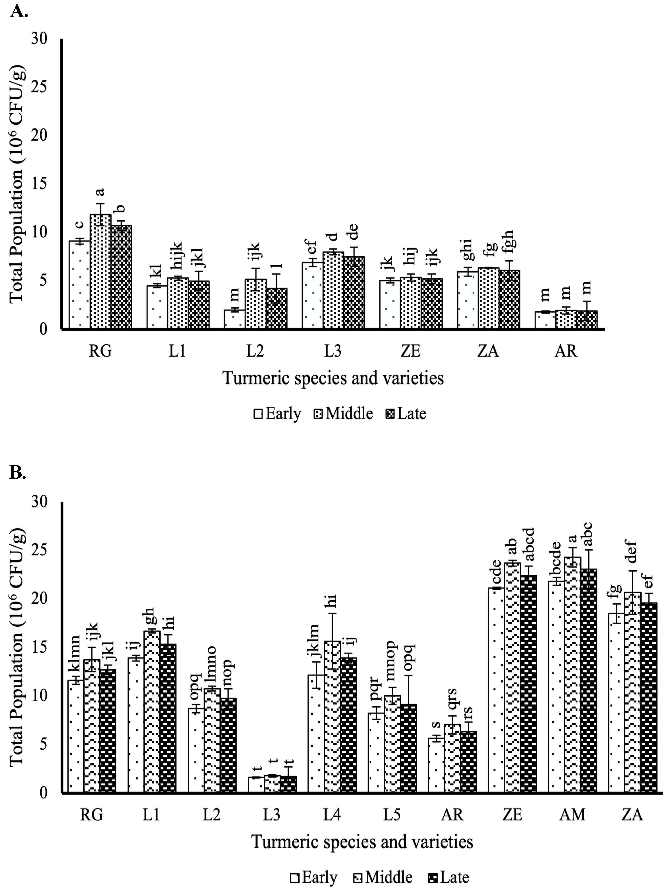
3.3. Effect of the Different Curcuma Growth Stages on Endophytic Bacteria Population in the Leaves

The population of endophytic bacteria in the leaves of 16 Curcuma species or strains varied depending on the species/strain and growth stage in both the experiments (Figure 3). In field-grown plants, bacterial populations ranged from 30.0 to 76.0 × 10⁴ CFU/g, while in plastic house-grown plants, the range was lower, from 7.3 to 23.3 × 10⁴ CFU/g.
Significant differences (p < 0.05) were observed among growth stages, with the lowest bacterial counts recorded during the early stage and the highest during the middle stage. The Curcuma strain RG exhibited the highest bacterial population in both experiments (76.0 × 10⁴ CFU/g in the field and 23.3 × 10⁴ CFU/g in the plastic house).

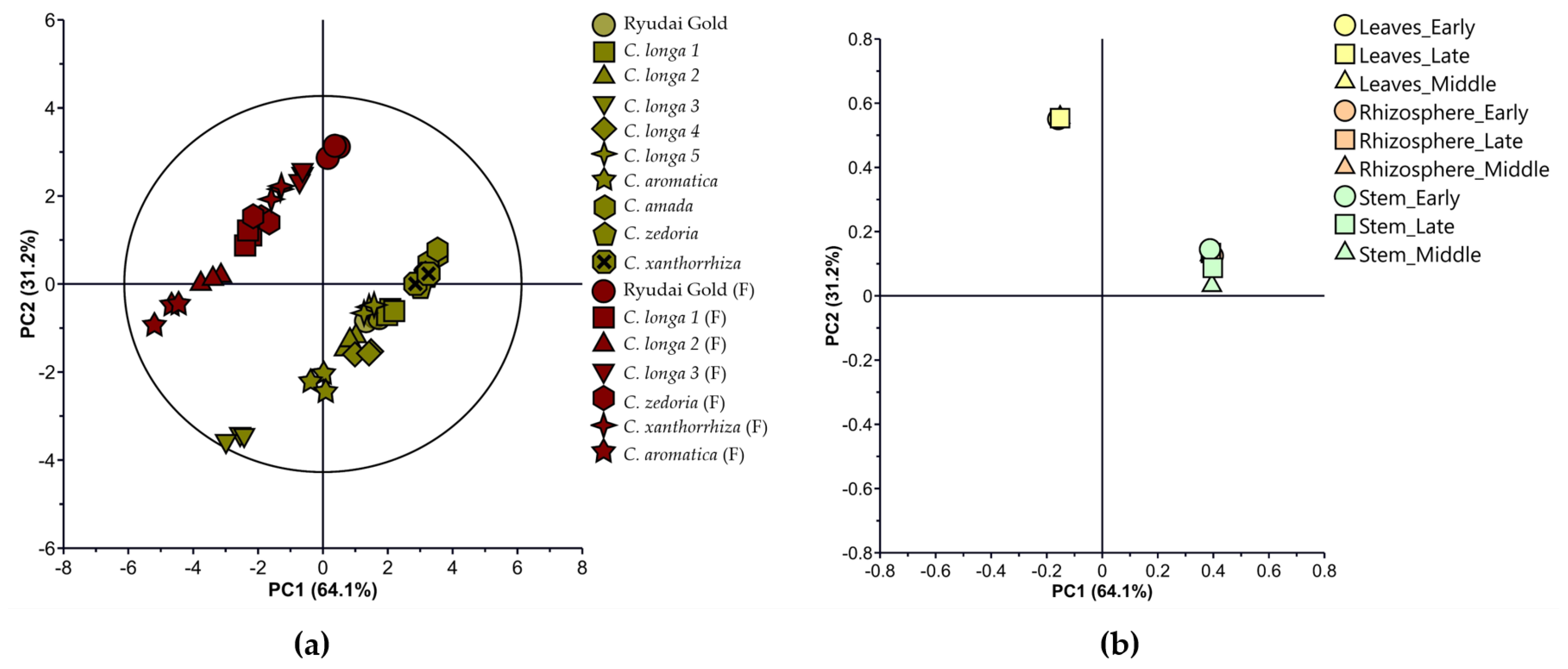
3.4. Multivariate Statistical Plot of the Bacterial Population in Curcuma Species Cultivated in the Field

Principal component analysis (PCA) of bacterial populations showed the variations across seven Curcuma species at different growth stages. PC1 and PC2 explain 94.1% and 3.1% of the total variance, respectively. Distinct clustering patterns are observed, with C. aromatica and C. longa strain L2 clearly separated from other species along the PC1 axis, indicating differences in bacterial community composition. Other species, including C. longa L1 and L3, C. zedoaria, and C. xanthorrhiza, are more tightly grouped, suggesting similar microbial profiles. The variations in bacterial populations in different growth stages of Curcuma species were further explained by multivariate analysis using the principal component analysis (PCA). The first two principal components (PCs) explained 97.2% of the total variance, accounted for by PC1 and PC2 (94.1 and 3.12%, respectively), showing that most of the separation between Curcuma species occurs along the PC1 axis. The PCA analysis revealed diverse grouping patterns for bacterial population data in the rhizosphere, leaves, and stems of seven species of Curcuma at different growth stages. C. aromatica seemed to cluster in the negative direction of PC1 and PC2, indicating distinct bacterial growth patterns compared to other species (Figure 4a). Similarly, C. longa strain L2 displayed a distinct clustering pattern, suggesting it harbors a bacterial population different from the other C. longa strain L1 and L3. In contrast, C. longa strain L1, C. zedoaria, C. xanthorrhiza, C. longa strain L3, and C. longa strain RG were more tightly grouped, suggesting similarities in their bacterial population trends.
Factor loading plots were used to investigate the relationships between the score plots and the microbial growth stages variables, providing insights into how each variable contributed to the observed clustering patterns. The corresponding factor loading plot (Figure 4b) revealed that the clustering observed along PC1 was primarily driven by sample type (rhizosphere, stem, and leaf), with rhizosphere samples showing the greatest contribution to PC1 variance. The growth stage also influenced the clustering along PC2, particularly in leaf and stem samples, which exhibited greater overlap, suggesting less variation in bacterial populations across growth stages in these tissues compared to the rhizosphere. These patterns highlight that sample type was the primary driver of bacterial community differentiation, while growth stage provided additional but lesser separation, particularly in non-rhizosphere tissues. Together, these results emphasize that both plant part and growth stage play key roles in shaping microbial communities in Curcuma, with the rhizosphere being the most variable and influential compartment in driving overall bacterial community structure.

3.5. Multivariate Statistical Plot of Bacterial Population of Curcuma Species Cultivated in the Plastic House

The first two PCs of the variations in bacterial populations in different growth stages of Curcuma species that were planted in the plastic house explained 97.8% of the total variance, accounted for by PC1 and PC2 (91.4 and 6.38%, respectively). C. longa strain L2, C. aromatica, C. longa strain L3, C. longa strain RG, and C. longa strain L1 form a relatively close cluster along PC1 and PC2 (Figure 5a). This indicates that these species share greater similarities in their bacterial population or growth data under plastic house conditions compared to others, such as C. zedoaria or C. xanthorrhiza that were plotted both in positive direction of PC1 and negative direction of PC2. Conversely, C. longa strain L5 exhibited a distinct separation in the score plot near to the origin (PC1 = 0, PC2 = 0), suggesting that this species has average or neutral characteristics in relation to the principal components. Thus, this indicates that its bacterial or growth data do not exhibit extreme variations and may fall somewhere in between the other species.
The factor loading plot (Figure 5b) revealed that the primary variable contributing to separation along PC1 was the bacterial population in the rhizosphere, especially at later growth stages. These variables had high positive loadings on PC1, indicating they drive separation of species like C. amada, which showed dominant rhizosphere bacterial populations. In contrast, bacterial populations in the leaves and stems—particularly of C. zedoaria and C. amada, contributed more to PC2 and were associated with separation in that dimension. This suggests that while PC1 reflects rhizosphere-driven variance, PC2 captures differences in endophytic bacterial populations in aerial parts. In conclusion, rhizosphere populations dominate the separation along PC1, while endophytic bacterial differences drive PC2 variability.

3.6. Multivariate Statistical Plot of Combined Data on Bacterial Population in Curcuma Species Cultivated in the Field and Plastic House

The comparison between the bacterial population structure based on the growth stages of Curcuma species in the field and plastic house experiments was further explored using PCA, which revealed that there were distinct separations between field and plastic house (Figure 6a). Accordingly, the first two PCs further explained 95.3% of the variance and were accounted for by PC1 and PC2 (64.1 and 31.2%, respectively). Based on the score plot, the Curcuma species that were cultivated in the field clustered distinctly from the plastic house along PC1, indicating that the bacterial populations in the field experiment differ significantly from those in the plastic house. Moreover, the plastic house samples tend to cluster more tightly, suggesting more uniform bacterial populations, likely influenced by conditions in the plastic house. The separation of field and plastic house samples along PC1 highlights the impact of the planting environment on bacterial community structure.
The overlap between the rhizosphere and stem in the loading plot, and the separation of leaves, can be explained by differences in the bacterial community composition and diversity driven by the plant part and its microenvironment. The loading plot (Figure 6b) further clarified the contribution of specific plant parts to the observed separation. Bacterial populations associated with leaves contributed predominantly to PC2 and clustered separately from the rhizosphere and stem samples, which partially overlapped. This suggests that leaf-associated bacterial communities are compositionally distinct, while rhizosphere and stem communities are more similar, likely due to closer spatial or physiological interactions. Overall, these results emphasize that different growth stage influences the cultivation places (field vs. plastic house) as the strongest driver of community separation (PC1), while plant part, particularly leaf versus non-leaf tissues, drives secondary variation (PC2). These findings underline the influence of both external growing conditions and internal plant microenvironments in shaping microbial community structures in Curcuma species.

4. Discussion

The bacterial populations in the rhizosphere, stems, and leaves of Curcuma species varied significantly across different growth stages in both the experiments, highlighting the influence of plant development and external conditions on microbial communities.
Across all growth stages, rhizosphere bacterial populations were consistently higher under plastic house conditions compared to the field. This trend likely reflects the benefits of stable environmental parameters, such as temperature, humidity, and soil moisture which promote microbial proliferation [20]. This observation aligns with previous studies showing higher microbial abundance under controlled environments due to reduced abiotic stress [21]. Notably, peak bacterial density occurred at the middle growth stage in both environments, coinciding with heightened root exudation, a key factor known to enrich microbial communities by supplying carbon-rich substrates [22,23]. Moreover, root anatomical changes such as increased vascular tissue and moisture content may further enhance microbial colonization [24]. These conditions collectively enhance microbial proliferation across the rhizosphere, stem, and leaf tissues [25]. The variations observed among different Curcuma species and strains may be due to differences in the quantity and composition of these exudates, as plants selectively shape their rhizosphere microbiome based on their metabolic profile [26]. This is evident in the significantly higher bacterial population in Ryudai Gold and C. amada, suggesting that these strains may release a more favorable set of exudates for microbial colonization. In this study, the rhizosphere bacterial density was 106 CFU/g, aligning with previous reports of rhizosphere populations ranging from 106 to 109 CFU/g [27]. The relatively low bacterial count observed may be influenced by the presence of bioactive compounds in turmeric, such as curcumin and essential oils, which are known to exhibit antimicrobial properties [28]. These compounds could potentially inhibit certain microbial populations in the rhizosphere, leading to lower overall bacterial density. Similarly, a previous study indicated that the population density of rhizosphere bacteria associated with Zea mays L. at different growth stages ranged from 109 to 1013 CFU/g, highlighting the potential variability in bacterial populations depending on plant species, environmental factors, and growth stages [29]. Moreover, previous research on the rhizosphere of five different Acacia species also revealed that each sample possessed a unique microbial population and community structure [30]. The rhizosphere is the area surrounding plant roots, characterized by high biological activity and a limited spatial extent. Plant roots secrete various compounds that attract microbial colonization in this region, significantly influencing bacterial colonization [31]. The carbon fixed during plant photosynthesis is partially transported to the roots and released as exudates, creating a nutrient-rich environment that fosters bacterial growth [26,31].
Similarly, endophytic bacterial populations in Curcuma stems and leaves also exhibited differences depending on growth stage, cultivation conditions, and plant species or strain. Endophytic bacteria may influence Curcuma physiology in several ways. Some strains can enhance curcumin production by inducing plant defense pathways and secondary metabolite biosynthesis [32]. Additionally, endophytes that produce phytohormones can stimulate root development and branching, thereby improving nutrient acquisition [33]. Although the overall bacterial densities in stems (105 CFU/g) were approximately ten times higher than those in leaves (104 CFU/g), this difference is relatively moderate. It may be attributed to anatomical and environmental factors; stems are generally more enclosed and structurally supportive, potentially offering a more stable microenvironment for bacterial colonization. In contrast, leaves are more exposed to sunlight, temperature fluctuations, and desiccation, which may limit bacterial persistence [34]. This may be due to differences in structural and chemical barriers, as well as the presence of specific compounds that support bacterial proliferation in stems but not in leaves [35]. Moreover, the higher bacterial populations in plastic house conditions compared to field conditions, especially in C. amada, indicate that environmental stability and reduced exposure to external stressors enhance bacterial colonization within plant tissues. The middle growth stage consistently showed the highest endophytic bacterial populations across both stems and leaves, mirroring trends observed in the rhizosphere. However, the rhizosphere bacteria populations could also differ depending on their growth stages and the specific Curcuma species or strains [36]. This pattern suggests that the internal plant environment at this stage is particularly conducive to bacterial growth [37]. Physiological changes in the plant, such as increased vascular activity and greater nutrient transport, likely facilitate bacterial movement and proliferation [38]. The early stage showed relatively low bacterial numbers, likely due to underdeveloped vascular structures limiting microbial infiltration, while the late stage exhibited a slight decline, possibly due to structural lignification or reduced nutrient flow [39]. Additionally, as plants grow, their immune responses fluctuate, and at the middle stage, they may provide a transiently permissive environment for bacterial colonization before later adopting more robust defense mechanisms [39,40].
A key observation from this study is the significantly higher bacterial population in the stems and leaves of C. amada and Ryudai Gold turmeric compared to other Curcuma species or strains. This suggests that these strains possess specific traits, such as enhanced production of bacterial-attracting metabolites or structural features that facilitate bacterial entry and colonization. Previous studies have demonstrated that different plant species, and even cultivars within the same species, harbor distinct microbial communities due to variations in metabolic composition [41,42].
The differences in endophytic bacterial populations between stems and leaves further highlight the influence of plant physiology on microbial colonization. Stems, being directly connected to roots through the vascular system, likely serve as the primary conduit for bacterial migration from the rhizosphere [43]. Once inside the plant, bacteria may move through the xylem, assisted by transpiration flow and bacterial motility mechanisms such as flagella [23]. Conversely, the lower bacterial population in leaves may be attributed to their relatively harsher environment, with exposure to UV radiation, fluctuating temperatures, and stronger plant defense responses [44]. The possibility of bacterial colonization through stomatal entry or wounds suggests an additional route for endophytes in leaves, but this appears to be less efficient than root-mediated colonization [45].
In addition to environmental factors, the morphological and phenotypic differences among the Curcuma species and strains studied may contribute to the observed variations in microbial populations [46]. For example, strains such as C. amada and C. longa exhibited larger rhizomes and thicker stems compared to other species, which could provide more extensive surface area and a richer microenvironment for microbial colonization [47]. These differences in plant structure may create unique niches that favor specific bacterial communities in the rhizosphere and within the plant tissues. Furthermore, distinct leaf morphology in species such as C. zedoaria and C. xanthorrhiza could influence the types of microbial communities present in the leaves [48]. The more compact or waxy surface of the leaves may alter microbial attachment and nutrient availability, potentially resulting in a different microbiome compared to species with thinner or more porous leaves [49].
The PCA analysis further reinforced the influence of growth stage on bacterial population distribution in Curcuma species cultivated in field and plastic house, as reflected in the distinct separations observed in the score and loading plots. In the field, C. aromatica clustered distinctly in the negative direction of both PC1 and PC2, suggesting highly unique bacterial populations, likely driven by specific root exudates and secondary metabolites that vary across growth stages. Similarly, C. longa strain L2 separated from strains L1 and L3, indicating that even within a species, bacterial population differences may arise due to genetic traits and growth stage influences [39]. The loading plot further underscored the impact of anatomical parts on bacterial diversity. Rhizosphere bacterial populations were strongly associated with high positive loadings along PC1, emphasizing their dominance and distinct contribution to microbial composition. In contrast, bacterial populations from leaves and stems showed greater overlap, indicating less distinct microbial communities in these anatomical parts compared to the rhizosphere [19].
In the plastic house, the interaction between growth stage and anatomical parts was less pronounced due to the controlled conditions reducing environmental variability. C. longa strains L1, L2, L3, RG, and C. aromatica formed a close cluster along PC1 and PC2, indicating a convergence of bacterial population trends that could be influenced by consistent soil, temperature, and moisture conditions [50]. Despite this homogenization, the rhizosphere continued to serve as the microbial hotspot, with strong positive loadings along PC1 across all species and growth stages. PCA results showed that C. amada rhizosphere samples contributed strongly to the separation along the positive PC1 and PC2 axes, indicating distinct patterns in bacterial community structure. In contrast, bacterial populations in leaves and stems contributed less to the variation, possibly reflecting the reduced microbial differentiation in these parts under controlled conditions [51]. Interestingly, C. amada and C. zedoaria demonstrated higher bacterial populations in their stem and leaf samples compared to other species, reflecting potential growth stage-specific shifts in microbial recruitment.
Overall, the microbial populations across different growth stages in different Curcuma species exhibit distinct variations between the field experiment and plastic house experiment. The distinct clustering patterns observed in both experiments highlight the critical role of the growth stage in shaping bacterial populations across anatomical parts. In the field, the dynamic shifts in bacterial diversity at different growth stages were dominated by early-stage opportunists, mid-stage metabolic activity, and late-stage specialization, as evidenced by the broader distribution of Curcuma species along PC1 and PC2. This variability reflects the influence of heterogeneous environmental factors and stage-specific plant physiological changes on microbial recruitment [52]. In contrast, the plastic house experiment reduced this variability, as seen in the tighter clustering among Curcuma species. However, rhizosphere bacterial populations remained a key driver of separation, while leaf and stem populations showed greater overlap across growth stages and species. This pattern highlights that while the rhizosphere remains highly dynamic, the controlled conditions homogenize bacterial communities associated with aerial parts. The strong alignment between PCA clustering and bacterial count data provides robust support for the hypothesis that differed growth stages could influence the bacterial population across all plant compartments and environmental conditions, synergistically shaping microbial diversity patterns.
While this study provides valuable insights into the microbial population dynamics in Curcuma species across different growth stages and planting environments, the limitations should be acknowledged, including the absence of microbial community profiling using molecular techniques, and the lack of endophytic fungal identification, which may limit a deeper understanding of microbial diversity and functional interactions within the plant. This study demonstrates that even though different Curcuma species were grown under identical conditions (field and plastic house), the microbial populations can still differ significantly, highlighting the complex interactions between the species and their respective environments. The overall findings of this study describe how growth stage and cultivation environment play a crucial role in shaping bacterial populations associated with Curcuma species. The peak bacterial abundance observed in the middle stage across all compartments highlights the significance of plant metabolic activity in microbial recruitment. The differences in microbial composition between field and plastic house conditions further demonstrate the impact of external environmental factors on bacterial colonization patterns. These insights contribute to our understanding of plant–microbe interactions in medicinal plants and suggest potential strategies for optimizing Curcuma cultivation to enhance beneficial microbial associations.

5. Conclusions

This study evaluated the endophytic and rhizosphere bacterial populations across different growth stages in various Curcuma species and strains cultivated under field and plastic house conditions. The results revealed notable differences in bacterial populations, with higher densities of endophytic bacteria observed in the leaves and stems during the middle growth stages of all Curcuma species or strains. Rhizosphere bacterial populations also varied depending on growth stage and plant species or strain. Understanding these dynamics provides valuable insights for optimizing microbial inoculant application strategies and developing sustainable agricultural practices tailored to specific crop stages and species. Future studies should explore the functional roles of these microbial communities and their potential applications as biofertilizer and stimulant in promoting plant health and productivity. Moreover, incorporating molecular identification and community profiling techniques are needed to uncover the taxonomic and functional diversity of these microbial communities.

Author Contributions

Experiment management, data collection, data analysis and manuscript writing, N.I.R.; experiment planning and manuscript editing, M.A.H.; manuscript review and editing, H.A. All authors have read and agreed to the published version of the manuscript.

Funding

This research received no external funding.

Institutional Review Board Statement

Not applicable.

Data Availability Statement

The original contributions presented in this study are included in the article. Further inquiries can be directed to the corresponding author.

Acknowledgments

All the authors have approved that this manuscript is applicable for the doctoral degree of the first author, Neptu Islamy Raharja, and express their gratitude to MEXT, Japan, for providing a scholarship to the first author.

Conflicts of Interest

The authors declare that they have no conflicts of interest.

References

  1. Kaliyadasa, E.; Samarasinghe, B.A. A review on golden species of Zingiberaceae family around the world: Genus Curcuma. Afr. J. Agric. Res. 2019, 14, 519–531. [Google Scholar]
  2. Jyotirmayee, B.; Mahalik, G. A review on selected pharmacological activities of Curcuma longa L. Int. J. Food Prop. 2022, 25, 1377–1398. [Google Scholar] [CrossRef]
  3. de Oliveira Filho, J.G.; de Almeida, M.J.; Sousa, T.L.; dos Santos, D.C.; Egea, M.B. Bioactive compounds of turmeric (Curcuma longa L.). In Bioactive Compounds of Under-Utilized Vegetables and Legumes; Alexandru, M.G., Holban, A.M., Eds.; Academic Press: Cambridge, MA, USA, 2021; pp. 297–318. [Google Scholar]
  4. Gaiero, J.R.; McCall, C.A.; Thompson, K.A.; Day, N.J.; Best, A.S.; Dunfield, K.E. Inside the root microbiome: Bacterial root endophytes and plant growth promotion. Am. J. Bot. 2013, 100, 1738–1750. [Google Scholar] [CrossRef] [PubMed]
  5. Bacon, C.W.; White, J.F. Functions, mechanisms and regulation of endophytic and epiphytic microbial communities of plants. Symbiosis 2016, 68, 87–98. [Google Scholar] [CrossRef]
  6. Khan, S.; Ambika Rani, K.; Sharma, S.; Kumar, A.; Singh, S.; Singh, Y. Rhizobacterial-mediated interactions in Curcuma longa for plant growth and enhanced crop productivity: A systematic review. Front. Plant Sci. 2023, 14, 1231676. [Google Scholar] [CrossRef]
  7. Schrey, D.; Hartmann, A.; Hampp, R. Rhizosphere interactions. In Ecological Biochemistry: Environmental and Interspecies Interactions, 2nd ed.; Wink, M., Ed.; Wiley-Blackwell: Weinheim, Germany, 2014; pp. 292–311. [Google Scholar]
  8. Song, Q.; Deng, X.; Song, R.; Song, X. Plant growth-promoting rhizobacteria promote growth of seedlings, regulate soil microbial community, and alleviate damping-off disease caused by Rhizoctonia solani on Pinus sylvestris var. mongolica. Plant Dis. 2022, 106, 2730–2740. [Google Scholar] [CrossRef]
  9. Vocciante, M.; Grifoni, M.; Fusini, D.; Petruzzelli, G.; Franchi, E. The role of plant growth-promoting rhizobacteria (PGPR) in mitigating plant environmental stresses. Appl. Sci. 2022, 12, 1231. [Google Scholar] [CrossRef]
  10. Nadeem, S.M.; Ahmad, M.; Zahir, Z.A.; Javaid, A.; Ashraf, M. The role of mycorrhizae and plant growth-promoting rhizobacteria (PGPR) in improving crop productivity under stressful environments. Biotechnol. Adv. 2014, 32, 429–448. [Google Scholar] [CrossRef]
  11. Compant, S.; Cambon, M.C.; Vacher, C.; Mitter, B.; Samad, A.; Sessitsch, A. The plant endosphere world—Bacterial life within plants. Environ. Microbiol. 2021, 23, 1812–1829. [Google Scholar] [CrossRef]
  12. Sontsa-Donhoung, A.M.; Bahdjolbe, M.; Hawaou; Nwaga, D. Selecting endophytes for rhizome production, curcumin content, biocontrol potential, and antioxidant activities of turmeric (Curcuma longa). Biomed. Res. Int. 2022, 2022, 8321734. [Google Scholar] [CrossRef]
  13. Thapa, S.; Prasanna, R. Prospecting the characteristics and significance of the phyllosphere microbiome. Ann. Microbiol. 2018, 68, 229–245. [Google Scholar] [CrossRef]
  14. Manikandan, A.; Anandham, R.; Madhan, S.; Raghu, R.; Krishnamoorthy, R.; Senthilkumar, M. Exploring the phyllosphere: Microbial diversity, interactions, and ecological significance in plant health. In Plant–Microbe Interactions in Stress Management; Giri, B., Varma, A., Eds.; Springer: Singapore, 2024; pp. 29–49. [Google Scholar]
  15. Sumathi, C.S.; Balasubramanian, V.; Ramesh, N.; Kannan, V.R. Influence of biotic and abiotic features on Curcuma longa L. plantation under tropical condition. Middle-East J. Sci. Res. 2008, 3, 171–178. [Google Scholar]
  16. Harish, B.S.; Umesha, K.; Venugopalan, R.; Prasad, B.M. Photo-selective nets influence physiology, growth, yield and quality of turmeric (Curcuma longa L.). Ind. Crops Prod. 2022, 186, 115202. [Google Scholar] [CrossRef]
  17. Ishimine, Y.; Hossain, M.; Murayama, S. Optimal planting depth for turmeric (Curcuma longa L.) cultivation in dark red soil in Okinawa Island, southern Japan. Plant Prod. Sci. 2003, 6, 83–89. [Google Scholar] [CrossRef]
  18. Hossain, M.A. Effect of harvest time on shoot biomass and yield of turmeric (Curcuma longa L.) in Okinawa, Japan. Plant Prod. Sci. 2010, 13, 97–103. [Google Scholar] [CrossRef]
  19. Kumar, A.; Singh, R.; Yadav, A.; Giri, D.D.; Singh, P.K.; Pandey, K.D. Isolation and characterization of bacterial endophytes of Curcuma longa L. 3 Biotech 2016, 6, 60. [Google Scholar] [CrossRef]
  20. Song, Y.; Li, X.; Yao, S.; Yang, X.; Jiang, X. Correlations between soil metabolomics and bacterial community structures in the pepper rhizosphere under plastic greenhouse cultivation. Sci. Total Environ. 2020, 728, 138439. [Google Scholar] [CrossRef]
  21. Vives-Peris, V.; de Ollas, C.; Gomez-Cadenas, A.; Perez-Clemente, R.M. Root exudates: From plant to rhizosphere and beyond. Plant Cell Rep. 2020, 39, 3–17. [Google Scholar] [CrossRef]
  22. Chaparro, J.M.; Badri, D.V.; Vivanco, J.M. Rhizosphere microbiome assemblage is affected by plant development. ISME J. 2014, 8, 790–803. [Google Scholar] [CrossRef]
  23. Compant, S.; Clément, C.; Sessitsch, A. Plant growth-promoting bacteria in the rhizo- and endosphere of plants: Their role, colonization, mechanisms involved and prospects for utilization. Soil Biol. Biochem. 2010, 42, 669–678. [Google Scholar] [CrossRef]
  24. Reinhold-Hurek, B.; Bünger, W.; Burbano, C.S.; Sabale, M.; Hurek, T. Roots shaping their microbiome: Global hotspots for microbial activity. Annu. Rev. Phytopathol. 2015, 53, 403–424. [Google Scholar] [CrossRef] [PubMed]
  25. Compant, S.; Duffy, B.; Nowak, J.; Clément, C.; Barka, E.A. Use of plant growth-promoting bacteria for biocontrol of plant diseases: Principles, mechanisms of action, and future prospects. Appl. Environ. Microbiol. 2005, 71, 4951–4959. [Google Scholar] [CrossRef] [PubMed]
  26. Lugtenberg, B.; Kamilova, F. Plant-growth-promoting rhizobacteria. Annu. Rev. Microbiol. 2009, 63, 541–556. [Google Scholar] [CrossRef] [PubMed]
  27. Bulgarelli, D.; Schlaeppi, K.; Spaepen, S.; Ver Loren van Themaat, E.; Schulze-Lefert, P. Structure and functions of the bacterial microbiota of plants. Annu. Rev. Plant Biol. 2013, 64, 807–838. [Google Scholar] [CrossRef]
  28. Kaskoos, R.A.; Ahamad, J.; Mir, S.R.; Javed, S.A. Antimicrobial properties of curcumin and its potential in the treatment of infections. Pharm. Biol. 2013, 51, 607–611. [Google Scholar]
  29. Cavaglieri, L.; Orlando, J.; Etcheverry, M. Rhizosphere microbial community structure at different maize plant growth stages and root locations. Microbiol. Res. 2009, 164, 391–399. [Google Scholar] [CrossRef]
  30. Yuan, Z.S.; Liu, F.; He, S.B.; Zhou, L.I.; Pan, H. Community structure and diversity characteristics of rhizosphere and root endophytic bacterial community in different Acacia species. PLoS ONE 2022, 17, e0262909. [Google Scholar] [CrossRef]
  31. Upadhyay, S.K.; Srivastava, A.K.; Rajput, V.D.; Chauhan, P.K.; Bhojiya, A.A.; Jain, D.; Chaubey, G.; Dwivedi, P.; Sharma, B.; Minkina, T. Root exudates: Mechanistic insight of plant growth-promoting rhizobacteria for sustainable crop production. Front. Microbiol. 2022, 13, 916488. [Google Scholar] [CrossRef]
  32. Singh, L.P.; Gill, S.S.; Tuteja, N. Unraveling the role of fungal endophytes in plant growth and stress tolerance. Plant Signal. Behav. 2020, 15, 1780043. [Google Scholar]
  33. Santoyo, G.; Moreno-Hagelsieb, G.; del Carmen Orozco-Mosqueda, M.; Glick, B.R. Plant growth-promoting bacterial endophytes. Microbiol. Res. 2016, 183, 92–99. [Google Scholar] [CrossRef]
  34. Kandel, S.L.; Joubert, P.M.; Doty, S.L. Bacterial endophyte colonization and distribution within plants. Microorganisms 2017, 5, 77. [Google Scholar] [CrossRef] [PubMed]
  35. Meng, F.C.; Zhou, Y.Q.; Ren, D.; Wang, R.; Wang, C.; Lin, L.G.; Zhang, X.Q.; Ye, W.C.; Zhang, Q.W. Turmeric: A review of its chemical composition, quality control, bioactivity, and pharmaceutical application. In Handbook of Food Bioengineering; Volume 7, Holban, A.M., Grumezescu, A.M., Eds.; Academic Press: Cambridge, MA, USA, 2018; pp. 299–350. [Google Scholar]
  36. Philippot, L.; Raaijmakers, J.M.; Lemanceau, P.; van der Putten, W.H. Going back to the roots: The microbial ecology of the rhizosphere. Nat. Rev. Microbiol. 2013, 11, 789–799. [Google Scholar] [CrossRef] [PubMed]
  37. Marag, P.S.; Suman, A. Growth stage and tissue-specific colonization of endophytic bacteria having plant growth-promoting traits in hybrid and composite maize (Zea mays L.). Microbiol. Res. 2018, 214, 101–113. [Google Scholar] [CrossRef] [PubMed]
  38. Compant, S.; Reiter, B.; Sessitsch, A.; Nowak, J.; Clément, C.; Barka, E.A. Endophytic colonization of Vitis vinifera L. by plant growth-promoting bacterium Burkholderia sp. strain PsJN. Appl. Environ. Microbiol. 2005, 71, 1685–1693. [Google Scholar] [CrossRef]
  39. Awin, T.; Mediani, A.; Leong, S.W.; Faudzi, S.M.M.; Shaari, K.; Abas, F. Phytochemical and bioactivity alterations of Curcuma species harvested at different growth stages by NMR-based metabolomics. J. Food Compos. Anal. 2019, 77, 66–76. [Google Scholar] [CrossRef]
  40. Mengistu, A.A. Endophytes: Colonization, behaviour, and their role in defense mechanisms. Int. J. Microbiol. 2020, 2020, 6927219. [Google Scholar] [CrossRef]
  41. Graner, G.; Paula, P.; Johan, M.; Sadhna, A. Microbial diversity in different cultivars of Brassica napus in relation to its wilt pathogen Verticillium longisporum. FEMS Microbiol. Lett. 2003, 224, 269–276. [Google Scholar] [CrossRef]
  42. Monika, S.; Prem, P.S.; Arun, K.P.; Singh, P.K.; Pandey, K.D. Enumeration of culturable endophytic bacterial population of different Lycopersicon esculentum L. varieties. Int. J. Curr. Microbiol. Appl. Sci. 2018, 7, 3344–3352. [Google Scholar]
  43. Wheatley, R.M.; Poole, P.S. Mechanisms of bacterial attachment to roots. FEMS Microbiol. Rev. 2018, 42, 448–461. [Google Scholar] [CrossRef]
  44. Beattie, G.A.; Lindow, S.E. The secret life of foliar bacterial pathogens on leaves. Annu. Rev. Phytopathol. 1995, 33, 145–172. [Google Scholar] [CrossRef]
  45. Jin, H.; Yang, X.Y.; Yan, Z.Q.; Liu, Q.; Li, X.Z.; Chen, J.X.; Qin, B. Characterization of rhizosphere and endophytic bacterial communities from leaves, stems, and roots of medicinal Stellera chamaejasme L. Syst. Appl. Microbiol. 2014, 37, 376–385. [Google Scholar] [CrossRef]
  46. Compant, S.; Samad, A.; Faist, H.; Sessitsch, A. A review on the plant microbiome: Ecology, functions, and emerging trends in microbial application. J. Adv. Res. 2019, 19, 29–46. [Google Scholar] [CrossRef] [PubMed]
  47. Reinhold-Hurek, B.; Hurek, T. Living inside plants: Bacterial endophytes. Curr. Opin. Plant Biol. 2011, 14, 435–443. [Google Scholar] [CrossRef]
  48. Hardoim, P.R.; van Overbeek, L.S.; van Elsas, J.D. Properties of bacterial endophytes and their proposed role in plant growth. Trends Microbiol. 2008, 16, 463–471. [Google Scholar] [CrossRef] [PubMed]
  49. Hunter, P.J.; Hand, P.; Pink, D.; Whipps, J.M.; Bending, G.D. Both leaf properties and microbe–microbe interactions influence within-species variation in bacterial population diversity and structure in the lettuce phyllosphere. Appl. Environ. Microbiol. 2010, 76, 8117–8125. [Google Scholar] [CrossRef] [PubMed]
  50. Kumar, A.; Singh, R.; Yadav, A.; Giri, D.D.; Singh, P.K.; Pandey, K.D. Diversity of bacterial endophytes from Curcuma longa L. and their role in plant growth promotion and antifungal activity. J. Appl. Microbiol. 2016, 120, 1653–1665. [Google Scholar]
  51. Aswathy, R.G.; Joshi, S.R.; Jha, D.K. Paenibacillus sp.: A novel endophyte from Curcuma longa with potential plant growth-promoting properties. Res. J. Microbiol. 2013, 8, 115–123. [Google Scholar]
  52. Gumiere, T.; Durrer, A.; Bohnen, H.; Vargas, L.K. Plant growth stage drives temporal and spatial dynamics of the bacterial microbiome in the rhizosphere of Vigna subterranea. Front. Microbiol. 2022, 13, 825377. [Google Scholar]
Figure 1. Bacterial population in the rhizosphere soil of Curcuma cultivated in field (A) and plastic house (B). The different letters above the bars represent that the data were significantly different based on the Tukey’s test p < 0.05. Note: RG, Ryudai gold; L1, C. longa strain 1; L2, C. longa strain 2; L3, C. longa strain 3; L4, C. longa strain 4; L5, C. longa strain 5; AR, C. aromatica; ZE, C. zedoaria; AM, C. amada; ZA, C. xanthorrhiza.
Agriculture 15 01092 g001
Figure 2. Bacterial population in the stems of Curcuma spp. or strains cultivated in field (A) and plastic house (B). The different letters above the bars represent that the data were significantly different based on the Tukey’s test at p < 0.05. Note: RG, Ryudai gold; L1, C. longa strain 1; L2, C. longa strain 2; L3, C. longa strain 3; L4, C. longa strain 4; L5, C. longa strain 5; AR, C. aromatica; ZE, C. zedoaria; AM, C. amada; ZA, C. xanthorrhiza.
Agriculture 15 01092 g002
Figure 3. Bacterial population in the leaves of Curcuma spp. or strains cultivated in field (A) and plastic house (B). The different letters above the bars represent that the data were significantly different based on the Tukey’s test at p < 0.05. Note: RG, Ryudai gold; L1, C. longa strain 1; L2, C. longa strain 2; L3, C. longa strain 3; L4, C. longa strain 4; L5, C. longa strain 5; AR, C. aromatica; ZE, C. zedoaria; AM, C. amada; ZA, C. xanthorrhiza.
Agriculture 15 01092 g003
Figure 4. PCA of bacterial population data from different Curcuma species or strains and growth stages cultivated in the field. (a) Score plot showing sample distribution. (b) Loading plot indicating the contribution of variables to the principal components.
Agriculture 15 01092 g004
Figure 5. PCA of bacterial population data from Curcuma species or strains and growth stages cultivated in a plastic house. (a) Score plot showing sample grouping. (b) Loading plot representing the influence of each variable on the principal components.
Agriculture 15 01092 g005
Figure 6. PCA based on combined bacterial population data from field and plastic house cultivation. (a) Score plot displaying overall sample distribution. (b) Loading plot illustrating the variable contributions to the principal components.
Agriculture 15 01092 g006
Disclaimer/Publisher’s Note: The statements, opinions and data contained in all publications are solely those of the individual author(s) and contributor(s) and not of MDPI and/or the editor(s). MDPI and/or the editor(s) disclaim responsibility for any injury to people or property resulting from any ideas, methods, instructions or products referred to in the content.

