Review

A Century of Vitamin E: Early Milestones and Future Directions in Animal Nutrition

Nutrition & Health Division, BASF SE, 67063 Ludwigshafen am Rhein, Germany
*
Author to whom correspondence should be addressed.
Agriculture 2023, 13(8), 1526; https://doi.org/10.3390/agriculture13081526
Submission received: 4 July 2023 / Revised: 23 July 2023 / Accepted: 26 July 2023 / Published: 31 July 2023
(This article belongs to the Section Farm Animal Production)

Abstract

:
Vitamin E, consisting of four tocopherols and four tocotrienols, with α-tocopherol as the most biologically active form, has a significant history in scientific research. It was first identified in the 1920s for its role in preventing neonatal mortality in rats. Over time, its chemical structure was elucidated, and its importance in the immune system, skin health, anti-inflammatory properties, and hormonal balance was revealed. Vitamin E production has evolved from natural sourcing to efficient synthesis with standardized potency units. Initially, animal studies focused on reproductive health and growth disorders, but later research highlighted vitamin E’s role in preventing encephalomalacia in domestic fowl and muscular dystrophy in various animals. Today, vitamin E is commonly used as a feed additive to enhance animal health and immune function. Despite substantial knowledge about its role in animal nutrition, several questions remain unanswered. Current research seeks to determine optimal supplementation levels, interactions with other nutrients, impacts on gene expression, cell signaling, and communication, as well as the effects of high dosages in livestock. Vitamin E continues to be a crucial component in improving animal health, and ongoing research aims to uncover its benefits and mechanisms of action further.

1. Introduction

Vitamin E plays a critical role in animal nutrition by serving as a potent lipid-soluble antioxidant as well as contributing to anti-inflammation, immune function, and gene expression regulation. As an antioxidant, it protects cell membranes and other lipid-containing structures from oxidative damage caused by free radicals [1]. Thereby, vitamin E is the major chain-breaking antioxidant inhibiting lipid peroxidation, a physiological function that is not provided by other dietary or endogenous antioxidants [2]. This makes it crucial for preserving cell integrity, particularly in tissues that are susceptible to oxidative stress, such as the liver, lungs, and muscles [3].
The significance of vitamin E in animal nutrition cannot be overestimated, as it has been recognized as an indispensable micronutrient for optimal health, growth, and development in livestock [4]. Throughout the past century, numerous studies and advancements have been made in understanding the crucial role vitamin E plays in livestock production. A deficiency of vitamin E can impair immune responses and increase the susceptibility of animals to infectious diseases. Furthermore, hypovitaminosis E has been linked to reduced reproductive performance in animals, including decreased fertility rates and increased embryonic mortality [5].
For livestock, optimizing vitamin E status is particularly important for animal health and production. In dairy cattle, supplementation with vitamin E has been demonstrated to enhance milk yield and lower the occurrence of mastitis [6,7,8]. In poultry, it has been associated with better growth rates, egg production, and hatchability [9,10]. Likewise, in swine, vitamin E supplementation has been proven to enhance meat quality, reduce stress, and increase growth rates [11,12,13,14].
To ensure that animals receive adequate amounts of vitamin E, it is common practice to add this micronutrient as synthetic dl-α-tocopheryl acetate to animal feeds. However, determining the optimal level of vitamin E supplementation can be challenging, as the requirements for this nutrient can vary depending on the species, age, and health status of the animal, as well as other factors [15].
In this review, we aim to provide a comprehensive overview of the early milestones in vitamin E research as well as its current understanding and future directions. By examining the historical advancements in vitamin E exploration, we hope to provide insight into the evolution of our knowledge and understanding of this essential micronutrient and how it has shaped animal production and health over the past century. Additionally, we will highlight the route of vitamin E chemical synthesis as well as recent and future research on the role of vitamin E in animal nutrition. Through this review, we aspire to emphasize the continuing importance of vitamin E in animal nutrition and the need for ongoing research to fully understand its potential in supporting animal health and productivity in the future.

2. Early Discoveries and Understanding of Vitamin E

Last year marked the 100th anniversary of the discovery of vitamin E in 1922, which was made by Herbert McLean Evans, an embryologist and endocrinologist, and his co-worker Kathrine Julia Scott Bishop, a medical physician and trained anatomist, while working at Berkeley University in California/USA. The two scientists observed that female rats fed on a purified diet had good growth and development and stayed healthy, but could not reproduce, as the embryos died and were resorbed after some 10 days of gravidity. However, when the semi-synthetic diet was supplemented with fresh green leaves of lettuce or dried alfalfa meal, a sudden restoration of fertility in previously sterile rats could be observed [16]. At first, the researchers believed that vitamin C, which had already been discovered at that time and was known not to be essential for growth, was necessary for pregnancy. However, they quickly realized that only the fat-soluble components of the leaves had led to a good result. After testing the hydro- and lipophilic extracts of various wheat by-products from a nearby flour mill, the two scientists discovered a new fat-soluble dietary lipophilic compound that causes sterility in rats when lacking in the feed [16].
The unknown dietary substance was initially called factor X, but it was soon renamed vitamin E by Barnett Sure [17] and Herbert Evans [18]. Evans and Bishop later demonstrated that male rats with diets lacking the new fat-soluble vitamin E also experienced sterility [18], leading to the vitamin’s subsequent designation as the “anti-sterility vitamin”. In the same year, Evans and his co-worker George Burr prepared a potent concentrate of vitamin E by saponification of wheat germ oil, which proved to possess high biological potency [19]. Wheat germ oil-based concentrates were used in many further experiments on vitamin E and served as a source for the development of the first commercial vitamin E products in the 1930s.
In 1936, Evans and his co-workers isolated two compounds with vitamin E activity from wheat germ oil, for which they proposed the names α-tocopherol and ß-tocopherol [20]. Soon afterward, a third active factor, γ-tocopherol, was found in cottonseed oil by Evans’ working group [21], and in 1947, a fourth tocopherol, named δ-tocopherol, was isolated from soybean oil [22]. In 1936, Evans and his co-workers suggested the nomenclature α-tocopherol, the childbirth-bearing alcohol, for the new compound based on the Greek terms “tokos” for childbirth, “phero” for to bear, and “-ol” indicating an alcohol. This designation was proposed by George Miller Calhoun, a classical philologist and professor of Greek at the University of California [20].
Therefore, the discovery of vitamin E by Evans and Bishop in 1922 [16] resulted in the identification and isolation of several tocopherols. Their dedication and contributions to the study of vitamin E will continue to be celebrated and studied for years to come.

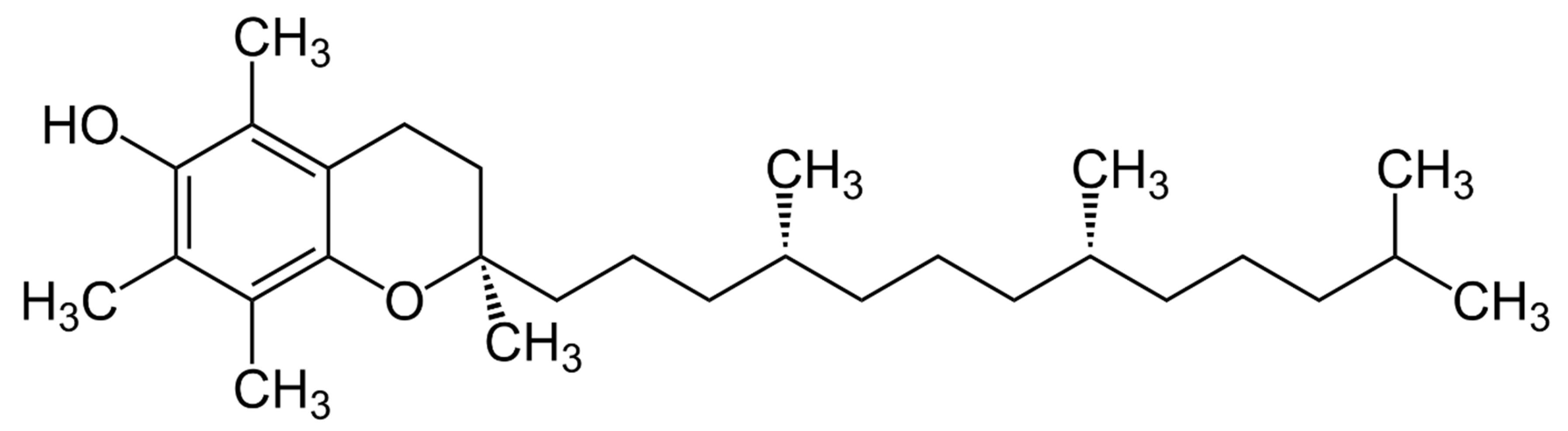
3. Vitamin E’s Chemical Structure and Biological Activity

The chemical structure of vitamin E was elucidated by the German chemist Erhard Fernholz in 1938 [23] while working in the USA. Fernholz proposed a structural formula that regarded α-tocopherol as a substituted 6-hydrocarbon with a long aliphatic sidechain attached to a pyran ring (Figure 1). Prior to this, in 1937, Fernholz [24] had studied the thermal decomposition of α-tocopherol and formed durohydro quinone and an aliphatic hydrocarbon. Shortly after Fernholz’s proposal, the Swiss chemist Paul Karrer achieved the chemical synthesis of α-tocopherol for the first time [25,26]. Karrer condensed trimethyl hydroquinone with phytol bromide derived from natural phytol, using zinc chloride as a catalyst. However, Karrer was not sure at that time regarding the chemical structure of the molecule he synthesized. He tended to assume a coumaran ring instead of the proposed chroman ring by Fernholz.
The first semi-synthetic tocopherol synthesized by Karrer consisted of two different stereoisomers and was initially called dl-α-tocopherol or 2-ambo-α-tocopherol. Shortly after the first synthesis of α-tocopherol, Bergel and co-workers of Lister-Institute in London, UK, and Lee Irvin Smith and co-workers of the University of Minnesota, Minneapolis, MN, USA, accomplished the synthesis of α-tocopherol as well [27,28].
The biological activity of the synthesized compound in the common rat resorption-gestation test was confirmed by Otto Isler [29], who accomplished an analog synthesis of vitamin E with Paul Karrer simultaneously.
Finally, the chroman ring as a constituent of α-tocopherol was confirmed with the help of UV spectra and other comparative model tests by Walter John at Göttingen University in Germany [30,31]. Furthermore, Walter John validated the chemical structure of α-tocopherol proposed by Fernholz and isolated ß-tocopherol simultaneously. John showed that ß-tocopherol differs from α-tocopherol only by one methyl group less at the chroman ring. He published more than 24 papers and book chapters on vitamin E-related topics in his short scientific career between 1937 and 1942.
In conclusion, the discovery of vitamin E’s chemical structure by Fernholz and the synthesis of α-tocopherol by Karrer were significant milestones for this essential micronutrient. Walter John’s confirmation of the chroman ring in α-tocopherol and work on synthesizing vitamin E derivatives contributed to scientific understanding, though his work is largely unrecognized outside of German journals.

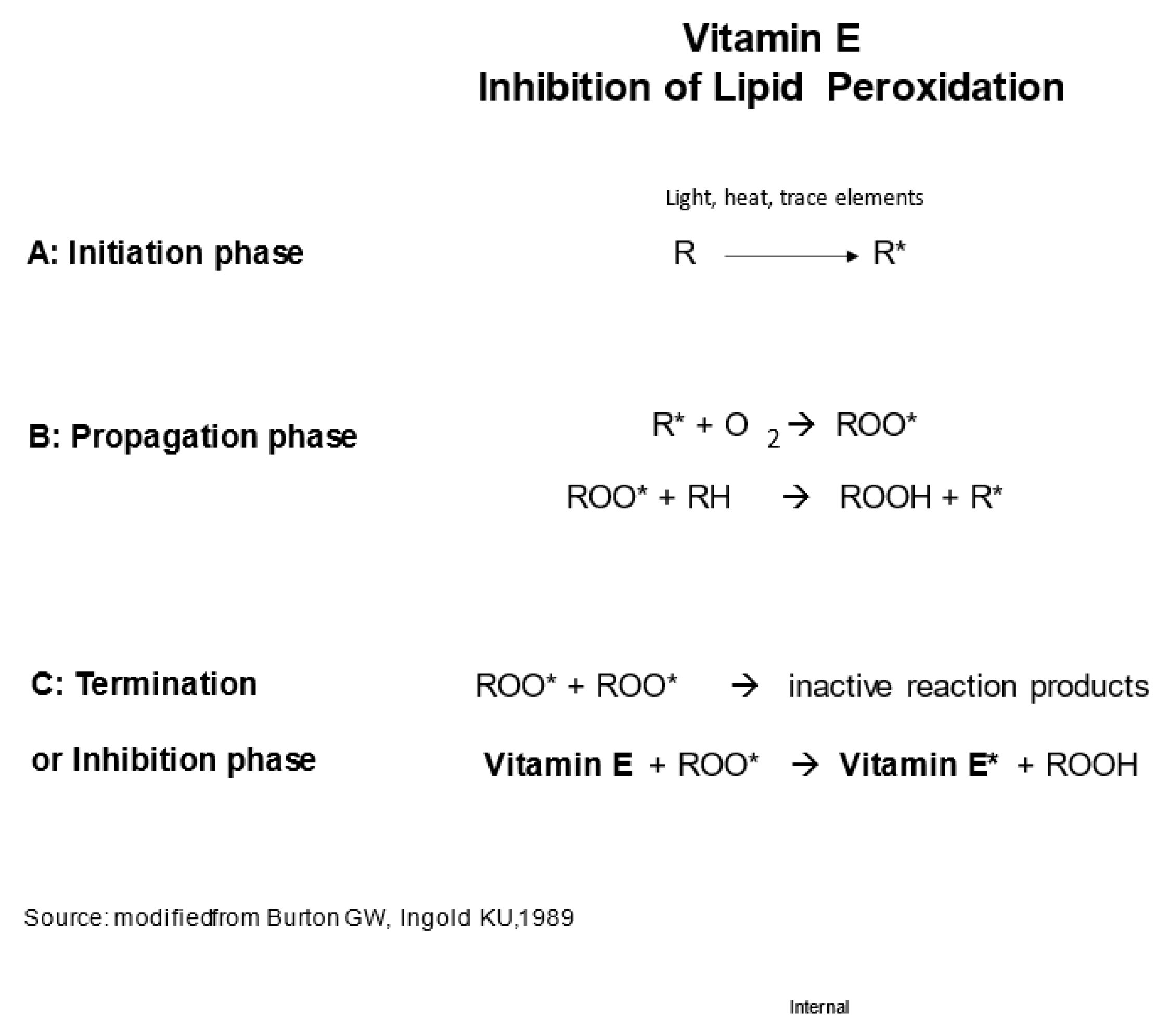
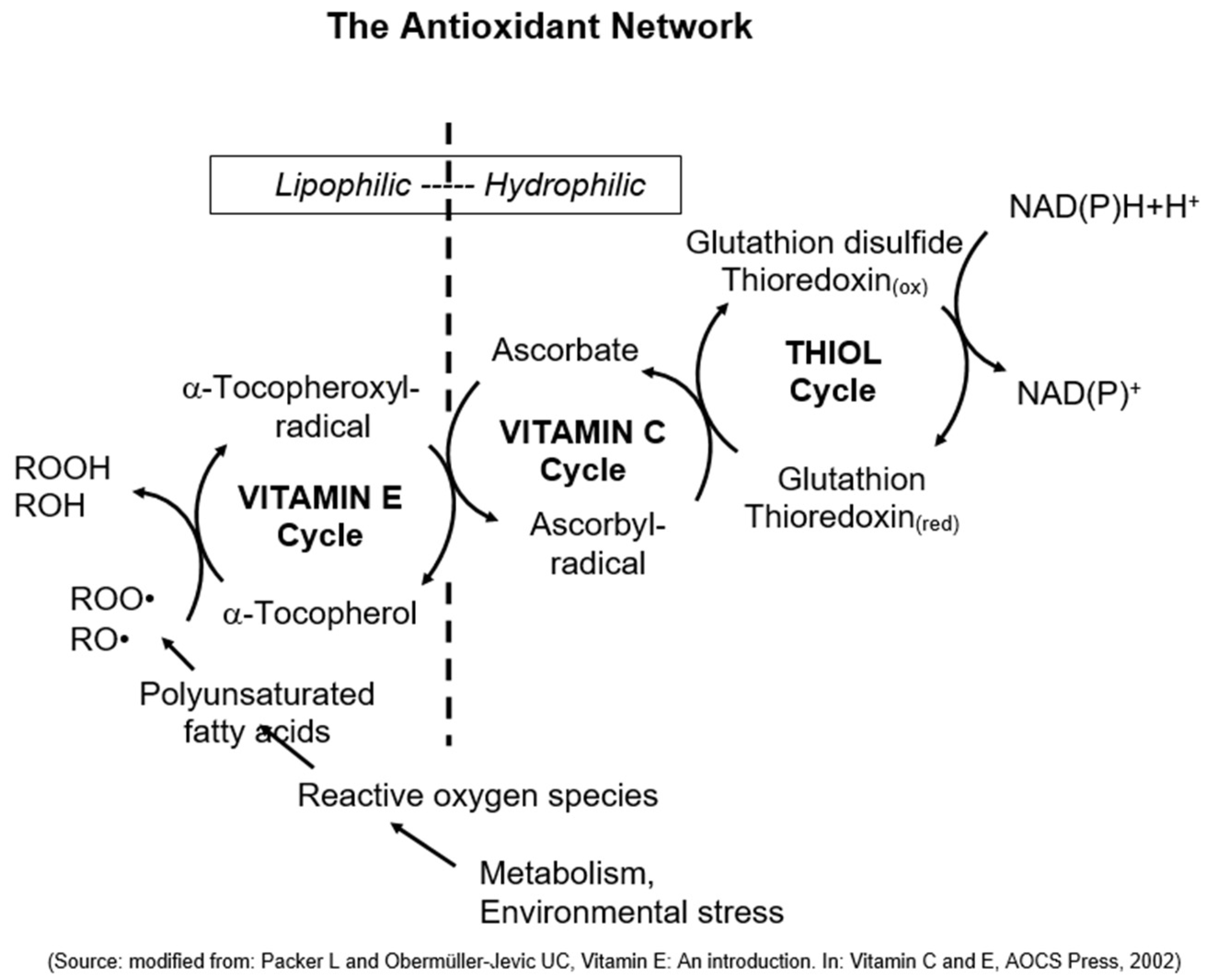
4. The Discovery of Vitamin E’s Unique Physiological Function as Chain-Breaking Antioxidant and the Antioxidant Network

In 1924, Henry Albright Mattill, a biochemist from the University of Iowa in Iowa City, IA, USA, conducted a study on the effects of milk consumption on reproduction. Along with his colleagues, he observed that rats became sterile when lard was added to their milk-based regimen. This led them to conclude that the fat content of a diet, in addition to vitamin E, affects reproduction. They proposed the hypothesis that the requirement for vitamin E increases with the amount of fat in the nutritional intake [32].
Three years later, in 1927, Mattill [33] reported another finding: The destruction of vitamin E in the presence of fat, particularly unsaturated fats. Building on this discovery, Mattill delved into further research on the autoxidation of fats. In collaboration with Marian Cummings, he put forward the idea that the oxidation of vitamin E could potentially safeguard other substances, such as vitamin A, from oxidation. They suggested that vitamin E possesses “antioxidant activity” and posited that its physiological role may lie in its ability to counteract oxidation [34]. It is worth noting that these early studies demonstrated the physiological consequences of the absence of antioxidant protection in lipids, namely, the sterility of rats.
A significant breakthrough in understanding the antioxidant role of vitamin E came from the research conducted by Aloys Tappel, a food biochemist at the University of California Davis/USA, during the 1950s. Alongside his colleagues, Tappel demonstrated that vitamin E effectively inhibits lipid peroxidation in living organisms. Through experiments conducted on isolated mitochondria and vitamin E-deficient animals, they observed elevated levels of lipid peroxidation in the liver, resulting in compromised mitochondrial stability [35,36].
In the early 1980s, Graham Burton and Kathrin Ingold, researchers from the National Research Council of Canada, conducted chemical investigations into the antioxidant properties of vitamin E and other phenolic compounds. They elucidated the chemical structure of α-tocopherol, which proved to be optimal for scavenging peroxyl radicals due to its hydroxylated chromanol ring with significant methylation. Furthermore, they noted that α-tocopherol possesses ideal characteristics for in vivo localization alongside lipids, thanks to its phytyl side chain [37]. Based on their findings, Burton and Ingold proposed that the primary, if not sole, function of α-tocopherol in living organisms is to act as an antioxidant. They even presented a reaction scheme for α-tocopherol in Figure 2 [38]. Subsequently, through studies involving individuals deficient in vitamin E, Burton and his colleagues demonstrated that α-tocopherol serves as the predominant chain-breaking antioxidant in vivo [2].
Finally, Lester Packer, a molecular and cell biologist from the University of California (Berkeley, CA, USA), made a significant observation regarding the combat against oxidative stress in cells. He recognized the importance of multiple antioxidants working together in what he referred to as “the antioxidant network” (Figure 3). Packer’s findings revealed that vitamin E and other antioxidants undergo oxidation but are subsequently recycled, forming a highly effective and precise defense system that adapts to oxidative stress [39,40,41].

5. The Synergy of Vitamin E, Vitamin C, and Selenium

In animal nutrition, the intricate relationship between vitamin E, vitamin C (ascorbic acid), and selenium (Se) plays a pivotal role in orchestrating antioxidant defenses and safeguarding cellular integrity [42]. Vitamin E, as a lipophilic antioxidant, primarily resides in biological membranes, where it acts as a chain-breaking antioxidant, protecting polyunsaturated fatty acids from peroxidative damage induced by free radicals and other reactive species [43]. The hydroxyl group on the chromanol ring of vitamin E is responsible for donating hydrogen atoms to lipid peroxyl radicals, leading to the formation of less reactive species and thus preventing lipid peroxidation chain reactions [44].
On the other hand, vitamin C, also known as L-ascorbic acid, is a hydrophilic antioxidant that inhabits the aqueous phase of cells and extracellular fluids [45]. It serves as a potent reducing agent, donating electrons to scavenge various reactive oxygen species, including superoxide radicals (O2•−), hydroxyl radicals (OH), and singlet oxygen (1O2) [46]. Moreover, vitamin C has the unique ability to regenerate the active form of vitamin E (α-tocopherol) from its oxidized form (α-tocopheroxyl radical), effectively recycling and extending the antioxidant capacity of vitamin E [47].
Selenium, an essential trace mineral, is a critical constituent of the selenoenzyme glutathione peroxidase (GPX). This enzyme plays a central role in the cellular defense mechanism against reactive oxygen species by reducing hydrogen peroxide (H2O2) and lipid hydroperoxides (LOOH) to their respective non-radical forms (H2O and LOH) while simultaneously converting GSH to its active reduced form [48]. Additionally, selenium is involved in the regulation of various selenoproteins that participate in redox homeostasis, cellular signaling, and thyroid hormone metabolism [49].
The interactions between these antioxidants extend beyond their individual activities. Selenium has been shown to enhance vitamin E absorption and retention in tissues [50], while selenium deficiency can impair vitamin E utilization and lead to increased lipid peroxidation [51,52]. Furthermore, vitamin C collaborates with vitamin E and selenium to protect cells from oxidative damage [53]. Vitamin C is capable of reducing α-tocopheroxyl radicals back to their native α-tocopherol form, which can then continue to neutralize lipid peroxyl radicals, thereby sustaining the antioxidant defense system [47]. Additionally, in the ascorbate-glutathione pathway, GSH plays a critical role in regenerating ascorbate by reducing dehydroascorbate [54]. This process further supports cellular antioxidant defenses. Deficiencies or imbalances of these essential micronutrients can lead to increased oxidative stress, which may have detrimental effects on animal health. Immunocompromised states, reproductive disorders, and cardiovascular anomalies are some of the consequences observed in animals deficient in vitamin E, vitamin C, or selenium [55,56].
In summary, the intricate cooperation between vitamin E, vitamin C, and selenium in animal nutrition harmonizes their antioxidant functions, optimizing cellular protection against oxidative damage. A well-balanced diet, providing sufficient levels of these micronutrients, is essential to maintaining optimal animal health and performance while mitigating the risks associated with oxidative stress. Understanding the specific roles and interactions of these antioxidants can lead to better dietary strategies to enhance overall animal well-being and productivity in various husbandry systems.

6. The Evolution of Vitamin E Production: From Natural Sources to Synthetic Pathways and Standardized Potency Units

Otto Isler realized that acetylation was necessary to stabilize tocopherol as the vitamin E alcohol was not stable enough for practical use. He collaborated with Paul Karrer, in August 1938, to scale up this process [29]. The method suggested by Paul Karrer involved synthesizing α-tocopherol, which was then reacted with acetic acid anhydride to produce α-tocopheryl acetate, a vitamin E ester.
Isler conducted several tests and proved that α-tocopheryl acetate maintained its biological potency over time, whereas non-acetylated α-tocopherol lost its biological effect during storage [29]. Subsequently, the world’s first synthetic vitamin E product (“Ephynal Acetate”, dl-α-tocopheryl acetate) was launched for the treatment of all diseases associated with vitamin E hypovitaminosis in 1939 [26]. It was only available with a prescription from a physician.
From today’s perspective, it is remarkable that it took only two years from the clarification of the chemical structure to the development of a chemical synthesis pathway and the launch of the very first commercial vitamin E product for medical purposes. However, the production of “Ephynal Acetate” relied on natural phytol as a precursor, which was painfully extracted from hundreds of kilos of stinging nettles [57]. Therefore, preparations made from wheat germ oil were initially used as a vitamin E source to combat hypovitaminosis and fertility problems in human and animal nutrition, as well as in human medicine and veterinary science.
The British pharmaceutical company Glaxo Laboratories Ltd. launched capsules of wheat germ oil extract in 1933, which were claimed to be 15–30 times as potent as wheat germ oil itself and assigned the brand name “Viteolin” in 1937 [58]. Initially, the main application of wheat germ oil-based vitamin E preparations in agriculture was to combat sterility in female animals. Two intramuscular injections of sterilized wheat germ oil were recommended, and they were reported to cure 75% of the affected animals [59]. In cattle, wheat germ oil was successfully used to combat brucellosis, also known as “Abortus Bang”, according to reports from Zurich University quoted by Karrer in 1939 [26].
In the 1930s, the marketing of various commercial vitamin E preparations, which were derived from wheat germ oil extract, required a standardized description of their vitamin E potency. To achieve this, three different biological dimensions were employed to describe and standardize the vitamin E potency of these products. All three were based on the fetal resorption-gestation test in rats, which determines the minimum dosage of a potential vitamin E source required for the restoration of fertility in sterile female rats. The Rat Unit according to Bomskov, the Fertility Dosage, and the Pacini-Linn Unit were used.
The Rat Unit, according to Bomskov, quantifies the single dosage of a preparation that ensures the normal gravidity of female rats fed on a vitamin E-free diet and prevents the resorption of embryos. The fertility dosage was defined as the minimum daily allowance orally administered to sterile female rats during gravidity that results in at least one living offspring. The Pacini-Linn Unit was calculated by the equation 1000 divided by the minimum daily allowance expressed in mg required to prevent or remove sterility in female rats fed on a vitamin E deficient diet [59].
These biological dimensions have become less relevant due to the reduced use of wheat germ oil-based vitamin E products and the increasing adoption of synthetic vitamin E preparations in recent years. In 1941, the Health Organization of the League of Nations established an international standard for vitamin E, similar to that for vitamins A, B, C, and D, based on fetal resorption-gestation tests in rats. The international standard adopted was synthetic dl-α-tocopheryl acetate derived from natural phythol, with a specific activity of 1 mg administered orally, defined as 1 international unit (IU) [60]. This definition remained in place until 1956, when the supply of the semi-synthetic compound ran out, causing the international standard for vitamin E to become obsolete [61].
Nevertheless, for labeling purposes, the concept of a unit of biological activity continues to be used, but it has been redefined as 1 mg of synthetic all-rac-α-tocopheryl acetate, which is the current USP reference standard, and renamed “USP Vitamin E-Unit”. This unit is numerically equal to the discontinued international unit, with a weight/unit relationship of 1 mg all-rac-α-tocopheryl acetate = 1 USP-Unit ([62]; Table 1).
In 1962, the Animal Nutrition Research Council (ANRC) in the United States selected a stabilized gelatin beadlet containing all-rac-α-tocopheryl acetate as the standard for animal nutrition [64].
In conclusion, after the chemical structure of vitamin E was clarified, the development of a synthesis pathway and the launch of the first commercial medical product happened quickly. Wheat germ oil-based vitamin E was used in human and animal nutrition, medicine, and veterinary science and standardized using three biological dimensions. However, as synthetic vitamin E became more popular and cheaper, the unit of biological activity was redefined and standardized.

7. Early Experiments on Vitamin E and Its Effects on Animal Health

Hypovitaminosis E in animal nutrition can result from a myriad of factors, ranging from dietary issues such as inadequate feed formulation and excessive polyunsaturated fatty acids to factors affecting absorption and utilization such as gut health disorders, parasitic infections, nutrient imbalances or deficiencies (e.g., dietary fat, other fat-soluble vitamins), stress, mycotoxins in feed, and certain digestive disorders [65,66,67]. Additionally, various interactions within the animal’s body can also play a role in contributing to this condition [68].
While various feedstuffs used in animal nutrition may contain some amount of vitamin E (Table 2), their contribution to meeting the vitamin E requirement is hindered by several factors. These factors include the highly variable content of vitamin E in feed components, the limited and unpredictable stability of natural vitamin E, and the challenge of accessing vitamin E from plant cells [69,70,71]. As a result, when formulating animal diets, the naturally occurring vitamin E levels in feed ingredients are generally not considered practical and reliable. Instead, the total vitamin E requirement is typically fulfilled through dietary supplementation of dl-α-tocopheryl acetate [69].
During the period from 1930–1950, several animal disorders were found to be caused by a deficiency in vitamin E. The first reports of nutritional muscular dystrophy in guinea pigs and rabbits, as well as encephalomalacia in chickens and ducklings, resulting from a vitamin E-deficient diet, were made by Marianne Goettsch and Alwin Pappenheimer of Columbia University in New York in 1931 and 1934 [82,83,84]. Dam and Glavind described the occurrence of exudative diathesis in chickens due to vitamin E deficiency a few years later, in 1938 [85]. However, it has been challenging to produce a single vitamin E-deficiency symptom without the presence of other symptoms. During the 1950s and early 1960s, several significant observations were made that clarified the nutritional factors involved. In 1961, Machlin and Gordon compiled the interacting nutrients affecting the development of exudative diathesis, encephalomalacia, and muscular dystrophy in chickens [86].
In a series of fetal resorption-gestation studies spanning over two years from 1945 to 1947 and involving more than 700 rats, Harris and Ludwig compared the biopotency of natural RRR-α-tocopherol with that of synthetic all-rac-α-tocopherol or their respective acetate esters. They found that the natural and synthetic sources had a relative substitution rate of 1.36:1, which has been used in animal nutrition to this day [87].
In the 1950s and early 1960s, several other compounds with vitamin E activity were discovered in plants by various scientific groups in the USA and Europe using fetal resorption-gestation tests in rats. These compounds have a chemical structure similar to that of tocopherol but have an unsaturated isoprenoid side chain instead of a saturated phytyl side chain. They are called tocotrienols, as proposed by Bunyan and colleagues [88]. Tocotrienols exist as α-, β-, γ-, and δ-homologs, depending on the number and position of their methyl groups in the chroman ring.
While tocotrienols have a lower biopotency than tocopherols, they do not have any practical relevance in animal nutrition since their effectiveness is considerably diminished in fetal resorption-gestation tests or erythrocytes hemolysis tests in rats (Table 3).
In the final analysis, the discovery of tocotrienols in the 1950s and 1960s provided further insight into the activity of vitamin E, but their lower biopotency limited their practical relevance in animal nutrition. However, the discovery of vitamin E deficiency as the cause of various animal disorders in the 1930s was a significant breakthrough that paved the way for further research on the nutritional factors involved. Table 4 presents a comprehensive list of diseases caused by vitamin E deficiency.

8. Current Status and Future Research of Vitamin E in Animal Nutrition

Currently, vitamin E is the second-most important vitamin in animal nutrition in terms of quantity. A recent market study indicates that global vitamin E consumption for animal nutrition was second only to choline chloride, with an estimated consumption of around 65,000 tons in 2020, equivalent to 130,000 tons of vitamin E 50% adsorbate [99]. In terms of turnover, vitamin E was ranked second only to vitamin A, with an estimated market size of approximately EUR 930 million in animal nutrition in 2020 [99]. It is evident that vitamin E is not only of great nutritional importance as an essential micronutrient but also commercially significant.
The latest research on vitamin E in animal nutrition has brought to light some intriguing and potentially revolutionary discoveries. A recent study featured in the Journal of Animal Science indicated that dietary vitamin E levels up to 160 IU/kg feed may impede the growth of intestinal epithelial cells in weaned piglets, affecting intestinal structure and performance [100]. In addition, the results of Choi et al.’s study in 2020 [101] suggest that a deficiency in vitamin E can alter the gut microbiota of animals. These discoveries have significant implications for animal nutrition and highlight the potential for further research into the use of vitamin E to enhance the health and overall well-being of animals.
In a recent study conducted by Calik et al. [102], it was demonstrated that including dietary vitamin E (250 mg/kg diet) and selenium (1 mg/kg diet) supplementation in the diets of heat-stressed broilers led to significant improvements in various aspects. The supplemented diets resulted in enhanced growth performance and carcass composition while also reducing heat-related mortality and core body temperature on day 35 of the study. Notably, the mRNA abundance of intestinal nutrient transporters remained unaffected by the supplementation. Furthermore, the researchers observed a remarkable enhancement in bone mineral content and bone mineral density on day 35. The authors attributed this improvement to the increased feed consumption and the antioxidant properties of vitamin E and selenium. These findings suggest that the leg bones may exhibit greater structural stiffness, indicating improved bone health under heat-stress conditions.
In a similar context, but in the absence of heat stress, Khalifa et al. [103] demonstrated that incorporating vitamin E (100 mg/kg diet) and/or selenium (0.3 mg/kg diet as sodium selenite) into the broiler’s diet significantly improved production parameters (p ≤ 0.05) without causing any adverse effects on the general health status of the birds, as evidenced by normal serum biochemical parameters. These treatments also had a positive impact on the expression of certain genes related to growth performance, such as the growth hormone receptor (GHR) and insulin-like growth factor 1 (IGF1), in the liver tissue of the broilers.
Recent research on broilers infected with Newcastle disease virus (NDV) showed that it causes oxidative stress and histopathological changes in the duodenum and jejunum of chickens [104]. However, supplementation of vitamin E at 50 or 100 IU/day/kg body weight partially or fully ameliorated these effects, highlighting its potential as a therapeutic approach to mitigate NDV-induced damage in poultry.
Jian et al. [105] investigated the non-antioxidant roles of vitamin E in protecting sheep hepatocytes from oxidative damage. They exposed primary sheep hepatocytes to different concentrations of hydrogen peroxide and found that pretreatment with 50, 100, and 200 μmol/L vitamin E significantly improved cell survival and reduced intracellular reactive oxygen species levels. The study concluded that vitamin E pretreatment protected the hepatocytes by regulating gene expression related to apoptosis and pyroptosis but not ferroptosis.
Another exciting area of research on vitamin E in animal nutrition involves its potential to improve the efficiency and well-being of ruminants. In a dose-response experiment using batch cultures, it was found that vitamin E supplementation had a positive impact on rumen fermentation, as evidenced by increased gas production and total VFA [106]. Furthermore, when α-tocopheryl acetate was supplied at 50 IU/animal/day as the source of vitamin E, protozoal activity was higher compared to α-tocopherol. Interestingly, α-tocopheryl acetate also resulted in an increase in feed degradability of 8% [106]. This effect may be attributed to the antioxidant properties of vitamin E, which led to higher levels of bacterial and protozoal activity in the rumen.
According to a study conducted by Wu et al. [107], high-dose vitamin E supplementation (12,000 IU/head/day) was found to have a positive impact on rumen fermentation and blood metabolism in dairy cows. This effect was achieved by modulating the relative abundance of rumen microorganisms, which helped to mitigate a range of adverse effects that are typically associated with subacute ruminal acidosis.
The biological mechanism behind the influence of vitamin E supplementation on the fatty acid profiles of intramuscular fat appears to be linked to its capacity to modify ruminal pathways of polyunsaturated fatty acid bio-hydrogenation in dairy and beef cattle [4]. Studies have shown that vitamin E can act as either an inhibitor of bacteria that produce trans-10C18:1 or as an electron acceptor for Butyrivibrio fibrisolvens, both of which play crucial roles in the rumen metabolism [108].
Besides, oral vitamin E supplementation up to 2700 mg/animal/day has been found to effectively reduce somatic cell counts (SCC) in cows during specific intervals before and after calving, leading to improved milk quality [4]. This nutritional approach can help decrease cow infections and enhance overall dairy herd health, contributing to increased profitability and sustainability of dairy operations.
Moreover, the use of vitamin E as a dietary supplement may also reduce the need for antibiotics and other pharmaceuticals in animal production, which can help address concerns around antimicrobial resistance and improve the overall sustainability of animal agriculture [109,110,111]. These emerging findings suggest that vitamin E may have important implications for the future of animal nutrition and production.
In the study conducted by Wang et al. [112], researchers investigated the effects of high vitamin E supplementation in sow diets during the last week of gestation and lactation on sow and piglet performance, milk composition, immune variables, and antioxidant parameters. The results demonstrated that adding 250 IU/kg of vitamin E per kg of sow feed improved piglet average daily gain and weaning weight, as well as increased immunoglobulin concentrations in sow plasma, colostrum, and milk. Furthermore, high vitamin E supplementation enhanced humoral immune function and antioxidant activity in both sows and piglets.
Comparably, dl-α-tocopherol acetate (50 mg/30 kg BW) and selenium (as sodium selenite, 1.5 mg/30 kg BW) and ascorbic acid (2 g/d/animal) supplementation to sows from late gestation to early lactation improved hemogram variables, serum protein levels, and IgG on different days of farrowing. It also reduced oxidative stress, lipid profile, and cortisol levels while increasing the average number of live piglets per sow at birth compared to the unsupplemented group [113].
Salinity poses a global threat to socioeconomic status, impacting soil, plants, and drinking water. Livestock farming on saline land offers a cost-effective solution, but animals face various stresses that lead to reduced performance. To address this, a study investigated the effects of vitamin E (1000 mg/kg BW) and selenium (3 mg/50 kg BW) supplementation on pregnant/nonpregnant goats [114]. Results showed improved antioxidant function, reduced oxidative stress, enhanced reproductive performance, and better growth of goat kids reared on saline land. Thus, vitamin E supplementation is advantageous in alleviating salinity-induced oxidative stress and improving animal performance on saline land.
In addition to its direct effects on animal health and productivity, vitamin E may also play a role in shaping the nutritional quality and safety of animal-derived foods. For example, research has shown that supplementing animal diets with vitamin E can increase the vitamin E content of meat, milk, and eggs (Table 5), improving their nutritional value for consumers [4]. Furthermore, vitamin E has been proven to have a protective effect against lipid oxidation, which can cause off-flavors and reduce the shelf life of animal-derived foods [115,116,117,118]. Notably, vitamin E indirectly influences lamb aroma development by mitigating protein and lipid oxidation [116]. The rate of vitamin E accumulation in muscle is influenced by the amount of supplementation, as higher dosages result in a more rapid deposition. For instance, after 6 weeks, lambs supplemented with a 150 IU/kg diet had a 2-fold increase in α-tocopherol content compared to those receiving 30 IU, and animals fed with the highest supplementation level of 400 IU showed a 3-fold increase [119]. This indicates that vitamin E may be an important tool for enhancing the quality and safety of animal-derived foods, particularly in the context of modern food systems where food safety and quality are major concerns. Overall, these recent findings suggest that vitamin E has a wide range of potential applications in animal nutrition, health, and food safety and highlight the need for continued research in this area.

9. Conclusions

Looking back at the historical perspective of the discovery and understanding of vitamin E provides a foundation for further research and development of vitamin E in various fields, including animal health, food quality, and animal production efficiency. More research could focus on synthesizing vitamin E compounds with more stability and practical use than tocopheryl acetate, which could improve the production, biological value, and availability of vitamin E for various applications.
In addition, further research could be conducted to explore the potential health benefits of vitamin E, including its antioxidant properties, anti-inflammatory effects, and role in immune function and gut health. This would help us understand the mechanisms of action of vitamin E and its potential advantages for a variety of applications.
Overall, the discovery of vitamin E and subsequent research have opened up new avenues for exploring its potential benefits and applications. Further research is needed to fully understand the scope of its upsides and to develop more effective and practical uses for this important micronutrient.

Author Contributions

Conceptualization, Y.S.; methodology, Y.S., U.O.-J. and W.P.; software, Y.S.; validation, U.O.-J. and W.P.; formal analysis, U.O.-J.; investigation, Y.S.; resources, Y.S., U.O.-J. and W.P.; data curation, Y.S.; writing—original draft preparation, Y.S.; writing—review and editing, U.O.-J. and W.P.; visualization, Y.S. and U.O.-J.; supervision, U.O.-J.; project administration, W.P. All authors have read and agreed to the published version of the manuscript.

Funding

This research received no external funding.

Institutional Review Board Statement

Not applicable.

Data Availability Statement

Not applicable.

Conflicts of Interest

The authors declare no conflict of interest. However, it is worth mentioning that all three authors of this work are affiliated with BASF, a manufacturer of vitamins and carotenoids, including vitamin E.

References

  1. Doğru Pekiner, B. Vitamin E as an antioxidant. J. Fac. Pharm. Ankara 2003, 32, 243–267. [Google Scholar] [CrossRef] [Green Version]
  2. Burton, G.W.; Joyce, A.; Ingold, K.U. First proof that vitamin E is major lipid-soluble, chain-breaking antioxidant in human blood plasma. Lancet 1982, 2, 327. [Google Scholar] [CrossRef] [PubMed]
  3. Traber, M.G.; Atkinson, J. Vitamin E, antioxidant and nothing more. Free Radic. Biol. Med. 2007, 43, 4–15. [Google Scholar] [CrossRef] [PubMed] [Green Version]
  4. Idamokoro, E.M.; Falowo, A.B.; Oyeagu, C.E.; Afolayan, A.J. Multifunctional activity of vitamin E in animal and animal products: A review. Anim. Sci. J. 2020, 91, e13352. [Google Scholar] [CrossRef] [PubMed]
  5. Xiao, J.; Khan, M.Z.; Ma, Y.; Alugongo, G.M.; Ma, J.; Chen, T.; Khan, A.; Cao, Z. The Antioxidant Properties of Selenium and Vitamin E Their Role in Periparturient Dairy Cattle Health Regulation. Antioxidants 2021, 10, 1555. [Google Scholar] [CrossRef] [PubMed]
  6. Hogan, J.S.; Weiss, W.P.; Smith, K.L. Role of Vitamin E and Selenium in Host Defense Against Mastitis. J. Dairy Sci. 1993, 76, 2795–2803. [Google Scholar] [CrossRef]
  7. Cusack, P.; McMeniman, N.; Rabiee, A.; Lean, I. Assessment of the effects of supplementation with vitamin E on health and production of feedlot cattle using meta-analysis. Prev. Vet. Med. 2009, 88, 229–246. [Google Scholar] [CrossRef]
  8. Chandra, G.; Aggarwal, A.; Singh, A.K.; Kumar, M.; Upadhyay, R.C. Effect of vitamin E and zinc supplementation on energy metabolites, lipid peroxidation, and milk production in peripartum sahiwal cows. Asian-Australas. J. Anim. Sci. 2013, 26, 1569–1576. [Google Scholar] [CrossRef] [Green Version]
  9. Rengaraj, D.; Hong, Y.H. Effects of dietary vitamin E on fertility functions in poultry species. Int. J. Mol. Sci. 2015, 16, 9910–9921. [Google Scholar] [CrossRef] [Green Version]
  10. Surai, P.F.; Fisinin, V.I.; Karadas, F. Antioxidant systems in chick embryo development. Part 1. Vitamin E, carotenoids and selenium. Anim. Nutr. 2016, 2, 1–11. [Google Scholar] [CrossRef]
  11. Cheah, K.S.; Cheah, A.M.; Krausgrill, D.I. Effect of dietary supplementation of vitamin E on pig meat quality. Meat Sci. 1995, 39, 255–264. [Google Scholar] [CrossRef]
  12. Corino, C.; Oriani, G.; Pantaleo, L.; Pastorelli, G.; Salvatori, G. Influence of dietary vitamin E supplementation on “heavy” pig carcass characteristics, meat quality, and vitamin E status. J. Anim. Sci. 1999, 77, 1755–1761. [Google Scholar] [CrossRef]
  13. Lu, T.; Harper, A.F.; Zhao, J.; Estienne, M.J.; Dalloul, R.A. Supplementing antioxidants to pigs fed diets high in oxidants: I. Effects on growth performance, liver function, and oxidative status. J. Anim. Sci. 2014, 92, 5455–5463. [Google Scholar] [CrossRef] [Green Version]
  14. Wang, D.; Dal Jang, Y.; Rentfrow, G.K.; Azain, M.J.; Lindemann, M.D. Effects of dietary vitamin E and fat supplementation in growing-finishing swine fed to a heavy slaughter weight of 150 kg: I. Growth performance, lean growth, organ size, carcass characteristics, primal cuts, and pork quality. J. Anim. Sci. 2022, 100, skac081. [Google Scholar] [CrossRef]
  15. McDowell, L.R. Vitamin nutrition of livestock animals: Overview from vitamin discovery to today. Can. J. Anim. Sci. 2006, 86, 171–179. [Google Scholar] [CrossRef]
  16. Evans, H.M.; Bishop, K.S. On the existence of a hitherto unrecognized dietary factor essential for reproduction. Science 1922, 56, 650–665. [Google Scholar] [CrossRef] [Green Version]
  17. Sure, B. Dietary requirements for reproduction. II. The existence of a specific vitamin for reproduction. J. Biol. Chem. 1924, 58, 693–703. [Google Scholar] [CrossRef]
  18. Evans, H.M. Invariable occurrence of male sterility with dietaries lacking fat soluble vitamine E. Proc. Natl. Acad. Sci. USA 1925, 11, 373–377. [Google Scholar] [CrossRef]
  19. Evans, H.M.; Burr, G.O. The anti-sterility fat soluble vitamin E. Proc. Natl. Acad. Sci. USA 1925, 11, 334–341. [Google Scholar] [CrossRef]
  20. Evans, H.M.; Emerson, O.H.; Emerson, G.A. The isolation from wheat germ oil of an alcohol, α-tocopherol, having the properties of vitamin E. J. Biol. Chem. 1936, 113, 319–332. [Google Scholar] [CrossRef]
  21. Emerson, O.H.; Emersox, G.A.; Mohammad, A.; Evans, H.M. The chemistry of vitamin E-tocopherols from various sources. J. Biol. Chem. 1937, 122, 99–107. [Google Scholar] [CrossRef]
  22. Stern, M.H.; Robeson, C.D.; Weisler, L.; Baxter, J.G. δ-Tocopherol. I. Isolation from Soybean Oil and Properties. J. Am. Chem. Soc. 1947, 69, 869–874. [Google Scholar] [CrossRef] [PubMed]
  23. Fernholz, E. On the constitution of α-tocopherol. J. Am. Chem. Soc. 1938, 60, 700–705. [Google Scholar] [CrossRef]
  24. Fernholz, E. The thermal decomposition of alpha-tocopherol. J. Am. Chem. Soc. 1937, 59, 1154–1155. [Google Scholar] [CrossRef]
  25. Karrer, P.; Fritzsehe, H.; Ringier, B.H.; Salomon, H. α-Tocopherol. Helv. Chim. Acta 1938, 21, 520–525. [Google Scholar] [CrossRef]
  26. Karrer, P. Vitamin E und verwandte Verbindungen. Helv. Chim. Acta 1939, 22, 334–350. [Google Scholar] [CrossRef]
  27. Bergel, F.; Jacob, A.; Todd, A.R.; Work, T.S. Vitamin E Synthesis of α-Tocopherol. Nature 1938, 142, 36. [Google Scholar] [CrossRef]
  28. Smith, L.I.; Ungnade, H.E.; Prichard, W.W. The Chemistry of Vitamin E. I. The Structure and Synthesis of α-Tocopherol. Science 1938, 88, 37–38. [Google Scholar] [CrossRef]
  29. Isler, O. Die Stabilisierung von d,l-α-Tocopherol. Helv. Chim. Acta 1938, 21, 1756–1759. [Google Scholar] [CrossRef]
  30. John, W. Über das Cumo-tokopherol, einen neuen Faktor der Vitamin E-Gruppe. Z. Physiol. Chem. 1937, 250, 11–24. [Google Scholar] [CrossRef]
  31. John, W. Zum Beweis der Chromanstruktur des α-Tokopherols. Naturwissenschaften 1938, 5, 21–22. [Google Scholar] [CrossRef]
  32. Mattill, H.A.; Carman, J.S.; Clayton, M.M. The nutritive properties of milk. III. The effectiveness of the X substance in preventing sterility in rats on milk rations high in fat. J. Biol. Chem. 1924, 61, 729–740. [Google Scholar] [CrossRef]
  33. Mattill, H.A. The oxidative destruction of vitamins A and E and the protective action of certain vegetable oils. J. Am. Med. Assoc. 1927, 89, 1505–1508. [Google Scholar] [CrossRef]
  34. Cummings, M.J.; Mattill, H.A. The auto-oxidation of fats with reference to their destructive effect on vitamin E. J. Nutr. 1931, 3, 421–432. [Google Scholar] [CrossRef]
  35. Tappel, A.L.; Zalkin, H. Inhibition of lipide peroxidation in mitochondria by vitamin E. Arch. Biochem. Biophys. 1959, 80, 333–336. [Google Scholar] [CrossRef]
  36. Tappel, A.L.; Zalkin, H. Inhibition of lipid peroxidation in microsomes by vitamin E. Nature 1960, 4705, 35. [Google Scholar] [CrossRef]
  37. Burton, G.W.; Ingold, K.U. Autoxidation of biological molecules. 1. The antioxidant activity of vitamin E and related chain-breaking phenolic antioxidants in vitro. J. Am. Chem. Soc. 1981, 103, 6472–6477. [Google Scholar] [CrossRef]
  38. Burton, G.W.; Ingold, K.U. Vitamin E as an in vitro and in vivo antioxidant. Ann. N. Y. Acad. Sci. 1989, 570, 7–22. [Google Scholar] [CrossRef]
  39. Packer, L.; Smith, J.R. Extension of the lifespan of cultured normal human diploid cells by vitamin E: A reevaluation. Proc. Natl. Acad. Sci. USA 1977, 74, 1640–1641. [Google Scholar] [CrossRef]
  40. Packer, L.; Landvik, S. Vitamin E: Introduction to biochemistry and health benefits. Ann. N. Y. Acad. Sci. 1989, 570, 1–6. [Google Scholar] [CrossRef]
  41. Packer, L.; Obermüller-Jevic, U.C. Vitamin E: An introduction. In The Antioxidant Vitamins C and E; Packer, L., Traber, M.G., Kramer, K., Eds.; AOCS Press: Champaign, IL, USA, 2002; pp. 133–151. [Google Scholar]
  42. Hondal, R.J. Selenium vitaminology: The connection between selenium, vitamin C, vitamin E, and ergothioneine. Curr. Opin. Chem. Biol. 2023, 75, 102328. [Google Scholar] [CrossRef] [PubMed]
  43. Diplock, A.T. The role of vitamin E in biological membranes. Ciba Found. Symp. 1983, 101, 45–55. [Google Scholar] [CrossRef] [PubMed]
  44. Liao, S.; Omage, S.O.; Börmel, L.; Kluge, S.; Schubert, M.; Wallert, M.; Lorkowski, S. Vitamin E and Metabolic Health: Relevance of Interactions with Other Micronutrients. Antioxidants 2022, 11, 1785. [Google Scholar] [CrossRef] [PubMed]
  45. Przybyło, M.; Lagner, M. On the physiological and cellular homeostais of ascorbate. Cell. Mol. Biol. Lett. 2020, 25, 32. [Google Scholar] [CrossRef]
  46. Kaźmierczak-Barańska, J.; Boguszewska, K.; Adamus-Grabicka, A.; Karwowski, B.T. Two Faces of Vitamin C-Antioxidative and Pro-Oxidative Agent. Nutrients 2020, 12, 1501. [Google Scholar] [CrossRef]
  47. Traber, M.G.; Stevens, J.F. Vitamins C and E: Beneficial effects from a mechanistic perspective. Free Radic. Biol. Med. 2011, 51, 1000–1013. [Google Scholar] [CrossRef] [Green Version]
  48. Lubos, E.; Loscalzo, J.; Handy, D.E. Glutathione peroxidase-1 in health and disease: From molecular mechanisms to therapeutic opportunities. Antioxid. Redox. Signal. 2011, 15, 1957–1997. [Google Scholar] [CrossRef] [Green Version]
  49. Kang, D.; Lee, J.; Wu, C.; Guo, X.; Lee, B.J.; Chun, J.S.; Kim, J.H. The role of selenium metabolism and selenoproteins in cartilage homeostasis and arthropathies. Exp. Mol. Med. 2020, 52, 1198–1208. [Google Scholar] [CrossRef]
  50. Desai, I.D.; Scott, M.L. Mode of action of selenium in relation to biological activity of tocopherols. Arch. Biochem. Biophys. 1965, 110, 309–315. [Google Scholar] [CrossRef]
  51. Hafeman, D.G.; Hoekstra, W.G. Lipid peroxidation in vivo during vitamin E and selenium deficiency in the rat as monitored by ethane evolution. J. Nutr. 1977, 107, 666–672. [Google Scholar] [CrossRef]
  52. Walsh, D.M.; Kennedy, S.; Blanchflower, W.J.; Goodall, E.A.; Kennedy, D.G. Vitamin E and selenium deficiencies increase indices of lipid peroxidation in muscle tissue of ruminant calves. Int. J. Vitam. Nutr. Res. 1993, 63, 188–194. [Google Scholar]
  53. Sokmen, B.; Basaraner, H.; Yanardag, R. Combined effects of treatment with vitamin C, vitamin E and selenium on the skin of diabetic rats. Hum. Exp. Toxicol. 2013, 32, 379–384. [Google Scholar] [CrossRef]
  54. Akbari, A.; Jelodar, G.; Nazif, S.; Sajedianfard, J. An overview of the characteristics and function of vitamin C in Various tissues: Relying on its antioxidant function. Zahedan J. Res. Med. Sci. 2016, 18, 4037. [Google Scholar] [CrossRef] [Green Version]
  55. Bendich, A. Vitamin E and immune functions. Basic Life Sci. 1988, 49, 615–620. [Google Scholar] [CrossRef]
  56. Kiani, A.K.; Dhuli, K.; Donato, K.; Aquilanti, B.; Velluti, V.; Matera, G.; Iaconelli, A.; Connelly, S.T.; Bellinato, F.; Gisondi, P.; et al. Main nutritional deficiencies. J. Prev. Med. Hyg. 2022, 63, E93–E101. [Google Scholar] [CrossRef]
  57. Eggersdorfer, M.; Laudert, D.; Létinois, U.; McClymont, T.; Medlock, J.; Netscher, T.; Bonrath, W. One Hundred Years of Vitamins—A Success Story of the Natural Sciences. Angew. Chem. Int. Ed. 2012, 51, 12960–12990. [Google Scholar] [CrossRef]
  58. Anonymous. Vitamin E. Nature 1940, 145, 345. [Google Scholar] [CrossRef] [Green Version]
  59. Grandel, F. Das Vitamin E, seine Bedeutung bei Mensch, Tier und Pflanze. Angew. Chem. 1939, 24, 420–426. [Google Scholar] [CrossRef]
  60. Hume, E.M. Standardization of vitamin E. Nature 1941, 148, 472–473. [Google Scholar] [CrossRef] [Green Version]
  61. Brubacher, G.; Wiss, O. Tocopherols: Standardization of Activity. In The Vitamins; Sebrell, H., Harris, R.S., Eds.; Academic Press: Cambridge, MA, USA, 1972; pp. 248–251. [Google Scholar] [CrossRef]
  62. Kappus, A.; Diplock, H.T. Tolerance and safety of Vitamin E: A toxicological position report. Free Radic. Biol. Med. 1992, 13, 55–74. [Google Scholar] [CrossRef]
  63. AWT (Arbeitsgemeinschaft für Wirkstoffe in der Tierernährung e.V.). Vitamins in Animal Nutrition: Vitamin E.; AgriMedia: Eisenberg, Germany, 2002; p. 15. ISBN 3-86037-167-3. [Google Scholar]
  64. Anonymous. ANRC acts on vitamin E, K standards. Feedstuffs 1962, 34, 34. [Google Scholar]
  65. Leeson, S.; Summers, J.D. Commercial Poultry Nutrition. In Scott’s Nutrition of the Chicken; Leeson, S., Ed.; Nottingham University Press: Nottingham, UK, 2005; p. 398. [Google Scholar]
  66. Kleyn, R.; Chrystal, P. Vitamins. In Broiler Nutrition: Masterclass; Context Products Ltd.: Leicestershire, UK, 2020; pp. 129–142. [Google Scholar]
  67. Haga, S.; Ishizaki, H.; Roh, S. The Physiological Roles of Vitamin E and Hypovitaminosis E in the Transition Period of High-Yielding Dairy Cows. Animals 2021, 11, 1088. [Google Scholar] [CrossRef] [PubMed]
  68. Van Vleet, J.F.; Ferrans, V.J. Etiologic factors and pathologic alterations in selenium-vitamin E deficiency and excess in animals and humans. Biol. Trace. Elem. Res. 1992, 33, 1–21. [Google Scholar] [CrossRef] [PubMed]
  69. Darroch, C.S. Selenium and Vitamin E in Swine Nutrition. In Swine Nutrition; Lewis, A.J., Southern, L.L., Eds.; CRC Press: New York, NY, USA, 2000; pp. 281–314. [Google Scholar]
  70. Nielsen, M.M.; Hansen, A. Stability of vitamin E in wheat flour and whole wheat flour during storage. Cereal Chem. 2008, 85, 716–720. [Google Scholar] [CrossRef] [Green Version]
  71. Tan, Y.; Zhou, H.; Zhang, Z.; McClements, D.J. Bioaccessibility of oil-soluble vitamins (A, D, E) in plant-based emulsions: Impact of oil droplet size. Food Funct. 2021, 12, 3883–3897. [Google Scholar] [CrossRef]
  72. NASEM (National Academies of Sciences, Engineering, and Medicine). Nutrient Requirements of Poultry, 9th ed.; National Academy Press: Washington, DC, USA, 1994. [Google Scholar]
  73. Chen, Y.F.; Huang, C.F.; Liu, L.; Lai, C.H.; Wang, F.L. Concentration of vitamins in the 13 feed ingredients commonly used in pig diets. Anim. Feed Sci. Technol. 2019, 247, 1–8. [Google Scholar] [CrossRef]
  74. Choo, Y.K.; Kwon, H.J.; Oh, S.T.; Kang, C.W.; Kim, H.K.; Hong, E.C.; Heo, K.N.; Lee, S.K.; An, B.K. Growth performance and carcass characteristics of Korean native ducks fed diets with varying levels of limiting amino acids. Asian-Aust. J. Anim. Sci. 2014, 27, 518–523. [Google Scholar] [CrossRef] [Green Version]
  75. Lee, K.H.; Qi, G.E.; Sim, J.S. Metabolizable energy and amino acid availability of full-fat seeds, meals, and oils of flax and canola. Poult. Sci. 1995, 74, 1341–1348. [Google Scholar] [CrossRef]
  76. Beeckman, A.; Vicca, J.; Van Ranst, G.; Janssens, G.P.J.; Fievez, V. Monitoring of vitamin E status of dry, early and mid-late lactating organic dairy cows fed conserved roughages during the indoor period and factors influencing forage vitamin E levels. J. Anim. Physiol. Anim. Nutr. 2010, 94, 736–746. [Google Scholar] [CrossRef]
  77. Mogensen, L.; Kristensen, T.; Søegaard, K.; Jensen, S.K.; Sehested, J. Alfa-tocopherol and beta-carotene in roughages and milk in organic dairy herds. Livest. Sci. 2012, 145, 44–54. [Google Scholar] [CrossRef]
  78. Lindqvist, H.; Nadeau, E.; Persson Waller, K.; Jensen, S.K.; Johansson, B. Effects of RRR-α-tocopheryl acetate supplementation during the transition period on vitamin status in blood and milk of organic dairy cows during lactation. Livest. Sci. 2011, 142, 155–163. [Google Scholar] [CrossRef]
  79. Johansson, B.; Persson Waller, K.; Jensen, S.K.; Lindqvist, H.; Nadeau, E. Status of vitamins E and a and β-carotene and health in organic dairy cows fed a diet without synthetic vitamins. J. Dairy Sci. 2014, 97, 1682–1692. [Google Scholar] [CrossRef] [Green Version]
  80. Höjer, A.; Adler, S.; Martinsson, K.; Jensen, S.K.; Steinhsamn, H.; Thuen, T.; Gustavsson, A.-M. Effect of legume–grass silages and a-tocopherol supplementation on fatty acid composition and a-tocopherol, b-carotene and retinol concentrations in organically produced bovine milk. Livest. Sci. 2012, 148, 268–281. [Google Scholar] [CrossRef]
  81. Kidane, A.; Nesheim, I.L.; Larsen, H.J.S.; Thuen, E.; Jensen, S.K.; Steinshamn, H. Effects of supplementing mid-lactation dairy cows with seaweed and vitamin E on plasma and milk α -tocopherol and antibody response to immunization. J. Agric. Sci. 2015, 153, 929–942. [Google Scholar] [CrossRef] [Green Version]
  82. Goettsch, M.; Pappenheimer, A.M. Nutritional muscular dystrophy in the guinea pig and rabbit. J. Exp. Med. 1931, 54, 145–165. [Google Scholar] [CrossRef] [Green Version]
  83. Pappenheimer, A.M.; Goettsch, M. A cerebellar disorder in chicks, apparently of nutritional origin. J. Exp. Med. 1931, 53, 11–26. [Google Scholar] [CrossRef]
  84. Goettsch, M.; Pappenheimer, A.M. Nutritional myopathy in ducklings. J. Exp. Med. 1934, 59, 35–42. [Google Scholar] [CrossRef] [Green Version]
  85. Dam, H.; Glavind, J. Alimentary Exudative Diathesis. Nature 1938, 142, 1077–1078. [Google Scholar] [CrossRef]
  86. Machlin, L.J.; Gordon, R.S. Etiology of Exudative Diathesis, Encephalomalacia, and Muscular Degeneration in the Chicken. Poult. Sci. 1961, 41, 473–477. [Google Scholar] [CrossRef]
  87. Harris, P.L.; Ludwig, M.I. Relative vitamin E potency of natural and of synthetic α-tocopherol. J. Biol. Chem. 1949, 179, 111–115. [Google Scholar] [CrossRef]
  88. Bunyan, J.; McHale, D.; Green, J.; Marcinkiewicz, S. Biological potencies of epsilon- and zeta-1-tocopherol and 5-methyltocol. Br. J. Nutr. 1961, 15, 253. [Google Scholar] [CrossRef] [PubMed] [Green Version]
  89. Marsh, J.A.; Dietert, R.R.; Combs, G.F. Influence of dietary selenium and vitamin E on the humoral immune response of chicks. Proc. Soc. Exp. Biol. Med. 1981, 166, 228–236. [Google Scholar] [CrossRef] [PubMed]
  90. Peplowski, M.A.; Mahan, D.C.; Murray, F.A.; Moxon, A.L.; Cantor, A.H.; Ekstrom, K.E. Effect of dietary and injectable vitamin E and selenium in weanling swine antigenically challenged with sheep red blood cells. J. Anim. Sci. 1981, 51, 344–351. [Google Scholar] [CrossRef] [PubMed]
  91. Blaxter, K.L. The significance of selenium and vitamin E in nutrition. Muscular dystrophy in farm animals: Its cause and prevention. Proc. Nutr. Soc. 1962, 21, 211–216. [Google Scholar] [CrossRef] [PubMed]
  92. Vos, J.; Hulstaert, C.E.; Molenaar, I. Nutritional myopathy in ducklings: A growth rate-dependent symptom of “tissue peroxidosis” due to a net nutritional shortage of vitamin E plus selenium in skeletal muscle. Ann. Nutr. Metab. 1981, 25, 299–306. [Google Scholar] [CrossRef]
  93. Menzies, P.; Langs, L.; Boermans, H.; Martin, J.; McNally, J. Myopathy and hepatic lipidosis in weaned lambs due to vitamin E deficiency. Can. Vet. J. 2004, 45, 244–247. [Google Scholar]
  94. Olson, R.E. Vitamin E and its relation to heart disease. Circulation 1973, 48, 179–184. [Google Scholar] [CrossRef] [Green Version]
  95. Scott, M.L. Advances in our understanding of vitamin E. Fed. Proc. Fed. Am. Soc. Exp. Biol. 1980, 39, 2736–2739. [Google Scholar]
  96. Gitler, C.; Sunde, M.L.; Baumann, C.A. Effect of certain necrosis-preventing factors on hemolysis in vitamin E-deficient rats and chicks. J. Nutr. 1958, 65, 397–407. [Google Scholar] [CrossRef]
  97. Fryer, M.J. Vitamin E as a protective antioxidant in progressive renal failure. Nephrology 2000, 5, 1–7. [Google Scholar] [CrossRef]
  98. Nafstad, I.; Tollersrud, S. The vitamin E-deficiency syndrome in pigs. I. Pathological changes. Acta Vet. Scand. 1970, 11, 452–480. [Google Scholar] [CrossRef]
  99. Hackett, M. Vitamins. In Chemical Economics Handbook; IHS Markit: London, UK, 2021; pp. 38–39. [Google Scholar]
  100. Chen, C.; Wang, Z.; Li, J.; Li, Y.; Huang, P.; Ding, X.; Yin, J.; He, S.; Yang, H.; Yin, Y. Dietary vitamin E affects small intestinal histomorphology, digestive enzyme activity, and the expression of nutrient transporters by inhibiting proliferation of intestinal epithelial cells within jejunum in weaned piglets. J. Anim. Sci. 2019, 97, 1212–1221. [Google Scholar] [CrossRef]
  101. Choi, Y.; Lee, S.; Kim, S.; Lee, J.; Ha, J.; Oh, H.; Lee, Y.; Kim, Y.; Yoon, Y. Vitamin E (α-tocopherol) consumption influences gut microbiota composition. Int. J. Food. Sci. 2020, 71, 221–225. [Google Scholar] [CrossRef]
  102. Calik, A.; Emami, N.K.; White, M.B.; Walsh, M.C.; Romero, L.F.; Dalloul, R.A. Influence of dietary vitamin E and selenium supplementation on broilers subjected to heat stress, Part I: Growth performance, body composition and intestinal nutrient transporters. Poult. Sci. 2022, 101, 101857. [Google Scholar] [CrossRef]
  103. Khalifa, O.A.; Al Wakeel, R.A.; Hemeda, S.A.; Abdel-Daim, M.M.; Albadrani, G.M.; El Askary, A.; Fadl, S.E.; Elgendey, F. The impact of vitamin E and/or selenium dietary supplementation on growth parameters and expression levels of the growth-related genes in broilers. BMC Vet. Res. 2021, 17, 251. [Google Scholar] [CrossRef]
  104. Rehman, Z.U.; Meng, C.; Sun, Y.; Mahrose, K.M.; Umar, S.; Ding, C.; Munir, M. Pathobiology of Avian avulavirus 1: Special focus on waterfowl. Vet. Res. 2018, 49, 94. [Google Scholar] [CrossRef] [Green Version]
  105. Jian, L.; Xue, Y.; Gao, Y.; Wang, B.; Qu, Y.; Li, S.; Li, H.; Li, Z.; Wang, B.; Luo, H. Vitamin E Can Ameliorate Oxidative Damage of Ovine Hepatocytes In Vitro by Regulating Genes Expression Associated with Apoptosis and Pyroptosis, but Not Ferroptosis. Molecules 2021, 26, 4520. [Google Scholar] [CrossRef]
  106. Belanche, A.; Kingston-Smith, A.H.; Newbold, C.J. An integrated multi-omics approach reveals the effects of supplementing grass or grass hay with vitamin E on the rumen microbiome and its function. Front. Microbiol. 2016, 7, 905. [Google Scholar] [CrossRef] [Green Version]
  107. Wu, Z.; Guo, Y.; Zhang, J.; Deng, M.; Xian, Z.; Xiong, H.; Liu, D.; Sun, B. High-Dose Vitamin E Supplementation Can Alleviate the Negative Effect of Subacute Ruminal Acidosis in Dairy Cows. Animals 2023, 13, 486. [Google Scholar] [CrossRef]
  108. Juárez, M.; Dugan, M.E.; Aalhus, J.L.; Aldai, N.; Basarab, J.A.; Baron, V.S.; McAllister, T.A. Effects of vitamin E and flaxseed on rumen-derived fatty acid intermediates in beef intramuscular fat. Meat Sci. 2011, 88, 434–440. [Google Scholar] [CrossRef]
  109. Lee, G.Y.; Han, S.N. The Role of Vitamin E in Immunity. Nutrients 2018, 10, 1614. [Google Scholar] [CrossRef] [PubMed] [Green Version]
  110. Hartmann, M.S.; Mousavi, S.; Bereswill, S.; Heimesaat, M.M. Vitamin E as promising adjunct treatment option in the combat of infectious diseases caused by bacterial including multi-drug resistant pathogens—Results from a comprehensive literature survey. Eur. J. Microbiol. Immunol. 2020, 10, 193–201. [Google Scholar] [CrossRef] [PubMed]
  111. Hosain, M.Z.; Kabir, S.M.L.; Kamal, M.M. Antimicrobial uses for livestock production in developing countries. Vet. World 2021, 14, 210–221. [Google Scholar] [CrossRef] [PubMed]
  112. Wang, L.; Xu, X.; Su, G.; Shi, B.; Shan, A. High concentration of vitamin E supplementation in sow diet during the last week of gestation and lactation affects the immunological variables and antioxidative parameters in piglets. J. Dairy Res. 2017, 84, 8–13. [Google Scholar] [CrossRef] [PubMed]
  113. Gaykwad, C.K.; De, U.K.; Jadhav, S.E.; Chethan, G.E.; Akhilesh; Sahoo, N.R.; Mondal, D.B.; Gaur, G.K.; Verma, M.R.; Chaudhuri, P. Adding α-tocopherol-selenium and ascorbic acid to periparturient sow diets influences hemogram, lipid profile, leptin, oxidant/antioxidant imbalance, performance and neonatal piglet mortality. Res. Vet. Sci. 2019, 125, 360–369. [Google Scholar] [CrossRef]
  114. Mahmood, N.; Hameed, A.; Hussain, T. Vitamin E and Selenium Treatment Alleviates Saline Environment-Induced Oxidative Stress through Enhanced Antioxidants and Growth Performance in Suckling Kids of Beetal Goats. Oxid. Med. Cell. Longev. 2020, 2020, 4960507. [Google Scholar] [CrossRef]
  115. Zdanowska-Sąsiadek, Ż.; Michalczuk, M.; Damaziak, K.; Niemiec, J.; Poławska, E.; Gozdowski, D.; Różańska, E. Effect of vitamin E supplementation on growth performance and chicken meat quality. Eur. Poult. Sci. 2016, 80, 152. [Google Scholar] [CrossRef]
  116. Bellés, M.; del Mar Campo, M.; Roncalés, P.; Beltrán, J.A. Supranutritional doses of vitamin E to improve lamb meat quality. Meat Sci. 2019, 149, 14–23. [Google Scholar] [CrossRef] [Green Version]
  117. Xu, L.; Wang, J.; Zhang, H.; Wu, S.; Yue, H.; Wan, X.; Yang, H.; Wang, Z.; Qi, G. Vitamin E Supplementation Enhances Lipid Oxidative Stability via Increasing Vitamin E Retention, Rather Than Gene Expression of MAPK-Nrf2 Signaling Pathway in Muscles of Broilers. Foods 2021, 10, 2555. [Google Scholar] [CrossRef]
  118. Trombetti, F.; Minardi, P.; Mordenti, A.L.; Badiani, A.; Ventrella, V.; Albonetti, S. The Evaluation of the Effects of Dietary Vitamin E or Selenium on Lipid Oxidation in Rabbit Hamburgers: Comparing TBARS and Hexanal SPME-GC Analyses. Foods 2022, 11, 1911. [Google Scholar] [CrossRef]
  119. Jose, C.G.; Jacob, R.H.; Pethick, D.W.; Gardner, G.E. Short term supplementation rates to optimise vitamin E concentration for retail colour stability of Australian lamb meat. Meat Sci. 2016, 111, 101–109. [Google Scholar] [CrossRef] [Green Version]
  120. Flachowsky, G. Vitamin E-transfer from feed into pig tissues. J. Appl. Anim. Res. 2000, 17, 69–80. [Google Scholar] [CrossRef] [Green Version]
  121. Berges, E. Importance of vitamin E in the oxidative stability of meat: Organoleptic qualities and consequences. In Feed Manufacturing in the Mediterranean Region: Recent Advances in Research and Technology; Brufau, J., Tacon, A., Eds.; CIHEAM: Zaragoza, Spain, 1999; pp. 347–363. [Google Scholar]
  122. Marusich, W.L.; De Ritter, E.; Ogrinz, E.F.; Keating, J.; Mitrovic, M.; Bunnell, R.H. Effect Supplemental Vitamin in Control of Rancidity in Poultry Meat; Animal Health Research Department and Product Development Department, Hoffmann-La Roche Inc.: Nutley, NJ, USA, 1994. [Google Scholar]
  123. Sünder, A.; Richter, G.; Flachowsky, G. EinfluB unterschiedlich hoher Vitamin E-Konzentrationen im Legehennenfutter auf den Vitamin E-Transfer ins Huhnerei. Proc. Soc. Nutr. Physiol. 1997, 6, 147. [Google Scholar]
  124. Yeargan, M.R.; Oshidari, S.; Mitchell, G.E., Jr.; Tucker, R.E.; Schelling, G.T.; Hemken, R.W. Mammary transfer of vitamin E in cows treated with vitamin A or linoleic acid. J. Dairy Sci. 1979, 62, 1734–1738. [Google Scholar] [CrossRef]
Figure 1. Structural formula for α-tocopherol.
Agriculture 13 01526 g001
Figure 2. Inhibition of lipid peroxidation by vitamin E [38]. In the first step, the initiation phase, a fatty acid radical (RO*) is produced upon exposure of a fatty acid to light, heat, or trace elements. In the second step, the propagation phase, RO* reacts with oxygen to form a highly reactive peroxyl radical (ROO*), which oxidizes an adjacent fatty acid, leading to a chain reaction. Ultimately, the chain reaction comes to an end when ROO* radicals react with each other, the termination phase, or when a chain-breaking antioxidant such as vitamin E reacts with ROO*, the inhibition phase.
Agriculture 13 01526 g002
Figure 3. The antioxidant network (modified from Packer and Obermüller-Jevic, 2002 [41]). In cells, several antioxidants are present in both lipophilic and hydrophilic compartments. Lipid peroxides and other radicals are scavenged and reduced by vitamin E, leading to the formation of vitamin E radicals. In a subsequent chain reaction, vitamin E gets recycled by vitamin C and other antioxidants.
Agriculture 13 01526 g003
Table 1. Active vitamin E substance in various chemical forms [63].
Active SubstanceUnitConversion Factors of Vitamin E Forms to Active Substance
Tocopherolmg1 mg dl-α-tocopheryl acetate= 1 IU
Bioequivalence of various tocopherols
1 mg d-α-tocopherol= 1.49 IU
1 mg dl-α-tocopherol= 1.10 IU
1 mg dl-α-tocopheryl acetate= 1.00 IU
1 mg dl-β-tocopherol= 0.33 IU
1 mg dl-δ-tocopherol= 0.25 IU
1 mg dl-γ-tocopherol= 0.01 IU
Table 2. Average vitamin E content in some feedstuffs for animals.
Feedstuffmg/kgReference
Corn18.7, 22.0[72,73]
Wheat7.3, 13.0[72,73]
Wheat bran19.8[73]
Soybean meal2.4, 2.5, 3.4[72,73,74]
Rapeseed meal8.7, 13.0[73,74,75]
Sunflower seed meal1[73]
Corn silage4.5, 13.0[76,77]
Hay4[76]
Fresh grass 30, 36[78,79]
Grass clover silage39[80,81]
Table 3. Biological efficiency of various vitamin E compounds [63].
FormRelative Efficiency
α-tocopherol100%
β-tocopherol15–40%
γ-tocopherol1–20%
δ-tocopherol1%
α-tocotrienol15–30%
β-tocotrienol1–5%
γ-tocotrienol1%
δ-tocotrienol1%
Table 4. Diseases associated with vitamin E deficiency in different animal species.
DisorderAnimal ModelCompromised Organ/TissueReference
Immune deficiencyChick, pigMononuclear phagocyte system[89,90]
Myopathic disordersRabbit, duck, lamb, calf, turkey, chickenHeart, skeletal muscles, gizzard[91,92,93]
Reproductive dysfunction
                    embryonic apoptosisHen, turkey, cowEmbryonic circulatory system[94]
                    infertility (male)Rooster, rabbitTestes[95]
Kidney, pancreas, liver, brain, blood
                    necrobiosisPigLiver[94]
                    erythrocyte hemolysisChick, calfRed blood cells[96]
                    hypoproteinemiaChick, turkeyRicin[94]
                    cerebral softeningChick, ducklingEncephalon[82]
                    hemorrhagic diathesisChick, turkeyVascular system[94]
                    nephrosisMink, ratRenal tubular[97]
                    yellow fat diseasePigAdipose tissue[98]
Table 5. The average transfer of dietary vitamin E into animal-origin products.
Form% of Dietary IntakeReference
Beef0.2[120]
Veal0.2–0.5[121]
Pork1.0[120]
Chicken meat2.0[120]
Turkey meat3.8[122]
EggsUp to 25[123]
Milk<1[124]
Disclaimer/Publisher’s Note: The statements, opinions and data contained in all publications are solely those of the individual author(s) and contributor(s) and not of MDPI and/or the editor(s). MDPI and/or the editor(s) disclaim responsibility for any injury to people or property resulting from any ideas, methods, instructions or products referred to in the content.

