Article

Effects of Acetaminophen Contamination on 5-Methylcytosine Content in Zea mays and Plant Physiological Parameters

1
Department of Botany and Plant Physiology, Faculty of Agrobiology, Food and Natural Resources, Czech University of Life Sciences Prague, Kamýcká 129, 165 00 Praha-Suchdol, Czech Republic
2
Department of Agroenvironmental Chemistry and Plant Nutrition, Faculty of Agrobiology, Food and Natural Resources, Czech University of Life Sciences Prague, Kamýcká 129, 165 00 Praha-Suchdol, Czech Republic
*
Author to whom correspondence should be addressed.
Agriculture 2023, 13(7), 1333; https://doi.org/10.3390/agriculture13071333
Submission received: 5 June 2023 / Revised: 22 June 2023 / Accepted: 28 June 2023 / Published: 29 June 2023
(This article belongs to the Special Issue Crop Responses to Abiotic Stress and Genetic Research)

Abstract

:
Zea mays L. plants were exposed to acetaminophen (APAP). Experiments were conducted in an experimental greenhouse with semi-controlled conditions. Experimental plants were grown in concentrations of APAP of 0, 200, 400, 600, 800, and 1000 mg L−1 for 14 days in an NFT hydroponic system. The impact of APAP contamination was observed on photosynthetic rate, water potential, proline content, and levels of 5-methylcytosine (5 mC%). The results showed that the selected parameters were influenced by different concentrations of APAP. High concentrations of APAP caused a decrease in transpiration rate, stomatal conductance, and water use efficiency. The water potential between the control and highest APAP concentration value increased by 388%. An upward trend of 5 mC% levels was observed, growing with APAP contamination. A 51% growth of 5 mC% was found between the control variant and the highest 1000 mg L−1 APAP contaminated variant. In most of the observed parameters, between 600 mg L−1 and 800 mg L−1 of APAP treatments, a turning point was shown with a noticeable increase in the stress in experimental plants according to the changes in the monitored parameters.

1. Introduction

Human and animal care requires the production of more than 4000 pharmaceutical compounds, produced in hundreds of tons worldwide [1,2]. Human consumption of pharmaceuticals more than doubled between the years of 2000 and 2019 [3], and since then, the numbers have been and still are rising. Wastewater and residues from wastewater treatment plants, such as sewage sludge, are common entrance gates for pharmaceutically active compounds (PACs) to the ecosystem [4,5,6]. Despite advanced technologies in wastewater treatment plants, it is almost impossible to purify water to a state that will pose no threat to the ecosystem at all [7]. By their nature, PACs can be potential environmental contaminants that may affect aquatic and terrestrial ecosystems [8,9,10,11].
Due to the mechanism of action and processes in organisms, PACs and their metabolites enter the environment through bodies of livestock cured by PACs. Agricultural production that uses manure as fertilizer is exposed to a higher risk of contamination by these chemicals [12,13]. Highly complex ecosystems, such as soil ecosystems containing a tremendous number of species, which are highly important for the function of whole system, are influenced by added PACs in various ways [14]. Most of the soil microorganisms and their interactions and biochemical pathways are not known well enough, so we can just assume what the final effect will be. The influence of the biogeochemical cycle is mentioned in connection with PACs in recent studies [15,16]. There is a possible risk of changes in plant development under the influence of PAC contamination, leading to modifications of plant growth [17]. Acetaminophen (APAP) is globally one of the most prescribed drugs for its antipyretic and analgesic properties [18]. A significant effect of APAP on plants was mentioned by [18] on plants of Lemna species where the number of fronds was decreased.
In the adaptation of plants to abiotic stress, epigenetic changes such as DNA methylation or demethylation have an important role. Different abiotic stress sources vary in the stimulation of enzymes active in the methylation/demethylation process—adding or removing methyl groups—predominantly on the fifth carbon position [19,20,21]. Methylation—the addition of a methyl group—is mediated by the DNA methyltransferase enzyme family, and demethylation—methyl group removal—by thymine-DNA glycosylase (TDG) and ten-eleven translocation (TET) enzyme families [22,23]. Increasing or lowering levels of DNA methylation affects gene expression. A gene expression increase is coupled with demethylation, and gene silencing with hypermethylation [24,25]. Epigenetic changes are manifested in phenotype changes like the induced or suppressed growth of plants or their parts and affects flowering, germination, and the fertility of plants in general [26,27,28,29].
The objectives of this study were to determine the effects of acetaminophen on maize (Zea mays L.) grown under hydroponic conditions, on the levels of 5-methylcytosine, proline content, changes in tissue, physiological parameters, and condition of the photosynthetic apparatus.

2. Materials and Methods

2.1. Plant Material and Experimental Conditions

Experimental maize (Zea mays L.) plants, RGT Exxotik cultivar (VP AGRO, Prague, Czech Republic), were grown in NFT hydroponic system with 6 different concentrations of APAP (0, 200, 400, 600, 800, and 1000 mg L−1) in the liquid growing medium in each grow tank. An amount of 15 L of growing media was used in each tank with APAP and control. Growing (Hoagland) solution was prepared with sterilized distilled water according to the Hoagland method [30,31,32]. Experimental greenhouse conditions were set up for 25 ± 2/20 ± 2 °C day/night air temperatures, 14/10 h of natural light without shading, and 65% minimum to 85% maximum air humidity. Seeds of maize were sown directly into rockwool cubes in NFT hydroponic system filled with Hoagland solution and then grown until BBCH 13–15 [33] stage before applying APAP. Sampling was provided in 14 days after exposure to APAP. The growing solution was stabilized at pH level 6.0 and EC level 1.8–2.0 with daily proof checks. Acetaminophen (APAP), chem.: N-acetyl-para-aminophenol (reference standard) with a guaranteed purity of 99.9%, was purchased from Sigma-Aldrich (Sigma-Aldrich, Germany). APAP dissolution in SDV was accelerated by using an ultrasonic homogenizer.

2.2. Photosynthesis and Gas Exchange Parameters

The net photosynthetic rate (Pn) was measured on the upper surface of the leaves (middle part of the leaf blade) using portable gas exchange system LCpro+ (ADC BioScientific Ltd., Hoddesdon, UK). Pn was measured under adjusted light and temperature conditions. The irradiance was 650 μmol m2 s−1 of photosynthetically active radiation and the chamber temperature was 25 °C. The gas exchange value and the stomatal conductivity (gs) were derived from Pn [34]. Based on the ratio of the net photosynthetic rate (Pn) and transpiration rate (E), the value of water use efficiency (WUE) was calculated [35].

2.3. Water Potential Analysis

Water potential as the energy status of the water in the system was determined using the dewpoint with a water potential meter (Decagon Devices, Inc., Pullman, WA, USA). Leaves were packed in plastic syringes and airtight-sealed with parafilm. Then, samples were frozen at −18 °C. After the thawing of samples at room temperature, drop of liquid was extracted from the syringe and used for measurements [36].

2.4. Proline Content Analysis

Proline content in the leaf tissues was measured via reaction with ninhydrin [37]. For colorimetric determinations, a solution of proline, ninhydrin acid, and glacial acetic acid (1:1:1) was incubated at 90 °C for 1 h. Then, the reaction was cooled in an ice bath. The chromophore was extracted using 2 mL of toluene and its absorbance at 520 nm was determined by Evolution™ 2000 UV–Visible Spectrophotometers (Thermo Fisher Scientific Inc., Waltham, MA, USA).

2.5. Determination of 5-Methylcytosine Content

The fronds were weighed, frozen in liquid nitrogen, and stored at −80 °C prior to the DNA methylation analysis. To isolate total DNA, fronds (1 g fresh weight) were ground to a fine powder in liquid nitrogen conditions by mortar and pestle. DNA was extracted from 100 mg of powdered tissue using a NucleoSpin Plant II molecular kit (Macherey-Nagel GmbH & Co., KG, Düren, Germany), as instructed in the user manual. The global DNA methylation status of DNA was determined using 100 ng of isolated DNA and a MethylFlash Methylated DNA Quantification Kit (Fluorometric; Epigentek Group Inc., Farmingdale, NY, USA) according to the manufacturer’s instructions. A SpectraMax MiniMax 300 Imaging Cytometer (Molecular Devices LLC, San Jose, CA, USA) with excitation at 530 nm was used to measure the fluorescence at 590 nm [38].

2.6. Microscopy

Microscopic sections of transversal cuts of roots were observed unstained with 200× magnification. Observations were performed using an optical microscope (Nikon Eclipse 50i with Nikon DS-Fi2 camera, Nikon Corporation, Tokyo, Japan). In the microscopic section, development of the primary cortex, vessel differentiation of the poles, and the vascular bundle (xylem development) were observed.

2.7. Statistical Analysis

The variability of differences in the monitored parameters in all treatments was tested by one-way ANOVA model (p < 0.05) and Tukey’s post hoc test for significant differences evaluation. The data were analyzed with Statistica 13.5 software (StatSoft, Tulsa, OK, USA). The impact of APAP on physiological parameters was analyzed with the regression method, using polynomial functions. Relationships between all the variables (E; gs; Pn; WUE; water potential; proline; 5 mC%) were evaluated via principal component analysis (PCA) with Canoco 5 [39].

3. Results

Primary metabolism is affected not only by internal factors, but also by the influence of the external environment including the negative effects of stress factors. Stressors in the environment can also be included among the anthropogenic stress factors, such as drugs and their metabolites. The effect of different APAP concentrations on the net photosynthetic rate is shown in (Table 1). The negative effect of increasing concentrations of APAP in solution on the net photosynthetic rate (Pn) of juvenile plants, as the lowest photosynthetic rate, was found at concentrations of 800 mg L−1 of APAP, which decreased by 72%, and 1000 mg L−1 of APAP, which decreased by 77% in comparison with the control variant. By contrast, plants showed the highest net photosynthetic rate from the control variant in the conditions with the lowest concentration of APAP in 200 mg L−1 of APAP solution, which decreased by 3%; in 400 mg L−1 of APAP solution, which decreased by 4%; and in 600 mg L−1 of APAP solution, which decreased by 10%. The most significant change in the net photosynthetic rate was found between 600 and 800 mg L−1 APAP variants, where there was a decrease jump.
Hazardous substances in the environment affect not only the primary metabolism of plants, but also the water regime of plants and nutrient uptake. One of the parameters of the water mode is the transpiration rate. Changes in the rate of transpiration are shown in Table 1. It shows the effect of APAP concentrations on the rate of leaf transpiration of maize plants. The highest transpiration rate was recorded in the control variant. Lower differences were found in lower-contamination treatments, with a concentration of 200 mg L−1 of APAP with a 9% decrease in transpiration rate, of 400 mg L−1 of APAP solution with a 17% decrease, and of 600 mg L−1 of APAP solution with a 27% decrease. Higher differences in transpiration rate were in higher concentrations of contamination. The 800 mg L−1 APAP treatment decreased by 78%, and the difference in the treatment of 1000 mg L−1 of APAP solution and the control was an 82% decrease. A trend change between 600 and 800 mg L−1 APAP treatments was found, where a steep decline in transpiration rate occurred.
Changes in the net transpiration rate are affected, among other things, by non-stomatal inhibition, which can also be caused by changes in the anatomical structure of plant roots. In the plants growing in high concentrations of APAP, there are structural changes in the radial vascular bundle (Figure 1). The changes are evident especially in the part of the vessels when they narrow and thus reduce water transport, as documented in variant 1000 mg L−1 (Figure 1b) compared to the control (Figure 1a). Based on these changes and values of the water potential in Table 1, the water potential of plants also decreases, and subsequently, the second occurrence of water deficit occurs.
Therefore, it can be stated that the symptoms of the highest doses of APAP in solution have similar manifestations in metabolism as the water deficit. Also, other anatomical changes were observed in the roots of maize (Figure 1)—the differentiation of poles of a vascular bundle of xylem, the necrotization of the parenchyma of the primary cortex, and local changes in the color of the parenchyma.
The rate of gas exchange is affected by stomatal conductivity due to internal factors; therefore, another measured characteristic was stomatal conductance. Its affect by APAP, presented in Table 1, shows that statistically significant differences were found between plants in the control variant and plants with APAP treatment. No significant differences were found between individual APAP treatments.
The effect of APAP concentration on water use efficiency (Table 1) pointed to growing WUE values together with APAP concentrations. In lower concentrations, the growth of WUE was faster. Compared with the control, in the 200 mg L−1 APAP variant, the WUE value increased by 7%, the 400 mg L−1 APAP variant by 16%, and the 600 mg L−1 APAP variant by 22%. In the highest APAP treatments, the largest increase in WUE values was in the 800 mg L−1 APAP variant by 25% and in the 1000 mg L−1 APAP variant by 26%, which was the largest increase, but for the highest variants, it was much more gradual compared to the lower contaminations. As well as the previous measurements, transpiration rate, and net photosynthetic rate, from which it is based, the growth rate was reduced between 600 and 800 mg L−1 APAP treatment variants.
The change in the water regime of plants can also be expressed by water potential values (Table 1). The water potential of plants detects the degree of water deficit of the plant, and the lower it is compared to 0 MPa, the higher the degree is. In APAP contaminations, water potential values decreased continuously against the control variant. In lower contaminations of APAP, the decrease was lower: a 9% decrease in 200 mg L−1, a 45% decrease in 400 mg L−1, and a 104% decrease in 600 mg L−1 APAP treatments. In higher APAP contaminations, the decrease in water potential values was higher: that in 800 mg L−1 had a 187% decrease and that in 1000 mg L−1 had a 388% decrease. The trend was similar to those of transpiration rate and net photosynthetic rate parameters, but in the water potential parameter, a breaking point was observed around the value of the 800 mg L−1 APAP treatment.
DNA methylation status (Figure 2) measured by 5-methylcytosine content showed that the relationship of methylated DNA content grew with the concentration of APAP. Methylation levels increased in all APAP-contaminated variants in methylated DNA values, pointing to APAP-induced hypermethylation as a general trend. A difference was found between the control variant and the lowest 200 mg L−1 contamination (23%); very low differences were between 200, 400, and 600 mg L−1 where values were very similar; an increase was observed with the 800 mg L−1 variant, which had a 33% increase against the control variant; and the most significant difference was found between the control variant and the highest contaminated 1000 mg L−1 treatment where an increase in methylated DNA of 51% was recorded.
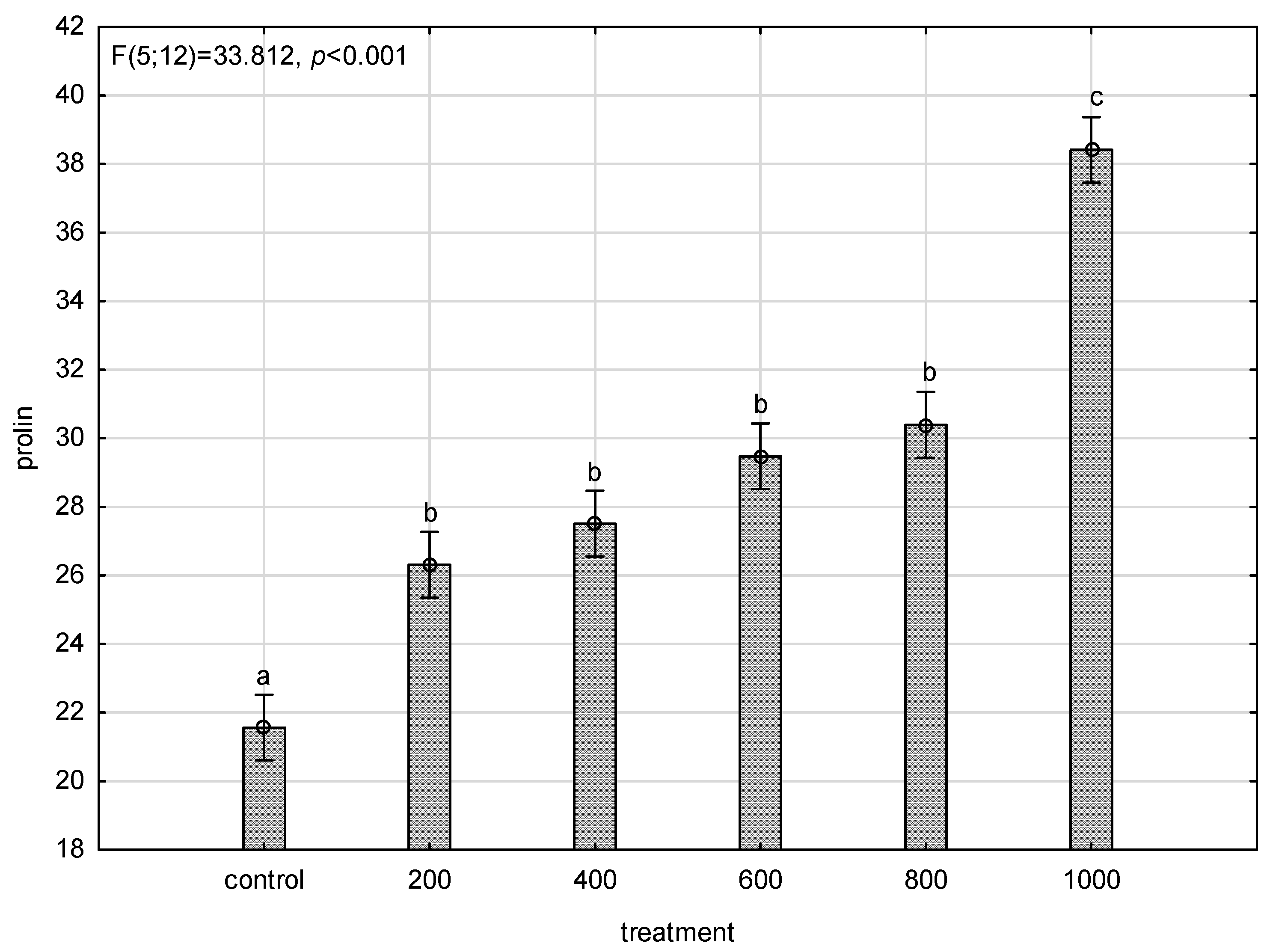
The proline content in the monitored plants is shown in Figure 3. An increase in proline by 78% was recorded in the plants grown in the highest concentration of APAP 1000 mg L−1 compared to the control variant. Thus, the proline content grew in all measured samples compared to the control, but the differences were not significant. In the 200 mg L−1 APAP treatment, the proline content increased by 22% against the control; in the 400 mg L−1 APAP treatment, it was changed by 28%; in the 600 mg L−1 APAP treatment, it was changed by 37%; and in the 800 mg L−1 APAP treatment, it was changed by 41%. This represented a slight shift in response to the contamination that was usually observed between 600 mg L−1 and 800 mg L−1 APAP treatments, but in proline content appeared up to between 800 mg L−1 and 1000 mg L−1 APAP treatments.
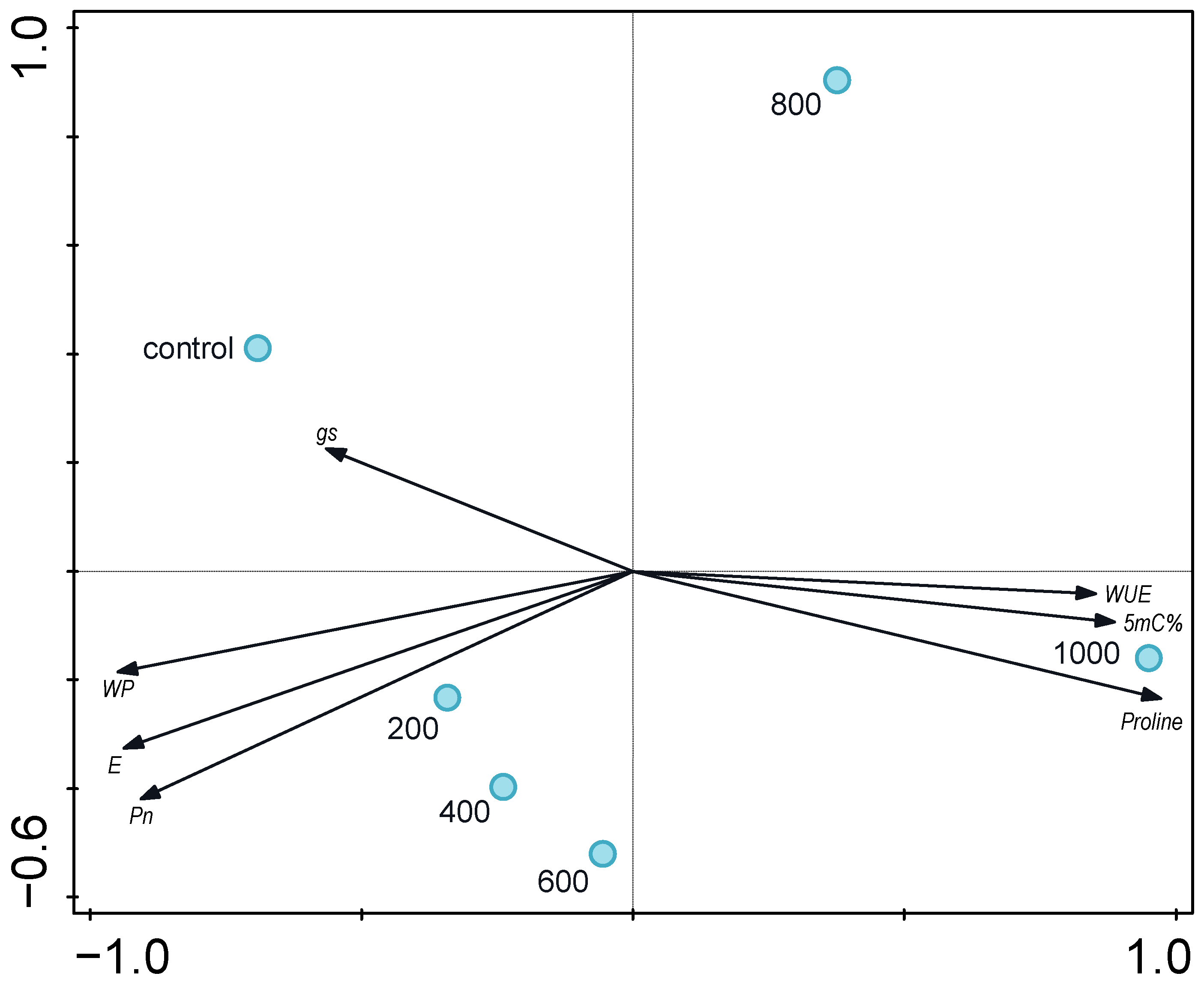
By principal component analysis evaluation (Figure 4), relationships were detected between the measured properties and treatments; the 1000 mg L−1 APAP treatment was shown as most stressful for plants by proline, WUE, and 5 mC% parameters, with the highest detected values against all other treatments and the control variant. Physiological parameters were less affected in the control variant and lower concentrations. Values of transpiration rate, stomatal conductance, and net photosynthetic rate were highest in the control variant, indicating the best photosynthetic apparatus condition. The breaking point concentration was between 600 and 800 mg L−1 of APAP where a significant change in the level of individual indicators was measured, pointing to unequivocal stress, into which the plants pass.

4. Discussion

Photosynthesis is the most important basic biological process supporting growth and nutrient uptake and affecting resistance to various types of biotic and abiotic stresses, and therefore responds sensitively to environmental contaminants such as amoxicillin, ibuprofen, and paracetamol [40,41,42,43,44,45,46,47]. Similarly with the results of [48,49] in lettuce and duckweed [50], it can be stated that low concentrations of paracetamol do not significantly reduce the rate of photosynthesis. This conclusion is probably related to the low rate of inhibition of electron transfer within photosystems and also to the assimilation of CO2. Furthermore, it may be a similar effect like with Brassica juncea [51]—paracetamol in low concentration has a similar effect as the optimal concentration of salicylic acid. This conclusion is also confirmed by [52,53]. In the case of high paracetamol concentrations, their inhibitory effect on the gas exchange rate of juvenile maize plants was confirmed. A similar conclusion was also confirmed for common bean plants [54]. A decrease in the rate of photosynthesis is associated with a reduced ability of the light reactions of photosynthesis. The responses of maize plants to higher concentrations of paracetamol correspond to the results presented by [51], when, according to this author, maize or soybean plants sprayed with higher concentrations of salicylic acid had an inhibitory effect on the rate of photosynthesis.
The rate of transpiration was affected by the concentration of paracetamol in the solution. In the case of high concentrations of paracetamol, a decrease in the rate of transpiration was observed. Changes in the rate of transpiration depending on the concentration of APAP are probably related to changes in the anatomical structure of vascular bundles, especially vessels. Furthermore, the vents are closed and thus the transport of gases and the transpiration flow are reduced. A similar effect occurs in the case of water deficit. This conclusion is presented in the work of [55], who studied the effect of ibuprofen on Vigna unguiculata plants. Similar findings were confirmed in the reaction of soy and corn to the application of salicylic acid and paracetamol [53,56]. Similarly, plants respond to the presence of risk elements in the environment. This effect has been confirmed, for example, in Lolium perenne in the presence of chromium [57] or in Setaria veridis in the presence of cadmium [58]. The reduction in the transpiration rate after exposure to higher concentrations of pollutants is related to their concentration, duration of exposure, and the functioning of the vents. The relationship between transpiration rate and net photosynthetic rate was confirmed by a very strong direct dependence (r > 0.9). This conclusion is in accordance with [59,60,61,62]. Plant sensitivity and changes in transpiration rate may also be influenced by the degree of ontogenesis, when plants in juvenile stages of development or health status usually react more sensitively to pollutants like tetracyclines and sulfonamides in cucumber, tomato, and lettuce [63] and metronidazole in soy [64].
The increased amino acid content of proline is commonly considered to be a side-effect of higher stressors on plants [65], which correlates with the measured results, where it can generally be said that with increasing APAP concentration, a higher production and a subsequent measurement of higher values of proline in biomass were observed. Proline is an osmolyte that also affects the water regime of plants [66], modulates cell redox homeostasis, removes reactive oxygen species [67], but also stabilizes subcellular structures and is a signaling substance [68,69]. The concentration of proline due to the action of a stressor is influenced genotypically, as evidenced in tomatoes [70]. Proline accumulates preferentially in leaves to maintain chlorophyll levels and cellular turgor to protect photosynthetic activity from, for example, salt stress [66] or water deficit [71]. This conclusion is also confirmed by work with diclofenac [72] or paclobutrazol [71]. The accumulation of proline in plants under stress is caused either by induction of the expression of proline biosynthesis genes (P5CS, P5CR) or by the repression of genes of its degradation pathway (PDH silencing) [73].
The DNA methylation status in maize was affected in all APAP-contaminated variants. Compared with other studies, where increased levels of methylated DNA were observed in plants grown in polluted environments [74,75,76,77,78], this trend was confirmed. DNA hypermethylation directly lowers gene expression [25]; specifically, the downregulation of a considerable part of the genes is connected with hormone signaling pathways and the metabolism of oxidative stress regulation [79]. That could be one of the reasons for the worse physiological and photosynthetic parameters of experimental plants.

5. Conclusions

APAP concentrations affected physiological and photosynthetic parameters of plants as well as DNA methylation levels. Changes in the rate of transpiration depending on the concentration of APAP are apparently related to changes in the anatomical structure of vascular bundles, especially vessels. Furthermore, the vents are closed and thus the transport of gases and the transpiration flow are reduced. A similar effect occurs in the case of water deficit. When comparing the values of the measured amount of proline in the final phase of the experiment, an increasing tendency is clearly visible with regard to increasing amounts of APAP. Although proline cannot be fully correlated with the stress level, when including other parameters as mentioned above, DNA methylation and changes in transpiration rate, they form a complex of physiological reactions that can already be perceived in this way. In a comparison of APAP treatments, it was found that in most of the observed parameters, a critical point existed between 600 and 800 mg L−1 APAP concentrations, where the manifestation of stress expressed by individual specific indicators began to intensify significantly.

Author Contributions

Conceptualization, J.K. (Jiří Kudrna); methodology, J.K. (Jiří Kudrna) and M.P.; formal analysis, P.V.; investigation, J.K. (Jiří Kudrna), M.P., F.H., M.L., J.K. (Jan Kubeš), J.Č. and B.T.; writing—original draft preparation, J.K. (Jiří Kudrna) and M.P.; writing—review and editing, J.K. (Jiří Kudrna), M.P. and V.Z. All authors have read and agreed to the published version of the manuscript.

Funding

This research was funded by the EU Project “NutRisk Centre” (grant No. CZ.02.1.01/0.0/0.0/16_019/0000845).

Institutional Review Board Statement

Not applicable.

Informed Consent Statement

Not applicable.

Data Availability Statement

The data presented in this study are available in this article.

Conflicts of Interest

The authors declare no conflict of interest.

References

  1. Ok, Y.S.; Kim, S.-C.; Kim, K.-R.; Lee, S.S.; Moon, D.H.; Lim, K.J.; Sung, J.-K.; Hur, S.-O.; Yang, J.E. Monitoring of selected veterinary antibiotics in environmental compartments near a composting facility in Gangwon Province, Korea. Environ. Monit. Assess. 2011, 174, 693–701. [Google Scholar] [CrossRef]
  2. Boxall, A.B.A.; Rudd, M.A.; Brooks, B.W.; Caldwell, D.J.; Choi, K.; Hickmann, S.; Innes, E.; Ostapyk, K.; Staveley, J.P.; Verslycke, T.; et al. Pharmaceuticals and Personal Care Products in the Environment: What Are the Big Questions? Environ. Health Perspect. 2012, 120, 1221–1229. [Google Scholar] [CrossRef]
  3. OECD. Available online: https://www.oecd-ilibrary.org/sites/5689c05c-en/index.html?itemId=/content/component/5689c05c-en (accessed on 16 May 2023).
  4. Doll, T.E.; Frimmel, F.H. Fate of pharmaceuticals––Photodegradation by simulated solar UV-light. Chemosphere 2003, 52, 1757–1769. [Google Scholar] [CrossRef]
  5. Jjemba, P.K. Excretion and ecotoxicity of pharmaceutical and personal care products in the environment. Ecotoxicol. Environ. Saf. 2006, 63, 113–130. [Google Scholar] [CrossRef]
  6. Choi, K.; Kim, Y.; Park, J.; Park, C.K.; Kim, M.; Kim, H.S.; Kim, P. Seasonal Variations of Several Pharmaceutical Residues in Surface Water and Sewage Treatment Plants of Han River, Korea. Sci. Total Environ. 2008, 405, 120–128. [Google Scholar] [CrossRef]
  7. Fatta-Kassinos, D.; Vasquez, M.I.; Kümmerer, K. Transformation products of pharmaceuticals in surface waters and wastewater formed during photolysis and advanced oxidation processes–degradation, elucidation of byproducts and assessment of their biological potency. Chemosphere 2011, 85, 693–709. [Google Scholar] [CrossRef]
  8. Brodin, T.; Piovano, S.; Fick, J.; Klaminder, J.; Heynen, M.; Jonsson, M. Ecological effects of pharmaceuticals in aquatic systems-impacts through behavioural alterations. Philos. Trans. R. Soc. B Biol. Sci. 2014, 369, 20130580. [Google Scholar] [CrossRef]
  9. Rehman, M.S.U.; Rashid, N.; Ashfaq, M.; Saif, A.; Ahmad, N.; Han, J.I. Global risk of pharmaceutical contamination from highly populated developing countries. Chemosphere 2015, 138, 1045–1055. [Google Scholar] [CrossRef] [PubMed]
  10. Couto, C.F.; Lange, L.C.; Amaral, M.C.S. Occurrence, fate and removal of pharmaceutically active compounds (PhACs) in water and wastewater treatment plants—A review. J. Water Process Eng. 2019, 32, 100927. [Google Scholar] [CrossRef]
  11. Zheng, C.; Liu, J.; Cai, Y.; Jing, C.; Jiang, R.; Zheng, X.; Lu, G. Pharmaceutically active compounds in biotic and abiotic media of rivers receiving urban sewage: Concentrations, bioaccumulation and ecological risk. Process Saf. Environ. Prot. 2022, 166, 491–499. [Google Scholar] [CrossRef]
  12. Kümmerer, K.; Steger-Hartmann, T.; Meyer, M. Biodegradability of the anti-tumour agent ifosfamide and its occurrence in hospital effluents and communal sewage. Water Res. 1997, 31, 2705–2710. [Google Scholar] [CrossRef]
  13. Kuhne, M.; Ihnen, D.; Moller, G.; Agthe, O. Stability of tetracycline in water and liquid manure. J. Vet. Med. Ser. 2000, 47, 379–384. [Google Scholar] [CrossRef] [PubMed]
  14. Torsvik, V.; Goksøyr, J.; Daae, F.L. High diversity in DNA of soil bacteria. Appl. Environ. Microbiol. 1990, 56, 782–787. [Google Scholar] [CrossRef] [PubMed]
  15. Westergaard, K.; Müller, A.K.; Christensen, S.; Bloem, J.; Sørensen, S.J. Effects of tylosin as a disturbance on the soil microbial community. Soil Biol. Biochem. 2001, 33, 2061–2071. [Google Scholar] [CrossRef]
  16. Leitão, I.; Martins, L.L.; Carvalho, L.; Oliveira, M.C.; Marques, M.M.; Mourato, M.P.; Spagnuolo, V.; Capozzi, F. Acetaminophen Induces an Antioxidative Response in Lettuce Plants. Plants 2021, 10, 1152. [Google Scholar] [CrossRef] [PubMed]
  17. Jjemba, P.K.; Robertson, B.K. Antimicrobial Agents with Improved Clinical Efficacy versus Their Persistence in the Environment: Synthetic 4-Quinolone as an Example. EcoHealth 2005, 2, 171–182. [Google Scholar] [CrossRef]
  18. Nunes, B.; Pinto, G.; Martins, L.; Gonçalves, F.; Antunes, S.C. Biochemical and standard toxic effects of acetaminophen on the macrophyte species Lemna minor and Lemna gibba. Environ. Sci. Pollut. Res. 2014, 21, 10815–10822. [Google Scholar] [CrossRef]
  19. Lukens, L.N.; Zhan, S. The plant genome’s methylation status and response to stress: Implications for plant improvement. Curr. Opin. Plant Biol. 2007, 10, 317–322. [Google Scholar] [CrossRef]
  20. Chen, M.; Lv, S.; Meng, Y. Epigenetic performers in plants. Dev. Growth Differ. 2010, 52, 555–566. [Google Scholar] [CrossRef]
  21. Grativol, C.; Hemerly, A.S.; Ferreira, P.C.G. Genetic and Epigenetic Regulation of Stress Responses in Natural Plant Populations. Biochim. Biophys. Acta-Gene Regul. Mech. 2012, 1819, 176–185. [Google Scholar] [CrossRef]
  22. Kohli, R.M.; Zhang, Y. TET Enzymes, TDG and the Dynamics of DNA Demethylation. Nature 2013, 502, 472–479. [Google Scholar] [CrossRef] [PubMed]
  23. Shen, L.; Wu, H.; Diep, D.; Yamaguchi, S.; D’Alessio, A.C.; Fung, H.L.; Zhang, K.; Zhang, Y. Genome-Wide Analysis Reveals TET- and TDG-Dependent 5-Methylcytosine Oxidation Dynamics. Cell 2013, 153, 692–706. [Google Scholar] [CrossRef]
  24. Espinas, N.A.; Saze, H.; Saijo, Y. Epigenetic control of defense signaling and priming in plants. Front. Plant Sci. 2016, 7, 1201. [Google Scholar] [CrossRef] [PubMed]
  25. Thiebaut, F.; Hemerly, A.S.; Ferreira, P.C.G. A Role for Epigenetic Regulation in the Adaptation and Stress Responses of Non-Model Plants. Front. Plant Sci. 2019, 10, 246. [Google Scholar] [CrossRef] [PubMed]
  26. Bossdorf, O.; Arcuri, D.; Richards, C.L.; Pigliucci, M. Experimental alteration of DNA methylation affects the phenotypic plasticity of ecologically relevant traits in Arabidopsis thaliana. Evol. Ecol. 2010, 24, 541–553. [Google Scholar] [CrossRef]
  27. Iwase, Y.; Shiraya, T.; Takeno, K. Flowering and Dwarfism Induced by DNA Demethylation in Pharbitis nil. Physiol. Plant. 2010, 139, 118–127. [Google Scholar] [CrossRef]
  28. Ba, Q.; Zhang, G.; Wang, J.; Niu, N.; Ma, S.; Wang, J. Gene expression and DNA methylation alterations in chemically induced male sterility anthers in wheat (Triticum aestivum L.). Acta Physiol. Plant. 2014, 36, 503–512. [Google Scholar] [CrossRef]
  29. Lechat, M.M.; Brun, G.; Montiel, G.; Véronési, C.; Simier, P.; Thoiron, S.; Pouvreau, J.B.; Delavault, P. Seed response to strigolactone is controlled by abscisic acid-independent DNA methylation in the obligate root parasitic plant, Phelipanche ramosa L. Pomel. J. Exp. Bot. 2015, 66, 3129–3140. [Google Scholar] [CrossRef]
  30. Hoagland, D.R.; Snyder, W.C. Nutrition of Strawberry Plant under Controlled Conditions. (a) Effects of Deficiencies of Boron and Certain Other Elements, (b) Susceptibility to Injury from Sodium Salts. Proc. Am. Soc. Hortic. Sci. 1933, 30, 288–294. [Google Scholar]
  31. Hoagland, D.R.; Arnon, D.I. Growing plants without soil by the water-culture method. Grow. Plants Without Soil Water-Cult. Method 1938, 347, 16. [Google Scholar]
  32. Hoagland, D.R.; Arnon, D.I. The water-culture method for growing plants without soil. Circ. Calif. Agric. Exp. Stn. 1950, 347, 32. [Google Scholar]
  33. Meier, U. Growth Stages of Mono- and Dicotyledonous Plants; BBCH Monograph: Berlin, Germany, 1997. [Google Scholar]
  34. Kuklová, M.; Hniličková, H.; Kukla, J.; Hnilička, F. Environmental impact of the Al smelter on physiology and macronutrient contents in plants and Cambisols. Plant Soil Environ. 2015, 61, 72–78. [Google Scholar] [CrossRef]
  35. Hatfield, J.L.; Dold, C. Water-use efficiency: Advances and challenges in a changing climate. Front. Plant Sci. 2019, 10, 103. [Google Scholar] [CrossRef]
  36. Banks, J.M.; Hirons, A.D. Alternative methods of estimating the water potential at turgor loss point in Acer genotypes. Plant Methods 2019, 15, 34. [Google Scholar] [CrossRef] [PubMed]
  37. Bates, L.S.; Waldren, R.P.; Teare, I.D. Rapid determination of free proline for water-stress studies. Plant Soil 1973, 39, 205–207. [Google Scholar] [CrossRef]
  38. Zemanová, V.; Popov, M.; Pavlíková, D.; Kotrba, P.; Hnilička, F.; Česká, J.; Pavlík, M. Effect of arsenic stress on 5-methylcytosine, photosynthetic parameters and nutrient content in arsenic hyperaccumulator Pteris cretica (L.) var. Albo-lineata. BMC Plant Biol. 2020, 20, 130. [Google Scholar] [CrossRef] [PubMed]
  39. Smilauer, P.; Leps, J. Multivariate Analysis of Ecological Data Using Canoco 5; Cambridge University Press: Cambridge, UK, 2014. [Google Scholar]
  40. Sabourin, L.; Duenk, P.; Bonte-Gelok, S.; Payne, M.; Lapen, D.R.; Topp, E. Uptake of pharmaceuticals, hormones and parabens into vegetables grown in soil fertilized with municipal biosolids. Sci. Total Environ. 2012, 431, 233–236. [Google Scholar] [CrossRef]
  41. Calderon-Preciado, D.; Matamoros, V.; Save, R.; Munoz, P.; Biel, C.; Bayona, J.M. Uptake of microcontaminants by crops irrigated with reclaimed water and groundwater under realfield greenhouse conditions. Environ. Sci. Pollut. Res. 2013, 20, 3629–3638. [Google Scholar] [CrossRef]
  42. Vasquez, M.I.; Lambrianides, A.; Schneider, M.; Kümmerer, K.; Fatta-Kassinos, D. Environmental side effects of pharmaceutical cocktails: What we know and what we should know. J. Hazard. Mater. 2014, 279, 169–189. [Google Scholar] [CrossRef]
  43. Schroeder, L.M.; Blackwell, B.; Klein, D.; Morse, A.N. Rate uptake of three common pharmaceuticals in celery. Apium Graveolens. Water Air Soil Pollut. 2015, 226, 123. [Google Scholar] [CrossRef]
  44. Azanu, D.; Mortey, C.; Darko, G.; Weisser, J.J.; Styrishave, B.; Abaidoo, R.C. Uptake of antibiotics from irrigation water by plants. Chemosphere 2016, 157, 107–114. [Google Scholar] [CrossRef]
  45. Christou, A.; Karaolia, P.; Hapeshi, E.; Michael, C.; Fatta-Kassinos, D. Long-term wastewater irrigation of vegetables in real agricultural systems: Concentration of pharmaceuticals in soil, uptake and bioaccumulation in tomato fruits and human health risk assessment. Water Res. 2017, 109, 24–34. [Google Scholar] [CrossRef] [PubMed]
  46. Di Baccio, D.; Pietrini, F.; Bertolotto, P.; Pérez, S.; Barcelò, D.; Zacchini, M.; Donati, E. Response of Lemna gibba L. to High and Environmentally Relevant Concentrations of Ibuprofen: Removal, Metabolism and Morpho-Physiological Traits for Biomonitoring of Emerging Contaminants. Sci. Total Environ. 2017, 584–585, 363–373. [Google Scholar] [CrossRef]
  47. Christou, A.; Kyriacou, M.C.; Georgiadou, E.C.; Papamarkou, R.; Hapeshi, E.; Karaolia, P.; Michael, C.; Fotopoulos, V.; Fatta-Kassinos, D. Uptake and bioaccumulation of three widely prescribed pharmaceutically active compounds in tomato fruits and mediated effects on fruit quality attributes. Sci. Total Environ. 2019, 647, 1169–1178. [Google Scholar] [CrossRef] [PubMed]
  48. Rede, D.; Santos, L.H.M.L.M.; Ramos, S.; Oliva-Teles, F.; Antão, C.; Sousa, S.R.; Delerue-Matos, C. Individual and mixture toxicity evaluation of three pharmaceuticals to the germination and growth of Lactuca sativa seeds. Sci. Total Environ. 2019, 673, 102–109. [Google Scholar] [CrossRef] [PubMed]
  49. Kudrna, J.; Hnilicka, F.; Kubes, J.; Vachova, P.; Hnilickova, H.; Kuklova, M. Effect of acetaminophen (APAP) on physiological Indicators in Lactuca sativa. Life 2020, 10, 303. [Google Scholar] [CrossRef]
  50. Kummerová, M.; Zezulka, S.; Babula, P.; Triska, J. Possible ecological risk of two pharmaceuticals diclofenac and paracetamol demonstrated on a model plant Lemna minor. J. Hazard. Mater. 2016, 302, 351–361. [Google Scholar] [CrossRef]
  51. Al-Muwayhi, M.A.R. Paracetamol Mediated Changes Modifies the Photosynthetic Efficiency of Vigna Radiata. Legum. Res. 2018, 41, 842–845. [Google Scholar] [CrossRef]
  52. Fariduddin, Q.; Hayat, S.; Ahmad, A. Salicylic Acid Influences Net Photosynthetic Rate, Carboxylation Efficiency, Nitrate Reductase Activity, and Seed Yield in Brassica juncea. Photosynthetica 2003, 41, 281–284. [Google Scholar] [CrossRef]
  53. Khan, W.; Prithiviraj, B.; Smith, D.L. Photosynthetic responses of corn and soybean to foliar application of salicylates. J. Plant Physiol. 2003, 160, 485–492. [Google Scholar] [CrossRef]
  54. Taschina, M.; Copolovici, D.M.; Bungau, S.G.; Andreea, L.P.; Copolovici, L.; Iovan, C. The influence of residual acetaminophen on Phaseolus vulgaris L. Secondary metabolites. Farmacia 2017, 65, 709–713. [Google Scholar]
  55. Mahouachi, J.; Socorro, A.R.; Talon, M. Responses of papaya seedlings (Carica papaya L.) to water stress and re-hydration: Growth, photosynthesis and mineral nutrient imbalance. Plant Soil 2006, 281, 137–146. [Google Scholar] [CrossRef]
  56. Kumar, P.; Lakshmi, N.J.; Mani, V.P. Interactive effects of salicylic acid and phytohormones on photosynthesis and grain yield of soybean (Glycine max L.). Physiol. Mol. Biol. Plants 2000, 6, 179–186. [Google Scholar]
  57. Vernay, P.; Gauthier-Moussard, C.; Hitmi, A. Interaction of bioaccumulation of heavy metal chromium with water relation, mineral nutrition and photosynthesis in developed leaves of Lolium perenne L. Chemosphere 2007, 68, 1563–1575. [Google Scholar] [CrossRef]
  58. Kaznina, N.M.; Titov, A.F.; Batova, Y.V.; Laidinen, G.F. The resistance of plants Setaria veridis (L.) Beauv. to the influence of cadmium. Biol. Bull. 2014, 41, 428–433. [Google Scholar] [CrossRef]
  59. Barcelo, J.; Vazquez, M.D.; Poschenrieder, C.H. Structural and ultrastructural disorders in cadmium-treated bush bean plants (Phaseolus vulgaris L.). New Phytol. 1988, 108, 37–49. [Google Scholar] [CrossRef] [PubMed]
  60. Steudle, E. Water uptake by plant roots: An integration of views. Plant Soil 2000, 226, 45–56. [Google Scholar] [CrossRef]
  61. Javot, H.; Maurel, C. The role of aquaporins in root water uptake. Ann. Bot. 2002, 90, 301–313. [Google Scholar] [CrossRef] [PubMed]
  62. Aggarwal, A.; Sharma, I.; Tripathi, B.; Munjal, A.; Baunthiyal, M.; Sharma, V. Metal toxicity and photosynthesis. In Photosynthesis: Overviews on Recent Progress and Future Perspectives; Itoh, S., Mohanty, P., Guruprasad, K.N., Eds.; IK International Publishing House: New Delhi, India, 2012; pp. 229–236. [Google Scholar]
  63. Ahmed, M.B.M.; Rajapaksha, A.U.; Lim, J.E.; Vu, N.T.; Kim, I.S.; Kang, H.M.; Lee, S.S.; Ok, Y.S. Distribution and Accumulative Pattern of Tetracyclines and Sulfonamides in Edible Vegetables of Cucumber, Tomato, and Lettuce. J. Agric. Food Chem. 2015, 63, 398–405. [Google Scholar] [CrossRef]
  64. Yakubu, O.H. Pharmaceutical wastewater effluent-source of contaminants of emerging concern: Phytotoxicity of metronidazole to soybean (Glycine max). Toxics 2017, 5, 10. [Google Scholar] [CrossRef]
  65. Liang, X.; Zhang, L.; Natarajan, S.K.; Becker, D.F. Proline Mechanisms of Stress Survival. Antioxid. Redox Signal. 2013, 19, 998–1011. [Google Scholar] [CrossRef] [PubMed]
  66. Silva-Ortega, C.O.; Ochoa-Alfaro, A.E.; Reyes-Agüero, J.A.; Aguado-Santacruz, G.A.; Jiménez-Bremont, J.F. Salt stress increases the expression of p5cs gene and induces proline accumulation in cactus pear. Plant Physiol. Biochem. 2008, 46, 82–92. [Google Scholar] [CrossRef] [PubMed]
  67. Soshinkova, T.N.; Radyukina, N.L.; Korolkova, D.V.; Nosov, A.V. Proline and functioning of the antioxidant system in Thellungiella salsuginea plants and cultured cells subjected to oxidative stress. Russ. J. Plant Physiol. 2012, 60, 41–54. [Google Scholar] [CrossRef]
  68. Szabados, L.; Savoure, A. Proline: A multifunctional amino acid. Trends Plant Sci. 2010, 15, 89–97. [Google Scholar] [CrossRef] [PubMed]
  69. Sharma, S.; Villamor, J.G.; Verslues, P.E. Essential role of tissue-specific proline synthesis and catabolism in growth and redox balance at low water potential. Plant Physiol. 2011, 157, 292–304. [Google Scholar] [CrossRef]
  70. Gharsallah, C.; Fakhfakh, H.; Grubb, D.; Gorsane, F. Effect of salt stress on ion concentration, proline content, antioxidant enzyme activities and gene expression in tomato cultivars. AoB Plants 2016, 8, plw055. [Google Scholar] [CrossRef]
  71. Jungklang, J.; Saengnil, K.; Uthaibutra, J. Effects of water-deficit stress and paclobutrazol on growth, relative water content, electrolyte leakage, proline content and some antioxidant changes in Curcuma alismatifolia Gagnep. cv. Chiang Mai Pink. Saudi J. Biol. Sci. 2017, 24, 1505–1512. [Google Scholar] [CrossRef]
  72. Martins, M.; Sousa, B.; Lopes, J.; Soares, C.; Machado, J.; Carvalho, S.; Fidalgo, F.; Teixeira, J. Diclofenac shifts the role of root glutamine synthetase and glutamate dehydrogenase for maintaining nitrogen assimilation and proline production at the expense of shoot carbon reserves in Solanum lycopersicum L. Environ. Sci. Pollut. Res. 2020, 27, 29130–29142. [Google Scholar] [CrossRef]
  73. Marco, F.; Bitrián, M.; Carrasco, P.; Rajam, M.V.; Alcázar, R.; Tiburcio, A.F. Genetic engineering strategies for abiotic stress tolerance in plants. In Plant Biology and Biotechnology; Springer: New Dehli, India, 2015; pp. 579–609. [Google Scholar]
  74. Khan, A.; Yadav, N.S.; Morgenstern, Y.; Zemach, A.; Grafi, G. Activation of Tag1 transposable elements in Arabidopsis dedifferentiating cells and their regulation by CHROMOMETHYLASE 3-mediated CHG methylation. Biochim. Biophys. Acta 2016, 1859, 1289–1298. [Google Scholar] [CrossRef]
  75. Wang, H.; He, L.; Song, J.; Cui, W.; Zhang, Y.; Jia, C.; Francis, D.; Rogers, H.J.; Sun, L.; Tai, P.; et al. Cadmium-induced genomic instability in Arabidopsis: Molecular toxicological biomarkers for early diagnosis of cadmium stress. Chemosphere 2016, 150, 258–265. [Google Scholar] [CrossRef]
  76. Hao, X.F.; Jin, Z.P.; Wang, Z.Q.; Qin, Q.S.; Pei, Y.X. Hydrogen sulfide mediates DNA methylation to enhance osmotic stress tolerance in Setaria italica L. Plant Soil 2020, 453, 355–370. [Google Scholar] [CrossRef]
  77. Chakrabarti, M.; Mukherjee, A. Investigating the Underlying Mechanism of Cadmium-Induced Plant Adaptive Response to Genotoxic Stress. Ecotoxicol. Environ. Saf. 2021, 209, 111817. [Google Scholar] [CrossRef] [PubMed]
  78. Luo, D.; Lu, H.; Wang, C.; Mubeen, S.; Cao, S.; Yue, Y.; Pan, J.; Wu, X.; Wu, Q.; Zhang, H.; et al. Physiological and DNA methylation analysis provides epigenetic insights into kenaf cadmium tolerance heterosis. Plant Sci. 2023, 331, 111663. [Google Scholar] [CrossRef] [PubMed]
  79. Di, X.; Zheng, F.; Norton, G.J.; Beesley, L.; Zhang, Z.; Lin, H.; Zhi, S.; Liu, X.; Ding, Y. Physiological responses and transcriptome analyses of upland rice following exposure to arsenite and arsenate. Environ. Exp. Bot. 2021, 183, 104366. [Google Scholar] [CrossRef]
Figure 1. Microscopic sections of roots in 200× magnification: (a) control variant; (b) 1000 mg L−1 APAP treatment.
Agriculture 13 01333 g001
Figure 2. Levels of 5-methylcytosine. The error bars indicate the standard errors, and the bar height is the mean value. The letters indicate significant differences based on the post hoc Tukey test, assuming p < 0.05.
Agriculture 13 01333 g002
Figure 3. Proline content. The error bars indicate the standard errors, and the bar height is the mean value. The letters indicate significant differences based on the post hoc Tukey test, assuming p < 0.05.
Agriculture 13 01333 g003
Figure 4. Relationships between the treatments and measured properties of the plants evaluated using PCA (principal component analysis) with supplementary variables. The treatments are displayed using color circles and the arrows are measured variables or properties of the plants. The first two axes explain 92.2% of the total variation in the plants’ properties.
Agriculture 13 01333 g004
Table 1. Effect of APAP on water potential (MPa) and photosynthetic parameters.
Water PotentialEgsPnWUE
control−0.95 ± 0.02 a0.98 ± 0.00 *f0.03 ± 0.01 b12.01 ± 0.01 e12.25 ± 0.01 a
200−1.04 ± 0.11 a0.89 ± 0.00 *e0.01 ± 0.00 *a11.60 ± 0.02 d13.08 ± 0.05 b
400−1.38 ± 0.15 a0.81 ± 0.00 *d0.02 ± 0.01 ab11.55 ± 0.02 d14.26 ± 0.02 c
600−1.94 ± 0.20 a0.72 ± 0.00 *c0.01 ± 0.00 *a10.78 ± 0.02 c14.98 ± 0.02 d
800−2.73 ± 0.30 ab0.22 ± 0.00 *b0.01 ± 0.00 *a3.37 ± 0.01 a15.32 ± 0.03 e
1000−4.64 ± 0.21 b0.18 ± 0.00 *a0.01 ± 0.00 *a2.78 ± 0.01 a15.44 ± 0.03 e
p<0.05<0.05<0.05<0.05<0.05
* SE < 0.005. E: transpiration rate (mmol CO2 m−2 s−1); gs: stomatal conductance (mol H2O m−2 s−1), Pn: net photosynthetic rate (μmol CO2 m−2 s−1); WUE: water use efficiency (Pn/E) (μmol CO2 mmol−1 H2O). The letters indicate significant differences based on the post hoc Tukey test, assuming p < 0.05.
Disclaimer/Publisher’s Note: The statements, opinions and data contained in all publications are solely those of the individual author(s) and contributor(s) and not of MDPI and/or the editor(s). MDPI and/or the editor(s) disclaim responsibility for any injury to people or property resulting from any ideas, methods, instructions or products referred to in the content.