Article

Impact of Ecological Factors on the Occurrence and Spatial-Taxonomic Structure of Keratinophilic Fungi and Their Co-Occurrence in Arable Soils

by
Justyna Bohacz
1,
Michał Możejko
1,*,
Teresa Korniłłowicz-Kowalska
1 and
Grzegorz Siebielec
2
1
Department of Environmental Microbiology, Faculty of Agrobioengineering, University of Life Sciences in Lublin, Leszczyńskiego 7 Street, 20-069 Lublin, Poland
2
Department of Soil Science Erosion and Land Protection, Institute of Soil Science and Plant Cultivation—State Research Institute, Czartoryskich 8, 24-100 Puławy, Poland
*
Author to whom correspondence should be addressed.
Agriculture 2022, 12(2), 194; https://doi.org/10.3390/agriculture12020194
Submission received: 2 December 2021 / Revised: 17 January 2022 / Accepted: 26 January 2022 / Published: 31 January 2022
(This article belongs to the Section Agricultural Soils)

Abstract

:
Fungi that decompose keratinized animal remains are an important component of the arable soil microbiome. The aim of the study is to characterize the communities of keratinophilic and co-inhabiting (non-keratinophilic) fungi in four cultivated soils that differ in physico-chemical properties, with particular emphasis on granulometric fractions, which have so far been omitted from studies concerning the ecology of these micromycetes. Fungi were isolated using the keratin-baiting method. Fungal species identification was carried out on the basis of their macro- and micromorphological features. The Simpson diversity index and Marczewski–Steinhaus similarity index were calculated for precise determination of the relationships between fungal communities. In the studied soils, Trichophyton ajelloi and Ctenomyces serratus dominated among keratinophilic fungi, while Purpureocillium lilacinum and Metacordyceps chlamydosporia, from the orders Eurotiales and Hypocreales, were dominant among non-keratinophilic fungi. The frequency of keratinophilic fungi was significantly positively correlated with pH and the content of two granulometric fractions, as opposed to non-keratinophilic fungi. This was reflected in the higher growth rates of keratinomycetes in loamy soil, chernozem, and rendzina, i.e., soils with a higher content of silt and clay fractions compared to sandy soil characterized by a high content of sand fractions. The species composition of both groups of fungi was most similar between loamy soil and chernozem, whereas the greatest differences were found for sandy soil and rendzina. Chernozem was characterized by the highest diversity of fungal species from both groups of fungi. The study, in addition to providing information about ecological factors, provided a collection of keratinomycete strains that can be used as a starting material for subsequent research stages regarding keratinolytic activity of these fungi and their potential use in agricultural practices.

1. Introduction

Fungi that colonize and decompose animal remains rich in keratin, i.e., proteins with high nitrogen and sulfur content, are present in various environments. Surface and deep soil layers containing keratin matter, most often the hair of small mammals (mainly rodents), bird feathers, and other keratinized animal remains, are the largest reservoirs and natural habitats of these fungi [1,2,3,4,5,6,7,8]. An additional source of native keratin in cultivated soils are organic fertilizers, e.g., manure containing keratin residues (bristles, hair, and feathers) of farm animals and domestic birds. Native keratin-decomposing fungi are involved in the circulation of elements, mainly nitrogen and sulfur, in natural ecosystems and agroecosystems [3,9]. Keratinophilic fungi play a key role among these organisms. This ecological Ascomycota group specializes in the decomposition of native keratin. The group comprises the so-called geophilic dermatophytes and related fungi from the Chrysosporium group [10,11]. They are saprotrophs, but some, e.g., Microsporum gypseum (Nannizzia gypsea), are potentially pathogenic to humans and animals as they can cause skin mycoses [10]. However, as reported by Simpanya [12], soil isolates of this dermatophyte, compared to those originating from animals, have low pathogenicity and only very virulent strains are able to cause infections. In addition to keratinophilic fungi, keratin residues in soils are colonized by various less-substrate-specialized fungi, which are conventionally referred to as non-keratinophilic species [13]. Their presence is associated with the heterogeneous composition of keratin residues, which, in addition to various forms of pure keratin, also contain various organic compounds, mainly non-keratin proteins [3]. The criterion for discriminating between keratinolytic and non-keratinolytic fungi was specified by Kunert [14] as the degree of degradation of native keratin (hair) used as the only source of C, N, S, and energy in 8-week liquid cultures. Fungi that decomposed at least 40% of native keratin in these conditions were classified as keratinolytic fungi, and the rest were referred to as non-keratinolytic species. Korniłłowicz-Kowalska [15] showed that typically keratinolytic fungi, such as the geophilic dermatophytes Arthroderma quadrifidum (Trichophyton terrestre teleomorph) and Chrysosporium keratinophilum (Aphanoascus keratinophilus teleomorph), decomposed 80–90% of the substrate in 8-week liquid cultures, incubated at 20 °C, containing chicken feathers as the only source of C, N, S, and energy. In turn, non-keratinolytic fungi, e.g., Paecilomyces lilacinus, decomposed slightly more than 20% of the feather mass [15]. In a study on the decomposition of waste feathers by geophilic fungi from the Chrysosporium group (Aphanoascus fulvescens and Chrysosporium articulatum), Bohacz [16] reported that approximately 20% of total nitrogen from feather keratin was released in the form of peptides and amino acids, and 26% to 46% was released in the form of ammonium ions in the cultures of all fungal strains.
The ability of keratinophilic fungi to biodegrade native keratin is determined by the secretion of substrate-specific proteases called keratinases [9,17,18] and the ability to break keratin disulfide bridges responsible for its resistance to “normal” proteolytic enzymes [14]. In contrast to non-keratinophilic species, keratinophilic fungi produce specific morphological structures composed of vegetative mycelium, which is responsible for the mechanical destruction of native keratin, and secrete lytic factors involved in keratinolysis [3].
Keratin residues, which are the only source of C, N, and S for keratinophilic fungi, not only provide these fungi with nutrients and energy, but also create a less competitive environment facilitating their survival [18]. The distribution and diversity of keratinolytic mycobiota in soil depends not only on the presence of keratinized animal remains, but also on many other ecological factors [2,19,20]. Garg et al. [19] included here: (i) climatic factors such as temperature and light, especially UV radiation and seasonal variations, (ii) soil factors, i.e., soil pH, abundance of carbonate, nitrogen and sulfur, moisture, humus, presence of heavy metals, fatty acids and fats, salts and agrochemicals, and (iii) biotic factors, i.e., birds and animals—the source of keratin substrate. CaCO3 and available phosphorus are listed among other ecological factors determining the growth of keratinomycetes, including geophilic dermatophytes in cultivated soils [18,21]. Soil pH is one of the most important of the aforementioned factors [12,18,19,20,22,23,24,25,26]. The research conducted so far has shown that humus-rich soils with neutral pH are characterized by the highest keratinomycete species richness, while humus-poor acidic soils are colonized by fungal biota with a low diversity of species composition [2,18,25]. Studies on the ecological determinants of the community structure of fungi colonizing and decomposing keratinized animal remains in cultivated soils (arable soils) have rarely been conducted [12,26].
Since there is a lack of in-depth information on this subject, the main aim of the present study is to carry out a comprehensive assessment of the diversity of fungi inhabiting keratinized biomass in cultivated soils differing in their physico-chemical properties, i.e., granulometric composition, organic matter and nitrogen content, pH, and several other parameters. The adopted hypothesis assumes that the presence of keratinophilic fungi and co-occurrence of non-keratinophilic fungi in arable soils was determined by chemical and physical soil properties, especially the granulometric composition. Several indicators of the frequency and diversity of organisms colonizing the environment were used to achieve the research objectives. Correlation analysis was performed to determine their relationships with soil environmental parameters. An analysis of variance was also performed to show significant differences between individual soils in a wider spectrum. Additionally, as part of the study, a collection of keratinomycete strains was obtained that can be used as a starting material for further research concerning keratinolytic activity of these fungi and their potential applications in agricultural practices.

2. Materials and Methods

2.1. Collection of Soil Samples

The experimental material consisted of four soils with different physico-chemical properties. They were sampled in the Lublin province (south-eastern Poland) on two private farms in Sobieszyn (soil I: 51°59′85.73 N, 22°13′34.30 E and II: 51°59′68.00 N, 22°13′46.96 E) and Żulice (soil III: 50°53′57.20 N, 23°78′26.85 E) and on a farm in Bezek (soil IV: 51°19′ N, 23°25′ E) belonging to the University of Life Sciences in Lublin. Soil samples were collected in early autumn in 2018 (at the turn of the month from September to October). A representative sample from each soil was collected into sterile foil bags from 10 evenly distributed sites of a given field, from a depth of 0–20 cm, and mixed. Each soil was placed in a separate bag. The final mass of the collected representative sample from each soil was approx. 10 kg. After transfer to the laboratory, the soil was sieved through a 2 mm mesh and mixed.

2.2. Chemical Determinations

Air-dried soil samples were ground in a porcelain mortar, sieved through a ø2 mm mesh, milled, homogenized, and stored in paper bags at 20 °C. Chemical determinations were performed according to certified reference materials (CRM) and internal laboratory standards.
Soil pH was measured in H2O using a combined glass electrode in both water and 1 M KCl slurry at a soil/solution ratio of 1:2 (v/v). Total carbon (C tot.) and nitrogen (N tot.) were analyzed by combustion using a CN analyzer (Elementar Analysensysteme GmbH, Hanau, Germany). Total organic C (C org.) was determined after sulfochromic oxidation, followed by titration of excess K2Cr2O7 with [FeSO4(NH4)2SO4] 6H2O (PN-ISO 14235, 2003).
Soil texture was determined using a Mastersizer 2000 laser diffraction particle size analyzer (Malvern Instruments, Worcestershire, UK).
Total P, K, Mg, and Ca contents were measured after sample digestion in a 3:1 mixture of concentrated HNO3:HCl in Teflon PFA vessels in a microwave accelerated reaction system (MarsXpress; CEM Corp., Matthews, NC, USA), followed by elemental analysis in the extracts using ICP-MS (Agilent 7500ce).
Assimilable phosphorus was measured using the Egner-Riehm colorimetric method after extraction with calcium lactate (0.02 M) in diluted HCl (0.01 M). Subsequently, colorimetric measurements, based on reaction with ammonium molybdate, were carried out in a Perkin Elmer Lambda 45 spectrometer. Assimilable potassium (K) was measured after the same extraction by AAS using AAnalyst 800 (Perkin Elmer, Waltham, MA, USA). Carbonates in the soils were determined by dissolving it with hydrochloric acid and measuring CO2 production.
Soil type and agricultural usability complex were determined using a digital soil map at a scale of 1:25,000, managed by the Institute of Soil Science and Plant Cultivation-State Research Institute, Poland, based on soil sampling location. The soil type derived from the national map was then translated into World Reference Base (WRB) classification (IUSS Working Group WRB [27]). Soil I and II were classified as Cambisols with particle size distribution of light loamy sand and sandy loam, respectively. In the text, these soils are conventionally defined as sandy and loamy soil, respectively. Soil III was classified as Chernozem with the texture of silt loam, while soil IV was identified as Leptosol with particle size distribution of silt loam. According to the data of the IUSS Working Group WRB [27], Leptosols on a limestone substrate are called Rendzina; therefore, Leptosol in this study was referred to as rendzina or calcareous soil.
In terms of the agricultural use classification, soil I represented poor rye complex 6, and soil III represented very good wheat complex 1, whereas soils II and IV belonged to defective wheat complex 3. The detailed characterization of the soils is presented in Table 1.

2.3. Mycological Analyses

2.3.1. Substrate

Broiler chicken feathers supplied by a poultry processing plant (Superdrob) in Lublin, Poland, were used as a substrate for the isolation of keratinophilic fungi. Feathers were thoroughly washed to remove any residual post-slaughter waste. Feathers were subsequently dried and shredded into fragments <5 mm. Feathers were sterilized by gassing to maintain the tertiary structure of keratin proteins (disulfide bridges).

2.3.2. Isolation of Keratinophilic Fungi

Keratinophilic fungi were isolated using the keratin bait method [2]. For each soil sample, 50 Petri dishes with a diameter of 180 mm were filled with approx. 30 g of soil, which corresponded to approx. 1/3 of the plate volume. In total, 1500 g portions of each soil were transferred onto the Petri dishes. Then, an even layer of chicken feathers was laid over the entire surface. The dishes were placed in a humid chamber, and the soil was periodically moistened as needed. Incubation was carried out at room temperature.
After 3–4 weeks of incubation in a humid chamber, the degree of fungal colonization of feathers was assessed. Mycelium visible to the naked eye was collected from each Petri dish, containing soil sampled from six randomly selected zones located at a distance, and transferred onto a plate with a Sabouraud medium for keratinolytic fungi (g dm−3): glucose (40), peptone (10), agar (20), 30 mg dm−3 streptomycin and 2 mg dm−3 chlortetracycline, and 500 mg dm−3 actidione (an antifungal antibiotic). The fungi were cultured in an incubator at 26 °C for 1–2 weeks (fast-growing fungi) or 2–3 weeks (slow-growing fungi). After this time, the mycelium was transferred to slants with Sabouraud medium without antibiotics and actidione and re-incubated at 26 °C for 2–3 weeks to obtain pure cultures, which were identified based on their macro- and micromorphological traits.

2.3.3. Identification of Fungi

The fungal genera and species were identified microscopically using an Olympus BS-41 microscope equipped with a CVIII4 camera with Cell-A software, and macroscopically by observing fungal growth on plates and slants. Microscopic evaluation consisted of the preparation of microcultures of pure fungal cultures and the observation of the shape and size of spores and other reproductive structures, e.g., fruiting bodies, and the ability to produce chlamydospores. Macroscopic evaluation was based on the observation of colony size and structure, color of the obverse and reverse, and the ability to release pigments into the substrate. In both cases, the systematic studies conducted by Domsch et al. [28], Kwaśna et al. [29], Nelson et al. [30], and van Oorschot [11] were used for final identification. The current nomenclature of the analyzed fungi was verified in the Index Fungorum: www.indexfungorum.org.

2.4. Result Analysis

The number of colonized soil samples, number of fungal genera, species and strains isolated from each soil sample, as well as the number of species per plate/soil sample were included in the analysis of the frequency of fungal occurrence. It was assumed that one soil sample was colonized by only one strain of a given species. To determine dominant species, the species dominance coefficients were calculated from the formula [31]:
D = 100 (Sa: S)
where S is the sum of isolates in a soil sample, and Sa is the sum of isolates of a given species.
The Simpson diversity index (D) [32] and the Marczewski–Steinhaus similarity index (S) [33] were calculated to precisely determine the relationships between fungal communities colonizing the analyzed soils.
The Simpson index, based on the probability theory, was calculated using the following formula:
D = 1 i = 1 S ( p i 2 )
where pi is the proportion of isolates of species “i” in a given fungal community, with pi = ni/N (ni is the number of isolates (strains) of species “i”, and N is the total number of strains). The values of the Simpson index can range from 0 (low diversity) to 1–1/S, where S is the number of species in the community. The greater the diversity, the greater the value of the Simpson index.
The index of species similarity of fungal communities was calculated using the formula:
S (1,2) = w/(a + b − w)
where a is the number of species in community 1, b is the number of species in community 2, and w is the number of species common to both communities
The spatial and taxonomic structure of fungal communities was also estimated, assuming, as in Korniłłowicz-Kowalska et al. [34], that the taxonomic structure of fungal communities is a proportional number of species from a given taxonomic group (orders) in relation to the total number of species expressed in %. In turn, the spatial structure is a proportional number of records (= number of strains) per representatives of the same taxonomic group (orders) in relation to the total number of records (strains) expressed in %. The following scale was used to assess the frequency of fungi (strains, species, genera, and orders): <1% is sporadic, 1–5% is rare; 6–10% is common, 11–25% is very common, 26–50% is abundant, and >50% is very abundant.

2.5. Statistical Analyses

Pearson’s r-correlation analysis was performed to determine the interrelationships between the studied indices, i.e., the number of isolates, species, orders, groups of microorganisms, and the physicochemical parameters of the soils. The data were correlated at three levels of significance: α = 0.05, 0.01, and 0.001 using Arstat (University of Life Sciences, Lublin, Poland) and Statistica software ver.13.3 (StatSoft, Kraków, Poland). One-way analysis of variance (ANOVA), followed by Tukey’s post hoc test, with a significance level of α = 0.05, was carried out to demonstrate the differences between the soils in terms of the frequency of occurrence of keratinophilic and non-keratinophilic fungi using Statistica software ver.13.3 (StatSoft, Kraków, Poland).

3. Results

3.1. Growth Indices of Keratinophilic and Non-Keratinophilic Fungi in the Analyzed Soils

The degree of colonization of the plates/soil samples by fungi growing on fragmented chicken feathers reached 95.5%, indicating that 191 out of 200 plates were colonized. Keratinophilic (= keratinolytic) fungi were detected in 76% of the samples (152 plates). Sandy soil exhibited the lowest degree of colonization by these fungi, i.e., only 24%. The other soils, i.e., loamy soil, chernozem, and rendzina, exhibited a very high degree of colonization by keratinophilic fungi in the range of 90–98%, with the highest value recorded for chernozem samples. Sandy soil, loamy soil and chernozem were characterized by a predominance of geophilic dermatophytes, which accounted for 22%, 90%, and 92%, respectively, of which 4%, 2%, and 64% were Chrysosporium representatives, respectively. In turn, keratinophilic fungal biota in rendzina samples was represented only by a monoculture of non-dermatophytic fungi constituting 90% of the colonizing species (Table 2). Chernozem samples were characterized by the highest richness of keratinophilic fungal taxa, as five genera and seven species were identified. These samples also showed the greatest abundance of keratinophilic species per one plate/soil sample. In contrast, the lowest abundance of keratinophilic mycobiota was found in sandy soil (Table 2). In comparison to other soil types, loamy soil was characterized by a higher taxon richness (genera and species) of non-keratinophilic fungi co-occurring with keratinomycetes (Table 2). Analysis of the variance of the number of isolated strains of keratinophilic fungi showed that the frequency of these fungi in rendzina was significantly different from that in chernozem and sandy soil, but not from that in loamy soil. The lowest frequency of occurrence of these fungi was recorded in sandy soil and the highest in chernozem. With respect to non-keratinophilic fungi, all four arable soils were found to be significantly different from each other. Among them, rendzina had the significantly lowest frequency of these fungi, while loamy soil the highest (Table 2).

3.2. General Characteristics of Keratinophilic and Non-Keratinophilic Species Composition

Table 3 presents the characteristics of the species composition (including the number of isolates) of keratinophilic and non-keratinophilic fungi isolated from the analyzed soils. In total, 1000 isolates of keratinophilic and non-keratinophilic fungi were obtained from the four studied soils; 430 isolates, constituting 43% of all isolated fungi, were keratinophilic species (20 isolates from sandy soil, 132 from loamy soil, 154 from chernozem, and 124 from calcareous soil). The percentage of keratinomycetes in fungal biota colonizing native feather keratin was 13%, 41%, 47.5%, and 64%, respectively. Based on phenotypic characteristics, the fungi were classified into five genera and seven species. Non-keratinophilic fungi (570 isolates) were represented by 19 genera and 19 species, and 106 isolates were not identified. Of the total number of isolated micromycetes, non-keratinophilic fungi accounted for 87%, 59%, 58%, and 36% in sandy soil, loamy soil, chernozem, and rendzina, respectively. Assuming that only one strain of a given species was isolated from one soil sample, we estimated that the overall frequency of keratinophilic fungi per 1 kg of fresh soil was 13 colony-forming units (CFU) in sandy soil, 89 CFU in loamy soil, 103 CFU in chernozem, and 83 CFU in rendzina. According to the same estimation principle, the values for non-keratinophilic fungi colonizing native keratin were 90, 128, 115, and 47 CFU kg−1 soil fresh weight, respectively.
Trichophyton ajelloi from the geophilic dermatophyte group and Ctenomyces serratus from the Chrysosporium group were the dominant keratinophilic fungi. Trichophyton ajelloi accounted for 90–93% of the isolated dermatophytic fungi (240 isolates in total). The highest number of isolates of this species was recorded in loamy soil (129 isolates), while sandy soil was colonized by the lowest number, i.e., 18 isolates. This species was not detected in rendzina samples. Another geophilic dermatophyte, Microsporum gypseum, was isolated from loamy soil and chernozem and constituted 1.5% and 6.5% of the isolates, respectively (Table 3).
Ctenomyces serratus was found in a total number of 166 isolates, which corresponded to 93% of Chrysosporium group representatives; it almost exclusively colonized calcareous soil samples (124 isolates), where it accounted for 100% of all keratinophilic fungi isolated from this soil. The lowest abundance of Ctenomyces serratus was noted in loamy soil (single isolate), and it did not occur in sandy soil (Table 3). Three other species from the Chrysosporium group, i.e., Chrysosporium tropicum, Ctenomyces vellereus, and Pseudogymnoascus pannorum (formerly Chrysosporium pannorum), and an unidentified Chrysosporium sp. were isolated from the tested soils. All these fungi represented approx. 7% of the Chrysosporium group.
In the group of non-keratinophilic (co-occurring) fungi, Metacordyceps chlamydosporia strains (formerly Verticillium chlamydosporium) were isolated most frequently. Populations of this species represented 23% and 13% of non-keratinophilic and all fungi, respectively (Table 3). Purpureocillium lilacinum (formerly Paecilomyces lilacinus) also occurred with a high frequency (101 isolates) and accounted for 18% of non-keratinophilic fungi and 10% of all fungi isolated from the soils. Fusarium spp. (10%) and Penicillium sp. (17%) belonged to frequently or very frequently detected non-keratinophilic fungi. Single isolates of five species, i.e., Acremonium rutilum, Cladosporium cladosporioides, Penicillium glabrum, Sarocladium kiliense, and Sarocladium strictum, were also isolated (Table 3).

3.3. Species Dominance Coefficients and the Taxonomic and Spatial Structure of Fungi Colonizing Native Keratin

Species identification of fungal strains isolated from the soils (Table 3) and species dominance coefficients calculated based on these data (Figure 1A) indicated that Penicillium simplicissimum was the most abundant fungus colonizing native feather keratin in sandy soil samples. This soil was colonized by 27 isolates of this fungus. They constituted 17% of all fungi in total and 20% of non-keratinophilic fungi. In comparison to other isolates, Trichophyton ajelloi and Purpureocillium lilacinum had a high frequency (18 isolates), accounting for 12% of total fungal isolates obtained from sandy soil. According to the adopted frequency scale, these species can be considered as very common. At the same time, Trichophyton ajelloi was a dominant species with 90% frequency in the group of keratinophilic fungi (Figure 1A). Lecanicillium psalliotae and Talaromyces purpureogenus were classified as common (8% frequency) in the group of non-keratinophilic fungi isolated from this soil (Figure 1A).
In loamy soil, 48% of all fungi isolated from native feather keratin (132 isolates) belonged to keratinophilic fungi (Figure 1B, Table 3). The dominant species was Trichophyton ajelloi (129 isolates), which accounted for 98% of the keratinomycete population and 40% of all fungi colonizing feathers on this soil (Figure 1B). In the group of non-keratinophilic fungi, a high frequency was recorded for three species, i.e., Purpureocillium lilacinum (54 isolates), Metacordyceps chlamydosporia (49 isolates), and Cunninghamella elegans (48 isolates). They represented 17%, 15%, and 15% of the total number of isolates, and 28%, 26%, and 25% of non-keratinophilic fungi, respectively (Figure 1B).
Similar to loamy soil, chernozem was the richest environment in terms of the number of species colonizing native feather keratin (Figure 1C). In total, 326 fungal isolates were isolated from this soil. Keratinophilic fungi accounted for almost half of the isolates (47%) and Trichophyton ajelloi was the dominant species. The Trichophyton ajelloi population constituted 29% of the fungal community colonizing feathers in chernozem samples. It represented 60% of the keratinomycete group, which classified this species as very abundant. The frequency of Ctenomyces serratus from the Chrysosporium group was also high (Figure 1C). The frequency of this species of all fungi isolated from feathers was 13% and 27% in the group of keratinophilic fungi; therefore, it could be considered an abundant species in the analyzed chernozem. Metacordyceps chlamydosporia was a non-keratinophilic species with the highest frequency in chernozem samples. It represented 18% of all fungi isolated from feathers and 38% of non-keratinophilic fungi (Figure 1C).
Rendzina was the only soil in which keratinophilic fungi dominated as a group (Figure 1D). This was evidenced by the fact that they accounted for over 64% of micromycete biota colonizing feathers. In addition, this calcareous soil was the only soil that was not colonized by geophilic dermatophytes. The samples contained only non-dermatophytic keratinophilic fungi represented by a Ctenomyces serratus monoculture (Figure 1D). In the group of non-keratinophilic fungi, Metacordyceps chlamydosporia was the most frequent species isolated from this soil, as in chernozem samples. The isolates of this fungus accounted for 11% of the total number of isolates and 30% in the group of non-keratinophilic fungi (Figure 1D).
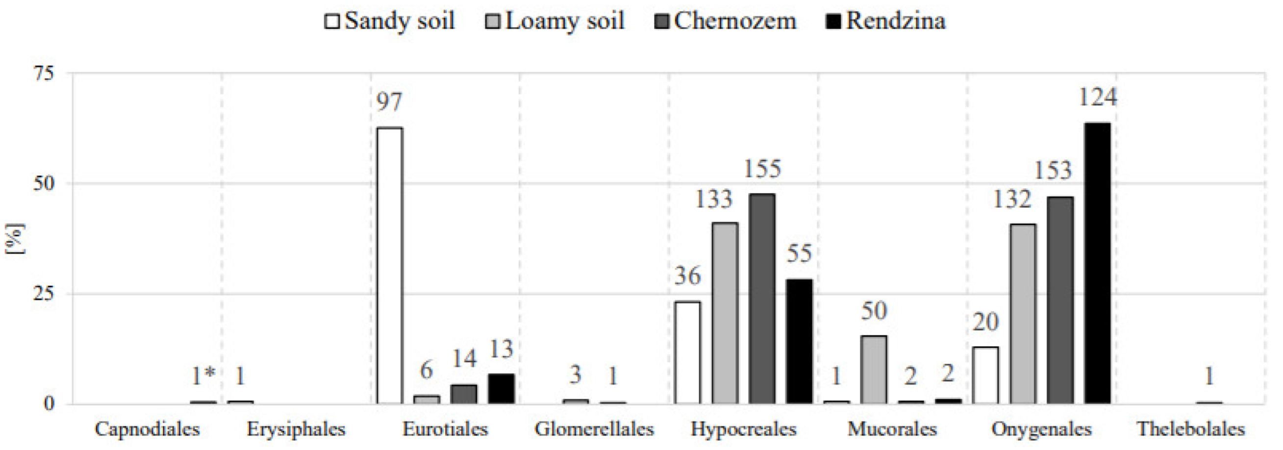
The data in Figure 2 and Figure 3 show that the fungal communities colonizing and decomposing native feather keratin in the analyzed soils represent seven Ascomycota orders: Capnodiales, Erysiphales, Eurotiales, Glomerellales, Hypocreales, Onygenales, and Thelebolales, and one order of Zygomycota (Mucorales). The order Onygenales was represented by keratinophilic fungi and the remaining orders were represented by non-keratinophilic fungi. In terms of the taxonomic structure, the order Hypocreales was characterized by the highest species richness, whereas Capnodiales, Erysiphales, and Thelebolales were represented by the lowest number of species (Figure 2). The species richness of the orders Eurotiales and Onygenales was lower, but not as low as the three orders mentioned above. Chernozem samples were found to have the richest taxonomic structure of native keratin-colonizing fungi. This was manifested by the presence of 15 species from six orders, including nine species from the order Hypocreales and six representatives of Onygenales. Other remaining soil samples contained keratin-colonizing species from five orders. Rendzina samples (12 species) showed the lowest species richness (Figure 2). In terms of the spatial structure, the largest number of records was recorded for Hypocreales from the group of non-keratinophilic fungi and Onygenales from the group of keratinomycetes (Figure 3). The highest number of records (orders) of keratinophilic and non-keratinophilic fungi was found in loamy soil and chernozem (324 and 326 isolates, respectively). They were mainly represented by Hypocreales and Onygenales. The spatial structure of mycobiota colonizing and decomposing native feather keratin in sandy soil and calcareous soil was dominated by Eurotiales and Onygenales, respectively (Figure 3).

3.4. Species Similarity and Diversity in the Analyzed Fungal Communities

The calculated value of the Marczewski–Steinhaus species similarity index (S) provided information on the similarity between the analyzed environments in terms of fungal species occurrence. It indicated that the species similarity in the group of fungi colonizing native feather keratin in the analyzed soils was relatively low, as it exceeded 50% in only one case. The highest number of common species was recorded for loamy soil and chernozem (53.80%), while the lowest (43.50%) was found for sandy soil and rendzina (Table 4).
Species diversity was analyzed using the Simpson index, taking into account species frequency. The lower the value of the Simpson species diversity index (D), the lower the diversity of the fungal community with the dominance of a single species. The values of this coefficient in the communities of keratinophilic and non-keratinophilic (co-occurring) fungi in the analyzed soils are listed in Table 5. Considering all micromycetes (both groups of fungi) colonizing native feather keratin, sandy soil and chernozem exhibited the greatest species diversity (D = 0.8057 and D = 0.8533, respectively), and the lowest value was observed for calcareous soil samples (D = 0.5753). Chernozem samples in the group of keratinophilic fungi were characterized by the greatest species diversity, whereas the lowest value of this coefficient was again obtained for rendzina samples (D = 0.5587 and D = 0.00, respectively) (Table 5). Rendzina and chernozem samples in the communities of non-keratinophilic fungi were characterized by greater species diversity than sandy and loamy soils (Table 5). In contrast to keratinophilic fungi, the values of the Simpson coefficient in this fungal group were high and ranged from 0.7617 to 0.8467 (Table 5).

3.5. Correlations between the Frequency of Occurrence of Fungi and Soil Properties

The analysis was performed based on the frequency of all keratinophilic fungi with a dominant species and all non-keratinophilic fungi.
The presence of the ecological group of keratinophilic fungi was significantly correlated with soil pH and its granulometric composition (Table 6). This was evidenced by an increase in the frequency of these fungi at higher pH values and in soils with silt (ø 0.05–0.002 mm) and clay (ø < 0.002 mm) fractions, while it decreased with increasing sand fraction content (ø 2.0–0.05 mm). Moreover, potassium content was found to significantly stimulate the frequency of this fungal group in soil. Non-keratinophilic fungi were more abundant in soils with a wider spectrum of macronutrients, i.e., total P, Ca, and Mg, and assimilable forms of P and K. Their occurrence in the soils was not supported by increased content of organic carbon compounds, organic carbon, total N and C, or the presence of CaCO3.
Correlation analysis of the effect of soil properties on the occurrence frequency of the most abundant keratinomycete species, i.e., Ctenomyces serratus from the Chrysosporium group and Trichophyton ajelloi, representing geophilic dermatophytes, revealed that the frequency of the former species was more strongly correlated with certain edaphic factors than the frequency of the latter species. The frequency of Ctenomyces serratus population increased with increasing pH, organic matter content, total C and N, assimilable phosphorus and potassium forms, CaCO3 accumulation, and clay fractions. In turn, the occurrence of this species was low in soils with a high proportion of sand fraction (r = −0.604*). Unfavorable factors affecting Trichophyton ajelloi occurrence included the presence of CaCO3 (negative correlation) and an increase in total carbon content (r = −603*). Additionally, high content of assimilable phosphorus and potassium in soil adversely affected the occurrence of this geophilic dermatophyte (negative correlation). This effect was demonstrated by correlation analyses between the frequency of Trichophyton ajelloi and total Mg and Ca content. Although no significant correlation was found between the frequency of Trichophyton ajelloi and soil pH, organic carbon, organic matter, and total N, as well as the negative values of the r coefficient calculated for these parameters (Table 6), suggested that the frequency of this fungus decreased with increasing pH and organic matter and nitrogen contents.
Correlation coefficients between the properties of the analyzed soils and the frequency of Onygenales, as a taxonomic group of keratinomycetes, confirmed the relationships observed for keratinophilic fungi as an ecological group (Table 7). It was found that the frequency of these fungi in soil was positively correlated with soil pH and silt and clay fractions, while negatively correlated with sand fraction content. Moreover, the abundance of Onygenales in soil increased with increasing in potassium content.
The occurrence of Hypocreales, i.e., the order represented by the majority of the isolated non-keratinophilic fungi colonizing native feather keratin, was positively correlated with silt fraction content and negatively correlated with sand fraction content. The occurrence of Eurotiales, i.e., the second most frequent order of non-keratinophilic fungi in the soils, was positively correlated with sand fraction levels, while negatively correlated with silt and clay fractions and soil pH.
Table 6 does not show values of the correlation coefficients (r) for the remaining keratinophilic fungal species because of the low number of records. Similarly, the values of taxonomic units (orders) with low frequency of occurrence are not included in Table 7.

4. Discussion

Most studies on the occurrence of keratinophilic fungi have focused on the epidemiological aspect [8,24,35,36,37], as soil is regarded as one of the reservoirs of pathogenic dermatophytes and other related fungal pathogens with keratinolytic properties [38]. However, from the perspective of soil environmental function, the fungal biota with the ability to decompose keratin proteins and other less assimilable fibrous proteins, such as elastin or collagen, is an important component of soil communities. It is involved in transformations of N and organic S, as well as in providing easily available forms of these elements for the microbiome of soil and plant roots.
The experiments conducted in this study showed the lowest frequency of keratinophilic fungi in sandy soil and the highest frequency in chernozem. Similarly, the highest fungal richness and diversity were observed in chernozem. This was confirmed by an analysis of variance, sandy soil, and chernozem that differed significantly from each other and from two other soils (loamy soil and rendzina—insignificant differences) in relation to the keratinomycetes inhabiting them. With respect to non-keratinophilic fungi, all soils were found to be significantly different from each other. As reported by Korniłłowicz-Kowalska and Bohacz [2], this was associated with the so-called animalization of this soil (concept introduced by Garg et al. [19]). This was indicated by the presence of numerous traces of animals living in this soil, e.g., rodent burrows, mole corridors, etc. Animalization contributes to the enrichment of chernozem soil with native keratin. Furthermore, the pH of these soils, usually slightly acidic or neutral, enhances the growth of most species of keratinophilic fungi [2,13,18,26]. This was confirmed in the present study by the positive correlation between the occurrence of these fungi and soil pH, as the frequency of the fungi increased with increasing soil pH. Additionally, it was demonstrated for the first time that the frequency of keratinomycetes was correlated with the content of sand, silt, and clay fractions. The total frequency of keratinomycetes was found to be higher with increasing content of silt (ø = 0.05–0.002) and clay (ø < 0.002 mm) fractions, but it decreased with an increase in the sand fraction proportion (ø = 2.0–0.05). Data in Table 1 demonstrated that sandy soil had the highest content of sand fractions and the lowest of clay and silt fractions. The lowest frequency of occurrence and low species diversity (Simpson’s coefficient = 0.1850) of keratinomycetes corresponded to these data. In turn (Table 1), chernozem was characterized by the lowest content of sand fractions, the highest of clay fractions, and the highest of silt. The highest frequency and species diversity (Simpson’s coefficient = 0.5587) of keratinophilic fungi were recorded in this soil. This suggests that the growth of keratinophilic fungi, as an ecological-physiological group, is enhanced in soils with a particle size distribution associated with an increased sorption capacity (clay fractions) and water-holding capacity (silt and clay fractions). Consequently, soil richness in nutrients, e.g., cations sorbed by clay minerals, and soil water retention are increased. The influence of granulometric fractions on the physico-mechanical and physico-chemical properties of soils has been well-described in numerous publications [39,40]. In contrast, the growth and species diversity of keratinomycetes are not promoted by excessively permeable and airy soils (sandy soils) because of their high levels of sand fractions and the weak sorption complex (very low content of clay minerals), and thus low nutrient levels. Similar preferences for the physical and chemical properties of soils were indicated by positive correlation coefficients for Onygenales-Ascomycota, i.e., a taxonomic group comprising all isolated species of dermatophytes and the Chrysosporium group (except for Pseudogymnoascus pannorum). The growth rates of keratinophilic fungi in the other two soil types, loamy soil and rendzina, were lower than in chernozem, but higher than in sandy soil, except for the fungal species richness and diversity in calcareous soil samples. The growth rates of these fungi in the latter soil were lower than in sandy soil (species monoculture). Therefore, it could be assumed that the nutritional and air-water conditions were generally favorable for keratinomycetes in both of these soil types because of the higher content of silt and clay fractions and lower of sand fractions compared to sandy soil. These observations are supported by the results of Bohacz and Korniłłowicz-Kowalska [18], who showed a positive correlation of the frequency of keratinophilic fungi in the soil containing fractions with a diameter less than 0.02 mm, i.e., silt and clay fractions. However, with regard to rendzina, the CaCO3 content and pH of this soil were of decisive importance for the occurrence of keratinomycetes, as presented later in the chapter.
The results of the present study indicated selection within the keratinomycete community. This was reflected in the dominance of two of seven recorded species of fungi: Trichophyton ajelloi from the group of geophilic dermatophytes and Ctenomyces serratus from the Chrysosporium group. Both of these species are characteristic of the temperate and cool climate zones. These mesophilic fungi do not grow at temperatures above 37 °C [28]. The frequency of occurrence of Trichophyton ajelloi and Ctenomyces serratus populations in the analyzed soils showed an opposite trend. The Trichophyton ajelloi population dominated in sandy soil, loamy soil, and chernozem, but was absent in rendzina samples. In turn, the Ctenomyces serratus population was the only keratinophilic species colonizing rendzina samples. It was abundant in chernozem samples, appeared occasionally in loamy soil, and was absent in sandy soil. A similar phenomenon was previously reported by Korniłłowicz [13] and Korniłłowicz-Kowalska and Bohacz [2]. Based on the significant negative correlation between the frequency of Trichophyton ajelloi and Ctenomyces serratus populations in 17 cultivated soils, the authors [2] indicated a potential antagonism between these fungi based on their different soil pH preferences. Trichophyton ajelloi is an acidophilic and acid-tolerant species, whereas Ctenomyces serratus is an alkaliphilic and alkali-tolerant species [19,41]. This has also been confirmed by the results of the present study. The correlation analysis proved that the frequency of Ctenomyces serratus increased with increasing soil pH and was significantly positively correlated with the content of CaCO3 and mineral colloids, organic matter levels, and total N, i.e., clay and silt fractions. The highest levels of these components were mainly determined in rendzina samples. It was the only soil with a CaCO3 content and a neutral pH value and the highest level of clay minerals, organic matter, and nitrogen, as well as assimilable phosphorus and potassium forms. Hence, we believe that Ctenomyces serratus prefers neutral or slightly alkaline macroelement-rich soils with a good sorption complex. In contrast, highly acidic, barren, excessively permeable, and airy soils with a high content of sand fractions and low content of organic matter and silt and clay fractions do not support the growth of this species of keratinomycetes. However, these soil properties did not limit the occurrence and growth of Trichophyton ajelloi populations in the soils tested. This species was clearly dominant in strongly acidic (sandy) to acidic (loamy soil and chernozem) soils with no CaCO3 content, and its occurrence did not depend significantly on the level of organic matter and nitrogen, but was positively influenced by macronutrient contents (P, Mg, Ca, and K).
Microsporum gypseum deserves attention in the group of the remaining five species of keratinophilic fungi, which accounted for only 5.6% in this group. This potentially pathogenic geophilic dermatophyte was isolated almost exclusively from chernozem samples. The presence of this fungus in this type of soil and its absence in other types of arable soil was previously reported by Korniłłowicz [13], as well as by Korniłłowicz-Kowalska and Bohacz [2]. These authors found that the presence of this fungus in chernozem was primarily determined by its animalization, as this fungal species often colonizes hair and habitats containing this type of native keratin. The abundance of Microsporum gypseum in soils containing animal hair was also demonstrated by Kačinová et al. [5].
Non-keratinophilic fungi, co-occurring in the soils with keratinomycetes on native keratin “debris”, represented a diverse microbiome in terms of abundance and taxonomy. In most of the analyzed soils, except for rendzina samples, non-keratinophilic mycobiota constituted a more abundant community of native keratin colonizers than the population of keratinomycetes colonizing this substrate. A greater number of non-keratinophilic fungi, as a group, compared to keratinophilic fungi, colonizing keratin residues in soils was previously reported by Moallaei et al. [7] and Zarrin and Haghgoo [42]. In this study, the overall frequency of non-keratinophilic fungi and the number of species per soil sample were in the following order: loamy soil> chernozem> sandy soil> rendzina. This distribution was slightly different in terms of the total species richness, i.e., chernozem> loamy soil> sandy soil> rendzina. Higher contents of phosphorus and monovalent and divalent cations (K, Mg, and Ca) were the only edaphic factors that exerted the most favorable effect on the overall abundance of these fungi. Other soil parameters, i.e., the level of organic nitrogen, CaCO3, or assimilable P and K forms, were significantly negatively correlated with the frequency of these fungi. These correlations were clearly different from the relationships observed in the group of keratinophilic fungi. In contrast to keratinomycetes, this group comprised mainly ubiquitous substrate-unspecialized fungi, widespread in various soil types. The species composition of these fungi was similar to that reported by Korniłłowicz [13] in a study on native keratin colonization in cultivated soils with different properties. Most of the genera and species of non-keratinophilic fungi detected on keratin residues in the soils tested in the present study, i.e., Penicillium spp., Fusarium spp., and Purpureocillium lilacinum, were also isolated by Moallaei et al. [7].
The present study demonstrated that edaphic factors had a more significant impact on the frequency (spatial structure) of the dominating taxonomic groups, i.e., Hypocreales and Eurotiales-Ascomycetes, in comparison to the overall frequency of non-keratinophilic fungi. The spatial structure (colonization frequency) of Eurotiales was reduced with an increase in soil fertility parameters (negative correlation with organic matter, silt and clay fractions, total N, and assimilable potassium) and soil pH. In contrast, the frequency of Eurotiales increased with increasing soil acidification and deterioration of soil fertility properties, e.g., trophic and air-water conditions (greater soil airiness and permeability related to a higher percentage of sand fractions). This corresponded to the accumulation of Eurotiales in sandy soil, mainly of the genera Penicillium and Purpureocillium. This can be explained by the fact that they mostly belong to acidophilic and xerophilic drought-resistant fungi (low water activity coefficient—aw) [28,43]. The spatial structure of Hypocreales showed a frequency shift toward the other studied soils: loamy soil, chernozem, and rendzina. In terms of the edaphic conditions, this was reflected in a significantly higher frequency with increasing silt fractions and decreasing sand fraction contents. The occurrence of these fungi was also favored by a higher level of such macronutrients as K, Mg, and Ca. This group included the genera Fusarium and Verticillium, which have higher nutritional and water requirements (higher aw values) compared to Penicillium and Purpureocillium [28,43].
In the species structure, the highest dominance coefficients in the group of 19 recorded non-keratinophilic species were obtained for Metacordyceps chlamydosporia (formerly Verticillium chlamydosporium) from the order Hypocreales-Ascomycota and Purpureocillium lilacinum (formerly Paecilomyces lilacinus) representing Eurotiales-Ascomycota. These species more frequently colonized native keratin in loamy soil and chernozem than in sandy soil and calcareous soil. Korniłłowicz [13], in a study on sandy, loamy, chernozem, and black earth soils, reported a similar tendency in soil population distribution of these non-keratinophilic feather colonizers. These fungi were also frequently isolated from keratin waste (chicken feathers) with damaged keratin structures [44]. Considering these data and the assumption that population size is an indicator of the metabolic activity of microbial species present in the community [34], we postulate that species co-occurring with keratinomycetes, e.g., Purpureocillium lilacinum and Metacordyceps chlamydosporia, are involved in native keratin decomposition in the analyzed arable soils. These fungi, especially Purpureocillium lilacinum, most likely degraded simpler proteins present in feathers and/or utilized protein products of keratinolysis because of their proteolytic abilities [28]. Most of the remaining non-keratinophilic species [28], less abundantly colonizing native keratin in the analyzed soils, were attributed with proteolytic abilities.
The analysis of species diversity of non-keratinophilic mycobiota using Simpson’s diversity indices, assigning particularly high importance to abundant species [32], revealed their high diversity in all soils, but the highest diversity was recorded in chernozem samples (0.8269). Similarly, the highest species diversity of all mycobiota (keratinophilic and non-keratinophilic fungi) colonizing native feather keratin was also found in chernozem. Therefore, this soil showed the greatest potential of its fungal biota involved in the colonization and decomposition of keratinized animal remains. This soil shared the highest number of species with loamy soil, as evidenced by the highest value of the Marczewski–Steinhaus similarity index for these soils compared to other soil pairs. The least common species were found by comparing the species composition of fungi colonizing native keratin in sandy soil and rendzina. The differences in pH, organic matter content, total N, fraction proportions, and macronutrients between loamy soil and chernozem were substantially smaller than between sandy soil and rendzina.
Therefore, we conclude that fungal communities colonizing and decomposing native keratin in different soils exhibit greater similarity when differences in soil properties are smaller, and vice versa: the larger the differences in soil properties, the lower the species similarity.
Considering the present results indicating a low degree of colonization of sandy soils by keratinophilic fungi, we propose that fertilization with composts containing keratin waste may be one of the methods of improving the condition of these soils. Bohacz and Korniłłowicz-Kowalska [45] and Bohacz [46,47] have demonstrated that composts produced from such keratin waste as chicken feathers together with plant material are rich in keratinophilic fungi and have a high content of assimilable nitrogen and sulfur forms, high humus-forming potential, and properties ensuring good air-water relations in soils. Moreover, as demonstrated by Bohacz [16], keratinophilic fungi can also serve as natural bio-fertilization agents. Since sandy soils, and especially loamy sands, constitute a large proportion (16.2%) of the structure of arable land in Poland [48], the use of such composts would not only improve their fertility, but also activate native keratinomycete biota, as in the case of natural soil “animalization”. Furthermore, it would also be a rational disposal method for insufficiently or improperly managed chicken-feather waste.
The collection of keratinophilic fungal strains obtained in this study also can be used as starting material for research regarding keratinolytic activity of these fungi and their potential use in agricultural practices.

Author Contributions

Conceptualization, J.B. and T.K.-K.; methodology, J.B., T.K.-K., M.M. and G.S.; validation, J.B., T.K.-K. and M.M.; formal analysis, M.M. and J.B.; investigation, M.M, J.B., T.K.-K. and G.S.; resources, J.B., T.K.-K., M.M. and G.S.; data curation, M.M. and J.B.; writing—original draft preparation, J.B. and M.M.; writing—review and editing, J.B., M.M. and T.K.-K.; visualization, M.M. and J.B.; project administration, J.B. and M.M.; funding acquisition, J.B. All authors have read and agreed to the published version of the manuscript.

Funding

This research was funded by subsidies from the Ministry of Science and Higher Education for the maintenance and development of research potential (research task RKM/S/21/2021).

Institutional Review Board Statement

Not applicable.

Informed Consent Statement

Not applicable.

Data Availability Statement

All data is contained within the article.

Acknowledgments

Research financed by subsidies from the Ministry of Science and Higher Education for the maintenance and development of research potential (research task RKM/S/21/2021).

Conflicts of Interest

The authors declare no conflict of interest.

References

  1. Chmel, L.; Vláčilíková, A. The ecology of keratinophilic fungi in different depths of soil. Sabouraudia 1975, 13, 185–191. [Google Scholar] [CrossRef] [PubMed]
  2. Korniłłowicz-Kowalska, T.; Bohacz, J. Some correlations between the occurrence frequency of keratinophilic fungi and selected soil properties. Acta Mycol. 2002, 37, 101–116. [Google Scholar] [CrossRef] [Green Version]
  3. Korniłłowicz-Kowalska, T.; Bohacz, J. Biodegradation of keratin waste: Theory and practical aspects. Waste Manag. 2011, 31, 1689–1701. [Google Scholar] [CrossRef] [PubMed]
  4. Javoreková, S.; Labuda, R.; Maková, J.; Novák, J.; Medo, J.; Majerčíková, K. Keratinophilic fungi isolated from soils of long-term fold-grazed, degraded pastures in National Parks of Slovakia. Mycopathologia 2012, 174, 239–245. [Google Scholar] [CrossRef] [PubMed]
  5. Kačinová, J.; Tančinová, D.; Labuda, R. Keratinophilic fungi in soils stressed by occurrence of animals. J. Microbiol. Biotechnol. Food Sci. 2013, 2, 1436–1446. [Google Scholar]
  6. Volleková, A. Keratinophilic fungi in rodents’ burrows and in their adjoining surroundings. Czech Mycol. 1985, 39, 97–105. [Google Scholar]
  7. Moallaei, H.; Zaini, F.; Pihet, M.; Mahmoudi, M.; Hashemi, J. Isolation of keratinophilic fungi from soil samples of forests and farm yards. Iran. J. Public Health 2006, 35, 62–69. [Google Scholar]
  8. Deshmukh, S.K.; Verekar, S.A.; Chavan, Y.G. Incidence of Keratinophilic fungi from the selected soils of Kaziranga National Park, Assam (India). Mycopathologia 2017, 182, 371–377. [Google Scholar] [CrossRef] [PubMed]
  9. Filipello-Marchisio, V. Keratinophilic fungi: Their role in nature and degradation of keratinic substrates. In Biology of Dermatophytes and Other Keratinophilic Fungi; Kushwaha, R.K.S., Guarro, J., Eds.; Revista Iberoamericana de Micología: Bilbao, Spain, 2000; Volume 17, pp. 86–92. [Google Scholar]
  10. Otčenášek, M.; Dvořák, J. Ecological classification of dermatophytes. Mycoses 1975, 18, 425–434. [Google Scholar] [CrossRef]
  11. Van Oorschot, C.A.N. A revision of Chrysosporium and allied genera. Stud. Mycol. 1980, 20, 1–89. [Google Scholar] [CrossRef]
  12. Simpanya, M.F. Dermatophytes: Their taxonomy, ecology and pathogenicity. In Biology of Dermatophytes and Other Keratinophilic Fungi; Kushwaha, R.K.S., Guarro, J., Eds.; Revista Iberoamericana de Micología: Bilbao, Spain, 2000; Volume 12, pp. 1–12. [Google Scholar]
  13. Korniłłowicz, T. The frequency of occurrence and distribution of keratinophilic fungi in some arable soils. Acta Mycol. 1993, 28, 3–17. [Google Scholar] [CrossRef]
  14. Kunert, J. Physiology of keratinophilic fungi. In Biology of Dermatophytes and Other Keratinophilic Fungi; Kushwaha, R.K.S., Guarro, J., Eds.; Revista Iberoamericana de Micología: Bilbao, Spain, 2000; Volume 17, pp. 77–85. [Google Scholar]
  15. Korniłłowicz-Kowalska, T. Studies on the decomposition of keratin wastes by saprotrophic microfungi. I. Criteria for evaluating keratinolytic activity. Acta Mycol. 1997, 32, 51–79. [Google Scholar] [CrossRef] [Green Version]
  16. Bohacz, J. Biodegradation of feather waste keratin by a keratinolytic soil fungus of the genus Chrysosporium and statistical optimization of feather mass loss. World J. Microbiol. Biotechnol. 2017, 33, 13. [Google Scholar] [CrossRef] [Green Version]
  17. Takiuchi, I.; Sei, Y.; Takagi, H.; Negi, M. Partial characterization of the extracellular keratinase from Microsporum canis. Sabouraudia 1984, 22, 219–224. [Google Scholar] [CrossRef]
  18. Bohacz, J.; Korniłłowicz-Kowalska, T. Species diversity of keratinophilic fungi in various soil types. Cent. Eur. J. Biol. 2012, 7, 259–266. [Google Scholar] [CrossRef]
  19. Garg, A.P.; Gandotra, S.; Mukrji, K.G.; Pugh, G.J.F. Ecology of keratinophilic fungi. Proc. Plant Sci. 1985, 94, 149–163. [Google Scholar] [CrossRef]
  20. Al-Musallam, A.A. Distribution of keratinophilic fungi in desert soil of Kuwait. Mycoses 1989, 32, 296–302. [Google Scholar] [CrossRef]
  21. Ciesielska, A.; Bohacz, J.; Korniłłowicz-Kowalska, T.; Stączek, P. Microsatellite-primed PCR for intra-species genetic relatedness in Trichophyton ajelloi strains isolated in Poland from various soil samples. Microbes Environ. 2014, 29, 178–183. [Google Scholar] [CrossRef] [PubMed] [Green Version]
  22. Böhme, H.; Ziegler, H. The distribution of geophilic dermatophytes and other keratinophilic fungi in relation to the pH of the soil. Mycopathol. Mycol. Appl. 1969, 38, 247–255. [Google Scholar] [CrossRef] [PubMed]
  23. Sharma, R.; Swati, K. Prevalence of keratinophilic fungi at various pH in different areas of Jaipur, Rajasthan. J. Microbiol. Biotechnol. Res. 2014, 4, 17–21. [Google Scholar]
  24. Kumawat, T.K.; Sharma, A.; Sharma, V.; Chandra, S.; Bhadauria, S. A study on the prevalence of keratinophilic fungal biota of semi-arid region of Rajasthan, India. J. King Saud Univ. Sci. 2020, 32, 1014–1020. [Google Scholar] [CrossRef]
  25. Chmel, L.; Hasilíková, A.; Hrašco, J.; Vláčilíková, A. The influence of some ecological factors on keratinophilic fungi in the soil. Sabouraudia 1972, 10, 26–34. [Google Scholar] [CrossRef] [PubMed]
  26. Korniłłowicz, T. Studies on mycoflora colonizing raw keratin wastes in arable soil. Acta Mycol. 1992, 27, 231–245, In Polish. [Google Scholar] [CrossRef] [Green Version]
  27. IUSS Working Group WRB. World Reference Base of Soil Resources. International Soil Classification System for Naming Soils and Creating Legends for Soil Maps; Update 2015; World Soil Recourses Reports No. 106; FAO: Rome, Italy, 2015. [Google Scholar]
  28. Domsch, K.H.; Gams, W.; Anderson, T.H. Compendium of Soil Fungi, 2nd ed.; IHW: Eching, Germany, 2007. [Google Scholar]
  29. Kwaśna, H.; Chełkowski, J.; Zajkowski, P. Polish Flora. Fungi (Mycota) v.22 (Deuteromycetes), (Hyphomycetales), (Fusarium); Polish Scientific Publishing House: Warsaw-Krakow, Poland, 1991. (In Polish) [Google Scholar]
  30. Nelson, P.E.; Toussoun, T.A.; Morasas, W.F.O. Fusarium Species: An Illustrated Manual for Identification; The Pennsylvania State University Press: University Park, PA, USA, 1983. [Google Scholar]
  31. Trojan, P. General Ecology; Polish Scientific Publishing House: Warsaw, Poland, 1975. (In Polish) [Google Scholar]
  32. Krebs, C.J. Ecology. The Experimental Analysis of Distribution and Abundance, 4th ed.; Harper Collins: New York, NY, USA, 1994. [Google Scholar]
  33. Szewczyk, W. Soil fungi communities from young Scots pine plantations affected with root rot. Acta Mycol. 2007, 42, 239–244. [Google Scholar] [CrossRef] [Green Version]
  34. Korniłłowicz-Kowalska, T.; Wojdyło-Kotwica, B.; Bohacz, J.; Możejko, M.T. Biodiversity of saprotrophic fungi of the root zone of grasses and clovers in a permanent meadow in a post-bog habitat in relation to the growing season and fertilization (Zaklęsłość Sosnowicka, Western Polesye). In Environmental Engineering in Polesye, Polish Polesye; Urban, D., Dobrowolski, R., Jeznach, J., Eds.; International Scientific Publications: Brest, Belarus; Rivne, Ukraine; Warsaw, Poland; Ryazan, Russia, 2020; pp. 485–538. (In Polish) [Google Scholar]
  35. Anane, S.; Hashim, M.; Al-Yasiri, Y.; Normand, A.C.; Ranque, S. Distribution of keratinophilic fungi in soil across Tunisia: A descriptive study and review of the literature. Mycopathologia 2015, 180, 61–68. [Google Scholar] [CrossRef]
  36. Anbu, P.; Hilda, A.; Gopinath, S.C.B. Keratinophilic fungi of poultry and feather dumping soil in Tamil Nadu, India. Mycopathologia 2004, 158, 303–309. [Google Scholar] [CrossRef]
  37. Agrawal, S.; Nandeibam, J.; Devi, I. Danger of exposure to keratinophilic fungi and other dermatophytes in recreational place in the northeast region of India. Aerobiologia 2021, 37, 755–766. [Google Scholar] [CrossRef]
  38. Ajello, I. Soil as a natural reservoir for human pathogenic fungi. Science 1956, 123, 876–879. [Google Scholar] [CrossRef]
  39. De Jong, R.; Campbell, C.A.; Nicholaichuk, W. Water retention equations and their relationship to soil organic matter and particle size distribution for disturbed samples. Can. J. Soil Sci. 1983, 63, 291–302. [Google Scholar] [CrossRef]
  40. Owczarzak, W.; Dębicki, R.; Mocek, A. Physical properties of soils. In Soil Science, 1st ed.; Mocek, A., Ed.; Polish Scientific Publishing House: Warsaw, Poland, 2015; pp. 131–188. (In Polish) [Google Scholar]
  41. Hubálek, Z. Keratinophilic fungi associated with free living mammals and birds. In Biology of Dermatophytes and Other Keratinophilic Fungi; Kushwaha, R.K.S., Guarro, J., Eds.; Revista Iberoamericana de Micología: Bilbao, Spain, 2000; Volume 17, pp. 104–108. [Google Scholar]
  42. Zarrin, M.; Haghgoo, R. Survey of keratinophilic fungi from soils in Ahvaz, Iran. Jundishapur J. Microbiol. 2011, 4, 191–194. [Google Scholar]
  43. Grant, C.; Hunter, C.A.; Flannigan, B.; Bravery, A.F. The moisture requirements of moulds isolated from domestic dwellings. Int. Biodeterior. 1989, 25, 259–284. [Google Scholar] [CrossRef]
  44. Korniłłowicz, T. Studies on the mycroflora colonizing keratin-bark-urea manure. Acta Mycol. 1993, 28, 19–30. [Google Scholar] [CrossRef] [Green Version]
  45. Bohacz, J.; Korniłłowicz-Kowalska, T. Fungal diversity and keratinolytic activity of fungi from lignocellulosic composts with chicken feathers. Process. Biochem. 2019, 80, 119–128. [Google Scholar] [CrossRef]
  46. Bohacz, J. Changes in mineral forms of nitrogen and sulfur and enzymatic activities during composting of lignocellulosic waste and chicken feathers. Environ. Sci. Pollut. Res. 2019, 26, 10333–10342. [Google Scholar] [CrossRef] [PubMed] [Green Version]
  47. Bohacz, J. Composts and water extracts of lignocellulosic composts in the aspect of fertilization, humus-forming, sanitary, phytosanitary and phytotoxicity value assessment. Waste Biomass Valorization 2019, 10, 2837–2850. [Google Scholar] [CrossRef] [Green Version]
  48. Krasowicz, S.; Oleszek, W.; Horabik, J.; Dębicki, R.; Jankowiak, J.; Stuczyński, T.; Jadczyszyn, J. Racjonalne gospodarowanie środowiskiem glebowym Polski. Pol. J. Agron. 2011, 7, 43–58. (In Polish) [Google Scholar]
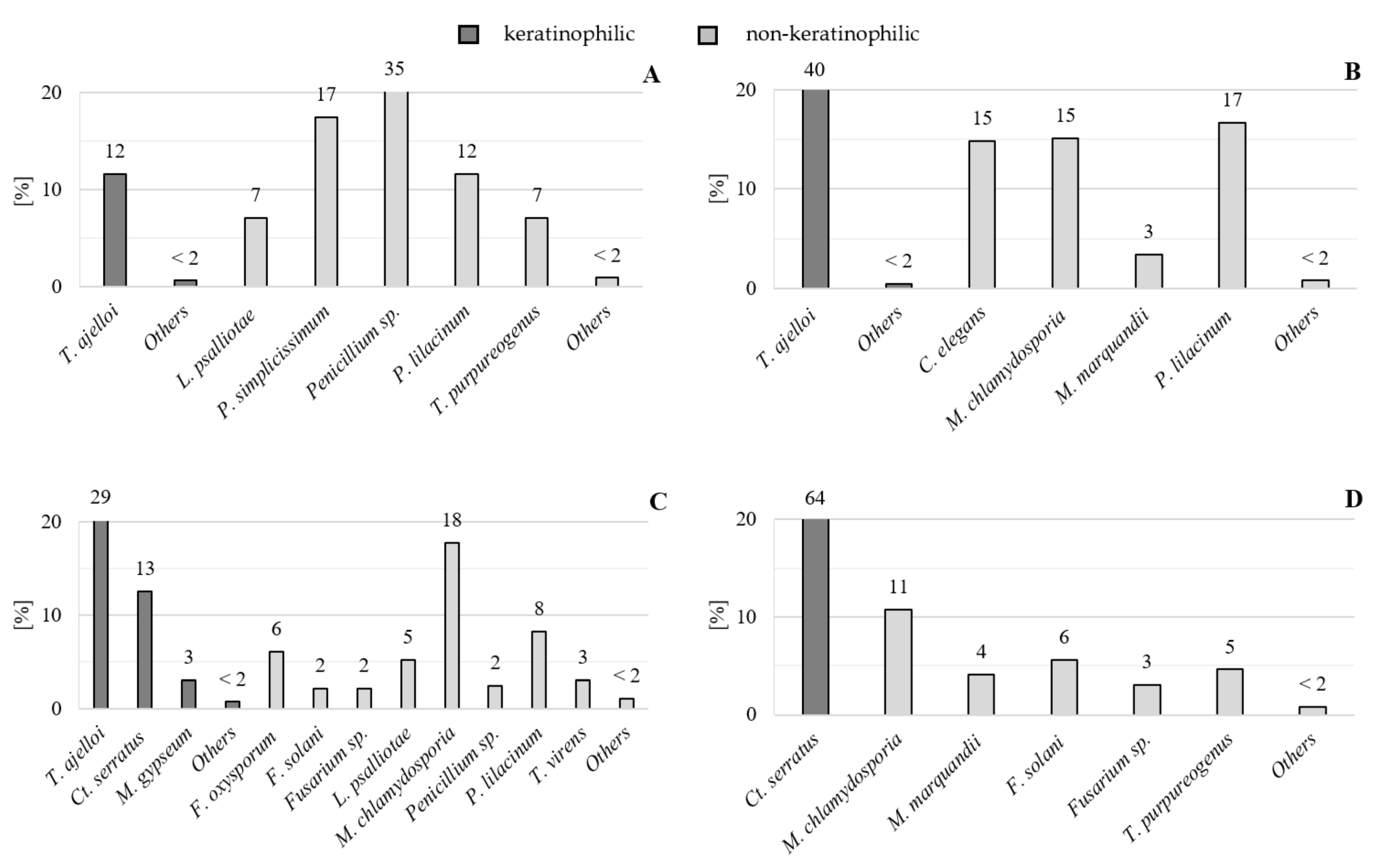
Figure 1. Species dominance coefficients [%] of keratinophilic and non-keratinophilic fungi in the analyzed soils (A) sandy soil; (B) loamy soil; (C) chernozem; (D) rendzina); explanations: Ct. serratus (Ct. serratus), F. oxysporum (Fusarium oxysporum), F. solani (Fusarium solani), L. psalliotae (Lecanicillium psalliotae), M. chlamydosporia (Metacordyceps chlamydosporia), M. gypseum (Microsporum gypseum), M. marquandii (Metarhizium marquandii), P. lilacinum (Purpureocillium lilacinum), P. simplicissimum (Penicillium simplicissimum), T. ajelloi (Trichophyton ajelloi), T. purpureogenus (Talaromyces purpureogenus), T. virens (Trichoderma virens), other fungi (fungi with dominance below 2).
Agriculture 12 00194 g001
Figure 2. Taxonomic structure [%] of keratinophilic and non-keratinophilic fungi in cultivated soils; explanations: * number of species.
Agriculture 12 00194 g002
Figure 3. Spatial structure [%] of keratinophilic and non-keratinophilic fungi in cultivated soils; explanations: * number of isolates.
Agriculture 12 00194 g003
Table 1. Particle size distribution and chemical characterization of the analyzed soils.
ParameterSoil Type/Particle Size Distribution
Cambisol/Loamy SandCambisol/Sandy LoamChernozem/Silt LoamLeptosol/Silt Loam
C org.
[%]
0.591.021.453.16
Organic matter
[%]
1.011.762.55.44
N tot.
[%]
0.0590.1070.1540.301
C tot.
[%]
0.6651.1961.69210.289
CaCO3
[%]
0.000.000.0057.96
P2O5
[mg kg−1]
102.0067.00114.00212.00
K2O
[mg kg−1]
80.00140.00207.00472.00
pHKCl3.45.45.06.9
P
[mg kg−1]
500.88481.44773.73994.65
Mg
[mg kg−1]
575.682851.902214.5919,300.12
K
[mg kg−1]
1079.875158.003886.997562.13
Ca
[mg kg−1]
578.782422.733604.02170,000.50
Ø 2–0.05 mm
[%]
84.0854.5717.3731.93
Ø 0.05–0.002 mm
[%]
14.6939.8375.5858.07
Ø < 0.002 mm
[%]
1.235.617.049.99
Table 2. Growth indices of keratinophilic and non-keratinophilic fungi in the analyzed soils and the analysis of variance (ANOVA) and homogenous groups (HSD–Tukey test) in the studied arable soils, including frequency of keratinophilic and non-keratinophilic isolates; the same letters (a, b, c and d) indicate means that do not differ significantly from each other (at the significance level of α = 0.05); means of the compared pairs with different letters (e.g., a and b) differ significantly (at the same significance level of α = 0.05).
Growth IndicesSoil
SandyLoamyChernozemRendzina
Number of Plates/Samples Colonized by:
Keratinophilic fungi12464945
Including
geophilic dermatophytes
1145460
Chrysosporium group213245
Non-keratinophilic fungi41504946
Total41505050
Number of Isolated
Genera
Keratinophilic2331
Non-keratinophilic12151114
Total14181615
Species
Geophilic dermatophytes1220
Chrysosporium group2151
Non-keratinophilic14161415
Total17192116
Strains
Geophilic dermatophytes1820 b131132 a103154 c0124a
Chrysosporium group2151124
Non-keratinophilic135 b192 d172 c71 a
Total155324326195
Number of Species per Plate/Sample
Geophilic dermatophytes0.362.622.060
Chrysosporium group0.040.021.022.48
Total0.42.643.082.48
Non-keratinophilic2.73.843.441.42
Table 3. List of species and number of records of keratinophilic and non-keratinophilic fungi isolated from the analyzed soils.
No.Fungal SpeciesSoil
SandyLoamyChernozemRendzinaTotal
Species Name Acc. to Index FungorumSpecies NameNumber of Records (Isolates)
Keratinophilic fungi
Chrysosporium group
1.Chrysosporium sp. (Onygenales)Chrysosporium sp. (Onygenales)10304
2.Chrysosporium tropicum J.W. Carmich. (Onygenales)Chrysosporium tropicum J.W. Carmich. (Onygenales)10506
3.Ctenomyces serratus Eidam (Onygenales)Ctenomyces serratus Eidam (Onygenales)0141124166
4.Ctenomyces vellereus (Sacc. & Speg.) P.M. Kirk (Onygenales)Myceliophthora vellerea (Sacc. & Speg.) Oorschot (Sordariales)00101
5.Pseudogymnoascus pannorum (Link) Minnis & D.L. Lindner (Thelebolales)Chrysosporium pannorum (Link) S. Hughes (Onygenales)00101
Geophilic dermatophytes
6.Microsporum gypseum (E. Bodin) Guiart & Grigoraki (Onygenales)Microsporum gypseum (E. Bodin) Guiart & Grigoraki (Onygenales)0210012
7.Trichophyton ajelloi (Vanbreus.) Ajello (Onygenales)Trichophyton ajelloi (Vanbreus.) Ajello (Onygenales)18129930240
Total keratinophilic20132154124430
Non-keratinophilic fungi
1.Acremonium rutilum W. Gams (Hypocreales)Acremonium rutilum W. Gams (Hypocreales)10011
2.Akanthomyces lecanii (Zimm.) Spatafora, Kepler & B. Shrestha (Hypocreales)Verticillium lecanii (Zimm.) Viégas (Glomerellales)12003
3.Cladosporium cladosporioides (Fresen.) G.A. de Vries (Capnodiales)Cladosporium cladosporioides (Fresen.) G.A. de Vries (Capnodiales)00011
4.Clonostachys rosea (Link) Schroers, Samuels, Seifert & W. Gams (Hypocreales)Gliocladium roseum Bainier (Hypocreales)256215
5.Cunninghamella elegans Lendn. (Mucorales)Cunninghamella elegans Lendn. (Mucorales)1482253
6.Fusarium oxysporum Schltdl. (Hypocreales)Fusarium oxysporum Schltdl. (Hypocreales)0220022
7.Fusarium solani (Mart.) Sacc. (Hypocreales)Fusarium solani (Mart.) Sacc. (Hypocreales)0071118
8.Fusarium sp. (Hypocreales)Fusarium sp. (Hypocreales)057618
9.Gliocladium sp. (Hypocreales)Gliocladium sp. (Hypocreales)03003
10.Lecanicillium psalliotae (Treschew) Zare & W. Gams (Hypocreales)Verticillium psalliotae Treschew (Glomerellales)11117231
11.Metacordyceps chlamydosporia (H.C. Evans) G.H. Sung, J.M. Sung, Hywel-Jones & Spatafora (Hypocreales)Verticillium chlamydosporium Goddard (Glomerellales)1495821129
12.Metarhizium marquandii (Massee) Kepler, S.A. Rehner & Humber (Hypocreales)Paecilomyces marquandii (Massee) S. Hughes (Eurotiales)2110821
13.Oidiodendron griseum Robak (Erysiphales)Oidiodendron griseum Robak (Erysiphales)10001
14.Paecilomyces sp. (Eurotiales)Paecilomyces sp. (Eurotiales)31116
15.Penicillium glabrum (Wehmer) Westling (Eurotiales)Penicillium frequentans Westling (Eurotiales)10001
16.Penicillium simplicissimum (Oudem.) Thom (Eurotiales)Penicillium janthinellum Biourge (Eurotiales)2700027
17.Penicillium sp. (Eurotiales)Penicillium sp. (Eurotiales)5548370
18.Purpureocillium lilacinum (Thom) Luangsa-ard, Houbraken, Hywel-Jones & Samson (Hypocreales)Paecilomyces lilacinus (Thom) Samson (Eurotiales)1854272101
19.Rhizopus stolonifer (Ehrenb.) Vuill. (Mucorales)Rhizopus nigricans Ehrenb. (Mucorales)02002
20.Sarocladium kiliense (Grütz) Summerb. (Hypocreales)Acremonium kiliense Grütz (Hypocreales)01001
21.Sarocladium strictum(W. Gams) Summerb. (Hypocreales)Acremonium strictum W. Gams (Hypocreales)00011
22.Talaromyces purpureogenus Samson, N. Yilmaz, Houbraken, Spierenb., Seifert, Peterson, Varga & Frisvad (Eurotiales)Penicillium purpureogenum Stoll (Eurotiales)1115926
23.Trichoderma sp. (Hypocreales)Trichoderma sp. (Hypocreales)10315
24.Trichoderma virens (J.H. Mill., Giddens & A.A. Foster) Arx (Hypocreales)Gliocladium virens J.H. Mill., Giddens & A.A. Foster (Hypocreales)0010010
25.Verticillium sp. (Glomerellales)Verticillium sp. (Glomerellales)03104
Total non-keratinophilic13519217271570
TOTAL1553243261951000
Table 4. Values of the Marczewski–Steinhaus species similarity index (S) calculated for the keratinophilic and non-keratinophilic (co-occurring) fungi in the analyzed soils; explanations: soil I, sandy soil; soil II, loamy soil; III, chernozem; and IV, rendzina.
Compared HabitatsMarczewski–Steinhaus Similarity Index (S)
Soil I–Soil II44.00%
Soil I–Soil III46.20%
Soil I–Soil IV43.50%
Soil II–Soil III53.80%
Soil II–Soil IV45.80%
Soil III–Soil IV48.00%
Table 5. Values of the Simpson species diversity index (D) calculated for the communities of keratinophilic and non-keratinophilic fungi in the analyzed soils.
Sandy SoilLoamy SoilChernozemRendzina
Keratinophilic and non-keratinophilic fungi (total)
0.80570.76670.85330.5753
Keratinophilic fungi
0.18500.04470.55870.000
Non-keratinophilic fungi
0.76170.78730.82690.8467
Table 6. Correlation coefficients (r) between the total frequency of keratinophilic fungi, non-keratinophilic fungi, and some keratinophilic species and soil properties; significance level α: 0.05 (*), 0.01 (**), and 0.001 (***).
ParametersKeratinophilicNon-keratinophilicGeophilic dermatophytesChrysosporium groupTrichophyton ajelloiCtenomyces serratus
C org.0.460−0.758 **−0.4530.976 ***−0.4630.986 ***
Organic matter0.461−0.759 **−0.4520.975 ***−0.4620.985 ***
N tot.0.509−0.721 **−0.4080.974 ***−0.4200.980 ***
C tot.0.267−0.864 ***−0.603 *0.938 ***−0.603 *0.962 ***
pH KCl0.698 *−0.450−0.0730.801 **−0.0720.818 **
P2O50.133−0.924 ***−0.738 **0.948 ***−0.752 **0.955 ***
K2O0.446−0.770 **−0.4690.978 ***−0.4780.988 ***
CaCO30.184−0.895 ***−0.657 *0.913 ***−0.653 *0.942 ***
P0.464−0.720 **−0.4600.981 ***−0.4900.965 ***
Mg0.281−0.841 ***−0.5710.918 ***−0.5670.946 ***
K0.695 *−0.415−0.0370.757 **−0.0310.778 **
Ca0.197−0.889 ***−0.648 *0.914 ***−0.644 *0.942 ***
ø 2–0.05−0.897 ***−0.064−0.250−0.651*−0.208−0.604 *
ø 0.05–0.0020.891 ***−0.0200.2820.605 *0.2380.554
ø < 0.0020.798 **−0.409−0.0320.856 ***−0.0490.849 ***
Table 7. Correlation coefficients (r) between the frequency of some taxonomic units (orders) and soil properties. significance level α: 0.05 (*), 0.01 (**), and 0.001 (***).
ParametersCapnodialesErysiphalesEurotialesGlomerellalesHypocrealesMucoralesOnygenalesThelebolales
C org.0.672 *−0.570−0.530−0.343−0.174−0.3020.460−0.062
Organic matter0.693 *−0.573−0.533−0.335−0.171−0.3020.463−0.061
N tot.0.694 *−0.612 *−0.570−0.307−0.112−0.2920.510−0.008
C tot.0.729 **−0.407−0.373−0.417−0.375−0.3180.270−0.257
pH KCl0.613 *−0.821 **−0.808 **0.0860.1320.1190.702 *−0.081
P2O50.723 **−0.232−0.177−0.638 *−0.452−0.597 *0.133−0.104
K2O0.656 *−0.557−0.517−0.349−0.184−0.3120.449−0.068
CaCO30.718 **−0.333−0.301−0.447−0.453−0.3230.188−0.333
P0.609 *−0.508−0.446−0.485−0.089−0.5440.4640.234
Mg0.699 *−0.430−0.401−0.363−0.363−0.2450.285−0.306
K0.543−0.824 ***−0.817 **0.1430.1450.1990.701 *−0.132
Ca0.746 **−0.346−0.313−0.441−0.440−0.3200.200−0.322
ø 2–0.05−0.2390.848 ***0.805 **−0.048−0.604*0.155−0.891 ***−0.677 *
ø 0.05–0.0020.218−0.827 ***−0.780 **0.0470.639*−0.1650.882 ***0.729 **
ø < 0.0020.533−0.865 ***−0.833 ***−0.0090.266−0.0440.796 **0.196
Publisher’s Note: MDPI stays neutral with regard to jurisdictional claims in published maps and institutional affiliations.