1. Introduction
Surgical interventions are prevalent globally. Minimally invasive surgeries are preferred over open surgeries because they involve smaller incisions and enable faster recovery, wherever applicable [
1]. Endoscopic and laparoscopic procedures are common examples of minimally invasive surgery. In these procedures, surgical instruments, along with a camera, are inserted through natural openings of the human body [
2]. Endoscopic sinus surgery and laparoscopic gallbladder removal are widely practiced examples [
3]. Despite the advantages of endoscopic surgical interventions, several limitations remain, including a restricted field of view and challenging visual conditions during live surgery [
4]. In this context, detecting and locating surgical instruments is critically important yet remains challenging. During live surgery, factors such as blur, blood obscuration, size variability, smoke, low contrast, glare, complex structure, and other artifacts complicate accurate pixel-level instrument detection [
5]. Addressing these challenges requires a comprehensive framework capable of precise surgical instrument detection at the pixel level.
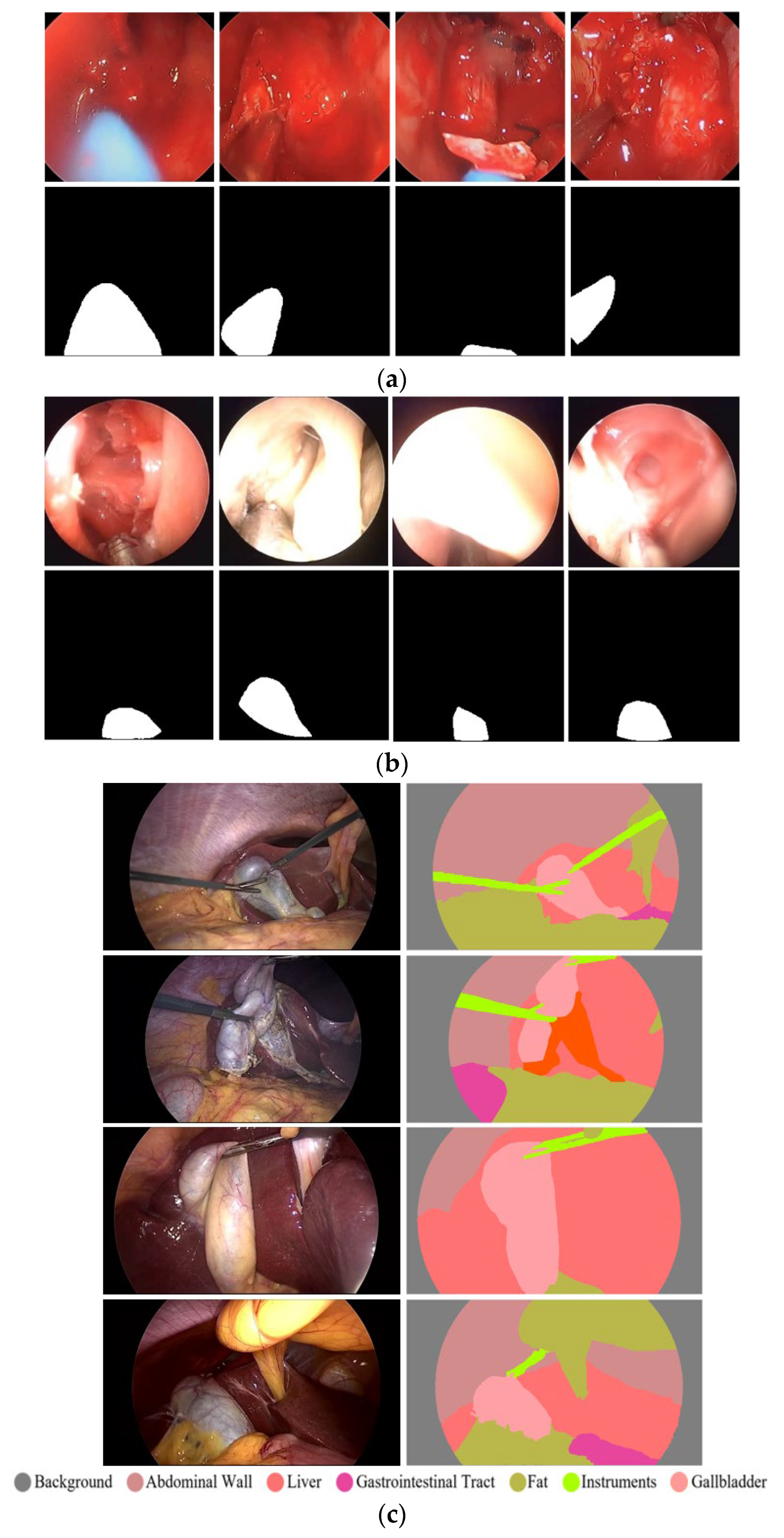
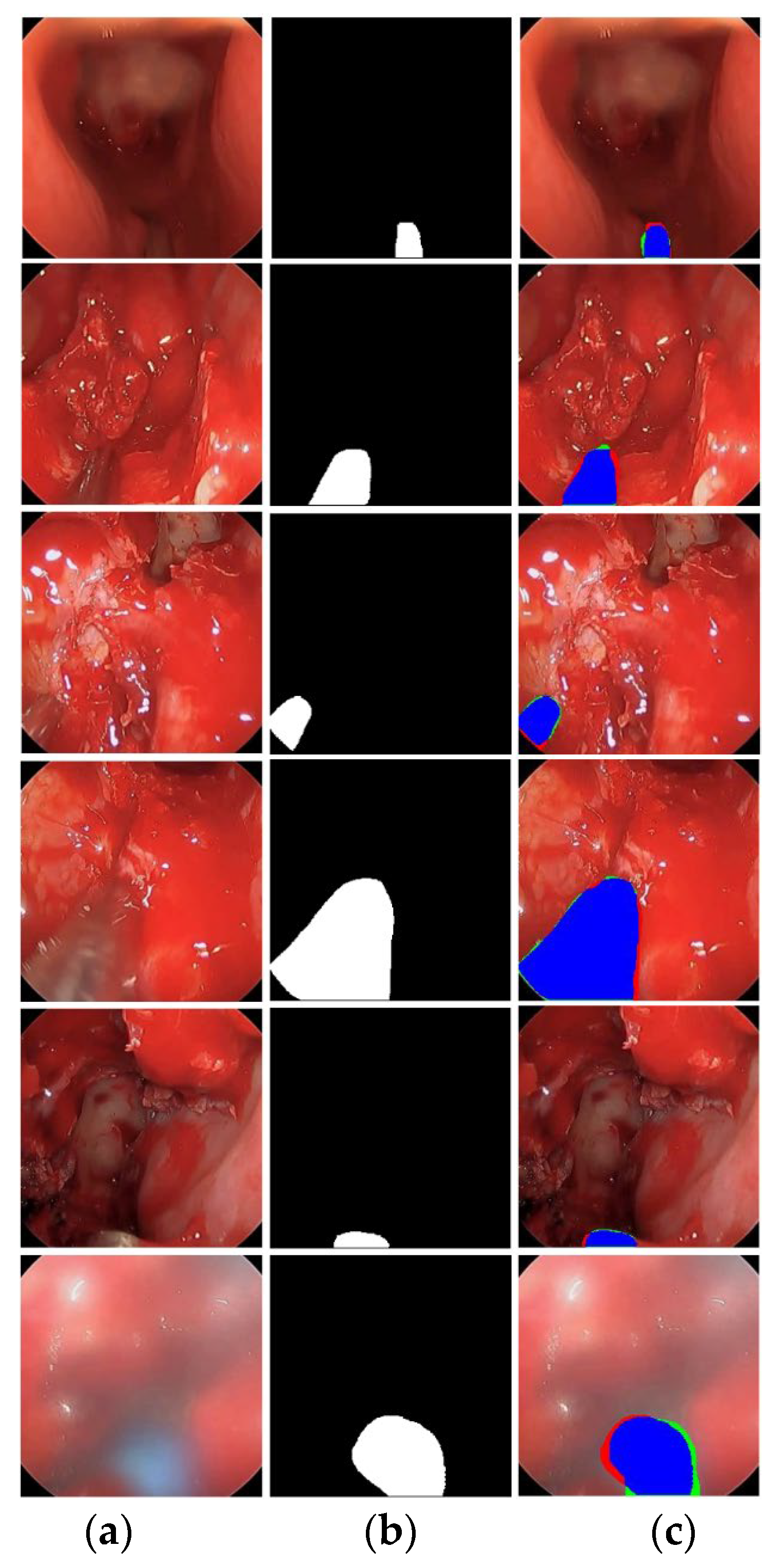
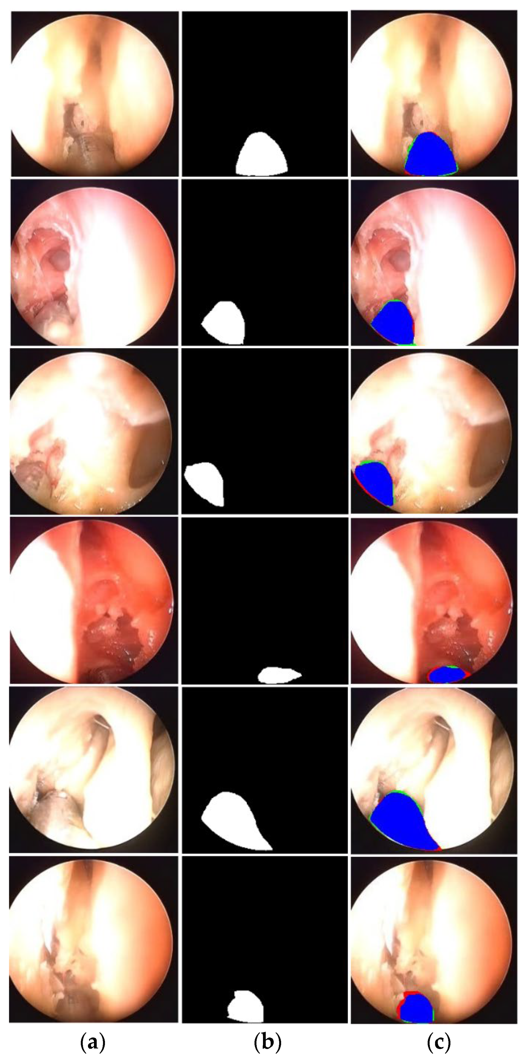
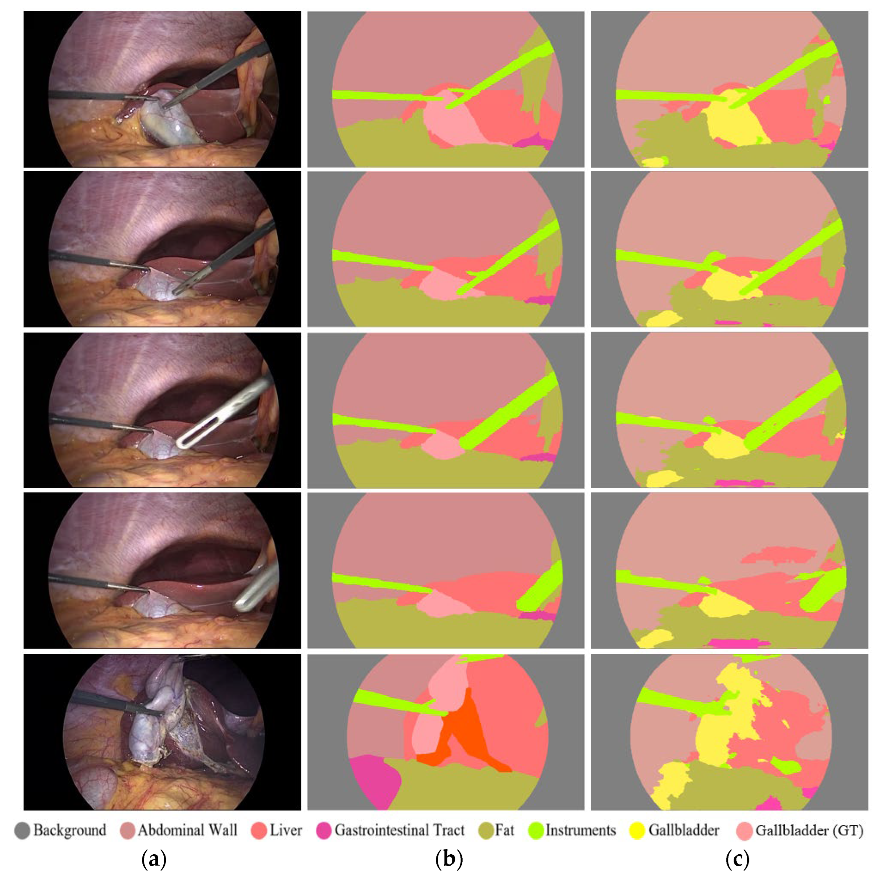
Accurate segmentation of surgical instrument boundaries is equally important and even more challenging under the aforementioned conditions. Surgical instrument segmentation is a key component of robot-assisted minimally invasive surgery, where precise spatial localization directly impacts procedural safety and effectiveness. Accordingly, we developed an advanced segmentation model designed to deliver robust performance under challenging imaging conditions. The proposed model is evaluated using three publicly available and diverse datasets. Sample images from these datasets are presented in
Figure 1. Specifically, this study uses UW-Sinus-surgery-live [
6], UW-Sinus-cadaveric [
6], and choelcSeg8k [
7]. As illustrated by the sample images, all three datasets exhibit substantial inter- and intra-dataset variability. In
Figure 1a, example images from the UW-Sinus-surgery-live dataset are shown. This dataset was created from live sinus surgery, and a substantial portion presents serious challenges for accurate surgical instrument segmentation. Sample images from UW-Sinus-surgery-live include blurred, blood-occluded, small-scale, and low-visibility surgical instruments. Similarly, example images from Sinus-cadaver and CholecSeg8k are shown in
Figure 1b,c, respectively. Sinus-cadaver is a binary-class dataset collected from endoscopic surgery. As illustrated by the sample images, UW-Sinus-cadaver exhibits large variations in illumination, along with challenges such as blur, glare, low contrast, and size variability. CholecSeg8k contains multi-class segmentation masks and is considered one of the most challenging datasets for surgical instrument segmentation. Dataset details are provided in
Section 3.1.
The rise of artificial intelligence is transforming and automating multiple sectors, including healthcare [
8,
9]. Deep learning is widely applied to disease diagnosis and assessment tasks, and has gained prominence by enabling effective early diagnosis and disease quantification. Extensive research is being conducted at the intersection of robotics and medicine [
10]. Medical robotics is shaping the future of healthcare and is expected to facilitate broader adoption and commercialization of robot-assisted minimally invasive surgery.
Surgical instrument segmentation remains an active research area due to the increasing adoption of robotic surgery. Existing research has explored both deep feature-based and handcrafted feature-based approaches for surgical instrument segmentation. A residual recurrent method proposed by Yang et al. [
6] employs densely connected attention mechanisms within a recurrent network to improve segmentation performance. This approach adopts U-Net as its backbone, which requires a large number of trainable parameters. Similarly, another study [
7] applies several state-of-the-art segmentation architectures to segment anatomical structures, including surgical instruments, during laparoscopic procedures. In that study, segmentation networks were evaluated on the challenging CholecSeg8k dataset, where U-Net++ [
11] reportedly outperformed other methods due to its semantic gap reduction capability. However, U-Net++ also exhibits computational limitations and requires a substantial number of parameters for training.
Another deep learning-based method [
12] employs multiscale feature aggregation and retention mechanisms to enhance segmentation performance for colorectal polyps and surgical instruments. That work proposes two architectures—a base network and a final network—to combine low-level and high-level information through multiscale retention, thereby improving segmentation accuracy. The method requires 4.97 million trainable parameters, indicating satisfactory computational performance; however, the authors acknowledge room for further improvement in computational efficiency.
A recent research work, namely adaptive SAM (Segment Anything), presented by Paranjape et al. [
13], extends SAM to improve performance, particularly on medical data. AdaptiveSAM introduces adaptable modifications to enable fast adaptability and prompt-based segmentation. However, AdaptiveSAM does not perform well on the surgical instrument segmentation task. Similarly, a recent study by Peng et al. [
14]. improves training by generating and incorporating synthetic images. The combination of active learning with synthetic images and blending and fusion strategies results in improved segmentation accuracy; however, this approach is limited to binary segmentation.
Lin et al. [
15] employ a generative adversarial network and image-to-image translation to reduce dependency on labeled data. They also introduce a customized loss function and evaluate their approach on surgical instrument segmentation from endoscopic images. Semantic inconsistency remains a minor limitation of this work. Another recent study by Jamal et al. [
16]. proposes a framework called SurgDepth that leverages a multimodal strategy using both RGB and depth information. SurgDepth employs a vision transformer as the encoder and is evaluated on multiple surgical instrument datasets. Although SurgDepth demonstrates strong performance, its computational requirements, with 101.3 million trainable parameters, represent a notable limitation. Similarly, Grammatikopoulou et al. [
17]. propose a neural architecture search-based model to optimize spatial and temporal processing for surgical segmentation. While this method achieves satisfactory segmentation performance, it requires a large number of trainable parameters (39.0 million).
We have divided previous studies into edge-aware and boundary-guided methods and briefly discussed them as follows.
Boundary-guided methods: Xu et al. [
18] presented a boundary guidance network for segmenting medical images. They primarily used a boundary extraction module to guide the decoding process. FFMS-Net uses frozen Sobel/Laplacian priors instead of a learned boundary extraction/guidance subnetwork. One more research work presented by Wang et al. [
19] introduced a boundary context neural network for the segmentation of medical images. During each encoding stage, they use pyramid edge extraction and explicit boundary/context coupling. FFMS-Net is lighter and avoids explicit boundary branches. Similarly, another research work by Zhang et al. [
20] uses boundary enhancements with the help of an edge detector for the semantic segmentation. However, unlike the proposed method, they also apply the edge detection throughout the encoder, and edge maps are not preserved using multiple frozen filters. Moreover, this work employs the U-Net as the baseline network, while the FFMS-Net does not rely on any other base network.
Edge-aware methods: In a study, Bui et al. [
21] presented a segmentation architecture with multiscale edge-guided attention. They explicitly explored the use of the Laplacian operator to preserve high-frequency edge information. Moreover, edge-guided attention is directly distributed to every stage of the decoder, making it computationally expensive (44.19 million parameters). FFMS-Net uses frozen Sobel + Laplacian priors in a frozen and learnable pipeline setting, without a separate edge-attention stack.
Another work presented by Peng et al. [
22] uses mainstream edge-guided information with a collaborative module throughout the encoder. Contrary to FFMS-Net, in this architecture, edge features are extracted from all of the encoder stages and fed to the decoder stage. Overall, this is a computationally heavy architecture that requires 149.5 million parameters.
The proposed efficient design is specifically motivated by endoscopic artifacts where boundary cues are present but easily attenuated by early downsampling. While multi-scale and dilated features-based modeling have already been explored, the design of modules such as tri-atrous blending, frozen and learnable feature pipeline, and the progressively structure-preserving decoder itself, and especially the combination of these modules, is novel. Ablation studies in this research also confirm the reinforcement effect of each module and the joint production of a reliable performance. According to the best of our knowledge, none of the FFMS-Net main modules are a known block/module from any other architecture. The detailed novelty is presented in the contribution at the end of
Section 1.
Researchers widely acknowledge the importance and scope of surgical instrument segmentation for enabling a smooth transition toward robot-assisted minimally invasive surgery [
6,
7]. Numerous state-of-the-art methods have been developed and evaluated for accurate surgical instrument segmentation. However, maintaining satisfactory segmentation performance under challenging imaging conditions remains difficult. Many existing approaches fail to deliver acceptable segmentation accuracy on challenging surgical data samples. Live endoscopic surgical procedures, characterized by a limited field of view, demand robust methods capable of handling unavoidable adverse imaging conditions. From a surgical perspective, accurate preservation of instrument boundaries and structural integrity is particularly important. Although achieving perfectly accurate boundary segmentation is unrealistic in challenging live surgical environments, attaining reliable performance for both structure and boundaries is highly desirable. Nevertheless, previous methods exhibit limitations in delivering consistent segmentation performance, particularly when predicting instrument boundaries and structural details.
Surgical datasets are often highly diverse, exhibiting substantial inter-dataset variability. Many existing methods rely on evaluation using a single dataset, which restricts the assessment of generalizability. Several existing approaches require a large number of trainable parameters, limiting their practical applicability. To address these challenges, we developed an advanced segmentation architecture with effective building blocks designed to perform robustly under challenging surgical conditions while requiring a very small number of trainable parameters (only 1.5 million).
The key contributions of the proposed research are summarized as follows.
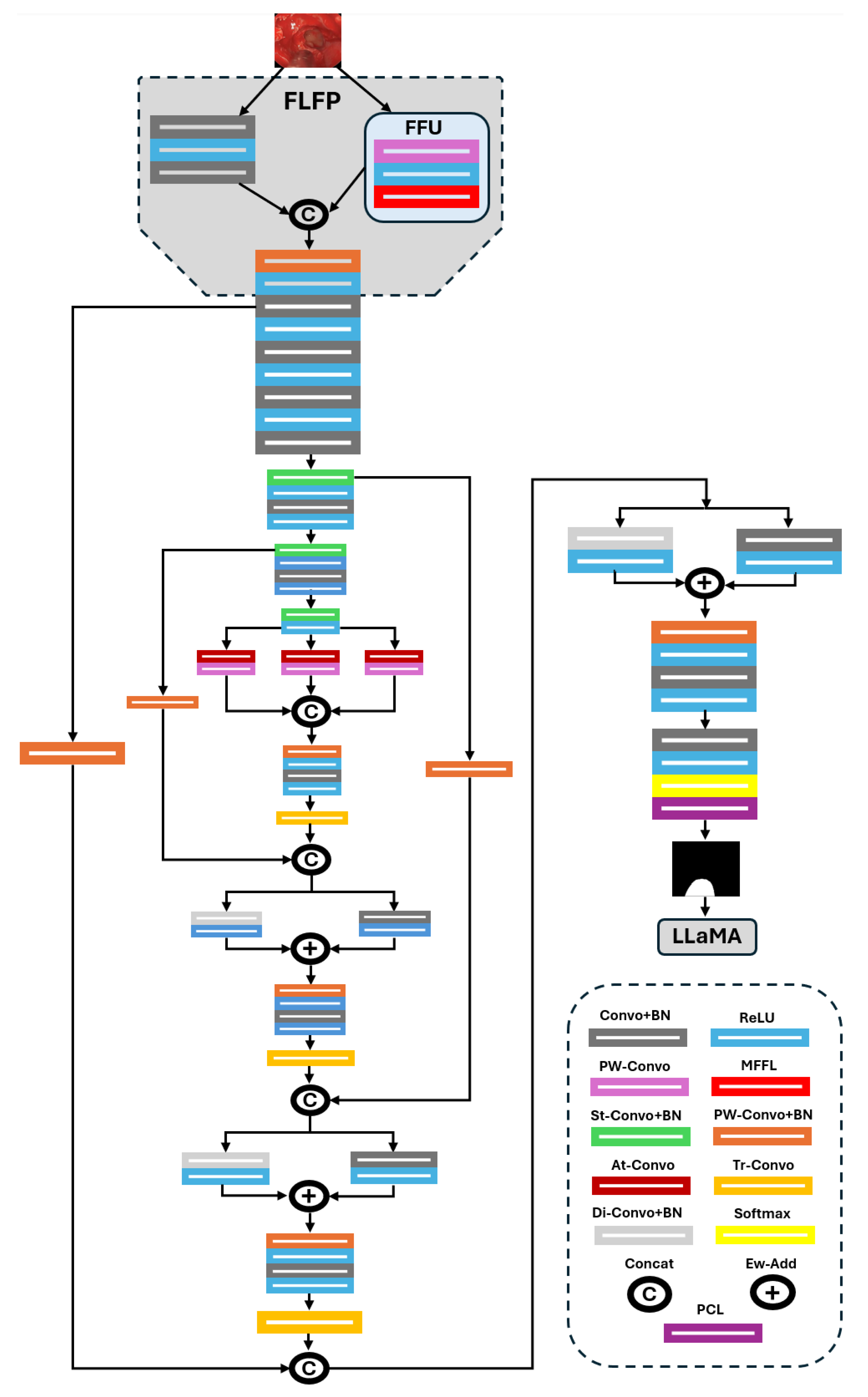
In this research work, an advanced segmentation architecture, namely the frozen-filters-based morphology-aware segmentation network (FFMS-Net), is developed to achieve reliable surgical instrument segmentation from endoscopic images. FFMS-Net introduces a frozen and learnable feature pipeline (FLFP) that simultaneously exploits frozen maps and learnable initial spatial features. The frozen component of FLFP, referred to as the frozen filter unit (FFU), is constructed using multiple frozen Sobel and Laplacian filters to preserve edge and structural information of the instruments, which is subsequently fused with learnable spatial features.
A tri-atrous blending (TAB) block is incorporated at the end of the encoding to process and fuse multi-receptive-field-based contextual information, enabling the network to preserve surgical instrument morphology. The multi-rate dilated context in TAB allows the architecture to retain contextual and morphological details even under challenging conditions such as blur, blood obscuration, and smoke.
A progressively structure-preserving decoder (PSPD) enables the model to learn and maintain the structural integrity of surgical instruments. In PSPD, features from different encoder stages are fused after each upsampling step, followed by aggregation of mildly dilated features. FFMS-Net is evaluated on three challenging datasets and outperforms state-of-the-art methods while requiring only 1.5 million trainable parameters.
The remainder of the paper is organized as follows.
Section 2 and
Section 3 present the proposed methodology and experimental results, respectively.
Section 4 discusses the findings, and
Section 5 concludes the study.
4. Discussion
The integration of artificial intelligence and robotics is transforming multiple sectors, including healthcare. Consequently, robot-assisted minimally invasive surgery has attracted substantial research interest. Surgical instruments are essential components of any surgical procedure; however, live endoscopic surgeries introduce numerous limitations and challenges for pixel-level detection of surgical instruments [
5]. The visual challenges associated with accurate surgical instrument segmentation have been discussed in previous sections. In addition to achieving high segmentation performance under challenging imaging conditions, computational efficiency, particularly the number of required trainable parameters, is also an important consideration.
Table 9 compares the number of trainable parameters required by FFMS-Net with those of existing methods. FFMS-Net requires only 1.5 million trainable parameters, which is substantially lower than that of other approaches. Moreover, to check hardware efficiency, we computed inference throughput on an NVIDIA GeForce RTX 4090 GPU. On the UW-Sinus-Surgery-Live dataset, the proposed model required an average of 0.0129 s per image, corresponding to 77.56 frames per second (FPS). The throughput of FFMS-Net is more than the typical surgical video frame rates (25–30 FPS). This shows that the model is computationally efficient and suitable for near-real-time or real-time surgical instrument segmentation.
From a translation perspective, dependable surgical instrument segmentation can support multiple sub-functions in robotic systems and surgical workflows. The predictions masks of the instrument can enable tool tracking, motion analysis, definition of collision-sensitive regions, and contextual analysis of surgical video streams. FFMS-Net’s compact design (1.5 million parameters) is advantageous for deployment in resource-constrained environments. However, for clinical application, prospective validation, robustness evaluation under severe occlusion, and careful safety analysis before clinical decision support or robotic platform integration are required.
4.1. Proof-of-Concept: Non-Clinical LLM-Based Scene Summarization
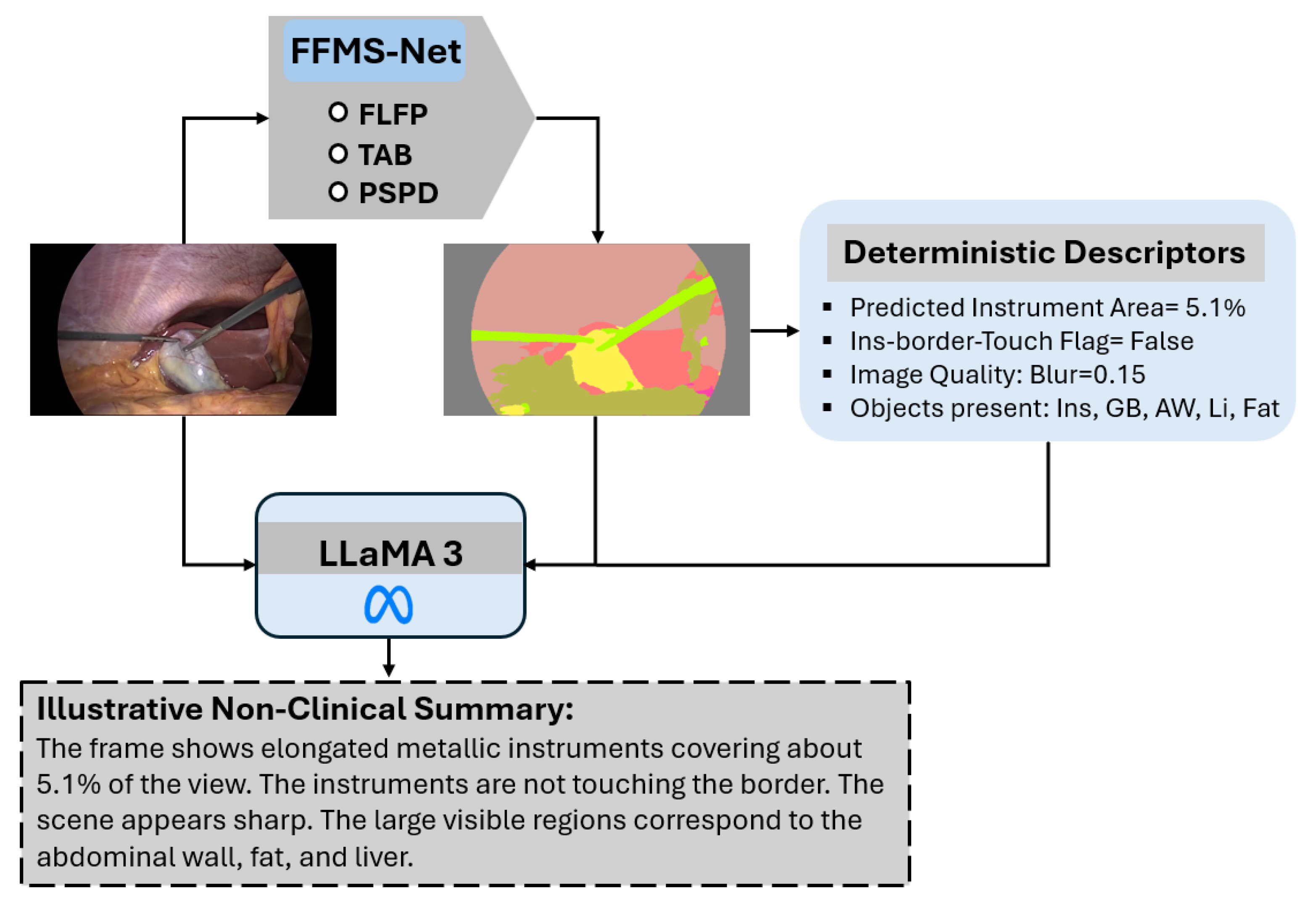
As shown in
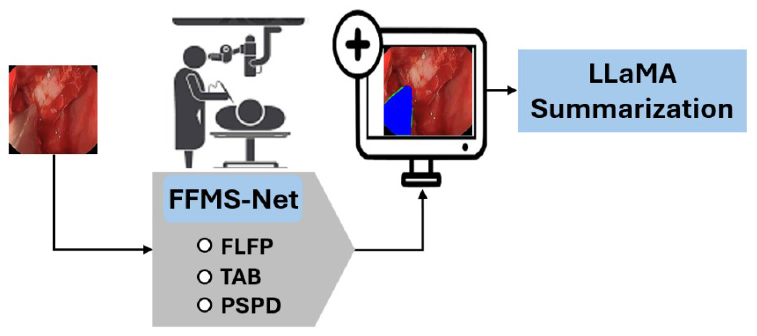
Figure 8, a lightweight LLM-based [
23] scene summarization module is included to illustrate one possible downstream application of the predicted masks. A small set of deterministic descriptors is computed from the prediction mask, including instrument area ratio, border-touch status, class presence, and a blur-related image quality indicator. These descriptors, together with the image and predicted mask, are provided to open-source LLaMA-3 to generate a short natural-language description of the scene. This component is included only as an exploratory proof-of-concept. The generated summaries are intended only for non-clinical interpretation; no expert validation, user study, or clinical decision-making assessment was performed in this work. The summary-quality evaluation and expert validation are explicitly noted as future work in
Section 4.3.
4.2. Visual Demonstration of FFMS-Net Focusing Instrument Features Using Grad-CAM
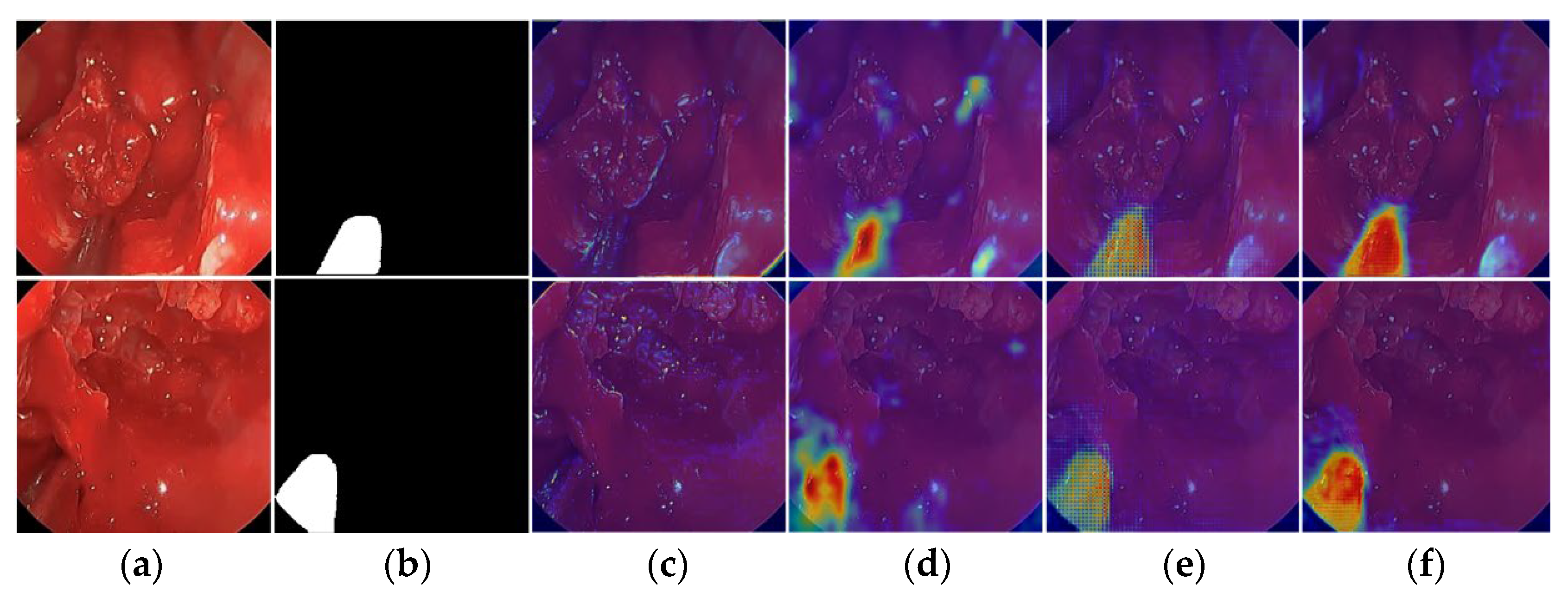
To demonstrate the progressive learning of meaningful features by FFMS-Net, heat activation maps are extracted using Grad-CAM [
42]. At early stages, the network attends to broader anatomical structures; at deeper stages, it increasingly focuses on features of the target class (surgical instrument). As shown in
Figure 9, Grad-CAM visualizations are extracted from different stages of FFMS-Net using challenging samples from the UW-Sinus-Surgery-Live dataset. In the deeper layers, particularly in
Figure 9d,e, FFMS-Net more clearly delineates the most relevant instrument features. As illustrated in
Figure 9f, FFMS-Net accurately concentrates on the spatial features and morphology of surgical instruments, even under low-contrast, blurred, and blood-occluded conditions. These mainly considered features of the surgical instruments by FFMS-Net are represented by Grad-CAM with reddish-orange color in
Figure 9f.
4.3. Limitations and Future Work
Despite the encouraging results, several limitations need to be mentioned. First, despite cross-dataset experimental results, domain shift between surgical environments remains a challenging task. Future research could explore more robust domain adaptation or augmentation strategies specifically designed to handle cross-domain evaluation. Second, the LLM-based scene summarization component was presented only as a proof-of-concept and has not been evaluated through user studies or clinical validation. Third, this study primarily focused on segmentation accuracy and computational efficiency; its clinical applicability could be further strengthened through extensive validation. These directions present important tasks for future research. Lastly, controlled random-seed experiments and repeated runs will be conducted to further quantify run-to-run variance in the future.
In
Table 4, cross-dataset evaluation results showed a significant performance degradation compared to testing with the same dataset, indicating that domain shift remains a major challenge. In the cases of the Sinus-live and Sinus-cadaver datasets, images exhibited distinct differences in tissue appearance, blood content, specular reflections, lighting stability, and motion-related artifacts, all of which impacted generalization. Although FFMS-Net maintained partial robustness under these shifts, the observed degradation suggests that morphology-aware design alone is insufficient for full cross-domain transfer. Therefore, future research should explore endoscope-specific data augmentation, domain adaptation, and domain generalization strategies to enhance robustness across the entire surgical environment.
5. Conclusions
Accurate surgical instrument segmentation is crucial for the advancement of robot-assisted minimally invasive surgery. Challenging imaging conditions during live procedures, such as blur, blood occlusion, low contrast, smoke, varying illumination, glare, and small instrument size, make reliable segmentation difficult. To address these challenges, we designed a robust architecture, FFMS-Net, to achieve reliable performance under adverse conditions. FFMS-Net employs an FLFP stem to process and fuse frozen and learnable features, preserving edge maps and structural information. In addition, a TAB block is incorporated at the end of the encoder to retain morphological and contextual information despite challenging imaging conditions.
Surgical instrument structures exhibit substantial variability; therefore, a progressively structure-preserving decoder is designed in which feature fusion followed by aggregation of slightly dilated features is performed after each upsampling step to maintain structural integrity. FFMS-Net is extensively evaluated on three datasets and demonstrates reliable performance while requiring only 1.5 million trainable parameters. Specifically, FFMS-Net achieves Dice scores of 89.30%, 93.02%, and 75.16% on the UW-Sinus-Surgery-Live, UW-Sinus-Surgery-Cadaver, and CholecSeg8k datasets, respectively, for surgical instrument segmentation. In the proposed framework, deterministic descriptors are computed from the prediction mask generated by FFMS-Net. The prediction mask, together with the original image and deterministic descriptors, is provided to an open-source LLaMA model for non-clinical natural-language summarization.