Carcinoid Heart Disease: The Role of Echocardiography in Raising the First Suspicion
 , , , , , , , , and
Abstract
1. Introduction
1.1. NET Classification
1.2. Carcinoid Heart Disease: The Pathophysiology
1.3. Biomarkers of CHD
1.4. Carcinoid Heart Disease: Why Cardiologists Should Care
2. Materials and Methods
3. The Role of Echocardiography and Cardiac Imaging in CHD
- Detailed evaluation of cardiac valves, with focus on the tricuspid and pulmonary valves.
- Assessment of right-sided cardiac chamber size and function.
- Evaluation for the presence of a PFO, particularly in patients with left-sided carcinoid heart disease and/or to identify those at high risk of CHD progression.
- Screening for myocardial metastases.
- Calculation of disease-specific score systems.
- 1.
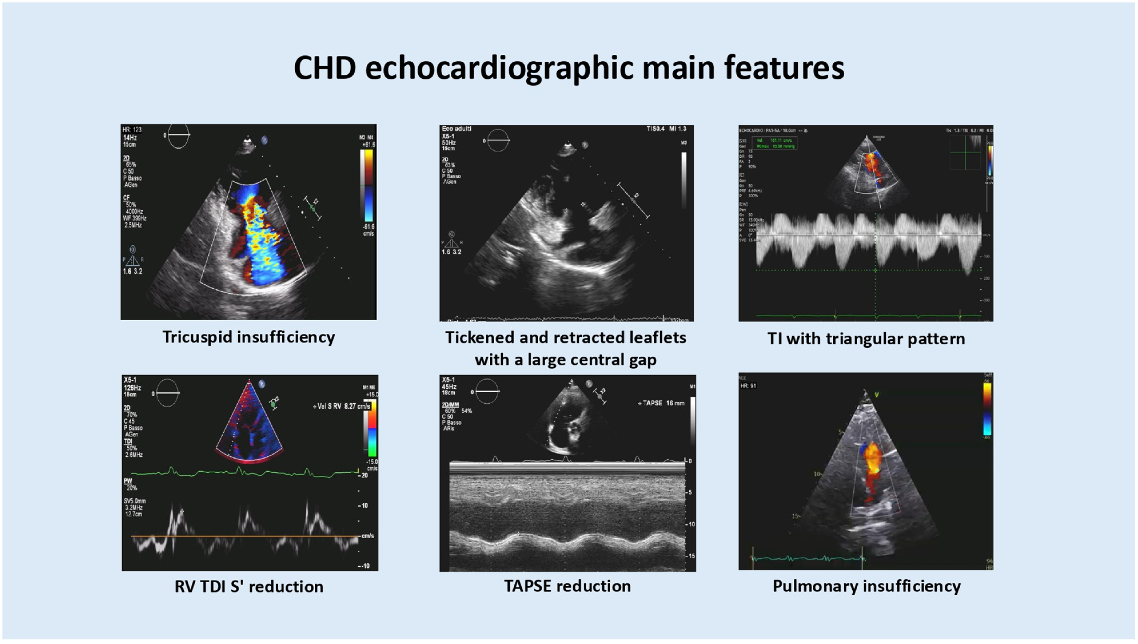
- Detailed evaluation of cardiac valves. Due to plaque-like fibrotic deposits, the valves become thickened, retracted and fixed, ultimately resulting in regurgitation and/or stenosis. The tricuspid valve is involved in up to 90% of cases, followed by the pulmonary (69%), mitral (29%), and aortic (27%) valves [13].In advanced cases, the tricuspid valve has a dilated annulus and extended thickened and retracted leaflets that fail to coapt during systole, resulting in a central gap and severe tricuspid regurgitation. On continuous-wave Doppler, the regurgitant signal appears densely saturated and, rather than exhibiting a parabolic contour, displays an early-peaking triangular configuration (often described as “dagger-shaped”). The peak regurgitant velocity is usually low (<2 m/s), suggesting that right ventricular systolic pressures are not elevated as in pulmonary hypertension. As a result, the right ventricle (RV) and the right atrium (RA) functionally behave as a single chamber.In the context of CHD, tricuspid stenosis is uncommon. According to the ENETS Carcinoid Heart Disease Task Force, its severity can be graded based on the tricuspid mean transvalvular gradient as mild (<5 mmHg), moderate (5–8 mmHg) and severe (>8 mmHg) [14].As observed for the tricuspid valve, pulmonary valve leaflets in CHD appear thickened and retracted and have reduced mobility, resulting in pulmonary regurgitation and/or stenosis. However, echocardiographic assessment of the pulmonary valve and accurate quantification of its regurgitation in CHD are challenging because of limited acoustic accessibility. Parasternal and subcostal right ventricular outflow tract views should, therefore, be carefully examined. Moreover, in patients with torrential tricuspid regurgitation, the pulmonary valve pathology (PV) is often masked, as both stenotic gradients and regurgitant volumes may be underestimated due to the reduced forward stroke volume reaching the PV. Thus, in this setting, the most informative parameter is the continuous-wave Doppler profile: a regurgitant jet ending before the onset of the next forward flow signal is suggestive of severe pulmonary regurgitation.The subcostal four-chamber view may be helpful for assessing RV free wall thickness, as the presence of RV hypertrophy may suggest pulmonary stenosis. Given the listed echocardiographic limitations, patients with CHD who are candidates for tricuspid valve replacement require a more accurate assessment of pulmonary valve pathology through cardiac magnetic resonance (CMR) [15].
- 2.
- Assessment of right-sided cardiac chamber size and function. The significant volume overload imposed by tricuspid and pulmonary regurgitation results in RV chamber dilation and diastolic ventricular septal flattening. Firstly, the RV preserves cardiac output thanks to compensatory mechanisms, which ultimately fail. The evaluation of right-sided cardiac chamber size and function includes tricuspid annular plane systolic excursion (TAPSE), systolic peak longitudinal tissue Doppler velocity at the RV free wall base (S′), RV fractional area change (FAC), three-dimensional ejection fraction (3D EF) and RV longitudinal strain. The following echocardiographic parameters are suggestive of RV dysfunction: TAPSE < 17 mm, RV TDI s’ < 10 cm/s, RV FAC ≤ 35%, 3D RV EF < 50%, RV free wall strain < 23% and RV global longitudinal strain < 21% [16].Of note, RV strain is reduced in patients with CHD, even when conventional systolic function parameters are initially preserved, and may be mildly impaired even before overt cardiac involvement in patients with elevated 5-hydroxyindoleacetic acid (5-HIAA) levels. However, a retrospective observational study by Alabdaljabar et al. [17], including 138 patients with confirmed CHD, demonstrated that reduced RV strain was not associated with increased mortality during a median follow-up of 5.0 years. In contrast, reduced right atrial (RA) reservoir strain emerged as an independent predictor of congestive heart failure development. These findings support the role of myocardial strain analysis in the early detection of cardiac involvement in patients with CS [18] and highlight the prognostic role of RA reservoir strain in reflecting disease-related morbidity in advanced CHD. Although echocardiography remains the first-line imaging modality, CMR is more accurate and highly reproducible in the assessment of RV volumes and function.
- 3.
- Evaluation for the presence of a PFO. PFO should be systematically assessed by contrast-enhanced transthoracic echocardiography in patients with left-sided carcinoid heart disease and/or to identify those at high risk of CHD progression. The exam consists of the injection of an agitated 5% glucose solution through an upper-extremity vein. A PFO is detected when contrast appears in the left atrium within the first three cardiac cycles, at rest, and after cough test or Valsalva maneuver. In patients who are candidates for percutaneous PFO closure, transesophageal echocardiography is required for preprocedural imaging assessment and device size selection.
- 4.
- Cardiac metastases in patients with CHD are uncommon (3.8%) and are usually located in the RA. Echocardiography may suggest their presence through deformation of the endocardial contour, but cardiac MRI is usually required for better characterization, including precise determination of lesion location, number, size, and spatial relationship to cardiac anatomical structures. Myocardial metastases have a high T2-weighted signal and isointense T1-weighted signal with postcontrast perfusion. Positron emission tomography (PET) represents an alternative imaging modality to CMR in the detection of metastatic lesions by showing their focal uptake.
- 5.
- Several echocardiographic scoring systems have been proposed to quantify the severity of CHD. The most comprehensive one was developed by Bhattacharyya S. et al., a 66-point assessment including evaluation and grading of the following echocardiographic characteristics: leaflet thickening, mobility, morphology, valvular stenosis, valvular regurgitation of all four valves, and RV diameter and function [19]. The scoring systems currently available in this field were compared by Dobson and colleagues in a prospective study enrolling 100 patients with metastatic NETs (21 with CHD) [20]. The five scoring systems were calculated in each patient. All resulted in discrimination between patients with and without CHD and all correlated with NT-proBNP and plasma 5-HIAA levels.
4. Treatment of Carcinoid Heart Disease
- A.
- Surgical and Transcatheter Management of CHD: which approach and when?
- B.
- Transcatheter Orthotopic Tricuspid Valve Replacement
Heterotopic Transcatheter Tricuspid Valve Replacement
- C.
- Treatment of cardiac metastases
5. Discussion
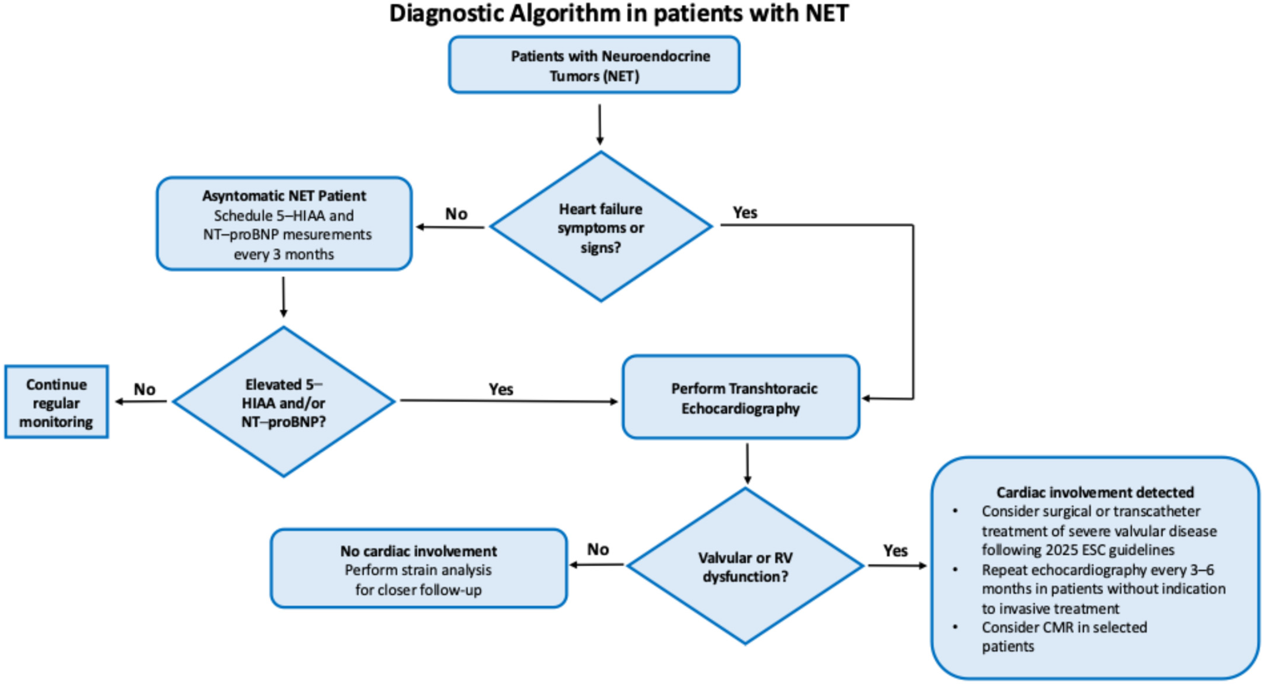
- Asymptomatic patients should undergo biomarker monitoring (urinary 5-HIAA and NT-proBNP) every three months. Patients with high or borderline levels should receive echocardiographic screening.
- (a)
- If no clear findings of CHD are detected, right-sided strain assessment is suggested to exclude early cardiac involvement.
- (b)
- If echocardiography screening confirms cardiac involvement, the exam should be repeated every 3–6 months, or sooner if signs of heart failure develop, to closely monitor disease progression. In patients with severe valvular disease, a surgical or transcatheter intervention should be considered, following the current ESC guideline recommendations.
- Symptomatic patients should promptly receive a transthoracic echocardiography to assess for CHD.
6. Conclusions
Author Contributions
Funding
Institutional Review Board Statement
Informed Consent Statement
Data Availability Statement
Conflicts of Interest
Abbreviations
| NETs | Neuroendocrine tumors |
| NENs | Neuroendocrine neoplasms |
| SEER | Epidemiological analyses from the Surveillance, Epidemiology, and End Results |
| ENETS | European Neuroendocrine Tumour Society |
| CHD | Carcinoid heart disease |
| 5-HIAA | 5-hydroxyindolacetic acid |
| CS | Carcinoid syndrome |
| NT-proBNP | N-terminal pro-B-type natriuretic peptide |
| 5-HT | 5-hydroxytryptamine |
| PFO | Patent foramen ovale |
| UKO | Unknown origin |
| CgA | Chromogranin A |
| u5-HIAA | Urinary 5-hydroxyindolacetic acid |
| ESC | European Society of Cardiology |
| RV | Right ventricle |
| RA | Right atrium |
| PV | Pulmonary valve |
| CMR | Cardiac magnetic resonance |
| TAPSE | Tricuspid annular plane systolic excursion |
| FAC | Fractional area change |
| PET | Positron emission tomography |
| PRRT | Peptide receptor radionuclide therapy |
| TTVR | Transcatheter tricuspid valve replacement |
| TPVR | Transcatheter pulmonary valve replacement |
References
- Dasari, A.; Wallace, K.; Halperin, D.M.; Maxwell, J.; Kunz, P.; Singh, S.; Chasen, B.; Yao, J.C. Epidemiology of Neuroendocrine Neoplasms in the US. JAMA Netw. Open 2025, 8, e2515798. [Google Scholar] [CrossRef] [PubMed]
- Borbath, I.; Garcia-Carbonero, R.; Bikmukhametov, D.; Jimenez-Fonseca, P.; Castaño, A.; Barkmanova, J.; Sedlackova, E.; Kollár, A.; Christ, E.; Kaltsas, G.; et al. The European Neuroendocrine Tumour Society registry, a tool to assess the prognosis of neuroendocrine neoplasms. Eur. J. Cancer 2022, 168, 80–90. [Google Scholar] [CrossRef] [PubMed]
- Sinnot, R.O.; Stell, A. The Patient-Oriented Neuroendocrine Tumor Registry for Australia. Stud. Health Technol. Inform. 2025, 329, 204–208. [Google Scholar] [CrossRef] [PubMed]
- Halperin, D.M.; Shen, C.; Dasari, A.; Xu, Y.; Chu, Y.; Zhou, S.; Shih, Y.T.; Yao, J.C. Frequency of carcinoid syndrome at neuroendocrine tumour diagnosis: A population-based study. Lancet Oncol. 2017, 18, 525–534. [Google Scholar] [CrossRef] [PubMed] [PubMed Central]
- Grozinsky-Glasberg, S.; Davar, J.; Hofland, J.; Dobson, R.; Prasad, V.; Pascher, A.; Denecke, T.; Tesselaar, M.E.T.; Panzuto, F.; Albåge, A.; et al. European Neuroendocrine Tumor Society (ENETS) 2022 Guidance Paper for Carcinoid Syndrome and Carcinoid Heart Disease. J. Neuroendocrinol. 2022, 34, e13146. [Google Scholar] [CrossRef] [PubMed] [PubMed Central]
- Clement, D.; Ramage, J.; Srirajaskanthan, R. Update on Pathophysiology, Treatment, and Complications of Carcinoid Syndrome. J. Oncol. 2020, 2020, 8341426. [Google Scholar] [CrossRef] [PubMed] [PubMed Central]
- Mansencal, N.; Mitry, E.; Pillière, R.; Lepère, C.; Gérardin, B.; Petit, J.; Gandjbakhch, I.; Rougier, P.; Dubourg, O. Prevalence of patent foramen ovale and usefulness of percutaneous closure device in carcinoid heart disease. Am. J. Cardiol. 2008, 101, 1035–1038. [Google Scholar] [CrossRef]
- Sabet, A.; Haghighiabyaneh, M.; Rajyaguru, C.; Raisinghani, A.; Kupsky, D.; DeMaria, A.N. Carcinoid heart disease. Struct. Heart 2020, 4, 78–86. [Google Scholar] [CrossRef]
- Calanchini, M.; Tadman, M.; Krogh, J.; Fabbri, A.; Grossman, A.; Shine, B. Measurement of urinary 5-HIAA: Correlation between spot versus 24-h urine collection. Endocr. Connect. 2019, 8, 1082–1088. [Google Scholar] [CrossRef] [PubMed] [PubMed Central]
- de Mestier, L.; Savagner, F.; Brixi, H.; Do Cao, C.; Dominguez-Tinajero, S.; Roquin, G.; Goichot, B.; Hentic, O.; Dubreuil, O.; Hautefeuille, V.; et al. Plasmatic and Urinary 5-Hydroxyindolacetic Acid Measurements in Patients with Midgut Neuroendocrine Tumors: A GTE Study. J. Clin. Endocrinol. Metab. 2021, 106, e1673–e1682. [Google Scholar] [CrossRef] [PubMed]
- Bhattacharyya, S.; Toumpanakis, C.; Caplin, M.E.; Davar, J. Usefulness of N-terminal pro-brain natriuretic peptide as a biomarker of the presence of carcinoid heart disease. Am. J. Cardiol. 2008, 102, 938–942. [Google Scholar] [CrossRef] [PubMed]
- Lyon, A.R.; López-Fernández, T.; Couch, L.S.; Asteggiano, R.; Aznar, M.C.; Bergler-Klein, J.; Boriani, G.; Cardinale, D.; Cordoba, R.; Cosyns, B.; et al. 2022 ESC Guidelines on cardio-oncology developed in collaboration with the European Hematology Association (EHA), the European Society for Therapeutic Radiology and Oncology (ESTRO) and the International Cardio-Oncology Society (IC-OS). Eur. Heart J. 2022, 43, 4229–4361, Erratum in Eur. Heart J. 2023, 44, 1621. https://doi.org/10.1093/eurheartj/ehad196. [Google Scholar] [CrossRef] [PubMed]
- Bhattacharyya, S.; Toumpanakis, C.; Burke, M.; Taylor, A.M.; Caplin, M.E.; Davar, J. Features of carcinoid heart disease identified by 2- and 3-dimensional echocardiography and cardiac MRI. Circ. Cardiovasc. Imaging 2010, 3, 103–111. [Google Scholar] [CrossRef] [PubMed]
- Hofland, J.; Lamarca, A.; Steeds, R.; Toumpanakis, C.; Srirajaskanthan, R.; Riechelmann, R.; Panzuto, F.; Frilling, A.; Denecke, R.; Christ, E.; et al. Synoptic reporting of echocardiography in carcinoid heart disease (ENETS Carcinoid Heart Disease Task Force). J. Neuroendocrinol. 2022, 34, e13060. [Google Scholar] [CrossRef]
- Baron, T.; Bergsten, J.; Albåge, A.; Lundin, L.; Sörensen, J.; Öberg, K.; Flachskampf, F.A. Cardiac Imaging in Carcinoid Heart Disease. JACC Cardiovasc. Imaging 2021, 14, 2240–2253. [Google Scholar] [CrossRef] [PubMed]
- Praz, F.; Borger, M.A.; Lanz, J.; Marin-Cuartas, M.; Abreu, A.; Adamo, M.; Ajmone Marsan, N.; Barili, F.; Bonaros, N.; Cosyns, B.; et al. 2025 ESC/EACTS Guidelines for the management of valvular heart disease: Developed by the task force for the management of valvular heart disease of the European Society of Cardiology (ESC) and the European Association for Cardio-Thoracic Surgery (EACTS). Eur. Heart J. 2025, 46, 4635–4736. [Google Scholar] [CrossRef]
- Alabdaljabar, M.S.; Alexandrino, F.; Al-Shakarchi, N.; Kucuk, H.O.; Lloyd, J.V.; Luis, S.A.; Pislaru, S.V.; Pellikka, P.A.; Connolly, H.M. Right atrial and ventricular strain analysis in patients with carcinoid heart disease. J. Am. Soc. Echocardiogr. 2024, 37, 1008–1010. [Google Scholar] [CrossRef]
- Haugaa, K.H.; Bergestuen, D.S.; Sahakyan, L.G.; Skulstad, H.; Aakhus, S.; Thiis-Evensen, E.; Edvardsen, T. Evaluation of right ventricular dysfunction by myocardial strain echocardiography in patients with intestinal carcinoid disease. J. Am. Soc. Echocardiogr. 2011, 24, 644–650. [Google Scholar] [CrossRef] [PubMed]
- Bhattacharyya, S.; Toumpanakis, C.; Chilkunda, D.; Caplin, M.E.; Davar, J. Risk factors for the development and progression of carcinoid heart disease. Am. J. Cardiol. 2011, 107, 1221–1226. [Google Scholar] [CrossRef] [PubMed]
- Dobson, R.; Cuthbertson, D.J.; Jones, J.; Valle, J.W.; Keevil, B.; Chadwick, C.; Poston, G.P.; Burgess, M.I. Determination of the optimal echocardiographic scoring system to quantify carcinoid heart disease. Neuroendocrinology 2014, 99, 85–93. [Google Scholar] [CrossRef] [PubMed]
- Korach, A.; Grozinsky-Glasberg, S.; Atlan, J.; Dabah, A.; Atlan, K.; Rudis, E.; Elami, A.; Gross, D.J.; Reardon, M.J.; Shapira, O.M. Valve Replacement in Patients with Carcinoid Heart Disease: Choosing the Right Valve at the Right Time. J. Heart Valve Dis. 2016, 25, 349–355. [Google Scholar]
- Sunjic, I.; Shin, D.; Sunjic, K.M.; Popat, J.V.; Tran, T.; Chae, S.H.; Caldeira, C.C.; Strosberg, J.R.; Sayad, D. Incidence of Atrioventricular Block After Valve Replacement in Carcinoid Heart Disease. Cardiol. Res. 2020, 11, 56–60. [Google Scholar] [CrossRef]
- Madhavan, M.V.; Agarwal, V.; Hahn, R.T. Transcatheter Therapy for the Tricuspid Valve: A Focused Review of Edge-to-Edge Repair and Orthotopic Valve Replacement. Curr. Cardiol. Rep. 2024, 26, 459–474. [Google Scholar] [CrossRef]
- Webb, J.G.; Chuang, A.M.; Meier, D.; von Bardeleben, R.S.; Kodali, S.K.; Smith, R.L.; Hausleiter, J.; Ong, G.; Boone, R.; Ruf, T.; et al. Transcatheter Tricuspid Valve Replacement with the EVOQUE System: 1-Year Outcomes of a Multicenter, First-in-Human Experience. JACC Cardiovasc. Interv. 2022, 15, 481–491. [Google Scholar] [CrossRef] [PubMed]
- Pereyra, M.; Farina, J.M.; Chao, C.J.; Ayoub, C.; Arsanjani, R. Percutaneous transcatheter pulmonary and tricuspid valve replacements in a patient with carcinoid heart disease. Eur. Heart J. Case Rep. 2023, 7, ytad511. [Google Scholar] [CrossRef] [PubMed]
- Dannenberg, L.; El Moayidi, Y.; Orban, M.; Braun, D.; Schaefer, A.; Mehilli, J. Simultaneous transcatheter treatment of carcinoid pulmonary and tricuspid valve disease using the Venus P-Valve and EVOQUE system. J. Invasive Cardiol. 2024, 36, E353–E356. [Google Scholar]
- Assafin, M. Transcatheter tricuspid valve replacement in severe carcinoid tricuspid regurgitation using the EVOQUE system. Presented at the New York Valves, New York, NY, USA, 25–27 June 2025; Available online: https://d14d5nk8lue86f.cloudfront.net/public/2025-06/83d635a1-081b-4797-a1c0-5fd8159a6274.pdf (accessed on 7 December 2025).
- Veas, N.; Verdugo, F.J.; Álvarez, J.; Springmuller, D.; Bontá, C.; Esteves, F.; Esteves, V.; Méndez, M. One-Stop Transcatheter Pulmonary and Tricuspid Valve Replacement for Carcinoid Heart Disease Treatment. JACC Case Rep. 2025, 31, 106224. [Google Scholar] [CrossRef] [PubMed]
- Stoltz, L.; Krompass, L.; Lubos, E.; von Bardeleben, R.S. Heterotopic transcatheter tricuspid valve replacement using the TricValve system in a high-risk patient with carcinoid heart disease. J. Cardiovasc. Dev. Dis. 2022, 9, 359. [Google Scholar] [CrossRef]
- Piscione, M.; Cammalleri, V.; Antonelli, G.; De Luca, V.M.; Carpenito, M.; Gaudio, D.; Cocco, N.; Nenna, A.; Dominici, C.; Bianchi, A.; et al. Cardiac Surgery and Transcatheter Intervention for Valvular Heart Disease in Carcinoid Syndrome: Risk Factors, Outcomes, and Evolving Therapeutic Strategies. J. Cardiovasc. Dev. Dis. 2024, 11, 359. [Google Scholar] [CrossRef]
- Bober, B.; Saracyn, M.; Kołodziej, M.; Kowalski, Ł.; Deptuła-Krawczyk, E.; Kapusta, W.; Kamiński, G.; Mozenska, O.; Bil, J. Carcinoid Heart Disease: How to Diagnose and Treat in 2020? Clin. Med. Insights Cardiol. 2020, 14, 1–8. [Google Scholar] [CrossRef] [PubMed] [PubMed Central]


Disclaimer/Publisher’s Note: The statements, opinions and data contained in all publications are solely those of the individual author(s) and contributor(s) and not of MDPI and/or the editor(s). MDPI and/or the editor(s) disclaim responsibility for any injury to people or property resulting from any ideas, methods, instructions or products referred to in the content. |
© 2026 by the authors. Licensee MDPI, Basel, Switzerland. This article is an open access article distributed under the terms and conditions of the Creative Commons Attribution (CC BY) license.
Share and Cite
Stavagna, S.; Manzi, G.; Angotti, D.; D’Amato, A.; Giannetta, E.; Badagliacca, R.; Ciccarelli, F.; Netti, L.; Severino, P.; Saade, W.; et al. Carcinoid Heart Disease: The Role of Echocardiography in Raising the First Suspicion. J. Clin. Med. 2026, 15, 1978. https://doi.org/10.3390/jcm15051978
Stavagna S, Manzi G, Angotti D, D’Amato A, Giannetta E, Badagliacca R, Ciccarelli F, Netti L, Severino P, Saade W, et al. Carcinoid Heart Disease: The Role of Echocardiography in Raising the First Suspicion. Journal of Clinical Medicine. 2026; 15(5):1978. https://doi.org/10.3390/jcm15051978
Chicago/Turabian StyleStavagna, Silvia, Giovanna Manzi, Danilo Angotti, Andrea D’Amato, Elisa Giannetta, Roberto Badagliacca, Federico Ciccarelli, Lucrezia Netti, Paolo Severino, Wael Saade, and et al. 2026. "Carcinoid Heart Disease: The Role of Echocardiography in Raising the First Suspicion" Journal of Clinical Medicine 15, no. 5: 1978. https://doi.org/10.3390/jcm15051978
APA StyleStavagna, S., Manzi, G., Angotti, D., D’Amato, A., Giannetta, E., Badagliacca, R., Ciccarelli, F., Netti, L., Severino, P., Saade, W., Vizza, C. D., & Maestrini, V. (2026). Carcinoid Heart Disease: The Role of Echocardiography in Raising the First Suspicion. Journal of Clinical Medicine, 15(5), 1978. https://doi.org/10.3390/jcm15051978