


Early (<6 Days) Extracranial Carotid Revascularization After Intravenous Thrombolysis for Stroke: A Scoping Review
 , , , and
Abstract
1. Introduction
2. Materials and Methods
2.1. Study Design
2.2. Research Question
2.3. Relevant Studies Identification
2.4. Studies Selection
2.5. Data Charting Process
2.6. Reporting the Results
3. Results
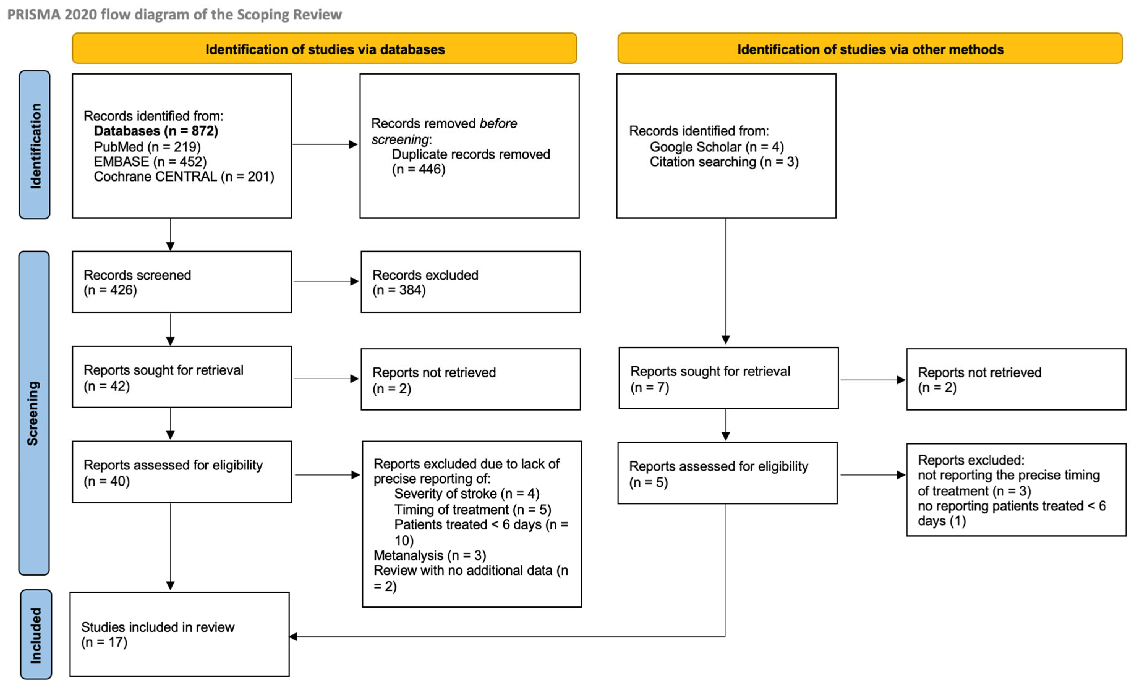
3.1. Search Results and Selection of the Studies
3.2. Study Characteristics

| Author Journal | Year | Country | Design of the Study and Duration | Population and Sample Size | Intervention Type | |
|---|---|---|---|---|---|---|
| 1 | Bellomo T. et al. [16] Annals of Vascular Surgery | 2024 | USA | Retrospective analysis of a single-center cohort (Mass. General Hospital). 2007–2019 | 56 stroke pts submitted to carotid revascularization after IVT (4.9%) CONTROL: 1083 symptomatic pts with no IVT submitted to carotid revascularization (94.1%) | 35 CEA, 21 CAS (15 catheter-based interventions for stroke: 9 before CEA and 6 before CAS) |
| 2 | Doo Hyuk Kwon et al. [17] Journal of Korean Medical Sciences | 2022 | Republic of Korea | Retrospective analysis of a cohort of emergent carotid intervention at a single center (Keimyung University School of Medicine). 2005–2020 | 19 pts with stroke submitted to emergent CAS after IVT (11 ICA occlusions excluded) No CONTROL Group | 19 CAS: 11 pts with ICA occlusion, 6 pts with tandem lesion (not considered in the review), 2 pts with ICA stenosis (included in the scoping review) |
| 3 | Johal A.S. et al. [29] European Journal of Vascular and Endovascular Surgery | 2021 | UK | Retrospective study using a large population-based dataset from the National Vascular Registry in the United Kingdom (UK-NVR). 2014–2019 | 1055 stroke pts submitted to IVT and CEA (11.7%) CONTROL: 7975 stroke pts with no IVT submitted to CEA (88.3%) | CEA (patients with CAS or carotid bypass were excluded from the study) |
| 4 | Deiana G. et al. [28] Annals of Vascular Surgery | 2020 | Italy | Retrospective analysis of a cohort of patients from two centers (Brotzu Hospital, Cagliari, Italy and Santissima Annunziata Hospital, Sassari, Italy). 2016–2018 | 11 stroke pts submitted to CEA after IVT (15.7%) CONTROL: 59 symptomatic pts with no IVT submitted to CEA (84.3%) | CEA under locoregional anesthesia in all patients |
| 5 | Ijäs P. et al. [8] Stroke | 2018 | Finland | Retrospective analysis of a single-center cohort of patients (Helsinki University Hospital, Helsinki, Finland). Comparison between pts operated <48 h after IVT and pts operated >48 h and <14 days. 2005–2016 | 128 pts submitted to CEA after IVT (15.7%) CONTROL: 777 stroke pts with no IVT submitted to CEA (84.3%) | All CEA, no data regarding surgical technique or anesthesia |
| 6 | Gunka I. et al. [18] Annals of Vascular Surgery | 2017 | Czech Republic | Retrospective analysis of a single-center cohort of patients (Charles University Hospital, Prague, Czech Repuplic). 2013–2016 | 13 pts submitted to CEA after IVT (14%)—4 patients with acute extracranial carotid occlusion CONTROL: 80 symptomatic pts with no IVT submitted to CEA (86%) | CEA under locoregional anesthesia (general anesthesia in selected patients) |
| 7 | Adachi K. et al. [19] Neurosurgical Review | 2017 | Japan | Retrospective analysis of a single-center cohort of patients treated with CAS < 2 wks of stroke (Fujita Health University, Japan). 2009–2014 | 16 patients undergoing urgent/emergent CAS for stroke/stroke in evolution—4 receiving IVT (25%) No CONTROL | CAS with embolic protection |
| 8 | Yamamoto Y. et al. [15] The Journal of Medical Investigation | 2016 | Japan | Case report on emergent CEA after IVT for deteriorating symptoms (Tokushima Prefectural Miyoshi Hospital, Japan). | One emergent CEA due to fluctuating symptoms | CEA under general anesthesia |
| 9 | Azzini C. et al. [20] European Journal of Vascular and Endovascular Surgery | 2016 | Italy | Retrospective analysis of a single-center cohort of patients treated with CEA (also <12 h) after IVT (Azienda Ospedaliera-Universitaria, Ferrara, Italy). 2009–2014 | 34 stroke pts submitted to CEA after IVT CONTROL: IVT pts without CEA | CEA under general anesthesia with routine shunt |
| 10 | Bazan H.A. et al. [21] Journal of Vascular Surgery | 2015 | USA | Retrospective analysis of a single-center cohort of patients treated with CEA/CAS after IVT (Ochsner Clinic, New Orleans, Louisiana). 2009–2015 | 31 stroke pts submitted to carotid revascularization after IVT CONTROL: 134 no IVT pts submitted to CEA (33% TIA pts) | IVT: CEA 25 − CAS 6 No IVT: CEA 110 − CAS 24 CEA under general anesthesia using standard patch angioplasty with routine intraoperative shunt |
| 11 | Vellimana A.K. et al. [22] Neurosurgery | 2014 | USA | Retrospective analysis of a single-center cohort of patients treated with CEA after IVT (Washington University School of Medicine, St. Louis, Missouri). 1995–2007 | 11 stroke pts submitted to carotid revascularization after IVT CONTROL: 131 no-IVT pts submitted to CEA (symptomatic—TIA included but percentage not specified) | CEA under general anesthesia. Intraoperative neuromonitoring with EEG. Selective shunting on EEG indication |
| 12 | Koraen-Smith L. et al. [30] Stroke | 2014 | Sweden | Retrospective analysis of a prospectively collected national database (Swedvasc and Riks-Stroke). 2008–2012 | 79 stroke pts submitted to carotid revascularization after IVT CONTROL: 3919 no IVT pts submitted to CEA (symptomatic stenosis) | IVT: CEA 71 − CAS 6 + 2 pts with aborted TEA due to high bifurcation No IVT: CEA 110 − CAS 24 |
| 13 | Yong Y.P. et al. [23] Journal of Vascular Surgery | 2013 | UK | Retrospective analysis of a single-center cohort of patients treated with CEA after IVT (Nottingham University Hospitals, Nottingham, UK). 2010–2012 | Seven stroke pts submitted to CEA after IVT No CONTROL | CEA under locoregional or general anesthesia |
| 14 | Sallustio F. et al. [24] Stroke Research and Treatment | 2012 | Italy | Retrospective analysis of a single-center cohort of patients treated with CAS after IVT (Policlinico Tor Vergata, Rome, Italy). 2006–2011 | Six stroke pts submitted to CAS after IVT No CONTROL | CAS under local anesthesia with embolic protection device (distal filter) |
| 15 | Leseche G. et al. [25] Journal of Vascular | 2012 | France | Retrospective analysis of a single-center cohort of patients with STROKE IN EVOLUTION (Bichat-Claude Bernard University Hospital, Paris, France). 2003–2010 | Seven pts with stroke in evolution (fluctuating symptoms) after IVT No CONTROL | CEA under locoregional anesthesia/general anesthesia with routine shunting |
| 16 | Bartoli M.A. et al. [26] European Journal of Vascular and Endovascular Surgery | 2008 | France | Retrospective analysis of a single-center cohort of patients treated with CEA after IVT (Hopital de la Timone, Service de Chirurgie Vasculaire, Marseille, France). 2005–2008 | 12 stroke pts submitted to CEA after IVT No CONTROL | IVT: CEA 12 CEA under general anesthesia (technique at surgeon’s discretion) |
| 17 | Abou-Chebl A. et al. [27] Stroke | 2005 | USA | Retrospective analysis of a single-center cohort of patients treated with CAS after IVT + GPIIb/IIIa Antagonists (Cleveland Clinic, Cleveland, Ohio). (Time period not specified) | 12 stroke pts submitted to emergent endovascular treatment and IVT + GPIIb/IIIa Antagonists (2 patients receiving CAS) No CONTROL | CAS in two patients associated with endovascular treatment of intracranial lesion |
3.3. Results
3.3.1. Severity of Stroke at Admission and Timing Between IVT and Carotid Revascularization
3.3.2. Ischemic Stroke at 30 Days
3.3.3. Asymptomatic Intracranial Bleeding (aICB) Plus Symptomatic Intracranial Hemorrhage (sICH) at 30 Days
3.3.4. Overall Stroke (Ischemic and Hemorrhagic) at 30 Days
3.3.5. Death at 30 Days
3.3.6. Wound Bleeding/Access Site Complications at 30 Days
4. Discussion
- Low risk of ischemic stroke and mortality after CEA: The incidence of postoperative ischemic stroke and death among patients undergoing CEA after IVT appears to be low.
- Higher complication risk with CAS, particularly sICH: Patients treated with CAS exhibit a higher risk of complications, notably sICH. However, this results should be interpreted with caution for several reasons. First, it is important to recognize that this cohort represents a distinct clinical subset, as CAS is frequently performed in the hyperacute phase (within hours) in patients with severe strokes and is often combined with intracranial thrombectomy for tandem lesions. These patients typically present with more severe neurological deficits and are exposed to the additional hemorrhagic risk of dual antiplatelet therapy [17,27], although the combination of IVT and dual antiplatelet therapy does not appear to be associated with an increased incidence of sICH, as suggested by recent findings from Cavalcante et al. [31]. Second, it must be emphasized that there is a marked numerical imbalance between the CEA (n = 1424) and CAS (n = 35) cohorts: this substantial volume disparity and inherent selection bias further limits any possible assumption.
- Wound complications after CEA: The reported incidence of wound-related complications following CEA after IVT is higher, largely influenced by the retrospective analysis by Johal et al. involving 1055 IVT patients [29]. Notably, this outcome encompassed bleeding and hematomas not necessarily requiring surgical reintervention, and the exact number of reoperations was not specified, leaving uncertainty regarding the clinical significance of these events.
- Limited data on infarct size as a selection criterion: The extent of cerebral infarction after IVT is rarely analyzed in the context of carotid revascularization and is primarily used as an exclusion criterion (typically, patients are considered for intervention if the infarct size is less than one-third of the middle cerebral artery territory and/or the ASPECT Score is ≥8). Although infarct size is a recognized risk factor for sICH [10], none of the patients who developed sICH had documented infarct dimensions in the reviewed studies. This limited characterization of the extent of brain lesions represents an intrinsic limitation of all available studies and substantially hampers the identification of potential risk factors that could aid in patient selection.
- Stroke severity as a determinant of complications: Stroke severity at admission was more consistently reported, and NIHSS scores were available for most patients who experienced sICH. In this subgroup, over 70% had NIHSS > 10, indicating moderate stroke severity; one patient had a modified Rankin Scale score of 5, reflecting a disabling stroke. Stroke severity is increasingly acknowledged as a major determinant of perioperative complications in the setting of carotid revascularization after stroke [4,32].
- Timing of revascularization and risk of sICH: Most postoperative sICH events occurred when revascularization was performed within 48–72 h of IVT, while the remaining occurred more than two weeks after IVT. No cases were observed between 4 and 6 days post-IVT in our analysis. The pooled sICH rate among patients undergoing CEA was 1.9%, comparable to rates reported in stroke patients undergoing CEA without prior IVT [10,33].
5. Conclusions
Author Contributions
Funding
Institutional Review Board Statement
Informed Consent Statement
Data Availability Statement
Acknowledgments
Conflicts of Interest
Appendix A
| Section | Item | PRISMA-ScR Checklist Item | Reported on Page # |
|---|---|---|---|
| TITLE | |||
| Title | 1 | Identify the report as a scoping review. | Page 1 |
| ABSTRACT | |||
| Structured summary | 2 | Provide a structured summary that includes (as applicable): background, objectives, eligibility criteria, sources of evidence, charting methods, results, and conclusions that relate to the review questions and objectives. | Page 1 |
| INTRODUCTION | |||
| Rationale | 3 | Describe the rationale for the review in the context of what is already known. Explain why the review questions/objectives lend themselves to a scoping review approach. | Page 2 |
| Objectives | 4 | Provide an explicit statement of the questions and objectives being addressed with reference to their key elements (e.g., population or participants, concepts, and context) or other relevant key elements used to conceptualize the review questions and/or objectives. | Page 2 |
| METHODS | |||
| Protocol and registration | 5 | Indicate whether a review protocol exists; state if and where it can be accessed (e.g., a Web address); and if available, provide registration information, including the registration number. | Page 3 |
| Eligibility criteria | 6 | Specify characteristics of the sources of evidence used as eligibility criteria (e.g., years considered, language, and publication status) and provide a rationale. | Page 3 |
| Information sources | 7 | Describe all information sources in the search (e.g., databases with dates of coverage and contact with authors to identify additional sources), as well as the date the most recent search was executed. | Page 3 |
| Search | 8 | Present the full electronic search strategy for at least one database, including any limits used, such that it could be repeated. | Appendix B |
| Selection of sources of evidence† | 9 | State the process for selecting sources of evidence (i.e., screening and eligibility) included in the scoping review. | Page 4 |
| Data charting process | 10 | Describe the methods of charting data from the included sources of evidence (e.g., calibrated forms or forms that have been tested by the team before their use, and whether data charting was performed independently or in duplicate) and any processes for obtaining and confirming data from investigators. | Page 4 |
| Data items | 11 | List and define all variables for which data were sought and any assumptions and simplifications made. | Page 4 |
| Critical appraisal of individual sources of evidence | 12 | If performed, provide a rationale for conducting a critical appraisal of included sources of evidence; describe the methods used and how this information was used in any data synthesis (if appropriate). | Not appropriate |
| Synthesis of results | 13 | Describe the methods of handling and summarizing the data that were charted. | Page 4 |
| RESULTS | |||
| Selection of sources of evidence | 14 | Give numbers of sources of evidence screened, assessed for eligibility, and included in the review, with reasons for exclusions at each stage, ideally using a flow diagram. | Figure 1 |
| Characteristics of sources of evidence | 15 | For each source of evidence, present characteristics for which data were charted and provide the citations. | Table 2 |
| Critical appraisal within sources of evidence | 16 | If performed, present data on critical appraisal of included sources of evidence (see item 12). | Not appropriate |
| Results of individual sources of evidence | 17 | For each included source of evidence, present the relevant data that were charted that relate to the review questions and objectives. | Page 19–20, Table 2 |
| Synthesis of results | 18 | Summarize and/or present the charting results as they relate to the review questions and objectives. | Page 19–20 |
| DISCUSSION | |||
| Summary of evidence | 19 | Summarize the main results (including an overview of concepts, themes, and types of evidence available), link to the review questions and objectives, and consider the relevance to key groups. | Page 21 |
| Limitations | 20 | Discuss the limitations of the scoping review process. | Page 22 |
| Conclusions | 21 | Provide a general interpretation of the results with respect to the review questions and objectives, as well as potential implications and/or next steps. | Page 23 |
| FUNDING | |||
| Funding | 22 | Describe sources of funding for the included sources of evidence, as well as sources of funding for the scoping review. Describe the role of the funders of the scoping review. | Not applicable |
Appendix B
References
- Powers, W.J.; Rabinstein, A.A.; Ackerson, T.; Adeoye, O.M.; Bambakidis, N.C.; Becker, K.; Biller, J.; Brown, M.; Demaerschalk, B.M.; Hoh, B.; et al. Guidelines for the Early Management of Patients with Acute Ischemic Stroke: 2019 Update to the 2018 Guidelines for the Early Management of Acute Ischemic Stroke: A Guideline for Healthcare Professionals from the American Heart Association/American Stroke Association. Stroke 2019, 50, e344–e418. [Google Scholar] [CrossRef] [PubMed]
- Suolang, D.; Chen, B.J.; Wang, N.-Y.; Gottesman, R.F.; Faigle, R. Temporal Trends in Stroke Thrombolysis in the US by Race and Ethnicity, 2009–2018. JAMA 2021, 326, 1741–1743. [Google Scholar] [CrossRef] [PubMed]
- Naylor, R.; Rantner, B.; Ancetti, S.; de Borst, G.J.; De Carlo, M.; Halliday, A.; Kakkos, S.K.; Markus, H.S.; McCabe, D.J.H.; Sillesen, H.; et al. Editor’s Choice—European Society for Vascular Surgery (ESVS) 2023 Clinical Practice Guidelines on the Management of Atherosclerotic Carotid and Vertebral Artery Disease. Eur. J. Vasc. Endovasc. Surg. 2023, 65, 7–111. [Google Scholar] [CrossRef] [PubMed]
- Hayson, A.; Burton, J.; Allen, J.; Sternbergh, W.C.; Fort, D.; Bazan, H.A. Impact of Presenting Stroke Severity and Thrombolysis on Outcomes Following Urgent Carotid Interventions. J. Vasc. Surg. 2023, 78, 702–710. [Google Scholar] [CrossRef]
- Kakkos, S.K.; Vega de Ceniga, M.; Naylor, R. A Systematic Review and Meta-Analysis of Peri-Procedural Outcomes in Patients Undergoing Carotid Interventions Following Thrombolysis. Eur. J. Vasc. Endovasc. Surg. 2021, 62, 340–349. [Google Scholar] [CrossRef]
- Nordanstig, A.; Gu, T.; Henze, A.; Wester, P.; Fox, A.J.; Johansson, E. Risk of Stroke Recurrence After Intravenous Thrombolysis in Patients with Symptomatic Carotid Stenosis. Can. J. Neurol. Sci. 2024, 51, 521–528. [Google Scholar] [CrossRef]
- Johansson, E.; Cuadrado-Godia, E.; Hayden, D.; Bjellerup, J.; Ois, A.; Roquer, J.; Wester, P.; Kelly, P.J. Recurrent Stroke in Symptomatic Carotid Stenosis Awaiting Revascularization: A Pooled Analysis. Neurology 2016, 86, 498–504. [Google Scholar] [CrossRef]
- Ijäs, P.; Aro, E.; Eriksson, H.; Vikatmaa, P.; Soinne, L.; Venermo, M. Prior Intravenous Stroke Thrombolysis Does Not Increase Complications of Carotid Endarterectomy. Stroke 2018, 49, 1843–1849. [Google Scholar] [CrossRef]
- Naylor, A.R. Thrombolysis and Expedited Carotid Revascularization. J. Cardiovasc. Surg. 2015, 56, 159–164. [Google Scholar]
- Hause, S.; Schönefuß, R.; Assmann, A.; Neumann, J.; Meyer, F.; Tautenhahn, J.; Schreiber, S.; Heinze, H.-J.; Halloul, Z.; Goertler, M. Editor’s Choice—Relevance of Infarct Size, Timing of Surgery, and Peri-Operative Management for Non-Ischaemic Cerebral Complications After Carotid Endarterectomy. Eur. J. Vasc. Endovasc. Surg. 2022, 63, 268–274. [Google Scholar] [CrossRef]
- Furlanis, G.; Ajčević, M.; Stragapede, L.; Lugnan, C.; Ridolfi, M.; Caruso, P.; Naccarato, M.; Ukmar, M.; Manganotti, P. Ischemic Volume and Neurological Deficit: Correlation of Computed Tomography Perfusion with the National Institutes of Health Stroke Scale Score in Acute Ischemic Stroke. J. Stroke Cerebrovasc. Dis. 2018, 27, 2200–2207. [Google Scholar] [CrossRef] [PubMed]
- Tong, D.C.; Yenari, M.A.; Albers, G.W.; O’Brien, M.; Marks, M.P.; Moseley, M.E. Correlation of Perfusion- and Diffusion-Weighted MRI with NIHSS Score in Acute (<6.5 h) Ischemic Stroke. Neurology 1998, 50, 864–870. [Google Scholar] [CrossRef]
- Tricco, A.C.; Lillie, E.; Zarin, W.; O’Brien, K.K.; Colquhoun, H.; Levac, D.; Moher, D.; Peters, M.D.J.; Horsley, T.; Weeks, L.; et al. PRISMA Extension for Scoping Reviews (PRISMA-ScR): Checklist and Explanation. Ann. Intern. Med. 2018, 169, 467–473. [Google Scholar] [CrossRef] [PubMed]
- Ouzzani, M.; Hammady, H.; Fedorowicz, Z.; Elmagarmid, A. Rayyan—A Web and Mobile App for Systematic Reviews. Syst. Rev. 2016, 5, 210. [Google Scholar] [CrossRef] [PubMed]
- Yamamoto, Y.; Okazaki, T.; Yoda, K.; Tada, Y.; Nagahiro, S. Successful Emergency Carotid Endarterectomy after Thrombolysis with Intravenous Recombinant Tissue-Type Plasminogen Activator. J. Med. Investig. 2016, 63, 300–304. [Google Scholar] [CrossRef]
- Bellomo, T.R.; DeCarlo, C.; Lella, S.K.; Jessula, S.; Feldman, Z.; Romero, J.M.; Eagleton, M.J.; Dua, A.; Zacharias, N. Complications of Carotid Interventions for Symptomatic Lesions after Systemic Thrombolysis. Ann. Vasc. Surg. 2024, 106, 377–385. [Google Scholar] [CrossRef]
- Kwon, D.H.; Jang, S.H.; Park, H.; Sohn, S.-I.; Hong, J.-H. Emergency Cervical Carotid Artery Stenting After Intravenous Thrombolysis in Patients with Hyperacute Ischemic Stroke. J. Korean Med. Sci. 2022, 37, e156. [Google Scholar] [CrossRef]
- Gunka, I.; Krajickova, D.; Lesko, M.; Renc, O.; Raupach, J.; Jiska, S.; Lojik, M.; Chovanec, V.; Maly, R. Safety of Early Carotid Endarterectomy after Intravenous Thrombolysis in Acute Ischemic Stroke. Ann. Vasc. Surg. 2017, 44, 353–360. [Google Scholar] [CrossRef]
- Adachi, K.; Sadato, A.; Hayakawa, M.; Maeda, S.; Hirose, Y. Acute Carotid Artery Stenting in Symptomatic High-Grade Cervical Carotid Artery Stenosis. Neurosurg. Rev. 2017, 40, 45–51. [Google Scholar] [CrossRef]
- Azzini, C.; Gentile, M.; Vito, A.D.; Traina, L.; Sette, E.; Fainardi, E.; Mascoli, F.; Casetta, I. Very Early Carotid Endarterectomy After Intravenous Thrombolysis. Eur. J. Vasc. Endovasc. Surg. 2016, 51, 482–486. [Google Scholar] [CrossRef][Green Version]
- Bazan, H.A.; Zea, N.; Jennings, B.; Smith, T.A.; Vidal, G.; Sternbergh, W.C. Urgent Carotid Intervention Is Safe after Thrombolysis for Minor to Moderate Acute Ischemic Stroke. J. Vasc. Surg. 2015, 62, 1529–1538. [Google Scholar] [CrossRef] [PubMed]
- Vellimana, A.K.; Yarbrough, C.K.; Blackburn, S.; Strom, R.G.; Pilgram, T.K.; Lee, J.-M.; Grubb, R.L.; Rich, K.M.; Chicoine, M.R.; Dacey, R.G.; et al. Intravenous Tissue-Type Plasminogen Activator Therapy Is an Independent Risk Factor for Symptomatic Intracerebral Hemorrhage after Carotid Endarterectomy. Neurosurgery 2014, 74, 254–261. [Google Scholar] [CrossRef] [PubMed]
- Yong, Y.P.; Saunders, J.; Abisi, S.; Sprigg, N.; Varadhan, K.; MacSweeney, S.; Altaf, N. Safety of Carotid Endarterectomy Following Thrombolysis for Acute Ischemic Stroke. J. Vasc. Surg. 2013, 58, 1671–1677. [Google Scholar] [CrossRef] [PubMed]
- Sallustio, F.; Koch, G.; Rocco, A.; Rossi, C.; Pampana, E.; Gandini, R.; Meschini, A.; Diomedi, M.; Stanzione, P.; Di Legge, S. Safety of Early Carotid Artery Stenting after Systemic Thrombolysis: A Single Center Experience. Stroke Res. Treat. 2012, 2012, 904575. [Google Scholar] [CrossRef]
- Leseche, G.; Alsac, J.-M.; Houbbalah, R.; Castier, Y.; Fady, F.; Mazighi, M.; Amarenco, P. Carotid Endarterectomy in the Acute Phase of Stroke-in-Evolution Is Safe and Effective in Selected Patients. J. Vasc. Surg. 2012, 55, 701–707. [Google Scholar] [CrossRef]
- Bartoli, M.A.; Squarcioni, C.; Nicoli, F.; Magnan, P.-E.; Malikov, S.; Berger, L.; Lerussi, G.B.; Branchereau, A. Early Carotid Endarterectomy after Intravenous Thrombolysis for Acute Ischaemic Stroke. Eur. J. Vasc. Endovasc. Surg. 2009, 37, 512–518. [Google Scholar] [CrossRef]
- Abou-Chebl, A.; Bajzer, C.T.; Krieger, D.W.; Furlan, A.J.; Yadav, J.S. Multimodal Therapy for the Treatment of Severe Ischemic Stroke Combining GPIIb/IIIa Antagonists and Angioplasty after Failure of Thrombolysis. Stroke 2005, 36, 2286–2288. [Google Scholar] [CrossRef]
- Deiana, G.; Baule, A.; Caneva, P.D.; Georgiev, G.G.; Morales, J.S.C.; Manca, A.; Camparini, S. Can Carotid Endarterectomy Be Performed Safely within 14 Days after Intravenous Thrombolysis for Acute Stroke? Ann. Vasc. Surg. 2020, 66, 385–389. [Google Scholar] [CrossRef]
- Johal, A.S.; Naylor, A.R.; Pherwani, A.D.; Li, Q.; Birmpili, P.; Waton, S.; O’Neill, R.; Boyle, J.R.; Cromwell, D.A. Carotid Endarterectomy Following Intravenous Thrombolysis in the UK. Eur. J. Vasc. Endovasc. Surg. 2021, 62, 9–15. [Google Scholar] [CrossRef]
- Koraen-Smith, L.; Troëng, T.; Björck, M.; Kragsterman, B.; Wahlgren, C.-M.; on behalf of the Swedish Vascular Registry and the Riks-Stroke Collaboration. Urgent Carotid Surgery and Stenting May Be Safe After Systemic Thrombolysis for Stroke. Stroke 2014, 45, 776–780. [Google Scholar] [CrossRef]
- Cavalcante, F.; Treurniet, K.; Kaesmacher, J.; Kappelhof, M.; Rohner, R.; Yang, P.; Liu, J.; Suzuki, K.; Yan, B.; van Elk, T.; et al. Intravenous Thrombolysis before Endovascular Treatment versus Endovascular Treatment Alone for Patients with Large Vessel Occlusion and Carotid Tandem Lesions: Individual Participant Data Meta-Analysis of Six Randomised Trials. Lancet Neurol. 2025, 24, 305–315. [Google Scholar] [CrossRef] [PubMed]
- Mastrorilli, D.; Mezzetto, L.; D’Oria, M.; Fiorini, R.; Lepidi, S.; Scorsone, L.; Veraldi, E.; Veraldi, G.F. National Institutes of Health Stroke Scale Score at Admission Can Predict Functional Outcomes in Patients with Ischemic Stroke Undergoing Carotid Endarterectomy. J. Vasc. Surg. 2022, 75, 1661–1669.e2. [Google Scholar] [CrossRef] [PubMed]
- Naylor, A.R. Delay May Reduce Procedural Risk, but at What Price to the Patient? Eur. J. Vasc. Endovasc. Surg. 2008, 35, 383–391. [Google Scholar] [CrossRef] [PubMed]
- Vellimana, A.K.; Washington, C.W.; Yarbrough, C.K.; Pilgram, T.K.; Hoh, B.L.; Derdeyn, C.P.; Zipfel, G.J. Thrombolysis Is an Independent Risk Factor for Poor Outcome After Carotid Revascularization. Neurosurgery 2018, 83, 922–930. [Google Scholar] [CrossRef]
- AbuRahma, A.F.; Avgerinos, E.D.; Chang, R.W.; Darling, R.C.; Duncan, A.A.; Forbes, T.L.; Malas, M.B.; Murad, M.H.; Perler, B.A.; Powell, R.J.; et al. Society for Vascular Surgery Clinical Practice Guidelines for Management of Extracranial Cerebrovascular Disease. J. Vasc. Surg. 2022, 75, 4S–22S. [Google Scholar] [CrossRef]
- Hasan, B.; Farah, M.; Nayfeh, T.; Amin, M.; Malandris, K.; Abd-Rabu, R.; Shah, S.; Rajjoub, R.; Seisa, M.O.; Saadi, S.; et al. A Systematic Review Supporting the Society for Vascular Surgery Guidelines on the Management of Carotid Artery Disease. J. Vasc. Surg. 2022, 75, 99S–108S.e42. [Google Scholar] [CrossRef]
- Squizzato, F.; Zivelonghi, C.; Menegolo, M.; Xodo, A.; Colacchio, E.C.; Massari, C.D.; Grego, F.; Piazza, M.; Antonello, M. A Systematic Review and Meta-Analysis on the Outcomes of Carotid Endarterectomy after Intravenous Thrombolysis for Acute Ischemic Stroke. J. Vasc. Surg. 2025, 81, 261–267.e2. [Google Scholar] [CrossRef]
- Fortin, W.; Chaput, M.; Elkouri, S.; Beaudoin, N.; Blair, J.-F. Carotid Endarterectomy after Systemic Thrombolysis in a Stroke Population. J. Vasc. Surg. 2020, 71, 1254–1259. [Google Scholar] [CrossRef]
- Hertzer, N.R. The Nationwide Inpatient Sample May Contain Inaccurate Data for Carotid Endarterectomy and Carotid Stenting. J. Vasc. Surg. 2012, 55, 263–266. [Google Scholar] [CrossRef][Green Version]
| Author Journal | n of Patients Treated Within 6 Days | Dimension of Brain Lesion/ASPECT Score | NIHSS/mRs n (%) | Overall Timing of Treatment | Outcome | Comment | |
|---|---|---|---|---|---|---|---|
| 1 | Bellomo T. et al. [16] Annals of Vascular Surgery | 48/56 (85.7%) | – | mRs 0:8 (14%) mRs 1:6 (11%) mRs 2:5 (9%) mRs 3:16 (29%) mRs 4:16 (29%) mRs 5:5 (9%) | IVT: Median 3 days (0–25) No IVT: 20 pts at day 0 (1.9%) | Ischemic stroke: 4% (2/56) ICH/Bleeding: 0% Wound bleeding/Reop.: 2% (1/56) Death: 5% (3/56) (statistically higher at univariate but not at multivariate) | Propensity score matching to no IVT patients. No difference in outcome between revascularization with IVT and without IVT (stroke, death, bleeding). |
| 2 | Doo Hyuk Kwon et al. [17] Journal of Korean Medical Sciences | All | ASPECTS ≥ 8 in all patients (<1/3 MCA in all patients) | NIHSS mean +/− SD: 15.1 +/− 6.4 | Emergent (<4 h from symptom) | Ischemic stroke: 0% ICH/Bleeding: 2/8 (25%, all asymptomatic ECASS HI Type 2) Death: 1/8 (12.5%, cardiac arrest) Access bleeding: 0% | Patients treated in an emergent setting. Very high NIHSS at admission. The two patients with no tandem lesion had no neurologic complication but one died of cardiac arrest at 3 days. Very long study time with small numbers. |
| 3 | Johal A.S. et al. [29] European Journal of Vascular and Endovascular Surgery | 342/1055 (32.4%) of which 199 (18.9%) between day 0 and 5 | – | mRS ≥ 3 (IVT 23.5% vs. no IVT 16.9%, p < 0.001). mRs 0:139 (13.2%) mRs 1:357 (33.8%) mRs 2:311 (29.5%) mRs 3:196 (18.6%) mRs 4:49 (4.6%) mRs 5:3 (0.3%) | IVT: Median + IQR 10 days (6–17) No IVT: Median + IQR 11 days (7–20) | Stroke at 30 days (ischemic and hemorrhagic): IVT 19/1055 (1.8%) vs. no IVT 170/7975 (2.1%) Death at 30 days: IVT 9/1055 (0.9%) vs. no IVT 53/7975 (0.7%) Neck hematoma: IVT 39/1055 (3.7%) vs. no IVT 182/7975 (2.3%) p < 0.05 For patients between 0 and 5 days: Stroke = IVT 2.0% vs. no IVT 2.3% Neck hematoma = IVT 4.5% vs. no IVT 2.0% p < 0.05 | No statistically different rate of stroke and death IVT vs. no IVT. Statistically significant higher rate of neck hematoma in patients treated < 5 days. The registry does not differentiate between postoperative hemorrhagic or ischemic stroke. Neck hematoma was a clinical condition; not all patients required reoperation. |
| 4 | Deiana G. et al. [28] Annals of Vascular Surgery | At least three but not specifically defined—all IVT patients treated within 14 days | – | mRS after IVT: 0–2 in 9 patients (81.8%) 3–5 in 2 patients (18.2%) mRs in no IVT: 0–2 in 54 patients (91.5%) 3–5 in 5 patients (8.5%) | IVT: Median + range 8 days (2–13) No IVT: Median + range 26 days (1–175) | Death at 30 days: IVT 0% vs. no IVT 0% Ischemic Stroke at 30 days: IVT 0% vs. no IVT 1.7% (1/59) ICH at 30 days: IVT 3/11 (27.2%) vs. no IVT 3.4% (2/59) Neck hematoma: – | Control group including all symptomatic patients (i.e., also TIA and amaurosis fugax). All patients with ICH were treated within 3 days from IVT. All ICH were radiologic findings with no clinical relevance |
| 5 | Ijäs P. et al. [8] Stroke | 7 pts (5.5%) < 24 h 20 pts (15.6%) < 48 h 28 pts (21.9%) < 72 h 87 pts (68%) < 2 wks | – | NIHSS Median + Range: 6 (0–20) | IVT: Median 9 days (Range: 0–349) | The rate of any ICH related to IVT (from hemorrhagic infarction to parenchymal hemorrhage) was 3.9%. Death at 90 days: IVT 0% vs. no IVT 0% Ischemic Stroke at 30 days: 3.9% (5/128) ICH/hemorrhagic transformation: 2.3% (3/128)—1 operated < 48 h, 1 at 19 days, 1 at 54 days Exploration because of neck hematoma: 8.6% (11/128) | Stroke recurrence in IVT patients was 5.5% at median 4 days after IVT (range, 0–8 days). Time between IVT and CEA was not associated with CEA-related complications. High rate of stroke recurrence during the waiting time for CEA. No clear definition of ICH. |
| 6 | Gunka I. et al. [18] Annals of Vascular Surgery | Four pts with ICA occlusion and failed IVT: immediate CEA (median 117.5 min) Two pts with unstable neurological status with CEA < 24 h Seven pts with stable neurological status with CEA < 2 wks | ASPECTS ≥ 8 in all patients (<1/3 MCA in all patients) | NIHSS median + range: 7 (3–18) | IVT: Median + Range 2 days (0–13) No IVT: Median + Range 6 days (0–14) | Stroke/Death at 30 days: IVT 7.7% (1/13) vs. no IVT 5% (4/80) Symptomatic ICH: IVT 0% vs. no IVT 1.2% (1/80) Exploration because of neck hematoma: IVT 0% vs. no IVT 3.8% (3/80) | Cohort of patients including four ICA occlusion. No difference between CEA + IVT and pts with CEA + no IVT. |
| 7 | Adachi K. et al. [19] Neurosurgical Review | Three out of four (75%) patients—two pts < 24 h, one pts < 24 h after symptom deteriotration, one pts atat 14 days | ASPECTS ≥ 8 in all patients (<1/3 MCA in all patients) | NIHSS: 7, 9, 14, 23 | All patients treated within 2 wks | Symptomatic ICH: 50% (2/4) | Severe ICH (one requiring decompression). These two pts all treated < 24 h, with the highest NIHSS (14, 23). |
| 8 | Yamamoto Y. et al. [15] The Journal of Medical Investigation | 10.5 h after IVT (atat admission ICA occlusion—recanalization after IVT) | Extensive lesion at MRI (>1/3 MCA territory) | NIHSS 14 | Single emergent CEA case | No ICH, no other complications | Amelioration of symptoms. Postoperative NIHSS 5. |
| 9 | Azzini C. et al. [20] European Journal of Vascular and Endovascular Surgery | 22 pts treated < 48 h: 11 emergent CEA < 12 h 11 pts with CEA > 12 h and <48 h | Emergent pts: ASPECT Score 10 in eight pts, 9 in two, 8 in one Evidence of a significant salvageable ischemic penumbra on perfusion CT | Emergent pts: mean NIHSS 10.0 +/− 6.3 (range 6.0–23.0) CEA > 12 h and <48 h: mean NIHSS 13.2 +/− 8.6 CEA > 48 h and <2 wks: mean NIHSS 7.0 +/− 2.5 | All 34 pts treated within 2 wks 11 emergent CEA < 12 h 11 pts with CEA > 12 h and <48 h 12 pts with CEA > 48 h and within 2 wks | Emergent pts: stroke 0%, ICH 0%, death-AMI 1/11 (9%) 11 pts with CEA > 12 h and <48 h: ICH/hemorrhagic transformation: 18% (2/11)—no ischemic stroke, no death 12 pts with CEA > 48 h and within 2 wks: ICH/hemorrhagic transformation: 8.3% (1/12)—no ischemic stroke, no death | No major complications, except one death due to AMI. Three cases of hemorrhagic transformation, all asymptomatic, described as petechial (two cases) and hematoma (one case). |
| 10 | Bazan H.A. et al. [21] Journal of Vascular Surgery | 17/31 (54.8%) pts treated < 72 h 14/31 pts 44.2%) treated > 72 h and <7 days | <1/3 of infarct volume on CT or MRI of the head (Infarct detected at MRI 80% of patients) | IVT mean NIHSS 6.6 (range 0–19) No IVT mean NIHSS 6.1 (range 0–26) | IVT: mean 2.2 days (range 0–7) No IVT: mean 2.2 days (range 0–15) | Death at 30 days: IVT 6.5% (2/31) vs. no IVT 1.5% (2/134) Ischemic Stroke at 30 days: IVT 0% vs. no IVT 0.7% (1/134) Symptomatic ICH: 3.2% (1/31) vs. 1.5% (2/134) Neck hematoma (not specified if requiring reoperation): IVT 3.2% (1/31) vs. 1.5% (2/134) | The stroke + death + AMI rates at 30 day: IVT 9.7% (3/31) vs. no IVT 4.5% (6/134—p = 0.38). No differences in individual complications between the two groups. 26% of pts in the IVT group with acute carotid occlusion. All ICH were symptomatic. The ICH in IVT group was a CAS < 72 h for ICA occlusion with NIHSS 15 No ICHs in pts with NIHSS score of <10 (both IVT and no IVT). |
| 11 | Vellimana A.K. et al. [22] Neurosurgery | IVT: 4/11 pts Not specified in the no IVT group | – | IVT mean NIHSS 10.2 +/− 8.7 (range 0–24) No IVT: – | IVT: mean 38.3 +/− 53.9 days (range 1–181) No IVT: – | Death at 30 days: – Ischemic Stroke at 30 days: n.a. Symptomatic ICH: IVT 18.2% (2/11) vs. no IVT 0.8% (1/131) Neck hematoma (not specified if requiring reoperation): n.a. | Very long study time with relatively low numbers (consecutive series). IVT as the only risk factor for ICH at multivariate analysis; however, the type of patients in no IVT is poorly described. Only study with negative results—all patients with ICH treated < 72 h. |
| 12 | Koraen-Smith L. et al. [30] Stroke | 8 within 72 h (58 within 2 weeks—38 within 10 days)—Patient-level data not available | – | IVT mean NIHSS 8 (range 2–25) No IVT mean NIHSS: n.a. | IVT: median 10 days (Range 0–108) No IVT: median 9 days (range 0–178) | Death at 30 days: IVT 0% vs. no IVT 31/3626 (0.9%) Ischemic stroke at 30 days: IVT 2/79 (2.5%) vs. no IVT 62/3626 (1.7%) Symptomatic ICH: 0% vs. 27/3626 (0.7%) Neck hematoma (requiring reoperation): IVT 3/79 (3.8%) vs. 119/3626 (3.3%) | No statistically significant difference between IVT and no IVT. The two strokes in the IVT were both minor. No correlation between postoperative complications and time from lysis to intervention. All complications in IVT occurring within 1 week (presenting symptom: four minor strokes, one major stroke) |
| 13 | Yong Y.P. et al. [23] Journal of Vascular Surgery | Four patients within 7 days | 5 small subacute infarction, 1 none, 1 small subacute with petechial hemorrhage | IVT median NIHSS 15 (range 10–22) | IVT: median 7 days (Range 2–12) | Death at 30 days: 0% Ischemic stroke at 30 days: 0% Symptomatic ICH: 1/7 (14.2%) Neck hematoma (requiring reoperation): 0% | One symptomatic ICH in one patient with NIHSS 10 operated at 48 h from IVT (hypertension at the end of CEA). No other complications. |
| 14 | Sallustio F. et al. [24] Stroke Research and Treatment | All patients treated within 7 days | <1/3 MCA in all patients Median DWI Lesions volume 26 cm3 (range 6–52) | Mean NIHSS 12 (IQR 9–16) | Median 48 h (IQR 30–94) | Death at 30 days: 0% Ischemic stroke at 30 days: 0% Symptomatic ICH: 0% Access site complication: 0% | Only study giving the extension of the lesion. Stents patent at 12 months. One recurrent stroke due to new onset atrial fibrillation |
| 15 | Leseche G. et al. [25] Journal of Vascular | 3/7(42.8%) pts < 6 days | <1/3 MCA in all patients | Mean NIHSS 9 (range 5–15) | Median 6 days (range 2–13) | Death at 30 days: 0% Ischemic Stroke at 30 days: 0% Symptomatic ICH: 0% Neck hematoma: 0% | Total of 7 pts in a group of 27 with stroke in evolution. The entire group had only one AMI as complication. |
| 16 | Bartoli M.A. et al. [26] European Journal of Vascular and Endovascular Surgery | 5/12 (41.7%) pts treated < 6 days | <1/3 of infarct volume on CT or MRI of the head | IVT mean NIHSS 12 (range 5–21) | IVT: median 8 days (range 1–16) | Death at 30 days: 0% Ischemic stroke at 30 days: 0% Symptomatic ICH: 1/12 (8.3%) Neck hematoma (not specified if requiring reoperation): 0% | ICH patient operated at 33 h from onset of symptoms due to floating thrombus and tight ICA stenosis (present also intracranial ICA/MCA occlusion). ICH after the patient became hypertensive. No other complications. |
| 17 | Abou-Chebl A. et al. [27] Stroke | 1 h, 3 h | – | NIHSS 16, 17 | All < 10 h | Death at 30 days: 2/12 (16.6%) Ischemic Stroke at 30 days: 0% ICH: 2/12 16.36%) Bleeding complications: 0% | In the subgroup treated with CAS for extracranial disease, there was no ICH and no asymptomatic bleeding. |
Disclaimer/Publisher’s Note: The statements, opinions and data contained in all publications are solely those of the individual author(s) and contributor(s) and not of MDPI and/or the editor(s). MDPI and/or the editor(s) disclaim responsibility for any injury to people or property resulting from any ideas, methods, instructions or products referred to in the content. |
© 2026 by the authors. Licensee MDPI, Basel, Switzerland. This article is an open access article distributed under the terms and conditions of the Creative Commons Attribution (CC BY) license.
Share and Cite
Coppi, G.; Milazzo, L.F.; Pipitone, M.D.; Zambello, G.; Zaraca, F.; Perkmann, R. Early (<6 Days) Extracranial Carotid Revascularization After Intravenous Thrombolysis for Stroke: A Scoping Review. J. Clin. Med. 2026, 15, 1174. https://doi.org/10.3390/jcm15031174
Coppi G, Milazzo LF, Pipitone MD, Zambello G, Zaraca F, Perkmann R. Early (<6 Days) Extracranial Carotid Revascularization After Intravenous Thrombolysis for Stroke: A Scoping Review. Journal of Clinical Medicine. 2026; 15(3):1174. https://doi.org/10.3390/jcm15031174
Chicago/Turabian StyleCoppi, Giovanni, Luiz Felippe Milazzo, Marco Damiano Pipitone, Giovanni Zambello, Francesco Zaraca, and Reinhold Perkmann. 2026. "Early (<6 Days) Extracranial Carotid Revascularization After Intravenous Thrombolysis for Stroke: A Scoping Review" Journal of Clinical Medicine 15, no. 3: 1174. https://doi.org/10.3390/jcm15031174
APA StyleCoppi, G., Milazzo, L. F., Pipitone, M. D., Zambello, G., Zaraca, F., & Perkmann, R. (2026). Early (<6 Days) Extracranial Carotid Revascularization After Intravenous Thrombolysis for Stroke: A Scoping Review. Journal of Clinical Medicine, 15(3), 1174. https://doi.org/10.3390/jcm15031174

