Suitability of Single-Branched Thoracic Endografts for the Treatment of Acute Type B Aortic Dissection—An Anatomical Feasibility and Comparative Study

 , ,
, ,  and
and 
Abstract
1. Introduction
2. Patients and Methods
2.1. Inclusion Criteria
- Diagnosis of acute Stanford type B aortic dissection (TBAD), defined as symptom onset ≤ 14 days before presentation.
- Complicated or uncomplicated TBAD.
- Availability of baseline computed tomography angiography (CTA) at presentation.
- Provision of written informed consent for the use of clinical data.
2.2. Exclusion Criteria
- Stanford type A aortic dissection or non-A non-B aortic dissection.
- Isolated abdominal aortic dissection.
- Intramural hematoma without intimal tear.
- Denied informed consent for data use.
2.3. Measurements
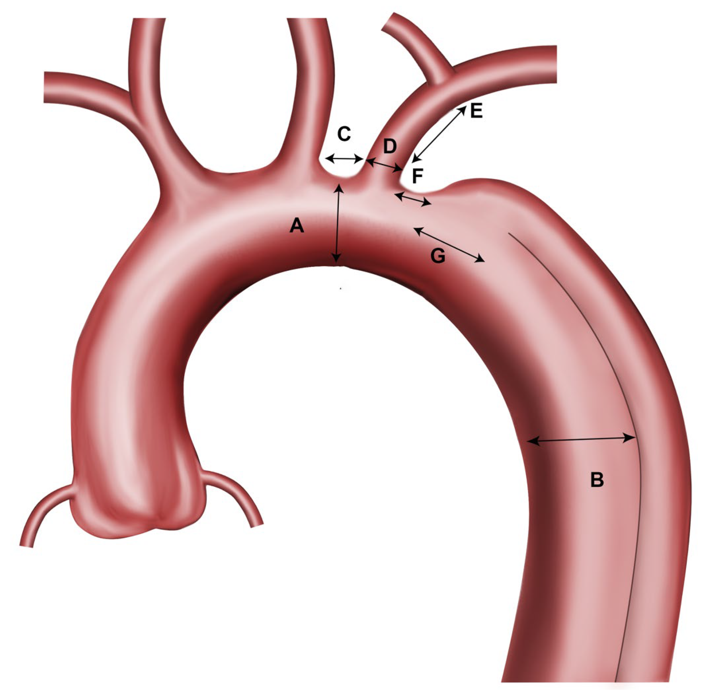
- Maximum aortic diameter at proximal sealing zone (just distal to the left common carotid artery, within zone 2 of the arch) (A).
- Maximum aortic diameter at the distal sealing zone/estimated distal stent graft end (zone 4–5) (B).
- Covered stent graft length proximal to the branch ostium (outer curvature) (C).
- Ideal length of the stent graft to seal the primary entry tear.
- LSA diameter (D).
- LSA length (till the offspring of the first branch/vertebral artery) (E).
- Distance from the distal end of the LSA ostium to the beginning of the aortic dissection (F).
- Distance from the distal end of the LSA ostium to the beginning of the primary entry tear of the aortic dissection (G).
- Minimum left and right iliac artery diameter.
- Aortic arch type according to Marrocco-Trischitta et al. [17].

2.4. Device Descriptions
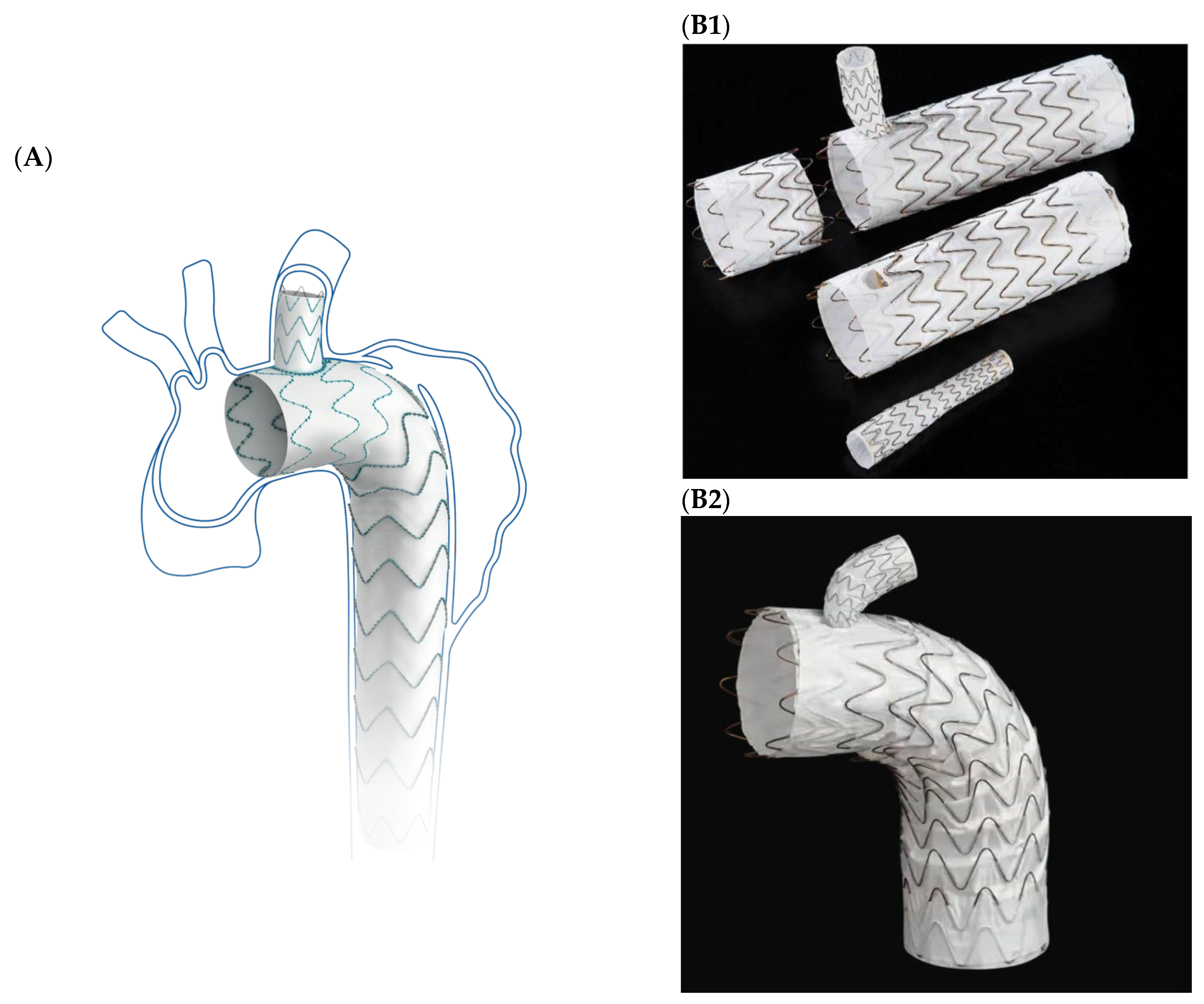
2.4.1. Castor
2.4.2. Gore TAG Thoracic Branch Endoprosthesis (TBE)
2.5. Suitability Assessment
- Oversizing of the LSA by 1–2 mm.
- Minimum proximal landing zone length: 15 mm.
- Minimum LSA landing zone length: 15 mm.
- Oversizing of the main body proximally and distally of <10%.
- No covering of the ostium of the left common carotid artery (LCCA).
- For the TBE the length of the main aortic graft in dissection needs to be calculated: Distance C + D + G + 10 cm (Figure 2).

| Parameter | Castor Single Branch Stent Graft | Thoracic Branch Endoprosthesis (TBE), 8 mm Portal | Thoracic Branch Endoprosthesis (TBE), 12 mm Portal |
|---|---|---|---|
| Aortic diameter at the proximal sealing zone | 23–41 mm | 16–42 mm | 24–42 |
| Covered stent graft length proximal to the branch | 5–30 mm | ≥15–20 mm * | 33.5–36 mm * |
| LSA diameter | 6–13 mm | 6–15 mm | 11–18 mm |
| LSA length | >25 mm | >25 mm | >25 mm |
| Adequate access vessel caliber | 8 mm | 7.5–9.5 mm | 8.2–9.5 mm |
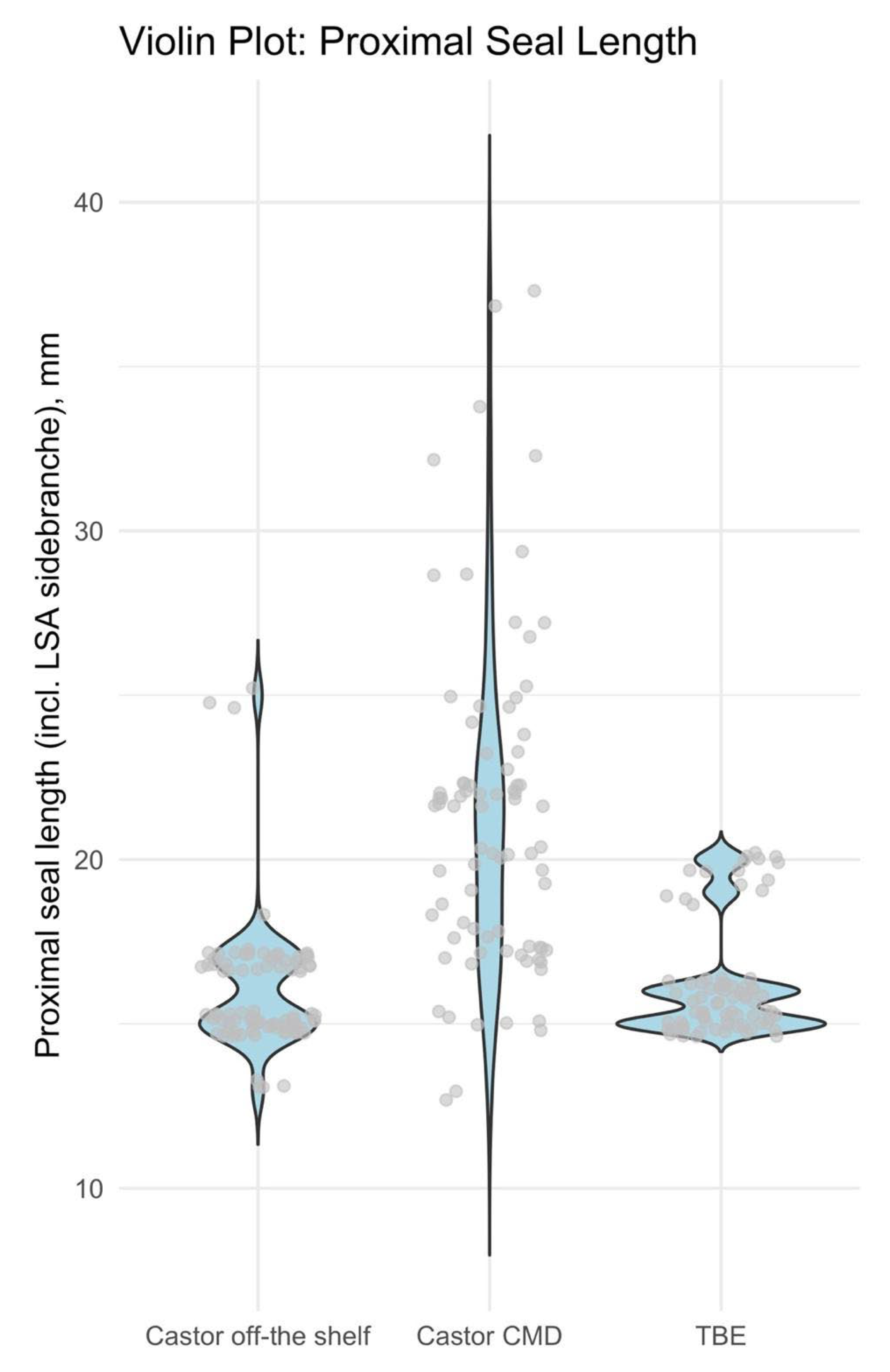
2.6. Proximal Sealing Length
2.7. Outcomes
2.8. Statistical Methods
3. Results
3.1. Patient and Anatomic Characteristics
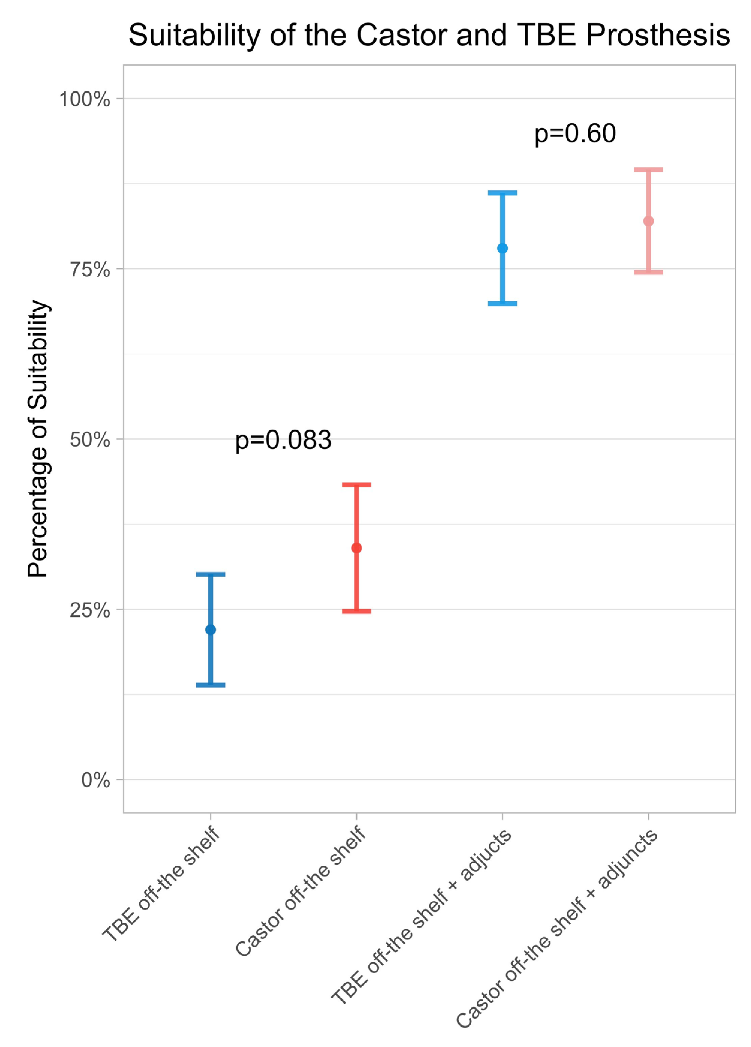
3.2. Suitability
3.2.1. Castor
3.2.2. TBE
3.3. Optimal Off-the Shelf-Stock
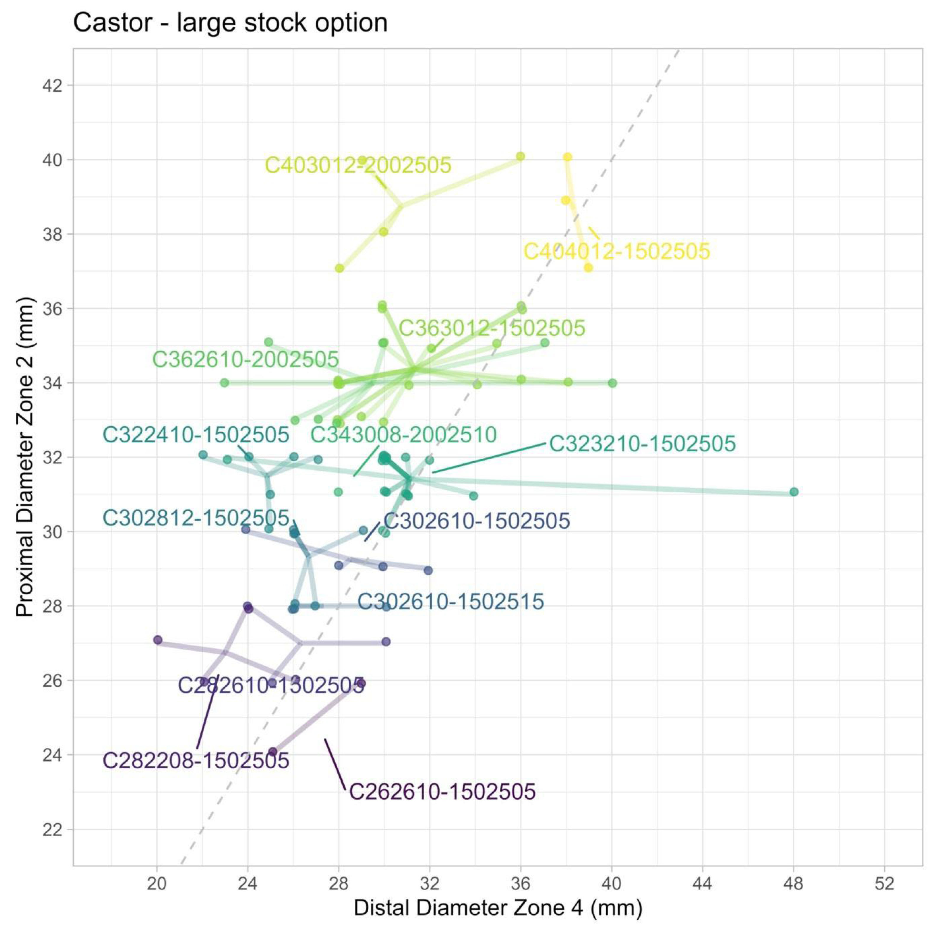
3.3.1. Castor—Optimal Off-the-Shelf Stock
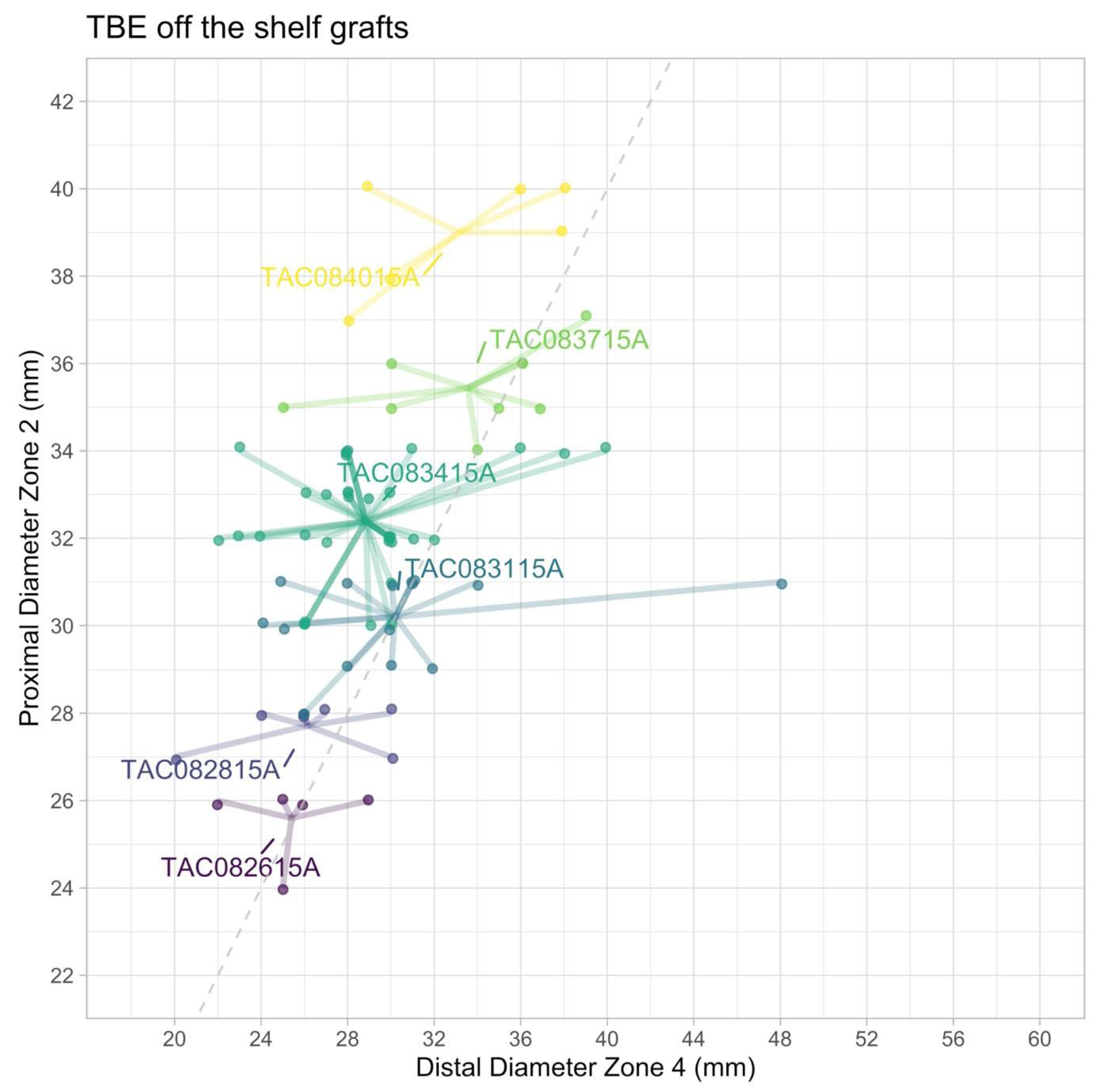
3.3.2. TBE—Optimal Off-the-Shelf Stock
3.4. Comparison Off-the-Shelf Suitability and Proximal Sealing
4. Discussion
4.1. Clinical Applications
4.2. Future Directions
4.3. Limitations
5. Conclusions
Supplementary Materials
Author Contributions
Funding
Institutional Review Board Statement
Informed Consent Statement
Data Availability Statement
Acknowledgments
Conflicts of Interest
Abbreviations
| CMD | custom-made device |
| CTA | Computer-tomography angiography |
| LCCA | Left common carotid artery |
| LSA | Left subclavian artery |
| TBAD | Acute Type B aortic dissection |
| TBE | Thoracic Branch Endoprosthesis |
| TEVAR | Thoracic endovascular aortic repair |
References
- Perone, F.; Guglielmo, M.; Coceani, M.; La Mura, L.; Dentamaro, I.; Sabatino, J.; Gimelli, A. The Role of Multimodality Imaging Approach in Acute Aortic Syndromes: Diagnosis, Complications, and Clinical Management. Diagnostics 2023, 13, 650. [Google Scholar] [CrossRef] [Scilit] [PubMed]
- Riambau, V.; Böckler, D.; Brunkwall, J.; Cao, P.; Chiesa, R.; Coppi, G.; Czerny, M.; Fraedrich, G.; Haulon, S.; Jacobs, M.; et al. Editor’s Choice—Management of Descending Thoracic Aorta Diseases. Eur. J. Vasc. Endovasc. Surg. 2017, 53, 4–52. [Google Scholar] [CrossRef] [Scilit] [PubMed]
- Oberhuber, A.; Maßmann, A.; Betge, S.; Raddatz, A.; Ott, C.; Ploenes, C.; Ito, W.; Janosi, R.A.; Langheim, E.; Czerny, M.; et al. Leitlinie—S2K Typ B Aortendissektion. pp. 1–103. Available online: https://register.awmf.org/assets/guidelines/004-034l_S2k_Typ_B_Aortendissektion_2022-05.pdf (accessed on 14 October 2022).
- MacGillivray, T.E.; Gleason, T.G.; Patel, H.J.; Aldea, G.S.; Bavaria, J.E.; Beaver, T.M. The Society of Thoracic Surgeons/American Association for Thoracic Surgery Clinical Practice Guidelines on the Management of Type B Aortic Dissection. Ann. Thorac. Surg. 2022, 163, 1231–1249. [Google Scholar] [CrossRef] [Scilit] [PubMed]
- Mazzolai, L.; Teixido-Tura, G.; Lanzi, S.; Boc, V.; Bossone, E.; Brodmann, M. 2024 ESC Guidelines for the management of peripheral arterial and aortic diseases: Developed by the task force on the management of peripheral arterial and aortic diseases of the European Society of Cardiology (ESC) Endorsed by the European Association for Cardio-Thoracic Surgery (EACTS), the European Reference Network on Rare Multisystemic Vascular Diseases (VASCERN), and the European Society of Vascular Medicine (ESVM). Eur. Heart J. 2024, 45, 3538–3700. [Google Scholar] [PubMed]
- Mesar, T.; Alie-Cusson, F.S.; Rathore, A.; Dexter, D.J.; Stokes, G.K.; Panneton, J.M. A more proximal landing zone is preferred for thoracic endovascular repair of acute type B aortic dissections. J. Vasc. Surg. 2021, 75, 38–46. [Google Scholar] [CrossRef] [Scilit] [PubMed]
- Dueppers, P.; Meuli, L.; Reutersberg, B.; Hofmann, M.; Messmer, F.; Zimmermann, A. Early and Mid-Term Outcomes of Open versus Endovascular Left Subclavian Artery Debranching for Thoracic Aortic Diseases. Ann. Thorac. Cardiovasc. Surg. 2021, 75, 38–46. [Google Scholar] [CrossRef] [Scilit] [PubMed]
- Feezor, R.J.; Lee, W.A. Management of the Left Subclavian Artery during TEVAR. Semin. Vasc. Surg. 2009, 22, 159–164. [Google Scholar] [CrossRef] [Scilit] [PubMed]
- Garg, K.; Maldonado, T.S. Further consideration for subclavian revascularization with TEVAR. Semin. Vasc. Surg. 2012, 25, 232–237. [Google Scholar] [CrossRef] [Scilit] [PubMed]
- Ye, P.; Miao, H.; Zeng, Q.; Chen, Y. Comparison of total percutaneous in situ microneedle puncture and chimney technique for left subclavian artery fenestration in thoracic endovascular aortic repair for type B aortic dissection. Eur. Radiol. 2024, 34, 7136–7144. [Google Scholar] [CrossRef] [Scilit] [PubMed]
- Brusa, J.; Lutz, E.; Schoenhoff, F.S.; Weiss, S.; Schmidli, J.; Makaloski, V. One-Year Outcome of Postoperative Stroke and Nerve Injury After Supraclavicular Revascularization of The Left Subclavian Artery for Proximal Landing Zone Extension in Thoracic Endovascular Aortic Repair. Ann. Vasc. Surg. 2022, 83, 265–274. [Google Scholar] [CrossRef] [Scilit] [PubMed]
- Huang, Q.; Chen, X.M.; Yang, H.; Lin, Q.N.; Qin, X. Effect of Left Subclavian Artery Revascularisation in Thoracic Endovascular Aortic Repair: A Systematic Review and Meta-analysis. Eur. J. Vasc. Endovasc. Surg. 2018, 56, 644–651. [Google Scholar] [CrossRef] [Scilit] [PubMed]
- Leone, N.; Andreoli, F.; Bartolotti, L.A.M.; Migliari, M.; Baresi, G.F.; Saitta, G.; Silingardi, R.; Gennai, S. Anatomical feasibility of a ‘semi-custom’ unibody single-branch endograft in previous zone 2 thoracic endovascular aortic repair. Eur. J. Cardiothorac. Surg. 2023, 64, ezad290. [Google Scholar] [CrossRef] [Scilit] [PubMed]
- Magee, G.A.; Veranyan, N.; Kuo, E.C.; Ham, S.W.; Ziegler, K.R.; Weaver, F.A.; Fleischman, F.; Bowdish, M.E.; Han, S.M. Anatomic suitability for “off-the-shelf” thoracic single side-branched endograft in patients with type B aortic dissection. J. Vasc. Surg. 2019, 70, 1776–1781. [Google Scholar] [CrossRef] [Scilit] [PubMed]
- Leone, N.; Andreoli, F.; Bartolotti, L.A.M.; Ferri, A.; Silingardi, R.; Gennai, S. Anatomical Suitability of a Standard Subclavian Branched Endograft in Previous Zone 2 Thoracic Endovascular Aortic Repair. J. Endovasc. Ther. 2025, 16, 15266028251316153. [Google Scholar] [CrossRef] [Scilit] [PubMed]
- Yoon, W.J.; Mani, K.; Wanhainen, A.; Rodriguez, V.M.; Mell, M.W. Anatomic feasibility of off-the-shelf thoracic single side-branched endograft in patients with blunt traumatic thoracic aortic injury. J. Vasc. Surg. 2021, 74, 1456–1463.e2. [Google Scholar] [CrossRef] [Scilit] [PubMed]
- Marrocco-Trischitta, M.M.; de Beaufort, H.W.; Secchi, F.; van Bakel, T.M.; Ranucci, M.; van Herwaarden, J.A.; Moll, F.L.; Trimarchi, S. A geometric reappraisal of proximal landing zones for thoracic endovascular aortic repair according to aortic arch types. J. Vasc. Surg. 2017, 65, 1584–1590. [Google Scholar] [CrossRef] [Scilit] [PubMed]
- Rizza, A.; Trimarchi, G.; Di Sibio, S.; Bastiani, L.; Murzi, M.; Palmieri, C.; Foffa, I.; Berti, S. Preliminary Outcomes of Zone 2 Thoracic Endovascular Aortic Repair Using Castor Single-Branched Stent Grafts: A Single-Center Experience. J. Clin. Med. 2023, 12, 7593. [Google Scholar] [CrossRef] [Scilit] [PubMed]
- Fang, C.; Wang, C.; Liu, K.; Pang, X. Early Outcomes of Left Subclavian Artery Revascularization Using Castor Single-Branched Stent-Graft in the Treatment of Type B Aortic Dissection or Intramural Hematoma. Ann. Thorac. Cardiovasc. Surg. 2021, 27, 251–259. [Google Scholar] [CrossRef] [Scilit] [PubMed]
- Paratz, E.D.; Nadel, J.; Humphries, J.; Rowe, S.; Fahy, L.; La Gerche, A.; Prior, D.; Celermajer, D.; Strange, G.; Playford, D. The aortic paradox: A nationwide analysis of 523 994 individual echocardiograms exploring fatal aortic dissection. Eur. Heart J. Cardiovasc. Imaging 2024, 25, 1423–1431. [Google Scholar] [CrossRef] [Scilit] [PubMed]


| Castor | TBE | |
|---|---|---|
| Design type | Unibody | Bi-Modular |
| Branch type | LSA branch incorporated in Unibody | Single inner-branch for LSA |
| Availability | Custom-made | Off-the-shelf |
| Sizes of the main body (diameter and length) | 26–44 mm 6–21 cm | 21–45 mm 10, 15, 20 cm |
| Branch sizes (diameter) | 6–14 mm | 8–20 mm |
| Tapering | Possible | Not possible |
| Possible number of configurations | 351,000 possible configurations | 110 possible configurations: 26 different main grafts: 16 × 8 mm Portal and 10 × 12 mm Portal 8 different branch sizes: 5 for the 8 mm portal and 3 for the 12 mm portal) |
| Market availability | Approved in China and CE-marked | FDA-approved (U.S.) and CE-marked |
| Delivery system | 24 F | 20–26 F |
| Proximal extension | Not available | 8 different proximal Extensions: diameter 21–45 mm, length 3.6–4.6 cm |
| Total (n = 100) | |
|---|---|
| Patient characteristics | |
| Age in years, median (Q1, Q3) | 63.0 (55.1, 71.3) |
| Male Sex | 75 (75%) |
| BMI, median (Q1, Q3) | 26.1 (23.5, 30.1) |
| Hypertension | 78 (78%) |
| Diabetes | 11 (11.2%) |
| Smoking | 40 (40.0%) |
| COPD | 11 (11%) |
| Coronary heart disease | 31 (31%) |
| Dyslipidemia | 18 (18%) |
| Chronic kidney disease | 23 (23%) |
| Anatomical characteristics | |
| Max proximal aortic diameter, mm | 32 (30, 34) |
| Max distal aortic diameter, mm | 30 (26, 31) |
| Max LSA diameter, mm | 11 (9, 12) |
| LCCA to LSA length, mm | 10 (8, 14) |
| LSA to dissection length, mm | 3 (0, 28) |
| LSA to primary entry tear length, mm | 31 (8, 84) |
| Iliac access not suitable | 1% (1.2%) |
| Aortic arch type | |
| Type 1 | 8% (9.0%) |
| Type 2 | 42% (47.2%) |
| Type 3 | 39% (43.8%) |
| Variable, mm, Median (Q1, Q3) | Suitable (n = 82) | Unsuitable (n = 18) | p-Value |
|---|---|---|---|
| Proximal aortic diameter | 32.0 (30.0, 34.0) | 31.0 (26.0, 36.0) | 0.848 |
| Distal aortic diameter | 29.5 (26.0, 32.0) | 28.0 (24.5, 31.5) | 0.110 |
| LSA distal diameter | 11.0 (10.0, 12.0) | 10.5 (8.8, 11.5) | 0.404 |
| LCCA to LSA length | 11.0 (8.0, 14.2) | 8.0 (7.0, 12.0) | 0.047 |
| LSA to dissection length | 6.0 (0.0, 31.5) | 0.0 (0.0, 11.5) | 0.463 |
| LSA to primary entry tear length | 38.0 (10.0, 95.5) | 15.0 (0.0, 52.5) | 0.491 |
| Variable, mm, median (Q1, Q3) | Suitable (n = 22) | Unsuitable (n = 78) | p-Value |
|---|---|---|---|
| Proximal aortic diameter | 31 (28, 34) | 32 (30, 34) | 0.508 |
| Distal aortic diameter | 31 (28, 34) | 28 (26, 30) | 0.045 |
| LSA distal diameter | 11 (9, 12) | 11 (9, 12) | 0.589 |
| LCCA to LSA length | 10 (8, 15) | 10 (8, 13) | 0.692 |
| LSA to dissection length | 12 (0, 25) | 2 (0, 28) | 0.940 |
| LSA to primary entry length | 40 (30, 67) | 25 (7, 90) | 0.644 |
Disclaimer/Publisher’s Note: The statements, opinions and data contained in all publications are solely those of the individual author(s) and contributor(s) and not of MDPI and/or the editor(s). MDPI and/or the editor(s) disclaim responsibility for any injury to people or property resulting from any ideas, methods, instructions or products referred to in the content. |
© 2026 by the authors. Licensee MDPI, Basel, Switzerland. This article is an open access article distributed under the terms and conditions of the Creative Commons Attribution (CC BY) license.
Share and Cite
Lang, J.; Meuli, L.; Dueppers, P.; Zimmerman, A.; Reutersberg, B. Suitability of Single-Branched Thoracic Endografts for the Treatment of Acute Type B Aortic Dissection—An Anatomical Feasibility and Comparative Study. J. Clin. Med. 2026, 15, 558. https://doi.org/10.3390/jcm15020558
Lang J, Meuli L, Dueppers P, Zimmerman A, Reutersberg B. Suitability of Single-Branched Thoracic Endografts for the Treatment of Acute Type B Aortic Dissection—An Anatomical Feasibility and Comparative Study. Journal of Clinical Medicine. 2026; 15(2):558. https://doi.org/10.3390/jcm15020558
Chicago/Turabian StyleLang, Julius, Lorenz Meuli, Philip Dueppers, Alexander Zimmerman, and Benedikt Reutersberg. 2026. "Suitability of Single-Branched Thoracic Endografts for the Treatment of Acute Type B Aortic Dissection—An Anatomical Feasibility and Comparative Study" Journal of Clinical Medicine 15, no. 2: 558. https://doi.org/10.3390/jcm15020558
APA StyleLang, J., Meuli, L., Dueppers, P., Zimmerman, A., & Reutersberg, B. (2026). Suitability of Single-Branched Thoracic Endografts for the Treatment of Acute Type B Aortic Dissection—An Anatomical Feasibility and Comparative Study. Journal of Clinical Medicine, 15(2), 558. https://doi.org/10.3390/jcm15020558