

Article

Heterogeneity and Detection Rate of Hypertrophic Cardiomyopathy Phenotype in China: A Multicenter Echocardiography Study


1
Department of Cardiology, Peking University First Hospital, Beijing 100034, China
2
Echocardiography Core Lab, Institute of Cardiovascular Disease, Peking University First Hospital, Beijing 100034, China
3
Department of Cardiology, Sir Run Run Shaw Hospital, School of Medicine, Zhejiang University, Hangzhou 310016, China
4
Department of Cardiology, Shanghai Tenth People’s Hospital, Shanghai 200072, China
5
Department of Cardiology, First Affiliated Hospital of Sun Yat-sen University, Guangzhou 510080, China
6
Department of Cardiology, Xuzhou Central Hospital, Xuzhou 221009, China
7
Department of Cardiology, The First Affiliated Hospital of Xi’an Jiaotong University, Xi’an 710061, China
8
Department of Cardiology, Taiyuan Central Hospital, Taiyuan 030009, China
9
Department of Cardiology, The People’s Hospital of Liaoning Province, Shenyang 110016, China
10
Department of Cardiology, Union Hospital, Tongji Medical College, Huazhong University of Science and Technology, Wuhan 430022, China
*
Authors to whom correspondence should be addressed.
These authors contributed equally to this work.
J. Clin. Med. 2026, 15(16), 6230; https://doi.org/10.3390/jcm15166230
Submission received: 28 June 2026 / Revised: 29 July 2026 / Accepted: 8 August 2026 / Published: 12 August 2026

Abstract

Background: Contemporary data on the clinical detection rate and profile of phenotypical hypertrophic cardiomyopathy (HCM) in major Chinese healthcare settings are limited. This multicenter study aimed to determine the detection rate and echocardiographic features of the HCM phenotype in a large Chinese cohort. Methods: This cross-sectional study analyzed echocardiography databases from nine medical centers across China, including adult patients examined during 2023. HCM phenotype was defined as end-diastolic wall thickness ≥15 mm in the left ventricle. Patients with moderate to severe aortic stenosis were excluded. Subcategories included phenotypes of obstructive HCM and apical hypertrophy. Results: Among 655,383 examinations, 2610 patients met the criteria of the HCM phenotype, yielding a detection rate of 0.40% (≈1 in 250). The mean age was 60.2 years with male predominance (70.3%). Asymmetric septal hypertrophy was present in 53.3% of patients. The most commonly involved site with maximal wall thickness was the interventricular septum (57.6%), followed by the apex (20.9%) and the basal septum (17.7%). The overall intra-left ventricular obstruction rate was 16.2%; left ventricular outflow tract obstruction (LVOTO) accounted for 12.1%. LVOTO patients had greater septal thickness, smaller left ventricular diastolic dimensions, and more mitral regurgitation. Female sex was associated with a significantly higher LVOTO rate than males (18.2% vs. 9.6%, p < 0.001). Pure apical hypertrophy was identified in 4.5% of patients, with an increasing detection rate in older age groups. Conclusions: This large-scale, multicenter study confirms a high clinical detection rate for the HCM phenotype (≈1 in 250) in major Chinese centers. Substantial phenotypic heterogeneity across age and sex, coupled with the identification of key factors associated with LVOTO, highlights the need for sex- and age-specific diagnostic strategies and therapeutic planning in clinical practice.

1. Introduction

Hypertrophic cardiomyopathy (HCM) is the most common inherited cardiac disease, characterized by unexplained left ventricular (LV) hypertrophy in the absence of obvious loading conditions that could account for the observed wall thickness. As a monogenic disorder primarily caused by mutations in sarcomeric protein genes, HCM exhibits remarkable clinical and morphological heterogeneity, presenting a spectrum of disease that ranges from asymptomatic hypertrophy to severe heart failure, malignant ventricular arrhythmias, and sudden cardiac death [1]. The clinical importance of HCM is thus underscored by its role as a leading cause of sudden cardiac death in young people and a significant contributor to morbidity across all age groups.
Historically, the prevalence of HCM has been cited as 1 in 500 in the general population [2]. However, advanced diagnostic techniques, including improved echocardiography and genetic screening, coupled with increased clinical awareness, have led to revised contemporary estimates suggesting that the true prevalence may be closer to 1 in 200 [3]. This shift in understanding highlights that HCM is more common than previously recognized and suggests that a significant portion of the population remains undiagnosed. Accurate identification and characterization of the disease, particularly the presence and location of left ventricular outflow tract obstruction (LVOTO), remain paramount, as these morphological and hemodynamic features dictate both prognosis and therapeutic strategies, including septal reduction therapy [4].
While extensive research has mapped the clinical course and genotype–phenotype associations of HCM, large-scale, contemporary, real-world data from diverse patient populations remain critical for refining management guidelines and public health strategies. Specifically, contemporary epidemiological and morphological data for the HCM phenotype in China, derived from multiple centers, is limited. Given the phenotypic variance observed in Asian cohorts, particularly concerning apical hypertrophy [5,6,7], a large-scale analysis is warranted to accurately assess the current clinical burden and phenotypic distribution.
Therefore, the aim of this study was to conduct a large, multicenter, cross-sectional analysis using echocardiographic databases from nine medical centers in mainland China. The primary objective was to determine the detection rate of the echocardiographically defined hypertrophic cardiomyopathy phenotype (HCM phenotype; phenotypical changes in cardiac structure indicative of HCM) within this large cohort during the year 2023. Secondary objectives included establishing a detailed profile of the HCM phenotype, identifying the detection rate of obstructive and pure apical hypertrophy subtypes, and characterizing significant differences in presentation when stratified by sex and age groups.

2. Methods

Data were collected from the echocardiography databases of nine participating centers, including all outpatients, emergency patients, and inpatients aged ≥18 years who underwent echocardiography from 1 January 2023 to 31 December 2023. All adult patients presenting with hypertrophic cardiomyopathy (HCM) phenotype identified by echocardiography were enrolled. For patients who underwent multiple echocardiographic examinations, the results from the first examination within the study period were used for analysis.
According to current American Society of Echocardiography criteria for detecting the presence of HCM (on two-dimensional echocardiography), an end-diastolic wall thickness ≥15 mm anywhere in the left ventricle was identified as an HCM phenotype [1,8,9]. Since this study only extracted data from an echocardiographic database, lacking detailed clinical information, some HCM phenocopies, such as hypertensive left ventricular hypertrophy, Anderson–Fabry disease and cardiac amyloidosis, cannot be fully ruled out through echocardiography. Patients with moderate and severe aortic stenosis on echocardiography were excluded. Therefore, the patients enrolled in our study may include other HCM phenocopies. So, we use the terminology “HCM phenotype” to include all patients who only meet the echocardiographic criteria for HCM.
The following subcategories of HCM phenotype were then identified, where possible, according to the availability of additional data:
  • Asymmetric septal hypertrophy (ASH): Septal-to-posterior wall thickness ratio >1.3.
  • Obstructive HCM (oHCM) phenotype: Increase in the peak pressure gradient (≥30 mmHg) anywhere in the left ventricle (either on a resting transthoracic echocardiogram or during stress testing, such as the Valsalva maneuver, when documented) on continuous wave (CW) or pulsed wave (PW) Doppler was assumed to indicate intra-ventricular obstruction. Based on the location of the obstruction in the left ventricle, the oHCM phenotype is subdivided into two subtypes:
    • Left ventricular outflow tract obstruction (LVOTO): Overt obstruction (LVOT peak pressure gradient ≥30 mmHg at rest) and latent obstruction (LVOT peak pressure gradient <30 mmHg at rest but ≥30 mmHg during stress test);
    • Mid-ventricular obstruction.
  • Non-obstructive HCM (nHCM) phenotype: Peak pressure gradient <30 mmHg anywhere in the left ventricle both at rest or during a stress test.
  • Pure apical hypertrophy: Hypertrophy localized exclusively to the apical region, while other myocardial segments remain non-hypertrophied.
  • Mixed apical hypertrophy: The apex, in conjunction with other myocardial segments, is hypertrophic.
Data that were collected for this study included patient age when undergoing the examination and gender. Echocardiographic data included left atrium and left ventricular dimensions, left ventricular systolic and diastolic function measurements, the location of the maximal LV wall thickness, the peak pressure gradient of LVOT, the mid-ventricle or LV apex (when available), and the degree of mitral regurgitation and aortic stenosis.
Ethical approvals were obtained from all relevant human research ethics committees. Institutional Review Board approval was granted for this research by the Ethics Committee of Peking University First Hospital (lead ethics committee).

Statistical Analysis

Continuous variables are shown as mean ± standard deviation (SD) or median (interquartile range) according to different distributions. Categorical variables are presented as numbers (percentages). Differences in variables between two groups were compared by a two-sample t-test, the Wilcoxon rank-sum test, or a chi-square test (or Fisher’s exact test when appropriate).
We also performed multivariable logistic regression analysis to identify independent risk factors for left ventricular outflow tract obstruction. Candidate predictors were first preselected using Least Absolute Shrinkage and Selection Operator (LASSO) regression with 10-fold cross-validation. Variables with non-zero coefficients were then entered into a multivariable logistic regression model, with forced adjustment for possible confounding factors. The results are presented as adjusted odds ratios (aOR) with 95% confidence intervals (CIs). Detailed procedures regarding variable selection and model specifications are provided in the Supplementary Materials (Supplementary Methods).
All statistical analyses were conducted using the R software, version 3.6.2. All statistical analyses were 2-tailed, and p < 0.05 was considered statistically significant.

3. Results

3.1. Overview of the Hypertrophic Cardiomyopathy Phenotype Cohort

Among 655,383 echocardiographic examinations conducted in 2023 across nine medical centers in mainland China, 2610 patients met the diagnostic criteria for hypertrophic cardiomyopathy (HCM) phenotype. Thus, the detection rate for the HCM phenotype in this cohort was approximately 0.40%. Among patients with the HCM phenotype, the mean age was 60.2 ± 15.1 years, with male dominance (70.3%).
The HCM phenotype within this cohort was characterized by increased interventricular septal wall thickness (interventricular septal wall thickness: 16.5 ± 4.4 mm), and more than half (1392/2610, 53.3%) of patients experienced asymmetric septal hypertrophy. The maximum LV wall thickness averaged 19.0 ± 3.8 mm. The left ventricular (LV) diastolic dimension was mostly normal (46.4 ± 6.1 mm, n = 2532), with normal LV systolic function but impaired diastolic function, and increased left atrial diameter (Table 1).
A stress test was performed on 14.3% (373/2610) of all patients. Among all patients, 423/2610 (16.2%) had intra-left ventricular obstruction, 317/2610 (12.1%) had left ventricular outflow tract (LVOT) obstruction, and 128/2610 (4.9%) had mid-ventricular obstruction.

3.2. Comparison of Echocardiographic Characteristics Between Sexes

We then compared the differences between patients of different genders. The male patients were younger than females (average age, male vs. female: 58.6 ± 14.8 years vs. 64.0 ± 15.2 years, p < 0.001). A higher proportion of male patients exhibited a left ventricular posterior wall thickness greater than 15 mm, while the proportion of asymmetric septal hypertrophy was lower compared to females. There was no statistically significant difference in maximum wall thickness between male and female patients. Females had a smaller left ventricular end-diastolic diameter and a higher proportion of preserved left ventricular ejection fraction compared to males. The proportions of apical aneurysm, pure apical hypertrophy, and concomitant right ventricular hypertrophy were comparable between genders. A higher proportion of female patients presented with left ventricular outflow tract obstruction (female vs. male: 18.2% vs. 9.6%, p < 0.001), most of which (85.1%) were classified as overt obstructions (Table 1).

3.3. Comparison of Echocardiographic Characteristics Between Age Groups

To compare the clinical characteristics across different age groups, patients were categorized into three cohorts: 18–49 years, 50–74 years, and ≥75 years. The mean ages of the three groups were 38.3 ± 7.5 years, 62.8 ± 6.9 years, and 81.0 ± 5.1 years, respectively. The youngest group (18–49 years) demonstrated the greatest thickness of the interventricular septum and left ventricular posterior wall, as well as the highest prevalence of asymmetric septal hypertrophy. The proportion of patients with pure apical hypertrophy was higher in the ≥75 years group compared to the other two age groups, whereas concomitant right ventricular hypertrophy was most frequently observed in the 18–49 years group. The detection rate of left ventricular outflow tract obstruction was comparable across all three age groups, while mid-ventricular obstruction was more prevalent in 18–49 years and 49–74 years than ≥75 years (Table 2).
In our cohort of patients with a hypertrophic cardiomyopathy phenotype, involvement was observed in various left ventricular segments, either individually or in combination. Figure 1 displays the distribution of the left ventricular segments exhibiting maximal wall thickness. The segments were categorized as follows: interventricular septum, basal septum, anterior wall, lateral wall, posterior wall, inferior wall, apex, and diffuse left ventricular wall. In the overall population, the most commonly involved site was the interventricular septum (57.6%), followed by the apex (20.9%) and the basal septum (17.7%). The proportion of basal septal involvement was higher in patients aged ≥75 years (29.7%) than in the other two age groups, while apical involvement was more frequent in patients aged ≥50 years compared with those under 50 years of age (Figure 1 and Supplementary Table S1).

3.4. Comparison of Patients with Left Ventricular Outflow Tract Obstruction Versus Non-Obstructive Phenotype

Out of all patients, 317/2610 (12.1%) met the diagnostic criteria for left ventricular outflow tract obstruction (LVOTO), and lower male predominance (55.5%) was observed in patients with LVOTO compared to those with non-obstructive HCM (nHCM) (72.2%). Compared to nHCM patients, patients with LVOTO presented with greater interventricular septal thickness, a higher detection rate for asymmetric septal hypertrophy, and greater maximum left ventricular wall thickness, which were more frequently located at the basal IV septum and less at the apex. Patients with LVOTO also had a smaller LV diastolic diameter, a higher LV ejection fraction, and a higher incidence of moderate to severe mitral regurgitation than nHCM patients. More than half of patients with LVOTO (192/317, 60.6%) exhibited an LVOT peak pressure gradient of ≥50 mmHg during either rest or stress tests. In total, 22/317 (6.9%) patients with LVOTO had concomitant mid-ventricular obstructions (Table 3).
Multivariable logistic regression analysis identified six independent factors associated with LVOTO (all p < 0.05). Female sex (aOR: 1.54, 95% CI: 1.16–2.05), increased interventricular septal thickness (aOR: 1.13 per unit increase, 95% CI: 1.08–1.18), maximal wall thickness located to basal interventricular septal (aOR: 2.27, 95% CI: 1.65–3.14), preserved ejection fraction (LVEF ≥55%, aOR: 3.95, 95% CI: 1.69–9.23), and moderate to severe mitral regurgitation (aOR: 5.65, 95% CI: 4.25–7.52) were identified as independent risk factors. In contrast, larger left ventricular diastolic diameter emerged as an independent protective factor (aOR: 0.93, 95% CI: 0.91–0.95). Detailed results are presented in the Supplementary Materials (Supplementary Table S2).

3.5. Echocardiographic Characteristics of Pure Apical Hypertrophy

Pure apical hypertrophy represents a unique phenotypic variant of HCM, defined by hypertrophy localized exclusively to the apical region, while other myocardial segments remain non-hypertrophied. As such, we delineated this specific phenotype within our study population for a separate description of its echocardiographic features.
The pure apical hypertrophy phenotype was identified in 117 out of 2610 (4.5%) patients with the HCM phenotype. This subgroup had a mean age of 63.2 ± 15.4 years, with a male predominance (67.5%). The majority of these patients exhibited relatively normal left ventricular diastolic dimensions and left ventricular ejection fraction. Apical aneurysm was present in two patients (1.7%), while mid-ventricular obstruction was observed in seven (6.0%) patients (Table 4).

4. Discussion

This multicenter, cross-sectional study provides a contemporary snapshot of the echocardiographically identified hypertrophic cardiomyopathy (HCM) phenotype in a large Chinese cohort from nine medical centers in the year 2023. Our primary finding is a notable detection rate of 0.40% (approximately 1 in 250) among patients undergoing echocardiography in 2023. We also established a detailed morphological, hemodynamic and demographic profile of this large cohort, revealing a male predominance, a high rate of asymmetric septal hypertrophy, and unique differences in disease presentation when stratified by sex and age.

4.1. The Detection Rate of Hypertrophic Cardiomyopathy (HCM) Phenotype

The detection rate of 0.40% found in our cohort is significantly higher than the widely cited historical prevalence of HCM (1 in 500), first reported nearly three decades ago [2]. Our finding aligns more closely with contemporary estimates, suggesting a true HCM population prevalence of 1 in 200 [3]. Among the factors contributing to the revised estimate of the disease being more common than 1 in 500 were the identification of gene carriers who are negative for the HCM phenotype; enhanced clinical identification of the HCM phenotype with advanced imaging; recognition that because of the autosomal-dominant inheritance pattern, multiple relatives of probands (and carriers) could be affected by HCM; and recognition that up to 0.6% of the population may carry HCM-causing sarcomere mutations [3].
As for the prevalence of HCM in China, an earlier HCM screening study of patients 18–74 years old found a prevalence of 0.16% [10]. A recent study in 2022 calculated that the prevalence of HCM was 0.0069% in 2010, rising to 0.076% in 2019 based on the whole Beijing population [11].
However, it is imperative to acknowledge that our figure represents a detection rate within an echocardiographic population undergoing diagnostic testing, rather than a true population prevalence derived from systematic screening. The nine participating centers are large medical institutions, likely serving as referral centers for patients with cardiac symptoms or known murmurs, leading to a substantial selection bias. Nonetheless, this high detection rate of phenotypical hypertrophy underscores two critical points: first, it supports the notion that HCM remains substantially underdiagnosed in the general population, and second, it highlights the significant burden of the disease within the healthcare system, necessitating increased clinical awareness and resource allocation.

4.2. Morphologic and Hemodynamic Features

The morphological characteristics of our cohort were generally consistent with the established phenotype of HCM [1]. Asymmetric septal hypertrophy (ASH) was the most common subtype, observed in 53.3% of patients, with the interventricular septum being the most frequently involved segment (57.6%). The overall intra-left ventricular obstruction detection rate was 16.2%, with 12.1% experiencing left ventricular outflow tract obstruction (LVOTO). This obstruction rate is lower than the 20–30% typically cited in specialized HCM centers and a nationwide echocardiographic database [12,13,14]. When the Valsalva maneuver and exercise echocardiography were widely performed, the proportion of LVOTO rose to about 70% [15]. This may be explained by our study’s inclusion of a broader, less selected patient population from both specialized and general cardiology practices. Furthermore, the fact that only 14.3% of patients underwent a stress test suggests that the true detection rate of latent obstruction is likely underestimated. To our knowledge, the lower stress test rate in our cohort reflects several practical challenges in China: (1) variable awareness of hypertrophic obstructive cardiomyopathy and stress tests among echocardiographers across different hospitals; (2) insufficient technical proficiency or patient cooperation, leading to unsuccessful or unreported tests; and (3) limited access to exercise stress echocardiography in some institutions. This highlights a significant gap in real-world clinical practice and emphasizes the need for more systematic stress tests to unmask latent gradients. In response, the Chinese Society of Echocardiography and seven other ultrasound societies jointly published the Guidelines for Clinical Application of Provocation/Stress Echocardiography in Patients with Hypertrophic Cardiomyopathy in 2024 [16], aiming to standardize procedures, promote broader adoption, and improve detection of latent obstruction. The implementation of these guidelines is expected to promote the standardized use of provocation testing across China, which may in turn enhance the detection of latent obstruction in clinical practice.
In our multivariable model, moderate-to-severe mitral regurgitation emerged as the strongest independent correlate of LVOTO. Among structural parameters, increased basal septal thickness and greater interventricular septal thickness confirmed the mechanical role of basal septal hypertrophy in narrowing the outflow tract. Notably, preserved LVEF (≥55%) was identified as a risk factor, suggesting that hyperdynamic contraction drives systolic anterior motion (SAM) and gradient formation. Female sex was associated with higher risk, possibly due to smaller left ventricular cavity dimensions, whereas larger LV diastolic diameter appeared protective, likely by diluting the relative impact of septal hypertrophy on outflow compromise. Collectively, LVOTO is not a single anatomical abnormality but a multifactorial phenomenon involving valvular function, regional hypertrophy, global contractile status, and chamber geometry. In clinical practice, patients with HCM phenotype with preserved or supranormal LVEF and coexisting mitral regurgitation warrant heightened vigilance for significant LVOT obstruction.

4.3. Variations According to Sex and Age Group

Subgroup analysis revealed clinically relevant age and sex differences. Male patients presented at a younger age but had a lower detection rate of LVOTO compared to females. Conversely, female patients, though older, demonstrated a higher rate of LVOTO (18.2% vs. 9.6%) and moderate to severe mitral regurgitation. This observation is consistent with recent studies reporting phenotypic differences between sexes, where female HCM patients may present later in life, often with a more adverse clinical course and higher rates of obstruction, possibly due to a smaller cavity size that promotes systolic anterior motion (SAM) of the mitral valve or distinct genetic backgrounds [17,18,19,20,21]. Both biological and social factors can affect the phenotypic presentation in female patients. Endocrine functions related to female gender may delay the development of hypertrophy or its clinical manifestations [22]. Also, disparities in healthcare-seeking behavior and referral patterns may delay the disease presentation and diagnosis [17]. All of the above underscores the importance of heightened suspicion for HCM in women.
Stratification by age showed that the youngest group (18–49 years) exhibited the greatest wall thickness and the highest detection rate of asymmetric septal hypertrophy (ASH). This suggests that the most severe, likely genetic, forms of HCM manifest earlier in life.

4.4. Apical Hypertrophy

Apical hypertrophic cardiomyopathy is conventionally classified into pure apical HCM, characterized by isolated apical involvement, and mixed apical HCM, which involves the apex in conjunction with hypertrophy of other myocardial segments. This phenotypic variant is notably more prevalent in Asian populations, accounting for approximately 25% of HCM cases among Asians, compared to 1–10% in non-Asians [23]. Data from multiple single-center studies and one multicenter study in China indicate that apical HCM constitutes between 10% and 22.8% of all HCM cases [5,24,25,26,27]. In our cohort, apical involvement was observed in 20.9% of all patients with an HCM phenotype, which is consistent with previous reports. However, owing to its distinctive localization, the limited routine use of left ventricular contrast echocardiography, and the lack of cardiac MRI data in our study, the pure apical hypertrophy may be underdiagnosed in clinical practice.
Several single-center studies in China have identified elderly age as an independent risk factor for cardiovascular events in patients with apical HCM [24,25,27]. Of note, our study found that the detection rate of hypertrophy localized to the apex increased with advancing age. Apical hypertrophic cardiomyopathy patients had a more favorable prognosis, compared with other subtypes of HCM, but not for all apical HCM patients [7,23,28]. The characteristics of the pure apical hypertrophy subgroup in our study further contribute to the understanding of HCM heterogeneity.

4.5. Limitations

Several limitations must be considered. First, given the multicenter, cross-sectional design utilizing echocardiography databases, the study cohort is subject to selection bias, which is inherent in non-population-based studies. Thus, findings cannot be generalized to the general Chinese population. Our “HCM phenotype” definition (≥15 mm thickness) is strictly echocardiographic, and without clinical data, we could not definitively rule out phenocopies such as hypertensive cardiac hypertrophy, Anderson–Fabry disease or cardiac amyloidosis. This classification is a pragmatic compromise for large-scale database research but acknowledges that our cohort may be heterogeneous in underlying etiology. Also, our relatively strict morphological criteria for probable or definite HCM (maximal left ventricular wall thickness ≥15 mm) could have excluded those occasional patients with mild morphological expressions of the disease and “borderline” wall thickness of only 13 or 14 mm. Furthermore, the assessment of dynamic obstruction was limited, as a stress test was performed on only 14.3% of the patients. Consequently, the true prevalence of latent LVOTO may be underestimated.

5. Conclusions

Our study provides crucial, large-scale, contemporary data on the detection rate and echocardiographic characteristics of the HCM phenotype in a major Chinese clinical setting. The detection rate of 1 in 250 confirms that the HCM phenotype is a common condition. The observed differences in presentation across age and sex underscore the phenotypic heterogeneity of the disease. These findings highlight the need for enhanced screening protocols, particularly in the outpatient setting, and provide essential epidemiological data that can inform public health strategies and the allocation of specialized resources for the diagnosis and management of HCM in China. Future work should focus on integrating clinical, genetic, and cardiac magnetic resonance imaging (CMR) data to better delineate true HCM from its phenocopies and to validate the prognostic significance of the different echocardiographic subtypes observed.

Supplementary Materials

The following supporting information can be downloaded at https://www.mdpi.com/article/10.3390/jcm15166230/s1. Supplementary Methods: Detailed statistical procedures for variable selection and multivariable modeling. Supplementary Table S1. Location of maximal wall thickness stratified by age group. Supplementary Table S2. Multivariable logistic regression analysis of factors associated with left ventricular outflow tract obstruction (LVOTO).

Author Contributions

Conceptualization, W.M. and Y.Y.; methodology, W.M., Y.Y. and B.W. (Beining Wang); formal analysis, B.W. (Beining Wang); investigation, B.W. (Beining Wang), B.W. (Bei Wang), H.S., M.Z., F.Y., W.L., S.W., J.W., Y.S. and M.X.; resources, all authors; data curation, B.W. (Beining Wang), B.W. (Bei Wang), H.S., M.Z., F.Y., W.L., S.W., J.W., Y.S. and M.X.; writing—original draft preparation, B.W. (Beining Wang); writing—review and editing, B.W. (Bei Wang), H.S., M.Z., F.Y., W.L., S.W., J.W., Y.S., M.X., Y.Y. and W.M.; visualization, B.W. (Beining Wang); supervision, W.M., Y.Y. and M.X.; project administration, B.W. (Bei Wang), H.S., M.Z., F.Y., W.L., S.W., J.W., Y.S., M.X., Y.Y. and W.M. All authors have read and agreed to the published version of the manuscript.

Funding

This work was supported by the National Key Research and Development Program of China (Grant No. 2021YFC2501106) and Corxel Pharmaceuticals.

Institutional Review Board Statement

The study was conducted in accordance with the Declaration of Helsinki and approved by the Ethics Committee of Peking University First Hospital (protocol code HCM-2024 and date of approval: 23 April 2024).

Informed Consent Statement

Patient consent was waived because the data used in this study was derived from previous clinical routine practice, and the subjects can no longer be contacted. This research project does not involve personal privacy or commercial interests.

Data Availability Statement

The raw data supporting the conclusions of this article will be made available by the authors upon request. The data that support the findings of this study are not publicly available due to ethical and privacy restrictions, as they contain sensitive patient information (including echocardiographic measurements). The institutional review board imposed restrictions on the public sharing of these data to protect patient confidentiality. De-identified data may be available from the corresponding author upon reasonable request and with the permission of the institutional ethics committee, subject to a data access agreement.

Acknowledgments

No artificial intelligence (AI) technologies or tools were used in this study.

Conflicts of Interest

The authors declare no conflicts of interest.

Abbreviations

The following abbreviations are used in this manuscript:
ASHasymmetric septal hypertrophy
AVaortic valve
CMRcardiac magnetic resonance
CWcontinuous wave
HCMhypertrophic cardiomyopathy
IVinterventricular
LAleft atrium
LVleft ventricle
LVEFleft ventricular ejection fraction
LVOTleft ventricular outflow tract
LVOTOleft ventricular outflow tract obstruction
nHCMnon-obstructive hypertrophic cardiomyopathy
oHCMobstructive hypertrophic cardiomyopathy
PWpulsed wave
RVright ventricle
SAMsystolic anterior motion
SDstandard deviation

References

  1. Ommen, S.R.; Ho, C.Y.; Asif, I.M.; Balaji, S.; Burke, M.A.; Day, S.M.; Dearani, J.A.; Epps, K.C.; Evanovich, L.; Ferrari, V.A.; et al. 2024 AHA/ACC/AMSSM/HRS/PACES/SCMR Guideline for the Management of Hypertrophic Cardiomyopathy: A Report of the American Heart Association/American College of Cardiology Joint Committee on Clinical Practice Guidelines. Circulation 2024, 149, e1239–e1311. [Google Scholar] [CrossRef] [PubMed]
  2. Maron, B.J.; Gardin, J.M.; Flack, J.M.; Gidding, S.S.; Kurosaki, T.T.; Bild, D.E. Prevalence of hypertrophic cardiomyopathy in a general population of young adults. Echocardiographic analysis of 4111 subjects in the CARDIA Study. Coronary Artery Risk Development in (Young) Adults. Circulation 1995, 92, 785–789. [Google Scholar] [CrossRef] [PubMed]
  3. Semsarian, C.; Ingles, J.; Maron, M.S.; Maron, B.J. New perspectives on the prevalence of hypertrophic cardiomyopathy. J. Am. Coll. Cardiol. 2015, 65, 1249–1254. [Google Scholar] [CrossRef] [PubMed]
  4. Cui, H.; Schaff, H.V.; Wang, S.; Lahr, B.D.; Rowin, E.J.; Rastegar, H.; Hu, S.; Eleid, M.F.; Dearani, J.A.; Kimmelstiel, C.; et al. Survival Following Alcohol Septal Ablation or Septal Myectomy for Patients With Obstructive Hypertrophic Cardiomyopathy. J. Am. Coll. Cardiol. 2022, 79, 1647–1655. [Google Scholar] [CrossRef] [PubMed]
  5. Chen, Q.F.; Zou, J.; Katsouras, C.S.; You, S.; Zhou, J.; Ge, H.B.; Liu, C.; Zhou, X.; Ni, C.; Peng, Y.; et al. Clinical Characteristics and Outcomes in Patients With Apical and Nonapical Hypertrophic Cardiomyopathy. J. Am. Heart Assoc. 2024, 13, e036663. [Google Scholar] [CrossRef] [PubMed]
  6. Kubo, T.; Kitaoka, H.; Okawa, M.; Hirota, T.; Hoshikawa, E.; Hayato, K.; Yamasaki, N.; Matsumura, Y.; Yabe, T.; Nishinaga, M.; et al. Clinical profiles of hypertrophic cardiomyopathy with apical phenotype--comparison of pure-apical form and distal-dominant form. Circ. J. Off. J. Jpn. Circ. Soc. 2009, 73, 2330–2336. [Google Scholar] [CrossRef] [PubMed]
  7. Moon, J.; Shim, C.Y.; Ha, J.W.; Cho, I.J.; Kang, M.K.; Yang, W.I.; Jang, Y.; Chung, N.; Cho, S.Y. Clinical and echocardiographic predictors of outcomes in patients with apical hypertrophic cardiomyopathy. Am. J. Cardiol. 2011, 108, 1614–1619. [Google Scholar] [CrossRef] [PubMed]
  8. Ommen, S.R.; Mital, S.; Burke, M.A.; Day, S.M.; Deswal, A.; Elliott, P.; Evanovich, L.L.; Hung, J.; Joglar, J.A.; Kantor, P.; et al. 2020 AHA/ACC Guideline for the Diagnosis and Treatment of Patients With Hypertrophic Cardiomyopathy: A Report of the American College of Cardiology/American Heart Association Joint Committee on Clinical Practice Guidelines. Circulation 2020, 142, e558–e631. [Google Scholar] [CrossRef] [PubMed]
  9. Nagueh, S.F.; Phelan, D.; Abraham, T.; Armour, A.; Desai, M.Y.; Dragulescu, A.; Gilliland, Y.; Lester, S.J.; Maldonado, Y.; Mohiddin, S.; et al. Recommendations for Multimodality Cardiovascular Imaging of Patients with Hypertrophic Cardiomyopathy: An Update from the American Society of Echocardiography, in Collaboration with the American Society of Nuclear Cardiology, the Society for Cardiovascular Magnetic Resonance, and the Society of Cardiovascular Computed Tomography. J. Am. Soc. Echocardiogr. 2022, 35, 533–569. [Google Scholar] [CrossRef] [PubMed]
  10. Zou, Y.; Song, L.; Wang, Z.; Ma, A.; Liu, T.; Gu, H.; Lu, S.; Wu, P.; Zhang, Y.; Shen, L.; et al. Prevalence of idiopathic hypertrophic cardiomyopathy in China: A population-based echocardiographic analysis of 8080 adults. Am. J. Med. 2004, 116, 14–18. [Google Scholar] [CrossRef] [PubMed]
  11. Bai, Y.; Zheng, J.P.; Lu, F.; Zhang, X.L.; Sun, C.P.; Guo, W.H.; Zou, Y.X.; Lip, G.Y.H.; Shi, X.B. Prevalence, incidence and mortality of hypertrophic cardiomyopathy based on a population cohort of 21.9 million in China. Sci. Rep. 2022, 12, 18799. [Google Scholar] [CrossRef] [PubMed]
  12. Maron, M.S.; Olivotto, I.; Betocchi, S.; Casey, S.A.; Lesser, J.R.; Losi, M.A.; Cecchi, F.; Maron, B.J. Effect of left ventricular outflow tract obstruction on clinical outcome in hypertrophic cardiomyopathy. N. Engl. J. Med. 2003, 348, 295–303. [Google Scholar] [CrossRef] [PubMed]
  13. Autore, C.; Bernabò, P.; Barillà, C.S.; Bruzzi, P.; Spirito, P. The prognostic importance of left ventricular outflow obstruction in hypertrophic cardiomyopathy varies in relation to the severity of symptoms. J. Am. Coll. Cardiol. 2005, 45, 1076–1080. [Google Scholar] [CrossRef] [PubMed]
  14. Playford, D.; Strange, G.A.; Atherton, J.J.; Harris, S.; Chan, Y.K.; Stewart, S. Clinical to Population Prevalence of Hypertrophic Cardiomyopathy Phenotype: Insights From the National Echo Database Australia. Heart Lung Circ. 2024, 33, 212–221. [Google Scholar] [CrossRef] [PubMed]
  15. Maron, M.S.; Olivotto, I.; Zenovich, A.G.; Link, M.S.; Pandian, N.G.; Kuvin, J.T.; Nistri, S.; Cecchi, F.; Udelson, J.E.; Maron, B.J. Hypertrophic cardiomyopathy is predominantly a disease of left ventricular outflow tract obstruction. Circulation 2006, 114, 2232–2239. [Google Scholar] [CrossRef] [PubMed]
  16. Chinese Society of Echocardiography; Chinese Society of Ultrasound in Medicine; National Quality Control Center for Ultrasonic Diagnosis; Ultrasound Medicine Professional Committee of China Medical Education Association; Echocardiogram Professional Committee of Chinese Society of Ultrasound Medical Engineering; Ultrasound Medicine Branch of Cross-Straits Medicine Exchange Association; Medical Imaging Center Branch of China Hospital Association; Ultrasonic Medicine Branch of Shandong Medical Association; Zhang, M.; Zhang, Y.; et al. Guidelines for clinical application of provocation/stress echocardiography in patients with hypertrophic cardiomyopathy (2024 edition). Chin. J. Ultrason. 2024, 33, 645–658. (In Chinese) [Google Scholar] [CrossRef]
  17. Olivotto, I.; Maron, M.S.; Adabag, A.S.; Casey, S.A.; Vargiu, D.; Link, M.S.; Udelson, J.E.; Cecchi, F.; Maron, B.J. Gender-related differences in the clinical presentation and outcome of hypertrophic cardiomyopathy. J. Am. Coll. Cardiol. 2005, 46, 480–487. [Google Scholar] [CrossRef] [PubMed]
  18. Batzner, A.; Aicha, D.; Pfeiffer, B.; Neugebauer, A.; Seggewiss, H. Sex-related differences in symptomatic patients with hypertrophic obstructive cardiomyopathy—Time for a new definition? Int. J. Cardiol. 2021, 328, 117–121. [Google Scholar] [CrossRef] [PubMed]
  19. Huurman, R.; Schinkel, A.F.L.; de Jong, P.L.; van Slegtenhorst, M.A.; Hirsch, A.; Michels, M. Impact of sex on timing and clinical outcome of septal myectomy for obstructive hypertrophic cardiomyopathy. Int. J. Cardiol. 2021, 323, 133–139. [Google Scholar] [CrossRef] [PubMed]
  20. Huang, F.Y.; Shah, J.P.; Pu, X.B.; Hagar, A.; Chen, S.J. Influence of Gender on Clinical Characteristics and Outcomes in Chinese Patients With Hypertrophic Cardiomyopathy. Am. J. Med. Sci. 2020, 360, 517–524. [Google Scholar] [CrossRef] [PubMed]
  21. Geske, J.B.; Ong, K.C.; Siontis, K.C.; Hebl, V.B.; Ackerman, M.J.; Hodge, D.O.; Miller, V.M.; Nishimura, R.A.; Oh, J.K.; Schaff, H.V.; et al. Women with hypertrophic cardiomyopathy have worse survival. Eur. Heart J. 2017, 38, 3434–3440. [Google Scholar] [CrossRef] [PubMed]
  22. Schwartzbauer, G.; Robbins, J. Matters of sex: Sex matters. Circulation 2001, 104, 1333–1335. [Google Scholar] [CrossRef]
  23. Yang, K.; Song, Y.Y.; Chen, X.Y.; Wang, J.X.; Li, L.; Yin, G.; Zheng, Y.C.; Wei, M.D.; Lu, M.J.; Zhao, S.H. Apical hypertrophic cardiomyopathy with left ventricular apical aneurysm: Prevalence, cardiac magnetic resonance characteristics, and prognosis. Eur. Heart J. Cardiovasc. Imaging 2020, 21, 1341–1350. [Google Scholar] [CrossRef] [PubMed]
  24. Yan, L.; Wang, Z.; Xu, Z.; Li, Y.; Tao, Y.; Fan, C. Two hundred eight patients with apical hypertrophic cardiomyopathy in china: Clinical feature, prognosis, and comparison of pure and mixed forms. Clin. Cardiol. 2012, 35, 101–106. [Google Scholar] [CrossRef] [PubMed]
  25. Yin, Y.; Hu, W.; Zhang, L.; Wu, D.; Yang, C.; Ye, X. Clinical, echocardiographic and cardiac MRI predictors of outcomes in patients with apical hypertrophic cardiomyopathy. Int. J. Cardiovasc. Imaging 2022, 38, 643–651. [Google Scholar] [CrossRef] [PubMed]
  26. Ma, H.; Zhou, Y.; He, Y.; Yu, C.; Liao, Q.; Xi, H.; Luo, R.; Liu, M.; Tao, J.; Liu, T.; et al. Prognosis for patients with apical hypertrophic cardiomyopathy: A multicenter cohort study based on propensity score matching. Kardiol. Pol. 2023, 81, 1247–1256. [Google Scholar] [CrossRef] [PubMed]
  27. Guo, M.; Liu, C.; Ye, J.; Liu, J. Clinical characteristics and long-term outcomes in patients with apical hypertrophic cardiomyopathy. ESC Heart Fail. 2025, 12, 2887–2897. [Google Scholar] [CrossRef] [PubMed]
  28. Klarich, K.W.; Attenhofer Jost, C.H.; Binder, J.; Connolly, H.M.; Scott, C.G.; Freeman, W.K.; Ackerman, M.J.; Nishimura, R.A.; Tajik, A.J.; Ommen, S.R. Risk of death in long-term follow-up of patients with apical hypertrophic cardiomyopathy. Am. J. Cardiol. 2013, 111, 1784–1791. [Google Scholar] [CrossRef] [PubMed]
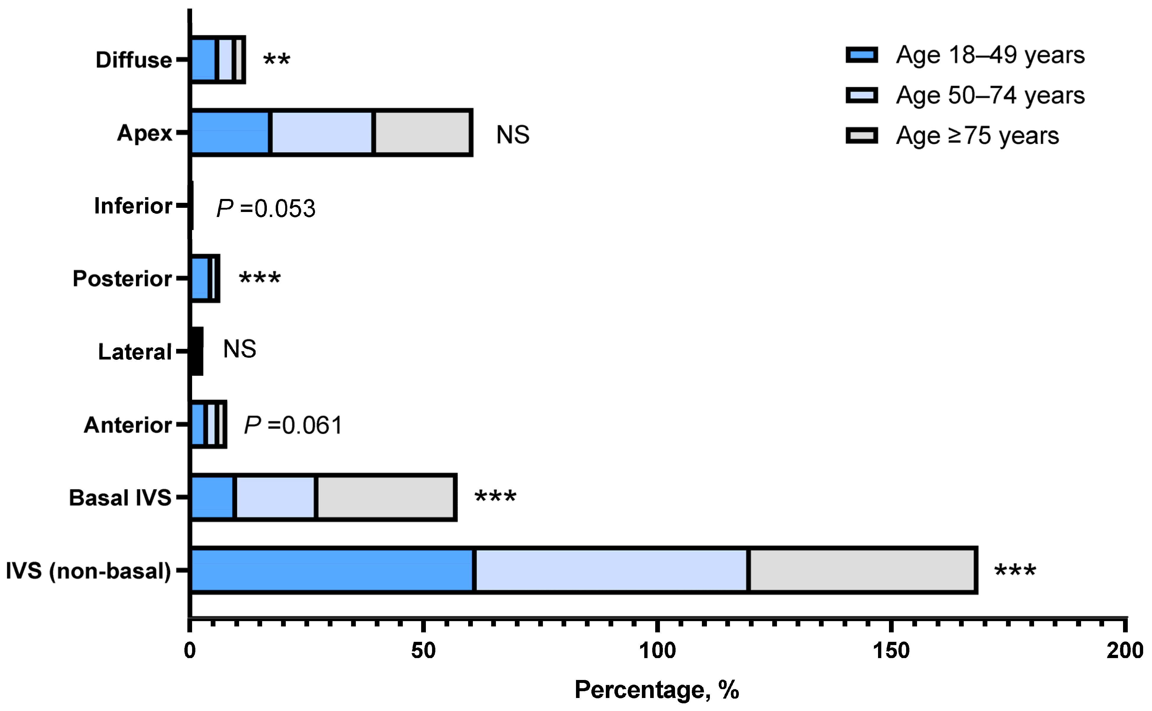
Figure 1. Distribution of location of maximal wall thickness across different age groups. Patients were categorized into three cohorts: 18–49 years, 50–74 years, and ≥75 years. The left ventricular segments involved were categorized as: interventricular septum, basal septum, anterior wall, lateral wall, posterior wall, inferior wall, apex, and diffuse left ventricular wall. p values were calculated using the chi-square test (or Fisher’s exact test when appropriate) for comparisons across the three age groups. IVS, interventricular septum; NS, non-significant. ** p < 0.01; *** p < 0.001.
Jcm 15 06230 g001
Table 1. Characteristics of hypertrophic cardiomyopathy phenotype patients.
CharacteristicsTotalMaleFemalep Value
n26101834776
Demographic
    Age, y60.2 ± 15.158.6 ± 14.864.0 ± 15.2<0.001
    Male, n (%)1834 (70.3%)1834 (100%)0
LV dimensions/function
    IV septal thickness, mm16.5 ± 4.416.6 ± 4.416.5 ± 4.50.688
    IV septal thickness ≥15 mm, n (%)1858 (71.2%)1324 (72.2%)534 (68.8%)0.082
    LV posterior wall thickness, mm11.7 ± 2.611.9 ± 2.511.1 ± 2.7<0.001
    LV posterior wall thickness ≥15 mm, n (%)332 (12.7%)266 (14.5%)66 (8.5%)<0.001
    Septal to posterior wall ratio1.5 ± 0.51.4 ± 0.51.5 ± 0.5<0.001
    Asymmetric septal hypertrophy, n (%)1392 (53.3%)927 (50.5%)465 (59.9%)<0.001
    Maximal wall thickness, mm19.0 ± 3.819.0 ± 3.919.0 ± 3.70.254
    LV diastolic diameter, mm(n = 2532) 46.4 ± 6.1(n = 1779) 47.4 ± 6.0(n = 753) 43.8 ± 5.4<0.001
       LV diastolic diameter <55 mm, n (%)(n = 2532) 2317 (91.5%)1584 (89.0%)733 (97.3%)<0.001
    LVEF, %65.6 ± 9.265.2 ± 9.366.6 ± 8.80.001
       LVEF ≥55%, n (%)2410 (92.3%)1680 (91.6%)730 (94.1%)0.03
       LVEF 41–49%, n (%)52 (2.0%)41 (2.2%)11 (1.4%)0.172
       LVEF ≤40%, n (%)52 (2.0%)42 (2.3%)10 (1.3%)0.094
    Apical aneurysm38 (1.5%)26 (1.4%)12 (1.5%)0.802
Pure apical hypertrophy117 (4.5%)79 (4.3%)38 (4.9%)0.506
RV hypertrophy, n (%)107 (4.1%)76 (4.1%)31 (4.0%)0.861
Stress test, n (%)373 (14.3%)244 (13.3%)129 (16.6%)0.027
LVOT/AV peak gradient at rest, mmHg(n = 619) 20.0 (9.0–49.0)(n = 365) 17.0 (7.5–36.0)(n = 254) 27.0 (10.4–66.0)<0.001
LVOT/AV peak gradient on stress test, mmHg(n = 267) 29.0 (14.5–48.0)(n = 178) 29.0 (13.0–47.5)(n = 89) 30.0 (17.0–51.0)0.383
Intra-left ventricular obstruction423 (16.2%)256 (14.0%)167 (21.5%)<0.001
    LVOT obstruction317 (12.1%)176 (9.6%)141 (18.2%)<0.001
       Latent LVOT obstruction(n = 317) 82 (25.9%)61 (34.5%)21 (14.9%)<0.001
       Overt LVOT obstruction(n = 317) 235 (74.1%)115 (65.0%)120 (85.1%)<0.001
       LVOT/AV peak gradient ≥50 mmHg, n (%)(n = 317) 192 (60.6%)92 (5.0%)100 (13.0%)<0.001
    Mid-ventricular obstruction128 (4.9%)92 (5.0%)36 (4.6%)0.683
Mitral E wave velocity, cm/s(n = 1944) 72.2 ± 24.0(n = 1354) 70.9 ±23.1(n = 590) 75.2 ± 25.8<0.001
    Mitral E wave velocity >90 cm/s, n (%)358 (18.4%)221 (16.3%)137 (23.2%)<0.001
Mitral A wave velocity, cm/s(n = 1576) 82.9 ± 24.9(n = 1120) 80.8 ± 23.8(n = 456) 87.9 ± 26.9<0.001
LV septal e’ velocity, cm/s(n = 939) 5.2 ± 1.8(n = 635) 5.4 ± 1.8(n = 304) 4.9 ± 1.8<0.001
    LV septal e’ velocity <9 cm/s, n (%)901 (96.0%)605 (95.3%)296 (97.4%)0.128
Mitral E: e’ ratio(n = 925) 14.5 ± 5.9(n = 628) 13.7 ± 5.4(n = 297) 16.2 ± 6.5<0.001
    Mitral E: e’ ratio >9, n (%)798 (86.3%)524 (83.4%)274 (92.3%)<0.001
LV lateral annulus e’ velocity, cm/s(n = 876) 7.5 ± 2.9(n = 591) 7.8 ± 3.0(n = 285) 6.8 ± 2.6<0.001
LA anterior-posterior diameter, mm(n = 2607) 41.3 ± 6.5(n = 1833) 41.6 ± 6.5(n = 774) 40.6 ± 6.5<0.001
Moderate to severe mitral regurgitation, n (%)429 (16.4%)252 (13.7%)177 (22.8%)<0.001
Mild aortic valve stenosis, n (%)49 (1.9%)36 (2.0%)13 (1.7%)0.621
Estimated pulmonary artery systolic pressure, mmHg(n = 1366) 31.0 (26.0–37.9)(n = 924) 30.0 (25.3–36.0)(n = 442) 33.1 (27.7–40.8)<0.001
LV, left ventricle; IV, interventricular; LVEF, left ventricular ejection fraction; RV, right ventricle; LVOT, left ventricular outflow tract; AV, aortic valve; LA, left atrium.
Table 2. Characteristics of hypertrophic cardiomyopathy phenotype patients across different age groups.
CharacteristicsAge 18–49 YearsAge 50–74 YearsAge ≥ 75 Yearsp Value
n5871599424
Demographic
    Age, y38.3 ± 7.562.8 ± 6.981.0 ± 5.1<0.001
    Male, n (%)458 (78.0%)1155 (72.2%)221 (52.1%)<0.001
LV dimensions/function
    IV septal thickness, mm17.7 ± 4.716.5 ± 4.315.0 ± 3.9<0.001
    IV septal thickness ≥15 mm, n (%)474 (80.7%)1144 (71.5%)240 (56.6%)<0.001
    LV posterior wall thickness, mm12.1 ± 3.211.6 ± 2.511.3 ± 2.10.003
    LV posterior wall thickness ≥15 mm, n (%)126 (21.5%)178 (11.1%)28 (6.6%)<0.001
    Septal to posterior wall ratio1.6 ± 0.61.5 ± 0.51.3 ± 0.4<0.001
    Asymmetric septal hypertrophy, n (%)338 (57.6%)866 (54.2%)188 (44.3%)<0.001
    Maximal wall thickness, mm19.8 ± 4.419.0 ± 3.817.7 ± 2.8<0.001
    LV diastolic diameter, mm(n = 582) 46.6 ± 7.0(n = 1547) 46.6 ± 5.8(n = 403) 45.0 ± 5.5<0.001
       LV diastolic diameter <55 mm, n (%)510 (87.6%)1429 (92.4%)378 (93.8%)<0.001
    LVEF, %65.2 ± 10.065.8 ± 8.865.5 ± 9.30.814
       LVEF ≥55%, n (%)532 (90.6%)1486 (92.9%)392 (92.5%)0.199
       LVEF 41–49%, n (%)17 (2.9%)30 (1.9%)5 (1.2%)0.135
       LVEF ≤40%, n (%)16 (2.7%)23 (1.4%)13 (3.1%)0.051
    Apical aneurysm5 (0.9%)29 (1.8%)4 (0.9%)0.158
Pure apical hypertrophy24 (4.1%)64 (4.0%)29 (6.8%)0.037
RV hypertrophy, n (%)36 (6.1%)60 (3.8%)11 (2.6%)0.011
Stress test, n (%)102 (17.4%)228 (14.3%)43 (10.1%)0.005
LVOT/AV peak gradient at rest, mmHg(n = 160) 18.0 (9.0–42.5)(n = 377) 20.0 (8.1–47.0)(n = 82) 25.0 (9.2–55.0)0.415
LVOT/AV peak gradient on stress test, mmHg(n = 73) 30.0 (15.8–52.0)(n = 163) 29.0 (13.0–48.0)(n = 31) 30.7 (19.0–46.3)0.569
Intra-left ventricular obstruction103 (17.5%)266 (16.6%)54 (12.7%)0.093
    LVOT obstruction79 (13.5%)192 (12.0%)46 (10.8%)0.439
       Latent LVOT obstruction28 (35.0%)45 (23.4%)9 (19.6%)0.081
       Overt LVOT obstruction51 (63.7%)147 (76.6%)37 (80.4%)0.05
       LVOT/AV peak gradient ≥50 mmHg, n (%)51 (8.8%)114 (7.2%)27 (6.4%)0.301
    Mid-ventricular obstruction30 (5.1%)87 (5.4%)11 (2.6%)0.053
Mitral E wave velocity, cm/s(n = 455) 75.7 ± 23.0(n = 1184) 70.2 ± 23.3(n = 305) 74.9 ± 27.1<0.001
    Mitral E wave velocity >90 cm/s, n (%)97 (21.3%)191 (16.1%)70 (23.0%)0.004
Mitral A wave velocity, cm/s(n = 402) 70.4 ± 23.2(n = 951) 84.6 ± 22.4(n = 223) 97.7 ± 27.6<0.001
LV septal e’ velocity, cm/s(n = 254) 6.0 ± 2.0(n = 593) 5.0 ± 1.7(n = 92) 4.7 ± 1.5<0.001
    LV septal e’ velocity <9 cm/s, n (%)234 (92.1%)576 (97.1%)91 (98.9%)0.001
Mitral E: e’ ratio(n = 252) 13.7 ± 5.7(n = 583) 14.7 ± 5.9(n = 90) 15.8 ± 6.10.001
    Mitral E: e’ ratio >9, n (%)217 (86.1%)502 (86.1%)79 (87.8%)0.909
LV lateral annulus e’ velocity, cm/s(n = 234) 8.7 ± 3.3(n = 558) 7.1 ± 2.6(n = 84) 6.6 ± 2.7<0.001
LA anterior-posterior diameter, mm(n = 586) 40.3 ± 6.6(n = 1598) 41.5 ± 6.5(n = 423) 41.8 ± 6.3<0.001
Moderate to severe mitral regurgitation, n (%)81 (13.8%)278 (17.4%)70 (16.5%)0.134
Mild aortic valve stenosis, n (%)5 (0.9%)31 (1.9%)13 (3.1%)0.036
Estimated pulmonary artery systolic pressure, mmHg(n = 234) 28.1 (24.2–35.0)(n = 847) 30.9 (26.0–37.8)(n = 285) 33.4 (28.5–40.9)<0.001
LV, left ventricle; IV, interventricular; LVEF, left ventricular ejection fraction; RV, right ventricle; LVOT, left ventricular outflow tract; AV, aortic valve; LA, left atrium.
Table 3. Comparison of characteristics of non-obstructive hypertrophic cardiomyopathy (nHCM) patients and patients with left ventricular outflow tract obstruction (LVOTO).
CharacteristicsnHCMLVOTOp Value
n2187317
Demographic
    Age, y60.5 ± 15.259.1 ± 14.70.098
    Male, n (%)1578 (72.2%)176 (55.5%)<0.001
LV dimensions/function
    IV septal thickness, mm16.2 ± 4.218.6 ± 4.7<0.001
    IV septal thickness ≥15 mm, n (%)1527 (69.8%)264 (83.3%)<0.001
    LV posterior wall thickness, mm11.7 ± 2.611.9 ± 2.40.036
    LV posterior wall thickness ≥15 mm, n (%)288 (13.2%)36 (11.4%)0.369
    Septal to posterior wall ratio1.4 ± 0.51.6 ± 0.5<0.001
    Asymmetric septal hypertrophy, n (%)1102 (50.4%)225 (71.0%)<0.001
    Maximal wall thickness, mm18.6 ± 3.620.4 ± 4.3<0.001
    LV diastolic diameter, mm(n = 2120) 46.8 ± 6.1(n = 309) 43.9 ± 5.6<0.001
       LV diastolic diameter <55 mm, n (%)1916 (90.4%)301 (97.4%)<0.001
    LVEF, %65.1 ± 9.368.2 ± 7.5<0.001
       LVEF ≥55%, n (%)1996 (91.3%)311 (98.1%)<0.001
       LVEF 41–49%, n (%)52 (2.4%)0 (0.0%)0.006
       LVEF ≤40%, n (%)52 (2.4%)0 (0.0%)0.006
    Apical aneurysm26 (1.2%)2 (0.6%)0.377
Location of maximal wall thickness
    IV septal, n (%)1266 (57.9%)186 (58.7%)0.791
    Basal IV septum, n (%)365 (16.7%)87 (27.4%)<0.001
    LV anterior wall, n (%)54 (2.5%)9 (2.8%)0.694
    LV lateral wall, n (%)17 (0.8%)4 (1.3%)0.377
    LV posterior wall, n (%)49 (2.2%)4 (1.3%)0.258
    LV inferior wall, n (%)2 (0.1%)2 (0.6%)0.081
    LV apex, n (%)467 (21.4%)33 (10.4%)<0.001
    Diffuse LV hypertrophy, n (%)95 (4.3%)4 (1.3%)0.008
Pure apical hypertrophy110 (5.0%)0 (0.0%)<0.001
RV hypertrophy, n (%)89 (4.1%)12 (3.8%)0.81
Stress test, n (%)212 (9.7%)134 (42.3%)<0.001
LVOT/AV peak gradient at rest, mmHg(n = 300) 9.0 (5.8–16.0)(n = 299) 50.0 (31.0–78.5)<0.001
LVOT/AV peak gradient on stress test, mmHg(n = 124) 15.0 (9.0–21.6)(n = 133) 48.0 (39.0–70.0)<0.001
LVOT/AV peak gradient ≥ 50 mmHg0 (0.0%)192 (60.6%)<0.001
Mid-ventricular obstruction0 (0.0%)22 (6.9%)<0.001
Mitral E wave velocity, cm/s(n = 1643) 71.6 ± 23.5(n = 216) 78.1 ± 27.50.001
    Mitral E wave velocity >90 cm/s, n (%)298 (18.1%)52 (24.1%)0.036
Mitral A wave velocity, cm/s(n = 1333) 81.7 ± 24.4(n = 172) 92.3 ± 26.5<0.001
LV septal e’ velocity, cm/s(n = 740) 5.2 ± 1.7(n = 131) 5.2 ± 2.40.25
    LV septal e’ velocity <9 cm/s, n (%)714 (96.5%)122 (93.1%)0.071
Mitral E: e’ ratio(n = 729) 14.0 ± 5.4(n = 128) 17.1 ± 7.7<0.001
    Mitral E: e’ ratio >9, n (%)619 (84.9%)118 (92.2%)0.029
LV lateral annulus e’ velocity, cm/s(n = 683) 7.6 ± 3.0(n = 123) 7.0 ± 2.70.026
LA anterior-posterior diameter, mm(n = 2184) 41.2 ± 6.4(n = 317) 42.1 ± 7.40.101
Moderate to severe mitral regurgitation, n (%)273 (12.5%)145 (45.7%)<0.001
Mild aortic valve stenosis, n (%)37 (1.7%)10 (3.2%)0.073
Estimated pulmonary artery systolic pressure, mmHg(n = 1170) 30.8 (25.7–37.3)(n = 161) 33.1 (28.0–41.0)<0.001
nHCM, non-obstructive hypertrophic cardiomyopathy; LV, left ventricle; IV, interventricular; LVEF, left ventricular ejection fraction; RV, right ventricle; LVOT, left ventricular outflow tract; LVOTO, left ventricular outflow tract obstruction; AV, aortic valve; LA, left atrium.
Table 4. Characteristics of patients with pure apical hypertrophy.
Characteristics
n117
Demographic
    Age, y63.2 ± 15.4
    Male, n (%)79 (67.5%)
LV dimensions/function
    IV septal thickness, mm10.1 ± 1.0
    LV posterior wall thickness, mm9.6 ± 1.1
    Maximal wall thickness, mm17.7 ± 2.4
    LV diastolic diameter, mm(n = 117) 47.6 ± 4.7
    LV diastolic diameter <55 mm, n (%)109 (93.2%)
    LVEF, %68.0 ± 7.3
     LVEF ≥55%, n (%)116 (99.1%)
     LVEF 41–49%, n (%)0 (0.0%)
     LVEF ≤40%, n (%)0 (0.0%)
    Apex aneurysm, n (%)2 (1.7%)
RV hypertrophy, n (%)1 (0.9%)
Stress test, n (%)4 (3.4%)
Intra-left ventricular obstruction7 (6.0%)
    LVOT obstruction, n (%)0 (0.0%)
    Midventricular obstruction, n (%)7 (6.0%)
Mitral E wave velocity, cm/s(n = 93) 72.4 ± 19.8
    Mitral E wave velocity >90 cm/s, n (%)15 (16.1%)
Mitral A wave velocity, cm/s(n = 81) 77.9 ± 25.6
LV septal e’ velocity, cm/s(n = 52) 6.0 ± 1.7
    LV septal e’ velocity <9 cm/s, n (%)49 (94.2%)
Mitral E: e’ ratio(n = 51) 12.3 ± 4.8
    Mitral E:e’ ratio >9, n (%)36 (70.6%)
LV lateral annulus e’ velocity, cm/s(n = 46) 9.2 ± 3.3
LA anterior-posterior diameter, mm(n = 117) 38.6 ± 6.0
Moderate to severe mitral regurgitation, n (%)12 (10.3%)
Mild aortic valve stenosis, n (%)(1.9%) 1 (0.9%)
Estimated pulmonary artery systolic pressure, mmHg(n = 47) 30.0 (25.8–39.4)
LV, left ventricle; IV, interventricular; LVEF, left ventricular ejection fraction; RV, right ventricle; LVOT, left ventricular outflow tract; LA, left atrium.
Disclaimer/Publisher’s Note: The statements, opinions and data contained in all publications are solely those of the individual author(s) and contributor(s) and not of MDPI and/or the editor(s). MDPI and/or the editor(s) disclaim responsibility for any injury to people or property resulting from any ideas, methods, instructions or products referred to in the content.


