Adult-Onset Alexander Disease Presenting as Atypical Parkinsonism and Autonomic Dysfunction: A Case Series

Abstract
1. Introduction
2. Case Descriptions
2.1. Case 1
2.2. Case 2
2.3. Case 3
2.4. Case 4
3. Discussion
4. Conclusions
Author Contributions
Funding
Institutional Review Board Statement
Informed Consent Statement
Data Availability Statement
Conflicts of Interest
References
- Sosunov, A.; Olabarria, M.; Goldman, J.E. Alexander disease: An astrocytopathy that produces a leukodystrophy. Brain Pathol. 2018, 28, 388–398. [Google Scholar] [CrossRef] [Scilit] [PubMed]
- Pajares, M.; Hernández-Gerez, E.; Pekny, M.; Pérez-Sala, D. Alexander disease: The road ahead. Neural Regen. Res. 2023, 18, 2156–2160. [Google Scholar] [CrossRef] [Scilit] [PubMed]
- Messing, A.; Brenner, M.; Feany, M.B.; Nedergaard, M.; Goldman, J.E. Alexander Disease. J. Neurosci. 2012, 32, 5017–5023. [Google Scholar] [CrossRef] [Scilit] [PubMed]
- Perng, M.D.; Su, M.; Wen, S.F.; Li, R.; Gibbon, T.; Prescott, A.R.; Brenner, M.; Quinlan, R.A. The Alexander Disease-Causing Glial Fibrillary Acidic Protein Mutant, R416W, Accumulates into Rosenthal Fibers by a Pathway That Involves Filament Aggregation and the Association of aB-Crystallin and HSP27. Am. J. Hum. Genet. 2006, 79, 197–213. [Google Scholar] [CrossRef] [Scilit]
- Hsiao, V.C.; Tian, R.; Long, H.; Der Perng, M.; Brenner, M.; Quinlan, R.A.; Goldman, J.E. Alexander-disease mutation of GFAP causes filament disorganization and decreased solubility of GFAP. J. Cell Sci. 2005, 118, 2057–2065. [Google Scholar] [CrossRef] [Scilit] [PubMed]
- Hagemann, T.L.; Powers, B.; Mazur, C.; Kim, A.; Wheeler, S.; Hung, G.; Swayze, E.; Messing, A. Antisense suppression of glial fibrillary acidic protein as a treatment for Alexander disease. Ann. Neurol. 2018, 83, 27–39. [Google Scholar] [CrossRef] [Scilit] [PubMed]
- Zavala, E.; Zimmerman, T. Alexander’s Disease: Potential Drug Targets and Future Directions. Mol. Neurobiol. 2025, 62, 15010–15023. [Google Scholar] [CrossRef] [Scilit] [PubMed]
- Yoshida, T. Clinical Characteristics of Alexander Disease. Neurodegener. Dis. Manag. 2020, 10, 325–333. [Google Scholar] [CrossRef] [Scilit] [PubMed]
- Grossi, A.; Rosamilia, F.; Carestiato, S.; Salsano, E.; Ceccherini, I.; Bachetti, T. A systematic review and meta-analysis of GFAP gene variants in Alexander disease. Sci. Rep. 2024, 14, 24341. [Google Scholar] [CrossRef] [Scilit] [PubMed]
- Balbi, P.; Salvini, S.; Fundarò, C.; Frazzitta, G.; Maestri, R.; Mosah, D.; Uggetti, C.; Sechi, G. The clinical spectrum of late-onset Alexander disease: A systematic literature review. J. Neurol. 2010, 257, 1955–1962. [Google Scholar] [CrossRef] [Scilit] [PubMed]
- Lynch, D.S.; Wade, C.; Carlson, A.K.; Barkhof, F.; Yoshida, T.; Collins, A.; Edwards, M.R.; Waldman, A.T. Diagnosing Alexander disease in adults. Pract. Neurol. 2025, 25, 507–515. [Google Scholar] [CrossRef] [Scilit] [PubMed]
- Park, J.; Park, S.-T.; Kim, J.; Kwon, K.-Y. A case report of adult-onset Alexander disease clinically presenting as Parkinson’s disease: Is the comorbidity associated with genetic susceptibility? BMC Neurol. 2020, 20, 27. [Google Scholar] [CrossRef] [Scilit] [PubMed]
- Watanabe, Y.; Tsukahara, Y.; Fujita, H.; Sakuramoto, H.; Shiina, T.; Suzuki, K. Adult-onset Alexander disease mimicking multiple system atrophy predominant cerebellar ataxia. J. Clin. Neurosci. 2021, 87, 150–152. [Google Scholar] [CrossRef] [Scilit] [PubMed]
- Pareyson, D.; Fancellu, R.; Mariotti, C.; Romano, S.; Salmaggi, A.; Carella, F.; Girotti, F.; Gattellaro, G.; Carriero, M.R.; Farina, L.; et al. Adult-onset Alexander disease: A series of eleven unrelated cases with review of the literature. Brain 2008, 131, 2321–2331. [Google Scholar] [CrossRef] [Scilit] [PubMed]
- Graff-Radford, J.; Schwartz, K.; Gavrilova, R.H.; Lachance, D.H.; Kumar, N. Neuroimaging and clinical features in type II (late-onset) Alexander disease. Neurology 2014, 82, 49–56. [Google Scholar] [CrossRef] [Scilit] [PubMed]
- Kota, V.; Sahoo, L.K.; Mishra, A.P.; Panda, A.K.; Veerapaneni, H.; Dash, D.; Karan, M. Tadpole Brainstem Atrophy in Adult-Onset Alexander Disease: A Case Report and Review of Literature. Ann. Indian Acad. Neurol. 2024, 27, 738–740. [Google Scholar] [CrossRef] [Scilit] [PubMed]
- Gagliardi, D.; Wade, C.; Tucci, A.; Houlden, H.; Chataway, J.; Barkhof, F.; Lynch, D.S. Analysis of GFAP variants in UK Biobank suggests underdiagnosis or incomplete penetrance of adult-onset Alexander disease. J. Neurol. Neurosurg. Psychiatry 2025, 96, 728–735. [Google Scholar] [CrossRef] [Scilit] [PubMed]
- Spritzer, S.D.; Zarkou, S.; Ireland, S.P.; Carter, J.L.; Goodman, B.P. Autonomic dysfunction in adult-onset alexander disease: A case report and review of the literature. Clin. Auton. Res. 2013, 23, 333–338. [Google Scholar] [CrossRef] [Scilit] [PubMed]
- Namekawa, M.; Takiyama, Y.; Honda, J.; Shimazaki, H.; Sakoe, K.; Nakano, I. Adult-onset Alexander disease with typical “tadpole” brainstem atrophy and unusual bilateral basal ganglia involvement: A case report and review of the literature. BMC Neurol. 2010, 10, 21. [Google Scholar] [CrossRef] [Scilit] [PubMed]
- Farina, L.; Pareyson, D.; Minati, L.; Ceccherini, I.; Chiapparini, L.; Romano, S.; Gambaro, P.; Fancellu, R.; Savoiardo, M. Can MR Imaging Diagnose Adult-Onset Alexander Disease? Am. J. Neuroradiol. 2008, 29, 1190–1196. [Google Scholar] [CrossRef] [Scilit] [PubMed]
- Kang, Y.-R.; Nam, T.-S.; Kim, J.-M.; Kang, K.W.; Lee, S.-H.; Choi, S.-M.; Kim, M.-K. Older adult-onset Alexander disease with atypical clinicoradiological features: A case report. Front. Neurol. 2023, 14, 1139047. [Google Scholar] [CrossRef] [Scilit] [PubMed]
- Yoshida, T.; Mizuta, I.; Yasuda, R.; Mizuno, T. Clinical and radiological characteristics of older-adult-onset Alexander disease. Eur. J. Neurol. 2021, 28, 3760–3767. [Google Scholar] [CrossRef] [Scilit] [PubMed]
- Prust, M.; Wang, J.; Morizono, H.; Messing, A.; Brenner, M.; Gordon, E.; Hartka, T.; Sokohl, A.; Schiffmann, R.; Gordish-Dressman, H.; et al. GFAP mutations, age at onset, and clinical subtypes in Alexander disease. Neurology 2011, 77, 1287–1294. [Google Scholar] [CrossRef] [Scilit] [PubMed]
- Yoshida, T.; Yasuda, R.; Mizuta, I.; Nakagawa, M.; Mizuno, T.; JAPAN Alexander Disease Study Group. Quantitative Evaluation of Brain Stem Atrophy Using Magnetic Resonance Imaging in Adult Patients with Alexander Disease. Eur. Neurol. 2017, 77, 296–302. [Google Scholar] [CrossRef] [Scilit] [PubMed]
- Kang, Y.-R.; Lee, S.-H.; Lin, N.-H.; Lee, S.-J.; Yang, A.-W.; Chandrasekaran, G.; Kang, K.W.; Jin, M.S.; Kim, M.-K.; Perng, M.-D.; et al. A novel in-frame GFAP p.E138_L148del mutation in Type II Alexander disease with atypical phenotypes. Eur. J. Hum. Genet. 2022, 30, 687–694. [Google Scholar] [CrossRef] [Scilit] [PubMed]
- Brenner, M.; Johnson, A.B.; Boespflug-Tanguy, O.; Rodriguez, D.; Goldman, J.E.; Messing, A. Mutations in GFAP, encoding glial fibrillary acidic protein, are associated with Alexander disease. Nat. Genet. 2001, 27, 117–120. [Google Scholar] [CrossRef] [Scilit] [PubMed]
- Chen, Y.-S.; Lim, S.-C.; Chen, M.-H.; Quinlan, R.A.; Perng, M.-D. Alexander disease causing mutations in the C-terminal domain of GFAP are deleterious both to assembly and network formation with the potential to both activate caspase 3 and decrease cell viability. Exp. Cell Res. 2011, 317, 2252–2266. [Google Scholar] [CrossRef] [Scilit] [PubMed]
- Goerttler, T.; Zanetti, L.; Regoni, M.; Egger, K.; Kellner, E.; Deuschl, C.; Kleinschnitz, C.; Sassone, J.; Klebe, S. Adult-Onset Alexander Disease: New Causal Sequence Variant in the GFAP Gene. Neurol. Genet. 2022, 8, e681. [Google Scholar] [CrossRef] [Scilit] [PubMed]
- Quinlan, R.A.; Brenner, M.; Goldman, J.E.; Messing, A. GFAP and its role in Alexander disease. Exp. Cell Res. 2007, 313, 2077–2087. [Google Scholar] [CrossRef] [Scilit] [PubMed]
- Muthusamy, K.; Sivadasan, A.; Dixon, L.; Sudhakar, S.; Thomas, M.; Danda, S.; Wszolek, Z.K.; Wierenga, K.; Dhamija, R.; Gavrilova, R. Adult-onset leukodystrophies: A practical guide, recent treatment updates, and future directions. Front. Neurol. 2023, 14, 1219324. [Google Scholar] [CrossRef] [Scilit] [PubMed]
- Ziad, F.; Cypers, G.; Phillips, M.; Vanhoenacker, P.; Hostens, A.; Yadavraj, S.; Lamont, D.; Robertson, T. Adult-onset Alexander disease with unusual inflammatory features and a novel GFAP mutation in two patients. Neuropathol. Appl. Neurobiol. 2023, 49, e12927. [Google Scholar] [CrossRef] [Scilit] [PubMed]
- Wang, H.; Li, L.; Wu, T.; Hou, B.; Wu, S.; Qiu, Y.; Feng, F.; Cui, L. Increased cerebellar activation after repetitive transcranial magnetic stimulation over the primary motor cortex in patients with multiple system atrophy. Ann. Transl. Med. 2016, 4, 103. [Google Scholar] [CrossRef] [Scilit] [PubMed]
- Zhang, M.; He, T.; Wang, Q. Effects of Non-invasive Brain Stimulation on Multiple System Atrophy: A Systematic Review. Front. Neurosci. 2021, 15, 771090. [Google Scholar] [CrossRef] [Scilit] [PubMed]
- Wang, M.; Zhang, W.; Zang, W. Repetitive transcranial magnetic stimulation improves cognition, depression, and walking ability in patients with Parkinson’s disease: A meta-analysis. BMC Neurol. 2024, 24, 490. [Google Scholar] [CrossRef] [Scilit] [PubMed]
- Gagnier, J.J.; Kienle, G.; Altman, D.G.; Moher, D.; Sox, H.; Riley, D.; The CARE Group. The CARE guidelines: Consensus-based clinical case reporting guideline development. BMJ Case Rep. 2013, 2013, bcr2013201554. [Google Scholar] [CrossRef] [Scilit] [PubMed]
- Riley, D.S.; Barber, M.S.; Kienle, G.S.; Aronson, J.K.; Von Schoen-Angerer, T.; Tugwell, P.; Kiene, H.; Helfand, M.; Altman, D.G.; Sox, H.; et al. CARE guidelines for case reports: Explanation and elaboration document. J. Clin. Epidemiol. 2017, 89, 218–235. [Google Scholar] [CrossRef] [Scilit] [PubMed]



| GFAP Variant | Reported Adult Phenotype(s) | Comment | Key References |
|---|---|---|---|
| p.Arg416Trp (R416W) | Palatal tremor/palatal myoclonus, bulbar dysfunction, pyramidal and cerebellar signs, adult leukodystrophy phenotype | Best-described adult-associated variant; not fully phenotype-specific | [9,10] |
| p.Asp78Asn (D78N) | Familial adult-onset cerebellar/pseudobulbar syndrome | Supports variable expressivity in familial AOAD | [9] |
| p.Met74Thr (M74T) | Adult bulbospinal syndrome with dysarthria, dysphagia, spastic gait/tetraparesis, sometimes sensory symptoms or mild cognitive change | Illustrates myelopathic/bulbar adult presentation | [9,10] |
| p.Tyr257Cys (Y257C) | Progressive ataxia and palatal tremor | Case-based adult association | [9,10] |
| p.Met73Ile (M73I) | Dysarthria, gait ataxia, spastic tetraparesis | Representative bulbospinal/cerebellar adult phenotype | [9] |
| p.Leu58Pro (L58P) | Slowly progressive bulbospinal/ataxic phenotype | Newer adult causal variant; limited evidence | [9] |
| p.Arg70Trp (R70W) | Bulbar, cerebellar, autonomic, or parkinsonian features | Adult-associated pathogenic variant, not phenotype-defining | [9,14] |
| In-frame deletion variants | Mixed bulbar, pyramidal, and cerebellar manifestations | Phenotypic expansion rather than fixed association | [9] |
| Feature | Case 1 | Case 2 | Case 3 | Case 4 |
|---|---|---|---|---|
| Age (years)/Sex | 58/Female | 40/Male | 59/Female | 47/Female |
| Disease Duration | 5 years | 2 years | 6 years | 1 year |
| Primary Clinical Manifestations | Progressive lower limb weakness, gait instability (“walking on cotton”), urinary incontinence, dysarthria, and dizziness. | Left-sided limb weakness, dysarthria, limb rigidity, and dysphagia. | Parkinsonism (rigidity, bradykinesia), severe autonomic failure (OH, urinary incontinence), and cognitive decline. | Paroxysmal focal weakness and numbness (10–30 min episodes), choking, and severe insomnia. |
| Cognitive Status (MoCA) | 20/30 (mild impairment) | 29/30 (preserved) | 18/30 (significant impairment) | Mild subjective decline |
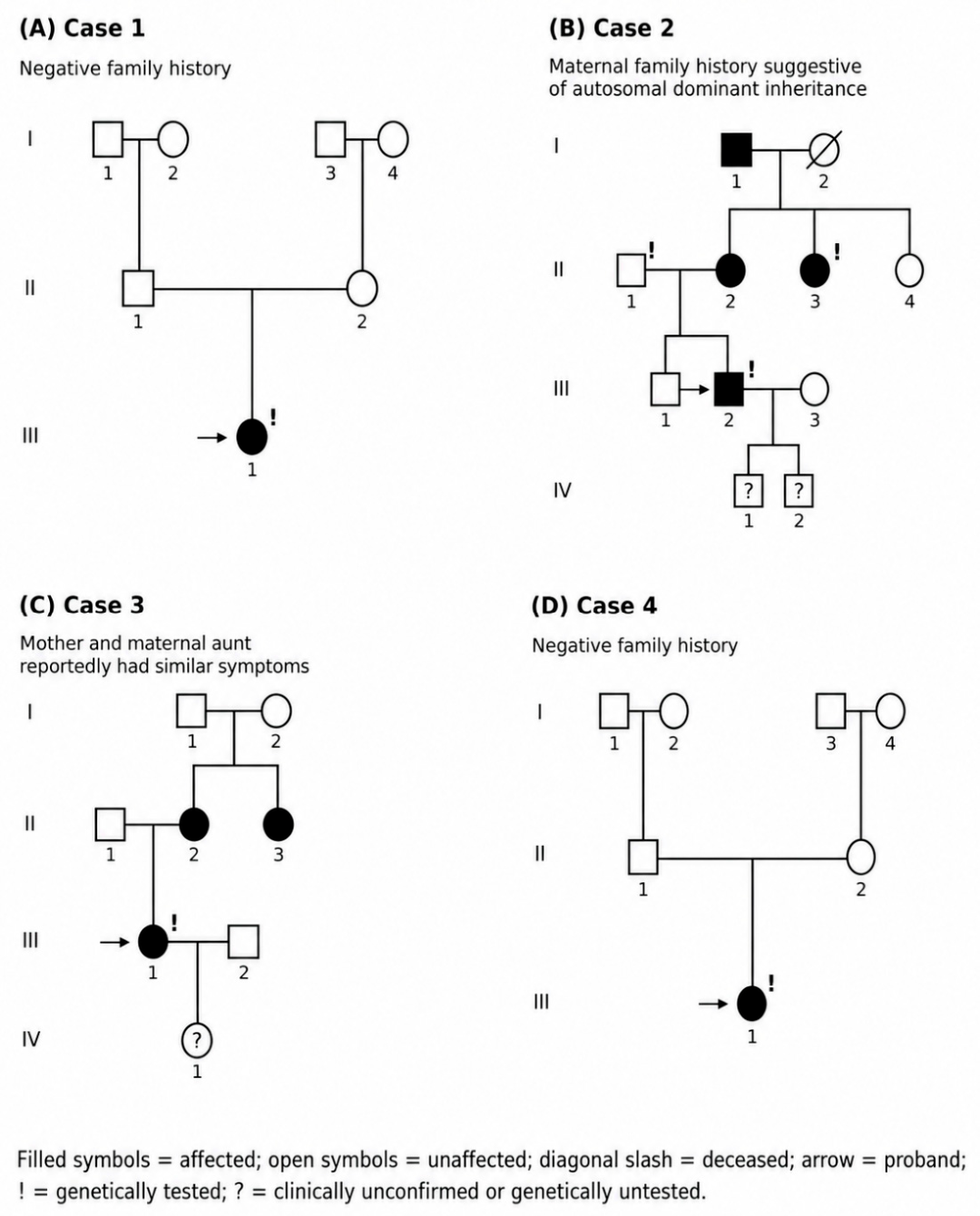
| Family History/Pedigree | Negative; three-generation family history obtained (Figure 2A). | Positive; three-generation maternal family history suggestive of autosomal dominant inheritance (Figure 2B). | Positive; mother and maternal aunt reportedly had similar symptoms; one daughter asymptomatic and genetically untested (Figure 2C). | Negative; three-generation family history obtained (Figure 2D). |
| Initial Misdiagnosis | Parkinsonism | Stroke/Hemiparesis | Multiple system atrophy (MSA) | Transient ischemic attack (TIA)/focal deficit |
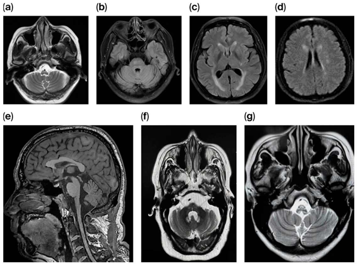
| Key MRI Findings | Medullary and upper cervical spinal cord atrophy (“Tadpole sign”). | Brainstem and medullary atrophy. | “Tadpole sign”; T2 hyperintensity in cerebellar dentate nuclei and basal ganglia. | Medullary thinning (“Tadpole sign”) and periventricular white matter signals. |
| GFAP Variant (HGVSc) | c.363_371del | c.1245G>A | c.208C>T | c.584A>T |
| GFAP Variant (HGVSp) | p.Glu122_Arg124del | p.Met415Ile | p.Arg70Trp | p.Glu195Val |
| Pathogenicity (ACMG) | VUS | VUS (co-segregation noted) | Likely pathogenic | VUS |
| Genetic testing site/DNA diagnostic method | Beijing Tiantan Hospital–Shenzhen BGI Medical Genetics Laboratory, Shenzhen, China; single-proband capture-based high-throughput whole-exome sequencing using peripheral-blood DNA. | Beijing Tiantan Hospital–Shenzhen BGI Medical Genetics Laboratory, Shenzhen, China; single-proband capture-based high-throughput clinical exome sequencing using peripheral-blood DNA. | Not documented in the available clinical records. | Jiaxing AccBio Medical Laboratory, Jiaxing, China; target-region capture followed by high-throughput sequencing using peripheral-blood DNA. |
| Validation/segregation information | No Sanger confirmation or trio-based analysis was documented in the available report. | Sanger sequencing confirmed the heterozygous variant in the proband; the variant was not detected in his father and was detected in the affected maternal aunt. Complete trio-based exome sequencing was not performed. | Sanger confirmation and trio-based analysis were not documented in the available clinical records. | No Sanger confirmation or trio-based analysis documented in the available report. |
Disclaimer/Publisher’s Note: The statements, opinions and data contained in all publications are solely those of the individual author(s) and contributor(s) and not of MDPI and/or the editor(s). MDPI and/or the editor(s) disclaim responsibility for any injury to people or property resulting from any ideas, methods, instructions or products referred to in the content. |
© 2026 by the authors. Licensee MDPI, Basel, Switzerland. This article is an open access article distributed under the terms and conditions of the Creative Commons Attribution (CC BY) license.
Share and Cite
Fang, J.; Wang, Z.; Feng, T.; Jiang, Y. Adult-Onset Alexander Disease Presenting as Atypical Parkinsonism and Autonomic Dysfunction: A Case Series. J. Clin. Med. 2026, 15, 5677. https://doi.org/10.3390/jcm15145677
Fang J, Wang Z, Feng T, Jiang Y. Adult-Onset Alexander Disease Presenting as Atypical Parkinsonism and Autonomic Dysfunction: A Case Series. Journal of Clinical Medicine. 2026; 15(14):5677. https://doi.org/10.3390/jcm15145677
Chicago/Turabian StyleFang, Jinping, Zhan Wang, Tao Feng, and Ying Jiang. 2026. "Adult-Onset Alexander Disease Presenting as Atypical Parkinsonism and Autonomic Dysfunction: A Case Series" Journal of Clinical Medicine 15, no. 14: 5677. https://doi.org/10.3390/jcm15145677
APA StyleFang, J., Wang, Z., Feng, T., & Jiang, Y. (2026). Adult-Onset Alexander Disease Presenting as Atypical Parkinsonism and Autonomic Dysfunction: A Case Series. Journal of Clinical Medicine, 15(14), 5677. https://doi.org/10.3390/jcm15145677