1. Introduction
Inflammatory bowel diseases (IBD), primarily represented by Crohn’s disease (CD) and ulcerative colitis (UC), are chronic, systemic conditions with multifactorial etiology, characterized by complex interactions between genetic predisposition, intestinal dysbiosis, environmental factors, and dysregulated immune responses. The incidence and prevalence of IBD are continuously increasing worldwide, reaching approximately 0.8–1% in many developed countries, with an upward trend also observed in developing regions, resulting in a significant burden both at the individual level and on public healthcare systems [
1,
2,
3].
IBD is characterized by recurrent inflammation of the gastrointestinal tract and is frequently associated with extraintestinal manifestations (EIMs), reflecting its systemic nature. EIMs are complications affecting organs and systems outside the gastrointestinal tract, including musculoskeletal, cutaneous, ocular, hepatobiliary, and cardiovascular structures, contributing significantly to the overall disease burden [
4,
5]. They may precede the onset of digestive symptoms, evolve concurrently with intestinal disease activity, or persist independently of it, including during periods of remission, thereby underscoring the systemic nature of inflammatory bowel diseases [
4,
5].
According to the 2024 ECCO Consensus, EIMs are classified based on their pathogenic mechanisms and relationship with disease activity into classical manifestations, complications of chronic systemic inflammation, associated manifestations, and treatment-induced manifestations, highlighting their heterogeneity and complexity. Within this framework, the modern approach transcends a strictly organ-specific perspective, integrating systemic manifestations into the disease spectrum. This approach advocates multidisciplinary evaluation and the use of standardized diagnostic and monitoring criteria [
6].
Mesenteric panniculitis (MP) is a chronic fibroinflammatory condition of the mesenteric adipose tissue, characterized by non-specific inflammation, adipocyte necrosis, and variable degrees of fibrosis. It is classified within the spectrum of sclerosing mesenteritis, which reflects the relative proportion of these histopathological components. Its clinical, imaging, and histological expression is heterogeneous, leading to difficulties in definition and diagnostic standardization. It is increasingly interpreted as an immune-mediated process of the mesentery [
7,
8,
9].
MP has been reported in association with a wide range of conditions, including chronic inflammatory diseases, autoimmune disorders, infections, and neoplasms, without identification of a single unifying etiology, supporting its multifactorial and potentially reactive nature [
7,
8,
9].
Interest in the association between MP and IBD stems from the recognition of the mesentery as an active participant in systemic inflammatory processes. In this context, MP is interpreted as an expression of a chronic fibroinflammatory mesenteric response, possibly correlated with persistent intestinal inflammation, suggesting a potential relationship with IBD. Nevertheless, available data remain limited and heterogeneous, precluding the establishment of a clear causal relationship, primarily due to the lack of standardization of diagnostic criteria and incomplete reporting of patients’ clinical characteristics [
7,
10,
11].
In parallel, the possible co-occurrence of MP in patients receiving biological therapies for IBD, including gut-selective agents, has not been systematically investigated and remains poorly documented in the literature.
In this context, MP emerges as an entity at the intersection of imaging, gastroenterology, and immunopathology, with its clinical significance and position within the IBD framework incompletely defined.
Against this background, the present report aims to describe a rare case of MP occurring in a patient with UC in deep remission during vedolizumab therapy, and to provide a critical narrative review of the available evidence on the association between MP and IBD. The rationale for this report stems from the paucity of data specifically linking MP to UC, the limited published evidence in the context of gut-selective biologic therapy, and the unresolved question of whether MP may represent a mesenteric fibroinflammatory phenomenon related to IBD, independent of mucosal disease activity. Given the frequently idiopathic nature of MP and the absence of histopathological confirmation in the present case, no causal relationship between MP, UC, and vedolizumab therapy can be established. Accordingly, the observations presented herein should be regarded as hypothesis-generating and warrant further dedicated investigation.
2. Case Presentation
We report the case of a 32-year-old female patient with normal body weight and a history of UC diagnosed at age 27, with pancolonic involvement (E3) and a corticosteroid-dependent disease course. Biological therapy with infliximab was initiated, but secondary loss of response occurred, and the patient was later switched to vedolizumab three years prior to the current presentation. The course of IBD under biological therapy was favorable, with clinical, endoscopic, and histological remission.
In July 2025, approximately four weeks after the last scheduled vedolizumab infusion (administered at the standard maintenance interval of eight weeks), the patient presented to the emergency department with diffuse crampy abdominal pain, associated with nausea, vomiting, and heartburn. On physical examination, the abdomen was moderately distended, with diminished bowel sounds and diffuse tenderness on both superficial and deep palpation, with no signs of peritoneal irritation. The remainder of the physical examination was within normal limits.
The principal laboratory, immunological, endoscopic, and histological findings at the time of presentation are summarized in
Table 1.
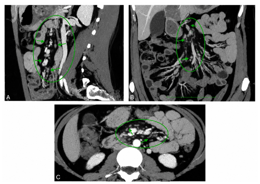
Contrast-enhanced abdominal and pelvic computed tomography (CT) revealed diffuse infiltration of the mesenteric root adipose tissue, extending into the retroperitoneal vascular compartment, associated with sub-centimeter lymphadenopathy and a pseudotumoral appearance, findings suggestive of MP. The imaging features are illustrated in
Figure 1.
Tissue biopsy was not performed, as the patient elected to defer the procedure at initial presentation and opted for medical treatment. Accordingly, treatment was initiated with prednisone (40 mg/day, followed by a gradual dose reduction of 5 mg/week after the first four weeks) in combination with tamoxifen (10 mg twice daily for 12 weeks), with concomitant continuation of vedolizumab therapy.
This regimen was selected based on the available evidence supporting corticosteroid–tamoxifen combination therapy in symptomatic MP [
12,
13].
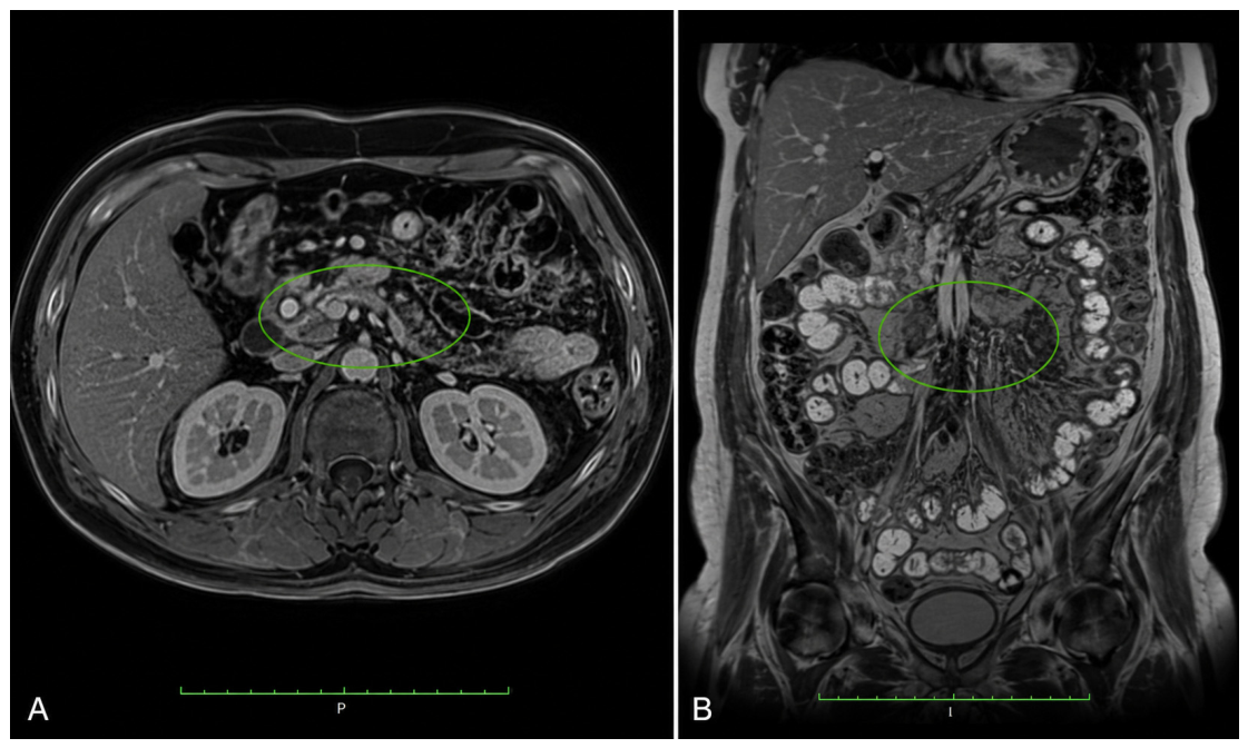
The clinical course was favorable, with complete resolution of gastrointestinal symptoms and normalization of inflammatory markers. Follow-up magnetic resonance imaging (MRI) performed at one month demonstrated complete resolution of mesenteric inflammatory changes, confirming clinical and radiological remission (
Figure 2).
The distinctive feature of this case lies in the occurrence of MP in a patient with UC in clinical, endoscopic, and histological remission on vedolizumab therapy. To the best of our knowledge, no previous report has described this specific clinical scenario.
Whether MP represents a coincidental idiopathic process, a mesenteric fibroinflammatory phenomenon related to the systemic immune dysregulation associated with IBD, or a temporally associated event unrelated to the underlying disease remains an open question that cannot be resolved on the basis of a single case observation.
3. Mesenteric Panniculitis—General Overview
3.1. Definition and Epidemiology
MP is a chronic benign condition of the mesenteric adipose tissue, characterized histopathologically by the variable combination of non-specific inflammation, adipocyte necrosis, and fibrosis, with predominant involvement of the small bowel mesenteric root. It is currently classified within the spectrum of sclerosing mesenteritis, which reflects a pathological continuum determined by the relative proportion of these components, rather than the existence of distinct entities [
11,
14,
15].
From a historical perspective, this condition was first described in 1924 by Jura as retractile sclerosing mesenteritis. Subsequent clinicopathological studies progressively unified the various terms used to describe this entity, leading to the current concept of sclerosing mesenteritis as a pathological continuum encompassing different manifestations of the same disease process [
16,
17,
18,
19].
The development and widespread use of abdominal CT have contributed significantly to the increasing recognition of this condition, suggesting that MP is likely underdiagnosed in routine clinical practice and more prevalent than previously considered [
7,
20].
The reported prevalence varies widely, ranging from 0.16% to 7.83%, with differences attributed to the heterogeneity of imaging criteria and methodological variability across studies [
11,
13,
21,
22,
23].
CT-based studies reported prevalence rates of approximately 0.6% in the initial series, while the adoption of more sensitive radiological criteria led to higher estimates, reaching up to 7.83%, highlighting the impact of the diagnostic definition on reported frequency [
7,
19,
23].
Large-scale retrospective analyses report intermediate prevalence rates, ranging from 1.1% to 2.5%, supporting the notion that this entity is more prevalent than previously recognized [
20,
24,
25].
From a demographic standpoint, the condition predominantly affects middle-aged and elderly adults, with a peak incidence between 50 and 70 years of age, and is rarely described in the pediatric population [
7,
14].
Most studies report a moderate male predominance of approximately 2:1, although this finding is not consistent across cohorts [
20,
26,
27].
Overall, the characteristic epidemiological profile is that of an incidental diagnosis in adult patients, in the context of the widespread use of abdominal CT, while the true frequency of the condition remains difficult to estimate precisely due to the methodological heterogeneity of available studies [
22,
24,
28,
29].
3.2. Etiopathogenesis
The etiopathogenesis of mesenteric panniculitis remains incompletely elucidated and is currently considered the expression of an idiopathic, probably immune-mediated, fibroinflammatory process of the mesenteric adipose tissue. In the absence of a single etiological determinant, multiple potential factors have been implicated. These include abdominal trauma, surgical interventions, mesenteric ischemia, chronic infections, autoimmune diseases, neoplastic processes, and drug reactions. Direct causal relationships have not been demonstrated; however, supporting the multifactorial and heterogeneous nature of the disease [
19,
30,
31].
The central pathogenic mechanism involves immunometabolic dysfunction of the mesenteric adipose tissue, which acts as an active immunological organ. Resident adipocytes and macrophages secrete pro-inflammatory cytokines and adipokines, including TNF-α, IL-6, and IL-8, promoting cellular recruitment, low-grade chronic inflammation, and fibrotic remodeling. The transformation of macrophages into foam cells and the activation of PPAR-γ-dependent pathways suggest mechanisms shared with visceral adipose tissue inflammation and atherosclerotic processes [
32,
33,
34].
Histopathologically, MP is characterized by chronic inflammation, adipocyte necrosis, and progressive fibrosis in variable proportions, justifying its classification within the sclerosing mesenteritis spectrum [
14,
15,
34].
The immunometabolic model is supported by clinical associations with metabolic syndrome, visceral obesity, and metabolic dysfunction-associated steatotic liver disease (MASLD, formerly termed non-alcoholic fatty liver disease [NAFLD]), suggesting that systemic low-grade inflammation may promote mesenteric compartment activation. Additionally, associations with chronic infections, systemic fibroinflammatory diseases, and secondary postoperative or iatrogenic forms have been reported, including in the context of modern therapies, indicating the existence of a common inflammatory response to various triggering factors [
8,
14,
32,
35,
36,
37,
38].
In the context of IBD, the mesentery is recognized as an active immunological compartment involved in the perpetuation of inflammation. Mesenteric hypertrophy, fibroadipose proliferation, and the secretion of pro-inflammatory mediators support the existence of an intestino-mesenteric continuum, in which inflammation extends beyond the intestinal mucosa to involve adjacent mesenteric structures [
39,
40,
41,
42].
Overall, MP may be interpreted as the result of the interaction between chronic immune activation, immunometabolic dysfunction, and local triggering factors.
In at least a subset of patients, MP may represent the expression of a mesenteric fibroinflammatory response occurring in the context of chronic immune activation associated with IBD [
7,
10,
28,
29,
43,
44,
45,
46,
47]. This interpretation, however, remains speculative in the absence of dedicated mechanistic studies specifically addressing the MP–IBD relationship.
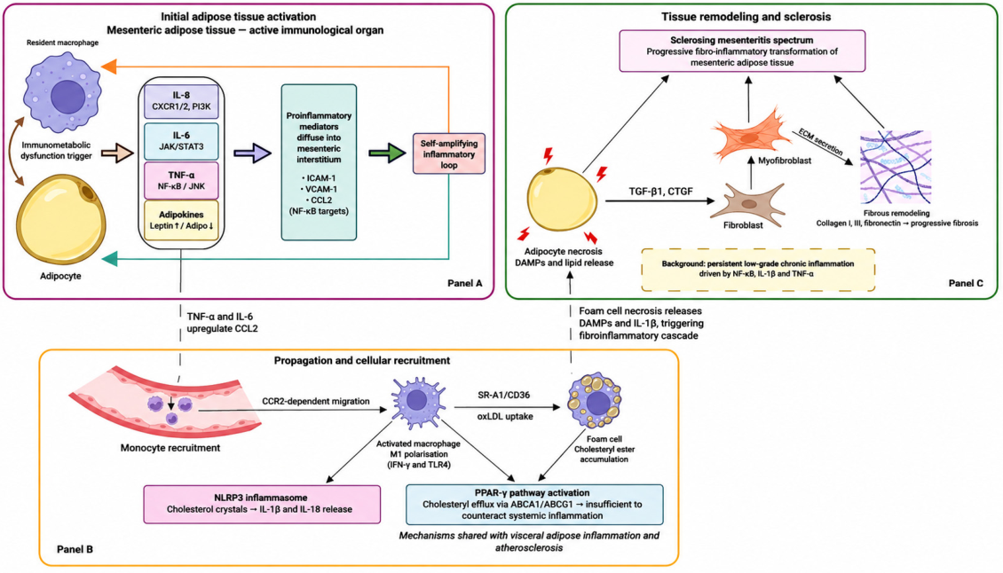
The pathophysiological mechanisms are illustrated in
Figure 3.
The main etiological factors and pathogenic mechanisms implicated in MP are summarized in
Table 2.
3.3. Diagnosis of Mesenteric Panniculitis
3.3.1. Positive Diagnosis
The clinical diagnosis of MP is challenging, as the presentation is non-specific and heterogeneous. Patients may be asymptomatic or may present with vague abdominal symptoms, most commonly abdominal pain without distinctive features, occasionally associated with bowel habit disturbances, nausea, or weight loss. Systemic manifestations, such as fever or asthenia, may be present but are inconsistent. Physical examination is often normal; however, in some cases, it may reveal nonspecific abdominal tenderness or, less frequently, a palpable mass [
13,
18,
52].
Laboratory investigations are non-specific, potentially revealing an inflammatory syndrome of variable intensity or values within normal limits, which limits the diagnostic value of clinical assessment alone [
52].
In this context, the diagnosis of MP is predominantly imaging-based, with contrast-enhanced multidetector CT representing the method of choice. In the majority of cases, the diagnosis can be established on the basis of a typical imaging pattern, without the need for histological confirmation, in the absence of clinical or imaging red flags for malignancy [
11,
14,
29].
The characteristic CT appearance consists of a hyperattenuating mesenteric fat area, typically located at the level of the small bowel mesenteric root, frequently involving the jejunal mesentery, associated with displacement of bowel loops and encasement of vascular structures without direct invasion. Mesenteric lymphadenopathy is common and, in the majority of cases, sub-centimeter in size [
7,
11,
53].
Suggestive imaging findings include hyperattenuation of the mesenteric fat, small mesenteric nodules, the “fat ring” sign (preservation of perivascular fat), and a peripheral pseudocapsule. The combination of these features supports the benign inflammatory nature of the condition and contributes to differentiation from neoplastic infiltrations [
8,
54,
55].
The imaging criteria proposed by Coulier are widely used and define the diagnosis when at least three of the following are present: a well-defined mesenteric fat mass, increased fat attenuation, mesenteric nodules, the “fat ring” sign, and a pseudocapsule [
19,
56].
No specific serological markers exist, and biopsy is not routinely required in typical presentations. Histological confirmation is reserved for cases with atypical imaging appearance or suspicion of malignancy [
11,
14].
3.3.2. Differential Diagnosis
The differential diagnosis of MP is broad and includes neoplastic, inflammatory, infectious, and vascular processes. Differentiation relies on the correlation of clinical and imaging findings and on the identification of red flags, such as bulky lymphadenopathy, increased fluorodeoxyglucose (FDG) uptake on positron emission tomography-computed tomography (PET-CT), or invasion of adjacent structures (
Table 3) [
13,
52].
3.4. Treatment of Mesenteric Panniculitis
The management of MP is individualized based on symptom presence and severity, clinical impact, and the occurrence of complications, in the absence of standardized therapeutic recommendations. In the majority of asymptomatic cases, a conservative approach is preferred, given the frequently benign and stable nature of the disease, while pharmacological treatment is reserved primarily for symptomatic forms. Available data support a symptom-guided strategy, in which the primary objective is clinical improvement rather than imaging normalization, as radiological changes may persist despite a favorable symptomatic response [
13,
19,
44].
Therapeutic options reported in the literature include clinical observation, systemic corticosteroid therapy, antifibrotic and anti-inflammatory therapies, and, in selected cases, immunosuppressive agents, immunomodulatory therapies, or surgical treatment. The main indications and characteristics of these approaches are summarized in
Table 4 [
9,
29,
60,
61].
4. Association of MP with IBD
4.1. Epidemiology
Epidemiological data on the association between MP and IBD remain limited and heterogeneous. In CT-based series, IBD is reported as a relatively rare comorbidity, frequently without differentiation between CD and UC and without systematic correlation with disease activity or the temporal relationship to the diagnosis of MP, precluding precise estimation of the true frequency or specificity of the association [
20,
21,
26].
Of the two major forms of IBD, the association appears better supported for CD than for UC. In the retrospective analysis by Protin-Catteau et al., CD was identified in 4.2% of patients with MP, suggesting a non-random overlap between mesenteric inflammation and chronic intestinal inflammatory disease [
44].
Additional data derived from clinical observations and small series reporting CD among the autoimmune or inflammatory comorbidities associated with MP, supporting the existence of a common immunological background, albeit without demonstrating a direct causal relationship [
45,
64].
In contrast, for UC, data remain fragmentary, limited to isolated observations or mentions within heterogeneous cohorts. In the cohort analyzed by Küpeli et al. [
43], UC was reported among the most common accompanying benign disorders in 3 out of 309 MP patients (0.97%), without dedicated analysis of this association. The narrative review by Ayala Gutiérrez and de Ramón Garrido [
21], which synthesized data from 202 published articles encompassing 1305 patients with MP, identified UC as a personal history in 1 of 710 patients (0.14%). Additionally, IBD was considered a possible etiological factor in 7 out of 1243 patients (0.6%), without differentiation between CD and UC. Collectively, these data suggest a possible but rare and insufficiently characterized association between MP and UC [
21,
43].
Overall, the available literature suggests that the association between MP and IBD is likely underrecognized and insufficiently defined epidemiologically. To date, the relationship appears more plausible and better supported for CD, whereas for UC, the evidence remains limited to isolated reports and small retrospective series [
28,
29].
Studies reporting the association between MP and IBD are summarized in
Table 5.
4.2. Common Pathogenic Mechanisms
Interest in the association between MP and IBD stems from recognition of the mesentery as an active immunological compartment directly involved in perpetuating inflammation and fibrotic remodeling. In CD, inflammation is not confined to the intestinal mucosa but may become transmural and extramural, involving adjacent mesenteric lymphatic, vascular, and adipose structures, thereby providing a plausible biological framework for interpreting MP as an expression of the same persistent inflammatory activation [
65,
66].
In CD, imaging and experimental data indicate that mesenteric fat hypertrophy, fibroadipose proliferation, and lymphadenopathy correlate with transmural inflammation and postoperative recurrence, supporting the concept that the mesentery may serve as an active source of inflammation and fibrogenesis [
39,
67,
68].
Mechanistically, mesenteric adipose tissue is an active immunometabolic organ that secretes pro-inflammatory cytokines and adipokines, including TNF-α, IL-1β, IL-6, leptin, and other profibrogenic mediators. In this model, the interaction between mesenteric adipocytes, macrophages, mesenteric lymph nodes, and the gut microbiota may sustain an autonomous inflammatory microenvironment capable of perpetuating both intestinal inflammation and fibroinflammatory remodeling of the mesentery [
34,
69,
70].
Specific profibrotic mechanisms, including activation of the autotaxin–lysophosphatidic acid axis and mesenteric lymphatic dysfunction, may further amplify fibrotic remodeling even in the absence of overt mucosal disease activity. These data support the concept of an intestino-mesenteric continuum, in which MP may represent a fibroinflammatory mesenteric phenotype potentially integrable, at least in some settings, within the spectrum of IBD, particularly CD [
40,
58,
71].
For UC, the biological arguments linking MP to the underlying disease are less compelling than for CD. Unlike Crohn’s disease, UC is primarily characterized by inflammation confined to the mucosa and submucosa, without transmural extension, and mesenteric involvement is correspondingly limited. Accordingly, mesenteric hypertrophy, creeping fat, and extensive fibroadipose remodeling, which are characteristic features of CD, are not observed in UC, providing a biologically plausible explanation for the rarity of MP in this setting [
41]. Nevertheless, UC is recognized as a systemic immune-mediated disease, with inflammatory mechanisms extending beyond the intestinal mucosa and giving rise to well-established extraintestinal immune manifestations [
47]. Based on these observations, one possible interpretation is that MP occurring in a patient with UC may represent either a coincidental idiopathic fibroinflammatory process or, alternatively, an immune-mediated mesenteric response developing in the setting of systemic immune dysregulation rather than as a consequence of direct transmural intestinal inflammation. However, dedicated mechanistic studies specifically addressing MP in UC are currently lacking, and this hypothesis remains speculative pending prospective validation [
11].
4.3. Diagnostic Pitfalls
One of the main challenges in interpreting MP in patients with IBD is the imaging overlap between primary mesenteric inflammation and secondary mesenteric changes due to intestinal disease. Increased mesenteric fat attenuation, described under the term “misty mesentery,” may occur in active CD through focal inflammation adjacent to affected intestinal segments, lymphadenopathy, and occasionally mesenteric abscesses, potentially mimicking idiopathic MP [
53,
55].
In certain settings, mesenteric inflammation may even precede overt intestinal manifestations. The report by Nuzzo et al. demonstrated that isolated ileal CD may simulate idiopathic MP over an extended period, in the context of repeatedly normal endoscopies and non-specific biological markers, with the diagnosis being clarified only after progression of mesenteric changes and subsequent identification of a mildly inflammatory and stricturing ileal segment at laparoscopy [
72]. Of note, a case of IgG4-related sclerosing mesenteritis associated with CD was previously reported by Kim et al., representing one of the earliest documented cases of overlap between mesenteric fibroinflammatory conditions and IBD [
73]. Together with the report by Nuzzo et al., these observations suggest that the association between mesenteric fibroinflammatory conditions and CD, while biologically plausible, remains exceptionally rare and insufficiently characterized in the literature. This observation underscores the necessity of systematically ruling out small bowel involvement before labeling MP as idiopathic in patients with suspected IBD [
72].
Another important source of diagnostic error is represented by fibroinflammatory or neoplastic processes that may reproduce the same imaging pattern. IgG4-related sclerosing mesenteritis, lymphoma, and other mesenteric infiltrations may mimic both MP and IBD-associated mesenteric involvement, with histological confirmation being required in atypical or pseudotumoral presentations [
52,
74].
Furthermore, in imaging-based cohorts, IBD is frequently reported without stratification between CD and UC, which may generate additional diagnostic confusion and limit the interpretation of the true relationship between the two entities. In this context, accurate differentiation requires careful integration of clinical, endoscopic, and imaging data, as well as the active exclusion of small bowel pathology, malignancy, and IgG4-related disease in atypical presentations [
21,
26].
5. Mesenteric Panniculitis as an Immunological Reaction/Drug-Induced Adverse Effect
Iatrogenic cases of MP have been described, particularly in the context of modern oncological therapies. Immune checkpoint inhibitors and targeted therapies may induce aberrant inflammatory reactions, including in mesenteric adipose tissue, through excessive immune activation and loss of peripheral tolerance. These forms fall within the spectrum of immune-mediated adverse reactions and may occur in isolation or in association with other systemic inflammatory manifestations [
38,
51].
Similarly, drug reactions are mentioned as potential triggering factors of mesenteric inflammation, although direct causal relationships remain difficult to demonstrate, and the majority of available data derive from case reports and small clinical series, in the absence of controlled studies confirming this association [
30,
31].
Vedolizumab is a humanized monoclonal antibody that acts through selective blockade of the α4β7 integrin, inhibiting the migration of gut-tropic T lymphocytes towards the intestinal mucosa by interfering with its interaction with MAdCAM-1, an adhesion molecule preferentially expressed on intestinal endothelium [
75].
Although vedolizumab’s gut-selective mechanism of action is associated with a favorable systemic safety profile, clinical observations have documented the de novo occurrence of extraintestinal inflammatory manifestations in patients receiving this therapy [
76,
77]. The underlying mechanisms remain incompletely understood, and whether these events reflect a direct drug-related effect, loss of prior systemic immunosuppression following anti-TNF discontinuation, or coincidental immune phenomena cannot be determined from available data [
78].
Observational clinical data have reported de novo EIMs in 26.7% of patients treated with vedolizumab in a retrospective study of 71 patients, with articular and cutaneous manifestations being the most frequently reported extraintestinal manifestations [
76].
Consistently, the French multicenter GETAID OBSERV-IBD cohort, which included 294 patients followed up to week 54, reported 34 incident cases (13.8%) of arthralgias or inflammatory arthritis and 14 cases (4.8%) of paradoxical cutaneous manifestations occurring de novo under treatment [
78].
However, the interpretation of these observations is complicated by important confounding factors, particularly prior exposure to anti-TNF-α therapies [
77].
In the absence of previous reports explicitly documenting an association between vedolizumab and MP, the potential relationship observed in the present case should be interpreted with caution. Although the onset of mesenteric panniculitis occurred during ongoing vedolizumab therapy, complete clinical and radiological remission was achieved without treatment discontinuation, which argues against a direct drug-induced mechanism. Nevertheless, a potential contribution of vedolizumab cannot be entirely excluded. Several alternative explanations remain equally plausible: idiopathic mesenteric panniculitis, an unidentified infectious trigger, an immune-mediated process independent of current biological therapy, or the potential influence of prior pharmacological exposure. Consequently, any potential association with vedolizumab should be interpreted with caution and should not be considered indicative of a drug-induced adverse effect.
6. Strengths, Limitations, and Future Perspectives
To the best of our knowledge, this article describes a rare case of MP associated with UC in a patient receiving vedolizumab therapy, in the context of biological, endoscopic, and histological remission of IBD. The originality of this case lies in the dissociation between mucosal inflammatory control and the occurrence of mesenteric inflammation. The article also provides a structured synthesis of available literature data on the association between MP and IBD, offering a critical appraisal of existing studies and highlighting the low level of evidence in this field.
The limitations of this article stem from its case report design, which precludes establishing a causal relationship between MP and UC, or between MP and vedolizumab therapy.
In addition to the inherent limitations of a single case report, several case-specific diagnostic limitations must be acknowledged. The diagnosis of MP was established on the basis of characteristic contrast-enhanced computed tomography criteria, in the absence of histopathological confirmation. Following complete clinical and radiological remission after medical treatment, the mesenteric inflammatory changes had resolved, precluding subsequent histopathological confirmation. Although imaging-based diagnosis is widely accepted in patients with typical radiological features, the absence of tissue confirmation limits the definitive exclusion of alternative fibroinflammatory conditions, including IgG4-related disease and disorders within the retroperitoneal fibrosis spectrum. This limitation is particularly relevant given the novelty of the reported association and the hypothesis-generating nature of the present observation.
Although the diagnosis was supported by characteristic imaging findings and an extensive diagnostic work-up—including microbiological, immunological, endoscopic, and histological investigations, as summarized in
Table 1—several additional evaluations could not be performed. Specifically, extractable nuclear antigen (ENA) antibody profiling and serum complement levels (C3 and C4) are not routinely available at our institution. Similarly, vedolizumab trough levels and anti-drug antibody testing were not available. Positron emission tomography-computed tomography (PET-CT) was not performed because this imaging modality is unavailable at our institution; however, the contrast-enhanced CT findings were consistent with established radiological criteria for MP and showed no features suggestive of occult malignancy.
The interpretation of the available literature is further limited by the predominance of retrospective studies, small case series, heterogeneous imaging criteria, inconsistent terminology, and the lack of systematic histopathological confirmation, all of which reduce diagnostic standardization and comparability across studies [
11,
19]. Selection bias introduced through retrospective radiological searches, the non-uniform use of biopsy, and the absence of standardized follow-up protocols additionally hamper the differentiation of clinically relevant forms from incidental findings and limit clarification of the prognostic significance of this entity [
26,
27]. Current therapeutic recommendations remain largely empirical, based on limited clinical experience rather than randomized controlled trials [
14,
61], while the reported associations of MP with both malignancy and IBD remain insufficiently characterized because of methodological heterogeneity and the absence of dedicated prospective studies, particularly for UC [
10,
47,
79]. Furthermore, it remains unclear whether mesenteric adipose tissue remodeling represents a cause or a consequence of intestinal inflammation, limiting the precise definition of the mesentery as a potential therapeutic target [
41,
71].
Taken together, these case-specific and literature-related limitations indicate that the observed association should be interpreted with caution. In the absence of histopathological confirmation and given the inherent limitations of a single-case report, no causal relationship between MP, UC, and vedolizumab therapy can be established. Furthermore, the delayed onset of MP—approximately three years after vedolizumab initiation and four weeks after the last infusion—constitutes an additional argument against a direct pharmacological causal relationship with vedolizumab.
Future prospective multicenter studies integrating clinical, imaging, histopathological, immunological, and molecular data are needed to standardize diagnostic criteria, clarify the relationship between mesenteric and intestinal inflammation in IBD, and better define the clinical significance of mesenteric involvement. Such studies may also help determine whether MP represents an IBD-associated mesenteric fibroinflammatory phenomenon or a distinct fibroinflammatory disorder.
7. Conclusions
MP is a fibroinflammatory condition of the mesentery characterized by variable clinical expression and predominantly imaging-based diagnosis, with its clinical significance incompletely defined. Available data suggest that MP and IBD may share common immunological and fibroinflammatory pathways. However, current clinical evidence supporting this association is substantially stronger for CD than for UC, in which published evidence remains limited.
The concept of the intestine–mesentery continuum does not exclude the possibility that inflammatory processes may predominantly involve the mesentery while intestinal disease remains clinically quiescent. This observation supports the need to evaluate mesenteric pathology independently of intestinal disease activity in selected clinical scenarios.
Although immune-mediated reactions, including paradoxical manifestations, have been reported in association with biological therapies, establishing a causal relationship between MP and vedolizumab—or any biological agent—remains challenging in the absence of controlled data and dedicated mechanistic studies.
In this context, clarification of these associations requires dedicated prospective studies systematically integrating clinical, imaging, histopathological, and biological data, including in the context of gut-selective biological therapies.
The present case, describing the co-occurrence of MP and UC in a patient receiving vedolizumab during sustained clinical, endoscopic, and histological remission, contributes to the limited available evidence regarding this association. Although it does not establish a causal relationship between MP, UC, or vedolizumab therapy, it supports further prospective investigation of the mesenteric compartment in IBD while underscoring the hypothesis-generating nature of this single observation.
Author Contributions
Conceptualization, C.A. and C.M.; methodology, A.-E.J.; investigation, C.V.M., I.R.M. and A.C.P.; formal analysis, I.R.M.; writing—original draft preparation, C.A., A.-E.J. and G.E.S.; writing—review and editing, G.E.S., B.-V.Ș., M.D., O.N. and G.E.G.-B.; visualization, C.V.M.; supervision, G.E.G.-B., C.C.P. and C.M.; project administration, C.C.P. and C.M. All authors have read and agreed to the published version of the manuscript.
Funding
This research received no external funding.
Institutional Review Board Statement
Our manuscript describes a single anonymized case report based on routine clinical care, accompanied by a narrative review of the published literature. No research-related interventions or procedures beyond standard clinical management were performed. Therefore, formal Institutional Review Board approval was not required for this type of publication.
Informed Consent Statement
Written informed consent has been obtained from the patient(s) to publish this paper.
Data Availability Statement
The original contributions presented in the study are included in the article; further inquiries can be directed to the corresponding author.
Acknowledgments
We would like to express our gratitude to “Sf. Spiridon” County Emergency Clinical Hospital and our colleagues in the gastroenterology, radiology, and surgery departments, as well as all the staff involved in the care of this patient.
Conflicts of Interest
The authors declare no conflicts of interest.
Abbreviations
The following abbreviations are used in this manuscript:
| MP | Mesenteric panniculitis |
| IBD | Inflammatory bowel disease |
| UC | Ulcerative colitis |
| CD | Crohn’s disease |
| EIM | Extraintestinal manifestation |
| CT | Computed tomography |
| MRI | Magnetic resonance imaging |
| CRP | C-reactive protein |
| FDG | Fluorodeoxyglucose |
| PET-CT | Positron emission tomography-computed tomography |
| PET | Positron emission tomography |
| MASLD | Metabolic dysfunction-associated steatotic liver disease |
| NAFLD | Non-alcoholic fatty liver disease |
| NSAIDs | Non-steroidal anti-inflammatory drugs |
| PPAR-γ | Peroxisome proliferator-activated receptor gamma |
| MAdCAM-1 | Mucosal addressin cell adhesion molecule-1 |
| ICI | Immune checkpoint inhibitors |
| ATX-LPA | Autotaxin–lysophosphatidic acid axis |
| TGF-β1 | Transforming growth factor beta-1 |
| CTGF | Connective tissue growth factor |
| NF-κB | Nuclear factor kappa B |
| IL-1β | Interleukin-1 beta |
| IL-6 | Interleukin-6 |
| IL-8 | Interleukin-8 |
| IL-18 | Interleukin-18 |
| TNF-α | Tumor necrosis factor alpha |
| CCL2 | C-C motif chemokine ligand 2 |
| NLRP3 | NLR family pyrin domain containing 3 |
| DAMPs | Damage-associated molecular patterns |
| ECM | Extracellular matrix |
| MDCT | Multidetector computed tomography |
| ICAM-1 | Intercellular adhesion molecule-1 |
| VCAM-1 | Vascular cell adhesion molecule-1 |
| COX-2 | Cyclooxygenase-2 |
| JAK/STAT3 | Janus kinase/signal transducer and activator of transcription 3 |
| ABCA1/ABCG1 | ATP-binding cassette transporter A1/G1 |
| SR-A1/CD36 | Scavenger receptor A1/CD36 |
| ANA | Antinuclear antibodies |
| ANCA | Anti-neutrophil cytoplasmic antibodies |
| RF | Rheumatoid factor |
| ENA | Extractable nuclear antigen |
| IgG4 | Immunoglobulin G subclass 4 |
| GDH | Glutamate dehydrogenase |
| CXCR1/2 | C-X-C motif chemokine receptor 1/2 |
| PI3K | Phosphoinositide 3-kinase |
| JNK | c-Jun N-terminal kinase |
| oxLDL | Oxidized low-density lipoprotein |
| IFN-γ | Interferon gamma |
| TLR4 | Toll-like receptor 4 |
References
- Ng, S.C.; Bernstein, C.N.; Vatn, M.H.; Lakatos, P.L.; Loftus, E.V.; Tysk, C.; O’Morain, C.; Moum, B.; Colombel, J.-F. Geographical Variability and Environmental Risk Factors in Inflammatory Bowel Disease. Gut 2013, 62, 630–649. [Google Scholar] [CrossRef] [PubMed]
- Kaplan, G.G.; Windsor, J.W. The Four Epidemiological Stages in the Global Evolution of Inflammatory Bowel Disease. Nat. Rev. Gastroenterol. Hepatol. 2021, 18, 56–66. [Google Scholar] [CrossRef] [PubMed]
- Ferrari, A.J.; Santomauro, D.F.; Aali, A.; Abate, Y.H.; Abbafati, C.; Abbastabar, H.; Abd ElHafeez, S.; Abdelmasseh, M.; Abd-Elsalam, S.; Abdollahi, A. Global Incidence, Prevalence, Years Lived with Disability (YLDs), Disability-Adjusted Life-Years (DALYs), and Healthy Life Expectancy (HALE) for 371 Diseases and Injuries in 204 Countries and Territories and 811 Subnational Locations, 1990–2021: A Systematic Analysis for the Global Burden of Disease Study 2021. Lancet 2024, 403, 2133–2161. [Google Scholar] [CrossRef] [PubMed]
- Malik, T.F.; Aurelio, D.M. Extraintestinal Manifestations of Inflammatory Bowel Disease. In StatPearls [Internet]; StatPearls Publishing: Treasure Island, FL, USA, 2024. [Google Scholar]
- Vavricka, S.R.; Schoepfer, A.; Scharl, M.; Lakatos, P.L.; Navarini, A.; Rogler, G. Extraintestinal Manifestations of Inflammatory Bowel Disease. Inflamm. Bowel Dis. 2015, 21, 1982–1992. [Google Scholar] [CrossRef] [PubMed]
- Gordon, H.; Burisch, J.; Ellul, P.; Karmiris, K.; Katsanos, K.; Allocca, M.; Bamias, G.; Barreiro-de Acosta, M.; Braithwaite, T.; Greuter, T. ECCO Guidelines on Extraintestinal Manifestations in Inflammatory Bowel Disease. J. Crohn’s Colitis 2024, 18, 1–37. [Google Scholar] [CrossRef] [PubMed]
- Daskalogiannaki, M.; Voloudaki, A.; Prassopoulos, P.; Magkanas, E.; Stefanaki, K.; Apostolaki, E.; Gourtsoyiannis, N. CT Evaluation of Mesenteric Panniculitis: Prevalence and Associated Diseases. Am. J. Roentgenol. 2000, 174, 427–431. [Google Scholar] [CrossRef] [PubMed]
- Horton, K.M.; Lawler, L.P.; Fishman, E.K. CT Findings in Sclerosing Mesenteritis (Panniculitis): Spectrum of Disease. Radiographics 2003, 23, 1561–1567. [Google Scholar] [CrossRef] [PubMed]
- Buragina, G.; Magenta Biasina, A.; Carrafiello, G. Clinical and Radiological Features of Mesenteric Panniculitis: A Critical Overview. Acta Biomed. 2019, 90, 411–422. [Google Scholar] [CrossRef] [PubMed]
- Eze, V.N.; Halligan, S. Mesenteric Panniculitis: A Clinical Conundrum. Br. J. Radiol. 2023, 96, 20211369. [Google Scholar] [CrossRef] [PubMed]
- Sulbaran, M.; Chen, F.K.; Farraye, F.A.; Hashash, J.G. A Clinical Review of Mesenteric Panniculitis. Gastroenterol. Hepatol. 2023, 19, 211–218. [Google Scholar]
- Akram, S.; Murray, J.A.; Pardi, D.S.; Alexander, G.L.; Schaffner, J.A.; Russo, P.A.; Abraham, S.C. Adult Autoimmune Enteropathy: Mayo Clinic Rochester Experience. Clin. Gastroenterol. Hepatol. 2007, 5, 1282–1290. [Google Scholar] [CrossRef] [PubMed]
- Danford, C.J.; Lin, S.C.; Wolf, J.L. Sclerosing Mesenteritis. Am. J. Gastroenterol. 2019, 114, 867–873. [Google Scholar] [CrossRef] [PubMed]
- Worthington, M.T.; Wolf, J.L.; Crockett, S.D.; Pardi, D.S. AGA Clinical Practice Update on Sclerosing Mesenteritis: Commentary. Clin. Gastroenterol. Hepatol. 2025, 23, 902–907. [Google Scholar] [CrossRef] [PubMed]
- Emory, T.S.; Monihan, J.M.; Carr, N.J.; Sobin, L.H. Sclerosing Mesenteritis, Mesenteric Panniculitis and Mesenteric Lipodystrophy: A Single Entity? Am. J. Surg. Pathol. 1997, 21, 392–398. [Google Scholar] [CrossRef] [PubMed]
- Jura, V. Sulla Mesenterite Retratile e Sclerosante. Policlinico 1924, 31, 575–581. [Google Scholar]
- Ogden, W.W.; Bradburn, D.M.; Rives, J.D. Mesenteric Panniculitis: Review of 27 Cases. Ann. Surg. 1965, 161, 864–875. [Google Scholar] [CrossRef] [PubMed]
- Kipfer, R.E.; Moertel, C.G.; Dahlin, D.C. Mesenteric Lipodystrophy. Ann. Intern. Med. 1974, 80, 582–588. [Google Scholar] [CrossRef] [PubMed]
- Coulier, B. Mesenteric Panniculitis Part 1: MDCT—Pictorial Review. J. Belg. Soc. Radiol. 2011, 94, 229–240. [Google Scholar] [CrossRef] [PubMed]
- Van Putte-Katier, N.; Van Bommel, E.F.H.; Elgersma, O.E.; Hendriksz, T.R. Mesenteric Panniculitis: Prevalence, Clinicoradiological Presentation and 5-Year Follow-Up. Br. J. Radiol. 2014, 87, 20140451. [Google Scholar] [CrossRef] [PubMed]
- Ayala Gutiérrez, M.D.M.; De Ramón Garrido, E. Paniculitis Mesentérica. Med. Clínica 2016, 146, 497–505. [Google Scholar] [CrossRef] [PubMed]
- Hussain, I.; Ishrat, S.; Aravamudan, V.M.; Khan, S.R.; Mohan, B.P.; Lohan, R.; Abid, M.B.; Ang, T.L. Mesenteric Panniculitis Does Not Confer an Increased Risk for Cancers: A Systematic Review and Meta-Analysis. Medicine 2022, 101, e29143. [Google Scholar] [CrossRef] [PubMed]
- Coulier, B. Mesenteric Panniculitis Part 2: Prevalence and Natural Course: MDCT Prospective Study. J. Belg. Soc. Radiol. 2011, 94, 241–246. [Google Scholar] [CrossRef] [PubMed]
- Saha, B.; Tome, J.; Wang, X.J. Sclerosing Mesenteritis: A Concise Clinical Review for Clinicians. Mayo Clin. Proc. 2024, 99, 812–820. [Google Scholar] [CrossRef] [PubMed]
- Obeidat, N.; Al-Omari, M.; Fataftah, J.; AlQararha, K.; Al-Qaralleh, M. Mesenteric Panniculitis: Is It Really Associated with Cancer? A Retrospective Imaging-Based Study. Future Sci. OA 2025, 11, 2573615. [Google Scholar] [CrossRef] [PubMed]
- Smith, Z.L.; Sifuentes, H.; Deepak, P.; Ecanow, D.B.; Ehrenpreis, E.D. Relationship between Mesenteric Abnormalities on Computed Tomography and Malignancy: Clinical Findings and Outcomes of 359 Patients. J. Clin. Gastroenterol. 2013, 47, 409–414. [Google Scholar] [CrossRef] [PubMed]
- Buchwald, P.; Diesing, L.; Dixon, L.; Wakeman, C.; Eglinton, T.; Dobbs, B.; Frizelle, F. Cohort Study of Mesenteric Panniculitis and Its Relationship to Malignancy. Br. J. Surg. 2016, 103, 1727–1730. [Google Scholar] [CrossRef] [PubMed]
- Wagner, C.; Dachman, A.; Ehrenpreis, E.D. Mesenteric Panniculitis, Sclerosing Mesenteritis and Mesenteric Lipodystrophy: Descriptive Review of a Rare Condition. Clin. Colon Rectal Surg. 2022, 35, 342–348. [Google Scholar] [CrossRef] [PubMed]
- Azar, C.; Ibrahim, M.A.; El Kouzi, Z.; El Mokahal, A.; Omran, N.; Muallem, N.; Sharara, A.I. Mesenteric Panniculitis. Inflamm. Intest. Dis. 2024, 9, 157–164. [Google Scholar] [CrossRef]
- Issa, I.; Baydoun, H. Mesenteric Panniculitis: Various Presentations and Treatment Regimens. World J. Gastroenterol. 2009, 15, 3827–3830. [Google Scholar] [CrossRef] [PubMed]
- Dahiya, D.S.; Kichloo, A.; Singh, J.; Albosta, M.S.; Wani, F.; Aljadah, M.; Haq, K.F. Acute Recurrent Exacerbations of Mesenteric Panniculitis with Immunosuppressive Therapy: A Case Report and a Brief Review. J. Investig. Med. High. Impact Case Rep. 2020, 8, 2324709620969581. [Google Scholar] [CrossRef] [PubMed]
- Schäffler, A.; Schölmerich, J.; Büchler, C. Mechanisms of Disease: Adipocytokines and Visceral Adipose Tissue—Emerging Role in Nonalcoholic Fatty Liver Disease. Nat. Rev. Gastroenterol. Hepatol. 2005, 2, 273–280. [Google Scholar] [CrossRef] [PubMed]
- Ahmed, M. A Clinician’s Perspective on the New Organ Mesentery and Non-Vascular Mesenteropathies. Front. Physiol. 2024, 15, 1336908. [Google Scholar] [CrossRef] [PubMed]
- Hussein, M.R.A.; Abdelwahed, S.R. Mesenteric Panniculitis: An Update. Expert. Rev. Gastroenterol. Hepatol. 2015, 9, 67–78. [Google Scholar] [CrossRef] [PubMed]
- Gunes, S.O.; Akturk, Y.; Guldogan, E.S.; Yilmaz, K.B.; Ergun, O.; Hekimoglu, B. Association between Mesenteric Panniculitis and Non-Neoplastic Disorders. Clin. Imaging 2021, 79, 219–224. [Google Scholar] [CrossRef] [PubMed]
- Wang, J.; Li, B. CT Findings of Idiopathic Mesenteric Panniculitis and Analysis of Related Factors. Eur. J. Radiol. 2023, 167, 111071. [Google Scholar] [CrossRef] [PubMed]
- Schweistein, H.; Weintraub, Y.; Hornik-Lurie, T.; Haskiya, H.; Levin, S.; Ringel, Y.; Naftali, T. Mesenteric Panniculitis Is Associated with Cardiovascular Risk-Factors: A Case-Control Study. Dig. Liver Dis. 2022, 54, 1657–1661. [Google Scholar] [CrossRef] [PubMed]
- Gray, E.J.; Darvishzadeh, A.; Sharma, A.; Ganeshan, D.; Faria, S.C.; Lall, C. Cancer Therapy-Related Complications in the Bowel and Mesentery: An Imaging Perspective. Abdom. Radiol. 2016, 41, 2031–2047. [Google Scholar] [CrossRef] [PubMed]
- Li, X.-H.; Feng, S.-T.; Cao, Q.-H.; Coffey, J.C.; Baker, M.E.; Huang, L.; Fang, Z.-N.; Qiu, Y.; Lu, B.-L.; Chen, Z.-H. Degree of Creeping Fat Assessed by Computed Tomography Enterography Is Associated with Intestinal Fibrotic Stricture in Patients with Crohn’s Disease: A Potentially Novel Mesenteric Creeping Fat Index. J. Crohn’s Colitis 2021, 15, 1161–1173. [Google Scholar] [CrossRef] [PubMed]
- Huang, L.; Qian, W.; Xu, Y.; Guo, Z.; Yin, Y.; Guo, F.; Zhu, W.; Li, Y. Mesenteric Adipose Tissue Contributes to Intestinal Fibrosis in Crohn’s Disease Through the ATX–LPA Axis. J. Crohn’s Colitis 2022, 16, 1124–1139. [Google Scholar] [CrossRef] [PubMed]
- Gu, P.; Dube, S.; McGovern, D.P.B. Medical and Surgical Implications of Mesenteric Adipose Tissue in Crohn’s Disease: A Review of the Literature. Inflamm. Bowel Dis. 2023, 29, 458–469. [Google Scholar] [CrossRef] [PubMed]
- Yin, Y.; Zhu, Z.-X.; Li, Z.; Chen, Y.-S.; Zhu, W.-M. Role of Mesenteric Component in Crohn’s Disease: A Friend or Foe? World J. Gastrointest. Surg. 2021, 13, 1536–1549. [Google Scholar] [CrossRef] [PubMed]
- Küpeli, A.; Akdoğan Kayhan, M.; Ünsal, A.; Hekimoğlu, B. Evaluation of Mesenteric Panniculitis with Computed Tomography: Benign Condition or Paraneoplastic Syndrome? Turk. J. Med. Sci. 2018, 48, 42–47. [Google Scholar] [CrossRef] [PubMed]
- Protin-Catteau, L.; Thiéfin, G.; Barbe, C.; Jolly, D.; Soyer, P.; Hoeffel, C. Mesenteric Panniculitis: Review of Consecutive Abdominal MDCT Examinations with a Matched-Pair Analysis. Acta Radiol. 2016, 57, 1438–1444. [Google Scholar] [CrossRef] [PubMed]
- García Cruz, E.J.; Romero, A.R.; Núñez, F.M.; Rosas, J.M.P.; Guzmán, N.I.B. Exploring Mesenteric Panniculitis: Etiology, Clinical Features, and Advances in Diagnosis and Treatment. Int. J. Med. Sci. Clin. Res. Stud. 2023, 3, 1598–1604. [Google Scholar] [CrossRef]
- Lu, W.; Cen, J.; Dai, Q.; Tao, H.; Peng, L. Gut Microbiota Does Not Play a Mediating Role in the Causal Association between Inflammatory Bowel Disease and Several Its Associated Extraintestinal Manifestations: A Mendelian Randomization Study. Front. Immunol. 2024, 14, 1296889. [Google Scholar] [CrossRef] [PubMed]
- Danese, S.; Fiocchi, C. Ulcerative Colitis. N. Engl. J. Med. 2011, 365, 1713–1725. [Google Scholar] [CrossRef] [PubMed]
- Rosu, O.-M.; Gimiga, N.; Stefanescu, G.; Ioniuc, I.; Tataranu, E.; Balan, G.G.; Ion, L.-M.; Plesca, D.A.; Schiopu, C.G.; Diaconescu, S. The Effectiveness of Different Eradication Schemes for Pediatric Helicobacter Pylori Infection—A Single-Center Comparative Study from Romania. Children 2022, 9, 1391. [Google Scholar] [CrossRef] [PubMed]
- Avincsal, M.O.; Otani, K.; Kanzawa, M.; Fujikura, K.; Jimbo, N.; Morinaga, Y.; Hirose, T.; Itoh, T.; Zen, Y. Sclerosing Mesenteritis: A Real Manifestation or Histological Mimic of IgG4-Related Disease? Pathol. Int. 2016, 66, 158–163. [Google Scholar] [CrossRef] [PubMed]
- Ehrenpreis, E.D.; Roginsky, G.; Gore, R.M. Clinical Significance of Mesenteric Panniculitis-Like Abnormalities on Abdominal Computerized Tomography in Patients with Malignant Neoplasms. World J. Gastroenterol. 2016, 22, 10601–10608. [Google Scholar] [CrossRef] [PubMed]
- Drews, M.A.; Baumgarten, A.; Zensen, S.; Opitz, M.; Bos, D.; Zimmer, L.; Ugurel, S.; Haubold, J.; Schadendorf, D.; Livingstone, E. Adverse Effects of Systemic Advanced Melanoma Therapies—Do BRAF/MEK Inhibitors Increase the Incidence of Mesenteric Panniculitis? Eur. Radiol. 2025, 35, 7298–7306. [Google Scholar] [CrossRef] [PubMed]
- Guettrot-Imbert, G.; Boyer, L.; Piette, J.-C.; Delèvaux, I.; André, M.; Aumaître, O. Panniculite Mésentérique. La Rev. Méd. Interne 2012, 33, 621–627. [Google Scholar] [CrossRef] [PubMed]
- McLaughlin, P.D.; Filippone, A.; Maher, M.M. The “Misty Mesentery”: Mesenteric Panniculitis and Its Mimics. Am. J. Roentgenol. 2013, 200, W116–W123. [Google Scholar] [CrossRef] [PubMed]
- Sabaté, J.M.; Torrubia, S.; Maideu, J.; Franquet, T.; Monill, J.M.; Pérez, C. Sclerosing Mesenteritis: Imaging Findings in 17 Patients. Am. J. Roentgenol. 1999, 172, 625–629. [Google Scholar] [CrossRef] [PubMed]
- Filippone, A.; Cianci, R.; Di Fabio, F.; Storto, M.L. Misty Mesentery: A Pictorial Review of Multidetector-Row CT Findings. La Radiol. Medica 2011, 116, 351–365. [Google Scholar] [CrossRef] [PubMed]
- Green, M.S.; Chhabra, R.; Goyal, H. Sclerosing Mesenteritis: A Comprehensive Clinical Review. Ann. Transl. Med. 2018, 6, 336. [Google Scholar] [CrossRef] [PubMed]
- Gögebakan, Ö.; Osterhoff, M.; Albrecht, T. Mesenteric Panniculitis (MP): A Frequent Coincidental CT Finding of Debatable Clinical Significance. In Fortschritte auf dem Gebiet der Röntgenstrahlen und der Nuklearmedizin; Georg Thieme Verlag KG: Stuttgart, Germany, 2018; Volume 190, pp. 1044–1052. [Google Scholar] [CrossRef] [PubMed]
- Rivera, E.D.; Coffey, J.C.; Walsh, D.; Ehrenpreis, E.D. The Mesentery, Systemic Inflammation, and Crohn’s Disease. Inflamm. Bowel Dis. 2019, 25, 226–234. [Google Scholar] [CrossRef] [PubMed]
- Wat, S.Y.J.; Harish, S.; Winterbottom, A.; Choudhary, A.K.; Freeman, A.H. The CT Appearances of Sclerosing Mesenteritis and Associated Diseases. Clin. Radiol. 2006, 61, 652–658. [Google Scholar] [CrossRef] [PubMed]
- Cortés, P.; Ghoz, H.M.; Mzaik, O.; Alhaj Moustafa, M.; Bi, Y.; Brahmbhatt, B.; Daoud, N.; Pang, M. Colchicine as an Alternative First-Line Treatment of Sclerosing Mesenteritis: A Retrospective Study. Dig. Dis. Sci. 2022, 67, 2403–2412. [Google Scholar] [CrossRef] [PubMed]
- Alsuhaimi, M.A.; Alshowaiey, R.A.; Alsumaihi, A.S.; Aldhafeeri, S.M. Mesenteric Panniculitis Various Presentations and Management: A Single Institute Ten Years Experience. Ann. Med. Surg. 2022, 80, 104203. [Google Scholar] [CrossRef] [PubMed]
- Sahin, A.; Artas, H.; Eroglu, Y.; Tunc, N.; Demirel, U.; Bahcecioglu, I.H.; Yalniz, M. An Overlooked Potentially Treatable Disorder: Idiopathic Mesenteric Panniculitis. Med. Princ. Pract. 2017, 26, 567–572. [Google Scholar] [CrossRef] [PubMed]
- ÇeviK, B.; Kasapoğlu, B.; Yozgat, A.; KekiLli, M. Mesenteric Panniculitis Clinical Presentations, Management, and Outcomes: A Single Institute Experience of 89 Patients. J. Health Sci. Med./JHSM 2023, 6, 1272–1276. [Google Scholar] [CrossRef]
- Nyberg, L.; Björk, J.; Björkdahl, P.; Ekberg, O.; Sjöberg, K.; Vigren, L. Sclerosing Mesenteritis and Mesenteric Panniculitis—Clinical Experience and Radiological Features. BMC Gastroenterol. 2017, 17, 75. [Google Scholar] [CrossRef] [PubMed]
- Abraham, C.; Cho, J.H. Inflammatory Bowel Disease. N. Engl. J. Med. 2009, 361, 2066–2078. [Google Scholar] [CrossRef] [PubMed]
- Bsat, M.; Chapuy, L.; Rubio, M.; Wassef, R.; Richard, C.; Schwenter, F.; Loungnarath, R.; Soucy, G.; Mehta, H.; Sarfati, M. Differential Pathogenic Th17 Profile in Mesenteric Lymph Nodes of Crohn’s Disease and Ulcerative Colitis Patients. Front. Immunol. 2019, 10, 1177. [Google Scholar] [CrossRef] [PubMed]
- Koifman, E.; Cohen, I.S.; Lopez-Alonso, R.; Hur, D.B.; Ilivitzki, A.; Waterman, M. Mesenteric Fat Proliferation on Magnetic Resonance Enterography Is a Predictor of Long-Term Disease Complications in Crohn’s Disease. BMC Gastroenterol. 2025, 25, 593. [Google Scholar] [CrossRef] [PubMed]
- Meng, J.; Mao, Y.; Zhou, J.; Chen, Z.; Huang, S.; Wang, Y.; Huang, L.; Zhang, R.; Shen, X.; Lv, W.; et al. Mesenteric Abnormalities Play an Important Role in Grading Intestinal Fibrosis in Patients with Crohn’s Disease: A Computed Tomography and Clinical Marker-Based Nomogram. Ther. Adv. Gastroenterol. 2022, 15, 17562848221122504. [Google Scholar] [CrossRef] [PubMed]
- Seifarth, C.; Hering, N.A.; Arndt, M.; Lehmann, K.S.; Stroux, A.; Weixler, B.; Kreis, M.E. Increased Proinflammatory Cytokines in Mesenteric Fat in Major Surgery and Crohn’s Disease. Surgery 2021, 169, 1328–1332. [Google Scholar] [CrossRef] [PubMed]
- Hasnaoui, A.; Trigui, R.; Giuffrida, M. Gut Microbiota and Mesenteric Adipose Tissue Interactions in Shaping Phenotypes and Treatment Strategies for Crohn’s Disease. World J. Gastroenterol. 2024, 30, 4969–4976. [Google Scholar] [CrossRef] [PubMed]
- Shen, W.; Li, Y.; Zou, Y.; Cao, L.; Cai, X.; Gong, J.; Xu, Y.; Zhu, W. Mesenteric Adipose Tissue Alterations in Crohn’s Disease Are Associated with the Lymphatic System. Inflamm. Bowel Dis. 2019, 25, 283–293. [Google Scholar] [CrossRef] [PubMed]
- Nuzzo, A.; Zappa, M.; Cazals-Hatem, D.; Bouhnik, Y. Crohn’s Disease Mistaken for Long-Standing Idiopathic Mesenteric Panniculitis: A Case Report and Management Algorithm. Medicine 2016, 95, e5073. [Google Scholar] [CrossRef] [PubMed]
- Kim, E.J.; Kim, E.Y.; Song, J.E.; Lee, H.C.; Bae, G.H.; Oh, H.K.; Lee, T.S. A Case of IgG4-Related Sclerosing Mesenteritis Associated with Crohn’s Disease. Korean J. Gastroenterol. 2014, 63, 176. [Google Scholar] [CrossRef] [PubMed]
- Zheng, Y.; Elsayes, K.M.; Waranch, C.; Abdelaziz, A.; Menias, C.O.; Sandrasegaran, K.; Shaaban, A.M.; Gaballah, A.H. IgG4-Related Disease in the Abdomen and Pelvis: Atypical Findings, Pitfalls, and Mimics. Abdom. Radiol. 2020, 45, 2485–2499. [Google Scholar] [CrossRef] [PubMed]
- Wyant, T.; Fedyk, E.; Abhyankar, B. An Overview of the Mechanism of Action of the Monoclonal Antibody Vedolizumab. J. Crohn’s Colitis 2016, 10, 1437–1444. [Google Scholar] [CrossRef] [PubMed]
- Diaz, L.I.; Keihanian, T.; Schwartz, I.; Kim, S.B.; Calmet, F.; Quintero, M.A.; Abreu, M.T. Vedolizumab-Induced De Novo Extraintestinal Manifestations. Gastroenterol. Hepatol. 2020, 16, 75–81. [Google Scholar]
- Hanzel, J.; Ma, C.; Vande Casteele, N.; Khanna, R.; Jairath, V.; Feagan, B.G. Vedolizumab and Extraintestinal Manifestations in Inflammatory Bowel Disease. Drugs 2021, 81, 333–347. [Google Scholar] [CrossRef] [PubMed]
- Tadbiri, S.; Peyrin-Biroulet, L.; Serrero, M.; Filippi, J.; Pariente, B.; Roblin, X.; Buisson, A.; Stefanescu, C.; Trang-Poisson, C.; Altwegg, R. Impact of Vedolizumab Therapy on Extra-Intestinal Manifestations in Patients with Inflammatory Bowel Disease: A Multicentre Cohort Study Nested in the OBSERV-IBD Cohort. Aliment. Pharmacol. Ther. 2018, 47, 485–493. [Google Scholar] [CrossRef] [PubMed]
- Halligan, S.; Plumb, A.; Taylor, S. Mesenteric Panniculitis: Systematic Review of Cross-Sectional Imaging Findings and Risk of Subsequent Malignancy. Eur. Radiol. 2016, 26, 4531–4537. [Google Scholar] [CrossRef] [PubMed]
Figure 1.
Contrast-enhanced computed tomography findings of mesenteric panniculitis. (A) Sagittal CT image demonstrating multiple sub-centimeter lymph nodes (green arrows) at the level of the mesenteric root. (B) Coronal CT image showing densification of the mesenteric fat with encasement of mesenteric vessels without direct invasion (green oval), and sub-centimeter lymphadenopathies (green arrows). (C) Axial CT image revealing diffuse hyperattenuating mesenteric fat infiltration (green oval) and associated lymph nodes (green arrows), suggestive of mesenteric panniculitis.
Figure 1.
Contrast-enhanced computed tomography findings of mesenteric panniculitis. (A) Sagittal CT image demonstrating multiple sub-centimeter lymph nodes (green arrows) at the level of the mesenteric root. (B) Coronal CT image showing densification of the mesenteric fat with encasement of mesenteric vessels without direct invasion (green oval), and sub-centimeter lymphadenopathies (green arrows). (C) Axial CT image revealing diffuse hyperattenuating mesenteric fat infiltration (green oval) and associated lymph nodes (green arrows), suggestive of mesenteric panniculitis.
Figure 2.
Mesenteric panniculitis—follow-up MRI at one month. (A) Axial opposed-phase T1-weighted image demonstrating complete resolution of the previously observed mesenteric fat infiltration, with no residual inflammatory lymph nodes at the mesenteric root (green oval). (B) Coronal T2-weighted fat-saturated image confirming complete resolution of the mesenteric fat infiltration and absence of residual inflammatory lymph nodes (green oval), consistent with complete radiological remission.
Figure 2.
Mesenteric panniculitis—follow-up MRI at one month. (A) Axial opposed-phase T1-weighted image demonstrating complete resolution of the previously observed mesenteric fat infiltration, with no residual inflammatory lymph nodes at the mesenteric root (green oval). (B) Coronal T2-weighted fat-saturated image confirming complete resolution of the mesenteric fat infiltration and absence of residual inflammatory lymph nodes (green oval), consistent with complete radiological remission.
Figure 3.
Immunometabolic pathogenesis of mesenteric panniculitis. (Panel A)—Initial activation of mesenteric adipose tissue as an active immunological organ. Resident macrophages and adipocytes respond to immunometabolic triggers by releasing pro-inflammatory cytokines (IL-6/JAK-STAT3, TNF-α/NF-κB/JNK, IL-8/CXCR1/2-PI3K) and adipokines (leptin ↑, adiponectin ↓), promoting inflammatory mediator release and CCL2-dependent monocyte recruitment. (Panel B)—Propagation of inflammation through CCR2-dependent monocyte migration, M1 macrophage polarization, oxLDL uptake, foam cell formation, NLRP3 inflammasome activation, and IL-1β/IL-18 release. PPAR-γ-mediated cholesterol efflux via ABCA1/ABCG1 is insufficient to counterbalance the inflammatory response. (Panel C)—Progressive fibroinflammatory remodeling. Adipocyte and foam cell necrosis release DAMPs and IL-1β, activating the TGF-β1/CTGF pathway, fibroblast-to-myofibroblast transition, extracellular matrix secretion, and collagen I/III and fibronectin deposition, leading to fibrosis within the sclerosing mesenteritis spectrum. Background: persistent low-grade chronic inflammation driven by NF-κB, IL-1β, and TNF-α sustains all three phases.
Figure 3.
Immunometabolic pathogenesis of mesenteric panniculitis. (Panel A)—Initial activation of mesenteric adipose tissue as an active immunological organ. Resident macrophages and adipocytes respond to immunometabolic triggers by releasing pro-inflammatory cytokines (IL-6/JAK-STAT3, TNF-α/NF-κB/JNK, IL-8/CXCR1/2-PI3K) and adipokines (leptin ↑, adiponectin ↓), promoting inflammatory mediator release and CCL2-dependent monocyte recruitment. (Panel B)—Propagation of inflammation through CCR2-dependent monocyte migration, M1 macrophage polarization, oxLDL uptake, foam cell formation, NLRP3 inflammasome activation, and IL-1β/IL-18 release. PPAR-γ-mediated cholesterol efflux via ABCA1/ABCG1 is insufficient to counterbalance the inflammatory response. (Panel C)—Progressive fibroinflammatory remodeling. Adipocyte and foam cell necrosis release DAMPs and IL-1β, activating the TGF-β1/CTGF pathway, fibroblast-to-myofibroblast transition, extracellular matrix secretion, and collagen I/III and fibronectin deposition, leading to fibrosis within the sclerosing mesenteritis spectrum. Background: persistent low-grade chronic inflammation driven by NF-κB, IL-1β, and TNF-α sustains all three phases.

Table 1.
Laboratory investigations, infectious workup, autoimmune serological screening, and endoscopic assessment at the time of presentation.
Table 1.
Laboratory investigations, infectious workup, autoimmune serological screening, and endoscopic assessment at the time of presentation.
| Investigation | Result |
|---|
| Laboratory investigations |
| Leukocyte count | 14,258/mm3 (reference: 4000–10,000/mm3) |
| C-reactive protein (CRP) | 5.23 mg/dL (reference: <0.5 mg/dL) |
| Fecal calprotectin | 20.3 µg/g (reference: <50 µg/g) |
Liver function tests (AST, ALT, ALP, GGT,
bilirubin) | Within normal limits |
| Renal function tests (serum creatinine, urea) | Within normal limits |
| Serum electrolytes | Within normal limits |
| Infectious workup |
| Clostridioides difficile: GDH, toxins A and B | Negative |
| Stool culture | Negative |
| Stool parasitology | Negative |
| Urine culture | Negative |
| Blood cultures | Negative |
| Chest X-ray | No pathological findings |
| Autoimmune and immunological investigations |
| IgG4 | 0.5 g/L (reference: 0.03–2.01 g/L) |
| Antinuclear antibodies (ANA) | Negative (index <0.7; reference: <0.7) |
| Anti-neutrophil cytoplasmic antibodies (ANCA) | Negative (titre <1:10; reference: <1:10) |
| Rheumatoid factor (RF) | 2 IU/mL (reference: <14 IU/mL) |
| Assessment of ulcerative colitis activity |
| Endoscopic Mayo Score | 0 (endoscopic remission) |
| Nancy Histological Index | 0 (histological remission) |
Table 2.
Etiology and associated factors in mesenteric panniculitis (MP).
Table 2.
Etiology and associated factors in mesenteric panniculitis (MP).
| Etiological Category | Representative Examples | Proposed Mechanism | References |
|---|
Mechanical/
postoperative factors | Abdominal surgery; abdominal trauma | Aberrant tissue repair | [8,20] |
| Vascular factors | Mesenteric ischemia, vasculitis | Secondary inflammatory activation | [30] |
| Infectious factors | Chronic gastrointestinal infections, (including Helicobacter pylori) | Persistent immune activation | [34,48] |
Autoimmune/
fibroinflammatory factors | Retroperitoneal fibrosis, sclerosing cholangitis, Riedel’s thyroiditis | Shared immunological substrate | [8,20] |
| IgG4-related disease | Forms of IgG4-associated sclerosing mesenteritis | Immune-mediated fibroinflammation | [14,49] |
| Neoplastic factors | Lymphomas, solid neoplasms (e.g., prostate) | paraneoplastic phenomenon or detection bias | [19,22,50] |
| Metabolic factors | Visceral obesity; metabolic syndrome; MASLD | metabolic inflammation | [35,36,37] |
| Immunometabolic factors | Secretion of TNF-α, IL-6, IL-8, leptin, MCP-1; macrophage activation | mesenteric fibrotic remodeling | [32] |
Drug-induced/
iatrogenic factors | Oncological therapies, immune checkpoint inhibitors, targeted therapies | Drug-induced immune reactions | [14,38,51] |
| Intestinal inflammatory factors | CD, UC | intestinal-mesenteric immune activation | [39,40,41,42] |
| Idiopathic factors | Cases without identifiable cause | Unknown mechanism | [30] |
Table 3.
Differential diagnosis of MP.
Table 3.
Differential diagnosis of MP.
| Entity | Suggestive Features | Differentiating Features | References |
|---|
| Mesenteric lymphoma | Bulky confluent
lymphadenopathy | Enlarged (>10–12 mm) FDG-avid lymph nodes | [53,57] |
| Peritoneal carcinomatosis | Peritoneal nodules,
ascites | Peritoneal dissemination,
metastases | [30] |
| Neuroendocrine tumors (carcinoid) | Mesenteric mass with desmoplastic reaction | Calcifications; retractile fibrosis | [30] |
| Desmoid tumors | Infiltrative solid mass | Absence “fat ring” sign;
progressive growth | [30] |
| Liposarcoma | Bulky adipose mass | Solid component, heterogeneity | [30] |
| IgG4-related disease | Fibroinflammatory mass | Multiorgan involvement,
elevated serum IgG4 | [49] |
| Peritoneal tuberculosis | Lymphadenopathy,
ascites | Evidence of systemic infection | [52] |
| CD (mesenteric) | “Creeping fat”; bowel wall thickening | Active intestinal lesions | [58] |
| Mesenteric edema | Diffuse fat stranding | Underlying Cardiac/hepatic disease; reversibility | [59] |
| Mesenteric venous thrombosis | Edema, congestion | Evident vascular thrombosis | [59] |
Table 4.
Therapeutic options in MP.
Table 4.
Therapeutic options in MP.
| Therapeutic option | Examples | Indications | Efficacy/Remarks | References |
|---|
| Watchful waiting | Clinical monitoring | Asymptomatic patients | Often stable or self-limited | [13,14] |
| Corticosteroid therapy | Prednisone | Symptomatic forms | First-line treatment;
frequent clinical response | [13,14] |
| Antifibrotic therapy | Tamoxifen | Combined with corticosteroid therapy | Reported benefit in combination therapy | [13,14] |
| Anti-inflammatory therapy | Colchicine | Alternative/
adjuvant | Reported benefit in selected series | [29,60,61] |
| Immunosuppressive agents | Azathioprine, methotrexate | Refractory forms | Limited evidence | [29,60,61] |
| Other therapies | Pentoxifylline,
thalidomide,
biologics | Selected cases | Limited evidence | [29,60,61] |
| Symptomatic treatment | NSAIDs, antibiotic therapy | Mild forms | Possibly self-limiting course | [62,63] |
| Surgical treatment | — | Complications | Limited indications | [9,13] |
Table 5.
Studies reporting the association between MP and IBD.
Table 5.
Studies reporting the association between MP and IBD.
| Study | Study Design | Patients (n) | MP Cases | Key Findings |
|---|
| Smith et al., 2013 [26] | Retrospective CT-based cohort | 359 | 359 | IBD reported as a rare comorbidity in 6/359 patients (1.6%), without differentiation between CD and UC |
| Protin-Catteau et al., 2016 [44] | Retrospective CT-based matched-pair analysis | 3054 | 96 | CD was identified in 4.2% of patients with MP |
| Ayala Gutiérrez & de Ramón Garrido, 2016 [21] | Narrative
review of 202 published
articles | 1305 | 1305 | UC identified as a personal history in 1/710 patients (0.14%); IBD considered a possible etiological factor in 7/1243 patients (0.6%),
CD and UC were not differentiated |
| Nyberg et al., 2017 [64] | Retrospective clinical and
imaging cohort | 31 | 27 | CD reported in 1/27 MP patients (3.7%) among autoimmune comorbidities; no dedicated statistical analysis was performed |
| Küpeli et al., 2018 [43] | Retrospective CT-based cohort | 22,033 | 309 | UC reported among benign comorbidities in 3/309 patients (0.97%), without a dedicated analysis of the association |
| Disclaimer/Publisher’s Note: The statements, opinions and data contained in all publications are solely those of the individual author(s) and contributor(s) and not of MDPI and/or the editor(s). MDPI and/or the editor(s) disclaim responsibility for any injury to people or property resulting from any ideas, methods, instructions or products referred to in the content. |




 ,
,