Invasive Evaluation of Coronary Artery Disease in Severe Aortic Stenosis—A Narrative Review

Abstract
1. Background
2. Pathophysiology of Concomitant CAD and AS
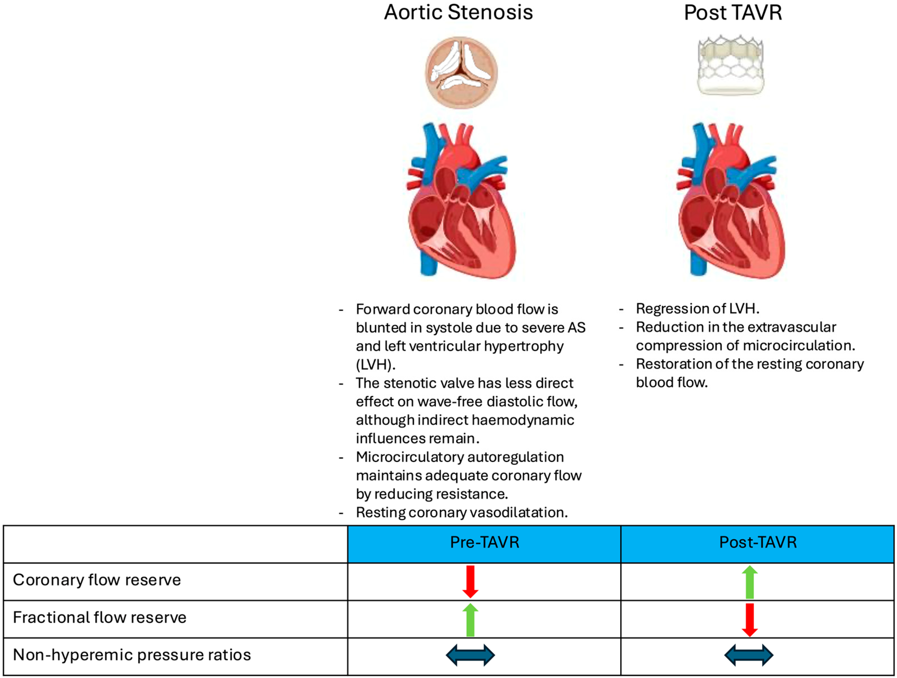
Current Conundrums in Assessing Coronary Physiology in AS
3. Invasive Evaluation of Coronary Lesions in Severe Aortic Stenosis
4. CT Coronary Angiography-Derived Haemodynamic Assessment
4.1. Feasibility
4.2. Diagnostic Performance
4.3. Prognostic Implications
4.4. Clinical Application and Limitations
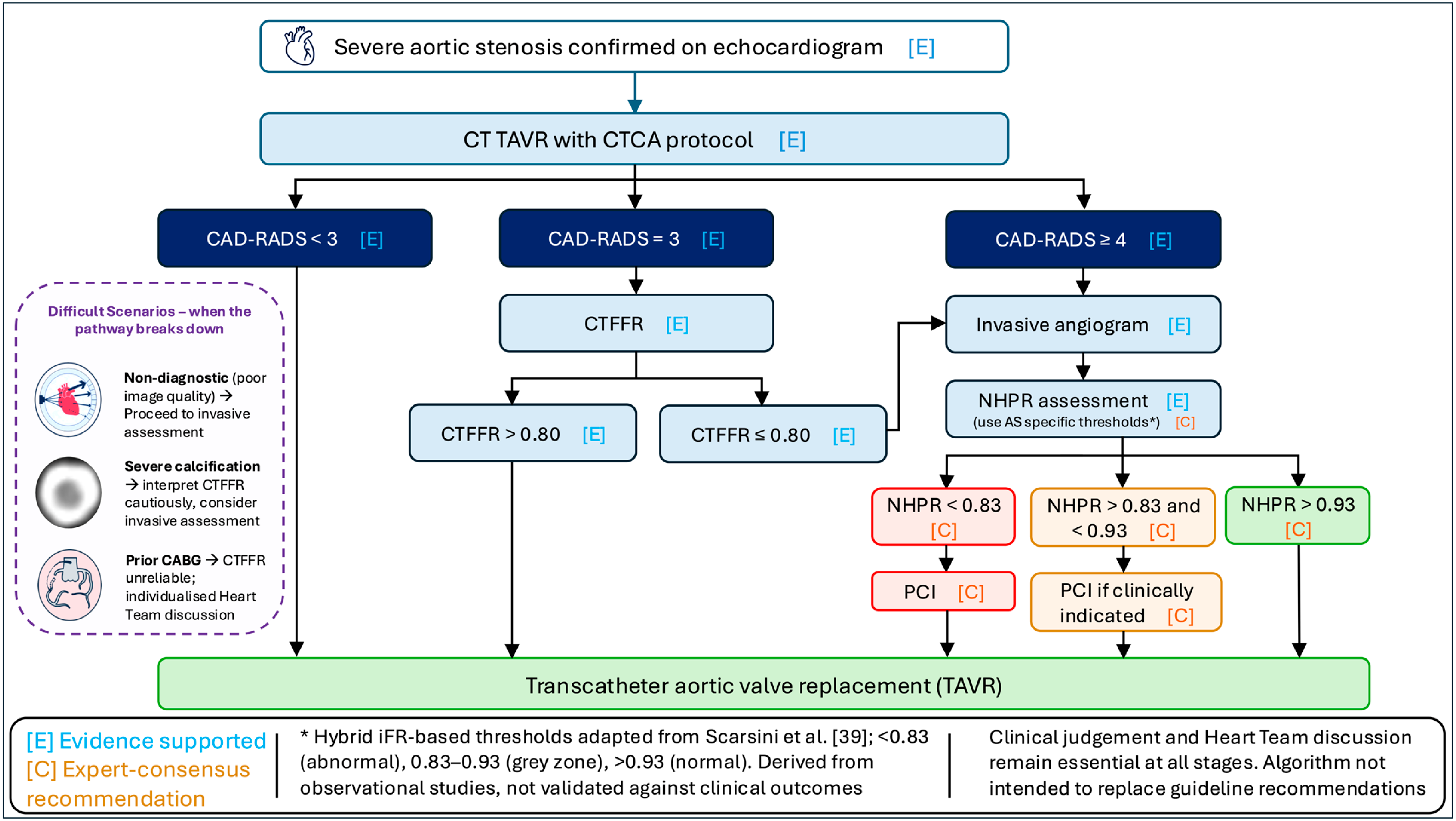
5. Proposed Management Algorithm and Perspectives Based on Expert Opinion
6. Revascularisation of Concomitant Coronary Artery Disease
6.1. Identifying Significant Lesions
6.2. Does Revascularisation Improve Outcomes?
7. Future Research Priorities
7.1. Priority 1: Validation of Thresholds Against Clinical Outcomes
7.2. Priority 2: Determining Whether Improved Diagnosis Translates into Improved Outcomes
7.3. Priority 3: Defining the Role of Non-Wire-Based Physiological Assessment
8. Conclusions
Author Contributions
Funding
Institutional Review Board Statement
Informed Consent Statement
Data Availability Statement
Acknowledgments
Conflicts of Interest
References
- Osnabrugge, R.L.; Mylotte, D.; Head, S.J.; Van Mieghem, N.M.; Nkomo, V.T.; LeReun, C.M.; Bogers, A.J.; Piazza, N.; Kappetein, A.P. Aortic stenosis in the elderly: Disease prevalence and number of candidates for transcatheter aortic valve replacement: A meta-analysis and modeling study. J. Am. Coll. Cardiol. 2013, 62, 1002–1012. [Google Scholar] [CrossRef] [PubMed]
- Freeman, R.V.; Otto, C.M. Spectrum of calcific aortic valve disease: Pathogenesis, disease progression, and treatment strategies. Circulation 2005, 111, 3316–3326. [Google Scholar] [CrossRef] [PubMed]
- Pibarot, P.; Dumesnil, J.G. Improving assessment of aortic stenosis. J. Am. Coll. Cardiol. 2012, 60, 169–180. [Google Scholar] [CrossRef] [PubMed]
- Stewart, B.F.; Siscovick, D.; Lind, B.K.; Gardin, J.M.; Gottdiener, J.S.; Smith, V.E.; Kitzman, D.W.; Otto, C.M. Clinical factors associated with calcific aortic valve disease. Cardiovascular Health Study. J. Am. Coll. Cardiol. 1997, 29, 630–634. [Google Scholar] [CrossRef] [PubMed]
- Danson, E.; Hansen, P.; Sen, S.; Davies, J.; Meredith, I.; Bhindi, R. Assessment, treatment, and prognostic implications of CAD in patients undergoing TAVI. Nat. Rev. Cardiol. 2016, 13, 276–285. [Google Scholar] [CrossRef] [PubMed]
- Smith, C.R.; Leon, M.B.; Mack, M.J.; Miller, D.C.; Moses, J.W.; Svensson, L.G.; Tuzcu, E.M.; Webb, J.G.; Fontana, G.P.; Makkar, R.R.; et al. Transcatheter versus surgical aortic-valve replacement in high-risk patients. N. Engl. J. Med. 2011, 364, 2187–2198. [Google Scholar] [CrossRef] [PubMed]
- Leon, M.B.; Smith, C.R. Transcatheter Aortic-Valve Replacement. N. Engl. J. Med. 2016, 375, 700–701. [Google Scholar] [CrossRef] [PubMed]
- Mack, M.J.; Leon, M.B.; Thourani, V.H.; Makkar, R.; Kodali, S.K.; Russo, M.; Kapadia, S.R.; Malaisrie, S.C.; Cohen, D.J.; Pibarot, P.; et al. Transcatheter Aortic-Valve Replacement with a Balloon-Expandable Valve in Low-Risk Patients. N. Engl. J. Med. 2019, 380, 1695–1705. [Google Scholar] [CrossRef] [PubMed]
- Popma, J.J.; Deeb, G.M.; Yakubov, S.J.; Mumtaz, M.; Gada, H.; O’Hair, D.; Bajwa, T.; Heiser, J.C.; Merhi, W.; Kleiman, N.S.; et al. Transcatheter Aortic-Valve Replacement with a Self-Expanding Valve in Low-Risk Patients. N. Engl. J. Med. 2019, 380, 1706–1715. [Google Scholar] [CrossRef] [PubMed]
- Sundt, T.M.; Jneid, H. Guideline Update on Indications for Transcatheter Aortic Valve Implantation Based on the 2020 American College of Cardiology/American Heart Association Guidelines for Management of Valvular Heart Disease. JAMA Cardiol. 2021, 6, 1088–1089. [Google Scholar] [CrossRef] [PubMed]
- Vahanian, A.; Beyersdorf, F.; Praz, F.; Milojevic, M.; Baldus, S.; Bauersachs, J.; Capodanno, D.; Conradi, L.; De Bonis, M.; De Paulis, R.; et al. 2021 ESC/EACTS Guidelines for the management of valvular heart disease. Eur. Heart J. 2022, 43, 561–632. [Google Scholar] [CrossRef] [PubMed]
- Otto, C.M.; Nishimura, R.A.; Bonow, R.O.; Carabello, B.A.; Erwin, J.P., III; Gentile, F.; Jneid, H.; Krieger, E.V.; Mack, M.; McLeod, C.; et al. 2020 ACC/AHA Guideline for the Management of Patients with Valvular Heart Disease: A Report of the American College of Cardiology/American Heart Association Joint Committee on Clinical Practice Guidelines. Circulation 2021, 143, e72–e227. [Google Scholar] [CrossRef] [PubMed]
- Tarantini, G.; Tang, G.; Nai Fovino, L.; Blackman, D.; Van Mieghem, N.M.; Kim, W.K.; Karam, N.; Carrilho-Ferreira, P.; Fournier, S.; Pręgowski, J.; et al. Management of coronary artery disease in patients undergoing transcatheter aortic valve implantation. A clinical consensus statement from the European Association of Percutaneous Cardiovascular Interventions in collaboration with the ESC Working Group on Cardiovascular Surgery. EuroIntervention 2023, 19, 37–52. [Google Scholar] [CrossRef] [PubMed]
- Marwick, T.H. Stress echocardiography. Heart 2003, 89, 113–118. [Google Scholar] [CrossRef] [PubMed]
- Magne, J.; Lancellotti, P.; Pierard, L.A. Exercise testing in asymptomatic severe aortic stenosis. JACC Cardiovasc. Imaging 2014, 7, 188–199. [Google Scholar] [CrossRef] [PubMed]
- Ahmad, Y.; Gotberg, M.; Cook, C.; Howard, J.P.; Malik, I.; Mikhail, G.; Frame, A.; Petraco, R.; Rajkumar, C.; Demir, O.; et al. Coronary Hemodynamics in Patients with Severe Aortic Stenosis and Coronary Artery Disease Undergoing Transcatheter Aortic Valve Replacement: Implications for Clinical Indices of Coronary Stenosis Severity. JACC Cardiovasc. Interv. 2018, 11, 2019–2031. [Google Scholar] [CrossRef] [PubMed]
- Comella, A.; Chan, J.; Thakkar, H.V.; Kim, J.; Michail, M.; Nicholls, S.J.; Gooley, R.; Ko, B.; Cameron, J.D.; Brown, A.J. Agreement between iFR and other non-hyperaemic pressure ratios in severe aortic stenosis. Cardiovasc. Revasc. Med. 2022, 41, 47–52. [Google Scholar] [CrossRef] [PubMed]
- Otto, C.M.; Kuusisto, J.; Reichenbach, D.D.; Gown, A.M.; O’Brien, K.D. Characterization of the early lesion of ‘degenerative’ valvular aortic stenosis. Histological and immunohistochemical studies. Circulation 1994, 90, 844–853. [Google Scholar] [CrossRef] [PubMed]
- Otto, C.M. Calcific aortic stenosis—Time to look more closely at the valve. N. Engl. J. Med. 2008, 359, 1395–1398. [Google Scholar] [CrossRef] [PubMed]
- Stary, H.C.; Chandler, A.B.; Dinsmore, R.E.; Fuster, V.; Glagov, S.; Insull, W., Jr.; Rosenfeld, M.E.; Schwartz, C.J.; Wagner, W.D.; Wissler, R.W. A definition of advanced types of atherosclerotic lesions and a histological classification of atherosclerosis. A report from the Committee on Vascular Lesions of the Council on Arteriosclerosis, American Heart Association. Circulation 1995, 92, 1355–1374. [Google Scholar] [CrossRef] [PubMed]
- Carabello, B.A.; Paulus, W.J. Aortic stenosis. Lancet 2009, 373, 956–966. [Google Scholar] [CrossRef] [PubMed]
- Otto, C.M.; Lind, B.K.; Kitzman, D.W.; Gersh, B.J.; Siscovick, D.S. Association of aortic-valve sclerosis with cardiovascular mortality and morbidity in the elderly. N. Engl. J. Med. 1999, 341, 142–147. [Google Scholar] [CrossRef] [PubMed]
- Hansson, G.K. Inflammation, atherosclerosis, and coronary artery disease. N. Engl. J. Med. 2005, 352, 1685–1695. [Google Scholar] [CrossRef] [PubMed]
- van Nunen, L.X.; Zimmermann, F.M.; Tonino, P.A.; Barbato, E.; Baumbach, A.; Engstrom, T.; Klauss, V.; MacCarthy, P.A.; Manoharan, G.; Oldroyd, K.G.; et al. Fractional flow reserve versus angiography for guidance of PCI in patients with multivessel coronary artery disease (FAME): 5-year follow-up of a randomised controlled trial. Lancet 2015, 386, 1853–1860. [Google Scholar] [CrossRef] [PubMed]
- Carabello, B.A. The relationship of left ventricular geometry and hypertrophy to left ventricular function in valvular heart disease. J. Heart Valve Dis. 1995, 4, S132–S138; discussion S138–S139. [Google Scholar]
- Rajappan, K.; Rimoldi, O.E.; Dutka, D.P.; Ariff, B.; Pennell, D.J.; Sheridan, D.J.; Camici, P.G. Mechanisms of coronary microcirculatory dysfunction in patients with aortic stenosis and angiographically normal coronary arteries. Circulation 2002, 105, 470–476. [Google Scholar] [CrossRef] [PubMed]
- Galiuto, L.; Lotrionte, M.; Crea, F.; Anselmi, A.; Biondi-Zoccai, G.G.; De Giorgio, F.; Baldi, A.; Baldi, F.; Possati, G.; Gaudino, M.; et al. Impaired coronary and myocardial flow in severe aortic stenosis is associated with increased apoptosis: A transthoracic Doppler and myocardial contrast echocardiography study. Heart 2006, 92, 208–212. [Google Scholar] [CrossRef] [PubMed]
- Miyagawa, S.; Masai, T.; Fukuda, H.; Yamauchi, T.; Iwakura, K.; Itoh, H.; Sawa, Y. Coronary microcirculatory dysfunction in aortic stenosis: Myocardial contrast echocardiography study. Ann. Thorac. Surg. 2009, 87, 715–719. [Google Scholar] [CrossRef] [PubMed]
- Cioffi, G.; Faggiano, P.; Vizzardi, E.; Tarantini, L.; Cramariuc, D.; Gerdts, E.; de Simone, G. Prognostic effect of inappropriately high left ventricular mass in asymptomatic severe aortic stenosis. Heart 2011, 97, 301–307. [Google Scholar] [PubMed]
- Hongo, M.; Goto, T.; Watanabe, N.; Nakatsuka, T.; Tanaka, M.; Kinoshita, O.; Yamada, H.; Okubo, S.; Sekiguchi, M. Relation of phasic coronary flow velocity profile to clinical and hemodynamic characteristics of patients with aortic valve disease. Circulation 1993, 88, 953–960. [Google Scholar] [CrossRef] [PubMed]
- Spaan, J.A. Mechanical determinants of myocardial perfusion. Basic Res. Cardiol. 1995, 90, 89–102. [Google Scholar] [CrossRef] [PubMed]
- Davies, J.E.; Whinnett, Z.I.; Francis, D.P.; Manisty, C.H.; Aguado-Sierra, J.; Willson, K.; Foale, R.A.; Malik, I.S.; Hughes, A.D.; Parker, K.H.; et al. Evidence of a dominant backward-propagating “suction” wave responsible for diastolic coronary filling in humans, attenuated in left ventricular hypertrophy. Circulation 2006, 113, 1768–1778. [Google Scholar] [CrossRef] [PubMed]
- Pijls, N.H.; De Bruyne, B.; Peels, K.; Van Der Voort, P.H.; Bonnier, H.J.; Bartunek, J.; Koolen, J.J.; Koolen, J.J. Measurement of fractional flow reserve to assess the functional severity of coronary-artery stenoses. N. Engl. J. Med. 1996, 334, 1703–1708. [Google Scholar] [CrossRef] [PubMed]
- Sen, S.; Escaned, J.; Malik, I.S.; Mikhail, G.W.; Foale, R.A.; Mila, R.; Tarkin, J.; Petraco, R.; Broyd, C.; Jabbour, R.; et al. Development and validation of a new adenosine-independent index of stenosis severity from coronary wave-intensity analysis: Results of the ADVISE (Adenosine Vasodilator Independent Stenosis Evaluation) study. J. Am. Coll. Cardiol. 2012, 59, 1392–1402. [Google Scholar] [CrossRef] [PubMed]
- Vendrik, J.; Ahmad, Y.; Eftekhari, A.; Howard, J.P.; Wijntjens, G.W.M.; Stegehuis, V.E.; Cook, C.; Terkelsen, C.J.; Christiansen, E.H.; Koch, K.T.; et al. Long-Term Effects of Transcatheter Aortic Valve Implantation on Coronary Hemodynamics in Patients with Concomitant Coronary Artery Disease and Severe Aortic Stenosis. J. Am. Heart Assoc. 2020, 9, e015133. [Google Scholar] [CrossRef] [PubMed]
- Neumann, F.J.; Sousa-Uva, M.; Ahlsson, A.; Alfonso, F.; Banning, A.P.; Benedetto, U.; Byrne, R.A.; Collet, J.P.; Falk, V.; Head, S.J.; et al. 2018 ESC/EACTS Guidelines on myocardial revascularization. Eur. Heart J. 2019, 40, 87–165. [Google Scholar] [CrossRef] [PubMed]
- Lawton, J.S.; Tamis-Holland, J.E.; Bangalore, S.; Bates, E.R.; Beckie, T.M.; Bischoff, J.M.; Bittl, J.A.; Cohen, M.G.; DiMaio, J.M.; Don, C.W.; et al. 2021 ACC/AHA/SCAI Guideline for Coronary Artery Revascularization: A Report of the American College of Cardiology/American Heart Association Joint Committee on Clinical Practice Guidelines. Circulation 2022, 145, e18–e114. [Google Scholar] [CrossRef] [PubMed]
- Lunardi, M.; Scarsini, R.; Venturi, G.; Pesarini, G.; Pighi, M.; Gratta, A.; Gottin, L.; Barbierato, M.; Caprioglio, F.; Piccoli, A.; et al. Physiological Versus Angiographic Guidance for Myocardial Revascularization in Patients Undergoing Transcatheter Aortic Valve Implantation. J. Am. Heart Assoc. 2019, 8, e012618. [Google Scholar] [CrossRef] [PubMed]
- Scarsini, R.; Pesarini, G.; Lunardi, M.; Piccoli, A.; Zanetti, C.; Cantone, R.; Bellamoli, M.; Ferrero, V.; Gottin, L.; Faggian, G.; et al. Observations from a real-time, iFR-FFR “hybrid approach” in patients with severe aortic stenosis and coronary artery disease undergoing TAVI. Cardiovasc. Revasc. Med. 2018, 19, 355–359. [Google Scholar] [CrossRef] [PubMed]
- Scarsini, R.; Pesarini, G.; Zivelonghi, C.; Piccoli, A.; Ferrero, V.; Lunardi, M.; Barbierato, M.; Caprioglio, F.; Vassanelli, C.; Ribichini, F. Coronary physiology in patients with severe aortic stenosis: Comparison between fractional flow reserve and instantaneous wave-free ratio. Int. J. Cardiol. 2017, 243, 40–46. [Google Scholar] [CrossRef] [PubMed]
- Stundl, A.; Shamekhi, J.; Bernhardt, S.; Starke, M.; Al-Kassou, B.; Weber, M.; Sedaghat, A.; Treede, H.; Grube, E.; Nickenig, G.; et al. Fractional flow reserve in patients with coronary artery disease undergoing TAVI: A prospective analysis. Clin. Res. Cardiol. 2020, 109, 746–754. [Google Scholar] [CrossRef] [PubMed]
- Pesarini, G.; Scarsini, R.; Zivelonghi, C.; Piccoli, A.; Gambaro, A.; Gottin, L.; Rossi, A.; Ferrero, V.; Vassanelli, C.; Ribichini, F. Functional Assessment of Coronary Artery Disease in Patients Undergoing Transcatheter Aortic Valve Implantation: Influence of Pressure Overload on the Evaluation of Lesions Severity. Circ. Cardiovasc. Interv. 2016, 9, e004088. [Google Scholar] [CrossRef] [PubMed]
- Kleczynski, P.; Dziewierz, A.; Rzeszutko, L.; Dudek, D.; Legutko, J. Hyperemic versus non-hyperemic indexes for coronary physiology assessment in patients with severe aortic stenosis. Adv. Med. Sci. 2021, 66, 366–371. [Google Scholar] [CrossRef] [PubMed]
- Arashi, H.; Yamaguchi, J.; Ri, T.; Tanaka, K.; Otsuki, H.; Nakao, M.; Kamishima, K.; Jujo, K.; Minami, Y.; Ogawa, H.; et al. Evaluation of the cut-off value for the instantaneous wave-free ratio of patients with aortic valve stenosis. Cardiovasc. Interv. Ther. 2019, 34, 269–274. [Google Scholar] [PubMed]
- Scarsini, R.; Lunardi, M.; Venturi, G.; Pighi, M.; Tavella, D.; Pesarini, G.; Ribichini, F. Long-term variations of FFR and iFR after transcatheter aortic valve implantation. Int. J. Cardiol. 2020, 317, 37–41. [Google Scholar] [CrossRef] [PubMed]
- Scarsini, R.; Pesarini, G.; Zivelonghi, C.; Piccoli, A.; Ferrero, V.; Lunardi, M.; Gottin, L.; Zanetti, C.; Faggian, G.; Ribichini, F. Physiologic evaluation of coronary lesions using instantaneous wave-free ratio (iFR) in patients with severe aortic stenosis undergoing transcatheter aortic valve implantation. EuroIntervention 2018, 13, 1512–1519. [Google Scholar] [CrossRef] [PubMed]
- Ahmad, Y.; Vendrik, J.; Eftekhari, A.; Howard, J.P.; Cook, C.; Rajkumar, C.; Malik, I.; Mikhail, G.; Ruparelia, N.; Hadjiloizou, N.; et al. Determining the Predominant Lesion in Patients with Severe Aortic Stenosis and Coronary Stenoses: A Multicenter Study Using Intracoronary Pressure and Flow. Circ. Cardiovasc. Interv. 2019, 12, e008263. [Google Scholar] [CrossRef] [PubMed]
- Comella, A.; Michail, M.; Chan, J.; Thakur, U.; Gooley, R.; Ko, B.; Cameron, J.D.; Brown, A.J. Discordance Between Hyperemia and Nonhyperemia Pressure Indexes in Patients with Severe Aortic Stenosis. JACC Cardiovasc. Interv. 2021, 14, 356–359. [Google Scholar] [CrossRef] [PubMed]
- Stoller, M.; Gloekler, S.; Zbinden, R.; Tueller, D.; Eberli, F.; Windecker, S.; Wenaweser, P.; Seiler, C. Left ventricular afterload reduction by transcatheter aortic valve implantation in severe aortic stenosis and its prompt effects on comprehensive coronary haemodynamics. EuroIntervention 2018, 14, 166–173. [Google Scholar] [CrossRef] [PubMed]
- Yamanaka, F.; Shishido, K.; Yokota, S.; Moriyama, N.; Ochiai, T.; Yamada, T.; Hayashi, T.; Miyashita, H.; Yokoyama, H.; Yamanaga, K.; et al. Discordance between fractional flow reserve and instantaneous wave-free ratio in patients with severe aortic stenosis: A retrospective cohort study. J. Cardiol. 2023, 81, 138–143. [Google Scholar] [CrossRef] [PubMed]
- Jo, H.H.; Kang, D.Y.; Lee, J.M.; Lim, S.M.; Park, Y.S.; Choi, Y.; Kim, H.; Lee, J.; Ahn, J.M.; Park, D.W.; et al. Evaluation of Instantaneous Wave-Free Ratio and Fractional Flow Reserve in Severe Aortic Valve Stenosis. Circ. Cardiovasc. Interv. 2024, 17, e013237. [Google Scholar] [CrossRef] [PubMed]
- Minten, L.; McCutcheon, K.; Vanhaverbeke, M.; Wouters, L.; Bezy, S.; Lesizza, P.; Jentjens, S.; Frederiks, P.; Bringmans, T.; Voigt, J.U.; et al. Coronary Physiological Indexes to Evaluate Myocardial Ischemia in Patients With Aortic Stenosis Undergoing Valve Replacement. JACC Cardiovasc. Interv. 2025, 18, 201–212. [Google Scholar] [CrossRef] [PubMed]
- Fezzi, S.; Pesarini, G.; Flaim, M.; Ding, D.; Del Sole, P.A.; Huang, J.; Tavella, D.; Prado, G.; Wijns, W.; Ribichini, F.; et al. Influence of physiological patterns of coronary disease on fractional flow reserve and instantaneous wave-free ratio changes in patients undergoing TAVI. Int. J. Cardiol. 2025, 420, 132761. [Google Scholar] [CrossRef] [PubMed]
- Dziewierz, A.; Zdzierak, B.; Rzeszutko, L.; Niewiara, L.; Legutko, J.; Kleczynski, P. Angiographic phenotypes predict FFR/iFR discordance in severe aortic stenosis: A cluster analysis. Cardiovasc. Revasc. Med. 2025, in press. [Google Scholar] [CrossRef] [PubMed]
- Yuta, F.; Kawamori, H.; Toba, T.; Hiromasa, T.; Sasaki, S.; Hamana, T.; Fujii, H.; Osumi, Y.; Iwane, S.; Yamamoto, T.; et al. Diagnostic accuracy of Murray law-based quantitative flow ratio in patients with severe aortic stenosis undergoing transcatheter aortic valve replacement. Heart Vessel. 2024, 39, 735–745. [Google Scholar] [CrossRef]
- Sejr-Hansen, M.; Christiansen, E.H.; Ahmad, Y.; Vendrik, J.; Westra, J.; Holm, N.R.; Thim, T.; Seligman, H.; Hall, K.; Sen, S.; et al. Performance of quantitative flow ratio in patients with aortic stenosis undergoing transcatheter aortic valve implantation. Catheter. Cardiovasc. Interv. 2022, 99, 68–73. [Google Scholar] [PubMed]
- Mejia-Renteria, H.; Nombela-Franco, L.; Paradis, J.M.; Lunardi, M.; Lee, J.M.; Amat-Santos, I.J.; Veiga Fernandez, G.; Kalra, A.; Bansal, E.J.; de la Torre Hernandez, J.M.; et al. Angiography-based quantitative flow ratio versus fractional flow reserve in patients with coronary artery disease and severe aortic stenosis. EuroIntervention 2020, 16, e285–e292. [Google Scholar] [CrossRef] [PubMed]
- Kleczynski, P.; Dziewierz, A.; Rzeszutko, L.; Dudek, D.; Legutko, J. Quantitative flow ratio for evaluation of borderline coronary lesions in patients with severe aortic stenosis. Rev. Esp. Cardiol. 2022, 75, 472–478. [Google Scholar] [CrossRef] [PubMed]
- Zasada, W.; Mikolajczyk, F.; Jedrychowska, M.; Malinowski, K.; Bobrowska, B.; Partyka, L.; Bartuś, S.; Dziewierz, A. Comparison of FFR, iFR, and QFR assessment in patients with severe aortic stenosis and coronary heart disease. Postep. Kardiol. Interwanencyjnej 2022, 18, 118–121. [Google Scholar] [CrossRef]
- Dowling, C.; Michail, M.; Zhang, J.M.; Comella, A.; Thakur, U.; Gooley, R.; McCormick, L.; Brown, A.J.; Wong, D.T.L. Diagnostic performance of quantitative flow ratio, non-hyperaemic pressure indices and fractional flow reserve for the assessment of coronary lesions in severe aortic stenosis. Cardiovasc. Diagn. Ther. 2022, 12, 314–324. [Google Scholar] [CrossRef] [PubMed]
- Min, J.K.; Taylor, C.A.; Achenbach, S.; Koo, B.K.; Leipsic, J.; Norgaard, B.L.; Pijls, N.J.; De Bruyne, B. Noninvasive Fractional Flow Reserve Derived from Coronary CT Angiography: Clinical Data and Scientific Principles. JACC Cardiovasc. Imaging 2015, 8, 1209–1222. [Google Scholar] [CrossRef] [PubMed]
- Gohmann, R.F.; Lauten, P.; Seitz, P.; Krieghoff, C.; Lucke, C.; Gottschling, S.; Mende, M.; Weiß, S.; Wilde, J.; Kiefer, P.; et al. Combined Coronary CT-Angiography and TAVI-Planning: A Contrast-Neutral Routine Approach for Ruling-out Significant Coronary Artery Disease. J. Clin. Med. 2020, 9, 1623. [Google Scholar] [CrossRef] [PubMed]
- Zhang, Y.; Xiong, T.Y.; Li, Y.M.; Huang, F.Y.; Peng, Y.; Li, Q.; Wei, J.F.; He, S.; Cao, K.L.; Feng, Y.; et al. Variation of computed tomographic angiography-based fractional flow reserve after transcatheter aortic valve implantation. Eur. Radiol. 2021, 31, 6220–6229. [Google Scholar] [CrossRef] [PubMed]
- Michiels, V.; Andreini, D.; Conte, E.; Tanaka, K.; Belsack, D.; Nijs, J.; Van Loo, I.; Argacha, J.F.; Vandeloo, B.; Roosens, B.; et al. Long term effects of surgical and transcatheter aortic valve replacement on FFRCT in patients with severe aortic valve stenosis. Int. J. Cardiovasc. Imaging 2022, 38, 427–434. [Google Scholar] [PubMed]
- Michail, M.; Ihdayhid, A.R.; Comella, A.; Thakur, U.; Cameron, J.D.; McCormick, L.M.; Gooley, R.P.; Nicholls, S.J.; Mathur, A.; Hughes, A.D.; et al. Feasibility and Validity of Computed Tomography-Derived Fractional Flow Reserve in Patients With Severe Aortic Stenosis: The CAST-FFR Study. Circ. Cardiovasc. Interv. 2021, 14, e009586. [Google Scholar] [CrossRef] [PubMed]
- Aquino, G.J.; Abadia, A.F.; Schoepf, U.J.; Emrich, T.; Yacoub, B.; Kabakus, I.; Violette, A.; Wiley, C.; Moreno, A.; Sahbaee, P.; et al. Coronary CT Fractional Flow Reserve before Transcatheter Aortic Valve Replacement: Clinical Outcomes. Radiology 2022, 302, 50–58. [Google Scholar] [CrossRef] [PubMed]
- Brandt, V.; Schoepf, U.J.; Aquino, G.J.; Bekeredjian, R.; Varga-Szemes, A.; Emrich, T.; Bayer, R.R., II; Schwarz, F.; Kroencke, T.J.; Tesche, C.; et al. Impact of machine-learning-based coronary computed tomography angiography-derived fractional flow reserve on decision-making in patients with severe aortic stenosis undergoing transcatheter aortic valve replacement. Eur. Radiol. 2022, 32, 6008–6016. [Google Scholar] [CrossRef] [PubMed]
- Peper, J.; Becker, L.M.; van den Berg, H.; Bor, W.L.; Brouwer, J.; Nijenhuis, V.J.; van Ginkel, D.J.; Rensing, B.J.M.W.; Ten Berg, J.M.; Timmers, L.; et al. Diagnostic Performance of CCTA and CT-FFR for the Detection of CAD in TAVR Work-Up. JACC Cardiovasc. Interv. 2022, 15, 1140–1149. [Google Scholar] [CrossRef] [PubMed]
- Steyer, A.; Puntmann, V.O.; Nagel, E.; Leistner, D.M.; Koch, V.; Vasa-Nicotera, M.; Kumar, P.; Booz, C.; Vogl, T.J.; Mas-Peiro, S.; et al. Coronary Artery Disease Assessment via On-Site CT Fractional Flow Reserve in Patients Undergoing Transcatheter Aortic Valve Replacement. Radiol. Cardiothorac. Imaging 2024, 6, e230096. [Google Scholar] [CrossRef] [PubMed]
- Sasaki, S.; Kawamori, H.; Toba, T.; Takeshige, R.; Fukuyama, Y.; Hiromasa, T.; Fujii, H.; Hamana, T.; Osumi, Y.; Iwane, S.; et al. Diagnostic Accuracy of Pre-Transcatheter Aortic Valve Replacement Nitroglycerin-Free Fractional Flow Reserve-Computed Tomography-Based Physiological Assessment in Patients with Severe Aortic Stenosis for Predicting Post-Transcatheter Aortic Valve Replacement Ischemia. Circ. J. 2024, 88, 501–509. [Google Scholar] [CrossRef] [PubMed]
- Thakkar, H.V.; Tan, S.; Chan, J.; Ihdayhid, A.R.; Michail, M.; Brown, A.J.; Ko, B. Assessing the feasibility and accuracy of an on-site prototype workstation in assessing CT derived fractional flow reserve in severe aortic stenosis. J. Cardiovasc. Comput. Tomogr. 2025, 19, 267–268. [Google Scholar] [CrossRef] [PubMed]
- Carerj, M.L.; Restelli, D.; Poleggi, C.; Di Bella, G.; Zito, C.; Manganaro, R.; Piccione, M.C.; Trimarchi, G.; Farina, A.; Micari, A.; et al. The Role of Imaging in Cardiovascular Prevention: A Comprehensive Review. J. Cardiovasc. Echogr. 2025, 35, 8–18. [Google Scholar] [CrossRef] [PubMed]
- van den Boogert, T.P.W.; Claessen, B.; Opolski, M.P.; Kim, W.K.; Hamdan, A.; Andreini, D.; Pugliese, F.; Möllmann, H.; Delewi, R.; Baan, J.; et al. DEtection of ProxImal Coronary stenosis in the work-up for Transcatheter aortic valve implantation using CTA (from the DEPICT CTA collaboration). Eur. Radiol. 2022, 32, 143–151. [Google Scholar] [PubMed]
- Ding, Y.; Li, Q.; Zhang, Y.; Tang, Y.; Zhang, H.; Yang, Q.; Shou, X.; Ye, Y.; Zhao, X.; Ye, Y.; et al. Diagnostic accuracy of noninvasive fractional flow reserve derived from computed tomography angiography in ischemia-specific coronary artery stenosis and indeterminate lesions: Results from a multicenter study in China. Front. Cardiovasc. Med. 2023, 10, 1236405. [Google Scholar] [CrossRef] [PubMed]
- Tao, Y.; Gao, Y.; Wu, X.; Cheng, Y.; Yan, X.; Gao, Y.; Liu, Y.; Tang, Y.; Li, Z. Diagnostic performance of coronary computed tomography (CT) angiography derived fractional flow reserve (CTFFR) in patients with coronary artery calcification: Insights from multi-center experiments in China. Ann. Transl. Med. 2022, 10, 788. [Google Scholar] [CrossRef] [PubMed]
- Paolisso, P.; Belmonte, M.; Gallinoro, E.; Scarsini, R.; Bergamaschi, L.; Portolan, L.; Armillotta, M.; Esposito, G.; Moscarella, E.; Montalto, C.; et al. Impact of SGLT2-inhibitors on acute kidney injury in diabetic patients with severe aortic stenosis undergoing transcatheter aortic valve implantation (TAVI). Cardiovasc. Diabetol. 2025, 24, 221. [Google Scholar] [CrossRef] [PubMed]
- Patterson, T.; Clayton, T.; Dodd, M.; Khawaja, Z.; Morice, M.C.; Wilson, K.; Kim, W.K.; Meneveau, N.; Hambrecht, R.; Byrne, J.; et al. ACTIVATION (PercutAneous Coronary inTervention prIor to transcatheter aortic VAlve implantaTION): A Randomized Clinical Trial. JACC Cardiovasc. Interv. 2021, 14, 1965–1974. [Google Scholar] [CrossRef] [PubMed]
- Sankaramangalam, K.; Banerjee, K.; Kandregula, K.; Mohananey, D.; Parashar, A.; Jones, B.M.; Jobanputra, Y.; Mick, S.; Krishnaswamy, A.; Svensson, L.G.; et al. Impact of Coronary Artery Disease on 30-Day and 1-Year Mortality in Patients Undergoing Transcatheter Aortic Valve Replacement: A Meta-Analysis. J. Am. Heart Assoc. 2017, 6, e006092. [Google Scholar] [CrossRef] [PubMed]
- D’Ascenzo, F.; Verardi, R.; Visconti, M.; Conrotto, F.; Scacciatella, P.; Dziewierz, A.; Stefanini, G.G.; Paradis, J.M.; Omedè, P.; Kodali, S.; et al. Independent impact of extent of coronary artery disease and percutaneous revascularisation on 30-day and one-year mortality after TAVI: A meta-analysis of adjusted observational results. EuroIntervention 2018, 14, e1169–e1177. [Google Scholar] [PubMed]
- Ribichini, F.L.; Scarsini, R.; Pesarini, G.; Fabris, T.; Barbierato, M.; D’Amico, G.; Zanchettin, C.; Gregori, D.; Lorenzoni, G.; Piva, T.; et al. Physiology vs angiography-guided percutaneous coronary intervention in transcatheter aortic valve implantation: The FAITAVI trial. Eur. Heart J. 2025, 47, 3257–3266. [Google Scholar] [CrossRef]
- Lonborg, J.; Jabbari, R.; Sabbah, M.; Veien, K.T.; Niemela, M.; Freeman, P.; Linder, R.; Ioanes, D.; Terkelsen, C.J.; Kajander, O.A.; et al. PCI in Patients Undergoing Transcatheter Aortic-Valve Implantation. N. Engl. J. Med. 2024, 391, 2189–2200. [Google Scholar] [CrossRef] [PubMed]



| Authors | Citation | Study Design and Hypothesis | Number of Patients | Number of Coronary Lesions | Conclusions |
|---|---|---|---|---|---|
| Ahmad et al. [16] | JACC: Cardiovascular Interventions 2018 | Prospective, multicentre observational study. To assess the effects of treating AS on FFR, iFR and coronary flow. | 28 | 30 | iFR did not change post TAVR (0.88 vs. 0.88, p = 0.73), FFR decreased significantly post TAVR (0.87 vs. 0.85, p = 0.001). |
| Comella et al. [17] | Cardiovascular Revascularization Medicine, 2022 | Prospective observational study. To assess agreement between iFR and other non-hyperaemic pressure ratios. | 42 | 67 | iFR and the other NHPR have good correlation with FFR but have a lower optimal cutoff than non-AS patients. |
| Vendrik et al. [35] | Journal of American Heart Association, 2020 | Prospective, observational study. To determine effects of TAVR on coronary blood flow and coronary physiology. | 13 | - | TAVR acutely improves whole-cycle hyperaemic coronary flow with ongoing improvements at 6 months follow-up, ongoing decrease in FFR at 6 months; conversely, no change in iFR on serial measurements. |
| Lunardi et al. [38] | Journal of American Heart Association 2019 | Retrospective analysis of prospective registry. Angiographic-guided v physiology-guided (FFR cutoff value—0.8) revascularisation in patients undergoing TAVR. | 122 in Angiography arm and 94 in FFR arm | 184 in Angiography arm and 142 in FFR arm | FFR-guided group had better MACE-free survival as compared with Angio-guided (92.6% vs. 82%, HR 0.4, p = 0.035). |
| Scarsini et al. [39] | Cardiovascular Revascularization Medicine, 2018 | Prospective, observational study. To assess the hybrid real-time iFR-FFR approach to physiologically evaluate CAD in severe AS. | 62 | 141 | A “defer” iFR value of >0.93 yielded an NPV of 98.4% to exclude FFR non-significant lesions and a “treatment” iFR value of <0.83 had a PPV of 91.3% to identify FFR significant lesions. |
| Scarsini et al. [40] | International Journal of Cardiology, 2017 | Prospective observational study. To compare iFR and FFR in patients with severe AS and CAD. | 85 | 179 | Conventional iFR cutoff, 0.89, had lower diagnostic agreement with FFR. A lower threshold of 0.83 had better agreement with FFR < 0.8. |
| Stundl et al. [41] | Clinical Research in Cardiology, 2020 | Prospective observational. To detect changes in haemodynamic significance of coronary lesions post TAVR and explore outcomes of FFR-positive CAD. | 131 AS with CAD; 31 had pre-TAVR FFR | 38 lesions in 31 patients | No significant difference in FFR values pre and post TAVR and no significant event in the follow-up period. |
| Pesarini et al. [42] | Circulation Cardiovascular Interventions, 2016 | Prospective observational study. To assess changes in FFR values immediately pre and post TAVR as well as 30-day outcomes. | 54 patients | 133 lesions | No significant overall change in FFR (0.89 vs. 0.89, p = 0.73). No patient with FFR positive at baseline had clinical events at time of procedure and at 30 days. |
| Kleczynski et al. [43] | Advances in Medical Sciences, 2021 | Prospective registry. To assess diagnostic agreement between FFR and iFR in the setting of severe AS. | 221 | 416 | FFR and iFR have a good diagnostic agreement (ICC 0.83), with lower optimal thresholds than non-AS patients. |
| Arashi et al. [44] | Cardiovascular Intervention and Therapeutics, 2019 | Retrospective study. To compare utility of iFR in severe AS using FFR as a comparison. | 158 | 217 | iFR showed good correlation with FFR in AS. Cutoff value for iFR was 0.73 to predict FFR < 0.8. |
| Scarsini et al. [45] | International Journal of Cardiology, 2020 | Prospective observational pilot study. To assess long-term variations in FFR and iFR after TAVR. | 14 | 23 | FFR remained stable if clearly normal (>0.85) but decreased if borderline or abnormal at baseline. iFR did not show a trend at long term post TAVR. |
| Scarsini et al. [46] | EuroIntervention, 2018 | Prospective observational study. To assess changes in iFR values before and after TAVR in severe AS and CAD. | 66 | 145 | iFR values did not change overall but showed significant and erratic individual variations after valve replacement. The diagnostic accuracy of iFR in predicting FFR < 0.8 was poor (65%). |
| Ahmad et al. [47] | Circulation: Cardiovascular Interventions, 2019 | Prospective observational study. To quantify the effects of severe AS on coronary microcirculation and determine if this is influenced by concomitant CAD. This was then compared to the effect of coronary stenoses on coronary microcirculation. | 55 patients with severe AS and CAD compared with 85 patients with CAD but no AS | - | TAVR improves microcirculatory function regardless of coronary lesion severity and this improvement is equivalent to haemodynamic benefit of stenting coronary lesions with iFR < 0.74. |
| Comella et al. [48] | JACC: Cardiovascular Interventions, 2021 | Prospective, observational study. To assess the discordance between FFR and NHPRs in patients with severe AS. | 41 | - | There is discordance between FFR and NHPR in 20% patients, with a very distinct pattern being FFR−/NHPR+. |
| Stoller et al. [49] | EuroIntervention, 2018 | Prospective observational study. To assess changes in coronary haemodynamics with LV afterload reduction in severe AS by TAVR. | 40 patients with severe AS, of which 26 had CAD and 14 no CAD. | - | CFR does not appear to be acutely affected by TAVR, but there is an improvement in FFR. |
| Yamanaka et al. [50] | Journal of Cardiology, 2022 | Retrospective cohort study. To assess discordance between FFR and iFR in severe AS. | 140 | 164 | There is discordance between FFR and iFR in 29% patients and predominantly FFR−/iFR+. |
| Jo et al. [51] | Circulation: Cardiovascular Interventions, 2024 | Retrospective analysis of FFR and iFR in patients with and without severe AS. | 293 with severe AS and 1882 without | 395 lesions in severe AS and 2257 without | FFR is less affected by severe AS and is associated with prognosis, iFR may overestimate functional severity without prognostic significance. |
| Minten et al. [52] | JACC: Cardiovascular Interventions, 2025 | Prospective analysis to assess relationship between FFR and RFR, long-term changes post AVR on these indices and to determine ischaemic cutoffs. | 116 | 146 | There was a discordance of 42% (FFR−/RFR+) at baseline. Six months after AVR, FFR decreases and RFR increases significantly (−0.03 vs +0.04, p < 0.0001 for both). Best ischaemic cutoffs FFR ≤ 0.83 and RFR ≤ 0.85. |
| Fezzi et al. [53] | International Journal of Cardiology, 2025 | To determine predominant physiological pattern of coronary disease in severe AS and to assess the impact of TAVR on pre and post FFR and iFR. | 67 | 136 | Diffuse CAD without major gradients was the predominant physiological pattern. Post TAVR, FFR decreased in vessels with major focal gradients, while iFR changes were more unpredictable. |
| Dziewierz et al. [54] | Cardiovascular Revascularization Medicine, 2025 | To identify angiographic predictors of FFR/iFR discordance and define angiographic phenotypes using machine learning. | 221 | 401 | FFR/iFR discordance occurred in 7.5% lesions with %diameter stenosis being the only independent predictor of discordance. The discordance manifests solely as FFR−/iFR+ pattern. |
| Authors | Citation | Study Design and Hypothesis | Number of Patients | Number of Coronary Legions | Conclusions |
|---|---|---|---|---|---|
| Yuta et al. [55] | Heart and Vessels, 2024 | Prospective analysis to evaluate the diagnostic performance of pre-TAVR QFR, μQFR and iFR using post-TAVR FFR ≤ 0.80 as reference. | 25 | 38 | μQFR significantly correlated with post-TAVR FFR (r = 0.73, p < 0.001) with an accuracy, sensitivity, specificity, PPV and NPV of 84.2%, 61.6%, 96%, 88.9% and 82.8%. Best cutoff value was 0.88. For iFR they were 76.5%, 90.9%, 69.6%, 58.8% and 94.1% respectively. Best cutoff value was 0.89. For QFR they were 81/5%, 69.2%, 88%, 75% and 84.6%. Best cutoff value was 0.91. |
| Sejr-Hansen et al. [56] | Catheter Cardiovascular Interventions, 2022 | Retrospective, multicentre, investigator-initiated study. To assess diagnostic performance of pre-TAVR QFR using post-TAVR FFR and post-TAVR iFR as references. | 28 | 29 | Pre-TAVR QFR showed a good diagnostic performance using post-TAVR FFR as reference (83%) as compared with post-TAVR iFR (52%); p = 0.008. |
| Mejia-Renteria et al. [57] | EuroIntervention, 2020 | Retrospective, international, multicentre study. To assess diagnostic performance of QFR as compared with FFR in severe AS and CAD. | 115 | 138 | Compared with FFR, QFR has good diagnostic yield and is superior to angiography alone. |
| Kleczynski et al. [58] | Rev Esp Cardiol, 2021 | Prospective observational study. To assess diagnostic performance of QFR with Pd/PA, FFR and iFR. | 221 | 416 | QFR had good agreement with FFR. However, its diagnostic accuracy was better when iFR was used as the reference. |
| Zasada et al. [59] | Advances in Interventional Cardiology, 2022 | Prospective, observational study. To compare FFR, iFR and QFR in intermediate lesions in severe AS. | 12 | 13 | There was 100% agreement between FFR and iFR. Agreement between FFR/iFR with QFR was 69%. |
| Dowling et al. [60] | Cardiovascular Diagnosis and Therapy, 2022 | Prospective, observational study. To determine the diagnostic accuracy of QFR in severe AS. | 35 | 57 | QFR demonstrated acceptable diagnostic performance as compared with FFR (AUC 0.92), iFR (AUC 0.92), dPR (AUC 0.90) and Pd/Pa (AUC 0.89). |
| Authors | Citation | Study Design and Hypothesis | Number of Patients | Number of Coronary Lesions | Conclusions |
|---|---|---|---|---|---|
| Gohmann et al. [62] | Journal of Clinical Medicine, 2020 | Prospective study to evaluate the ability of onsite CT-FFR to correctly classify cases without significant CAD on CCTA as compared to invasive angiogram. | 109 | 436 | Unselectively applied, CT-FFR may vastly increase the number of false-positive ratings of CAD compared to morphological scoring. |
| Zhang et al. [63] | European Radiology, 2021 | Retrospective analysis. Pre TAVR-CT used to calculate CT-FFR. The aim was to evaluate the impact of TAVR on CT-FFR values. | 190 each pre and post TAVR, 80 at 1-year mark | 568 each pre and post TAVR, 243 at 1-year mark | TAVR improves CT-FFR values in patients with compromised coronary blood flow. |
| Michiels et al. [64] | International Journal of Cardiovascular Imaging, 2021 | Prospective study to assess the effects of SAVR or TAVR on CT-FFR. | 25 | 75 | CT-FFR is not subject to the confounding effect of LV mass regression after SAVR or TAVR. Despite significant LV mass regression at 6 months after SAVR or TAVR, CT-FFR values remained constant. |
| Michail et al. [65] | Circulation: Cardiovascular Interventions, 2021 | Prospective evaluation of safety, feasibility and validity of offsite CT-FFR in severe AS. | 39 | 60 | CT-FFR is safe and feasible with a sensitivity, specificity, PPV and NPV of 73.9%, 78.4%, 68% and 82.9%, and an accuracy of 76.7%. |
| Aquino et al. [66] | Radiology, 2022 | Retrospective study to evaluate the predictive value of onsite CT-FFR for adverse clinical outcomes in TAVR patients. | 196 | - | CT-FFR was associated with MACE and improved the predictive value of coronary CT angiography assessment. |
| Brandt et al. [67] | European Radiology, 2022 | Retrospective analysis to evaluate feasibility and diagnostic performance of onsite CT-FFR for detection of significant CAD and decision-making in patient with severe AS, to potentially avoid additional invasive angiogram. | 95 | - | Combination of CT-FFR and CAD-RADS can identify significant CAD pre-TAVR with a sensitivity, specificity, PPV and NPV of 100%, 78%, 40% and 100% respectively, potentially decreasing the number of ICAs by 68%. |
| Peper et al. [68] | JACC: Cardiovascular Interventions, 2022 | Retrospective analysis to assess the diagnostic performance of onsite CT-FFR for the diagnosis of CAD pre TAVR. | 338 | 977 | CT-FFR significantly improves the diagnostic accuracy of CCTA is diagnosing significant CAD on ICA with a sensitivity, specificity, PPV and NPV of 84.6%, 88.3%, 63.2% and 96% with a diagnostic accuracy of 87.6% on a per patient level. |
| Steyer et al. [69] | Radiology: Cardiothoracic Imaging, 2024 | Retrospective evaluation to examine clinical feasibility of workstation-based CT-FFR system to evaluate CAD and predict MACE within 24 months post TAVR. | 112 | - | Compared with conventional CAD markers, CT-FFR better predicted adverse outcomes after TAVR. |
| Sasaki et al. [70] | Circulation Journal, 2024 | Prospective analysis to evaluate diagnostic performance of pre-TAVR FFRCT and iFR to predict post-TAVR FFR ≤ 0.8. | 21 | 34 | The diagnostic accuracy of CTFFR to predict post-TAVR FFR ≤ 0.8 is 82% with a sensitivity, specificity, PPV and NPV of 83%, 82%, 71% and 90%. The optimal pre-TAVR CTFFR was 0.78 and pre-TAVR iFR was 0.89 to predict post-TAVR FFR ≤ 0.8. |
| Thakkar et al. [71] | Journal of Cardiovascular Computed Tomography, 2025 | To assess the feasibility and diagnostic accuracy of an onsite workstation to detect FFR ≤ 0.8 in patients with severe AS. | 38 | 59 | There was moderate correlation between CTFFR and FFR (r = 0.65, p < 0.001) with Bland–Altman analysis indicating a mean bias ± SD of 0.04 ± 0.12. Sensitivity, specificity, positive and negative predictive values of 79%, 62%, 79%, and 62%, with an accuracy of 73% and AUC of 0.79. |
Disclaimer/Publisher’s Note: The statements, opinions and data contained in all publications are solely those of the individual author(s) and contributor(s) and not of MDPI and/or the editor(s). MDPI and/or the editor(s) disclaim responsibility for any injury to people or property resulting from any ideas, methods, instructions or products referred to in the content. |
© 2026 by the authors. Licensee MDPI, Basel, Switzerland. This article is an open access article distributed under the terms and conditions of the Creative Commons Attribution (CC BY) license.
Share and Cite
Thakkar, H.V.; Samady, H.; Ko, B.; Brown, A.J. Invasive Evaluation of Coronary Artery Disease in Severe Aortic Stenosis—A Narrative Review. J. Clin. Med. 2026, 15, 5354. https://doi.org/10.3390/jcm15145354
Thakkar HV, Samady H, Ko B, Brown AJ. Invasive Evaluation of Coronary Artery Disease in Severe Aortic Stenosis—A Narrative Review. Journal of Clinical Medicine. 2026; 15(14):5354. https://doi.org/10.3390/jcm15145354
Chicago/Turabian StyleThakkar, Harsh V., Habib Samady, Brian Ko, and Adam J. Brown. 2026. "Invasive Evaluation of Coronary Artery Disease in Severe Aortic Stenosis—A Narrative Review" Journal of Clinical Medicine 15, no. 14: 5354. https://doi.org/10.3390/jcm15145354
APA StyleThakkar, H. V., Samady, H., Ko, B., & Brown, A. J. (2026). Invasive Evaluation of Coronary Artery Disease in Severe Aortic Stenosis—A Narrative Review. Journal of Clinical Medicine, 15(14), 5354. https://doi.org/10.3390/jcm15145354