Relationship Between Routine Preoperative Chest CT-Based Cardiac Parameters and Recanalization After Mechanical Thrombectomy in Patients with Acute Ischemic Stroke

Abstract
1. Introduction
2. Method
2.1. Patients
2.2. Clinical Variables and Outcome Indicators
2.3. Cardiac Imaging Variables
2.4. Statistical Analyses
3. Result
4. Discussion
Limitation
5. Conclusions
Author Contributions
Funding
Institutional Review Board Statement
Informed Consent Statement
Data Availability Statement
Acknowledgments
Conflicts of Interest
References
- Tannock, G.A.; Griffith, I.P. Degradation of myxovirus virion RNA by periodate. Intervirology 1975, 5, 150–161. [Google Scholar] [CrossRef] [Scilit] [PubMed]
- Bamford, J.; Sandercock, P.; Dennis, M.; Warlow, C.; Burn, J. Classification and natural history of clinically identifiable subtypes of cerebral infarction. Lancet 1991, 337, 1521–1526. [Google Scholar] [CrossRef] [Scilit]
- Powers, W.J.; Rabinstein, A.A.; Ackerson, T.; Adeoye, O.M.; Bambakidis, N.C.; Becker, K.; Biller, J.; Brown, M.; Demaerschalk, B.M.; Hoh, B.; et al. Guidelines for the Early Management of Patients with Acute Ischemic Stroke: 2019 Update to the 2018 Guidelines for the Early Management of Acute Ischemic Stroke: A Guideline for Healthcare Professionals from the American Heart Association/American Stroke Association. Stroke 2019, 50, e344–e418. [Google Scholar] [CrossRef] [Scilit]
- Jadhav, A.P.; Desai, S.M.; Jovin, T.G. Indications for Mechanical Thrombectomy for Acute Ischemic Stroke: Current Guidelines and Beyond. Neurology 2021, 97, S126–S136. [Google Scholar] [CrossRef] [Scilit]
- Saver, J.L.; Goyal, M.; Bonafe, A.; Diener, H.C.; Levy, E.I.; Pereira, V.M.; Albers, G.W.; Cognard, C.; Cohen, D.J.; Hacke, W.; et al. Stent-retriever thrombectomy after intravenous t-PA vs. t-PA alone in stroke. N. Engl. J. Med. 2015, 372, 2285–2295. [Google Scholar] [CrossRef] [Scilit]
- Munich, S.A.; Mokin, M.; Snyder, K.V.; Siddiqui, A.H.; Hopkins, L.N.; Levy, E.I. Guest Editorial: An Update on Stroke Intervention. Neurosurgery 2015, 77, 313–320. [Google Scholar] [CrossRef] [Scilit]
- Jang, K.M.; Nam, T.K.; Ko, M.J.; Choi, H.H.; Kwon, J.T.; Park, S.W.; Byun, J.S. Thrombolysis in Cerebral Infarction Grade 2C or 3 Represents a Better Outcome than 2B for Endovascular Thrombectomy in Acute Ischemic Stroke: A Network Meta-Analysis. World Neurosurg. 2020, 136, e419–e439. [Google Scholar] [CrossRef] [Scilit]
- Goyal, M.; Menon, B.K.; van Zwam, W.H.; Dippel, D.W.; Mitchell, P.J.; Demchuk, A.M.; Dávalos, A.; Majoie, C.B.; van der Lugt, A.; de Miquel, M.A.; et al. Endovascular thrombectomy after large-vessel ischaemic stroke: A meta-analysis of individual patient data from five randomised trials. Lancet 2016, 387, 1723–1731. [Google Scholar] [CrossRef] [Scilit]
- van Horn, N.; Kniep, H.; Leischner, H.; McDonough, R.; Deb-Chatterji, M.; Broocks, G.; Thomalla, G.; Brekenfeld, C.; Fiehler, J.; Hanning, U.; et al. Predictors of poor clinical outcome despite complete reperfusion in acute ischemic stroke patients. J. Neurointerv. Surg. 2021, 13, 14–18. [Google Scholar] [CrossRef] [Scilit] [PubMed]
- Wang, L.R.; Li, B.H.; Zhang, Q.; Cheng, X.D.; Jia, L.J.; Zhou, S.; Yang, S.; Wang, J.H.; Yu, N.W. Predictors of futile recanalization after endovascular treatment of acute ischemic stroke. BMC Neurol. 2024, 24, 207. [Google Scholar] [CrossRef] [Scilit] [PubMed]
- Nicolini, E.; Iacobucci, M.; De Michele, M.; Ciacciarelli, A.; Berto, I.; Petraglia, L.; Falcou, A.; Cirelli, C.; Biraschi, F.; Lorenzano, S.; et al. No-reflow phenomenon in acute ischemic stroke: An angiographic evaluation. Neurol. Sci. 2023, 44, 3939–3948. [Google Scholar] [CrossRef] [Scilit]
- Ni, H.; Wang, B.; Hang, Y.; Liu, S.; Jia, Z.Y.; Shi, H.B.; Zhao, L.B. Predictors of Futile Recanalization in Patients with Intracranial Atherosclerosis-Related Stroke Undergoing Endovascular Treatment. World Neurosurg. 2023, 171, e752–e759. [Google Scholar] [CrossRef] [Scilit]
- Zhu, G.; Michel, P.; Jovin, T.; Patrie, J.T.; Xin, W.; Eskandari, A.; Zhang, W.; Wintermark, M. Prediction of recanalization in acute stroke patients receiving intravenous and endovascular revascularization therapy. Int. J. Stroke 2015, 10, 28–36. [Google Scholar] [CrossRef] [Scilit]
- Kortman, H.G.; Smit, E.J.; Oei, M.T.; Manniesing, R.; Prokop, M.; Meijer, F.J. 4D-CTA in neurovascular disease: A review. Am. J. Neuroradiol. 2015, 36, 1026–1033. [Google Scholar] [CrossRef] [Scilit]
- Battaglini, D.; Robba, C.; Lopes da Silva, A.; Dos Santos Samary, C.; Leme Silva, P.; Dal Pizzol, F.; Pelosi, P.; Rocco, P.R.M. Brain-heart interaction after acute ischemic stroke. Crit. Care 2020, 24, 163. [Google Scholar] [CrossRef] [Scilit] [PubMed]
- Prosser, J.; MacGregor, L.; Lees, K.R.; Diener, H.C.; Hacke, W.; Davis, S. Predictors of early cardiac morbidity and mortality after ischemic stroke. Stroke 2007, 38, 2295–2302. [Google Scholar] [CrossRef] [Scilit]
- Wolf, P.A.; Abbott, R.D.; Kannel, W.B. Atrial fibrillation as an independent risk factor for stroke: The Framingham Study. Stroke 1991, 22, 983–988. [Google Scholar] [CrossRef] [Scilit]
- Fuhrer, H.; Reinhard, M.; Niesen, W.D. Paradigm Change? Cardiac Output Better Associates with Cerebral Perfusion than Blood Pressure in Ischemic Stroke. Front. Neurol. 2017, 8, 706. [Google Scholar] [CrossRef] [Scilit]
- Miller, J.; Chaudhry, F.; Tirgari, S.; Calo, S.; Walker, A.P.; Thompson, R.; Nahab, B.; Lewandowski, C.; Levy, P. Cardiac Stroke Volume Index Is Associated with Early Neurological Improvement in Acute Ischemic Stroke Patients. Front. Physiol. 2021, 12, 689278. [Google Scholar] [CrossRef] [Scilit]
- Chen, Z.; Venkat, P.; Seyfried, D.; Chopp, M.; Yan, T.; Chen, J. Brain-Heart Interaction: Cardiac Complications After Stroke. Circ. Res. 2017, 121, 451–468. [Google Scholar] [CrossRef] [Scilit] [PubMed]
- Doherty, J.U.; Kort, S.; Mehran, R.; Schoenhagen, P.; Soman, P. ACC/AATS/AHA/ASE/ASNC/HRS/SCAI/SCCT/SCMR/STS 2019 Appropriate Use Criteria for Multimodality Imaging in the Assessment of Cardiac Structure and Function in Nonvalvular Heart Disease: A Report of the American College of Cardiology Appropriate Use Criteria Task Force, American Association for Thoracic Surgery, American Heart Association, American Society of Echocardiography, American Society of Nuclear Cardiology, Heart Rhythm Society, Society for Cardiovascular Angiography and Interventions, Society of Cardiovascular Computed Tomography, Society for Cardiovascular Magnetic Resonance, and the Society of Thoracic Surgeons. J. Nucl. Cardiol. 2019, 26, 1392–1413. [Google Scholar] [CrossRef] [Scilit] [PubMed]
- Choy, G.; Kröpil, P.; Scherer, A.; El-Sherief, A.H.; Chung, J.; Rojas, C.A.; Abbara, S. Pertinent reportable incidental cardiac findings on chest CT without electrocardiography gating: Review of 268 consecutive cases. Acta Radiol. 2013, 54, 396–400. [Google Scholar] [CrossRef] [Scilit]
- Lyden, P. Using the National Institutes of Health Stroke Scale: A Cautionary Tale. Stroke 2017, 48, 513–519. [Google Scholar] [CrossRef] [Scilit]
- Teasdale, G.; Jennett, B. Assessment of coma and impaired consciousness. A practical scale. Lancet 1974, 2, 81–84. [Google Scholar] [CrossRef] [Scilit]
- Xia, W.; Yu, H.; Chen, W.; Chen, B.; Huang, Y. A Radiological Nomogram to Predict 30-day Mortality in Patients with Acute Pulmonary Embolism. Acad. Radiol. 2022, 29, 1169–1177. [Google Scholar] [CrossRef] [Scilit]
- Liu, M.; Ma, Z.H.; Guo, X.J.; Wang, S.K.; Chen, X.Y.; Yang, Y.H.; Wang, C. A septal angle measured on computed tomographic pulmonary angiography can noninvasively estimate pulmonary vascular resistance in patients with chronic thromboembolic pulmonary hypertension. J. Thorac. Imaging 2012, 27, 325–330. [Google Scholar] [CrossRef] [Scilit]
- Johns, C.S.; Wild, J.M.; Rajaram, S.; Tubman, E.; Capener, D.; Elliot, C.; Condliffe, R.; Charalampopoulos, A.; Kiely, D.G.; Swift, A.J. Identifying At-Risk Patients with Combined Pre- and Postcapillary Pulmonary Hypertension Using Interventricular Septal Angle at Cardiac MRI. Radiology 2018, 289, 61–68. [Google Scholar] [CrossRef] [Scilit]
- Nagayama, Y.; Nakamura, N.; Itatani, R.; Oda, S.; Kusunoki, S.; Takahashi, H.; Nakaura, T.; Utsunomiya, D.; Yamashita, Y. Epicardial fat volume measured on nongated chest CT is a predictor of coronary artery disease. Eur. Radiol. 2019, 29, 3638–3646. [Google Scholar] [CrossRef] [Scilit]
- Kandil, H.; Soliman, A.; Alghamdi, N.S.; Jennings, J.R.; El-Baz, A. Using Mean Arterial Pressure in Hypertension Diagnosis versus Using Either Systolic or Diastolic Blood Pressure Measurements. Biomedicines 2023, 11, 849. [Google Scholar] [CrossRef] [Scilit] [PubMed]
- Goda, T.; Oyama, N.; Kitano, T.; Iwamoto, T.; Yamashita, S.; Takai, H.; Matsubara, S.; Uno, M.; Yagita, Y. Factors Associated with Unsuccessful Recanalization in Mechanical Thrombectomy for Acute Ischemic Stroke. Cerebrovasc. Dis. Extra 2019, 9, 107–113. [Google Scholar] [CrossRef] [Scilit] [PubMed]
- Ribo, M.; Flores, A.; Rubiera, M.; Pagola, J.; Mendonca, N.; Rodriguez-Luna, D.; Piñeiro, S.; Meler, P.; Alvarez-Sabin, J.; Molina, C.A. Difficult catheter access to the occluded vessel during endovascular treatment of acute ischemic stroke is associated with worse clinical outcome. J. Neurointerv. Surg. 2013, 5, i70–i73. [Google Scholar] [CrossRef] [Scilit]
- Acampa, M.; Romano, D.G.; Lazzerini, P.E.; Leonini, S.; Guideri, F.; Tassi, R.; Casseri, T.; Bracco, S.; Martini, G. Increased Arterial Stiffness is Associated with Poor Collaterals in Acute Ischemic Stroke from Large Vessel Occlusion. Curr. Neurovascular Res. 2018, 15, 34–38. [Google Scholar] [CrossRef] [Scilit]
- Kondo, T.; Abdul-Rahim, A.H.; Talebi, A.; Abraham, W.T.; Desai, A.S.; Dickstein, K.; Inzucchi, S.E.; Køber, L.; Kosiborod, M.N.; Martinez, F.A.; et al. Predicting stroke in heart failure and reduced ejection fraction without atrial fibrillation. Eur. Heart J. 2022, 43, 4469–4479. [Google Scholar] [CrossRef] [Scilit]
- de Havenon, A.; Stoddard, G.; Saini, M.; Wong, K.H.; Tirschwell, D.; Bath, P. Increased blood pressure variability after acute ischemic stroke increases the risk of death: A secondary analysis of the Virtual International Stroke Trial Archive. JRSM Cardiovasc. Dis. 2019, 8, 2048004019856496. [Google Scholar] [CrossRef] [Scilit]
- Kang, J.; Hong, J.H.; Jang, M.U.; Choi, N.C.; Lee, J.S.; Kim, B.J.; Han, M.K.; Bae, H.J. Change in blood pressure variability in patients with acute ischemic stroke and its effect on early neurologic outcome. PLoS ONE 2017, 12, e0189216. [Google Scholar] [CrossRef] [Scilit]
- Manning, L.S.; Rothwell, P.M.; Potter, J.F.; Robinson, T.G. Prognostic Significance of Short-Term Blood Pressure Variability in Acute Stroke: Systematic Review. Stroke 2015, 46, 2482–2490. [Google Scholar] [CrossRef] [Scilit] [PubMed]
- Brito, J.; Alves da Silva, P.; Inácio Cazeiro, D.; Azaredo Raposo, M.; Lousada, N.; Inácio, J.; Guimarães, T.; Almeida, A.G.; Pinto, F.J.; Plácido, R. Multidimensional CT approach to predict hemodynamics in pulmonary hypertension. Clin. Radiol. 2024, 79, 921–930. [Google Scholar] [CrossRef] [Scilit]
- Olsson, K.M.; Nickel, N.P.; Tongers, J.; Hoeper, M.M. Atrial flutter and fibrillation in patients with pulmonary hypertension. Int. J. Cardiol. 2013, 167, 2300–2305. [Google Scholar] [CrossRef] [Scilit]
- Hassoun, P.M. Pulmonary Arterial Hypertension. N. Engl. J. Med. 2021, 385, 2361–2376. [Google Scholar] [CrossRef] [Scilit] [PubMed]
- Gan, C.; Lankhaar, J.W.; Marcus, J.T.; Westerhof, N.; Marques, K.M.; Bronzwaer, J.G.; Boonstra, A.; Postmus, P.E.; Vonk-Noordegraaf, A. Impaired left ventricular filling due to right-to-left ventricular interaction in patients with pulmonary arterial hypertension. Am. J. Physiol. Heart Circ. Physiol. 2006, 290, H1528–H1533. [Google Scholar] [CrossRef] [Scilit] [PubMed]
- McKay, R.G.; Pfeffer, M.A.; Pasternak, R.C.; Markis, J.E.; Come, P.C.; Nakao, S.; Alderman, J.D.; Ferguson, J.J.; Safian, R.D.; Grossman, W. Left ventricular remodeling after myocardial infarction: A corollary to infarct expansion. Circulation 1986, 74, 693–702. [Google Scholar] [CrossRef] [Scilit] [PubMed]
- Anzai, T. Inflammatory Mechanisms of Cardiovascular Remodeling. Circ. J. 2018, 82, 629–635. [Google Scholar] [CrossRef] [Scilit]
- Xi, Q.; Liu, Z.; Xiong, C.; Luo, Q.; Zhao, Z.; Zhao, Q.; Yang, T.; Zeng, Q.; Li, P.; Qiu, L. Pulmonary artery dilatation in different causes of pulmonary hypertension. Pulm. Circ. 2023, 13, e12313. [Google Scholar] [CrossRef] [Scilit]
- Su, M.; Chen, Z.; Chen, X.; Huang, J.; Li, Z.; Zhou, Y.; Xu, G. Venous Flow Profiles on Perfusion CT are Associated with Futile Recanalization After Thrombectomy. Neuropsychiatr. Dis. Treat. 2022, 18, 933–942. [Google Scholar] [CrossRef] [Scilit]
- Lee, S.H.; Seo, J.B.; Kang, J.W.; Chae, E.J.; Park, S.H.; Lim, T.H. Incidental cardiac and pericardial abnormalities on chest CT. J. Thorac. Imaging 2008, 23, 216–226. [Google Scholar] [CrossRef] [Scilit] [PubMed]
- Lu, M.T.; Cai, T.; Ersoy, H.; Whitmore, A.G.; Levit, N.A.; Goldhaber, S.Z.; Rybicki, F.J. Comparison of ECG-gated versus non-gated CT ventricular measurements in thirty patients with acute pulmonary embolism. Int. J. Cardiovasc. Imaging 2009, 25, 101–107. [Google Scholar] [CrossRef] [Scilit] [PubMed]
- Sacks, D.; Baxter, B.; Campbell, B.C.V.; Carpenter, J.S.; Cognard, C.; Dippel, D.; Eesa, M.; Fischer, U.; Hausegger, K.; Hirsch, J.A.; et al. Multisociety Consensus Quality Improvement Revised Consensus Statement for Endovascular Therapy of Acute Ischemic Stroke. Int. J. Stroke 2018, 13, 612–632. [Google Scholar] [CrossRef] [Scilit]
- Pickhardt, P.J. Value-added Opportunistic CT Screening: State of the Art. Radiology 2022, 303, 241–254. [Google Scholar] [CrossRef] [Scilit]



| Variables | Overall Cohort (n = 215) |
|---|---|
| Demographics | |
| Age, median (IQR) | 72 (62, 78) |
| Gender (male/total) | 63.7% (n = 137/215) |
| Clinical variables | |
| Current drinking | 35.3% (n = 76/215) |
| Current smoking | 35.3% (n = 76/215) |
| Hypertension | 65.1% (n = 140/215) |
| Diabetes mellitus | 18.6% (n = 40/215) |
| History of cardiac surgery | 10.7% (n = 23/214) |
| Previous stroke | 8.8% (n = 19/214) |
| Use of antiplatelet drugs | 5.1% (n = 11/214) |
| Use of anticoagulation drugs | 7.4% (n = 16/215) |
| Lengths-of-stay in hospital, median (IQR) | 13 (9, 22) |
| Coexisting disease | |
| Atrial fibrillation | 25.1% (n = 54/214) |
| Coronary artery disease | 9.3% (n = 20/215) |
| Pericardial effusion | 15.8% (n = 34/211) |
| Pleural effusion | 56.3% (n = 121/215) |
| Emphysema | 9.3% (n = 20/215) |
| Lung cancer | 0.9% (n = 2/215) |
| Laboratory index | |
| White blood cell counts, median (IQR) | 8.66 (6.73, 10.78) |
| Red blood cell counts, median (IQR) | 4.51 (4.21, 4.89) |
| Platelet counts, median (IQR) | 203 (166, 252.25) |
| Random glucose, median (IQR) | 7.10 (6.30, 9.15) |
| Prothrombin time, median (IQR) | 13.20 (12.60, 13.90) |
| Prothrombin activity, median (IQR) | 98 (87, 109) |
| International normalized ratio, median (IQR) | 1.01 (0.95, 1.09) |
| Fibrinogen, median (IQR) | 3.24 (2.68, 3.75) |
| Activated partial thromboplastin time, median (IQR) | 34.30 (30.90, 37.50) |
| APTT ratio, median (IQR) | 0.95 (0.85, 1.04) |
| Thrombin time, median (IQR) | 17 (16.20, 18.30) |
| D-dimer, median (IQR) | 0.73 (0.43, 1.62) |
| Admission systolic blood pressure, median (IQR) | 133 (116, 155) |
| Admission diastolic blood pressure, median (IQR) | 78 (68, 90) |
| Mean arterial pressure, mean ± SD | 40.3 ± 6.5 |
| Temperature, median (IQR) | 37.40 (37.10, 27.80) |
| Heart rate, median (IQR) | 76.86 (67.21, 92.27) |
| Abnormal electrocardiogram | 85.6% (n = 184/204) |
| Admission NIHSS score, median (IQR) | 15 (10, 20) |
| Admission GCS score, median (IQR) | 10 (7, 13) |
| Cardiac imaging variables | |
| Maximum left ventricular diameter, median (IQR) | 65.75 (61.42, 70.95) |
| Maximum right ventricular diameter, mean ± SD | 40.26 ± 6.48 |
| Maximum the left atrium diameter, mean ± SD | 46.87 ± 9.66 |
| Maximum the right atrium diameter, mean ± SD | 52.69 ± 8.86 |
| Maximum ascending aorta diameter, mean ± SD | 36.63 ± 4.27 |
| Aortic arch calcification | 80% (n = 172/215) |
| Main pulmonary artery diameter, median (IQR) | 30.07 (27.24, 32.68) |
| Right pulmonary artery diameter, median (IQR) | 23.07 (20.99, 25.26) |
| Left pulmonary artery diameter, median (IQR) | 21.25 (19.59, 23.30) |
| Superior vena cava diameter, mean ± SD | 21.8 ± 3.4 |
| Inferior vena cava diameter, median (IQR) | 24.29 (22.46, 28.01) |
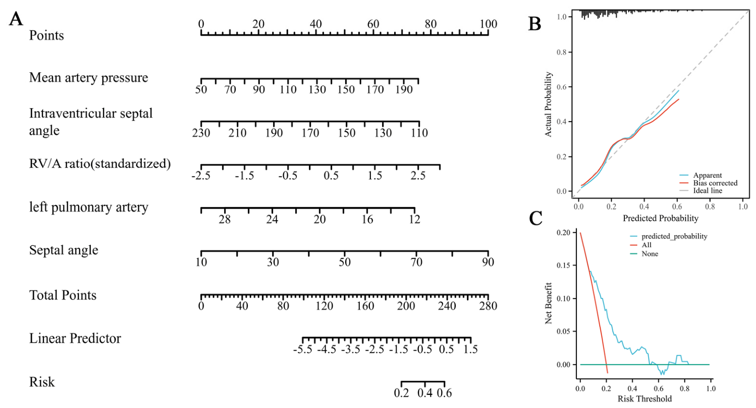
| Septal angle, mean ± SD | 44.05 ± 10.87 |
| Intraventricular septal angle, median (IQR) | 150.51 (142.55, 161.92) |
| Epicardial fat volume (cm3), median (IQR) | 81.60 (52.77, 112.58) |
| Coronary artery calcification | 64.2% (n = 138/215) |
| Mitral valve calcification | 9.3% (n = 20/213) |
| Aortic valve calcification | 8.4% (n = 18/214) |
| History of lung resection surgery | 0.5% (n = 1/215) |
| Variables | Unsuccessful Recanalization (USR) (n = 43/215) | Successful Recanalization (SR) (n = 172/215) | p Value |
|---|---|---|---|
| Demographics | |||
| Age, median (IQR) | 71 (58.75, 74.25) | 69 (61, 77) | 0.335 |
| Gender (male/total) | 60.5% (n = 26/43) | 64.5% (n = 111/172) | 0.62 |
| Clinical variables | |||
| Current drinking | 37.2% (n = 16/43) | 34.9% (n = 60/172) | 0.775 |
| Current smoking | 30.2% (n = 13/43) | 36.6% (n = 63/172) | 0.433 |
| Hypertension | 67.4% (n = 29/43) | 64.5% (n = 111/172) | 0.721 |
| Diabetes mellitus | 11.6% (n = 5/43) | 20.3% (n = 35/172) | 0.189 |
| History of cardiac surgery | 14% (n = 6/43) | 9.9% (n = 17/172) | 0.628 |
| Previous stroke | 4.7% (n = 2/43) | 9.9% (n = 17/172) | 0.429 |
| Use of antiplatelet drugs | 9.3% (n = 4/43) | 4.1% (n = 7/171) | 0.319 |
| Use of anticoagulation drugs | 7% (n = 3/43) | 7.6% (n = 13/172) | 1 |
| Lengths-of-stay in hospital, median (IQR) | 16 (10, 25) | 15 (10, 28) | 0.642 |
| Coexisting disease | |||
| Atrial fibrillation | 27.9% (n = 12/43) | 24.6% (n = 42/172) | 0.652 |
| Coronary artery disease | 4.7% (n = 2/43) | 10.5% (n = 18/172) | 0.379 |
| Pericardial effusion | 9.3% (n = 4/43) | 19.8% (n = 34/172) | 0.108 |
| Pleural effusion | 46.5% (n = 20/43) | 58.7% (n = 101/172) | 0.149 |
| Emphysema | 11.6% (n = 5/43) | 8.7% (n = 15/172) | 0.769 |
| Lung cancer | 0% (n = 0/43) | 1.2% (n = 2/172) | 1 |
| Laboratory index | |||
| White blood cell counts, median (IQR) | 8.68 (8.14, 10.46) | 8.21 (6.33, 10.33) | 0.219 |
| Red blood cell counts, median (IQR) | 4.38 (4.22, 4.84) | 4.53 (4.21, 4.91) | 0.75 |
| Platelet counts, median (IQR) | 227.5 (181, 299) | 204 (164, 251) | 0.156 |
| Random glucose, median (IQR) | 6.7 (6.05, 8.3) | 7 (6, 9.2) | 0.158 |
| Prothrombin time, median (IQR) | 13.3 (12.75, 13.93) | 13.1 (12.3, 13.8) | 0.502 |
| Prothrombin activity, median (IQR) | 95.5 (86.5, 105.25) | 100 (90, 113) | 0.54 |
| International normalized ratio, median (IQR) | 1.03 (0.97, 1.1) | 1 (0.93, 1.07) | 0.519 |
| Fibrinogen, median (IQR) | 3.12 (2.45, 3.51) | 3.21 (2.73, 3.78) | 0.916 |
| Activated partial thromboplastin time, median (IQR) | 33.2 (30.85, 36.28) | 33.9 (30.2, 36) | 0.212 |
| APTT ratio, median (IQR) | 0.92 (0.86, 1.01) | 0.93 (0.84, 1) | 0.188 |
| Thrombin time, median (IQR) | 17.05 (16.13, 17.8) | 16.7 (15.7, 17.7) | 0.321 |
| Thrombin time ratio, median (IQR) | 1.01 (0.95, 1.04) | 0.98 (0.92, 1.04) | 0.345 |
| D-dimer, median (IQR) | 0.61 (0.45, 1.75) | 0.71 (0.32, 1.08) | 0.388 |
| Mean arterial pressure, mean ± SD | 105.86 ± 24.1 | 96.85 ± 17.73 | 0.006 * |
| Temperature, median (IQR) | 37.6 (37.15, 38) | 37.4 (37.2, 37.7) | 0.417 |
| Heart rate, median (IQR) | 84.64 (70.1, 121.54) | 75.21 (66.45, 85.31) | 0.217 |
| Abnormal electrocardiogram | 95% (n = 38/40) | 89% (n = 146/164) | 0.399 |
| Admission NIHSS score, median (IQR) | 23 (14.25, 26.25) | 13 (8, 19) | 0.186 |
| Admission GCS score, median (IQR) | 7 (6.75, 11.25) | 10 (7, 12) | 0.312 |
| Chest imaging variables | |||
| Maximum left ventricular diameter, median (IQR) | 64.89 (63.66, 67.98) | 67.97 (62.73, 75.53) | 0.392 |
| Maximum right ventricular diameter, mean ± SD | 41.19 ± 7.18 | 40.08 ± 6.31 | 0.292 |
| Maximum diameter of the left atrium, mean ± SD | 45.76 ± 8.83 | 47.16 ± 9.86 | 0.395 |
| Maximum diameter of the right atrium, mean ± SD | 51 ± 10 | 53.12 ± 8.57 | 0.163 |
| Maximum diameter of ascending aorta, mean ± SD | 37.34 ± 4.23 | 36.48 ± 4.28 | 0.222 |
| Calcification of the aortic arch | 79.1% (n = 34/43) | 80.2% (n = 138/172) | 0.865 |
| Main pulmonary artery diameter, median (IQR) | 30.83 (28, 32.94) | 30.46 (28.23, 33.17) | 0.050 * |
| Right pulmonary artery diameter, median (IQR) | 22.59 (19.98, 25.9) | 23.01 (20.64, 24.51) | 0.082 |
| Left pulmonary artery diameter, median (IQR) | 20.43 (18.51, 22.03) | 21.3 (19.76, 23.52) | 0.008 * |
| Superior vena cava diameter, mean ± SD | 21.03 ± 2.81 | 21.98 ± 3.56 | 0.106 |
| Inferior vena cava diameter, median (IQR) | 24.49 (22.74, 28.1) | 24.24 (22.34, 27.96) | 0.774 |
| RV/A, mean ± SD | 0.82 ± 0.15 | 0.77 ± 0.15 | 0.028 * |
| LV/A, median (IQR) | 1.51 (1.29, 1.69) | 1.51 (1.33, 1.76) | 0.947 |
| RV/LV, mean ± SD | 0.63 ± 0.12 | 0.60 ± 0.10 | 0.173 |
| RA/LA, median (IQR) | 1.1 (0.99, 1.22) | 1.17 (1.02, 1.3) | 0.383 |
| MPA/AA, mean ± SD | 0.78 ± 0.14 | 0.84 ± 0.13 | 0.009 * |
| Septal angle, mean ± SD | 46.56 ± 11.67 | 43.43 ± 10.63 | 0.09 |
| Intraventricular septal angle, median (IQR) | 147.75 (137.79, 160.75) | 145.02 (139.22, 159.01) | 0.019 * |
| Epicardial fat volume (cm3), median (IQR) | 66.57 (40.98, 128.58) | 67 (41.16, 100.8) | 0.555 |
| Coronary artery calcification | 62.8% (n = 27/43) | 64.5% (n = 111/172) | 0.831 |
| Mitral valve calcification | 4.8% (n = 2/43) | 10.5% (n = 18/172) | 0.394 |
| Aortic valve calcification | 7.1% (n = 3/43) | 8.7% (n = 15/172) | 0.984 |
| History of lung resection surgery | 0% (n = 0/43) | 0.6% (n = 1/172) | 1 |
Disclaimer/Publisher’s Note: The statements, opinions and data contained in all publications are solely those of the individual author(s) and contributor(s) and not of MDPI and/or the editor(s). MDPI and/or the editor(s) disclaim responsibility for any injury to people or property resulting from any ideas, methods, instructions or products referred to in the content. |
© 2026 by the authors. Licensee MDPI, Basel, Switzerland. This article is an open access article distributed under the terms and conditions of the Creative Commons Attribution (CC BY) license.
Share and Cite
Xia, W.; Huang, Y.; Chen, Q.; Wang, X.; Yan, Z.; Zhang, W. Relationship Between Routine Preoperative Chest CT-Based Cardiac Parameters and Recanalization After Mechanical Thrombectomy in Patients with Acute Ischemic Stroke. J. Clin. Med. 2026, 15, 4446. https://doi.org/10.3390/jcm15124446
Xia W, Huang Y, Chen Q, Wang X, Yan Z, Zhang W. Relationship Between Routine Preoperative Chest CT-Based Cardiac Parameters and Recanalization After Mechanical Thrombectomy in Patients with Acute Ischemic Stroke. Journal of Clinical Medicine. 2026; 15(12):4446. https://doi.org/10.3390/jcm15124446
Chicago/Turabian StyleXia, Weizhi, Yingbao Huang, Qi Chen, Xue Wang, Zhihan Yan, and Wenru Zhang. 2026. "Relationship Between Routine Preoperative Chest CT-Based Cardiac Parameters and Recanalization After Mechanical Thrombectomy in Patients with Acute Ischemic Stroke" Journal of Clinical Medicine 15, no. 12: 4446. https://doi.org/10.3390/jcm15124446
APA StyleXia, W., Huang, Y., Chen, Q., Wang, X., Yan, Z., & Zhang, W. (2026). Relationship Between Routine Preoperative Chest CT-Based Cardiac Parameters and Recanalization After Mechanical Thrombectomy in Patients with Acute Ischemic Stroke. Journal of Clinical Medicine, 15(12), 4446. https://doi.org/10.3390/jcm15124446