The Role of Virtual and Augmented Reality in Transsphenoidal Surgical Approaches to the Sellar and Parasellar Area—A Systematic Review
 ,
,  , , ,
, , ,
Abstract
1. Introduction
2. Materials and Methods
- ❖
- Population (P): patients undergoing transsphenoidal pituitary surgery;
- ❖
- Intervention (I): application of virtual or augmented reality (VR/AR), based on imaging diagnostics (CT/MRI);
- ❖
- Comparison (C): standard methods for preoperative planning without VR/AR;
- ❖
- Outcome (O): improved spatial orientation, reduced surgical complications, shortened operative time, and increased educational effect.
AI Statement
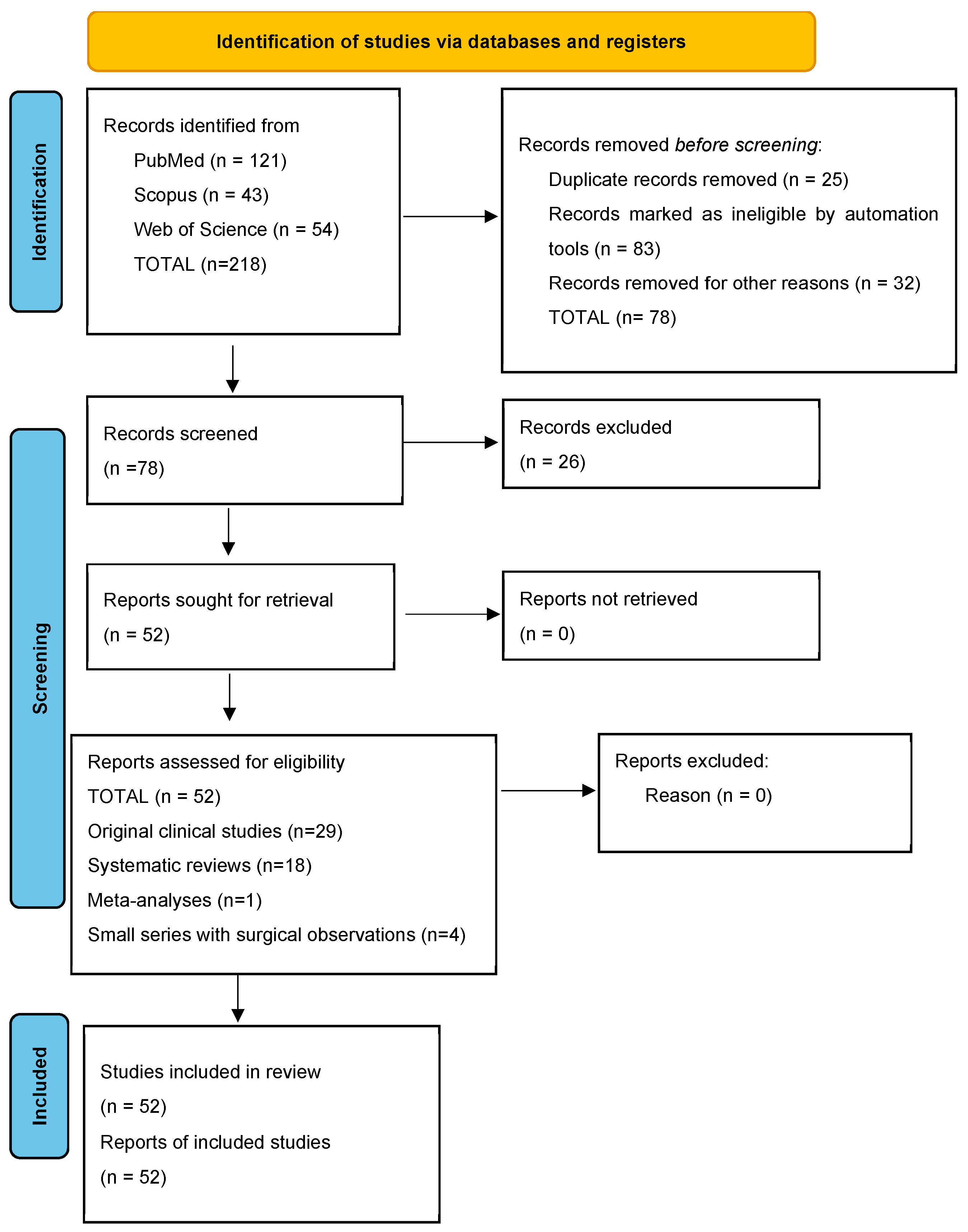
3. Results
- ❖
- VR/AR technological and simulation studies (n = 30);
- ❖
- Morphological and anatomical studies (n = 10);
- ❖
- Clinical and surgical studies (n = 12).
3.1. Effects of Virtual Reality (VR)
3.2. Effects of Augmented Reality (AR)
- ❖
- Reduction in time to reach the sellar region;
- ❖
- More precise identification of critical anatomical structures such as the internal carotid artery and optic nerve;
- ❖
- Reduction in unintended instrument movements, improvement in surgical precision;
- ❖
- Some studies have reported a reduction in operative time by approximately 20–25%, which highlights the clinical relevance of AR technology.
3.3. Subgroup Analysis (VR vs. AR)
3.4. Morphological Factors and Clinical Significance
3.5. Limitations of the Available Data
- ❖
- Limited samples;
- ❖
- Heterogeneity in methodology;
- ❖
- Lack of standardized protocols;
- ❖
- Insufficient number of randomized controlled trials.
4. Discussion
4.1. Study Heterogeneity and Evidence Hierarchy
4.2. Anatomical Context and Implications for VR/AR Integration
4.3. Virtual Reality: Preoperative Planning and Surgical Training
4.4. Augmented Reality: Intraoperative Navigation
4.5. Clinical Translation, Implementation Barriers, and Comparison with Conventional Neuronavigation
4.6. Limitations
4.7. Future Directions
5. Conclusions
Supplementary Materials
Author Contributions
Funding
Institutional Review Board Statement
Informed Consent Statement
Data Availability Statement
Acknowledgments
Conflicts of Interest
Abbreviations
| VR | virtual reality |
| AR | augmented reality |
| CT | computed tomography |
| MRI | magnetic resonance imaging |
| PRISMA | Preferred Reporting Items for Systematic Reviews and Meta-Analyses |
| NOS | Newcastle–Ottawa Scale |
| RoB | Risk of Bias Tool |
| AI | Augmented Learning |
| XR | Extended Reality |
| TORS | Transoral robotic surgery |
| CBCT | Cone Beam Computed Tomography |
| CTA | Computed Tomography Angiography |
| FESS | Functional Endoscopic Sinus Surgery |
| ICA | Internal carotid artery |
| O-arm | Intraoperative 3D Imaging System |
References
- Grunert, R.; Snyderman, C.-H.; Gardner, P.; Busse, M.; Ahner, L.; Kropla, F.; Möbius, R.; Jung, S.; Scholz, S.; Güresir, E.; et al. NextLens—The Next Generation of Surgical Navigation: Proof of Concept of an Augmented Reality System for Surgical Navigation. J. Neurol. Surg. B Skull Base 2023, 85, 363–369. [Google Scholar] [CrossRef]
- Hanson, M.; Li, H.; Geer, E.; Karimi, S.; Tabar, V.; Cohen, M.A. Perioperative management of endoscopic transsphenoidal pituitary surgery. World J. Otorhinolaryngol. Head Neck Surg. 2020, 6, 84–93. [Google Scholar] [CrossRef]
- Fang, C.H.; Friedman, R.; Schild, S.D.; Goldstein, I.M.; Baredes, S.; Liu, J.K.; Eloy, J.A. Purely endoscopic endonasal surgery of the craniovertebral junction: A systematic review. Int. Forum Allergy Rhinol. 2015, 5, 754–760. [Google Scholar] [CrossRef] [PubMed]
- Chauvet, D.; Hans, S. Robot-Assisted Pituitary Surgery. In Neurosurgical Robotics; Marcus, H.J., Payne, C.J., Eds.; Springer: New York, NY, USA, 2021; pp. 145–159. [Google Scholar] [CrossRef]
- Saher, L.; Asif, A.; Mehmood, Z.; Fatima, S.S.; Nizam, R. Virtual Reality in Neurosurgery: Advancing Training, Education, and Surgical Planning. Asian J. Neurosurg. 2025, 21, 26–37. [Google Scholar] [CrossRef] [PubMed]
- Inoue, A.; Ohnishi, T.; Kohno, S.; Harada, H.; Nishikawa, M.; Ozaki, S.; Matsumoto, S.; Ohue, S. Utility of three-dimensional computed tomography for anatomical assistance in endoscopic endonasal transsphenoidal surgery. Neurosurg. Rev. 2015, 38, 559–565. [Google Scholar] [CrossRef]
- Bue, E.L.; Pesaresi, A.; Lacroce, P.; Portonero, I.; Antico, A.; De Marco, R.; Garbossa, D.; Penner, F.; Zenga, F. Anatomical Differences in Sphenoid Sinus During Endoscopic Transsphenoidal Surgery: Comparison Between Nonfunctioning Pituitary Neuroendocrine Tumor (PiTNET) and Growth Hormone-Secreting PiTNET. World Neurosurg. 2024, 190, e701–e706. [Google Scholar] [CrossRef]
- Muslu, M.; Karaca, Ö.; Kökçe, A.; Acer, N. A Computed Tomography-Based Morphometric Assessment of the Foramen Lacerum in a Turkish Population Using the 3D Slicer Method. Medicina 2025, 61, 943. [Google Scholar] [CrossRef]
- Ahmadipour, Y.; Lemonas, E.; Maslehaty, H.; Goericke, S.; Stuck, B.A.; El Hindy, N.; Sure, U.; Mueller, O. Critical analysis of anatomical landmarks within the sphenoid sinus for transsphenoidal surgery. Eur. Arch. Otorhinolaryngol. 2016, 273, 3929–3936. [Google Scholar] [CrossRef] [PubMed]
- Khan, D.Z.; Hanrahan, J.G.; Baldeweg, S.E.; Dorward, N.L.; Stoyanov, D.; Marcus, H.J. Current and Future Advances in Surgical Therapy for Pituitary Adenoma. Endocr. Rev. 2023, 44, 947–959. [Google Scholar] [CrossRef]
- Singh, P.; Hung, K.; Ajmera, D.H.; Yeung, A.W.K.; von Arx, T.; Bornstein, M.M. Morphometric characteristics of the sphenoid sinus and potential influencing factors: A retrospective assessment using cone beam computed tomography (CBCT). Anat. Sci. Int. 2021, 96, 544–555. [Google Scholar] [CrossRef]
- Bopp, M.H.A.; Saß, B.; Pojskić, M.; Corr, F.; Grimm, D.; Kemmling, A.; Nimsky, C. Use of Neuronavigation and Augmented Reality in Transsphenoidal Pituitary Adenoma Surgery. J. Clin. Med. 2022, 11, 5590. [Google Scholar] [CrossRef]
- Shao, X.; Yuan, Q.; Qian, D.; Ye, Z.; Chen, G.; Zhuang, K.L.; Jiang, X.; Jin, Y.; Qiang, D. Virtual reality technology for teaching neurosurgery of skull base tumor. BMC Med. Educ. 2020, 20, 3. [Google Scholar] [CrossRef] [PubMed]
- Santona, G.; Madoglio, A.; Mattavelli, D.; Rigante, M.; Ferrari, M.; Lauretti, L.; Mattogno, P.; Parrilla, C.; De Bonis, P.; Galli, J.; et al. Training models and simulators for endoscopic transsphenoidal surgery: A systematic review. Neurosurg. Rev. 2023, 46, 248. [Google Scholar] [CrossRef]
- Novák, V.; Hrabálek, L.; Valošek, J.; Jablonský, J.; Hoza, J.; Korčáková, I.; Hampl, M.; Stejskal, P.; Hučko, C. The use of an O-arm in endonasal endoscopic operations of the skull base. BMC Surg. 2021, 21, 58. [Google Scholar] [CrossRef] [PubMed]
- Campisi, B.M.; Costanzo, R.; Gulino, V.; Avallone, C.; Noto, M.; Bonosi, L.; Brunasso, L.; Scalia, G.; Iacopino, D.G.; Maugeri, R. The Role of Augmented Reality Neuronavigation in Transsphenoidal Surgery: A Systematic Review. Brain Sci. 2023, 13, 1695. [Google Scholar] [CrossRef] [PubMed]
- Kim, D.H.; Kim, Y.; Park, J.-S.; Kim, S.W. Virtual Reality Simulators for Endoscopic Sinus and Skull Base Surgery: The Present and Future. Clin. Exp. Otorhinolaryngol. 2019, 12, 12–17. [Google Scholar] [CrossRef]
- Jaworek-Troć, J.; Zarzecki, M.; Bonczar, A.; Kaythampillai, L.N.; Rutowicz, B.; Mazur, M.; Urbaniak, J.; Przybycień, W.; Piątek-Koziej, K.; Kuniewicz, M.; et al. Sphenoid bone and its sinus-anatomo-clinical review of the literature including application to FESS. Folia Med. Cracov. 2019, 59, 45–59. [Google Scholar]
- Hudise, J.Y.; Mojiri, M.E.; Shawish, A.M.; Majrashi, K.A.; Ayoub, A.Y.; Alshammakhi, A.M.; Akoor, F.A.; Madkhali, K.A.; Fathi, M.A.; Areeshi, R.A.; et al. The Role of Virtual Reality in Advancing Surgical Training in Otolaryngology: A Systematic Review. Cureus 2024, 16, e71222. [Google Scholar] [CrossRef]
- Mishra, R.; Narayanan, K.; Umana, G.E.; Montemurro, N.; Chaurasia, B.; Deora, H. Virtual Reality in Neurosurgery: Beyond Neurosurgical Planning. Int. J. Environ. Res. Public Health 2022, 19, 1719. [Google Scholar] [CrossRef]
- Newall, N.; Khan, D.Z.; Hanrahan, J.G.; Booker, J.; Borg, A.; Davids, J.; Nicolosi, F.; Sinha, S.; Dorward, N.; Marcus, H.J. High fidelity simulation of the endoscopic transsphenoidal approach: Validation of the UpSurgeOn TNS Box. Front. Surg. 2022, 9, 1049685. [Google Scholar] [CrossRef]
- Scott, H.; Griffin, C.; Coggins, W.; Elberson, B.; Abdeldayem, M.; Virmani, T.; Larson-Prior, L.J.; Petersen, E. Virtual Reality in the Neurosciences: Current Practice and Future Directions. Front. Surg. 2022, 8, 807195. [Google Scholar] [CrossRef]
- Filimonov, A.; Zeiger, J.; Goldrich, D.; Nayak, R.; Govindaraj, S.; Bederson, J.; Shrivastava, R.; Iloreta, A.M.C. Virtual reality surgical planning for endoscopic endonasal approaches to the craniovertebral junction. Am. J. Otolaryngol. 2022, 43, 103219. [Google Scholar] [CrossRef]
- Heredia-Pérez, S.A.; Harada, K.; Padilla-Castañeda, M.A.; Marques-Marinho, M.; Márquez-Flores, J.A.; Mitsuishi, M. Virtual reality simulation of robotic transsphenoidal brain tumor resection: Evaluating dynamic motion scaling in a master-slave system. Int. J. Med. Robot. 2019, 15, e1953. [Google Scholar] [CrossRef]
- Kawamata, T.; Iseki, H.; Shibasaki, T.; Hori, T. Endoscopic augmented reality navigation system for endonasal transsphenoidal surgery to treat pituitary tumors: Technical note. Neurosurgery 2002, 50, 1393–1397. [Google Scholar] [CrossRef]
- Rosseau, G.; Bailes, J.; del Maestro, R.; Cabral, A.; Choudhury, N.; Comas, O.; Debergue, P.; De Luca, G.; Hovdebo, J.; Jiang, D.; et al. The Development of a Virtual Simulator for Training Neurosurgeons to Perform and Perfect Endoscopic Endonasal Transsphenoidal Surgery. Neurosurgery 2013, 73, S85–S93. [Google Scholar] [CrossRef]
- Cho, J.H.; Kim, J.K.; Lee, J.-G.; Yoon, J.-H. Sphenoid sinus pneumatization and its relation to bulging of surrounding neurovascular structures. Ann. Otol. Rhinol. Laryngol. 2010, 119, 646–650. [Google Scholar] [CrossRef]
- Lee, D.; Mennillo, L.; Burrows, E.; Chen, J.-E.; Khan, D.Z.; Starup-Hansen, J.; Stoyanov, D.; Clarkson, M.J.; Marcus, H.J.; Bano, S. 3D reconstruction in endonasal pituitary surgery. Int. J. Comput. Assist. Radiol. Surg. 2025, 20, 1153–1157. [Google Scholar] [CrossRef]
- Jaworek-Troć, J.; Walocha, J.A.; Skrzat, J.; Iwanaga, J.; Tubbs, R.S.; Mazur, M.; Lipski, M.; Curlej-Wądrzyk, A.; Gładysz, T.; Chrzan, R.; et al. A computed tomography comprehensive evaluation of the ostium of the sphenoid sinus and its clinical significance. Folia Morphol. 2022, 81, 694–700. [Google Scholar] [CrossRef] [PubMed]
- Wu, P.; Zeng, H.; Tan, X.; Ouyang, J.; Zhong, S. A Novel Method to Locate Intracranial Anatomic Landmarks of Sella Using Three-Dimensional Computed Tomography Reconstruction Images for Endoscopic Endonasal Transsphenoidal Surgery. J. Craniofac. Surg. 2015, 26, 2184–2186. [Google Scholar] [CrossRef]
- Skvortsova, A.; Stiel, S.; Afshar, K.; Röwer, H.A.; Bausewein, C.; Hartigan, I.; Saab, M.M.; Pereira, S.M.; Hernández-Marrero, P.; Hrdlička, J.; et al. Acceptability of virtual reality for training health professions students in serious illness communication: A cross-sectional study with educators. PEC Innov. 2025, 7, 100411. [Google Scholar] [CrossRef] [PubMed]
- Park, S.-H.; Hwang, J.-H. Arrested Pneumatization of the Sphenoid Sinus in the Skull Base. Brain Tumor Res. Treat. 2021, 9, 40–43. [Google Scholar] [CrossRef] [PubMed]
- Kenig, N.; Echeverria, J.M.; Vives, A.M. Artificial Intelligence in Surgery: A Systematic Review of Use and Validation. J. Clin. Med. 2024, 13, 7108. [Google Scholar] [CrossRef]
- Higa, J.; Nkatha, S.; Herrera, R.R.; Marcus, H.; Yoo, S.; Blandford, A.; Opie, J. Augmented reality for endoscopic transsphenoidal surgery: Evaluating design factors with neurosurgeons. Int. J. Comput. Assist. Radiol. Surg. 2025, 20, 131–136. [Google Scholar] [CrossRef]
- Meola, A.; Cutolo, F.; Carbone, M.; Cagnazzo, F.; Ferrari, M.; Ferrari, V. Augmented reality in neurosurgery: A systematic review. Neurosurg. Rev. 2017, 40, 537–548. [Google Scholar] [CrossRef]
- Carl, B.; Bopp, M.; Voellger, B.; Saß, B.; Nimsky, C. Augmented Reality in Transsphenoidal Surgery. World Neurosurg. 2019, 125, e873–e883. [Google Scholar] [CrossRef]
- Begagić, E.; Bečulić, H.; Pugonja, R.; Memić, Z.; Balogun, S.; Džidić-Krivić, A.; Milanović, E.; Salković, N.; Nuhović, A.; Skomorac, R.; et al. Augmented Reality Integration in Skull Base Neurosurgery: A Systematic Review. Medicina 2024, 60, 335. [Google Scholar] [CrossRef]
- Enkaoua, A.; Ramalhinho, J.; Islam, M.; Marcus, H.J.; Clarkson, M.J. Augmented reality system for endoscopic pituitary surgery with automatic registration. Int. J. Comput. Assist. Radiol. Surg. 2025, 20, 1301–1305. [Google Scholar] [CrossRef] [PubMed]
- Sung, H.; Kim, M.; Park, J.; Shin, N.; Han, Y. Effectiveness of Virtual Reality in Healthcare Education: Systematic Review and Meta-Analysis. Sustainability 2024, 16, 8520. [Google Scholar] [CrossRef]
- Goto, Y.; Kawaguchi, A.; Inoue, Y.; Nakamura, Y.; Oyama, Y.; Tomioka, A.; Higuchi, F.; Uno, T.; Shojima, M.; Kin, T.; et al. Efficacy of a Novel Augmented Reality Navigation System Using 3D Computer Graphic Modeling in Endoscopic Transsphenoidal Surgery for Sellar and Parasellar Tumors. Cancers 2023, 15, 2148. [Google Scholar] [CrossRef]
- Yang, J.; Wen, G.; Tang, C.; Zhong, C.; Zhu, J.; Cong, Z.; Ma, C. Evaluation of Surgical Freedom for One-and-a-Half Nostril, Mononostril, and Binostril Endoscopic Endonasal Transsphenoidal Approaches. J. Neurol. Surg. B Skull Base 2020, 82, 383–391. [Google Scholar] [CrossRef] [PubMed]
- Sanker, V.; Nordin, E.O.R.; Badary, A.; Eberle, A.T.; Ramanan, S.; Jensen, K.N.; Miller, M.E.; Catapano, J.S.; Huguenard, A.L.; Rahmani, R.; et al. Extended reality in skull base surgery: A systematic literature review. Front. Surg. 2025, 12, 1642033. [Google Scholar] [CrossRef]
- Munawar, A.; Li, Z.; Nagururu, N.; Trakimas, D.; Kazanzides, P.; Taylor, R.H.; Creighton, F.X. Fully immersive virtual reality for skull-base surgery: Surgical training and beyond. Int. J. Comput. Assist. Radiol. Surg. 2024, 19, 51–59. [Google Scholar] [CrossRef]
- Wada, K.; Moriyama, H.; Edamatsu, H.; Hama, T.; Arai, C.; Kojima, H.; Otori, N.; Yanagi, K. Identification of Onodi cell and new classification of sphenoid sinus for endoscopic sinus surgery. Int. Forum Allergy Rhinol. 2015, 5, 1068–1076. [Google Scholar] [CrossRef]
- Tortolero, L.B.; Hajtovic, S.A.; Gautreaux, J.; Lebowitz, R.; Placantonakis, D.G. Intraoperative Augmented Reality Visualization in Endoscopic Transsphenoidal Tumor Resection Using the Endoscopic Surgical Navigation Advanced Platform (EndoSNAP): A Technical Note and Retrospective Cohort Study. Cureus 2025, 17, e79714. [Google Scholar] [CrossRef]
- Mao, Z.; Das, A.; Islam, M.; Khan, D.Z.; Williams, S.C.; Hanrahan, J.G.; Borg, A.; Dorward, N.L.; Clarkson, M.J.; Stoyanov, D.; et al. PitSurgRT: Real-time localization of critical anatomical structures in endoscopic pituitary surgery. Int. J. Comput. Assist. Radiol. Surg. 2024, 19, 1053–1060. [Google Scholar] [CrossRef] [PubMed]
- Arrambide-Garza, F.J.; Zarate-Garza, P.P.; Aguilar-Morales, K.; Villarreal-Del-Bosque, I.S.; Quiroga-Garza, A.; Gómez-Sánchez, A.; Pinales-Razo, R.; Elizondo-Omaña, R.E.; Guzmán-Lopez, S. Safety window for the transsphenoidal approach for pituitary tumours: A computed tomographic angiography study. Folia Morphol. 2023, 82, 17–23. [Google Scholar] [CrossRef]
- Baker, K.E.; Robbins, A.C.; Wasson, R.G.; McCandless, M.G.; Lirette, S.T.; Kimball, R.J.; Washington, C.W.; Luzardo, G.D.; Stringer, S.P.; Zachariah, M.A. Side-firing intraoperative ultrasound applied to resection of pituitary macroadenomas and giant adenomas: A single-center retrospective case-control study. Front. Oncol. 2022, 12, 1043697. [Google Scholar] [CrossRef] [PubMed]
- Baloiu, A.I.; Filipoiu, F.; Toader, C.; Covache-Busuioc, R.-A.; Munteanu, O.; Serban, M. Sphenoid sinus hyperpneumatization: Anatomical variants, molecular blueprints, and AI-augmented roadmaps for skull base surgery. Front. Endocrinol. 2025, 16, 1634206. [Google Scholar] [CrossRef]
- Zhang, J.; Shi, Z.; Kang, Y.; Wu, B.; Liu, X.; Zhu, H. The application of augmented reality technology in endoscopic pituitary adenoma surgery via nasal approach. Gland Surg. 2025, 14, 1318–1335. [Google Scholar] [CrossRef]
- Nillahoot, N.; Pillai, B.M.; Chumnanvej, S.; Suthakorn, J. The development of a virtual simulator for a novel design surgical tool in endoscopic endonasal transsphenoidal surgery. Bull. Electr. Eng. Inform. 2021, 10, 1368–1379. [Google Scholar] [CrossRef]
- Thavarajasingam, S.G.; Vardanyan, R.; Rad, A.A.; Thavarajasingam, A.; Khachikyan, A.; Mendoza, N.; Nair, R.; Vajkoczy, P. The use of augmented reality in transsphenoidal surgery: A systematic review. Br. J. Neurosurg. 2022, 36, 457–471. [Google Scholar] [CrossRef]
- Pušnik, L.; Cuzak, M.M.; Umek, N.; Jerman, A. Simulation of suprazygomatic and infrazygomatic approaches for maxillary nerve block in children. J. Anesth. 2026. [Google Scholar] [CrossRef] [PubMed]
- Page, M.J.; McKenzie, J.E.; Bossuyt, P.M.; Boutron, I.; Hoffmann, T.C.; Mulrow, C.D.; Shamseer, L.; Tetzlaff, J.M.; Akl, E.A.; Brennan, S.E.; et al. The PRISMA 2020 statement: An updated guideline for reporting systematic reviews. BMJ 2021, 372, n71. [Google Scholar] [CrossRef] [PubMed]


| Database | Hits (n) | Search String |
|---|---|---|
| PubMed/MEDLINE | 121 | “transsphenoidal” [MeSH Terms] OR “transsphenoidal surgery” [Title/Abstract] OR “endonasal approach” [Title/Abstract] OR “endonasal transsphenoidal” [Title/Abstract] AND “virtual reality” [MeSH Terms] OR “augmented reality” [MeSH Terms] OR “virtual reality” [Title/Abstract] OR “augmented reality” [Title/Abstract] OR “extended reality” [Title/Abstract] OR “mixed reality” [Title/Abstract] OR “surgical simulation” [Title/Abstract] OR “surgical simulator” [Title/Abstract] OR “3D reconstruction” [Title/Abstract] AND “pituitary” [MeSH Terms] OR “sella turcica” [MeSH Terms] OR “sellar region” [Title/Abstract] OR “parasellar” [Title/Abstract] OR “skull base” [Title/Abstract] OR “sphenoid sinus” [MeSH Terms] OR “sphenoid sinus” [Title/Abstract] OR “pituitary surgery” [Title/Abstract] AND “1 January 2015” [Date—Publication]: “31 December 2025” [Date—Publication] |
| Scopus | 43 | TITLE-ABS-KEY “transsphenoidal” OR “endonasal approach” OR “transsphenoidal surgery” OR “endonasal transsphenoidal” AND “virtual reality” OR “augmented reality” OR “extended reality” OR “mixed reality” OR “surgical simulation” OR “surgical simulator” OR “3D reconstruction” AND (“pituitary” OR “sellar” OR “parasellar” OR “skull base” OR “sphenoid sinus” OR “pituitary surgery” AND PUBYEAR > 2014 AND PUBYEAR < 2026 |
| Web of Science | 54 | TS = “transsphenoidal” OR “endonasal approach” OR “transsphenoidal surgery” OR “endonasal transsphenoidal” AND “virtual reality” OR “augmented reality” OR “extended reality” OR “mixed reality” OR “surgical simulation” OR “surgical simulator” OR “3D reconstruction” AND “pituitary” OR “sellar” OR “parasellar” OR “skull base” OR “sphenoid sinus” OR “pituitary surgery” AND PY = (2015–2025) |
| No. | Author (Year) | Study Design | Technology | Clinical Application | N | LoE | Primary Outcome Metric | Key Quantitative Result/Main Finding |
|---|---|---|---|---|---|---|---|---|
| 1 | Lee et al. (2025) [28] | Clinical | VR/3D | Preoperative planning | 4 | 3D landmark visualization accuracy | Improved spatial orientation (qualitative) | |
| 2 | Jaworek-Troć et al. (2022) [29] | Morphometric | CT | Anatomy | 4 | Sphenoid ostium dimensions (mm) | Clinically significant anatomical variants identified | |
| 3 | Muslu et al. (2025) [8] | Morphometric | CT | Anatomy | 4 | Foramen lacerum dimensions (mm) | Population-level morphometric differences documented | |
| 4 | Wu et al. (2015) [30] | Technical note | 3D CT | Preoperative planning | 4/5 | Intracranial landmark localization | Accurate sellar localization confirmed | |
| 5 | Skvortsova et al. (2025) [31] | Cross-sectional | VR | Training | 4 | VR acceptability score (Likert) | High acceptability among health educators | |
| 6 | Bue et al. (2024) [7] | Retrospective clinical | CT | Anatomy | 3 | Sinus morphology (CT parameters) | Significant differences between PiTNET subtypes | |
| 7 | Park & Hwang (2021) [32] | Narrative overview | CT | Anatomy | 5 | N/A (descriptive) | Pneumatization patterns described | |
| 8 | Kenig et al. (2024) [33] | Systematic review | AI | General | studies | 1a | AI validation metrics (accuracy, sensitivity) | AI validated in surgical applications |
| 9 | Higa et al. (2025) [34] | Experimental | AR | Intraoperative | N = 11 (2 + 4 + 3 + 4 participants across phases) | 4 | Design factor ratings (qualitative) | AR design factors evaluated by neurosurgeons |
| 10 | Meola et al. (2017) [35] | Systematic review | AR | Navigation | studies | 1a | AR navigation accuracy (qualitative) | AR improves accuracy; evidence heterogeneous |
| 11 | Carl et al. (2019) [36] | Clinical | AR | Intraoperative | N = 47 patients (from series of 288) | 3 | Landmark ID accuracy; OR time | AR reliable tool for complicated TSS |
| 12 | Begagić et al. (2024) [37] | Systematic review | AR | Navigation | 19 studies | 1a | Navigation accuracy; complication rate | AR improves visualization and orientation |
| 13 | Enkaoua et al. (2025) [38] | Technological | AR | Intraoperative | 5 | Registration accuracy (mm) | Automatic AR registration feasible | |
| 14 | Ahmadipour et al. (2016) [9] | Clinical | CT | Anatomy | 4 | Anatomical landmark identification rate (%) | Critical landmarks characterized | |
| 15 | Khan et al. (2023) [10] | Narrative review | — | Clinical | 5 | N/A (narrative) | Current advances in pituitary surgery reviewed | |
| 16 | Sung et al. (2024) [39] | Meta-analysis | VR | Training | 45 RCTs | 1a | Pooled skill performance outcomes | High VR training effectiveness confirmed |
| 17 | Goto et al. (2023) [40] | Clinical | AR | Navigation | N = 15 patients | 3 | OR time (min); efficacy score (5-point scale) | Mean efficacy score 4.7/5; ~20–25% OR time reduction |
| 18 | Kawamata et al. (2002) [25] | Technical note | AR | Navigation | 5 | Navigation feasibility | First endonasal AR navigation system described | |
| 19 | Yang et al. (2021) [41] | Clinical | Endoscopy | Surgery | 4 | Surgical freedom (degrees) | Approach freedom quantified across nostril configs | |
| 20 | Sanker et al. (2025) [42] | Systematic review | XR | Combined | studies | 1a | XR clinical and training outcomes | Extended reality applications in skull base reviewed |
| 21 | Munawar et al. (2024) [43] | Experimental | VR | Training | 4 | Task completion accuracy (%); error rate | Improved technical skills with VR immersion | |
| 22 | Newall et al. (2022) [21] | Validation study | VR | Simulation | N = 15 participants (10 novice, 5 expert) | 4 | Face, content and construct validity scores | High fidelity VR simulator validated (mOSAT) |
| 23 | Wada et al. (2015) [44] | Clinical | CT | Anatomy | 4 | Onodi cell prevalence (%); classification | Novel sphenoid sinus classification proposed | |
| 24 | Chauvet et al. (2021) [4] | Book chapter | TORS | Neurosurgery | 5 | Feasibility (narrative) | TORS role in pituitary surgery defined | |
| 25 | Tortolero et al. (2025) [45] | Retrospective clinical | AR | Intraoperative | N = 18 patients | 3 | EOR (%); complication rate; OR time | Mean EOR 93.6%; AR visualization feasible (EndoSNAP) |
| 26 | Singh et al. (2021) [11] | Retrospective | CBCT | Anatomy | 4 | Sinus morphometric parameters (mm) | Morphometric characteristics described | |
| 27 | Grunert et al. (2023) [1] | Technological | AR | Navigation | 5 | Proof-of-concept feasibility | NextLens AR navigation system described | |
| 28 | Hanson et al. (2020) [2] | Clinical | — | Management | 5 | Perioperative management outcomes | Perioperative protocol for pituitary surgery described | |
| 29 | Mao et al. (2024) [46] | Technological | AI + AR | Intraoperative | 5 | Real-time localization accuracy | PitSurgRT real-time AI system described | |
| 30 | Fang et al. (2015) [3] | Systematic review | Endoscopy | Surgery | studies | 1a | Surgical outcomes (endoscopic approach) | Purely endoscopic craniovertebral approach effective |
| 31 | Arrambide-Garza et al. (2023) [47] | Clinical | CTA | Anatomy | 4 | Safety window dimensions (mm) | Safety parameters for transsphenoidal approach quantified | |
| 32 | Baker et al. (2022) [48] | Retrospective case–control | Ultrasound | Surgery | N = 27 (15 IOUS + 12 control) | 3 | Extent of resection (%) | IOUS improves macroadenoma resection |
| 33 | Jaworek-Troć et al. (2019) [18] | Narrative review | — | Anatomy | 5 | N/A (narrative) | Sphenoid anatomy for FESS reviewed | |
| 34 | Baloiu et al. (2025) [49] | Narrative review | AI | Anatomy | 5 | N/A (narrative) | Hyperpneumatization and AI roles reviewed | |
| 35 | Cho et al. (2010) [27] | Morphometric | CT | Anatomy | 4 | Pneumatization rates (%); ICA contact freq. (%) | Septa contact ICA bony shell in ~28% of cases | |
| 36 | Zhang et al. (2025) [50] | Clinical | AR | Navigation | N = 5 patients (3M/2F) | 3 | Target registration error (mm); spatial accuracy | TRE 2.23 ± 0.57 mm; AR enhanced spatial orientation |
| 37 | Nillahoot et al. (2021) [51] | Technological | VR | Simulation | 5 | Simulator performance metrics | Novel endonasal VR simulator developed and tested | |
| 38 | Rosseau et al. (2013) [26] | Educational | VR | Training | 5 | Skill acquisition metrics | Foundational VR training simulator developed | |
| 39 | Campisi et al. (2023) [16] | Systematic review | AR | Navigation | studies | 1a | Complication rate; navigation accuracy | AR associated with reduced intraoperative complications |
| 40 | Hudise et al. (2024) [19] | Systematic review | VR | Training | studies | 1a | Training outcomes; learnability | High VR efficacy in otolaryngology surgical training |
| 41 | Novák et al. (2021) [15] | Clinical | O-arm | Navigation | N = 6 patients | 3 | Navigation accuracy (mm deviation) | O-arm intraoperative imaging feasible and accurate in TSS |
| 42 | Thavarajasingam et al. (2022) [52] | Systematic review | AR | Clinical | studies | 1a | AR clinical outcomes vs. standard navigation | AR outcomes comparable to conventional neuronavigation |
| 43 | Santona et al. (2023) [14] | Systematic review | VR | Training | studies | 1a | Simulator validity; training outcomes | VR simulators effective for transsphenoidal training |
| 44 | Bopp et al. (2022) [12] | Clinical | AR | Navigation | N = 165 patients (84 AR/81 control) | 3 | Intraoperative orientation accuracy | AR straightforwardly integrated; improved orientation |
| 45 | Inoue et al. (2015) [6] | Clinical | 3D CT | Navigation | 4 | Anatomical identification accuracy (%) | 3D CT utility in endonasal approaches confirmed | |
| 46 | Saher et al. (2025) [5] | Narrative overview | VR | Training | 5 | N/A (narrative) | VR applications in neurosurgery reviewed | |
| 47 | Mishra et al. (2022) [20] | Narrative overview | VR | Planning | 5 | N/A (narrative) | VR potential beyond surgical planning outlined | |
| 48 | Scott et al. (2022) [22] | Narrative overview | VR | General | 5 | N/A (narrative) | VR in neurosciences—future directions | |
| 49 | Heredia-Pérez et al. (2019) [24] | Experimental | VR | Robotics | 4 | Motion scaling accuracy in VR | Dynamic motion scaling in robotic TSS evaluated | |
| 50 | Kim et al. (2019) [17] | Narrative overview | VR | Simulation | 5 | N/A (narrative) | VR simulators for ENT and skull base reviewed | |
| 51 | Filimonov et al. (2022) [23] | Clinical | VR | Planning | N = 5 patients | 4 | Planning feasibility; trajectory accuracy | VR planning for craniovertebral junction in 5 cases |
| 52 | Shao et al. (2020) [13] | Educational | VR | Training | N = 30 students (15 VR/15 control) | 4 | Knowledge test scores; skill improvement | VR training significantly effective vs. traditional |
| Author (Year) | Study Design | Assessment Tool | Key Methodological Limitations | Overall Quality Judgment |
|---|---|---|---|---|
| Lee et al. (2025) [28] | Clinical | NOS | Small N (NR); retrospective design; single centre; no control group; outcome assessment not blinded | Fair (5/9) |
| Bue et al. (2024) [7] | Retrospective clinical | NOS | Single centre; retrospective; no formal sample size calculation; CT assessment not blinded; potential selection bias | Fair (6/9) |
| Carl et al. (2019) [36] | Clinical cohort | NOS | Single surgeon series; retrospective subset (N = 47/288); no randomisation; fiducial vs. iCT registration not stratified; no long-term follow-up | Fair (6/9) |
| Goto et al. (2023) [40] | Prospective clinical | NOS | Small N (n = 15); no control group; self-reported efficacy scale; single centre; potential performance bias | Fair (5/9) |
| Yang et al. (2021) [41] | Clinical/cadaveric | NOS | Mixed clinical and cadaveric design; small sample; outcomes not blinded; limited generalisability | Fair (5/9) |
| Tortolero et al. (2025) [45] | Retrospective cohort | NOS | Small N (n = 18); no control group; single surgeon; retrospective; EOR assessed without blinding; no comparator arm | Poor (4/9) |
| Baker et al. (2022) [48] | Retrospective case-ctrl | NOS | Small N (n = 27); single centre; historical control group; potential selection bias; no blinding of outcome assessors | Fair (5/9) |
| Zhang et al. (2025) [50] | Clinical | NOS | Very small N (n = 5); no control group; proof-of-concept design; limited generalisability; single institution | Poor (3/9) |
| Novák et al. (2021) [15] | Clinical | NOS | Very small N (n = 6); no randomisation; single centre; no blinding; limited statistical power; pilot study | Poor (3/9) |
| Bopp et al. (2022) [12] | Retrospective cohort | NOS | Single surgeon; retrospective allocation to AR/non-AR; no randomisation; potential temporal bias; good sample size (n = 165) | Fair (6/9) |
| Inoue et al. (2015) [6] | Clinical | NOS | Small sample; retrospective; single centre; no formal control group; outcome measures not standardised | Fair (5/9) |
| Filimonov et al. (2022) [23] | Clinical case series | NOS | Very small N (n = 5); no control; descriptive design; limited to single anatomical subtype (CVJ); single institution | Poor (3/9) |
| Jaworek-Troć et al. (2022) [29] | Morphometric | NOS | Retrospective CT; bilateral measurements; adequate sample; no clinical outcome correlation; single rater | Fair (6/9) |
| Muslu et al. (2025) [8] | Morphometric | NOS | Retrospective CT; single population (Turkish); 3D Slicer methodology transparent; no clinical correlation | Fair (6/9) |
| Skvortsova et al. (2025) [31] | Cross-sectional | NOS | Convenience sample (educators); self-reported outcomes; no objective performance measure; limited to acceptability | Fair (5/9) |
| Ahmadipour et al. (2016) [9] | Clinical | NOS | Retrospective; landmark assessment not blinded; single centre; no standardised imaging protocol | Fair (5/9) |
| Newall et al. (2022) [21] | Validation study | NOS | Multi-centre; N = 15; structured validity assessment (mOSAT); expert/novice stratification; limited to simulation setting | Good (7/9) |
| Wada et al. (2015) [44] | Morphometric/clinical | NOS | Retrospective; single centre; no inter-rater reliability reported; CT-based classification lacks prospective validation | Fair (6/9) |
| Singh et al. (2021) [11] | Retrospective CBCT | NOS | CBCT-based; adequate sample; single centre; no clinical outcome correlation; population-specific results | Fair (6/9) |
| Arrambide-Garza et al. (2023) [47] | CTA morphometric | NOS | Retrospective CTA; no clinical outcome correlation; single centre; safety window not validated prospectively | Fair (5/9) |
| Cho et al. (2010) [27] | Morphometric | NOS | Retrospective; outside primary search period; no blinding; single centre; historical data; adequate N | Fair (6/9) |
| Shao et al. (2020) [13] | Educational RCT | RoB 2.0 | Blinding of participants not feasible; outcome assessors not blinded; small N (n = 30); limited to students; single institution | Some concerns |
| Kenig et al. (2024) [33] | Systematic review | AMSTAR 2 | Broad scope (AI in surgery generally); limited PICO specificity; heterogeneous primary studies; risk of bias not formally graded | Moderate |
| Meola et al. (2017) [35] | Systematic review | AMSTAR 2 | Early publication; no protocol registration reported; limited database coverage; heterogeneous included studies; no meta-analysis | Low |
| Begagić et al. (2024) [37] | Systematic review | AMSTAR 2 | PRISMA-compliant; dual screening; 19 included studies; risk of bias discussed narratively; no registration reported | Moderate |
| Sung et al. (2024) [39] | Meta-analysis | AMSTAR 2 | 45 RCTs included; RoB 2.0 applied; comprehensive search; broad scope (healthcare education); not transsphenoidal-specific | Moderate–High |
| Sanker et al. (2025) [42] | Systematic review | AMSTAR 2 | PRISMA-compliant; recent publication; heterogeneous XR technologies; no meta-analysis; no protocol registration reported | Moderate |
| Fang et al. (2015) [3] | Systematic review | AMSTAR 2 | Older search; limited databases; no quality grading of included studies; heterogeneous designs; no protocol registration | Low |
| Campisi et al. (2023) [16] | Systematic review | AMSTAR 2 | PRISMA-compliant; transsphenoidal-specific; risk of bias discussed; no registration; heterogeneous platforms | Moderate |
| Hudise et al. (2024) [19] | Systematic review | AMSTAR 2 | Otolaryngology-focused; PRISMA-compliant; dual screening; heterogeneous training outcomes; no protocol registration | Moderate |
| Thavarajasingam et al. (2022) [52] | Systematic review | AMSTAR 2 | Transsphenoidal-specific; dual screening; heterogeneous AR systems; risk of bias narrative only; no registration reported | Moderate |
| Santona et al. (2023) [14] | Systematic review | AMSTAR 2 | Training-focused; comprehensive search; PRISMA-compliant; heterogeneous simulators; no quantitative synthesis; no registration | Moderate |
| Wu et al. (2015) [30] | Technical note | Descriptive | Proof-of-concept design; small N; no clinical validation; single centre; methodology not reproducible | Low quality |
| Park & Hwang (2021) [32] | Narrative overview | Descriptive | No systematic search; narrative; no quality assessment of cited studies; expert opinion level | Low quality |
| Higa et al. (2025) [34] | Qualitative design study | Descriptive | Small N (n ≈ 11); qualitative phases; no clinical outcomes; context-specific AR prototype | Moderate quality |
| Enkaoua et al. (2025) [38] | Technological | Descriptive | Proof-of-concept; no clinical series; registration accuracy reported in phantom; external validity limited | Low quality |
| Khan et al. (2023) [10] | Narrative review | Descriptive | Narrative; no systematic search; no quality appraisal; expert opinion | Low quality |
| Kawamata et al. (2002) [25] | Technical note | Descriptive | Historical; outside search window; no formal evaluation; small N; no control | Low quality |
| Munawar et al. (2024) [43] | Experimental sim. | Descriptive | Small participant group; simulation only; no clinical transfer assessment; open-source system | Moderate quality |
| Chauvet et al. (2021) [4] | Book chapter | Descriptive | Secondary source; no original data; expert narrative; no methodology reported | Low quality |
| Grunert et al. (2023) [1] | Technological | Descriptive | Proof-of-concept; no clinical series; prototype stage; limited reproducibility data | Low quality |
| Hanson et al. (2020) [2] | Clinical review | Descriptive | Narrative; perioperative management focus; no systematic search; single-centre experience | Low quality |
| Mao et al. (2024) [46] | Technological | Descriptive | Technical feasibility only; video-based dataset; no prospective clinical testing; limited N | Low quality |
| Jaworek-Troć et al. (2019) [18] | Narrative review | Descriptive | Narrative; no systematic search; anatomical focus; no original data | Low quality |
| Baloiu et al. (2025) [49] | Narrative review | Descriptive | Narrative; no systematic search; no quality appraisal; AI roles discussed speculatively | Low quality |
| Nillahoot et al. (2021) [51] | Technological | Descriptive | Simulator development study; no participant validation; limited clinical relevance assessment | Low quality |
| Rosseau et al. (2013) [26] | Educational development | Descriptive | Outside search window; historical; small N; simulator development only; no clinical transfer | Low quality |
| Saher et al. (2025) [5] | Narrative overview | Descriptive | Narrative; no systematic search; no quality appraisal; broad scope | Low quality |
| Mishra et al. (2022) [20] | Narrative overview | Descriptive | Narrative; no systematic search; speculative future directions | Low quality |
| Scott et al. (2022) [22] | Narrative overview | Descriptive | Narrative; broad neurosciences scope; no systematic methodology | Low quality |
| Heredia-Pérez et al. (2019) [24] | Experimental sim. | Descriptive | Robotic simulation; small N; no clinical transfer; dynamic scaling evaluated in VR only | Low quality |
| Kim et al. (2019) [17] | Narrative overview | Descriptive | Narrative; no systematic search; ENT/skull base perspective; expert opinion | Low quality |
| Pušnik L et al. (2026) [53] | Retrospective | NOS | Retrospective CTA dataset; simulation only—no prospective clinical validation; bilateral measurements introduce within-subject dependency; paediatric-specific population limits generalisability; no clinical outcome correlation (safety assessed in silico only) | Fair (6/9) |
| Good/Low risk/High confidence | Fair/Some concerns/Moderate | Poor/High risk/Low confidence | N/A—Technical/Narrative | |
Disclaimer/Publisher’s Note: The statements, opinions and data contained in all publications are solely those of the individual author(s) and contributor(s) and not of MDPI and/or the editor(s). MDPI and/or the editor(s) disclaim responsibility for any injury to people or property resulting from any ideas, methods, instructions or products referred to in the content. |
© 2026 by the authors. Licensee MDPI, Basel, Switzerland. This article is an open access article distributed under the terms and conditions of the Creative Commons Attribution (CC BY) license.
Share and Cite
Bechev, K.; Markov, D.; Aleksiev, V.; Markov, G.; Poryazova, E.; Fasova, A. The Role of Virtual and Augmented Reality in Transsphenoidal Surgical Approaches to the Sellar and Parasellar Area—A Systematic Review. J. Clin. Med. 2026, 15, 4142. https://doi.org/10.3390/jcm15114142
Bechev K, Markov D, Aleksiev V, Markov G, Poryazova E, Fasova A. The Role of Virtual and Augmented Reality in Transsphenoidal Surgical Approaches to the Sellar and Parasellar Area—A Systematic Review. Journal of Clinical Medicine. 2026; 15(11):4142. https://doi.org/10.3390/jcm15114142
Chicago/Turabian StyleBechev, Kristian, Daniel Markov, Vladimir Aleksiev, Galabin Markov, Elena Poryazova, and Antoaneta Fasova. 2026. "The Role of Virtual and Augmented Reality in Transsphenoidal Surgical Approaches to the Sellar and Parasellar Area—A Systematic Review" Journal of Clinical Medicine 15, no. 11: 4142. https://doi.org/10.3390/jcm15114142
APA StyleBechev, K., Markov, D., Aleksiev, V., Markov, G., Poryazova, E., & Fasova, A. (2026). The Role of Virtual and Augmented Reality in Transsphenoidal Surgical Approaches to the Sellar and Parasellar Area—A Systematic Review. Journal of Clinical Medicine, 15(11), 4142. https://doi.org/10.3390/jcm15114142