Review

Evidence on Non-Invasive Respiratory Support During Flexible Bronchoscopy: A Narrative Review

by
María Hidalgo Sánchez
1,
Manel Luján
2,
Sergio Alcolea Batres
1,
Julia Álvarez del Vayo
3,
Pablo Mariscal-Aguilar
1,
Carlos Carpio
1,* and
Rodolfo Álvarez-Sala Walther
1
1
Pulmonology Department, Hospital Universitario La Paz-Cantoblanco-Carlos III, Faculty of Medicine, Universidad Autónoma de Madrid (UAM), IdiPAZ, 28046 Madrid, Spain
2
Pulmonology Department, Hospital Universitari Parc Taulí, 08208 Sabadell, Spain
3
Anaesthesiology and Intensive Care Department, Hospital Universitario La Paz-Cantoblanco-Carlos III, 28046 Madrid, Spain
*
Author to whom correspondence should be addressed.
J. Clin. Med. 2025, 14(18), 6658; https://doi.org/10.3390/jcm14186658
Submission received: 9 August 2025 / Revised: 15 September 2025 / Accepted: 15 September 2025 / Published: 22 September 2025

Abstract

Background: Flexible bronchoscopy (FB) is a widely used diagnostic and therapeutic procedure in patients with pulmonary disease, many of whom are at risk of gas exchange impairment. FB may exacerbate hypoxaemia due to increased airway resistance, alveolar derecruitment, and haemodynamic fluctuations. Objectives: To assess the effectiveness of non-invasive respiratory support strategies in preventing oxygen desaturation and respiratory complications during FB. Methods: A systematic review and meta-analysis were conducted using PubMed and Cochrane databases, covering studies from 2000 to 2024. Inclusion criteria focused on adult patients undergoing FB with any form of non-invasive oxygen support. Twelve high-quality studies were selected, including randomised trials and prospective cohorts. Results: High-flow therapy (HFT) was more effective than conventional oxygen therapy (COT) in maintaining oxygenation and reducing procedure interruptions, especially in patients with moderate hypoxaemia or risk factors such as obesity and obstructive sleep apnoea. Continuous positive airway pressure (CPAP) and non-invasive ventilation (NIV) offered superior oxygenation and ventilatory support in patients with more severe respiratory or cardiac compromise. Conclusions: Non-invasive respiratory support should be individualised based on patient risk and procedural complexity. HFT benefits mild-to-moderate cases, while CPAP or NIV is preferable in more severe conditions. Further multicentre randomised trials are needed to establish formal guidelines.

1. Introduction

FB is a diagnostic and/or therapeutic procedure frequently performed in patients with pulmonary diseases. Most individuals undergoing this procedure have underlying conditions that impair gas exchange, including pneumonia, interstitial lung disease, or lung cancer, which may result in the development or exacerbation of respiratory failure.
During the procedure, the partial pressure of oxygen in arterial blood (PaO2) may decrease by more than 10–20 mmHg, thereby elevating the risk of respiratory insufficiency. Consequently, some form of respiratory support is often required to prevent desaturation episodes. The bronchoscope occupies approximately 10% of the tracheal cross-sectional area and 15% of the cricoid ring. This increases airway resistance, elevating the work of breathing and potentially causing dynamic hyperinflation with an augmented functional residual capacity. When suction is applied, airway and alveolar pressures can fall to zero or even become negative, thereby reducing end-expiratory lung volume, promoting alveolar derecruitment and atelectasis, and ultimately impairing gas exchange [1,2].
These effects are even more pronounced during flexible bronchoscopy with bronchoalveolar lavage (BAL), as aliquots of sterile saline are instilled and only partially retrieved, leaving a substantial volume within the alveoli. This significantly reduces the end-expiratory volume of the affected lung parenchyma below the functional residual capacity [1,2], which may precipitate alveolar collapse and ventilation-perfusion mismatch [3,4]. Although such alterations are typically reversible following bronchoscopy, recovery may require anywhere from several minutes to several hours in patients with severe pulmonary disease.
Bronchoscopy also produces indirect haemodynamic effects. The increase in airway resistance and respiratory effort modifies intrathoracic pressures, which may compromise venous return and afterload, thereby reducing cardiac output. However, sympathetic activation typically results in an approximate 50% rise in cardiac output, which returns to baseline within 15 min following the procedure [5]. Electrocardiographic abnormalities have been reported in up to 21% of patients aged over 60 years [6,7].
Consequently, bronchoscopy represents a substantial cardiopulmonary risk in patients with respiratory or cardiac vulnerability, emphasising the need for an individualised oxygenation or respiratory support strategy.

2. Materials and Methods

A comprehensive systematic search was undertaken in the PubMed and Cochrane databases, covering the period from 2000 to 2024. The search employed the following keywords: Bronchoscop and Respiratory Support, Bronchoscop and Oxygen, Bronchoscop and Non Invasive. Only studies published in English and involving adult human participants receiving one or more modalities of oxygen support during flexible bronchoscopy, for either diagnostic or interventional purposes, were considered. No limitations were imposed regarding the type of bronchoscopic procedure or the degree of anaesthetic risk.
The studies that met the inclusion criteria consisted of randomised controlled trials, prospective and retrospective cohort studies, systematic reviews, and meta-analyses published in peer-reviewed, indexed journals between 2000 and 2024 (Figure 1). From these, twelve studies were selected for in-depth analysis based on their superior methodological quality, larger sample sizes, direct comparison of respiratory support modalities, and publication in high-impact medical journals (Table 1).
Exclusion criteria included articles published in languages other than English, case reports or case series lacking control groups, cross-sectional studies, procedures performed without moderate to deep sedation, animal studies, paediatric populations, and studies involving rigid bronchoscopy, upper airway interventions, tracheal pathology, or invasive mechanical ventilation.
Results are presented in a narrative synthesis, with interpretation and discussion of the published evidence.

3. Results

The main findings of the included trials are summarised in Table 1.

3.1. Modalities of Respiratory Support During Flexible Bronchoscopy

During flexible bronchoscopy, several respiratory support modalities are employed. COT delivers low-flow oxygen via nasal cannulae, simple masks, or Venturi masks.
HFT provides heated and humidified air/oxygen mixtures at flows up to 60 L/min, with an inspired oxygen fraction ranging from 21 to 100%. CPAP maintains positive end-expiratory pressure (PEEP) throughout the respiratory cycle via a mask, while NIV delivers ventilatory support through inspiratory and expiratory positive airway pressures (IPAP and EPAP). Currently, no formal consensus or clinical guidelines exist regarding the optimal respiratory support strategy during flexible bronchoscopy.
This study provides a narrative review of the current literature from PubMed and Cochrane, with the aim of outlining the respiratory support modalities most frequently used in clinical practice and offering recommendations adapted to patients’ specific clinical profiles.

3.1.1. Conventional Oxygen Therapy

COT consists of delivering a fixed flow of oxygen, which blends with ambient air to satisfy the patient’s inspiratory flow requirements. Its main advantages include accessibility, low cost, and simplicity of use; however, a key limitation is the variability and unpredictability of the fraction of inspired oxygen (FiO2) actually delivered. This variability can render COT inadequate in cases of severe hypoxaemia and is highly dependent on the patient’s breathing pattern. Nevertheless, COT remains the most frequently employed form of respiratory support in bronchoscopy settings.
When a patient’s peak inspiratory flow exceeds the oxygen flow provided by conventional systems, FiO2 becomes unpredictable [20,21]. Oxygen can be administered via nasal cannulae, simple masks with or without reservoirs, or Venturi masks. Nasal cannulae may be positioned either nasally or orally, with no significant difference in mean peripheral oxygen saturation (SpO2) observed during the procedure between these approaches [22]. Several studies have shown that COT offers advantages over no oxygen supplementation in patients undergoing FB who are at heightened risk of desaturation, including those with baseline SpO2 < 93%, obstructive ventilatory defects, or forced expiratory volume in one second (FEV1) below 1 litre [23]. Additional benefits have been documented in patients with chronic obstructive pulmonary disease and immunosuppression, who face a higher likelihood of requiring intubation within 24 h post-procedure [24]. Furthermore, COT is typically required during BAL or cytological brushing [8,25]. Accordingly, while not universally mandatory, COT is primarily recommended for patients at risk of desaturation—particularly those with baseline SpO2 below 93%—and during procedures involving BAL or brushing [26].
Nevertheless, owing to the rising number of severely ill patients with acute or chronic respiratory failure, advanced pulmonary disease, and cardiac comorbidities, conventional oxygen therapy frequently proves insufficient to maintain adequate oxygenation and prevent hypercapnia.

3.1.2. High-Flow Oxygen Therapy

High-flow therapy (HFT) has been adopted in clinical practice as an effective alternative to COT for respiratory support during bronchoscopy. Its principal advantage lies in providing a more stable FiO2 at flow rates of up to 60 L/min, which can accommodate or exceed the patient’s elevated inspiratory demand during and after the procedure.
Secondly, HFT produces a modest PEEP within the airways, influenced by flow rate, upper airway anatomy, the patient’s nasal or oral breathing pattern, and the cannula-to-nare size ratio. This generates alveolar distension that improves end-expiratory lung volume and oxygenation in patients with diverse forms of acute respiratory failure (ARF) [27,28]. Thirdly, HFT reduces anatomical dead space in the upper airway, thereby enhancing alveolar ventilation. Finally, it decreases the patient’s work of breathing and lowers upper airway resistance.
HFT promotes a CO2 washout from the pharyngeal dead space, which is proportional to the applied flow; each 1 L/min increment in flow corresponds to an approximate 1.8 mL/s increase in clearance within the nasal cavity. This washout effect is also influenced by respiratory rate, becoming more pronounced at lower rates owing to longer expiratory times [29]. Consequently, this mechanism reduces the patient’s respiratory effort compared with COT.
HFT has been effectively utilised to prevent the worsening of ARF during bronchoscopy.
Enhancements in SpO2 following the procedure, along with reductions in mucosal trauma and patient discomfort, have been reported, which are attributed to the delivery of warmed and humidified gas.
Moreover, HFT has been demonstrated to decrease episodes of oxygen desaturation and the requirement for airway interventions, such as jaw-thrust manoeuvres or bag-mask ventilation, in patients at risk of hypoxaemia undergoing flexible bronchoscopy under deep sedation, including individuals with obstructive sleep apnoea (OSA), male sex, obesity, advanced age, or hypertension [9,30].
Several studies have compared HFT with other oxygen delivery methods, particularly COT. In a randomised controlled trial involving lung transplant recipients undergoing flexible bronchoscopy for transbronchial biopsy, procedural interruptions occurred more frequently with low-flow nasal oxygen than with HFT [10]. Compared with Venturi masks, HFT at 60 L/min provided superior oxygenation, whereas no significant difference was observed between HFT and Venturi masks at 40 L/min [31]. Another randomised trial examining HFT during endobronchial ultrasound (EBUS) under conscious sedation in patients without acute respiratory failure found no significant reduction in intraprocedural desaturation, and procedure duration did not affect the outcomes [32].
During BAL, the efficacy and safety of HFT compared with COT have been assessed in patients with ARF. No significant adverse events, including endotracheal intubation within 24 h post-procedure, were reported, nor were there notable differences between groups in transient hypoxaemia, fever, or hypotension. HFT enhanced gas exchange during BAL by preserving end-expiratory lung volume and reducing diaphragmatic workload [33,34].
In a cohort of 40 patients undergoing flexible bronchoscopy via the oral route for suspected pneumonia requiring bronchial aspirate collection, HFT at 40 L/min was associated with smaller decreases in SpO2 and a lower incidence of desaturation compared with standard oxygen therapy [11].
More recent studies and meta-analyses have questioned the superiority of HFT over COT in improving oxygen saturation, minimising desaturation episodes, and preventing procedural interruptions [12]. The advantages of HFT seem most pronounced in patients with lower baseline oxygenation.
No significant effects on hypercapnia or the incidence of endotracheal intubation (ETI) have been observed. During flexible bronchoscopy with BAL, HFT appears more effective than COT in maintaining oxygen saturation in both ambulatory patients and hypoxaemic individuals in intensive care, without influencing ETI rates [35,36,37].
In certain populations, including lung transplant recipients, or during procedures such as EBUS or foreign body removal, HFT also seems more effective than COT in preventing hypoxaemia. Ben-Menachem et al. [13] compared HFT with COT during flexible bronchoscopy for transbronchial biopsy in lung transplant patients, reporting fewer mild-to-moderate desaturation episodes and reduced procedural interruptions with HFT.
In summary, the advantages of HFT over COT are most evident in patients with significant baseline hypoxaemia, lung transplant recipients, and those at heightened risk of hypoxaemia during flexible bronchoscopy under deep sedation—such as individuals with obstructive sleep apnea (OSA), male sex, obesity, advanced age, or hypertension—particularly when BAL or EBUS is performed. In these patients, HFT improves oxygen saturation, reduces desaturation events, minimizes procedural interruptions, and lessens the need for supplementary airway interventions, without affecting hypercapnia or the incidence of ETI.

3.1.3. Continuous Positive Airway Pressure

CPAP delivers a constant positive pressure throughout the respiratory cycle, promoting the recruitment of atelectatic lung regions, decreasing intrapulmonary shunting, and lowering the patient’s inspiratory effort. Its main limitation is the potential for poor tolerance of the interface and the requirement for specialised masks equipped with accessory ports to allow passage of the bronchoscope.
The main effects of CPAP include enhanced oxygenation through the reduction in intrapulmonary shunting and ventilation–perfusion mismatch, alongside a decrease in the work of breathing. CPAP delivered via a face mask has been shown to effectively recruit alveoli and improve gas exchange in hypoxaemic patients. In those with heart failure, additional benefits have been described, including redistribution of pulmonary oedema, improved left ventricular performance, and reduced respiratory effort.
The application of CPAP via a face mask during bronchoscopy has been assessed in patients with hypoxaemic acute respiratory failure (PaO2/FiO2 < 300 mmHg) by Maitre et al. [14]. Compared with conventional oxygen therapy, CPAP maintained higher SpO2 levels both during and after the procedure, and a smaller proportion of patients required additional respiratory support within six hours post-procedure. During BAL performed as part of flexible bronchoscopy, patients receiving oxygen therapy alone experienced a more pronounced decline in PaO2, highlighting the adverse impact of BAL on gas exchange in hypoxaemic individuals. By contrast, no significant reduction in PaO2 was observed in the CPAP group, supporting its potential benefit when BAL is indicated. Consequently, CPAP appears particularly advantageous for patients with moderate to severe hypoxaemia undergoing diagnostic procedures that markedly reduce PaO2, such as BAL.

3.1.4. Non-Invasive Mechanical Ventilation

NIV provides the benefits of CPAP while additionally delivering inspiratory pressure support, which diminishes the patient’s respiratory effort and maintains adequate ventilation during deeper levels of sedation. Its limitations, beyond discomfort associated with the interface, include challenges in achieving effective patient–ventilator synchrony due to leaks and interference from airway manipulation during the procedure.
Extensive literature and clinical guidelines indicate that NIV preserves normal minute ventilation in patients with impaired respiratory drive, prevents peripheral airway collapse and atelectasis, promotes alveolar recruitment, enhances vital capacity and pulmonary compliance, and decreases the work of breathing. NIV thus allows bronchoscopy to be performed safely for both diagnostic and therapeutic purposes in patients experiencing acute exacerbations of respiratory failure, as well as in those with moderate to severe chronic respiratory insufficiency [38,39,40].
NIV delivered via face mask was first applied during BAL in flexible bronchoscopy in 1996. In a cohort of immunocompromised patients with suspected pneumonia and severe hypoxaemia (PaO2/FiO2 < 100 mmHg), Antonelli et al. [15] reported that NIV was well tolerated, improved gas exchange, and prevented the need for intubation. Subsequently, its use via face mask was evaluated in immunocompetent patients with hypoxaemic acute respiratory failure of varying severity (PaO2/FiO2 < 200 mmHg) in comparison with conventional oxygen therapy. NIV maintained optimal gas exchange during and after bronchoscopy without causing haemodynamic instability, was associated with a low incidence of minor complications, and only a small proportion of patients required intubation within eight hours post-procedure. Moreover, NIV has been applied in more complex procedures, including transbronchial lung biopsy and interventional techniques such as balloon dilation, electrocautery, and argon plasma coagulation, with similar efficacy observed in patients with chronic and acute-on-chronic respiratory failure [41,42].
To evaluate the feasibility and safety of NIV during bronchoscopy in patients with chronic obstructive pulmonary disease (COPD) for whom spontaneous ventilation is contraindicated, Da Conceisao et al. [16] conducted a prospective, non-comparative clinical trial. NIV was found to be safe, maintaining adequate gas exchange in hypoxaemic and hypercapnic COPD patients undergoing BAL. SpO2 improved significantly during the procedure (from 91 ± 4.7 to 97 ± 1.7%) without falling below 90%. No significant changes in PaCO2 or PaO2 were observed during the hour following bronchoscopy, no complications occurred, and NIV was well tolerated, effectively reducing the risk of intubation within the first 24 h.
More recently, in 2023, Sharma VK et al. [43] conducted a randomised, controlled, three-arm trial including 90 COPD patients who were randomly assigned (1:1:1) to receive COT, HFT, or NIV. The study evaluated the incidence of hypoxia, nadir SpO2, electrocardiographic changes, and vital signs. Participants comprised moderate (n = 51), severe (n = 34), and very severe (n = 5) COPD according to Global Initiative for Chronic Obstructive Lung Disease (GOLD) criteria. SpO2 during bronchoscopy was lowest in the COT group (COT: 87.03 ± 5.7% vs. HFT: 95.57 ± 5.0% vs. NIV: 97.40 ± 1.6%, p < 0.001). Secondary outcomes were generally comparable, except for respiratory rate, which was higher in the COT group (COT: 20.23 ± 3.1 vs. HFT: 18.57 ± 4.1 vs. NIV: 16.80 ± 1.9, p < 0.001). Post-bronchoscopy arterial oxygen tension was greatest in the NIV group (NIV: 84.27 ± 21.6 mmHg vs. HFT: 69.03 ± 13.6 mmHg vs. COT: 69.30 ± 11.9 mmHg, p < 0.001), whereas arterial carbon dioxide tension did not differ significantly between groups. The authors concluded that both NIV and HFT are superior to COT in preventing hypoxia during bronchoscopy; however, NIV is associated with reduced patient tolerance and greater procedural complexity for the operator.
Attempts have been made to identify predictive thresholds for complications during bronchoscopy performed with NIV. Reported predictors include PaO2 of 69 ± 18.5 mmHg and PaCO2 of 49 ± 9.0 mmHg (p < 0.05), leukocyte counts of 10.0 ± 4.81 and 14.4 ± 3.10 (p < 0.05), and the presence of left heart disease (p = 0.046). At present, there are no absolute contraindications to NIV during bronchoscopy; however, it is not recommended for patients who are inherently unsuitable for NIV, such as those with severe acute hypoxaemic respiratory failure and a PaO2/FiO2 ratio < 150 mmHg. Precise thresholds for hypoxaemia and hypercapnia, as well as risk factors, ventilator settings, indications, and contraindications, require definition and validation in future prospective, multicentre studies [17].
Growing evidence supporting the use of HFT and NIV has prompted comparative studies in patients with mild to moderate hypoxaemic acute respiratory failure. Compared with HFT, NIV improved oxygenation before, during, and after bronchoscopy, and reduced the incidence of desaturation episodes below 90%, without affecting mortality or the need for intubation and invasive mechanical ventilation. Overall, NIV and HFT exhibited comparable efficacy in preventing hypoxaemia during bronchoscopy. Nevertheless, NIV provided superior oxygenation adequacy and stability in patients with a baseline PaO2 < 60 mmHg on room air, with more marked stability immediately and 30 min post-procedure. Vital parameters, including heart rate and respiratory rate, were also more stable with NIV than with HFT [18].
Regarding therapeutic procedures in which the FiO2 needs to be reduced to minimise the risk of fire during therapeutic flexible bronchoscopy under 40%, we identified only a single article addressing argon plasma and electrocautery performed via flexible bronchoscopy. This study reports a case series of 14 patients with relatively uncomplicated central airway lesions, considered to carry a low predictable risk of bleeding. Greater procedural safety was observed when these interventions were performed using non-invasive ventilation in CPAP mode with an FiO2 of 30%, compared with COT. No significant differences were noted in SpO2 levels or success rates, but improved haemodynamic control was achieved in terms of blood pressure and heart rate [44].
Therefore, NIV appears to provide superior oxygenation adequacy and cardiopulmonary parameter stability compared to HFT and COT in patients with moderate to severe hypoxaemia and a history of cardiac disease, provided PaO2/FiO2 > 150 mmHg.

4. Discussion

The respiratory support strategy should be individualised according to the patient’s type, risk factors, and the procedure to be performed. Some procedures (BAL, transbronchial biopsy, bronchial brushing, among others) can be carried out with extensive topical anaesthesia and superficial sedation. Others (EBUS, laser, etc.) may require deeper sedation, employing different pharmacological strategies including sedatives and analgesics. These drugs may alter the closing pressure of the upper airways, induce collapse, and affect both the respiratory pattern and ventilatory drive. Therefore, in cases of deep sedation, NIV may be necessary and preferable to other oxygenation strategies to ensure adequate ventilation and gas exchange.
Risk factors for respiratory failure following FB are not yet fully established. However, large retrospective analyses have identified several factors associated with adverse events, including extensive pulmonary infiltrates, PaO2 < 75 mmHg, SpO2 < 90%, and FEV1 < 1 L. In a series of 108 ventilated patients, Trouillet et al. [45] reported that the presence of acute respiratory distress syndrome (ARDS) and frequent procedural interruptions during FB were linked to hypoxaemic episodes. Similarly, Verra et al. observed that pre-procedural pulmonary function test results and radiographic findings were correlated with the lowest oxygen saturation recorded during bronchoscopy.
The European Society of Anaesthesiology (ESA) and the European Society of Intensive Care Medicine (ESICM) have issued guidelines regarding the use of non-invasive respiratory support in hypoxaemic patients during the perioperative and periprocedural periods. For hypoxaemic patients undergoing bronchoscopy, they recommend employing non-invasive respiratory support rather than COT, with a level of evidence of 2B. However, the guidelines do not provide specific recommendations on the choice between HFT, NIV, or CPAP.
HFT has been shown to be safe in most patients with mild to moderate ARF undergoing FB for diagnostic or therapeutic indications, including lung transplant recipients. In contrast, NIV provides more stable oxygenation during longer procedures or in patients with more severe ARF. Overall, HFT effectively enhances oxygenation in patients with lower oxygen demands, whereas NIV appears superior for those with severe ARF when compared with HFT.
A recent multicentre study including 142 patients at high risk of desaturation—such as those with morbid obesity, a narrow trachea, or baseline hypoxaemia/hypercapnia—compared HFT with COT and found that HFT significantly reduced the incidence of desaturation, procedural interruptions, and the need for escalated interventions during bronchoscopy [46].
Although the positive expiratory pressure generated by HFT diminishes substantially during oral breathing, potentially reducing its effects on alveolar distension and prevention of derecruitment, evidence indicates that HFT preserves oxygenation during BAL in FB by maintaining end-expiratory lung volume, preserving tidal volume, and reducing diaphragmatic effort compared with COT.
Bronchoscopy can induce cardiovascular alterations that may trigger adverse events in patients with underlying cardiac vulnerability. In such cases, both CPAP and NIV are recommended over HFT or COT as the preferred respiratory support strategies for patients with coexisting cardiac disease and/or heart failure. CPAP may be favoured over NIV in certain situations because it delivers a continuous flow that is simpler to implement; whereas both techniques maintain positive airway pressure throughout the respiratory cycle, NIV requires careful adjustment of ventilator settings to optimise patient–ventilator synchrony during inspiration.
Some interfaces for CPAP or NIV, including helmets and masks, have been specifically developed for use during FB, permitting bronchoscope insertion via the oral or nasal route. Nevertheless, air leaks around the interface or through the bronchoscope port may induce patient–ventilator asynchrony, which can be challenging to manage. The potential for microorganism dispersion should also be considered: when masks are well-fitted with minimal leaks, exhaled air disperses similarly to HFT, with reported distances of 172–332 mm. With helmet NIV, exhaled air escapes through the neck–helmet interface at 150–230 mm. By contrast, masks with intentional leaks via the exhalation port in single-limb circuits can produce exhaled air jets extending up to 916 mm [47].
NIV has emerged as a safe and effective modality during FB in patients with chronic respiratory conditions, particularly those with COPD or baseline hypercapnia. Unlike COT, which may deliver an inadequate FiO2 when the patient’s peak inspiratory flow surpasses the oxygen flow provided, NIV delivers positive airway pressure, thereby enhancing gas exchange, reducing the work of breathing, and preventing alveolar collapse during bronchoscopic procedures [48].
NIV not only ensures more effective maintenance of oxygenation during bronchoscopy but also mitigates CO2 retention, a clinically significant concern in hypoxaemic patients undergoing the procedure.
Randomised trials involving patients with acute exacerbations of COPD have shown that NIV is associated with lower rates of initial support failure and a reduced need for intubation compared with HFT (initial failure: 14% vs. 25%). Furthermore, a recent meta-analysis in hypercapnic COPD patients indicated that, while HFT and NIV demonstrated similar outcomes regarding mortality and intubation rates, the risk of treatment failure was significantly greater with HFT (RR 1.64; 95% CI: 1.04–2.60) [19].
The application of NIV during bronchoscopy appears to be the optimal strategy in patients with severe hypoxaemia or hypercapnia, such as those with advanced COPD. In summary, current evidence indicates that NIV in patients with chronic respiratory disease provides superior protection against both hypoxaemia and hypercapnia, lowers the risk of post-procedural ventilatory failure, and improves procedural safety compared with HFT or COT.
Currently, a multicentre, randomised, controlled clinical trial is in progress, enrolling 300 patients stratified according to the Horowitz index, to compare COT versus HFT, HFT versus NIV, and NIV versus invasive mechanical ventilation (IMV) [49].
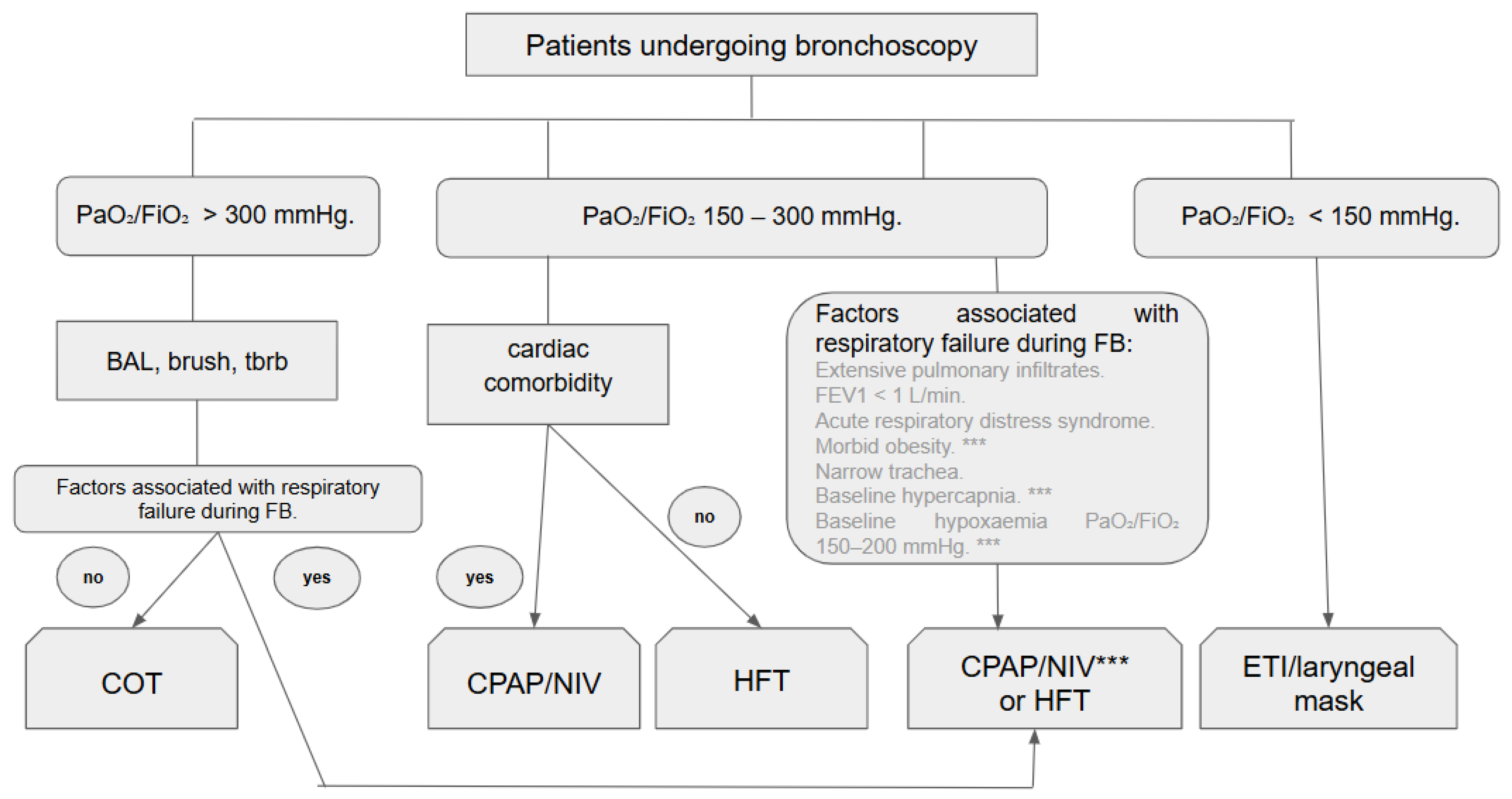
Based on the current literature, a possible approach is outlined in Figure 2.
Most studies have focused on the impact of oxygenation strategies on physiological parameters, with comparatively fewer addressing meaningful clinical outcomes. The quality of the reviewed literature is limited by small sample sizes and considerable heterogeneity among patient populations, precluding formal meta-analysis. Furthermore, most studies were conducted at single centres.
Moreover, there is considerable variability in the titration and settings of HFT, NIV, and CPAP, alongside marked differences in sedation protocols.
This review presents several limitations that warrant consideration. The literature search was confined to the PubMed and Cochrane databases, potentially omitting relevant studies indexed elsewhere. Given the limited number of rigorous trials directly comparing respiratory support strategies during flexible bronchoscopy, both randomised and high-quality observational studies were included. While this approach permitted a more comprehensive assessment of the available evidence, it may introduce a risk of bias. Furthermore, there remains a need for additional well-designed randomised controlled trials to facilitate direct comparisons among the various respiratory support modalities.
A key strength of this review lies in its current and comprehensive appraisal of the available evidence regarding diverse respiratory support strategies during bronchoscopy, rather than limiting the focus to comparisons between high-flow therapy and conventional oxygen therapy, which dominate existing literature. We conducted a comparative analysis of the three principal oxygen delivery modalities employed during bronchoscopy, examining their characteristics and potential advantages. This direct comparison offers a practical framework for clinicians in selecting the most appropriate oxygenation strategy for diagnostic bronchoscopy, as summarised in the algorithm presented in Figure 2, providing a rapid decision-making tool that is currently absent from the literature.

5. Conclusions

In conclusion, the selection of a respiratory support during flexible bronchoscopy should not be based on conventional oxygen therapy by default but must be guided by the type of procedure, the patient’s baseline status, and the anticipated risk of respiratory compromise. Consequently, the likelihood of respiratory deterioration during FB should be thoroughly assessed prior to the procedure [19]. Current evidence supports a structured, stepwise approach, as outlined in Figure 2.
For patients with mild to moderate oxygen requirements and no significant haemodynamic compromise, HFT is recommended over COT as it provides more stable oxygenation, reduces the incidence of desaturation, and minimises procedural interruptions.
In contrast, when bronchoscopy is undertaken in patients with more severe hypoxaemia, relevant cardiac comorbidities, or when deeper sedation is required, CPAP or NIV should be prioritised. Both modalities maintain positive airway pressure throughout the procedure, ensure adequate gas exchange, and contribute to greater cardiopulmonary stability.
Among patients with chronic respiratory disease, particularly COPD or hypercapnia, NIV offers further advantages over HFT and COT. These include superior protection against carbon dioxide retention, a lower risk of post-procedural ventilatory failure, and a reduced need for endotracheal intubation while maintaining more stable cardiopulmonary parameters. NIV unloads the respiratory muscles and counteracts dynamic hyperinflation, thereby preventing intubation and reducing in-hospital mortality in this group of patients [50,51].
Accordingly, the selection of respiratory support must be justified on clinical grounds: HFT is most appropriate for cases with mild to moderate oxygen demands, whereas CPAP or NIV should be reserved for patients with more severe respiratory or cardiac impairment or higher procedural risk. This stepwise framework, represented in Figure 2 and now detailed in the text, provides a pragmatic guide for clinicians to enhance safety and optimise outcomes during flexible bronchoscopy.

Author Contributions

Conceptualization, M.H.S., M.L., P.M.-A., S.A.B., and J.Á.d.V.; methodology, M.H.S., and M.L.; software, C.C.; validation R.Á.-S.W., C.C. and S.A.B.; resources, M.H.S., P.M.-A. and J.Á.d.V.; data curation, M.H.S., P.M.-A. and S.A.B.; writing—original draft preparation, M.H.S.; writing—review and editing, M.H.S., R.Á.-S.W., and C.C.; supervision, M.L., and R.Á.-S.W.; project administration, M.L. All authors have read and agreed to the published version of the manuscript.

Funding

This research received no external funding.

Institutional Review Board Statement

This study did not require ethical approval.

Informed Consent Statement

Not applicable.

Data Availability Statement

No new data were created or analyzed in this study. Data sharing is not applicable to this article.

Acknowledgments

During the preparation of this manuscript, the authors used ChatGPT (GPT-5, OpenAI, San Francisco, CA, USA; https://chat.openai.com) for assistance in language editing and improving English grammar and phrasing. The authors have reviewed and edited the content and take full responsibility for the final publication.

Conflicts of Interest

The authors declare no conflicts of interest.

Abbreviations

ARDSAcute respiratory distress syndrome
ARFAcute respiratory failure
BALBronchoalveolar lavage
BPBlood pressure
COPDChronic obstructive pulmonary disease
COTConventional oxygen therapy
CPAPContinuous positive airway pressure
EBUSEndobronchial ultrasound
ETIEndotracheal intubation
FBFlexible bronchoscopy
FEV1Forced expiratory volume in one second
FiO2Fraction of inspired oxygen
GOLDGlobal Initiative for Chronic Obstructive Lung Disease
HFTHigh-flow therapy
HRHeart rate
ICUIntensive care unit
IMVInvasive mechanical ventilation
IPAPInspiratory positive airway pressure
LOSLength of stay (hospital/ICU)
NDNot defined
NIVNon-invasive ventilation
NIPPVNon-invasive positive pressure ventilation
OSAObstructive sleep apnoea
PaCO2Partial pressure of carbon dioxide in arterial blood
PaO2Partial pressure of oxygen in arterial blood
PaO2/FiO2Ratio of arterial oxygen partial pressure to inspired oxygen fraction
PEEPPositive end-expiratory pressure
RRRespiratory rate
SpO2Peripheral capillary oxygen saturation

References

  1. Matsushima, Y.; Jones, R.L.; King, E.G.; Moysa, G.; Alton, J.D. Alterations in pulmonary mechanics and gas exchange during routine fiberoptic bronchoscopy. Chest 1984, 86, 184–188. [Google Scholar] [CrossRef]
  2. Dubrawsky, C.; Awe, R.J.; Jenkins, D.E. The effect of bronchofiberscopic examination on oxygenation status. Chest 1975, 67, 137–140. [Google Scholar] [CrossRef] [PubMed]
  3. Lindgren, S.; Odenstedt, H.; Erlandsson, K.; Grivans, C.; Lundin, S.; Stenqvist, O. Bronchoscopic suctioning may cause lung collapse: A lung model and clinical evaluation. Acta Anaesthesiol. Scand. 2008, 52, 209–218. [Google Scholar] [CrossRef] [PubMed]
  4. Brach, B.B.; Escano, G.G.; Harrell, J.H.; Moser, K.M. Ventilation-perfusion alterations induced by fiberoptic bronchoscopy. Chest 1976, 69, 335–337. [Google Scholar] [CrossRef]
  5. Randazzo, G.P.; Wilson, A.R. Cardiopulmonary changes during flexible fiberoptic bronchoscopy. Respiration 1976, 33, 143–149. [Google Scholar] [CrossRef] [PubMed]
  6. Shrader, D.L.; Lakshminarayan, S. The effect of fiberoptic bronchoscopy on cardiac rhythm. Chest 1978, 73, 821–824. [Google Scholar] [CrossRef] [PubMed]
  7. Davies, L.; Mister, R.; Spence, D.P.; Calverley, P.M.; Earis, J.E.; Pearson, M.G. Cardiovascular consequences of fiberoptic bronchoscopy. Eur. Respir. J. 1997, 10, 695–698. [Google Scholar] [CrossRef]
  8. Golpe, R.; Mateos, A. Supplemental oxygen during flexible bronchoscopy. Chest 2002, 121, 663–664. [Google Scholar] [CrossRef] [PubMed]
  9. Longhini, F.; Pelaia, C.; Garofalo, E.; Bruni, A.; Placida, R.; Iaquinta, C.; Arrighi, E.; Perri, G.; Procopio, G.; Cancelliere, A.; et al. High-flow nasal cannula oxygen therapy for outpatients undergoing flexible bronchoscopy: A randomised controlled trial. Thorax 2022, 77, 58–64. [Google Scholar] [CrossRef] [PubMed]
  10. Zhang, W.; Wang, J.L.; Fu, S.; Zhou, J.M.; Zhu, Y.J.; Cai, S.N.; Fang, J.; Xie, K.-J.; Chen, X.-Z. Incidence of oxygen desaturation using a high-flow nasal cannula versus a facemask during flexible bronchoscopy in patients at risk of hypoxemia: A randomised controlled trial. BMC Pulm. Med. 2022, 22, 389, Erratum in BMC Pulm. Med. 2022, 22, 451. [Google Scholar] [CrossRef] [PubMed] [PubMed Central]
  11. Arias-Sanchez, P.P.; Ledesma, G.; Cobos, J.; Tirape, H.; Jaramillo, B.; Ruiz, J.; Pacheco, L.; Martinez, J.; Maldonado, R.; Andrade, L.; et al. Changes in Oxygen Saturation During Fiberoptic Bronchoscopy: High-Flow Nasal Cannula versus Standard Oxygen Therapy. Respir. Care 2023, 68, 727–733. [Google Scholar] [CrossRef] [PubMed] [PubMed Central]
  12. Qin, H.; Li, J.; Wang, J.; Yang, Y.G.; Jing, G.Q.; Chen, R.Z.; Tan, W.; Zhang, Y.-Q.; Li, T.; Yang, J.-C.; et al. Comparison of High-Flow Nasal Cannula and Conventional Oxygen Therapy for High-Risk Patients During Bronchoscopy Examination: A Multi-Center Randomized Controlled Trial. Ann. Am. Thorac. Soc. 2025, 22, 1018–1026. [Google Scholar] [CrossRef] [PubMed]
  13. Ben-Menachem, E.; McKenzie, J.; O’Sullivan, C.; Havryk, A.P. High-flow nasal oxygen versus standard oxygen during flexible bronchoscopy in lung transplant patients: A randomized controlled trial. J. Bronchol. Interv. Pulmonol. 2020, 27, 259–265. [Google Scholar] [CrossRef]
  14. Maitre, B.; Jaber, S.; Maggiore, S.M.; Bergot, E.; Richard, J.C.; Bakthiari, H.; Housset, B.; Boussignac, G.; Brochard, L. Continuous positive airway pressure during fiberoptic bronchoscopy in hypoxemic patients. A randomized double-blind study using a new device. Am. J. Respir. Crit. Care Med. 2000, 162 Pt 1, 1063–1067. [Google Scholar] [CrossRef] [PubMed]
  15. Antonelli, M.; Conti, G.; Rocco, M.; Arcangeli, A.; Cavaliere, F.; Proietti, R.; Meduri, G.U. Noninvasive positive-pressure ventilation vs. conventional oxygen supplementation in hypoxemic patients undergoing diagnostic bronchoscopy. Chest 2002, 121, 1149–1154. [Google Scholar] [CrossRef] [PubMed]
  16. Da Conceiçao, M.; Genco, G.; Favier, J.C.; Bidallier, I.; Pitti, R. Fiberoptic bronchoscopy during noninvasive positive-pressure ventilation in patients with chronic obstructive lung disease with hypoxemia and hypercapnia. Ann. Fr. Anesth. Reanim. 2000, 19, 231–236. [Google Scholar] [CrossRef] [PubMed]
  17. Simon, M.; Braune, S.; Frings, D.; Wiontzek, A.-K.; Klose, H.; Kluge, S. High-flow nasal cannula oxygen versus non-invasive ventilation in patients with acute hypoxaemic respiratory failure undergoing flexible bronchoscopy-a prospective randomised trial. Crit. Care 2014, 18, 712. [Google Scholar] [CrossRef] [PubMed]
  18. Saksitthichok, B.; Petnak, T.; So-Ngern, A.; Boonsarngsuk, V. A prospective randomized comparative study of high-flow nasal cannula oxygen and non-invasive ventilation in hypoxemic patients undergoing diagnostic flexible bronchoscopy. J. Thorac. Dis. 2019, 11, 1929–1939. [Google Scholar] [CrossRef]
  19. Tan, D.; Wang, B.; Cao, P.; Wang, Y.; Sun, J.; Geng, P.; Walline, J.H.; Wang, Y.; Wang, C. High flow nasal cannula oxygen therapy versus non-invasive ventilation for acute exacerbations of chronic obstructive pulmonary disease with acute-moderate hypercapnic respiratory failure: A randomized controlled non-inferiority trial. Crit. Care 2024, 28, 250. [Google Scholar] [CrossRef] [PubMed] [PubMed Central]
  20. Bazuaye, E.A.; Stone, T.N.; Corris, P.A.; Gibson, G.J. Variability of inspired oxygen concentration with nasal cannulas. Thorax 1992, 47, 609–611. [Google Scholar] [CrossRef] [PubMed] [PubMed Central]
  21. Duprez, F.; Dupriez, F.; De Greef, J.; Gabriel, J.; Bruyneel, A.; Reychler, G.; De Terwangne, C.; Poncin, W. Performance of Different Low-Flow Oxygen Delivery Systems. Respir. Care 2022, 67, 322–330. [Google Scholar] [CrossRef] [PubMed]
  22. McCain, T.W.; Dunagan, D.P.; Adair, N.E.; Chin, R., Jr. Prospective randomized trial comparing oxygen administration during nasal flexible bronchoscopy: Oral vs. nasal delivery. Chest 2001, 120, 1671–1674. [Google Scholar] [CrossRef] [PubMed]
  23. Yserbyt, J.; De Maeyer, N.; Dooms, C.; Testelmans, D.; Muylle, I.; Bruyneel, M.; Ninane, V. The Feasibility of Tracheal Oxygen Supplementation during Flexible Bronchoscopy. Respiration 2016, 92, 48–52. [Google Scholar] [CrossRef] [PubMed]
  24. Bauer, P.R.; Chevret, S.; Yadav, H.; Mehta, S.; Pickkers, P.; Bukan, R.B.; Rello, J.; van de Louw, A.; Klouche, K.; Meert, A.-P.; et al. Diagnosis and outcome of acute respiratory failure in immunocompromised patients after bronchoscopy. Eur. Respir. J. 2019, 54, 1802442. [Google Scholar] [CrossRef] [PubMed]
  25. Pelaia, C.; Bruni, A.; Garofalo, E.; Rovida, S.; Arrighi, E.; Cammarota, G.; Navalesi, P.; Pelaia, G.; Longhini, F. Oxygenation strategies during flexible bronchoscopy: A review of the literature. Respir. Res. 2021, 22, 253. [Google Scholar] [CrossRef] [PubMed] [PubMed Central]
  26. Du Rand, I.A.; Blaikley, J.; Booton, R.; Chaudhuri, N.; Gupta, V.; Khalid, S.; Mandal, S.; Martin, J.; Mills, J.; Navani, N.; et al. British Thoracic Society Bronchoscopy Guideline Group. British Thoracic Society guideline for diagnostic flexible bronchoscopy in adults: Accredited by NICE. Thorax 2013, 68 (Suppl. 1), 1–44. [Google Scholar] [CrossRef] [PubMed]
  27. Corley, A.; Caruana, L.R.; Barnett, A.G.; Tronstad, O.; Fraser, J.F. Oxygen delivery through high-flow nasal cannulae increase end-expiratory lung volume and reduce respiratory rate in post-cardiac surgical patients. Br. J. Anaesth. 2011, 107, 998–1004. [Google Scholar] [CrossRef]
  28. Maggiore, S.M.; Idone, F.A.; Vaschetto, R.; Festa, R.; Cataldo, A.; Antonicelli, F.; Montini, L.; De Gaetano, A.; Navalesi, P.; Antonelli, M. Nasal high-flow versus Venturi mask oxygen therapy after extubation. Effects on oxygenation, comfort, and clinical outcome. Am. J. Respir. Crit. Care Med. 2014, 190, 282–288. [Google Scholar] [CrossRef]
  29. Moller, W.; Celik, G.; Feng, S.; Bartenstein, P.; Meyer, G.; Eickelberg, O.; Schmid, O.; Tatkov, S. Nasal high flow clears anatomical dead space in upper airway models. J. Appl. Physiol. 2015, 118, 1525–1532. [Google Scholar] [CrossRef] [PubMed] [PubMed Central]
  30. Rochwerg, B.; Einav, S.; Chaudhuri, D.; Mancebo, J.; Mauri, T.; Helviz, Y.; Goligher, E.C.; Jaber, S.; Ricard, J.-D.; Rittayamai, N.; et al. The role for high flow nasal cannula as a respiratory support strategy in adults: A clinical practice guideline. Intensive Care Med. 2020, 46, 2226–2237. [Google Scholar] [CrossRef] [PubMed]
  31. Lucangelo, U.; Vassallo, F.G.; Marras, E.; Ferluga, M.; Beziza, E.; Comuzzi, L.; Berlot, G.; Zin, W.A. High-flow nasal interface improves oxygenation in patients undergoing bronchoscopy. Crit. Care Res. Pract. 2012, 2012, 506382. [Google Scholar] [CrossRef]
  32. Irfan, M.; Ahmed, M.; Breen, D. Assessment of high flow nasal cannula oxygenation in endobronchial ultrasound bronchoscopy: A randomized controlled trial. J. Bronchol. Interv. Pulmonol. 2021, 28, 130–137. [Google Scholar] [CrossRef] [PubMed]
  33. Kim, E.J.; Jung, C.Y.; Kim, K.C. Effectiveness and safety of high-flow nasal cannula oxygen delivery during bronchoalveolar lavage in acute respiratory failure patients. Tuberc. Respir. Dis. 2018, 81, 319–329. [Google Scholar] [CrossRef]
  34. Verra, F.; Hmouda, H.; Rauss, A.; Lebargy, F.; Cordonnier, C.; Bignon, J.; Lemaire, F.; Brochard, L. Bronchoalveolar lavage in immunocompromised patients. Clinical and functional consequences. Chest 1992, 101, 1215–1220. [Google Scholar] [CrossRef] [PubMed]
  35. Roy, A.; Khanna, P.; Chowdhury, S.R.; Haritha, D.; Sarkar, S. The Impact of High-flow Nasal Cannula vs. Other Oxygen Delivery Devices during Bronchoscopy under Sedation: A Systematic Review and Meta-analyses. Indian J. Crit. Care Med. 2022, 26, 1131–1140. [Google Scholar] [CrossRef]
  36. Sampsonas, F.; Karamouzos, V.; Karampitsakos, T.; Papaioannou, O.; Katsaras, M.; Lagadinou, M.; Zarkadi, E.; Malakounidou, E.; Velissaris, D.; Stratakos, G.; et al. High-Flow vs. Low-Flow Nasal Cannula in Reducing Hypoxemic Events During Bronchoscopic Procedures: A Systematic Review and Meta-Analysis. Front. Med. 2022, 9, 815799. [Google Scholar] [CrossRef]
  37. Thiruvenkatarajan, V.; Sekhar, V.; Wong, D.T.; Currie, J.; Van Wijk, R.; Ludbrook, G.L. Effect of high-flow nasal oxygen on hypoxaemia during procedural sedation: A systematic review and meta-analysis. Anaesthesia 2023, 78, 81–92. [Google Scholar] [CrossRef]
  38. Corral-Blanco, M.; Sayas-Catalán, J.; Hernández-Voth, A.; Rey-Terrón, L.; Villena-Garrido, V. High-Flow Nasal Cannula Therapy as an Adjuvant Therapy for Respiratory Support during Endoscopic Techniques: A Narrative Review. J. Clin. Med. 2023, 13, 81. [Google Scholar] [CrossRef] [PubMed] [PubMed Central]
  39. Leone, M.; Einav, S.; Chiumello, D.; Constantin, J.M.; De Robertis, E.; De Abreu, M.G.; Gregoretti, C.; Jaber, S.; Maggiore, S.M.; Pelosi, P.; et al. Guideline contributors. Noninvasive respiratory support in the hypoxaemic peri-operative/periprocedural patient: A joint ESA/ESICM guideline. Intensiv. Care Med. 2020, 46, 697–713. [Google Scholar] [CrossRef] [PubMed] [PubMed Central]
  40. Cabrini, L.; Nobile, L.; Cama, E.; Borghi, G.; Pieri, M.; Bocchino, S.; Zangrillo, A. Non-invasive ventilation during upper endoscopies in adult patients. A systematic review. Minerva Anestesiol. 2013, 79, 683–694. [Google Scholar] [PubMed]
  41. Agarwal, R.; Khan, A.; Aggarwal, A.N.; Gupta, D. Bronchoscopic lung biopsy using noninvasive ventilatory support: Case series and review of literature of NIV-assisted bronchoscopy. Respir. Care 2012, 57, 1927–1936. [Google Scholar] [CrossRef]
  42. Chen, X.; Zhou, Y.; Yu, H.; Peng, Y.; Xia, L.; Liu, N.; Lin, H. Feasibility analysis of flexible bronchoscopy in conjunction with noninvasive ventilation for therapy of hypoxemic patients with Central Airway Obstruction: A retrospective study. PeerJ 2020, 8, e8687. [Google Scholar] [CrossRef] [PubMed]
  43. Sharma, V.K.; Singh, P.K.; Govindagoudar, M.B.; Thulasi, A.; Chaudhry, D.; Shriram, C.P.; Lalwani, L.K.; Ahuja, A. Efficacy of different respiratory supports to prevent hypoxia during flexible bronchoscopy in patients of COPD: A triple-arm, randomised controlled trial. BMJ Open Respir. Res. 2023, 10, e001524. [Google Scholar] [CrossRef] [PubMed] [PubMed Central]
  44. Ferrer, M.; Torres, A. Noninvasive Ventilation and High-Flow Nasal Therapy Administration in Chronic Obstructive Pulmonary Disease Exacerbations. Semin. Respir. Crit. Care Med. 2020, 41, 786–797. [Google Scholar] [CrossRef]
  45. Trouillet, J.L.; Guiguet, M.; Gibert, C.; Fagon, J.Y.; Dreyfuss, D.; Blanchet, F.; Chastre, J. Fiberoptic bronchoscopy in ventilated patients. Evaluation of cardiopulmonary risk under midazolam sedation. Chest 1990, 97, 927–933. [Google Scholar] [CrossRef] [PubMed]
  46. Qin, H.; Jing, G.Q.; Tan, W.; Wang, J.; Yin, Y.; Chen, R.; Zhang, W.; Li, J. Comparison of high-flow nasal cannula and conventional oxygen therapy for high-risk patients during bronchoscopy examination: Protocol for a randomized controlled trial. Trials 2023, 24, 12. [Google Scholar] [CrossRef]
  47. Avari, H.; Hiebert, R.J.; Ryzynski, A.A.; Levy, A.; Nardi, J.; Kanji-Jaffer, H.; Kiiza, P.; Pinto, R.; Plenderleith, S.W.; Fowler, R.A.; et al. Quantitative Assessment of Viral Dispersion Associated with Respiratory Support Devices in a Simulated Critical Care Environment. Am. J. Respir. Crit. Care Med. 2021, 203, 1112–1118. [Google Scholar] [CrossRef] [PubMed] [PubMed Central]
  48. Esquinas, A.; Zuil, M.; Scala, R.; Chiner, E. Bronchoscopy during non-invasive mechanical ventilation: Review of techniques and procedures. Arch. Bronconeumol. 2013, 49, 105–112. [Google Scholar] [CrossRef]
  49. Oraczewska, A.; Cofta, S.; Warcholiński, A.; Trejnowska, E.; Brożek, G.; Swinarew, A.; Stolz, D.; Scala, R.; Barczyk, A.; Skoczyński, S. The use of non-invasive respiratory assistance to facilitate bronchofiberoscopy performance in patients with hypoxemic (type one) respiratory failure—Study protocol. Adv. Med. Sci. 2023, 68, 474–481. [Google Scholar] [CrossRef]
  50. Skoczyński, S.; Ogonowski, M.; Tobiczyk, E.; Krzyżak, D.; Brożek, G.; Wierzbicka, A.; Trzaska-Sobczak, M.; Trejnowska, E.; Studnicka, A.; Swinarew, A.; et al. Risk factors of complications during noninvasive mechanical ventilation -assisted flexible bronchoscopy. Adv. Med. Sci. 2021, 66, 246–253. [Google Scholar] [CrossRef] [PubMed]
  51. Munshi, L.; Mancebo, J.; Brochard, L.J.; Hardin, C.C. Noninvasive respiratory support for adults with acute respiratory failure. N. Engl. J. Med. 2022, 387, 1688–1698. [Google Scholar] [CrossRef]
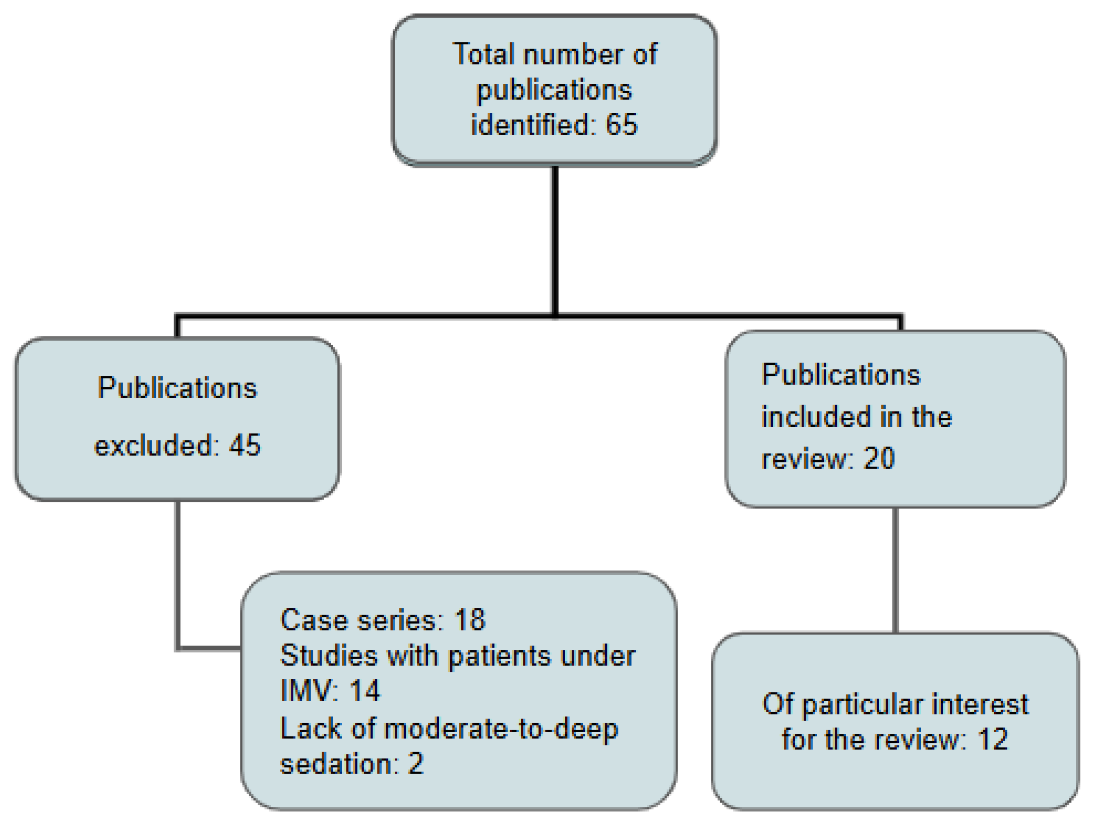
Figure 1. Flowchart for study selection: invasive mechanical ventilation (IMV).
Jcm 14 06658 g001
Figure 2. The algorithm for selection of respiratory support during flexible bronchoscopy. Abbreviations: PaO2/FIO2 (ratio of arterial oxygen pressure to inspired oxygen fraction), BAL (bronchoalveolar lavage), COT (conventional oxygen therapy), HFT (high-flow therapy), NIV (non-invasive ventilation), Tbrb (transbronchial biopsy), CPAP (continuous positive airway pressure), FEV1 (Forced Expiratory Volume in 1 s), ETI (endotracheal intubation), *** (in cases of morbid obesity, baseline hypercapnia, baseline hipoxaemia it would be highly recommended CPAP or NIV).
Jcm 14 06658 g002
Table 1. Main articles revised for this article: Abbreviations: ND: not defined. SpO2: oxygen saturation; PaO2: arterial oxygen pressure; PaCO2: arterial carbon dioxide pressure; HR: heart rate; BP: blood pressure; RR: respiratory rate; pH: measure of acidity or alkalinity in blood; ETI: endotracheal intubation; COT: conventional oxygen therapy; HFT: high-flow therapy; NIV: non-invasive ventilation; NIPPV: non-invasive positive pressure ventilation; CPAP: continuous positive airway pressure; PaO2/FiO2: ratio of arterial oxygen partial pressure (PaO2) to the fraction of inspired oxygen (Fi02), used as an index of oxygenation. LOS: Length of stay at hospital and intensive care unit.
ReferenceNo. of PatientsStudy TypeClinical StatusSupport During FBMain Objectives
Golpe, et al. [8]44ObservationalNDCOTSpO2
Longhini, et al. [9]36Randomised prospectiveChronicCOT vs. HFTSpO2, PaO2
Zhang, et al. [10]176Randomised prospectiveAcuteCOT vs. HFTSpO2, post-procedure respiratory support
Arias-Sánchez, et al. [11]40ObservationalAcuteCOT vs. HFTSpO2
Quin H et al. [12]142Multicentre randomised prospectiveChronicCOT vs. HFTSpO2, transcutaneous CO2, complications
Ben-Menachem, et al. [13]76Randomised prospectiveChronicCOT vs. HFTSpO2
Maitre, et al. [14]30Randomised prospectiveAcuteCOT vs. CPAPSpO2, PaO2, post-ventilatory support
Antonelli, et al. [15]26Randomised prospectiveAcuteCOT vs. NIPPVPaO2/FiO2, HR, BP, ETI
Da Conceição et al. [16]10ProspectiveAcuteNIPPVSpO2, PaO2, PaCO2, ETI, mortality
Simon, et al. [17]40Randomised prospectiveAcuteNIV vs. HFTSpO2, PaO2/FiO2, PaCO2, HR, BP, procedure time, ETI, mortality
Saksitthichok, et al. [18]51Randomised prospectiveAcuteNIV vs. HFTSpO2, PaO2, PaCO2, pH, RR, HR, BP, dyspnoea
Tan, et al. [19]225Randomised, open-label, non-inferiority trialAcute hypercapniaHFT vs. NIVETI, blood gases, ICU/Hospital LOS, 28-day mortality
Disclaimer/Publisher’s Note: The statements, opinions and data contained in all publications are solely those of the individual author(s) and contributor(s) and not of MDPI and/or the editor(s). MDPI and/or the editor(s) disclaim responsibility for any injury to people or property resulting from any ideas, methods, instructions or products referred to in the content.