Three-Dimensional Evaluation of Impacted Mandibular Canines and Adjacent Structures Using Cone Beam Computed Tomography: A Retrospective Study
Abstract
1. Introduction
2. Materials and Methods
- Type 1: Above the cementoenamel junction of the adjacent teeth.
- Type 2: At the height of the cervical third of the adjacent tooth roots.
- Type 3: At the height of the middle third of the adjacent tooth roots.
- Type 4: At the height of the apical third of the adjacent tooth roots.
- Type 5: Below the apices of the adjacent tooth roots.
- There is contact with the mental foramen.
- There is no contact with the mental foramen.
- There is no incisive mandibular canal.
- There is an incisive mandibular canal, but there is no contact.
- There is an incisive mandibular canal, and there is contact.
- (0)
- No resorption: root surface intact.
- (1)
- Slight resorption: root resorption extending to the pulp up to half the dentin thickness.
- (2)
- Moderate resorption: root resorption extending to more than half the pulpal distance, with the pulp margin preserved.
- (3)
- Severe resorption: root resorption reaching the pulp.
- No deciduous mandibular canine.
- No contact between the permanent and deciduous mandibular canines; no root resorption of the deciduous mandibular canine.
- There is contact between the permanent and deciduous mandibular canines, but no root resorption of the deciduous mandibular canines.
- There is no contact between the permanent and deciduous mandibular canines, but there is root resorption of the deciduous mandibular canines.
- There is contact between the permanent and deciduous mandibular canines, and there is root resorption of the deciduous mandibular canines.
2.1. Statistical Analyses
2.2. Evaluations of Measurement Error
3. Results
4. Discussion
5. Conclusions
Author Contributions
Funding
Institutional Review Board Statement
Informed Consent Statement
Data Availability Statement
Conflicts of Interest
Abbreviations
| CBCT | Cone beam computed tomography |
References
- Alqerban, A.; Jacobs, R.; Lambrechts, P.; Loozen, G.; Willems, G. Root resorption of the maxillary lateral incisor caused by impacted canine: A literature review. Clin. Oral Investig. 2009, 13, 247–255. [Google Scholar] [CrossRef] [PubMed]
- Grover, P.S.; Lorton, L. The incidence of unerupted permanent teeth and related clinical cases. Oral Surg. Oral Med. Oral Pathol. 1985, 59, 420–425. [Google Scholar] [CrossRef] [PubMed]
- Dalessandri, D.; Parrini, S.; Rubiano, R.; Gallone, D.; Migliorati, M. Impacted and transmigrant mandibular canines incidence, aetiology, and treatment: A systematic review. Eur. J. Orthod. 2017, 39, 161–169. [Google Scholar] [CrossRef] [PubMed]
- Ando, S.; Aizawa, K.; Nakashima, T.; Sanka, Y.; Shimbo, K.; Kiyokawa, K. Transmigration process of the impacted mandibular cuspid. J. Nihon Univ. Sch. Dent. 1964, 6, 66–71. [Google Scholar] [CrossRef]
- Miranti, R.; Levbarg, M. Extraction of a horizontally transmigrated impacted mandibular canine: Report of case. J. Am. Dent. Assoc. 1974, 88, 607–610. [Google Scholar] [CrossRef]
- Shapira, Y.; Mischler, W.A.; Kuftinec, M.M. The displaced mandibular canine. ASDC J. Dent. Child. 1982, 49, 362–364. [Google Scholar]
- Aras, M.H.; Halicioglu, K.; Yavuz, M.S.; Caglaroglu, M. Evaluation of surgical-orthodontic treatments on impacted mandibular canines. Med. Oral Patol. Oral Cir. Bucal 2011, 16, e925–e928. [Google Scholar] [CrossRef]
- Sinko, K.; Nemec, S.; Seemann, R.; Eder-Czembirek, C. Clinical Management of Impacted and Transmigrated Lower Canines. J. Oral Maxillofac. Surg. 2016, 74, 2142. [Google Scholar] [CrossRef]
- Chaushu, S.; Chaushu, G.; Becker, A. The use of panoramic radiographs to localize displaced maxillary canines. Oral Surg. Oral Med. Oral Pathol. Oral Radiol. Endod. 1999, 88, 511–516. [Google Scholar] [CrossRef]
- Chu, F.C.; Li, T.K.; Lui, V.K.; Newsome, P.R.; Chow, R.L.; Cheung, L.K. Prevalence of impacted teeth and associated pathologies—A radiographic study of the Hong Kong Chinese population. Hong Kong Med. J. 2003, 9, 158–163. [Google Scholar]
- Preda, L.; La Fianza, A.; Di Maggio, E.M.; Dore, R.; Schifino, M.R.; Campani, R.; Segu, C.; Sfondrini, M.F. The use of spiral computed tomography in the localization of impacted maxillary canines. Dentomaxillofac. Radiol. 1997, 26, 236–241. [Google Scholar] [CrossRef] [PubMed]
- Walker, L.; Enciso, R.; Mah, J. Three-dimensional localization of maxillary canines with cone-beam computed tomography. Am. J. Orthod. Dentofac. Orthop. 2005, 128, 418–423. [Google Scholar] [CrossRef] [PubMed]
- Cakir Karabas, H.; Ozcan, I.; Erturk, A.F.; Guray, B.; Unsal, G.; Senel, S.N. Cone-beam computed tomography evaluation of impacted and transmigrated mandibular canines: A retrospective study. Oral Radiol. 2021, 37, 403–411. [Google Scholar] [CrossRef] [PubMed]
- Strbac, G.D.; Foltin, A.; Gahleitner, A.; Bantleon, H.P.; Watzek, G.; Bernhart, T. The prevalence of root resorption of maxillary incisors caused by impacted maxillary canines. Clin. Oral Investig. 2013, 17, 553–564. [Google Scholar] [CrossRef]
- Bertl, M.H.; Frey, C.; Bertl, K.; Giannis, K.; Gahleitner, A.; Strbac, G.D. Impacted and transmigrated mandibular canines: An analysis of 3D radiographic imaging data. Clin. Oral Investig. 2018, 22, 2389–2399. [Google Scholar] [CrossRef]
- Yavuz, M.S.; Aras, M.H.; Buyukkurt, M.C.; Tozoglu, S. Impacted mandibular canines. J. Contemp. Dent. Pract. 2007, 8, 78–85. [Google Scholar]
- Lai, C.S.; Bornstein, M.M.; Mock, L.; Heuberger, B.M.; Dietrich, T.; Katsaros, C. Impacted maxillary canines and root resorptions of neighbouring teeth: A radiographic analysis using cone-beam computed tomography. Eur. J. Orthod. 2013, 35, 529–538. [Google Scholar] [CrossRef]
- Ericson, S.; Kurol, P.J. Resorption of incisors after ectopic eruption of maxillary canines: A CT study. Angle Orthod. 2000, 70, 415–423. [Google Scholar]
- Ericson, S.; Bjerklin, K.; Falahat, B. Does the canine dental follicle cause resorption of permanent incisor roots? A computed tomographic study of erupting maxillary canines. Angle Orthod. 2002, 72, 95–104. [Google Scholar]
- McDonnell, D.; Reza Nouri, M.; Todd, M.E. The mandibular lingual foramen: A consistent arterial foramen in the middle of the mandible. J. Anat. 1994, 184 Pt 2, 363–369. [Google Scholar]
- Sekerci, A.E.; Sisman, Y.; Payveren, M.A. Evaluation of location and dimensions of mandibular lingual foramina using cone-beam computed tomography. Surg. Radiol. Anat. 2014, 36, 857–864. [Google Scholar] [CrossRef] [PubMed]
- Sherwood, R.J.; Hlusko, L.J.; Duren, D.L.; Emch, V.C.; Walker, A. Mandibular symphysis of large-bodied hominoids. Hum. Biol. 2005, 77, 735–759. [Google Scholar] [CrossRef] [PubMed]
- Wang, Y.M.; Ju, Y.R.; Pan, W.L.; Chan, C.P. Evaluation of location and dimensions of mandibular lingual canals: A cone beam computed tomography study. Int. J. Oral Maxillofac. Surg. 2015, 44, 1197–1203. [Google Scholar] [CrossRef] [PubMed]
- Liu, D.G.; Zhang, W.L.; Zhang, Z.Y.; Wu, Y.T.; Ma, X.C. Localization of impacted maxillary canines and observation of adjacent incisor resorption with cone-beam computed tomography. Oral Surg. Oral Med. Oral Pathol. Oral Radiol. Endod. 2008, 105, 91–98. [Google Scholar] [CrossRef]
- MF, D.O.-A.; Arriola-Guillen, L.E.; Rodriguez-Cardenas, Y.A.; Ruiz-Mora, G.A. Skeletal and dentoalveolar bilateral dimensions in unilateral palatally impacted canine using cone beam computed tomography. Prog. Orthod. 2017, 18, 7. [Google Scholar] [CrossRef]
- Hanke, S.; Hirschfelder, U.; Keller, T.; Hofmann, E. 3D CT based rating of unilateral impacted canines. J. Craniomaxillofacal Surg. 2012, 40, e268–e276. [Google Scholar] [CrossRef]
- Aydın, M.; Uğurlu, M. Mandibular Gömülü Kaninlerin Konik Işinli Bilgisayarli Tomografi İle Açisal, Doğrusal Ölçümlerinin ve Deskriptif Özelliklerinin Üç Boyutlu Analizi. Atatürk Üniv. Diş Hek. Fak. Derg. 2020, 30, 212–218. [Google Scholar]
- Celikoglu, M.; Kamak, H.; Oktay, H. Investigation of transmigrated and impacted maxillary and mandibular canine teeth in an orthodontic patient population. J. Oral Maxillofac. Surg. 2010, 68, 1001–1006. [Google Scholar] [CrossRef]
- Qadeer, M.; Khan, H.; Najam, E.; Anwar, A.; Khan, T. Prevalence And Patterns Of Mandibular Impacted Canines. A Cbct Based Retrospective Study. Pak. Oral Dent. J. 2018, 38, 178–181. [Google Scholar]
- Oberoi, S.; Knueppel, S. Three-dimensional assessment of impacted canines and root resorption using cone beam computed tomography. Oral Surg. Oral Med. Oral Pathol. Oral Radiol. 2012, 113, 260–267. [Google Scholar] [CrossRef]
- Sajnani, A.K.; King, N.M. Impacted mandibular canines: Prevalence and characteristic features in southern Chinese children and adolescents. J. Dent. Child. 2014, 81, 3–6. [Google Scholar]
- Gilis, S.; Dhaene, B.; Dequanter, D.; Loeb, I. Mandibular incisive canal and lingual foramina characterization by cone-beam computed tomography. Morphologie 2019, 103, 48–53. [Google Scholar] [CrossRef] [PubMed]
- da Silva Ramos Fernandes, L.M.; Capelozza, A.L.; Rubira-Bullen, I.R. Absence and hypoplasia of the mental foramen detected in CBCT images: A case report. Surg. Radiol. Anat. 2011, 33, 731–734. [Google Scholar] [CrossRef] [PubMed]
- Ericson, S.; Bjerklin, K. The dental follicle in normally and ectopically erupting maxillary canines: A computed tomography study. Angle Orthod. 2001, 71, 333–342. [Google Scholar] [PubMed]
- Kim, Y.; Hyun, H.K.; Jang, K.T. Morphological relationship analysis of impacted maxillary canines and the adjacent teeth on 3-dimensional reconstructed CT images. Angle Orthod. 2017, 87, 590–597. [Google Scholar] [CrossRef]
- Yan, B.; Sun, Z.; Fields, H.; Wang, L.; Luo, L. Etiologic factors for buccal and palatal maxillary canine impaction: A perspective based on cone-beam computed tomography analyses. Am. J. Orthod. Dentofac. Orthop. 2013, 143, 527–534. [Google Scholar] [CrossRef]
- Sharhan, H.M.; Almashraqi, A.A.; Al-Fakeh, H.; Alhashimi, N.; Abdulghani, E.A.; Chen, W.; Al-Sosowa, A.A.; Cao, B.; Alhammadi, M.S. Qualitative and quantitative three-dimensional evaluation of maxillary basal and dentoalveolar dimensions in patients with and without maxillary impacted canines. Prog. Orthod. 2022, 23, 38. [Google Scholar] [CrossRef]
- Al-Nimri, K.; Gharaibeh, T. Space conditions and dental and occlusal features in patients with palatally impacted maxillary canines: An aetiological study. Eur. J. Orthod. 2005, 27, 461–465. [Google Scholar] [CrossRef]
- Cacciatore, G.; Poletti, L.; Sforza, C. Early diagnosed impacted maxillary canines and the morphology of the maxilla: A three-dimensional study. Prog. Orthod. 2018, 19, 20. [Google Scholar] [CrossRef]
- Langberg, B.J.; Peck, S. Adequacy of maxillary dental arch width in patients with palatally displaced canines. Am. J. Orthod. Dentofac. Orthop. 2000, 118, 220–223. [Google Scholar] [CrossRef]
- Kim, Y.; Hyun, H.K.; Jang, K.T. Interrelationship between the position of impacted maxillary canines and the morphology of the maxilla. Am. J. Orthod. Dentofac. Orthop. 2012, 141, 556–562. [Google Scholar] [CrossRef] [PubMed]
- Schindel, R.H.; Duffy, S.L. Maxillary transverse discrepancies and potentially impacted maxillary canines in mixed-dentition patients. Angle Orthod. 2007, 77, 430–435. [Google Scholar] [CrossRef]
- McConnell, T.L.; Hoffman, D.L.; Forbes, D.P.; Janzen, E.K.; Weintraub, N.H. Maxillary canine impaction in patients with transverse maxillary deficiency. ASDC J. Dent. Child. 1996, 63, 190–195. [Google Scholar]
- Louly, F.; Nouer, P.R.; Janson, G.; Pinzan, A. Dental arch dimensions in the mixed dentition: A study of Brazilian children from 9 to 12 years of age. J. Appl. Oral Sci. 2011, 19, 169–174. [Google Scholar] [CrossRef]


| n (%) | Mean ± SD | Median (IQR) | Min; Max | |
|---|---|---|---|---|
| Gender | ||||
| Female | 34 (63.0) | 16.06 ± 4.38 | 15.0 (5.0) | 12.0; 35.0 |
| Male | 20 (37.0) | 19.65 ± 7.88 | 17.0 (11.8) | 12.0; 36.0 |
| Impacted tooth: | ||||
| Right mandibular canine | 28 (51.9) | |||
| Left mandibular canine | 26 (48.1) |
| n (%) | ||
|---|---|---|
| Angulation of the canine tooth | Horizontal | 5 (9.3) |
| Mesioangular | 21 (38.8) | |
| Vertical | 23 (42.6) | |
| Distoangular | 5 (9.3) | |
| Labiolingual position of the canine crown | Labially impacted | 28 (51.9) |
| Lingually impacted | 10 (18.5) | |
| Medially impacted | 16 (29.6) | |
| Vertical position of the canine cusp tip | Type 1 | 9 (16.7) |
| Type 2 | 5 (9.3) | |
| Type 3 | 12 (22.2) | |
| Type 4 | 20 (37.0) | |
| Type 5 | 8 (14.8) | |
| Contact with the mental foramen | Yes | 6 (11.1) |
| No | 48 (88.9) | |
| Incisive mandibular canal | Yes | 46 (85.2) |
| No | 8 (14.8) | |
| Contact with the incisive mandibular canal | Yes | 3 (6.5) |
| No | 43 (93.5) | |
| Resorption of adjacent permanent tooth | No | 46 (85.2) |
| Slight resorption | 3 (37.5) | |
| Moderate resorption | 4 (50.0) | |
| Severe resorption | 1 (12.5) | |
| Localization of resorption | Cementoenamel junction | 1 (12.5) |
| Apical third | 3 (37.5) | |
| Middle third | 4 (50.0) | |
| Deciduous mandibular canine | No | 24 (44.4) |
| Yes | 30 (55.6) | |
| Resorption and contact in deciduous canine | Contact, resorption | 16 (53.4) |
| No contact, resorption | 6 (20.0) | |
| Contact, no resorption | 1 (3.3) | |
| No contact, no resorption | 7 (23.3) | |
| Cortical bone perforation | No | 9 (16.7) |
| Labial | 29 (64.4) | |
| Lingual | 12 (26.7) | |
| Labial and lingual | 4 (8.9) | |
| Follicle diameter | <3 mm | 41 (75.9) |
| ≥3 mm | 13 (24.1) |
| Adjacent Permanent Tooth Resorption | ||
|---|---|---|
| No n (%) | Yes n (%) | |
| Location | ||
| Labial | 23 (50.0) | 5 (62.5) |
| Lingual | 8 (17.4) | 2 (25.0) |
| Medial | 15 (32.6) | 1 (12.5) |
| Adjacent Permanent Tooth Resorption | ||||
|---|---|---|---|---|
| No n (%) | Yes n (%) | Test Statistic | ||
| χ2 | p | |||
| Follicle diameter | ||||
| <3 mm | 35 (85.4) | 6 (14.6) | 0.004 | 0.947 |
| >3 mm | 11 (84.6) | 2 (15.4) | ||
| Impacted Side | Non-Impacted Side | Test Statistic | ||
|---|---|---|---|---|
| Mean ± SD Median (IQR) | Mean ± SD Median (IQR) | z; t | p | |
| Mesiodistal width of the canine | 6.78 ± 0.41 | 6.56 ± 0.40 | t = 2.617 | 0.010 |
| 6.70 (0.50) | 6.60 (0.50) | |||
| Interpremolar width | 15.69 ± 2.29 | 16.45 ± 1.42 | z = 1.864 | 0.062 |
| 16.00 (2.78) | 16.80 (1.65) | |||
| Intermolar width | 21.86 ± 2.27 | 22.44 ± 2.06 | t = 1.393 | 0.166 |
| 22.15 (2.90) | 22.35 (2.80) | |||
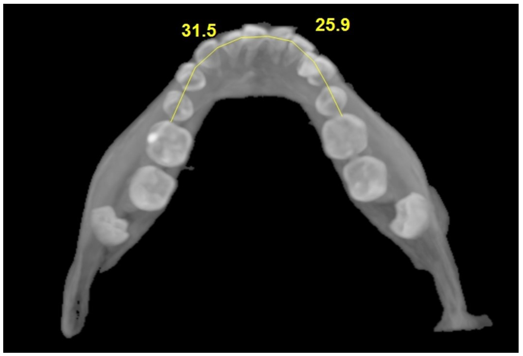
| Arch length | 29.92 ± 2.38 | 32.21 ± 2.18 | t = 5.194 | <0.001 |
| 30.05 (2.80) | 32.40 (3.00) | |||
| Variable | B (95% CI) | SE | p-Value |
|---|---|---|---|
| Age | −0.047 (−0.137, 0.043) | 0.046 | 0.312 |
| Mesiodistal width | −0.391 (−1.781, 0.999) | 0.709 | 0.584 |
| Interpremolar width | 0.204 (−0.168, 0.576) | 0.19 | 0.290 |
| Intermolar width | 0.570 (0.127, 1.013) | 0.226 | 0.015 |
| Vertical position: type 1 (ref = type 4) | 0.320 (−2.003, 2.643) | 1.152 | 0.784 |
| Vertical position: type 2 (ref = type 4) | 1.297 (−1.295, 3.889) | 1.317 | 0.329 |
| Vertical position: type 3 (ref = type 4) | 0.602 (−1.625, 2.829) | 1.123 | 0.594 |
| Vertical position: type 5 (ref = type 4) | 0.206 (−1.528, 1.940) | 0.88 | 0.820 |
| Location: lingual (ref = labial) | 0.391 (−1.158, 1.940) | 0.787 | 0.629 |
| Location: medial (ref = labial) | 0.603 (−1.214, 2.420) | 0.927 | 0.512 |
| Angulation: horizontal (ref = mesioangular) | −2.884 (−5.003, −0.765) | 1.081 | 0.011 |
| Angulation: vertical (ref = mesioangular) | −0.034 (−2.107, 2.039) | 1.041 | 0.973 |
| Angulation: distoangular (ref = mesioangular) | −0.102 (−2.011, 1.807) | 0.952 | 0.921 |
Disclaimer/Publisher’s Note: The statements, opinions and data contained in all publications are solely those of the individual author(s) and contributor(s) and not of MDPI and/or the editor(s). MDPI and/or the editor(s) disclaim responsibility for any injury to people or property resulting from any ideas, methods, instructions or products referred to in the content. |
© 2025 by the authors. Licensee MDPI, Basel, Switzerland. This article is an open access article distributed under the terms and conditions of the Creative Commons Attribution (CC BY) license (https://creativecommons.org/licenses/by/4.0/).
Share and Cite
Dogan, A.; Uslu, F.; Duman, S.B. Three-Dimensional Evaluation of Impacted Mandibular Canines and Adjacent Structures Using Cone Beam Computed Tomography: A Retrospective Study. J. Clin. Med. 2025, 14, 6372. https://doi.org/10.3390/jcm14186372
Dogan A, Uslu F, Duman SB. Three-Dimensional Evaluation of Impacted Mandibular Canines and Adjacent Structures Using Cone Beam Computed Tomography: A Retrospective Study. Journal of Clinical Medicine. 2025; 14(18):6372. https://doi.org/10.3390/jcm14186372
Chicago/Turabian StyleDogan, Ayhan, Filiz Uslu, and Suayip Burak Duman. 2025. "Three-Dimensional Evaluation of Impacted Mandibular Canines and Adjacent Structures Using Cone Beam Computed Tomography: A Retrospective Study" Journal of Clinical Medicine 14, no. 18: 6372. https://doi.org/10.3390/jcm14186372
APA StyleDogan, A., Uslu, F., & Duman, S. B. (2025). Three-Dimensional Evaluation of Impacted Mandibular Canines and Adjacent Structures Using Cone Beam Computed Tomography: A Retrospective Study. Journal of Clinical Medicine, 14(18), 6372. https://doi.org/10.3390/jcm14186372


