
The Path of Bronchiolitis Towards Intensive Care: Risk Factor Analysis in a Large Italian Cohort


 , , , ,
Abstract
1. Introduction
2. Materials and Methods
2.1. Study Design
2.2. Population
2.3. Outcomes
2.4. Methods
2.5. Statistical Analysis
3. Results
3.1. Characteristics of the Study Population
3.2. Comparison Between Two Seasons
3.3. Independent Risk Factors for Severe Course and Characteristics of the IC Group
4. Discussion
5. Conclusions
Author Contributions
Funding
Institutional Review Board Statement
Informed Consent Statement
Data Availability Statement
Acknowledgments
Conflicts of Interest
Abbreviations
| PICU | Pediatric Intensive Care Unit |
| NICU | Neonatal Intensive Care Unit |
| SpO2 | Peripheral Oxygen Saturation |
| IC | Intensive Care |
| RSV | Respiratory Syncytial Virus |
| CRP | C-Reactive Protein |
| OR | Odds Ratio |
| HFNC | High-Flow Nasal Cannula |
| IMV | Invasive Mechanical Ventilation |
References
- Stein, R.T.; Bont, L.J.; Zar, H.; Polack, F.P.; Park, C.; Claxton, A.; Borok, G.; Butylkova, Y.; Wegzyn, C. Respiratory syncytial virus hospitalization and mortality: Systematic review and meta-analysis. Pediatr. Pulmonol. 2017, 52, 556–569. [Google Scholar] [CrossRef]
- Wilkesmann, A.; Ammann, R.A.; Schildgen, O.; Eis-Hübinger, A.M.; Müller, A.; Seidenberg, J.; Stephan, V.; Rieger, C.; Herting, E.; Wygold, T.; et al. Hospitalized children with respiratory syncytial virus infection and neuromuscular impairment face an increased risk of a complicated course. Pediatr. Infect. Dis. J. 2007, 26, 485–491. [Google Scholar] [CrossRef] [PubMed]
- Guarnieri, V.; Palmas, G.; Trapani, S.; Mollo, A.; Macucci, C.; Gambini, I.; Mottola, V.; Rubino, C.; Moriondo, M.; Nieddu, F.; et al. Exploring Risk Factors Associated With Intensive Care Unit Admission in a Retrospective Cohort of 631 Children With Bronchiolitis. Pediatr. Pulmonol. 2025, 60, e27394. [Google Scholar] [CrossRef]
- Armarego, M.; Forde, H.; Wills, K.; Beggs, S.A. High-flow nasal cannula therapy for infants with bronchiolitis. Cochrane Database Syst. Rev. 2024, 3, CD009609. [Google Scholar] [PubMed]
- Wildenbeest, J.G.; Billard, M.N.; Zuurbier, R.P.; Korsten, K.; Langedijk, A.C.; van de Ven, P.M.; Snape, M.D.; Drysdale, S.B.; Pollard, A.J.; Robinson, H.; et al. The burden of respiratory syncytial virus in healthy term-born infants in Europe: A prospective birth cohort study. Lancet Respir. Med. 2023, 11, 341–353. [Google Scholar] [CrossRef]
- Li, Y.; Wang, X.; Blau, D.M.; Caballero, M.T.; Feikin, D.R.; Gill, C.J.; Madhi, S.A.; Omer, S.B.; Simões, E.A.F.; Campbell, H.; et al. Global, regional, and national disease burden estimates of acute lower respiratory infections due to respiratory syncytial virus in children younger than 5 years in 2019: A systematic analysis. Lancet 2022, 399, 2047–2064. [Google Scholar] [CrossRef] [PubMed]
- Shi, T.; McAllister, D.A.; O’Brien, K.L.; Simoes, E.A.F.; Madhi, S.A.; Gessner, B.D.; Polack, F.P.; Balsells, E.; Acacio, S.; Aguayo, C.; et al. Global, regional, and national disease burden estimates of acute lower respiratory infections due to respiratory syncytial virus in young children in 2015: A systematic review and modelling study. Lancet 2017, 390, 946–958. [Google Scholar] [CrossRef] [PubMed]
- Center for Drug Evaluation and Research Approval Package for: Application Number: 761328Orig1s000. Available online: https://www.fda.gov/media/169322/download (accessed on 10 April 2025).
- Jones, J.M.; Fleming-Dutra, K.E.; Prill, M.M.; Roper, L.E.; Brooks, O.; Sánchez, P.J.; Kotton, C.N.; Mahon, B.E.; Meyer, S.; Long, S.S.; et al. Use of Nirsevimab for the Prevention of Respiratory Syncytial Virus Disease Among Infants and Young Children: Recommendations of the Advisory Committee on Immunization Practices—United States, 2023. MMWR Morb. Mortal. Wkly Rep. 2023, 72, 920–925. [Google Scholar] [CrossRef]
- Giunta Regionale della Campania. Decreto Dirigenziale n 957 del 31.10.2024. Available online: https://www.regione.campania.it/regione/it/la-tua-campania/decreti-e-determine-dirigenziali (accessed on 29 April 2025).
- Bronchiolitis in Children: Diagnosis and Management, NICE Guideline NG 9. Available online: https://www.nice.org.uk/guidance/ng9 (accessed on 29 May 2025).
- Manti, S.; Staiano, A.; Orfeo, L.; Midulla, F.; Marseglia, G.L.; Ghizzi, C.; Zampogna, S.; Carnielli, V.P.; Favilli, S.; Ruggieri, M.; et al. UPDATE—2022 Italian guidelines on the management of bronchiolitis in infants. Ital. J. Pediatr. 2023, 49, 19. [Google Scholar] [CrossRef]
- de la Oliva, P.; Cambra-Lasaosa, F.J.; Quintana-Díaz, M.; Rey-Galán, C.; Sánchez-Díaz, J.I.; Martín-Delgado, M.C.; de Carlos-Vicente, J.C.; Hernández-Rastrollo, R.; Holanda-Peña, M.S.; Pilar-Orive, F.J.; et al. Guias de ingreso, alta y triage para las unidades de cuida dos intensivos pediátricos en España [Admission, discharge and triage guidelines for paediatric intensive care units in Spain]. An. Pediatr. (Engl. Ed.) 2018, 88, 235–246. [Google Scholar]
- Riccò, M.; Cascio, A.; Corrado, S.; Bottazzoli, M.; Marchesi, F.; Gili, R.; Giuri, P.G.; Gori, D.; Manzoni, P. Impact of Nirsevimab Immunization on Pediatric Hospitalization Rates: A Systematic Review and Meta-Analysis. Vaccines 2024, 12, 640. [Google Scholar] [CrossRef] [PubMed]
- Ares-Gómez, S.; Mallah, N.; Santiago-Pérez, M.I.; Pardo-Seco, J.; Pérez-Martínez, O.; Otero-Barrós, M.T.; Suárez-Gaiche, N.; Kramer, R.; Jin, J.; Platero-Alonso, L.; et al. Effectiveness and impact of universal prophylaxis with nirsevimab in infants against hospitalisation for respiratory syncytial virus in Galicia, Spain: Initial results of a population-based longitudinal study. Lancet Infect Dis. 2024, 24, 817–828. [Google Scholar] [CrossRef] [PubMed]
- Coma, E.; Martinez-Marcos, M.; Hermosilla, E.; Mendioroz, J.; Reñé, A.; Fina, F.; Perramon-Malavez, A.; Prats, C.; Cereza, G.; Ciruela, P.; et al. Effectiveness of nirsevimabimmunoprophylaxis against respiratory syncytial virus-related outcomes in hospital and primary care settings: A retrospective cohort study in infants in Catalonia (Spain). Arch. Dis. Child. 2024, 109, 736–741. [Google Scholar] [CrossRef] [PubMed]
- Maglione, M.; Tipo, V.; Barbieri, E.; Ragucci, R.; Ciccarelli, A.S.; Esposito, C.; Carangelo, L.; Giannattasio, A. Changes in Respiratory Viruses’ Activity in Children During the COVID-19 Pandemic: A Systematic Review. J. Clin. Med. 2025, 14, 1387. [Google Scholar] [CrossRef]
- Assad, Z.; Romain, A.S.; Aupiais, C.; Shum, M.; Schrimpf, C.; Lorrot, M.; Corvol, H.; Prevost, B.; Ferrandiz, C.; Giolito, A.; et al. Nirsevimab and Hospitalization for RSV Bronchiolitis. N. Engl. J. Med. 2024, 391, 144–154. [Google Scholar] [CrossRef]
- Ernst, C.; Bejko, D.; Gaasch, L.; Hannelas, E.; Kahn, I.; Pierron, C.; Del Lero, N.; Schalbar, C.; Do Carmo, E.; Kohnen, M.; et al. Impact of nirsevimab prophylaxis on paediatric respiratory syncytial virus (RSV)-related hospitalisations during the initial 2023/24 season in Luxembourg. Euro Surveill. 2024, 29, 2400033. [Google Scholar] [CrossRef]
- Pierri, L. Impatto della nuova profilassi anti-VRS sulle ospedalizzazioni in età pediatrica e neonatale: i risultati della prima stagione in Regione Campania. In Proceedings of the 80° Congresso Nazionale della Società Italiana di Pediatria, Naples, Italy, 28–31 May 2025. [Google Scholar]
- Bandeira, T.; Carmo, M.; Lopes, H.; Gomes, C.; Martins, M.; Guzman, C.; Bangert, M.; Rodrigues, F.; Januário, G.; Tomé, T.; et al. Burden and severity of children’s hospitalizations by respiratory syncytial virus in Portugal, 2015–2018. Influenza Other Respir. Viruses 2023, 17, e13066. [Google Scholar] [CrossRef]
- Ghirardo, S.; Ullmann, N.; Zago, A.; Ghezzi, M.; Minute, M.; Madini, B.; D’Auria, E.; Basile, C.; Castelletti, F.; Chironi, F.; et al. Increased bronchiolitis burden and severity after the pandemic: A national multicentric study. Ital. J. Pediatr. 2024, 50, 25. [Google Scholar] [CrossRef]
- Maglione, M.; Pascarella, A.; Botti, C.; Ricci, G.; Morelli, F.; Camelia, F.; Micillo, A.; Calì, C.; Savoia, F.; Tipo, V.; et al. Changing Epidemiology of Acute Viral Respiratory Infections in Hospitalized Children: The Post-Lockdown Effect. Children 2022, 9, 1242. [Google Scholar] [CrossRef]
- Cohen, R.; Levy, C.; Rybak, A.; Angoulvant, F.; Ouldali, N.; Grimprel, E. Immune debt: Recrudescence of disease and confirmation of a contested concept. Infect. Dis. Now. 2023, 53, 104638. [Google Scholar] [CrossRef]
- Ma, H.Y.; Lin, I.F.; Liu, Y.C.; Yen, T.Y.; Huang, K.A.; Shih, W.L.; Lu, C.Y.; Chang, L.Y.; Huang, L.M. Risk Factors for Severe Respiratory Syncytial Virus Infection in Hospitalized Children. Pediatr. Infect. Dis. J. 2024, 143, 487–492. [Google Scholar] [CrossRef] [PubMed]
- Kirolos, N.; Mtaweh, H.; Datta, R.R.; Farrar, D.S.; Seaton, C.; Bone, J.N.; Muttalib, F.; Kaziev, C.L.; Fortini, J.; Mahant, S.; et al. Risk Factors for Severe Disease Among Children Hospitalized With Respiratory Syncytial Virus. JAMA Netw. Open 2025, 8, 254666. [Google Scholar] [CrossRef] [PubMed]
- Englund, J.A.; Cohen, R.A.; Bianco, V.; Domachowske, J.B.; Langley, J.M.; Madhi, S.A.; Zaman, K.; Bueso, A.; Ceballos, A.; Cousin, L.; et al. Evaluation of Clinical Case Definitions for Respiratory Syncytial Virus Lower Respiratory Tract Infection in Young Children. J. Pediatric Infect. Dis. Soc. 2023, 12, 273–281. [Google Scholar] [CrossRef] [PubMed]


| Intensive Care | ||||
|---|---|---|---|---|
| Total 1 | n-IC Group (n = 945) | IC Group (n = 111) | p | |
| Season (n, %) | ||||
| 2023/2024 | 633 (59.9) | 564 (59.7) | 69 (62.2) | |
| 2024/2025 | 423 (40.1) | 381 (40.3) | 42 (37.8) | 0.614 |
| Age group (n, %) | ||||
| <30 days | 150 (14.2) | 120 (12.7) | 30 (27) | |
| 1–2 months | 500 (47.3) | 446 (47.2) | 54 (48.7) | |
| 3–5 months | 245 (23.2) | 227 (24) | 18 (16.2) | |
| 6–12 months | 161 (15.2) | 152 (16.1) | 9 (8.1) | <0.001 |
| Gender (n, %) | ||||
| Female | 475 (45) | 417 (44.1) | 58 (52.3) | |
| Male | 581 (55) | 528 (55.9) | 53 (47.8) | 0.104 |
| Preterm birth (n, %) | ||||
| No | 734 (69.5) | 665 (86.8) | 69 (80.2) | |
| Yes | 118 (11.2) | 101 (13.2) | 17 (19.8) | 0.904 |
| Comorbidities (n, %) | ||||
| No | 866 (82) | 777 (92.6) | 89 (80.9) | |
| Yes | 83 (7.9) | 62 (7.4) | 21 (19.1) | <0.001 |
| RSV 2 (n, %) | ||||
| No | 275 (26) | 261 (28.1) | 14 (12.6) | |
| Yes | 765 (72.4) | 668 (71.9) | 97 (87.4) | <0.001 |
| Coinfections 3 (n, %) | ||||
| No | 379 (35.9) | 328 (69) | 51 (66.2) | |
| Yes | 173 (16.4) | 147 (31) | 26 (33.8) | 0.621 |
| CRP 4 > 10 mg/dL (n, %) | ||||
| No | 735 (69.6) | 674 (77.6) | 61 (55.5) | |
| Yes | 244 (23.1) | 195 (22.4) | 49 (44.6) | <0.001 |
| Oxygen therapy (n, %) | ||||
| No | 410 (38.8) | 405 (42.9) | 5 (4.5) | |
| Yes | 646 (61.2) | 540 (57.1) | 106 (95.5) | <0.001 |
| Antibiotic therapy (n, %) | ||||
| No | 685 (64.9) | 657 (69.7) | 28 (25.2) | |
| Yes | 369 (34.9) | 286 (30.3) | 83 (74.8) | <0.001 |
| Inhaled steroids (n, %) | ||||
| No | 306 (29) | 283 (30) | 23 (20.7) | |
| Yes | 747 (70.7) | 659 (70) | 88 (79.3) | 0.041 |
| Oral steroids (n, %) | ||||
| No | 718 (68) | 629 (66.5) | 89 (80.2) | |
| Yes | 338 (32) | 316 (33.6) | 22 (19.8) | 0.003 |
| Parenteral steroids (n, %) | ||||
| No | 842 (80) | 799 (84.7) | 43 (39.1) | |
| Yes | 211 (20) | 144 (15.3) | 67 (60.9) | <0.001 |
| Inhaled adrenaline (n, %) | ||||
| No | 740 (70.3) | 685 (72.6) | 55 (50) | |
| Yes | 313 (29.7) | 258 (27.4) | 55 (50) | <0.001 |
| Chest X ray consolidation (n, %) | ||||
| No | 285 (67.2) | 241 (71.9) | 44 (49.4) | |
| Yes | 139 (32.8) | 94 (28.1) | 45 (50.6) | <0.001 |
| Hospital stay (days) * | 4 (3–6) | 4 (2–6) | 8 (5–11) | <0.001 |
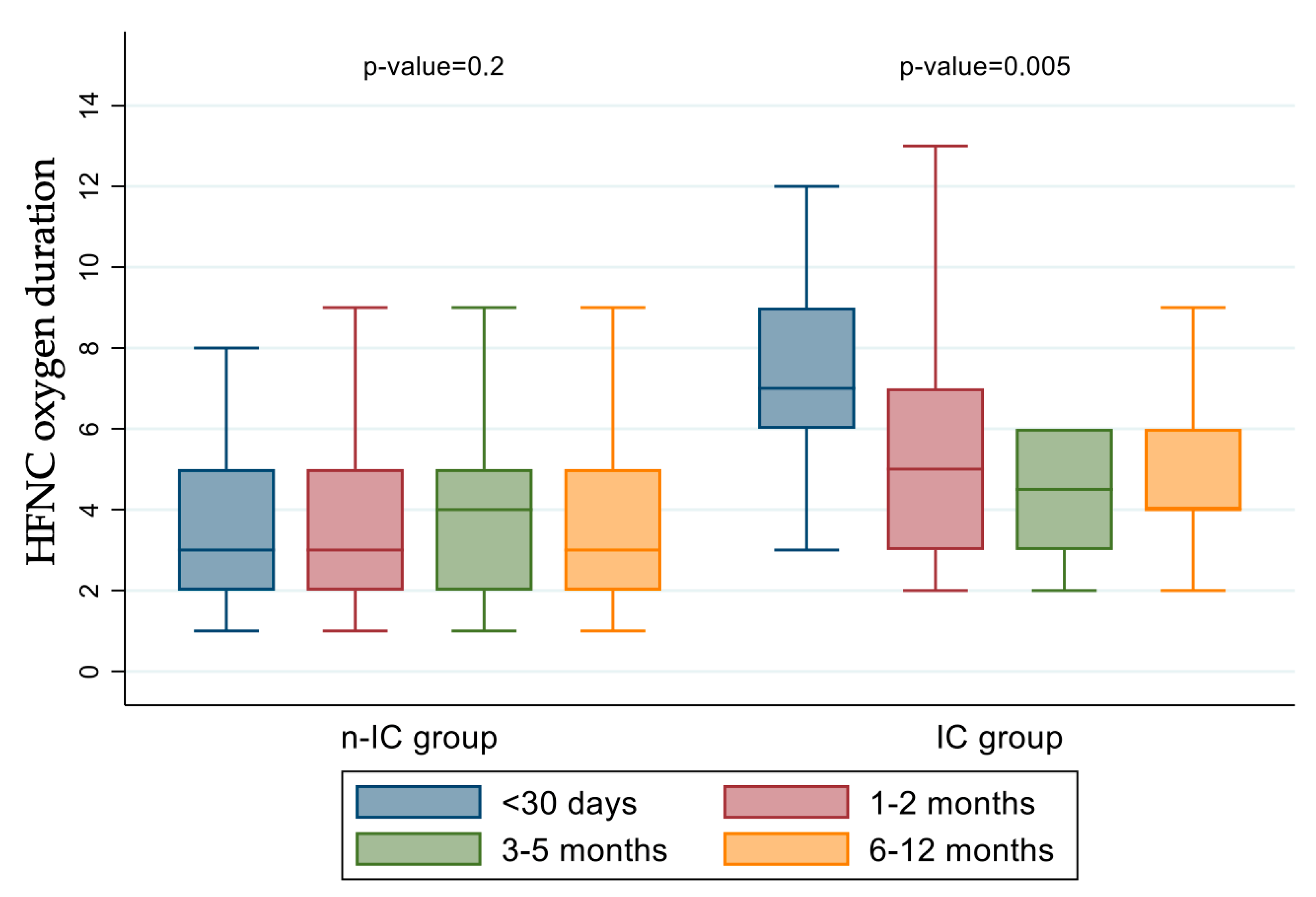
| HFNC 5 oxygen therapy (days) * | 4 (2–6) | 3 (2–5) | 6 (4–8) | <0.001 |
| Univariate Analysis | Multivariate Analysis | |||||||
|---|---|---|---|---|---|---|---|---|
| OR 1 | 95% CI | p | OR | 95% CI | p | |||
| Age group | ||||||||
| <30 days | ||||||||
| 1–2 months | 0.48 | 0.30 | 0.79 | <0.0001 | 0.37 | 0.22 | 0.64 | <0.0001 |
| 3–5 months | 0.32 | 0.17 | 0.59 | <0.0001 | 0.27 | 0.14 | 0.52 | <0.0001 |
| 6–12 months | 0.24 | 0.11 | 0.52 | <0.0001 | 0.20 | 0.09 | 0.46 | <0.0001 |
| Comorbidities | 2.96 | 1.72 | 5.08 | <0.0001 | 3.54 | 1.97 | 6.35 | <0.0001 |
| RSV 2 | 2.71 | 1.52 | 4.83 | <0.0001 | 3.30 | 1.77 | 6.13 | <0.0001 |
| CRP 3 > 10 mg/dL | 2.78 | 1.85 | 4.18 | <0.0001 | 2.61 | 1.70 | 4.01 | <0.0001 |
| Season | 0.90 | 0.60 | 1.35 | 0.61 | 0.77 | 0.50 | 1.19 | 0.25 |
Disclaimer/Publisher’s Note: The statements, opinions and data contained in all publications are solely those of the individual author(s) and contributor(s) and not of MDPI and/or the editor(s). MDPI and/or the editor(s) disclaim responsibility for any injury to people or property resulting from any ideas, methods, instructions or products referred to in the content. |
© 2025 by the authors. Licensee MDPI, Basel, Switzerland. This article is an open access article distributed under the terms and conditions of the Creative Commons Attribution (CC BY) license (https://creativecommons.org/licenses/by/4.0/).
Share and Cite
Maglione, M.; Pierri, L.; Savoia, F.; Calì, C.; Ragucci, R.; Sarno, M.; Ranucci, G.; Coppola, E.; Nunziata, F.; Di Toro, A.; et al. The Path of Bronchiolitis Towards Intensive Care: Risk Factor Analysis in a Large Italian Cohort. J. Clin. Med. 2025, 14, 5420. https://doi.org/10.3390/jcm14155420
Maglione M, Pierri L, Savoia F, Calì C, Ragucci R, Sarno M, Ranucci G, Coppola E, Nunziata F, Di Toro A, et al. The Path of Bronchiolitis Towards Intensive Care: Risk Factor Analysis in a Large Italian Cohort. Journal of Clinical Medicine. 2025; 14(15):5420. https://doi.org/10.3390/jcm14155420
Chicago/Turabian StyleMaglione, Marco, Luca Pierri, Fabio Savoia, Camilla Calì, Roberta Ragucci, Marco Sarno, Giulia Ranucci, Emma Coppola, Francesco Nunziata, Antonino Di Toro, and et al. 2025. "The Path of Bronchiolitis Towards Intensive Care: Risk Factor Analysis in a Large Italian Cohort" Journal of Clinical Medicine 14, no. 15: 5420. https://doi.org/10.3390/jcm14155420
APA StyleMaglione, M., Pierri, L., Savoia, F., Calì, C., Ragucci, R., Sarno, M., Ranucci, G., Coppola, E., Nunziata, F., Di Toro, A., Tipo, V., Giannattasio, A., & the BRAND Study. (2025). The Path of Bronchiolitis Towards Intensive Care: Risk Factor Analysis in a Large Italian Cohort. Journal of Clinical Medicine, 14(15), 5420. https://doi.org/10.3390/jcm14155420