


Comparison of Dental Findings with Computed Tomographic and Clinical Examination in Patients with End-Stage Heart Failure

 , ,
, ,  , , and
Abstract
1. Introduction
2. Materials and Methods
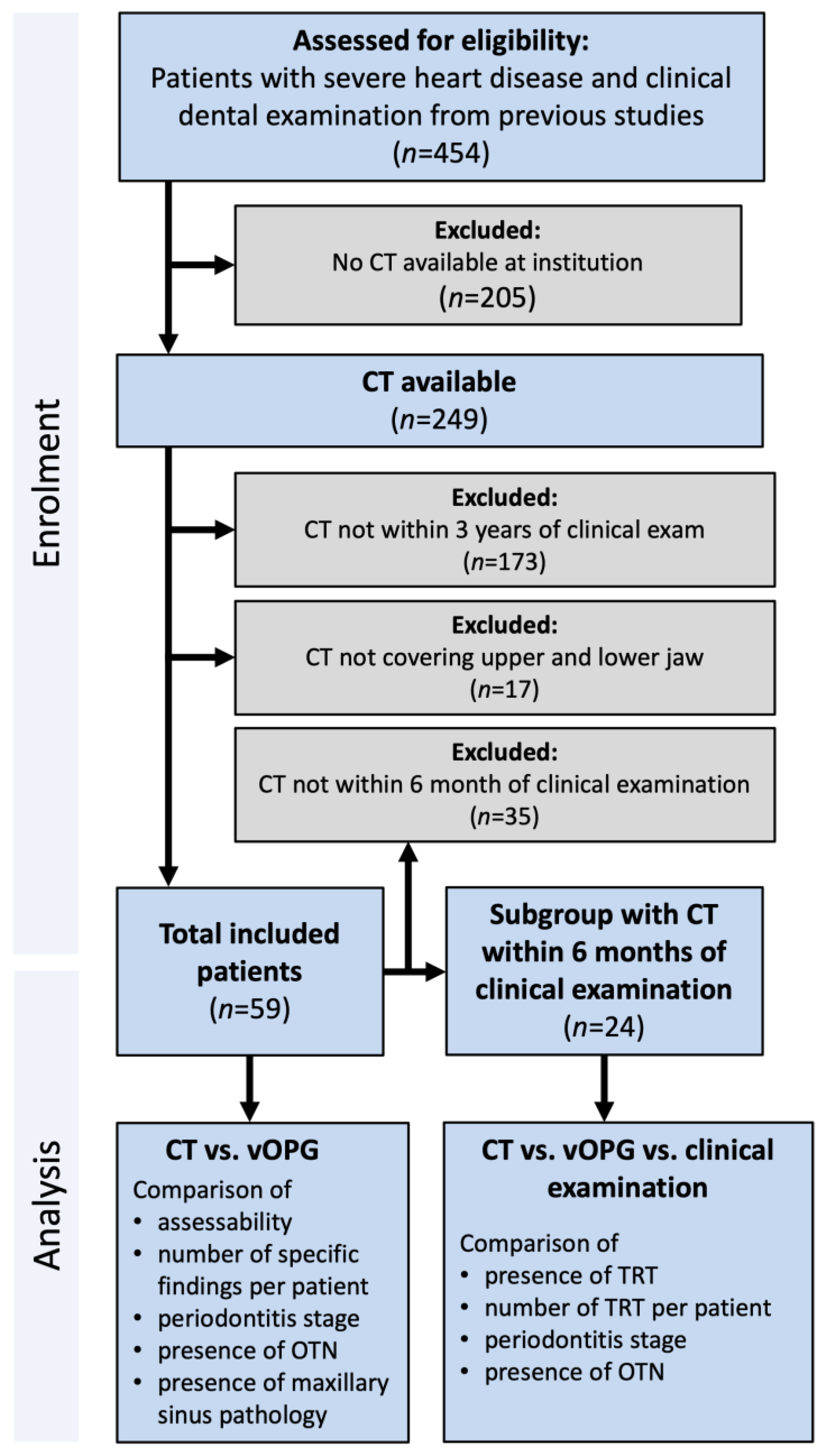
2.1. Patients
2.2. Image Acquisition, Reconstruction, and Radiographic Assessment
2.2.1. CT
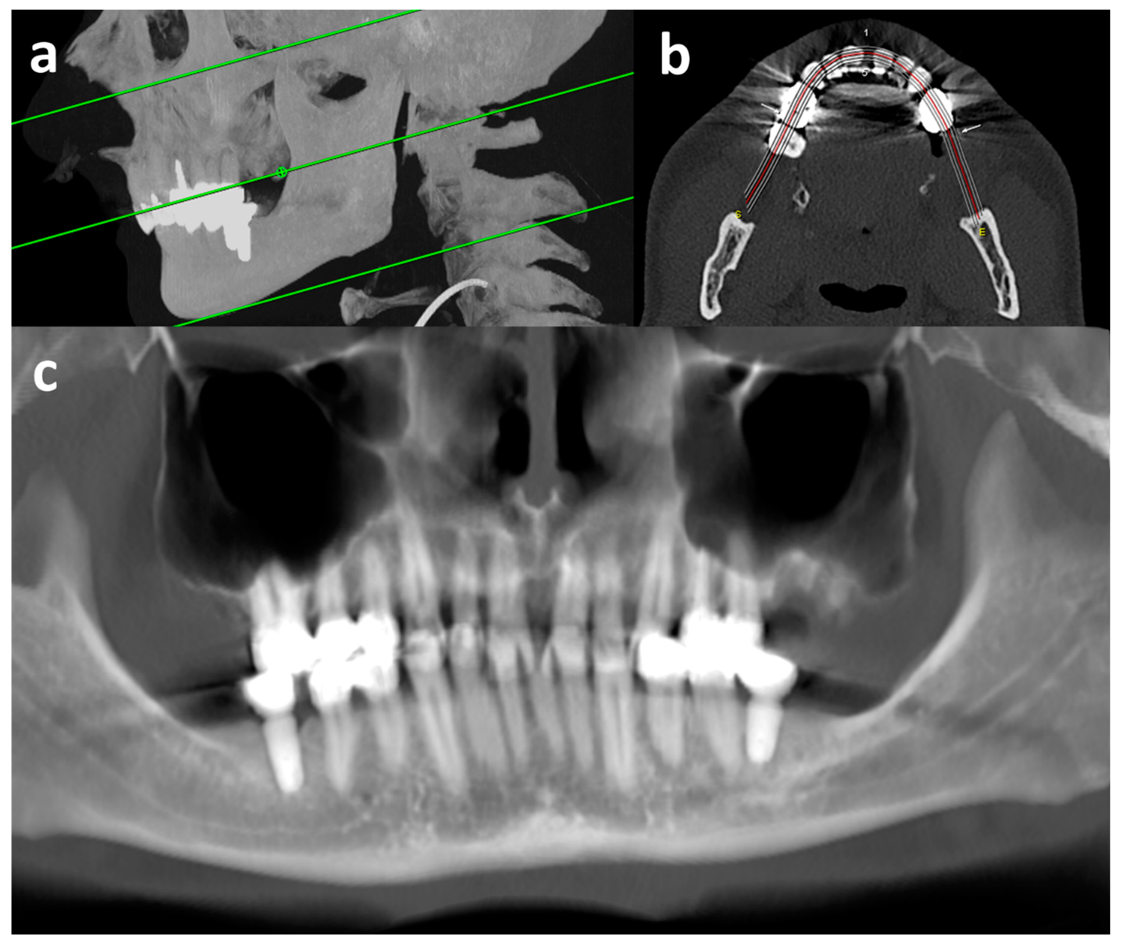
2.2.2. Virtual Orthopantomogram (vOPG)
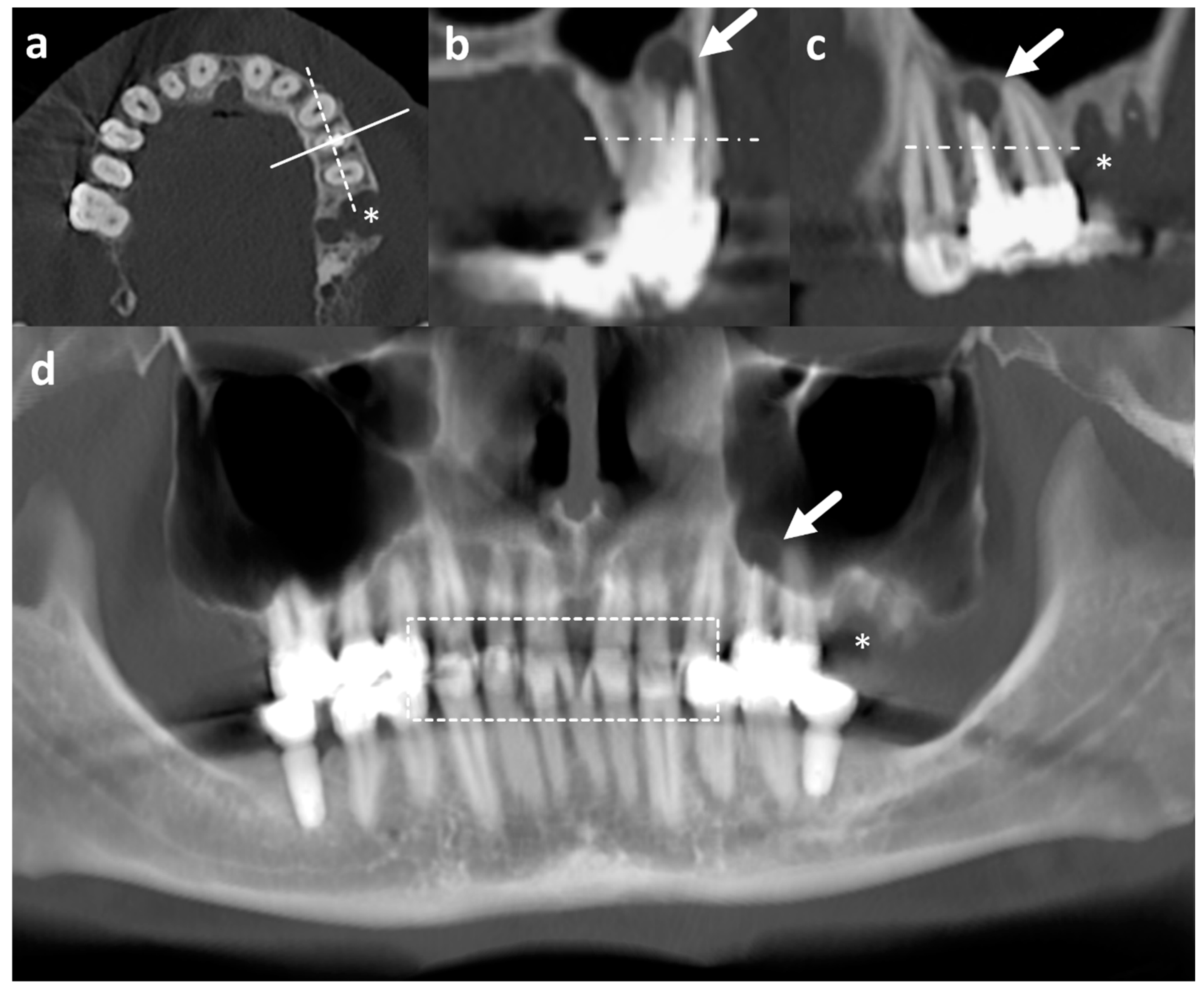
2.2.3. Radiological Evaluation
2.2.4. Assessability
2.3. Dental and Periodontal Treatment Need
2.3.1. CT and vOPG
- tooth with need for restoration due to caries or insufficient restorations;
- severely damaged tooth (tooth with more than 50% damage of the crown);
- remaining root(s) (tooth without crown);
- apical lesion at tooth with/without root canal treatment;
- tooth partial impacted by bone or soft tissue.
2.3.2. Clinical Examination
- number of teeth with carious cavitation (D-T) according to WHO [22] (including all stages with treatment need as decayed teeth, insufficient restorations, severely damaged teeth, and remaining roots);
- periodontal inflamed surface area (PISA) [23];
- periodontitis diagnosis based on detailed periodontal chart (periodontal probing depth, attachment loss, furcation involvement, tooth mobility) with staging and grading [24];
- presence of periodontal probing depth of over 3 mm [21] indicating periodontal treatment need;
2.4. Comparisons
2.5. Statistical Analysis
3. Results
3.1. Patients
3.2. Assessability
3.3. Dental Findings
4. Discussion
5. Conclusions
Author Contributions
Funding
Institutional Review Board Statement
Informed Consent Statement
Data Availability Statement
Conflicts of Interest
Abbreviations
| Clinic | clinical dental examination |
| CT | computed tomography |
| HF | heart failure |
| HTx | heart transplantation |
| LVAD | left ventricular assist device |
| OTN | overall treatment need (CT/vOPG: presence/number of teeth with TRT, apical lesion or partially impaction; clinic: presence of TRT or periodontal treatment need) |
| TRT | teeth requiring treatment (presence/number of teeth with restoration need due to caries, insufficient restorations, severe damage, or remaining roots) |
| vOPG | virtual orthopantomogram |
References
- Tomasoni, D.; Adamo, M.; Lombardi, C.M.; Metra, M. Highlights in heart failure. ESC Heart Fail 2019, 6, 1105–1127. [Google Scholar] [CrossRef] [PubMed]
- Bundesärztekammer (BÄK); Kassenärztliche Bundesvereinigung (KBV). National Disease Management Guideline: Chronic Heart Failure (in German). 2021. Available online: https://www.leitlinien.de/themen/herzinsuffizienz/3-auflage/kapitel-2 (accessed on 29 May 2022).
- Seferović, P.M.; Vardas, P.; Jankowska, E.A.; Maggioni, A.P.; Timmis, A.; Milinković, I.; Polovina, M.; Gale, C.P.; Lund, L.H.; Lopatin, Y.; et al. The Heart Failure Association Atlas: Heart Failure Epidemiology and Management Statistics 2019. Eur. J. Heart Fail 2021, 23, 906–914. [Google Scholar] [CrossRef] [PubMed]
- Crespo-Leiro, M.G.; Metra, M.; Lund, L.H.; Milicic, D.; Costanzo, M.R.; Filippatos, G.; Gustafsson, F.; Tsui, S.; Barge-Caballero, E.; De Jonge, N.; et al. Advanced heart failure: A position statement of the Heart Failure Association of the European Society of Cardiology. Eur. J. Heart Fail 2018, 20, 1505–1535. [Google Scholar] [CrossRef] [PubMed]
- Maniar, S.; Kondareddy, S.; Topkara, V.K. Left ventricular assist device-related infections: Past, present and future. Expert. Rev. Med. Devices 2011, 8, 627–634. [Google Scholar] [CrossRef]
- Leuck, A.-M. Left ventricular assist device driveline infections: Recent advances and future goals. J. Thorac. Dis. 2015, 7, 2151–2157. [Google Scholar] [CrossRef] [PubMed]
- Zhou, P.; Xiao, Z.; Zhu, P.; Nie, Z.; Pavan, D.; Zheng, S. Diabetes Mellitus Is Not a Risk Factor for Patients Supported with Left Ventricular Assist Device. Ann. Thorac. Surg. 2020, 109, 1614–1622. [Google Scholar] [CrossRef]
- Rustemeyer, J.; Bremerich, A. Necessity of surgical dental foci treatment prior to organ transplantation and heart valve replacement. Clin. Oral. Investig. 2007, 11, 171–174. [Google Scholar] [CrossRef]
- Joshy, G.; Arora, M.; Korda, R.J.; Chalmers, J.; Banks, E. Is poor oral health a risk marker for incident cardiovascular disease hospitalisation and all-cause mortality? Findings from 172 630 participants from the prospective 45 and Up Study. BMJ Open 2016, 6, e012386. [Google Scholar] [CrossRef]
- National Institute of Dental and Craniofacial Research. Dental Management of the Organ or Stem Cell Transplant Patient. 2016. Available online: https://nidcr.nih.gov/sites/default/files/2017-09/dental-management-organ-stem-cell-transplant.pdf (accessed on 29 May 2022).
- Kumar, A.; Rai, A. Oral Health Status, Health Behaviour and Treatment Needs of Patients Undergoing Cardiovascular Surgery. Braz. J. Cardiovasc. Surg. 2018, 33, 151–154. [Google Scholar] [CrossRef]
- Binner, C.; Wagner, J.; Schmalz, G.; Eisner, M.; Rast, J.; Kottmann, T.; Haak, R.; Oberbach, A.; Borger, M.A.; Garbade, J.; et al. Insufficient Oral Behaviour and the High Need for Periodontal Treatment in Patients with Heart Insufficiency and after Heart Transplantation: A Need for Special Care Programs? J. Clin. Med. 2019, 8, 1668. [Google Scholar] [CrossRef]
- Ziebolz, D.; Friedrich, S.; Binner, C.; Rast, J.; Eisner, M.; Wagner, J.; Schmickler, J.; Kottmann, T.; Haak, R.; Borger, M.A.; et al. Lack in Periodontal Care of Patients Suffering from Severe Heart Diseases-Results after 12 Months Follow-Up. J. Clin. Med. 2020, 9, 359. [Google Scholar] [CrossRef] [PubMed]
- Garbade, J.; Rast, J.; Schmalz, G.; Eisner, M.; Wagner, J.; Kottmann, T.; Oberbach, A.; Lehmann, S.; Haak, R.; Borger, M.A.; et al. Oral health and dental behaviour of patients with left ventricular assist device: A cross-sectional study. ESC Heart Fail 2020, 7, 1273–1281. [Google Scholar] [CrossRef] [PubMed]
- Slade, G.D.; Spencer, A.J. Development and evaluation of the Oral Health Impact Profile. Community Dent. Health 1994, 11, 3–11. [Google Scholar] [PubMed]
- Aziz, W.; Claridge, S.; Ntalas, I.; Gould, J.; de Vecchi, A.; Razeghi, O.; Toth, D.; Mountney, P.; Preston, R.; Rinaldi, C.A.; et al. Emerging role of cardiac computed tomography in heart failure. ESC Heart Fail 2019, 6, 909–920. [Google Scholar] [CrossRef]
- Cunqueiro, A.; Gomes, W.A.; Lee, P.; Dym, R.J.; Scheinfeld, M.H. CT of the Neck: Image Analysis and Reporting in the Emergency Setting. Radiographics 2019, 39, 1760–1781. [Google Scholar] [CrossRef]
- Avery, R.K. Recipient screening prior to solid-organ transplantation. Clin. Infect. Dis. 2002, 35, 1513–1519. [Google Scholar] [CrossRef]
- Stember, J.N.; Moonis, G.; Silva, C. Panoramic Dental Reconstruction for Faster Detection of Dental Pathology on Medical Non-dental CT Scans: A Proof of Concept from CT Neck Soft Tissue. J. Digit. Imaging 2021, 34, 959–966. [Google Scholar] [CrossRef]
- Pokorny, A.; Tataryn, R. Clinical and radiologic findings in a case series of maxillary sinusitis of dental origin. Int. Forum Allergy Rhinol. 2013, 12, 973–979. [Google Scholar] [CrossRef] [PubMed]
- Papapanou, P.N.; Sanz, M.; Buduneli, N.; Dietrich, T.; Feres, M.; Fine, D.H.; Flemmig, T.F.; Garcia, R.; Giannobile, W.V.; Graziani, F.; et al. Periodontitis: Consensus report of workgroup 2 of the 2017 World Workshop on the Classification of Periodontal and Peri-Implant Diseases and Conditions. J. Clin. Periodontol. 2018, 45 (Suppl. S20), S162–S170. [Google Scholar] [CrossRef]
- World Health Organization. Oral Health Surveys: Basic Methods, 4th ed.; World Health Organization: Geneva, Switzerland, 1997. [Google Scholar]
- Nesse, W.; Abbas, F.; van der Ploeg, I.; Spijkervet, F.K.L.; Dijkstra, P.U.; Vissink, A. Periodontal inflamed surface area: Quantifying inflammatory burden. J. Clin. Periodontol. 2008, 35, 668–673. [Google Scholar] [CrossRef]
- Caton, J.G.; Armitage, G.; Berglundh, T.; Chapple, I.L.C.; Jepsen, S.; Kornman, K.S.; Mealey, B.L.; Papapanou, P.N.; Sanz, M.; Tonetti, M.S. A new classification scheme for periodontal and peri-implant diseases and conditions-Introduction and key changes from the 1999 classification. J. Clin. Periodontol. 2018, 45 (Suppl. S20), S1–S8. [Google Scholar] [CrossRef] [PubMed]
- Kumar, P.S. Oral microbiota and systemic disease. Anaerobe 2013, 24, 90–93. [Google Scholar] [CrossRef] [PubMed]
- Zhang, W.; Daly, C.G.; Mitchell, D.; Curtis, B. Incidence and magnitude of bacteraemia caused by flossing and by scaling and root planing. J. Clin. Periodontol. 2013, 40, 41–52. [Google Scholar] [CrossRef] [PubMed]
- Tomás, I.; Diz, P.; Tobías, A.; Scully, C.; Donos, N. Periodontal health status and bacteraemia from daily oral activities: Systematic review/meta-analysis. J. Clin. Periodontol. 2012, 39, 213–228. [Google Scholar] [CrossRef] [PubMed]
- Ziebolz, D.; Jahn, C.; Pegel, J.; Semper-Pinnecke, E.; Mausberg, R.F.; Waldmann-Beushausen, R.; Mealey, B.L.; Papapanou, P.N.; Sanz, M.; Tonetti, M.S. Periodontal bacteria DNA findings in human cardiac tissue-Is there a link of periodontitis to heart valve disease? Int. J. Cardiol. 2018, 251, 74–79. [Google Scholar] [CrossRef]
- Fishman, J.A. Infection in Organ Transplantation. Am. J. Transplant. 2017, 17, 856–879. [Google Scholar] [CrossRef]
- Morimoto, Y.; Nakatani, T.; Yokoe, C.; Kudo, C.; Hanamoto, H.; Niwa, H. Haemostatic management for oral surgery in patients supported with left ventricular assist device—A preliminary retrospective study. Br. J. Oral. Maxillofac. Surg. 2015, 53, 991–995. [Google Scholar] [CrossRef]
- Lam, O.L.T.; Zhang, W.; Samaranayake, L.P.; Li, L.S.W.; McGrath, C. A systematic review of the effectiveness of oral health promotion activities among patients with cardiovascular disease. Int. J. Cardiol. 2011, 151, 261–267. [Google Scholar] [CrossRef]
- Herman, W.W.; Ferguson, H.W. Dental care for patients with heart failure: An update. J. Am. Dent. Assoc. 2010, 141, 845–853. [Google Scholar] [CrossRef]
- Nunn, P. Medical emergencies in the oral health care setting. J. Dent. Hyg. 2000, 74, 136–151. [Google Scholar]
- Schmalz, G.; Wendorff, H.; Berisha, L.; Meisel, A.; Widmer, F.; Marcinkowski, A.; Teschler, H.; Sommerwerck, U.; Haak, R.; Kollmar, O.; et al. Association between the time after transplantation and different immunosuppressive medications with dental and periodontal treatment need in patients after solid organ transplantation. Transpl. Infect. Dis. 2018, 20, e12832. [Google Scholar] [CrossRef] [PubMed]
- Kweon, H.H.-I.; Lee, J.-H.; Youk, T.-M.; Lee, B.-A.; Kim, Y.-T. Panoramic radiography can be an effective diagnostic tool adjunctive to oral examinations in the national health checkup program. J. Periodontal Implant. Sci. 2018, 48, 317–325. [Google Scholar] [CrossRef] [PubMed]
- Corbet, E.F.; Ho, D.K.L.; Lai, S.M.L. Radiographs in periodontal disease diagnosis and management. Aust. Dent. J. 2009, 54 (Suppl. S1), S27–S43. [Google Scholar] [CrossRef] [PubMed]
- Nikolic-Jakoba, N.; Spin-Neto, R.; Wenzel, A. Cone-Beam Computed Tomography for Detection of Intrabony and Furcation Defects: A Systematic Review Based on a Hierarchical Model for Diagnostic Efficacy. J. Periodontol. 2016, 87, 630–644. [Google Scholar] [CrossRef]
- Huettig, F.; Axmann, D. Reporting of dental status from full-arch radiographs: Descriptive analysis and methodological aspects. World J. Clin. Cases 2014, 2, 552–564. [Google Scholar] [CrossRef]
- Nekolla, E.A.; Schegerer, A.A.; Griebel, J.; Brix, G. Häufigkeit und Dosis diagnostischer und interventioneller Röntgenanwendungen: Trends zwischen 2007 und 2014. [Frequency and doses of diagnostic and interventional X-ray applications: Trends between 2007 and 2014]. Radiologe 2017, 57, 555–562. [Google Scholar] [CrossRef]
- Bushberg, J. National Council on Radiation Protection and Measurements. Alada (as low as diagnostically acceptable): 2014. In Proceedings of the NCRP Annual Meeting, Bethesda, MD, USA, 10–11 March 2014. [Google Scholar]
- Chapman, M.N.; Nadgir, R.N.; Akman, A.S.; Saito, N.; Sekiya, K.; Kaneda, T.; Sakai, O. Periapical lucency around the tooth: Radiologic evaluation and differential diagnosis. Radiographics 2013, 33, E15–E32. [Google Scholar] [CrossRef]
- Mohammad-Rahimi, H.; Motamedian, S.R.; Rohban, M.H.; Krois, J.; Uribe, S.E.; Mahmoudinia, E.; Rokhshad, R.; Nadimi, M.; Schwendicke, F. Deep learning for caries detection: A systematic review. J. Dent. 2022, 122, 104115. [Google Scholar] [CrossRef]
- Zanini, L.G.K.; Rubira-Bullen, I.R.F.; Nunes, F.L.D.S. A Systematic Review on Caries Detection, Classification, and Segmentation from X-Ray Images: Methods, Datasets, Evaluation, and Open Opportunities. J. Imaging Inf. Med. 2024, 37, 1824–1845. [Google Scholar] [CrossRef]
- Schulze, R.; Heil, U.; Gross, D.; Bruellmann, D.D.; Dranischnikow, E.; Schwanecke, U.; Schoemer, E. Artefacts in CBCT: A review. Dentomaxillofac Radiol. 2011, 40, 265–273. [Google Scholar] [CrossRef]
- Harris, D.; Buser, D.; Dula, K.; Grondahl, K.; Haris, D.; Jacobs, R.; Lekholm, U.; Nakielny, R.; Van Steenberghe, D.; Van Der Stelt, P. E.A.O. guidelines fo the use of diagnostic imaging in implant dentistry. A consensus workshop organized by the European Association for Osseointegration in Trinity College Dublin. Clin. Oral. Implant. Res. 2002, 13, 566–570. [Google Scholar] [CrossRef]
- Ludlow, J.B.; Davies-Ludlow, L.E.; Brooks, S.L.; Howerton, W.B. Dosimetry of 3 CBCT devices for oral and maxillofacial radiology: CB Mercuray, NewTom 3G and i-CAT. Dentomaxillofac Radiol. 2006, 35, 219–226. [Google Scholar] [CrossRef] [PubMed]
- Mandelaris, G.A.; Scheyer, E.T.; Evans, M.; Kim, D.; McAllister, B.; Nevins, M.L.; Rios, H.F.; Sarment, D. American Academy of Periodontology Best Evidence Consensus Statement on Selected Oral Applications for Cone-Beam Computed Tomography. J. Periodontol. 2017, 88, 939–945. [Google Scholar] [CrossRef]
- Kim, D.M.; Bassir, S.H. When Is Cone-Beam Computed Tomography Imaging Appropriate for Diagnostic Inquiry in the Management of Inflammatory Periodontitis? An American Academy of Periodontology Best Evidence Review. J. Periodontol. 2017, 88, 978–998. [Google Scholar] [CrossRef] [PubMed]
- Huumonen, S.; Kvist, T.; Gröndahl, K.; Molander, A. Diagnostic value of computed tomography in re-treatment of root fillings in maxillary molars. Int. Endod. J. 2006, 39, 827–833. [Google Scholar] [CrossRef]
- Constantine, S.; Clark, B.; Kiermeier, A.; Anderson, P.P. Panoramic radiography is of limited value in the evaluation of maxillary sinus disease. Oral. Surg. Oral. Med. Oral. Pathol. Oral. Radiol. 2019, 127, 237–246. [Google Scholar] [CrossRef] [PubMed]
- Chapple, I.L.C.; Mealey, B.L.; van Dyke, T.E.; Bartold, P.M.; Dommisch, H.; Eickholz, P.; Geisinger, M.L.; Genco, R.J.; Glogauer, M.; Goldstein, M.; et al. Periodontal health and gingival diseases and conditions on an intact and a reduced periodontium: Consensus report of workgroup 1 of the 2017 World Workshop on the Classification of Periodontal and Peri-Implant Diseases and Conditions. J. Periodontol. 2018, 89 (Suppl. S1), S74–S84. [Google Scholar] [CrossRef]
- Hong, C.H.L.; Hu, S.; Haverman, T.; Stokman, M.; Napeñas, J.J.; Braber, J.B.; Gerber, E.; Geuke, M.; Vardas, E.; Waltimo, T.; et al. A systematic review of dental disease management in cancer patients. Support. Care Cancer 2018, 26, 155–174. [Google Scholar] [CrossRef]
- Miki, K.; Kitamura, M.; Hatta, K.; Kamide, K.; Gondo, Y.; Yamashita, M.; Takedachi, M.; Nozaki, T.; Fujihara, C.; Kashiwagi, Y.; et al. Periodontal inflamed surface area is associated with hs-CRP in septuagenarian Japanese adults in cross-sectional findings from the SONIC study. Sci. Rep. 2021, 11, 14436. [Google Scholar] [CrossRef]
- Anil, K.; Vadakkekuttical, R.J.; Radhakrishnan, C.; Parambath, F.C. Correlation of periodontal inflamed surface area with glycemic status in controlled and uncontrolled type 2 diabetes mellitus. World J. Clin. Cases 2021, 9, 11300–11310. [Google Scholar] [CrossRef]
- Deman, P.; Atwal, P.; Duzenli, C.; Thakur, Y.; Ford, N.L. Dose measurements for dental cone-beam CT: A comparison with MSCT and panoramic imaging. Phys. Med. Biol. 2014, 59, 3201–3222. [Google Scholar] [CrossRef] [PubMed]
- Schmalz, G.; Hennecke, A.; Haak, R.; Kottmann, T.; Garbade, J.; Binner, C.; Ziebolz, D. Secondary analysis of potential associations between oral health and infection-related parameters in patients with severe heart failure-results of a German cohort. BMC Cardiovasc. Disord. 2023, 21, 573. [Google Scholar] [CrossRef] [PubMed]
- Schmalz, G.; Reuschel, F.; Bartl, M.; Schmidt, L.; Runge, J.; Haak, R.; Goralski, S.; Roth, A.; Ziebolz, D. One Third of Patients before Endoprosthesis Implantation Show an Oral Focus as Potential Source of Infectious Complication-The Value of Pre-Operative Dental Risk Stratification in a German Cohort. J. Clin. Med. 2022, 11, 3686. [Google Scholar] [CrossRef] [PubMed]

| Variables | Total Included Patients (n = 59) | Subgroup with CT ≤ 6 Months from Clinical Exam (n = 24) | p-Value | |
|---|---|---|---|---|
| Disease [n (% of patients)] | HTx | 11 (18.6) | 3 (12.5) | 0.12 |
| LVAD | 35 (59.3) | 14 (58.3) | 0.55 | |
| HF | 13 (22.0) | 7 (29.2) | 0.09 | |
| Age at clinical examination (years) [mean ± SD] | 54.9 ± 10.0 | 52.0 ± 11.8 | 0.67 | |
| Time from HTx to clinical examination (months) [mean ± SD] | 28.8 ± 20.2 | 20.4 ± 30.1 | 0.11 | |
| Time from LVAD to clinical examination (months) [mean ± SD] | 29.4 ± 26.9 | 30.1 ± 29.3 | 0.24 | |
| Sex (male) [mean ± SD] | 54 (91.5) | 23 (95.8) | 0.14 | |
| Smoker [mean ± SD] | 15 (25.4) | 6 (25) | 0.78 | |
| Time between CT and clinical examination (month) [mean ± SD] | 15.0 ± 13.4 | 3.3 ± 2.3 | ||
| Variables [n (% of Patients)] | Examination | n # | Assessable | Partially Assessable | Not Assessable | Intergroup Comparison (p-Value) |
|---|---|---|---|---|---|---|
| Caries | CT | 57 | 5 (8.8) | 49 (86.0) | 3 (5.3) | <0.01 |
| vOPG | 57 | 8 (14.0) | 24 (42.1) | 25 (43.9) | ||
| Sufficiency of restorations | CT | 50 | 0 (0.0) | 0 (0.0) | 50 (100.0) | 0.99 |
| vOPG | 52 | 0 (0.0) | 2 (3.8) | 50 (96.2) | ||
| Remaining roots | CT | 59 | 27 (45.8) | 31 (52.5) | 1 (1.7) | 0.66 |
| vOPG | 59 | 26 (44.1) | 30 (50.8) | 3 (5.1) | ||
| Apical lesions | CT | 57 | 31 (54.4) | 26 (45.6) | 0 (0.0) | <0.01 |
| vOPG | 57 | 9 (15.8) | 42 (73.7) | 6 (10.5) | ||
| Presence of root canal treatment | CT | 57 | 41 (71.9) | 16 (28.1) | 0 (0.0) | <0.01 |
| vOPG | 57 | 8 (14.0) | 37 (64.9) | 12 (21.1) | ||
| Quality of root canal treatment | CT | 24 | 0 (0.0) | 0 (0.0) | 24 (100.0) | 0.99 |
| vOPG | 33 | 0 (0.0) | 2 (6.1) | 31 (93.9) | ||
| Impacted teeth | CT | 59 | 38 (64.4) | 21 (35.6) | 0 (0.0) | <0.01 |
| vOPG | 59 | 24 (40.7) | 34 (57.6) | 1 (1.7) | ||
| Maximal horizontal periodontal bone loss | CT | 57 | 30 (52.6) | 26 (45.6) | 1 (1.8) | <0.01 |
| vOPG | 57 | 12 (21.1) | 39 (68.4) | 6 (10.5) | ||
| Periodontal vertical bone loss | CT | 57 | 24 (42.1) | 30 (52.6) | 3 (5.3) | <0.01 |
| vOPG | 57 | 8 (14.0) | 24 (42.1) | 25 (43.9) | ||
| Maxillary sinus pathology | CT | 59 | 59 (100.0) | 0 (0.0) | 0 (0.0) | <0.01 |
| vOPG | 59 | 21 (35.6) | 33 (55.9) | 5 (8.5) |
| Variables | CT | vOPG | ||||
|---|---|---|---|---|---|---|
| Slice Thickness | Kernel | Both | Slice Thickness | Kernel | Both | |
| Caries | 0.54 | 0.02 | <0.05 | 0.08 | 0.07 | 0.04 |
| Remaining roots | 0.17 | 0.47 | 0.03 | 0.79 | 0.20 | 0.35 |
| Apical lesions | 0.009 | 0.02 | 0.001 | 0.32 | 0.04 | <0.01 |
| Presence of root canal treatment | 0.25 | 0.37 | 0.21 | 0.69 | 0.006 | <0.01 |
| Impacted teeth | 0.06 | 1.00 | 0.15 | 0.17 | 0.65 | 0.66 |
| Maximal horizontal periodontal bone loss | <0.001 | 0.21 | 0.007 | 0.80 | 0.009 | 0.02 |
| Periodontal vertical bone loss | 0.002 | 0.24 | 0.02 | 0.09 | 0.07 | 0.04 |
| Variables | CT (n = 59) | vOPG (n = 59) | p-Value | ||
|---|---|---|---|---|---|
| Number of findings per patient [median [0.25 quartile, 0.75 quartile]] | |||||
| Restorations needed pp | 0 [0, 0] | 1 [0, 2] | <0.01 | ||
| Severely damaged teeth pp | 0 [0, 0] | 0 [0, 0] | 0.04 | ||
| Remaining roots pp | 0 [0, 0] | 0 [0, 0] | 0.08 | ||
| Apical lesions pp | with root treatment | 0 [0, 0] | 0 [0, 0] | 0.63 | |
| without root treatment | 0 [0, 1] | 0 [0, 1] | 0.89 | ||
| total | 0 [0, 1] | 0 [0, 1] | 0.77 | ||
| Partially impacted teeth pp | 0 [0, 0] | 0 [0, 0] | 0.62 | ||
| Complete impacted teeth pp | 0 [0, 0] | 0 [0, 0] | 0.05 | ||
| OTN pp | 2 [1, 4] | 3 [2, 5] | 0.002 | ||
| Presence of [n (% of patients)] | |||||
| OTN | 23 (39.0) | 47 (79.7) | <0.01 | ||
| Maximal horizontal periodontal bone loss | stage 1 | 2 (3.4) | 7 (11.9) | 0.58 | |
| stage 2 | 28 (47.5) | 21 (35.6) | |||
| stage 3 or 4 | 26 (44.1) | 28 (47.5) | |||
| Not assessable | 3 (5.1) | 3 (5.1) | |||
| Periodontal vertical bone loss | 6 (10.2) | 5 (8.5) | 0.99 | ||
| Maxillary sinus pathology | 27 (45.8) | 13 (22.0) | <0.01 | ||
| Variables | CT (n = 24) # | vOPG (n = 24) # | Clinic (n = 24) # | p-Value | |||
|---|---|---|---|---|---|---|---|
| CT vs. vOPG | CT vs. Clinic | vOPG vs. Clinic | |||||
| Presence of TRT [n (% of patients)] | 7 (29.2) | 15 (62.5) | 7 (29.2) | <0.01 | 1.00 | <0.01 | |
| Number of TRT per patient [median [0.25 quartile, 0.75 quartile]] | 0 [0, 1] | 1 [0, 2] | 0 [0, 1] | 0.05 | 1.00 | 0.05 | |
| Periodontitis stage [n (% of patients)] | 1 | 2 (8.3) | 4 (16.7) | 1 (4.2) | 0.38 | 0.04 | 0.01 |
| 2 | 11 (48.5) | 9 (37.5) | 5 (20.8) | ||||
| 3 or 4 | 10 (41.7) | 9 (37.5) | 18 (75.0) | ||||
| Not assessable | 1 (4.2) | 2 (8.3) | 0 | ||||
| Presence of OTN [n (% of patients)] | 8 (33.3) | 18 (75.0) | 22 (91.7) | <0.01 | <0.01 | 0.22 | |
Disclaimer/Publisher’s Note: The statements, opinions and data contained in all publications are solely those of the individual author(s) and contributor(s) and not of MDPI and/or the editor(s). MDPI and/or the editor(s) disclaim responsibility for any injury to people or property resulting from any ideas, methods, instructions or products referred to in the content. |
© 2024 by the authors. Licensee MDPI, Basel, Switzerland. This article is an open access article distributed under the terms and conditions of the Creative Commons Attribution (CC BY) license (https://creativecommons.org/licenses/by/4.0/).
Share and Cite
Merle, C.L.; Gocke, J.; Seitz, P.; Gutberlet, M.; Saeed, D.; Haak, R.; Ziebolz, D.; Gohmann, R.F.; Schmalz, G. Comparison of Dental Findings with Computed Tomographic and Clinical Examination in Patients with End-Stage Heart Failure. J. Clin. Med. 2024, 13, 5406. https://doi.org/10.3390/jcm13185406
Merle CL, Gocke J, Seitz P, Gutberlet M, Saeed D, Haak R, Ziebolz D, Gohmann RF, Schmalz G. Comparison of Dental Findings with Computed Tomographic and Clinical Examination in Patients with End-Stage Heart Failure. Journal of Clinical Medicine. 2024; 13(18):5406. https://doi.org/10.3390/jcm13185406
Chicago/Turabian StyleMerle, Cordula Leonie, Julia Gocke, Patrick Seitz, Matthias Gutberlet, Diyar Saeed, Rainer Haak, Dirk Ziebolz, Robin Fabian Gohmann, and Gerhard Schmalz. 2024. "Comparison of Dental Findings with Computed Tomographic and Clinical Examination in Patients with End-Stage Heart Failure" Journal of Clinical Medicine 13, no. 18: 5406. https://doi.org/10.3390/jcm13185406
APA StyleMerle, C. L., Gocke, J., Seitz, P., Gutberlet, M., Saeed, D., Haak, R., Ziebolz, D., Gohmann, R. F., & Schmalz, G. (2024). Comparison of Dental Findings with Computed Tomographic and Clinical Examination in Patients with End-Stage Heart Failure. Journal of Clinical Medicine, 13(18), 5406. https://doi.org/10.3390/jcm13185406