Article

The Value of Mandibular Indices on Cone Beam Computed Tomography in Secondary Causes of Low Bone Mass

by
Ioana Ruxandra Poiană
1,*,
Ramona Dobre
2,3,
Silviu-Mirel Pițuru
1 and
Alexandru Bucur
1
1
Faculty of Dentistry, “Carol Davila” University of Medicine and Pharmacy, 050474 Bucharest, Romania
2
Faculty of Medicine, “Carol Davila” University of Medicine and Pharmacy, 050474 Bucharest, Romania
3
Department of Endocrinology, National Institute of Endocrinology C. I. Parhon, 011853 Bucharest, Romania
*
Author to whom correspondence should be addressed.
J. Clin. Med. 2024, 13(16), 4854; https://doi.org/10.3390/jcm13164854
Submission received: 23 July 2024 / Revised: 8 August 2024 / Accepted: 15 August 2024 / Published: 16 August 2024
(This article belongs to the Section Endocrinology & Metabolism)

Abstract

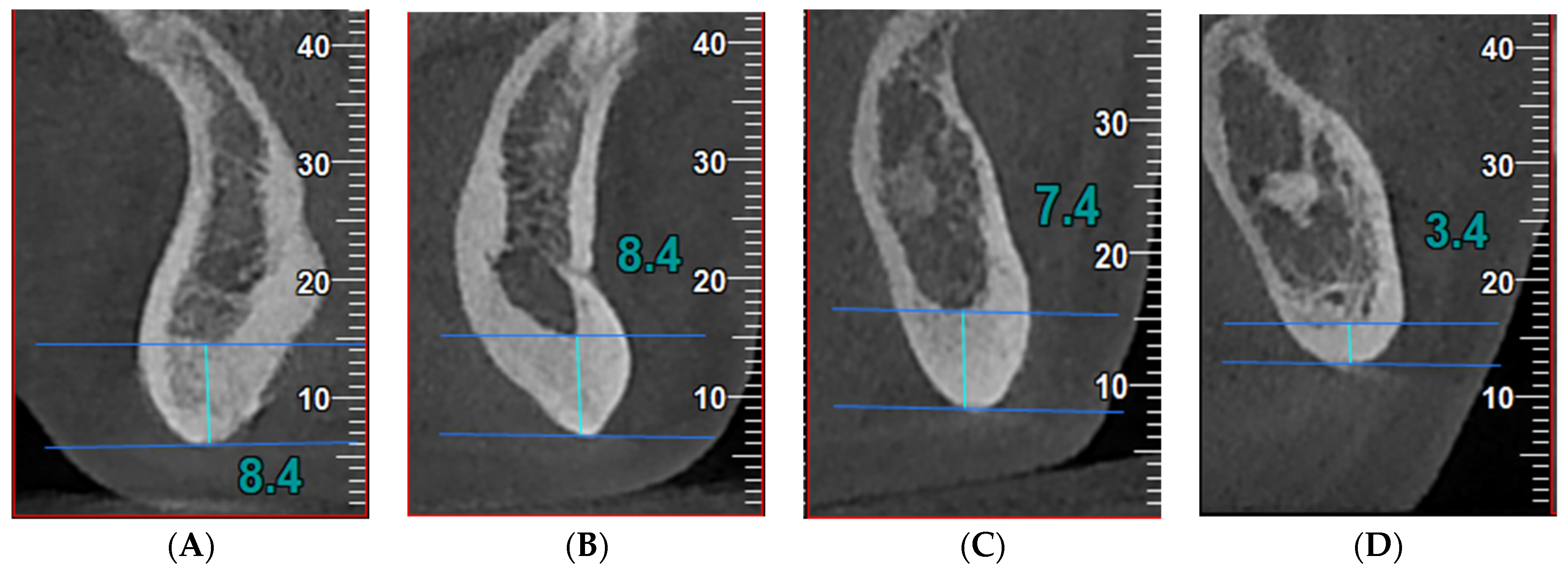
Background: As implant treatment cases increase, many cases of failure/side effects also occur. Generally, dental clinics determine the density of the jawbone using cone beam CT (CBCT). Considering the known potential role of this tool for bone assessment in primary osteoporosis, this study evaluated patients with secondary endocrine causes of low bone mass. Methods: The study included 83 patients with endocrine causes of osteoporosis who were evaluated by dual-energy X-ray absorptiometry (DXA), trabecular bone score (TBS), and mental foramen (MF) region CBCT. The following CBCT indices were measured: anterior (A)—thickness of inferior mandibular cortex 10 mm anterior from MF; molar (M)—thickness of inferior mandibular cortex 10 mm posterior from MF; posterior (P)—thickness of inferior mandibular cortex 25 mm posterior from MF; symphysis (S)—thickness of inferior mandibular cortex equidistant from the centers of right and left MF. Results: The highest correlation coefficient in the secondary causes group was between the A index and the lumbar BMD (r = 0.375, p = 0.001) and the P index and the femoral neck BMD (r = 0.38, p = 0.001). Hypercortisolism seems to be the most predictable cause of secondary osteoporosis using the A, M, and P indices. The A, M, and P indices showed predictive values of the bone micro-architecture that was evaluated using TBS score, and were statistically significant. The symphysis index does not significantly predict osteoporosis or impaired bone micro-architecture. Conclusions: These findings support the potential usefulness of A, M, and P CBCT-derived radiomorphometric mandibular indices in secondary osteoporosis, underlining the well-known effects of these pathologies on bone micro-architecture rather than bone quantity.
Keywords: secondary and primary osteoporosis; menopause; cone beam computed tomography; mandibular indices secondary and primary osteoporosis; menopause; cone beam computed tomography; mandibular indices