
Deep Brain Stimulation Electrode Reconstruction: Comparison between Lead-DBS and Surgical Planning System
 , and
, and
Abstract
1. Introduction
2. Materials and Methods
2.1. Patients and Preoperative Assessment
2.2. Surgical Procedures
2.3. Postoperative Electrode Localization
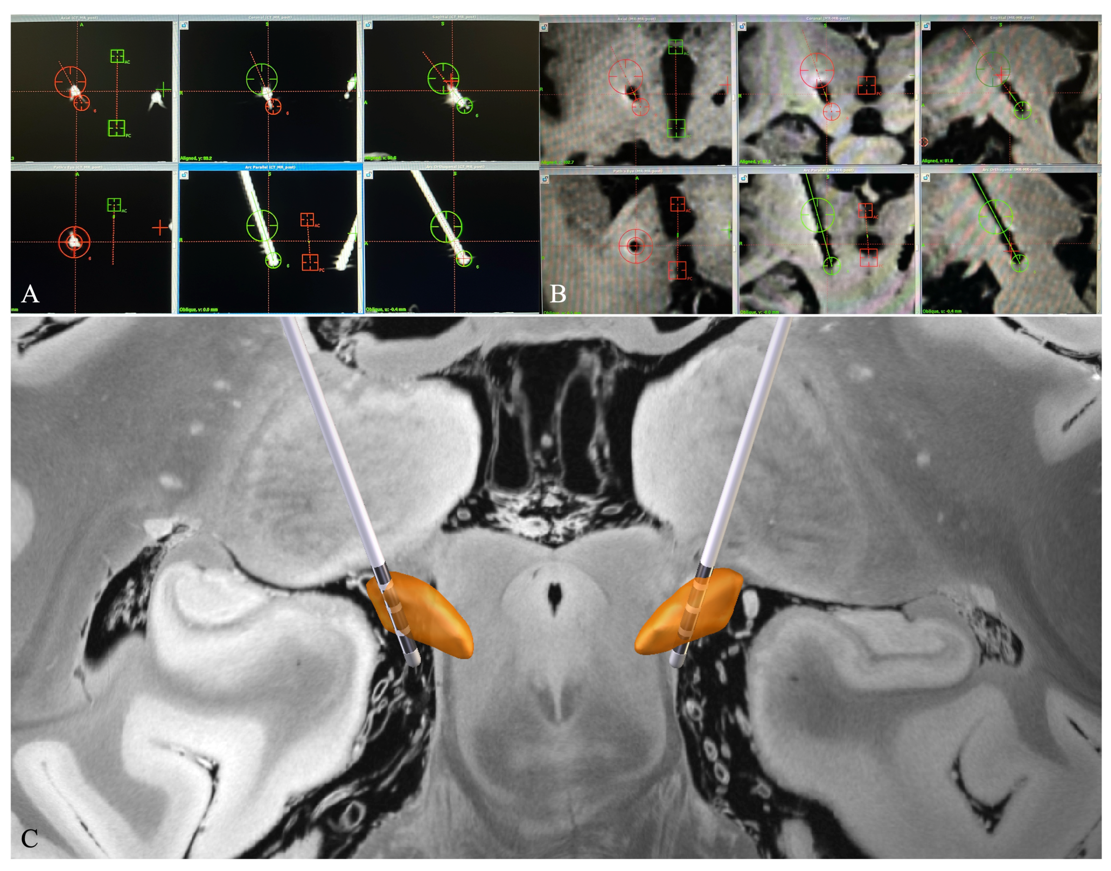
2.3.1. Electrode Reconstruction in Surgiplan
2.3.2. Electrode Reconstruction in Lead-DBS
2.4. Relative Position between the Electrode and the DBS Target
2.5. Euclidean Distance between the Electrode Tip and the Pre-Defined Target
2.6. DBS Parameter Programming and Patient Follow-Up
2.7. Data Analysis and Statistics
3. Results
3.1. Patient Characteristics
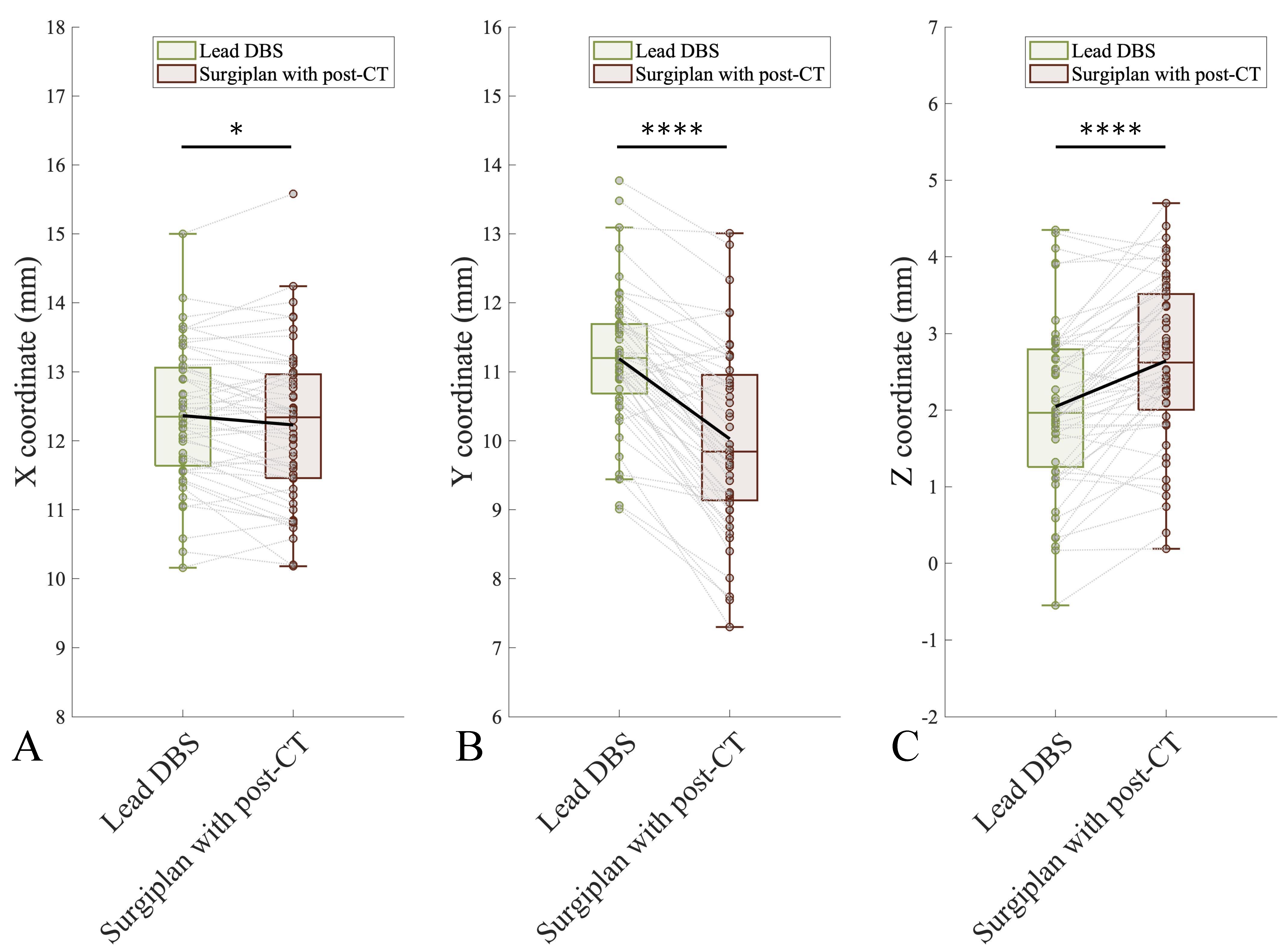
3.2. Comparison of the Coordinates between Lead-DBS and Surgiplan with Postoperative CT
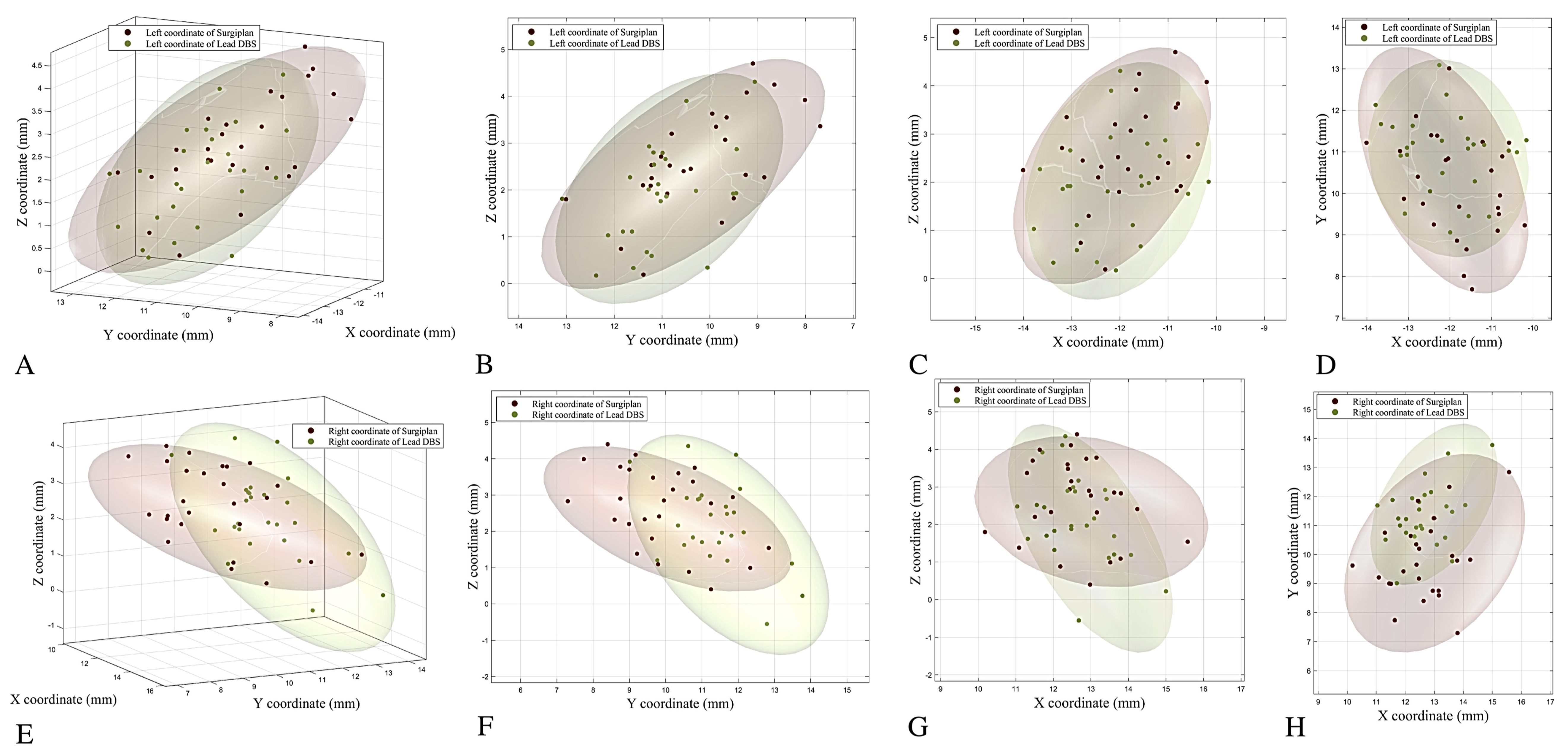
3.3. Comparison of Calculated Coordinates between Lead-DBS, Surgiplan with Postoperative CT, and Surgiplan with MRI
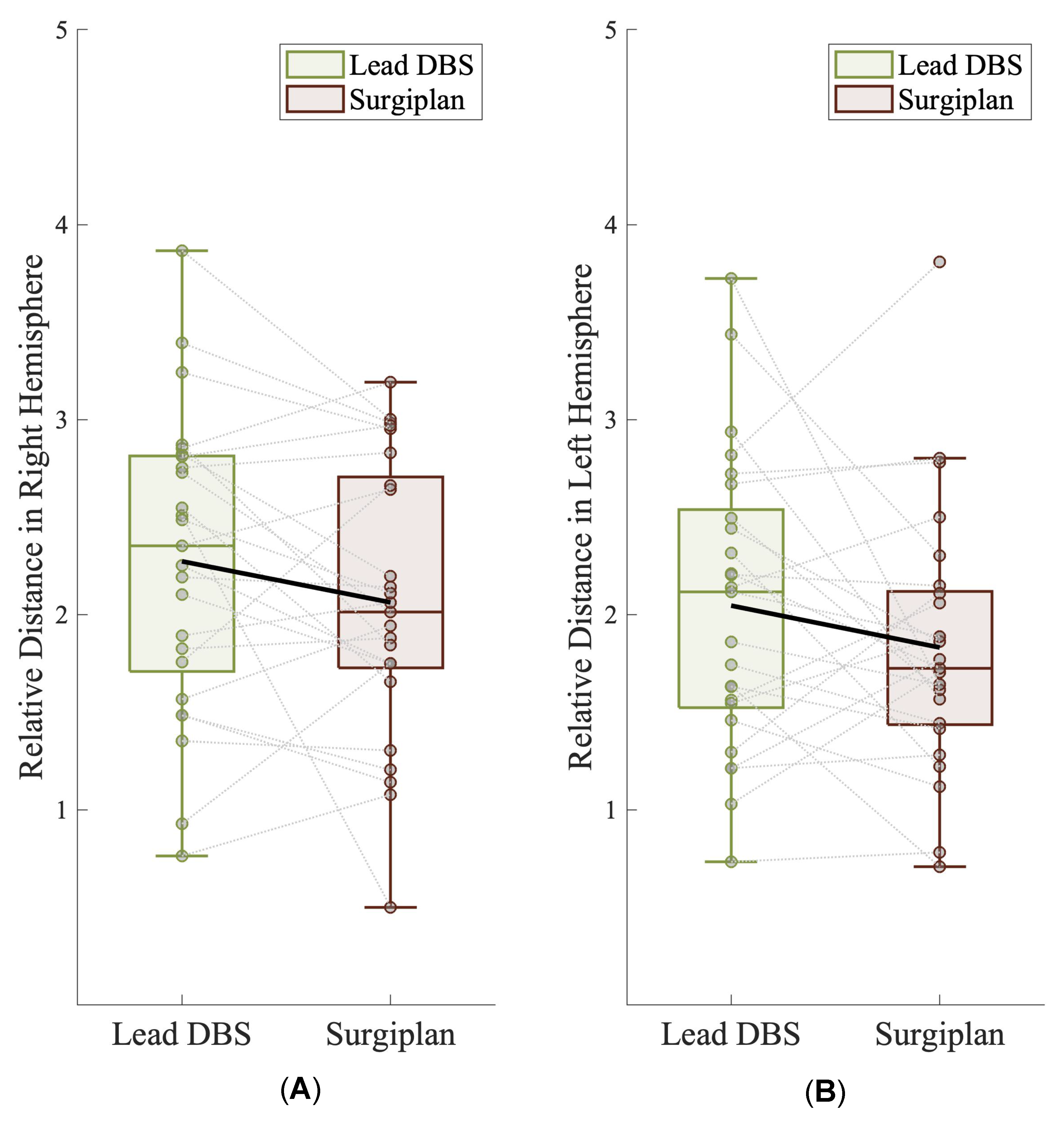
3.4. Relative Position of the Electrode and the DBS Target
3.5. Validation of Lead-DBS According to the Euclidean Distance and Clinical Follow-Up Data
4. Discussion
Supplementary Materials
Author Contributions
Funding
Institutional Review Board Statement
Informed Consent Statement
Data Availability Statement
Acknowledgments
Conflicts of Interest
References
- Krauss, J.K.; Lipsman, N.; Aziz, T.; Boutet, A.; Brown, P.; Chang, J.W.; Davidson, B.; Grill, W.M.; Hariz, M.I.; Horn, A.; et al. Technology of deep brain stimulation: Current status and future directions. Nat. Rev. Neurol. 2021, 17, 75–87. [Google Scholar] [CrossRef]
- Okun, M.S.; Tagliati, M.; Pourfar, M.; Fernandez, H.H.; Rodriguez, R.L.; Alterman, R.L.; Foote, K.D. Management of referred deep brain stimulation failures: A retrospective analysis from 2 movement disorders centers. Arch. Neurol. 2005, 62, 1250–1255. [Google Scholar] [CrossRef]
- Holl, E.M.; Petersen, E.A.; Foltynie, T.; Martinez-Torres, I.; Limousin, P.; Hariz, M.I.; Zrinzo, L. Improving targeting in image-guided frame-based deep brain stimulation. Neurosurgery 2010, 67, 437–447. [Google Scholar] [CrossRef]
- Haynes, W.I.; Haber, S.N. The organization of prefrontal-subthalamic inputs in primates provides an anatomical substrate for both functional specificity and integration: Implications for Basal Ganglia models and deep brain stimulation. J. Neurosci. 2013, 33, 4804–4814. [Google Scholar] [CrossRef]
- Mallet, L.; Schüpbach, M.; N’Diaye, K.; Remy, P.; Bardinet, E.; Czernecki, V.; Welter, M.L.; Pelissolo, A.; Ruberg, M.; Agid, Y.; et al. Stimulation of subterritories of the subthalamic nucleus reveals its role in the integration of the emotional and motor aspects of behavior. Proc. Natl. Acad. Sci. USA 2007, 104, 10661–10666. [Google Scholar] [CrossRef]
- Steigerwald, F.; Matthies, C.; Volkmann, J. Directional Deep Brain Stimulation. Neurotherapeutics 2019, 16, 100–104. [Google Scholar] [CrossRef]
- Dembek, T.A.; Reker, P.; Visser-Vandewalle, V.; Wirths, J.; Treuer, H.; Klehr, M.; Roediger, J.; Dafsari, H.S.; Barbe, M.T.; Timmermann, L. Directional DBS increases side-effect thresholds-A prospective, double-blind trial. Mov. Disord. 2017, 32, 1380–1388. [Google Scholar] [CrossRef] [PubMed]
- Barnaure, I.; Pollak, P.; Momjian, S.; Horvath, J.; Lovblad, K.O.; Boex, C.; Remuinan, J.; Burkhard, P.; Vargas, M.I. Evaluation of electrode position in deep brain stimulation by image fusion (MRI and CT). Neuroradiology 2015, 57, 903–908. [Google Scholar] [CrossRef]
- Pinsker, M.O.; Herzog, J.; Falk, D.; Volkmann, J.; Deuschl, G.; Mehdorn, M. Accuracy and distortion of deep brain stimulation electrodes on postoperative MRI and CT. Zentralbl. Neurochir. 2008, 69, 144–147. [Google Scholar] [CrossRef]
- Boutet, A.; Chow, C.T.; Narang, K.; Elias, G.J.B.; Neudorfer, C.; Germann, J.; Ranjan, M.; Loh, A.; Martin, A.J.; Kucharczyk, W.; et al. Improving Safety of MRI in Patients with Deep Brain Stimulation Devices. Radiology 2020, 296, 250–262. [Google Scholar] [CrossRef]
- Zhao, S.; Li, G.; Tong, C.; Chen, W.; Wang, P.; Dai, J.; Fu, X.; Xu, Z.; Liu, X.; Lu, L.; et al. Full activation pattern mapping by simultaneous deep brain stimulation and fMRI with graphene fiber electrodes. Nat. Commun. 2020, 11, 1788. [Google Scholar] [CrossRef]
- Horn, A.; Li, N.; Dembek, T.A.; Kappel, A.; Boulay, C.; Ewert, S.; Tietze, A.; Husch, A.; Perera, T.; Neumann, W.J.; et al. Lead-DBS v2: Towards a comprehensive pipeline for deep brain stimulation imaging. Neuroimage 2019, 184, 293–316. [Google Scholar] [CrossRef]
- Horn, A.; Kühn, A.A. Lead-DBS: A toolbox for deep brain stimulation electrode localizations and visualizations. Neuroimage 2015, 107, 127–135. [Google Scholar] [CrossRef]
- Alkemade, A.; Groot, J.M.; Forstmann, B.U. Do We Need a Human post mortem Whole-Brain Anatomical Ground Truth in in vivo Magnetic Resonance Imaging? Front. Neuroanat. 2018, 12, 110. [Google Scholar] [CrossRef]
- Ewert, S.; Horn, A.; Finkel, F.; Li, N.; Kühn, A.A.; Herrington, T.M. Optimization and comparative evaluation of nonlinear deformation algorithms for atlas-based segmentation of DBS target nuclei. NeuroImage 2019, 184, 586–598. [Google Scholar] [CrossRef] [PubMed]
- Vogel, D.; Shah, A.; Coste, J.; Lemaire, J.-J.; Wårdell, K.; Hemm, S. Anatomical brain structures normalization for deep brain stimulation in movement disorders. NeuroImage Clin. 2020, 27, 102271. [Google Scholar] [CrossRef] [PubMed]
- Lofredi, R.; Auernig, C.G.; Ewert, S.; Irmen, F.; Steiner, L.A.; Scheller, U.; van Wijk, B.C.M.; Oxenford, S.; Kuhn, A.A.; Horn, A. Interrater reliability of deep brain stimulation electrode localizations. Neuroimage 2022, 262, 119552. [Google Scholar] [CrossRef] [PubMed]
- Chen, S.; Gao, G.; Feng, T.; Zhang, J. Chinese expert consensus on programming deep brain stimulation for patients with Parkinson’s disease. Transl. Neurodegener. 2018, 7, 11. [Google Scholar] [CrossRef]
- Xiao, Y.; Lau, J.C.; Hemachandra, D.; Gilmore, G.; Khan, A.R.; Peters, T.M. Image Guidance in Deep Brain Stimulation Surgery to Treat Parkinson’s Disease: A Comprehensive Review. IEEE Trans. Biomed. Eng. 2021, 68, 1024–1033. [Google Scholar] [CrossRef]
- Fan, S.; Zhang, Q.; Meng, F.; Fang, H.; Yang, G.; Shi, Z.; Liu, H.; Zhang, H.; Yang, A.; Zhang, J.; et al. Comparison of dural puncture and dural incision in deep brain stimulation surgery: A simple but worthwhile technique modification. Front. Neurosci. 2022, 16, 988661. [Google Scholar] [CrossRef]
- Pollo, C.; Villemure, J.G.; Vingerhoets, F.; Ghika, J.; Maeder, P.; Meuli, R. Magnetic resonance artifact induced by the electrode Activa 3389: An in vitro and in vivo study. Acta Neurochir. 2004, 146, 161–164. [Google Scholar] [CrossRef]
- Avants, B.B.; Tustison, N.J.; Song, G.; Cook, P.A.; Klein, A.; Gee, J.C. A reproducible evaluation of ANTs similarity metric performance in brain image registration. Neuroimage 2011, 54, 2033–2044. [Google Scholar] [CrossRef]
- Ewert, S.; Plettig, P.; Li, N.; Chakravarty, M.M.; Collins, D.L.; Herrington, T.M.; Kuhn, A.A.; Horn, A. Toward defining deep brain stimulation targets in MNI space: A subcortical atlas based on multimodal MRI, histology and structural connectivity. Neuroimage 2018, 170, 271–282. [Google Scholar] [CrossRef]
- Horn, A.; Kühn, A.A.; Merkl, A.; Shih, L.; Alterman, R.; Fox, M. Probabilistic conversion of neurosurgical DBS electrode coordinates into MNI space. NeuroImage 2017, 150, 395–404. [Google Scholar] [CrossRef]
- Deuschl, G.; Schade-Brittinger, C.; Krack, P.; Volkmann, J.; Schäfer, H.; Bötzel, K.; Daniels, C.; Deutschländer, A.; Dillmann, U.; Eisner, W.; et al. A randomized trial of deep-brain stimulation for Parkinson’s disease. N. Engl. J. Med. 2006, 355, 896–908. [Google Scholar] [CrossRef]
- Draz, H.H.; Gabran, S.R.I.; Basha, M.; Mostafa, H.; Abu-Elyazeed, M.F.; Zaki, A. Comparative mechanical analysis of deep brain stimulation electrodes. BioMedical Eng. OnLine 2018, 17, 123. [Google Scholar] [CrossRef]
- Niederer, J.; Patriat, R.; Rosenberg, O.; Palnitkar, T.; Darrow, D.; Park, M.C.; Schrock, L.; Eberly, L.E.; Harel, N. Factors Influencing Electrode Position and Bending of the Proximal Lead in Deep Brain Stimulation for Movement Disorders. Stereotact. Funct. Neurosurg. 2020, 98, 300–312. [Google Scholar] [CrossRef]
- Contarino, M.F.; Bot, M.; Speelman, J.D.; de Bie, R.M.; Tijssen, M.A.; Denys, D.; Bour, L.J.; Schuurman, P.R.; van den Munckhof, P. Postoperative displacement of deep brain stimulation electrodes related to lead-anchoring technique. Neurosurgery 2013, 73, 681–688. [Google Scholar] [CrossRef]
- Goransson, N.; Johansson, J.D.; Wardell, K.; Zsigmond, P. Postoperative Lead Movement after Deep Brain Stimulation Surgery and the Change of Stimulation Volume. Stereotact. Funct. Neurosurg. 2021, 99, 221–229. [Google Scholar] [CrossRef]
- Lee, J.Y.; Kim, J.W.; Lee, J.Y.; Lim, Y.H.; Kim, C.; Kim, D.G.; Jeon, B.S.; Paek, S.H. Is MRI a reliable tool to locate the electrode after deep brain stimulation surgery? Comparison study of CT and MRI for the localization of electrodes after DBS. Acta Neurochir. 2010, 152, 2029–2036. [Google Scholar] [CrossRef]
- Roth, A.; Buttrick, S.S.; Cajigas, I.; Jagid, J.R.; Ivan, M.E. Accuracy of frame-based and frameless systems for deep brain stimulation: A meta-analysis. J. Clin. Neurosci. 2018, 57, 1–5. [Google Scholar] [CrossRef]
- Yelnik, J.; Damier, P.; Demeret, S.; Gervais, D.; Bardinet, E.; Bejjani, B.P.; François, C.; Houeto, J.L.; Arnule, I.; Dormont, D.; et al. Localization of stimulating electrodes in patients with Parkinson disease by using a three-dimensional atlas-magnetic resonance imaging coregistration method. J. Neurosurg. 2003, 99, 89–99. [Google Scholar] [CrossRef] [PubMed]
- Martinez-Santiesteban, F.M.; Swanson, S.D.; Noll, D.C.; Anderson, D.J. Magnetic field perturbation of neural recording and stimulating microelectrodes. Phys. Med. Biol. 2007, 52, 2073. [Google Scholar] [CrossRef] [PubMed]
- Boutet, A.; Rashid, T.; Hancu, I.; Elias, G.J.B.; Gramer, R.M.; Germann, J.; Dimarzio, M.; Li, B.; Paramanandam, V.; Prasad, S.; et al. Functional MRI Safety and Artifacts during Deep Brain Stimulation: Experience in 102 Patients. Radiology 2019, 293, 174–183. [Google Scholar] [CrossRef] [PubMed]
- Golestanirad, L.; Kirsch, J.; Bonmassar, G.; Downs, S.; Elahi, B.; Martin, A.; Iacono, M.I.; Angelone, L.M.; Keil, B.; Wald, L.L.; et al. RF-induced heating in tissue near bilateral DBS implants during MRI at 1.5 T and 3T: The role of surgical lead management. Neuroimage 2019, 184, 566–576. [Google Scholar] [CrossRef] [PubMed]
- Hamid, N.A.; Mitchell, R.D.; Mocroft, P.; Westby, G.W.; Milner, J.; Pall, H. Targeting the subthalamic nucleus for deep brain stimulation: Technical approach and fusion of pre- and postoperative MR images to define accuracy of lead placement. J. Neurol. Neurosurg. Psychiatry 2005, 76, 409–414. [Google Scholar] [CrossRef] [PubMed]
- van Rooijen, B.D.; Backes, W.H.; Schijns, O.E.; Colon, A.; Hofman, P.A. Brain imaging in chronic epilepsy patients after depth electrode (stereoelectroencephalography) implantation: Magnetic resonance imaging or computed tomography? Neurosurgery 2013, 73, 543–549. [Google Scholar] [CrossRef]
- Yoshida, F.; Miyagi, Y.; Morioka, T.; Hashiguchi, K.; Murakami, N.; Matsumoto, K.; Nagata, S.; Sasaki, T. Assessment of contact location in subthalamic stimulation for Parkinson’s disease by co-registration of computed tomography images. Stereotact. Funct. Neurosurg. 2008, 86, 162–166. [Google Scholar] [CrossRef]
- Wodarg, F.; Herzog, J.; Reese, R.; Falk, D.; Pinsker, M.O.; Steigerwald, F.; Jansen, O.; Deuschl, G.; Mehdorn, H.M.; Volkmann, J. Stimulation site within the MRI-defined STN predicts postoperative motor outcome. Mov. Disord. 2012, 27, 874–879. [Google Scholar] [CrossRef]
- Bot, M.; Schuurman, P.R.; Odekerken, V.J.J.; Verhagen, R.; Contarino, F.M.; De Bie, R.M.A.; van den Munckhof, P. Deep brain stimulation for Parkinson’s disease: Defining the optimal location within the subthalamic nucleus. J. Neurol. Neurosurg. Psychiatry 2018, 89, 493–498. [Google Scholar] [CrossRef]
- Roediger, J.; Dembek, T.A.; Wenzel, G.; Butenko, K.; Kuhn, A.A.; Horn, A. StimFit-A Data-Driven Algorithm for Automated Deep Brain Stimulation Programming. Mov. Disord. 2022, 37, 574–584. [Google Scholar] [CrossRef] [PubMed]
- Roediger, J.; Dembek, T.A.; Achtzehn, J.; Busch, J.L.; Kramer, A.P.; Faust, K.; Schneider, G.H.; Krause, P.; Horn, A.; Kuhn, A.A. Automated deep brain stimulation programming based on electrode location: A randomised, crossover trial using a data-driven algorithm. Lancet Digit. Health 2022, 5, E59–E70. [Google Scholar] [CrossRef] [PubMed]


| PD Patients | DT Patients | |
|---|---|---|
| Number of patients | 21 | 5 |
| Age (year, mean ± SD) | 58.4 ± 7.4 | 53.6 ± 13.0 |
| Gender (male/female) | 8/13 | 4/1 |
| Age of disease onset (year, mean ± SD) | 51.1 ± 7.4 | 47.4 ± 15.5 |
| Disease duration (year, mean ± SD) | 7.4 ± 3 | 6.2 ± 5.1 |
| LEDD (mg/day) | 1050.3 ± 589.1 | - |
| Preoperative assessment | ||
| UPDRS-III med off (mean ± SD) | 44.7 ± 18.9 | - |
| UPDRS-III med on (mean ± SD) | 20.5 ± 11.3 | - |
| UPDRS-III improvement (%) | 55.4 ± 16.7 | - |
| BFMDRS (mean ± SD) | - | 22.7 ± 15.1 |
| GRS (mean ± SD) | - | 23 ± 6.7 |
| Lead-DBS | Surgiplan | Mean Discrepancy | p Value | ||
|---|---|---|---|---|---|
| X coordinate | All | 12.36 ± 0.98 | 12.23 ± 1.11 | −0.13 | 0.0251 |
| Right | 12.62 ± 0.91 | 12.64 ± 1.12 | 0.02 | 0.8019 | |
| Left | −12.11 ± 1.00 | −11.82 ± 0.95 | −0.29 | 0.0002 | |
| Y coordinate | All | 11.19 ± 1.00 | 10.03 ± 1.29 | −1.16 | <0.0001 |
| Right | 11.38 ± 1.03 | 9.86 ± 1.35 | −1.52 | <0.0001 | |
| Left | −11.00 ± 0.94 | −10.19 ± 1.24 | −0.81 | <0.0001 | |
| Z coordinate | All | 2.05 ± 1.09 | 2.64 ± 1.08 | 0.59 | <0.0001 |
| Right | 2.18 ± 1.12 | 2.65 ± 1.106 | 0.47 | <0.0001 | |
| Left | −1.93 ± 1.05 | −2.64 ± 1.06 | 0.71 | 0.0012 |
Disclaimer/Publisher’s Note: The statements, opinions and data contained in all publications are solely those of the individual author(s) and contributor(s) and not of MDPI and/or the editor(s). MDPI and/or the editor(s) disclaim responsibility for any injury to people or property resulting from any ideas, methods, instructions or products referred to in the content. |
© 2023 by the authors. Licensee MDPI, Basel, Switzerland. This article is an open access article distributed under the terms and conditions of the Creative Commons Attribution (CC BY) license (https://creativecommons.org/licenses/by/4.0/).
Share and Cite
Xu, Y.; Qin, G.; Tan, B.; Fan, S.; An, Q.; Gao, Y.; Fan, H.; Xie, H.; Wu, D.; Liu, H.; et al. Deep Brain Stimulation Electrode Reconstruction: Comparison between Lead-DBS and Surgical Planning System. J. Clin. Med. 2023, 12, 1781. https://doi.org/10.3390/jcm12051781
Xu Y, Qin G, Tan B, Fan S, An Q, Gao Y, Fan H, Xie H, Wu D, Liu H, et al. Deep Brain Stimulation Electrode Reconstruction: Comparison between Lead-DBS and Surgical Planning System. Journal of Clinical Medicine. 2023; 12(5):1781. https://doi.org/10.3390/jcm12051781
Chicago/Turabian StyleXu, Yichen, Guofan Qin, Bojing Tan, Shiying Fan, Qi An, Yuan Gao, Houyou Fan, Hutao Xie, Delong Wu, Huanguang Liu, and et al. 2023. "Deep Brain Stimulation Electrode Reconstruction: Comparison between Lead-DBS and Surgical Planning System" Journal of Clinical Medicine 12, no. 5: 1781. https://doi.org/10.3390/jcm12051781
APA StyleXu, Y., Qin, G., Tan, B., Fan, S., An, Q., Gao, Y., Fan, H., Xie, H., Wu, D., Liu, H., Yang, G., Fang, H., Xiao, Z., Zhang, J., Zhang, H., Shi, L., & Yang, A. (2023). Deep Brain Stimulation Electrode Reconstruction: Comparison between Lead-DBS and Surgical Planning System. Journal of Clinical Medicine, 12(5), 1781. https://doi.org/10.3390/jcm12051781